Embed Size (px)
Citation preview
P
FEAT
UR
EA
RT
ICLES
Meandering Inferior Vena Cava in a Child WithPseudo-Scimitar SyndromeSanjeev Aggarwal, MD, Aparna Joshi, MD, Stephanie Bolger-Theut, MD, and
ooja Gupta, MD
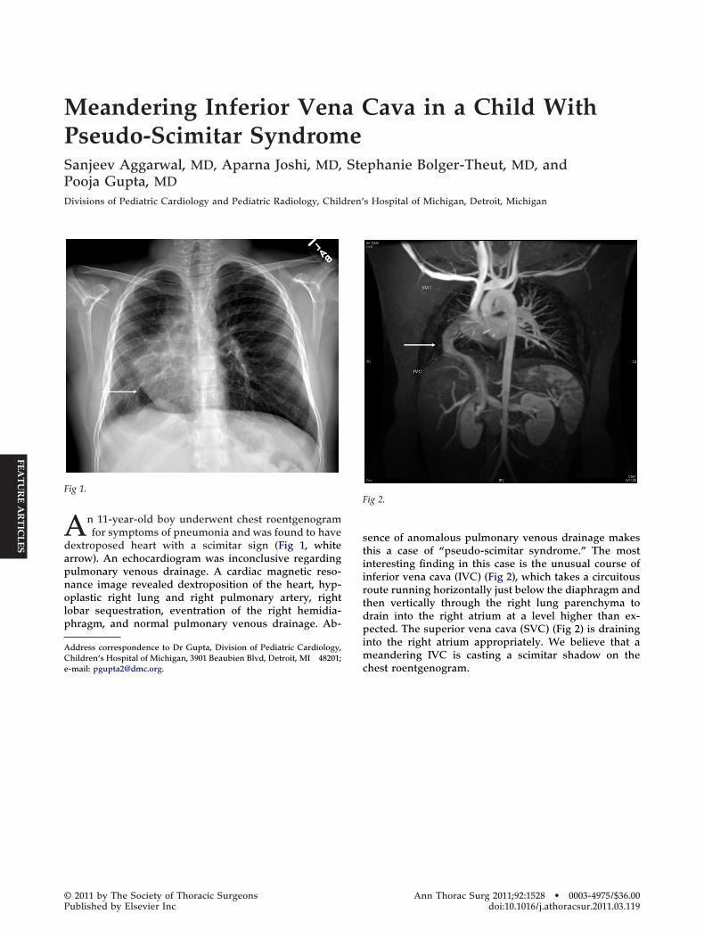
Divisions of Pediatric Cardiology and Pediatric Radiology, Children’s Hospital of Michigan, Detroit, MichiganAn 11-year-old boy underwent chest roentgenogramfor symptoms of pneumonia and was found to have
dextroposed heart with a scimitar sign (Fig 1, whitearrow). An echocardiogram was inconclusive regardingpulmonary venous drainage. A cardiac magnetic reso-nance image revealed dextroposition of the heart, hyp-oplastic right lung and right pulmonary artery, rightlobar sequestration, eventration of the right hemidia-phragm, and normal pulmonary venous drainage. Ab-
Address correspondence to Dr Gupta, Division of Pediatric Cardiology,
Fig 1.
Children’s Hospital of Michigan, 3901 Beaubien Blvd, Detroit, MI 48201;e-mail: [email protected].
© 2011 by The Society of Thoracic SurgeonsPublished by Elsevier Inc
sence of anomalous pulmonary venous drainage makesthis a case of “pseudo-scimitar syndrome.” The mostinteresting finding in this case is the unusual course ofinferior vena cava (IVC) (Fig 2), which takes a circuitousroute running horizontally just below the diaphragm andthen vertically through the right lung parenchyma todrain into the right atrium at a level higher than ex-pected. The superior vena cava (SVC) (Fig 2) is draininginto the right atrium appropriately. We believe that ameandering IVC is casting a scimitar shadow on the
Fig 2.
chest roentgenogram.
Ann Thorac Surg 2011;92:1528 • 0003-4975/$36.00doi:10.1016/j.athoracsur.2011.03.119