Embed Size (px)
Citation preview
m.d. Shydlovscky. A.V.
Classification of the surgical infections:1. Acute surgical infections (see appendix 16).2. Chronic surgical infections.Acute purulent surgical infections1. Acute purulent aerobic infections.2. Acute anaerobic infections.3. Acute specific infections.4. Viral infections.
Acute Purulent Aerobic Infections
The cause of the most frequently purulent surgical infections it's:
1. Staphylococcal infections.2. Streptococcal infections.3. Gram - negative infections.4. Mixed bacterial infections.
A large number of infections encountered in surgical practice are caused by Staphylococcus aureus. It is an important pathogen in postoperative wound infection and in infections following penetrating wounds. The lesions produced by S. aureus are characteristically localized with an indurated area of cellulitis that undergoes central necrosis and abscess formation with a thick, creamy, odorless, and yellow or cream-coloured pus. Bacteraemia may occur, with the development metastatic abscesses. Fever and leukocytosis are usually present. Antibiotic- resistant bacteria of increased virulence often cause those infections acquired during the course of hospitalization. Recent studies have shown that the emergence of new bacteriophage type Staphylococcus aureus may occur spontaneously.
A variety of streptococcal organisms produce infections seen in surgical practice. The most frequent of these is Streptococcus pyogenes, although others such as S. viridans, Pepto-streptococcus, aerophilic streptococcus, and S. faecalis (Group D enterococci) may be encountered.
The lesions caused by S. pyogenes are characteristically invasive with a rapid course. Full-blown infections are often seen within 12 to 24 hours after the time of contamination, but may occur as late as 1 or 2 weeks. The infections are characterized by diffuse cellulitis, lymphangitis, lymphadenitis, and extension of the inflammation along fascial planes. Thin, watery pus may develop, but frank abscess formation rarely occurs.
Several specific disease syndromes are related to streptococcal infections. Among these is erysipelas, which is most often produced by the haemolytic streptococci. It usually occurs in the epifascial tissues and skin although it may develop at other sites of trauma or surgical incisions. After an incubation period of 1 to 3 days, fever, chills, rapid pulse, and severe toxaemia develop, associated with a spreading superficial cellulitis that has a characteristic appearance with an indurated, raised, and irregular margin. These infections are often self-limited, and improvement is seen within a period of 4 to 8 days.

A variety of gram-negative bacteria indigenous to the genitourinary and gastrointestinal tracts of humans may cause surgical infection. Wound infection by these organisms usually results from operative contamination of spilled gastrointestinal content and may be related to improper surgical technique. In other instances of wound infection or invasive systemic infection, these organisms act as opportunistic invaders and most frequently cause infection when there is impairment of the host defense mechanisms, as previously discussed. They are frequent pathogens when there has been bacterial contamination from exogenous sources of incompletely removed devitalized tissue in burns and in infections associated with perforations of the gastrointestinal or genitourinary tract. Gram-negative infections are often polymicrobic, with both anaerobic and aerobic organisms, but are often not recognized as such because anaerobic cultures are infrequently done on a routine basis in clinical practice. Postoperative wound infections caused by enteric bacilli usually have a longer incubation period than those caused by the staphylococcus or streptococcus.
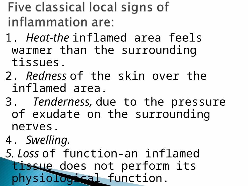
1. Heat-the inflamed area feels warmer than the surrounding tissues.
2. Redness of the skin over the inflamed area.
3. Tenderness, due to the pressure of exudate on the surrounding nerves.
4. Swelling.5. Loss of function-an inflamed tissue does not perform its physiological function.
Purulent infection penetrates through small injuries to the skin (so-called minor traumatism). In such cases a purulent process most frequently develops because the patients at first fail to pay proper attention to their affections and are not given timely and required aid. If aid is administered in due time (the wound is painted with iodine tincture and a sterile dressing is applied), i.e., the wound is protected against contamination, purulent diseases develop only in exceptional case.
The clinical picture of the disease develops according to the virulence of the microbes, which have gained entrance into the body and the state of the natural defensive powers of the latter.
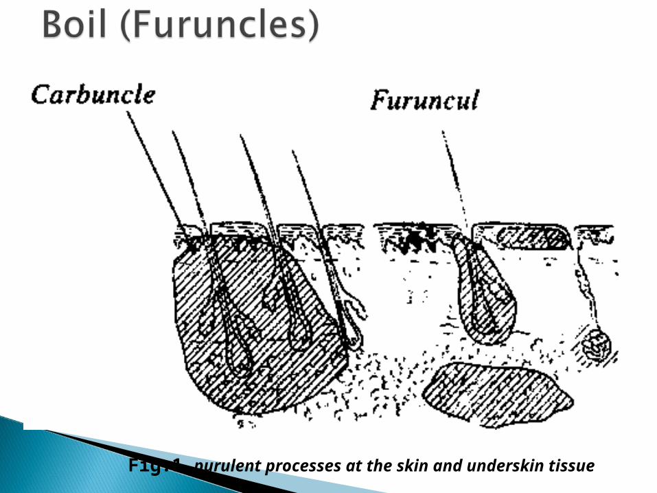
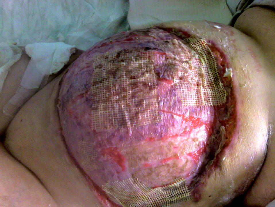
Fig.1 purulent processes at the skin and underskin tissue
Boil constitute one of the very widespread purulent diseases of the hair follicle and sebaceous (Fig.1). The disease begins with an appearance of a painful infiltrate in the skin in the form of an inflammatory node differing in size from a pea to a pigeon egg; the skin grows red over the swelling and local pyrexia is observed in the region of the focus. The disease develops over a period of 4 - 6 days, and a purulent blister (exfoliation of the epidermis by pus) is formed at the most protruding part of the swelling. A focus consisting of a necrotic gland with surrounding subcutaneous tissue is found under this blister; the subcutaneous tissue is subsequently discharged together with the pus (the core of the boil). The remaining small cavity fills with granulations and heals.
a) boil may lead to cellulilis, particularly in those whose power of immunity is less.
b) boils may also lead to infection of the neighbouring hair follicles where numbers of hair follicles are too many (e.g. axilla) leading to hidradenitis.
c) boils usually secondarily infect the regional lymph nodes.
1. The general health of the patient has to be improved, as boils often occur in individuals with debility and ill-health.
2. Incision is usually unnecessary as the pustule is very small. Only a touch of iodine on the skin pustule will hasten necrosis of the overlying skin and help the pus to drain out.
3. If escape of pus does not occur spontaneously or with application of iodine, removal of the affected hair allows ready escape of pus.
4. Antibiotic is usually not required. It is possible when multiple boils appear or if boils recur.
5. After escape of pus the part should be cleaned twice with a suitable disinfectant e.g hexachlorophene. This discourages development of further boils.
6. If boils are recurrent, diabetes should be excluded.
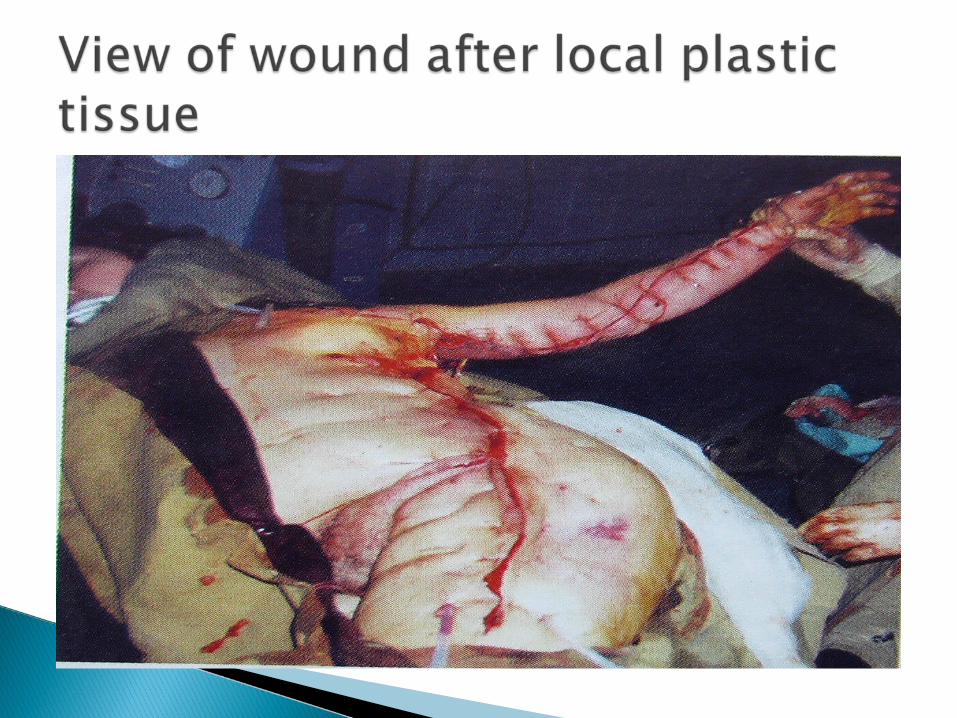
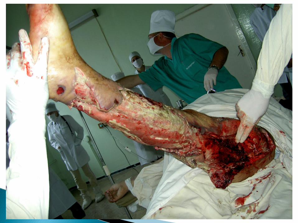
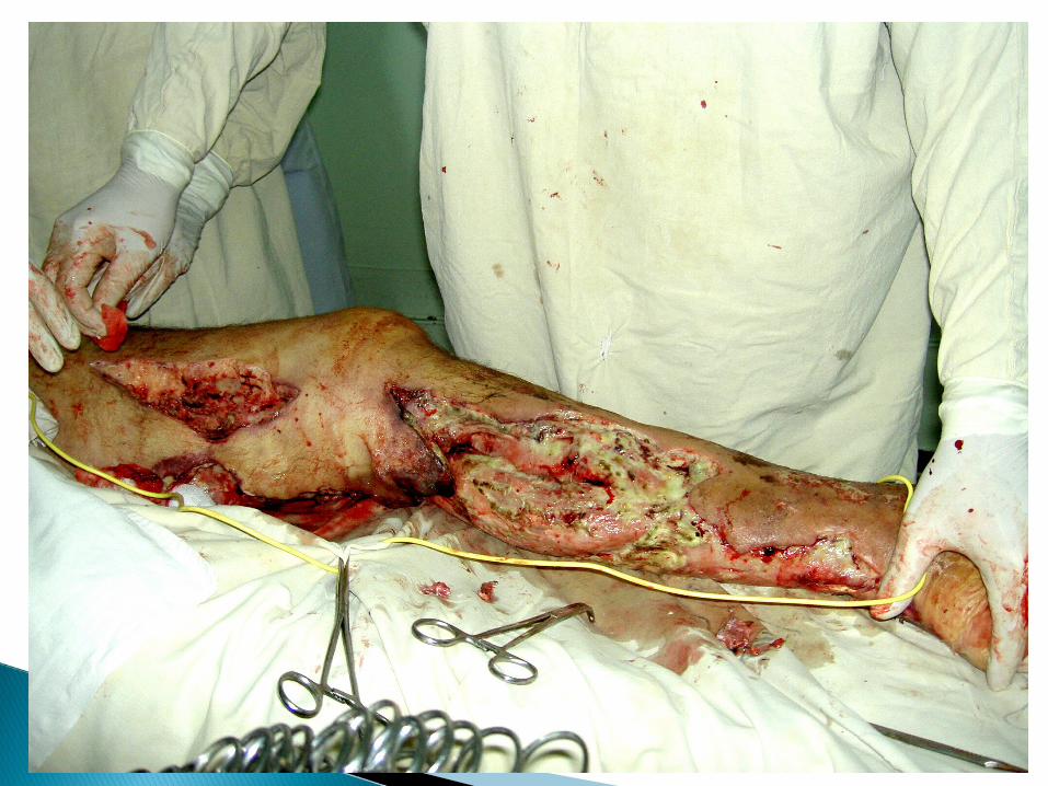
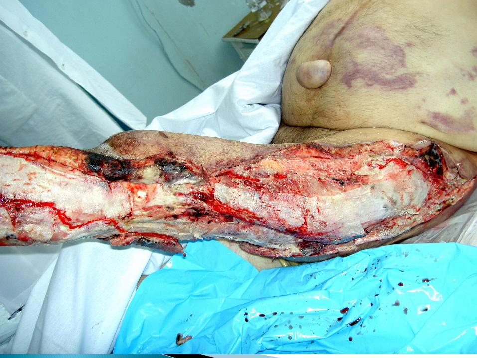
After penetration of pyogenic bacteria under the skin through hair follicles and sebaceous glands the process spreads in depth, if the conditions are unfavourable to the body, and affects considerable sections of subcutaneous tissue (Fig.1). It is an infective gangrene of the subcutaneous tissue due to Staphylococcal (Staphylococcus aureus) infection. Gram-negative bacilli and Streptococci may be found coincidently.
Sites. Carbuncles are mostly seen on the back, in the nape of the neck where the skin is coarse and vitality of the tissue is less. The shoulders, the cheek, dorsum of the hand are the other rare sites. Hirsute portions of the chest and abdomen may also be involved.
It commences as painful and stiff swelling which spreads very rapidly with marked induration. The overlying skin becomes red, dusky and oedematous. Subsequently the central part softens and a multiple of vesicles appear on the skin. Later these vesicles transform into pustules. These pustules subsequently burst allowing the discharge to come out through several openings in the skin producing a sieve-like or cribriform appearance. These openings enlarge and ultimately coalesce to produce an ulcer. At low floor of the ulcer lies the ashy-grey slough. Finally the slough separates leaving an excavated granulated fascia, which heals by itself. When the resistance of the individual is poor in diabetic subject, the sloughing process may extend deeply into the muscle or even bone.
Constitutional symptoms and toxaemia vary according to the degree of the resistance of the individual and Scarecy of the treatment.
a) improvement of the general health of the patient should be brought about.
b) proper antibiotic should be started immediately from the culture and sensitivity test. If the surface openings have not formed, a synthetic penicillin e.g. erythromycin may be used. At this time a paste composed of anhydrous magnesium sulphate and glycerin may be applied or S. S. Mag Sulph powder is used on a moisten cotton and placed on the affected area. This will exercise a valuable osmotic effect and will not only reduce oedema but also will help to burst the carbuncle. Hot compress is helpful before bursting. It may be supplemented by infra-red or short wave diathermy.
Operation may be required:a) when toxaemia and pain persist even after a course of
antibiotics andb) when the carbuncle is more than 2 inches in diameter. It
must be remembered that incision is never made unless there is softening in the centre.
Technique. A large cruciate incision is made extending up to the margin of the inflammatory zone. Sloughs should be cleared with a piece of gauze. Epices of the four skin flaps are generously excised. The wound is covered with vaseline gauze or sofratulle dressing. The part should be kept in perfect rest for a week and antibiotic is continued till resolution.
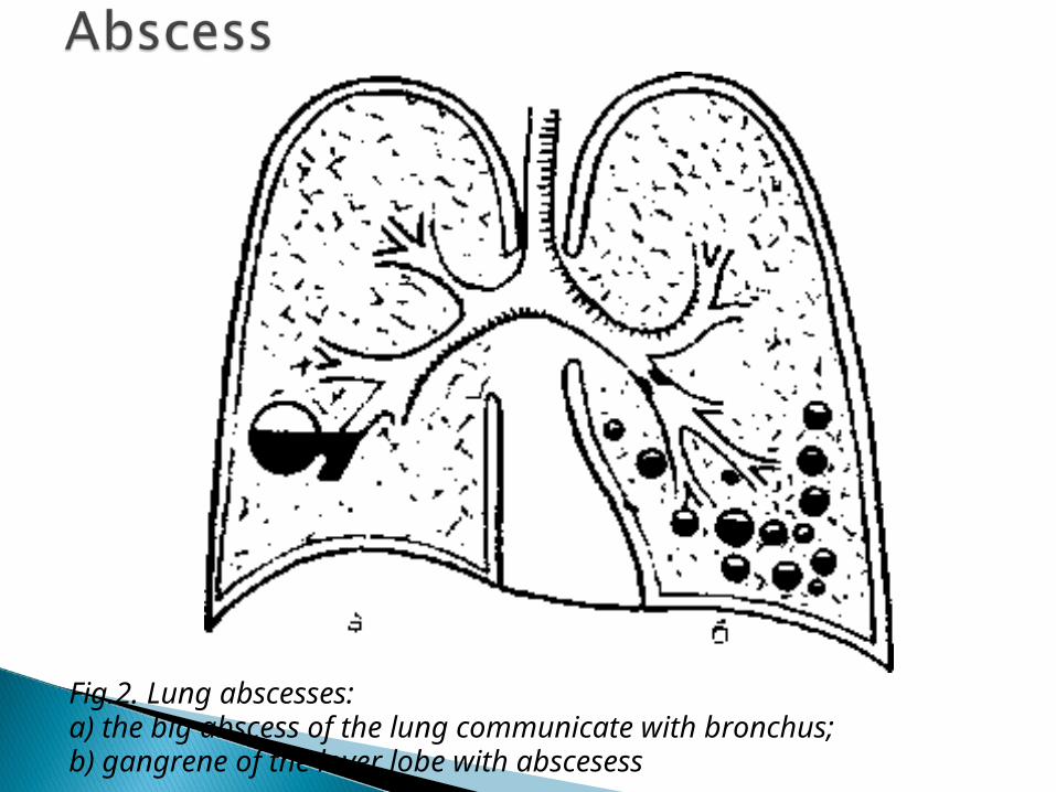
Fig.2. Lung abscesses:a) the big abscess of the lung communicate with bronchus;b) gangrene of the lover lobe with abscesess
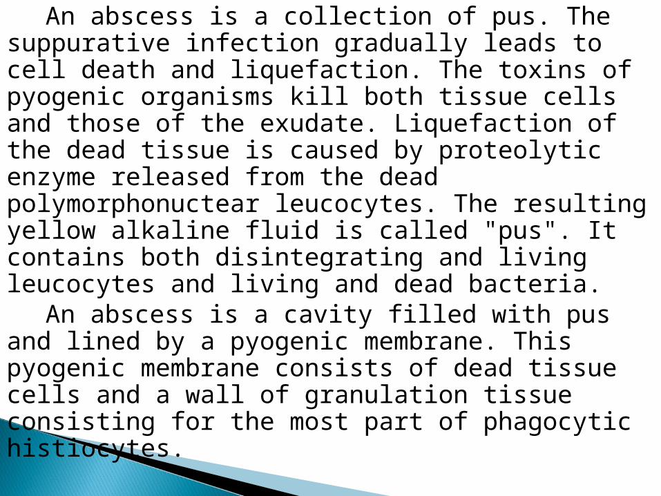
An abscess is a collection of pus. The suppurative infection gradually leads to cell death and liquefaction. The toxins of pyogenic organisms kill both tissue cells and those of the exudate. Liquefaction of the dead tissue is caused by proteolytic enzyme released from the dead polymorphonuctear leucocytes. The resulting yellow alkaline fluid is called "pus". It contains both disintegrating and living leucocytes and living and dead bacteria.
An abscess is a cavity filled with pus and lined by a pyogenic membrane. This pyogenic membrane consists of dead tissue cells and a wall of granulation tissue consisting for the most part of phagocytic histiocytes.
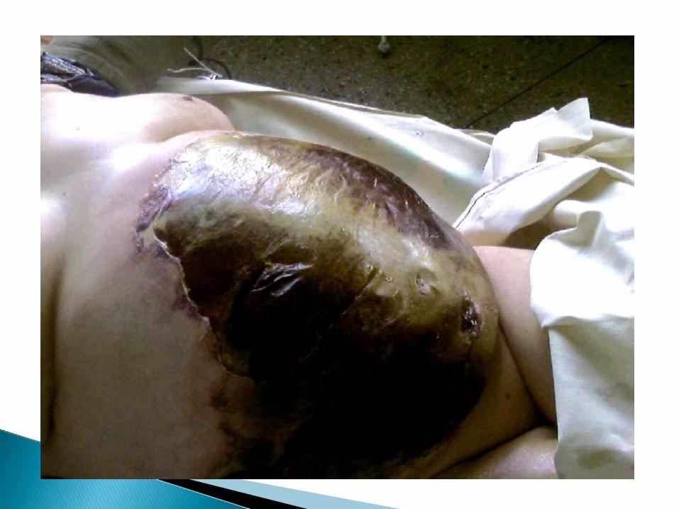
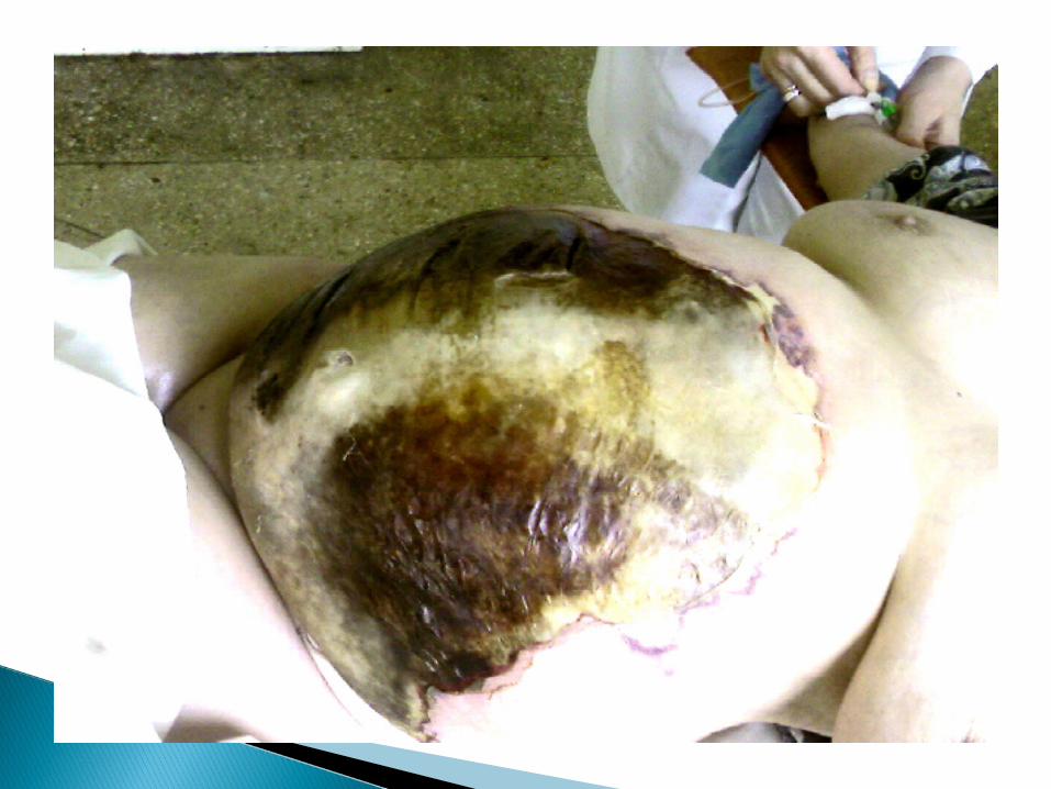

Cardinal features of acute inflammation are usually present. These are: a) redness or rubor—there is redness over the area particularly before localisation of the abscess. This is due to hyperaemia.
b) pain or dolor— a throbbing pain is characteristic of presence of pus.
c) heat or color— the inflamed area is hot due to hyperaemia (e.g. in cold abscess this is not present and that is why it is called "cold").
d) swelling or tumor— due to presence of pus inside the abscess cavity.
e) impairment of function — the function of the part is definitely impaired. This is more obvious when an abscess occurs near a joint, when movement of the joint will be painful and patient tries not to move the joint.
Nowadays various sophisticated investigations have been introduced to correctly located and accurately diagnosed abscess cavities in different parts of the body. The various methods are:
a) conventional radiology is only successful when there is air or gas with pus. This examination then reveals fluid levels, e.g. subphrenic abscess, lung abscess, etc. Sometimes presence of pus is suggested by opacity, e. g. in the nasal antrum, pleural cavity, etc.
b) isotope scanning is helpful in locating collection of pus or site of infection by accumulation of radioactive technique after its intravenous injection. This is mostly used as diagnostic tool in demonstrating brain abscess, hepatic abscess and osteomyelitis. Similarly radioactive gallium scan is sometimes used to detect pelvic, perinephric, mediastinal or subphrenic abscesses.
c) ultrasound is of considerable value in the diagnosis of gallbladder stones or empyema and also to detect abscesses in the liver or spleen.
d) CT scan is particularly helpful to distinguish between abscess and tumour by showing necrotic centre in case of abscess. It is helpful to locate abscess cavity inside the abdomen as also in the brain.
1. In the initial stage, when the pus is not localised, conservative treatment may be advised. The affected part is elevated and given rest. A suitable antibiotic should be started.
2. When the pus has been localised, it should be drained. The old adage holds true today also where there is pus, let it out.
So the basic principle of treatment of an abscess is: a) to drain the pus; b) to send a sample of pus for culture and
sensitivity test; c) to give proper antibiotic.
This method is chosen when there are plenty of important structures like nerves and vessels around the abscess cavity, which are liable to be injured. This is a particularly employed in place like neck, axilla or groin. In this technique the skin and subcutaneous tissue are incised on the most prominent and most dependent part of the abscess cavity. A pair of artery forceps or sinus forceps is forced through the deep fascia into the abscess cavity. The blades are gradually opened and the pus is seen to be extruded out. The forceps is now taken out with the jaws open to increase the opening in the deep fascia. A finger is introduced to explore the abscess cavity.
After the incision has been made up to the abscess cavity and some amount of pus has been extruded, a finger is inserted into the abscess cavity and all the walls of the loculi are broken. There must not be any loculus unbroken as this will lead to chronicity. All loculi are broken into one cavity for complete drainage.
When the most prominent part is not the most dependent part, complete drainage of pus is not possible with a single incision. So a counter-incision is required at the most dependent part to facilitate drainage by gravity. In this technique, through the first-made incision on the most prominent part, a sinus forceps is passed to the most dependent part. The blades are slightly made apart, then with a knife a fresh incision is made on the skin between the tips of the sinus forceps.
. A corrugated rubber drain is usually used for drainage of an abscess cavity. When counter-incision is used, the drain extends from the first incision to the counter - incision. When the surrounding granulation tissue is bleeding too much, a roller gauze should be packed inside the wound and it can be kept for 48 hours. Some surgeons believe in instilling local antibiotic into the abscess cavity.
Follow-up. Rest to the affected part is very important postoperative measure. This expedites healing.
Proper antibiotics selected by culture and sensitivity test should be started immediately.
After 48 hours the dressing or drain should be removed. Fresh dressing is done everyday with acriflavine lotion and sterile gauze. If the cavity has to be packed, the packing should be made gradually lighter to help the cavity to heal.
It is an acute inflammation of the lymphatics of the skin or mucous membrane. The causative organism is usually Streptococcus haemolyticus. The infection may be transmitted from one patient to another through the dressing material, hands of the medical personnel, instruments, etc. In erysipelas the disease begins with prodromal pheno mena—general indisposition suddenly followed by excessive chills and a temperature of 40 - 41°C; vomiting is sometimes observed. Subsequently the temperature either persists on a high level or from time to time drops.
The condition, which predisposes this disease are debilitating state and poor health. The condition commences as a rose-pink rash, which extends to the adjacent skin like a drop of grease spreading on a piece of paper. The vesicles appear sooner or later over the rash and rupture. Serous discharge comes out from these vesicles. Fever and other constitutional symptoms may be present with varying degrees. When it affects skin below which there is loose areolar tissues, e.g. orbit, scrotum etc., there is considerable swelling of the part due to oedema of the subcutaneous tissues and thus very much resembles cellulitis.
To distinguish between a true erysipelas and a cellulitis, the following points in favour of erysipelas should be born in mind:
a) the .typical rosy rash disappears on pressure and feels stiff;
b) the raised rash of erysipelas has a sharply defined margin, which is better felt than inspected;
c) the vesicles of erysipelas contain serum in contradistinction to the cellulitis in which they contain pus;
d) in case of the face, Milian's ear sign is significant in which erysipelas can spread into the pinna (being cuticular affection), whereas cellulitis cannot spread to the pinna due to close adhesion of skin to the cartilage of the ear (without any areolar tissue).
The patient must be confined to bed, all pressure or rubbing bandages must be removed and the tissues around the redness must be painted with iodine to prevent the disease from spreading. Irradiation by a sun lamp is effective. The wounds, which served as the source of infection must be examined to see if any pus is retained under their edges and if the pus is being well discharged.
Daily administration of antibiotics and sulfonylamides.
A spread of infection along the lymphatic system is manifested in a disease of the lymphatic vessels and lymph nodes. Inflammation of the lymphatic vessels (lymphangitis) is one of the frequent complications of infected wounds, especially during the first weeks following injury, and of local purulent diseases. Lymphangitis also develops in cases in which the discharge of pus from the wound is hampered, new infection gains entrance into the wound during dressing and during accelerated outflow of lymph, for example, as a result of untimely or vigorous movements of the affected organ.
In lymphangitis the local manifestations consist in appearance of longitudinal red lines on the skin along the course of the lymphatic vessels, i. e., inflamed superficial lymphatic vessels which are palpated as dense cords and are painful to touch. Simultaneously the adjacent lymph nodes (regional, for example inguinal or axillary) become swollen and painful; general phenomena in the form of chills and fever up to 40°C are also observed.
The treatment of lymphangitis consists primarily in elimination of its cause (incision of the abscess, pockets of the wound, etc.) and in giving the affected organ complete rest. Confinement of the patient to bed in lymphangitis of the leg and splint bandages if the disease affects the arm are obligatory. The red lines and the region of the swollen lymph, nodes are painted with iodine. Hot compresses and administration of antibiotics are recommended. If pus is retained in the region of the wound, an attempt may be made to remove it (the pockets are opened, the crusts are taken off and absorbent dressings are applied).
In purulent processes the infection spreads through the lymphatic vessels and penetrates into the lymph nodes where it is retained. In infected wounds this is very frequently manifested in swelling, enlargement and painfulness of the adjacent lymph nodes. If the disease develops on the arm, the elbow and axillary nodes enlarge; if the disease is on the leg or in the region of the perineum or the anus the inguinal lymph nodes become enlarged. The infection sometimes gains entrance into the lymph nodes after lymphangitis and sometimes develops spontaneously without any visible inflammatory phenomena in the lymphatic vessels.
The blood vessels constitute another avenue for the spread of purulent infection. Penetration of infection into the blood stream is not infrequently preceded by an inflammatory disease of the veins (phlebitis) with simultaneous thrombophlebitis. In thrombophlebitis the local manifestations are painfulness and induration along the course of the veins, the latter being palpable as dense cord is painful to touch. If large veins are affected, for example, the femoral vein, edema of the extremity and cyanosis develop
Treatment of thrombophlebitis consists primarily in giving the affected organ complete rest; to improve the conditions for blood outflow the organ must be placed in an elevated position. The patient may sometimes have to be in this position for several months, until the process has completely abated. It should be remembered that in thrombophlebitis any rubdowns and massages are strictly prohibited because they may induce the spread of the purulent process throughout the body and by carrying a disengaged blood clot in the blood stream may cause obstruction of important arteries (embolism), for example, the cerebral or pulmonary arteries. Leeches are not infrequently used in thrombophlebitis, five or six leeches being sticked to the skin of the extremity. Leeches clotting to prevent the progress of thrombosis by diminishing blood clotting, synthetic anticoagulants (dicoumarin, neodicoumarin, pelentan) are administered in thrombophlebitis. Since complications in the form of haemorrhages are possible during administration of anticoagulants, their use is permissible only with control of blood clotting (tests for prothombin) and systematic urinalyses.
Aetiology and pathogenesis. It is caused by mixed microflora (staphylococcus, enterococcus, esherichia coli, anaerobic microorganisms). It is usually observed at men. Appearing of the process is promoted by such things as chaps of the anus, inflammation of haemorrhoidal lymphatic nodes, damage of the mucous membrane of rectum.
Clinics. There are exist two forms of disease: diffuse (phlegmon of the pararectal cellular tissue) and limited. Phlegmon of the pararectal cellular tissue is characterised by the serious flow (fast distribution, necrosis of the tissue, marked intoxication); it is observed at shotgun wounds, decaying cancer of rectum, urinal phlegmons.
In the stage of infiltration conservative therapy b usually used (antibiotics, sparing thet). At the phlegmon or abscess there is indicated imidiate operation. It is frequently used the semilunar section 2 cm off of the external sphincter of the rectum. At submucous abscesses dissection of the abscess is made from the cavity of the rectum. At anaerobic paraproctitis there are indicated wide sections with the carving of necrodsed tissues and using of GBO. In the postoperative period there is used flowing bathing with the solutions of antiseptics (hydrogen peroxide, dioxidine), proteolytic enzymes, hip-baths with die solutions of antiseptics
Parotitis- is purulent inflammation of the parotic gland. It is occured in the result of infectioning the parotic gland haematogenously or lymphogenously or along the excretory ducts from the oral cavity of. It is arised at weaked persons with general infection or after big operations with dehydratation of the organism and bad cars of the oral cavity. Pathogenic microorganisms are often staphylococcuss and streptococcus. Limited abscesses are formed in the gland or phlegmon with spreading on the cellular tissue. At these persons we can see purulent swellings on the neck and in the temporal area.
Clinics. In the area of the parotic gland there is observed swelling and painfulness at palpation. It is accompanied by worsening of the general condition (chill, increasing of the temperature up to 39- 40°C, difficulties at swallowing and chewing).
In the area of swelling redness of the skin and fluctuation is seen. Swelling moves on the soft palate, neck, cheek, submandibular area. At some ill persons we can determine paresis of the facial nerve. Abscess can self-dissects with the formation of fistules through which secvestres of the necrodsed tissue of the gland come out. Serious complication is the generalization of infection(sepsis) which gives the high mortality.
Treatment. In the first stages usually use: antibiotics (semisynthetic penicilines, aminoglicosides, cephalosporines), warm procedures (compresses, UHF, sollux), and sanation of the oral cavity (rinsing with the solutions of antiseptics, massage of the mucous membrane of die oral cavity).
At the abscessing there, is indicated operation- dissection of the purulent focuses in the gland and formation of pus outflow. Dissection of the abscess must be made in the region of the most fluctuation with taking into coonsideration the direction of the basic branches of the facial nerve. We dissect the skin and capsule of the gland; than by the blunt way (corcang or finger) open the abscess in the glandular tissue. Then there is used draining, bathing with antiseptics, proteolytic enzymes. Localy are use antibiotics, water-soluble ointments (levosin, levomekol, dioxykol and other). There is indicated plentiful drinking, diet, and therapy with vitamins, protein preparations.
At parotitis several serious complications can occur haemorrhages from the vessels in the gland or carotic arteries at purulent leaking, phlegmons of arround-pharyngeous cellular tissue, profund phlegmons of the neck.
Mastitis- is inflammation of lactic gland tissue. There is distinguished lactation mastitis at nursing mothers, mastitis of newboms and period of pubescence.
Aethiology and pathogenesis. Pathogenic microorganisms arc often staphylococcus and enterobacteries. The ways of infectioning: chaps of the nipple, intracanalicular (at nursing mothers), haemotogenous, and lymphogeneous (at endogeneous infection). Promotional factors are: stagnation of the milk in the gland, bad care of gland in the period of nursing.
Clinics. Distinguish acute and chronic forms. Acute mastitises are mostly at the period of lactation. Chronic arc very rare appear in the result of wrong treatment of the acute one or in the result of specific damage (tuberculosis, syphilis).
Acute mastitises is divided into serous, infiltrative, abscessing, phlegmonous, gangrenous. It can be said diat dlis are the stages of the same process, which turn one into another.
Treatment. The kind of treatment depends on the stage of the process. At serous and infiltrative forms there is indicated concervative therapy: nursing must not be stoped, the milk must be strained off by hand or with milksucker off), antibioticotherapy (semisynthetic penicilines, aminoglicosides, macrolids, cephalosporines), physiotherapy (sollux, UHF, ultrasound, UVI (Ultra Violet Irradiation), novocaine-electrophoresis). There is also can be used retromamaric novocaine blockade with antibiotics. At abscessing form it is indicated operative treatment. Sections are made dependent on localization of abscesses: at subareolaric- semilunar, at intramamaric- radial sections along the lactic ducts, at retromamaric- arched section under the gland
Ill persons with phlegmonous and gangrenous form of mastitis need in the urgent operation (several radial sections of 8-10 cm, carving of necrotised tissues, draining, flowing bathing with antiseptics). Treatment is suplemented'by infusion therapy (antibiotics, transfusion of the blood, stimulators of immunity), desintoxication (UVTB, haemosorbtion, hyperbaric oxygenation)