Embed Size (px)
Citation preview
MCL ELBOW SPRAIN
WHAT IS A MEDIAL COLLATERAL LIGAMENT SPRAIN? The medial collateral ligament of the elbow (MCL), is not to be mistaken with the identically named ligament in the knee. A MCL sprain is defined as the overstretching or tearing of the ligament on the inner aspect of the elbow. ANATOMY: The medial collateral ligament is a strong band of connective tissue that joins the humerus (upper arm) to the inner forearm (ulna). The MCL gives structure and stability to the elbow and prevents medial forces being placed through the elbow. INJURY CLASSIFICATION: MCL tears are graded on the following scale:
Ø Grade 1: Fewer than 10% of fibres are damaged with no loss of ligamentous integrity;
Ø Grade 2: Incomplete tearing of the MCL with increased joint laxity;
Ø Grade 3: Complete rupture of the MCL, with gross laxity.
Often there are further structures injured in a grade 3 rupture including cartilage, the elbow capsule and the bony surfaces.
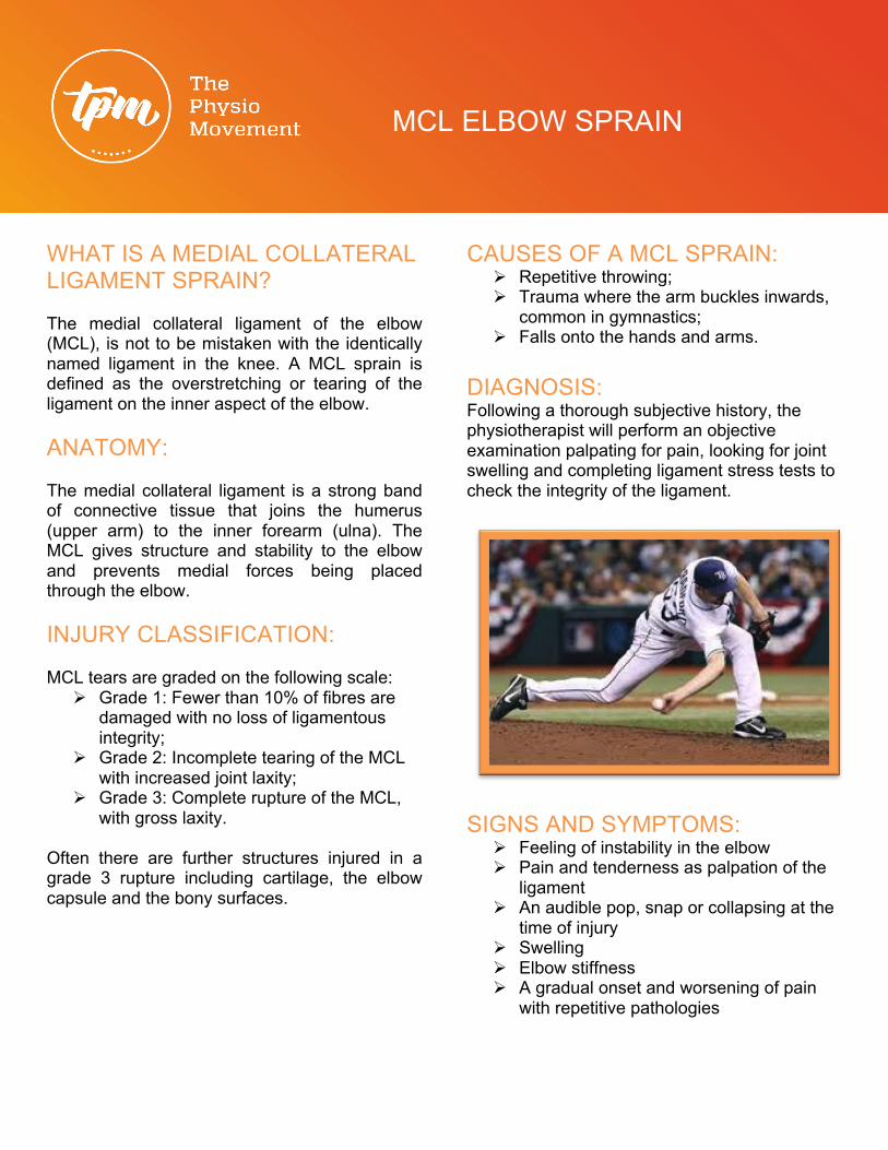
CAUSES OF A MCL SPRAIN: Ø Repetitive throwing; Ø Trauma where the arm buckles inwards,
common in gymnastics; Ø Falls onto the hands and arms.
DIAGNOSIS: Following a thorough subjective history, the physiotherapist will perform an objective examination palpating for pain, looking for joint swelling and completing ligament stress tests to check the integrity of the ligament.
SIGNS AND SYMPTOMS:
Ø Feeling of instability in the elbow Ø Pain and tenderness as palpation of the
ligament Ø An audible pop, snap or collapsing at the
time of injury Ø Swelling Ø Elbow stiffness Ø A gradual onset and worsening of pain
with repetitive pathologies
MCL ELBOW SPRAIN
PHYSIOTHERAPY TREATMENT OPTIONS:
Ø Massage Ø Exercise and
strengthening programs
Ø Activity reduction
Ø Splinting, bracing
Ø Neural glides Ø Anti-
inflammatory advice
Ø Dry Needling Ø Education Ø Electrotherapy Ø Facial releases Ø Joint mobility
techniques Ø Mobilisation
with movements
Ø Throwing technique modifications
PROGNOSIS: Grade 1-2 injuries will normally return to normal activities in 2-6 weeks and high-end sport at 8 weeks. Grade 3 ruptures will have a significantly longer rehabilitation depending on whether surgical intervention is required and how much associated damage is present.