Embed Size (px)
Citation preview
PROCEEDINGS Open Access
McCune-Albright syndrome and the extraskeletalmanifestations of fibrous dysplasiaMichael T Collins1*, Frederick R Singer2, Erica Eugster3
From International Meeting on Fibrous Dysplasia/McCune-Albright Syndrome and Cherubism: Best ClinicalPractice and Future ResearchBethesda, MD, USA. 3-5 October 2010
Abstract
Fibrous dysplasia (FD) is sometimes accompanied by extraskeletal manifestations that can include any combinationof café-au-lait macules, hyperfunctioning endocrinopathies, such as gonadotropin-independent precocious puberty,hyperthyroidism, growth hormone excess, FGF23-mediated renal phosphate wasting, and/or Cushing syndrome, aswell as other less common features. The combination of any of these findings, with or without FD, is known asMcCune-Albright syndrome (MAS). The broad spectrum of involved tissues and the unpredictable combination offindings owe to the fact that molecular defect is due to dominant activating mutations in the widely expressedsignaling protein, Gsa, and the fact these mutations arises sporadically, often times early in development, prior togastrulation, and can distribute across many or few tissues.The complexity can be mastered by a systematic screening of potentially involved tissues and cognizance that thepattern of involved tissues is established, to some degree, in utero. Thorough testing allows the clinician toestablish, often times at presentation, the full extent of the disease, and importantly as well what tissues areunaffected. Treatment and follow-up can then be focused on affected systems and a meaningful prognosis can beoffered to the patient and family. The authors outline screening and treatment strategies that allow for effectivemanagement of the extraskeletal manifestations of FD.
IntroductionThe original extraskeletal manifestations of fibrous dys-plasia (FD) reported by McCune [1] and Albright [2]were café-au-lait spots, precocious puberty, andhyperthyroidism. With time a number of manifestationswere added to the spectrum of findings that could beseen in association with FD. These included growth hor-mone (GH) excess [3], hypercortisolism [4], hypopho-sphatemia/osteomalacia [5], hepatic involvement [6],cardiac involvement [7], and others [8].
NIH cohortTo evaluate the extraskeletal manifestations observed inpatients with FD we reviewed all of the patients seen atthe National Institutes of Health over the last 24 years.
The evaluation included physical examination, imagingstudies (skeletal survey, head CT, nuclear medicine bonescan, ultrasound of the thyroid and gonads, and MRI ofthe pituitary), biochemical studies of skeletal metabolismand endocrine axes, and when available mutation analy-sis of affected tissue. There have been 140 patients eval-uated at the time of this review. Patients have beenfollowed from <1 – 24 years.
Prevalence of extraskeletal manifestationsThe relative prevalence of findings in MAS patients inthe NIH cohort are shown in Table 1. While these dataprobably reflect the relative prevalence of each of thesefindings, it is also likely that the NIH cohort represents amore severely affected group of patients than is typicallyfound in clinical practice. Therefore the likelihood of anindividual patient with FD having a given manifestationis probably lower than shown here.
1Skeletal Clinical Studies Unit, Craniofacial and Skeletal Diseases Branch,National Institute of Dental and Craniofacial Research, National Institutes ofHealth, Bethesda, MD, USAFull list of author information is available at the end of the article
Collins et al. Orphanet Journal of Rare Diseases 2012, 7(Suppl 1):S4http://www.ojrd.com/content/7/S1/S4
© 2012 Collins et al; licensee BioMed Central Ltd. This is an Open Access article distributed under the terms of the Creative CommonsAttribution License (http://creativecommons.org/licenses/by/2.0), which permits unrestricted use, distribution, and reproduction inany medium, provided the original work is properly cited.
In addition to the major and more common/classicfindings seen in association with FD as part of theMcCune-Albright syndrome, we have observed a num-ber of other findings in associated with the disease.These are shown in Table 2.
Timing of appearance of extraskeletalmanifestationsAn important consideration in terms of patient/familycounseling and the ability to give a prognosis for patients
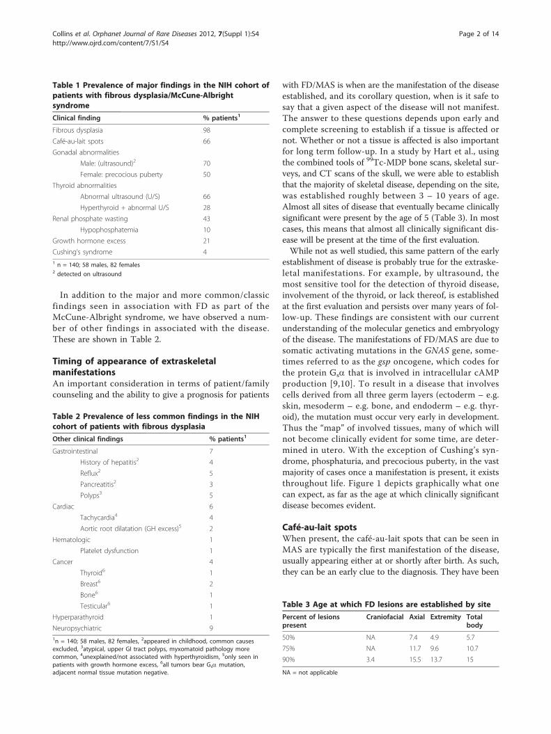
with FD/MAS is when are the manifestation of the diseaseestablished, and its corollary question, when is it safe tosay that a given aspect of the disease will not manifest.The answer to these questions depends upon early andcomplete screening to establish if a tissue is affected ornot. Whether or not a tissue is affected is also importantfor long term follow-up. In a study by Hart et al., usingthe combined tools of 99Tc-MDP bone scans, skeletal sur-veys, and CT scans of the skull, we were able to establishthat the majority of skeletal disease, depending on the site,was established roughly between 3 – 10 years of age.Almost all sites of disease that eventually became clinicallysignificant were present by the age of 5 (Table 3). In mostcases, this means that almost all clinically significant dis-ease will be present at the time of the first evaluation.While not as well studied, this same pattern of the early
establishment of disease is probably true for the extraske-letal manifestations. For example, by ultrasound, themost sensitive tool for the detection of thyroid disease,involvement of the thyroid, or lack thereof, is establishedat the first evaluation and persists over many years of fol-low-up. These findings are consistent with our currentunderstanding of the molecular genetics and embryologyof the disease. The manifestations of FD/MAS are due tosomatic activating mutations in the GNAS gene, some-times referred to as the gsp oncogene, which codes forthe protein Gsa that is involved in intracellular cAMPproduction [9,10]. To result in a disease that involvescells derived from all three germ layers (ectoderm – e.g.skin, mesoderm – e.g. bone, and endoderm – e.g. thyr-oid), the mutation must occur very early in development.Thus the “map” of involved tissues, many of which willnot become clinically evident for some time, are deter-mined in utero. With the exception of Cushing’s syn-drome, phosphaturia, and precocious puberty, in the vastmajority of cases once a manifestation is present, it existsthroughout life. Figure 1 depicts graphically what onecan expect, as far as the age at which clinically significantdisease becomes evident.
Café-au-lait spotsWhen present, the café-au-lait spots that can be seen inMAS are typically the first manifestation of the disease,usually appearing either at or shortly after birth. As such,they can be an early clue to the diagnosis. They have been
Table 1 Prevalence of major findings in the NIH cohort ofpatients with fibrous dysplasia/McCune-Albrightsyndrome
Clinical finding % patients1
Fibrous dysplasia 98
Café-au-lait spots 66
Gonadal abnormalities
Male: (ultrasound)2 70
Female: precocious puberty 50
Thyroid abnormalities
Abnormal ultrasound (U/S) 66
Hyperthyroid + abnormal U/S 28
Renal phosphate wasting 43
Hypophosphatemia 10
Growth hormone excess 21
Cushing’s syndrome 41 n = 140; 58 males, 82 females2 detected on ultrasound
Table 2 Prevalence of less common findings in the NIHcohort of patients with fibrous dysplasia
Other clinical findings % patients1
Gastrointestinal 7
History of hepatitis2 4
Reflux2 5
Pancreatitis2 3
Polyps3 5
Cardiac 6
Tachycardia4 4
Aortic root dilatation (GH excess)5 2
Hematologic 1
Platelet dysfunction 1
Cancer 4
Thyroid6 1
Breast6 2
Bone6 1
Testicular6 1
Hyperparathyroid 1
Neuropsychiatric 91n = 140; 58 males, 82 females, 2appeared in childhood, common causesexcluded, 3atypical, upper GI tract polyps, myxomatoid pathology morecommon, 4unexplained/not associated with hyperthyroidism, 5only seen inpatients with growth hormone excess, 6all tumors bear Gsa mutation,adjacent normal tissue mutation negative.
Table 3 Age at which FD lesions are established by site
Percent of lesionspresent
Craniofacial Axial Extremity Totalbody
50% NA 7.4 4.9 5.7
75% NA 11.7 9.6 10.7
90% 3.4 15.5 13.7 15
NA = not applicable
Collins et al. Orphanet Journal of Rare Diseases 2012, 7(Suppl 1):S4http://www.ojrd.com/content/7/S1/S4
Page 2 of 14

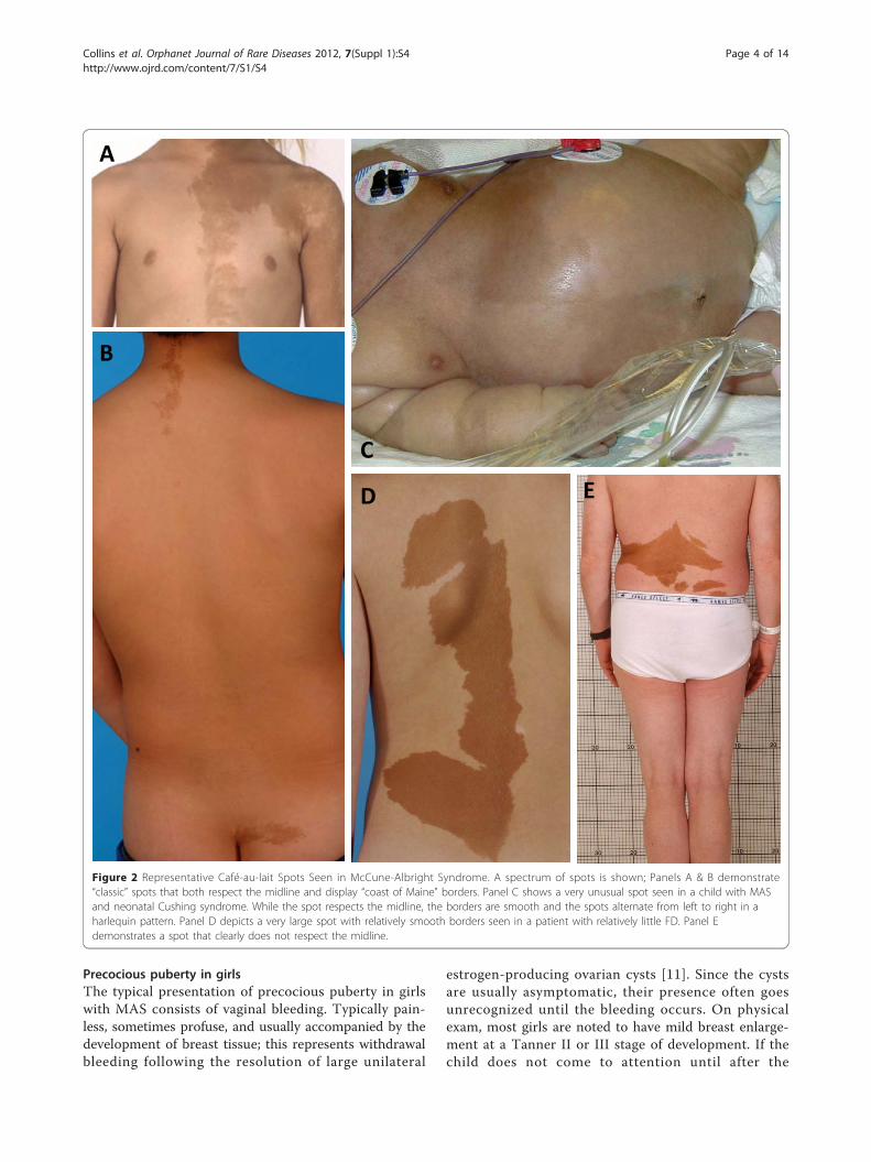
classically described as having a “coast of Maine” border,which refers to the jagged appearance of the Maine coast-line as it appears on maps. While this is usually the case, itis not always true. Examples of café-au-lait spots seen inMAS that both conform with and defy this dictum areshown in Fig. 2. Likewise, café-au-lait spots found in MASusually show some association with (“respect”) to the mid-line. Again, while this is often the case, there are frequentexceptions. Examples of these can be seen in Fig. 2 C & E.While these spots do cross the midline, they retain someassociation to the midline.Contrary to what has been previously reported, we have
not observed a correlation between the size of the spotsand the extent of the disease. Nor have we observed a cor-relation between side of the body on which the spot isfound and the side of the body on which the FD is found.The café-au-lait spots seen in association with FD are
the result of gsp-bearing melanocytes in which themutation brings about c-AMP-mediated tyrosinase geneactivation and melanin production in mutation-bearingcells [11]. There are no well-defined effective treatmentsfor the hyperpigmentation seen in MAS. Attempts tobleach areas of hyperpigmentation usually leave an areaof under pigmentation, which is usually unsatisfying to
the patient. A single report of the efficacy of Q-switchedruby laser in the treatment of the café-au-lait spots ofMAS has been reported [12], but further evidence ofefficacy is necessary before such a treatment can be rou-tinely recommended.
Precocious pubertyIntroductionPrecocious puberty is one of the defining manifestationsof McCune-Albright syndrome (MAS) (10). It arises dueto autonomous gsp-mediated gonadal function in cellsharboring the GNAS activating mutation. Thus, it is char-acterized as a form of peripheral precocious puberty, incontrast to the early hypothalamic-pituitary-gonadal(HPG) axis activation designated as central precociouspuberty. Although it might theoretically be expected toaffect girls and boys equally, precocious puberty in chil-dren with MAS is far more common in girls, in whom itis typically both the presenting feature as well as the onethat ultimately leads to the diagnosis being made. As theclinical characteristics, diagnosis and treatment are dis-tinctly different, precocious puberty in girls will be con-sidered separately from precocious puberty in boys withMAS.
Fibrous dysplasia
Café-au-lait
Precocious Puberty
Thyroid
Phosphaturia
Growth Hormone Excess
Cushings0 5 10 15 20 30 50
Age pre-clinical clinically evident
spontaneous resolution possible
persistent abnormal menses
Figure 1 The relative age at which any given aspect of the disease becomes clinically evident is depicted by a solid black bar. Preclinical diseaseis depicted by gray bars, and ages during which spontaneous resolution is possible for Cushing’s disease and phosphaturia are shown in openbars. The period of time during which abnormal menstruation can be expected is depicted by the stippled bar.
Collins et al. Orphanet Journal of Rare Diseases 2012, 7(Suppl 1):S4http://www.ojrd.com/content/7/S1/S4
Page 3 of 14
Precocious puberty in girlsThe typical presentation of precocious puberty in girlswith MAS consists of vaginal bleeding. Typically pain-less, sometimes profuse, and usually accompanied by thedevelopment of breast tissue; this represents withdrawalbleeding following the resolution of large unilateral
estrogen-producing ovarian cysts [11]. Since the cystsare usually asymptomatic, their presence often goesunrecognized until the bleeding occurs. On physicalexam, most girls are noted to have mild breast enlarge-ment at a Tanner II or III stage of development. If thechild does not come to attention until after the
A
B
C
D E
Figure 2 Representative Café-au-lait Spots Seen in McCune-Albright Syndrome. A spectrum of spots is shown; Panels A & B demonstrate“classic” spots that both respect the midline and display “coast of Maine” borders. Panel C shows a very unusual spot seen in a child with MASand neonatal Cushing syndrome. While the spot respects the midline, the borders are smooth and the spots alternate from left to right in aharlequin pattern. Panel D depicts a very large spot with relatively smooth borders seen in a patient with relatively little FD. Panel Edemonstrates a spot that clearly does not respect the midline.
Collins et al. Orphanet Journal of Rare Diseases 2012, 7(Suppl 1):S4http://www.ojrd.com/content/7/S1/S4
Page 4 of 14

resolution of the initial episode, the breast tissue mayhave resolved and on casual inspection involuted breasttissue may be missed. Similarly, the finding of obviousand classic café-au-lait macules in girls with MAS isquite variable and, even if present, their significancemay go unrecognized. Therefore, it is not unusual forgirls to present initially to an emergency department orprimary care clinic and have the treating physicians failto include MAS in the differential diagnosis. As some ofthe clinical and radiographic findings overlap with thoseof juvenile granulosa cell tumors, girls with MAS some-times end up undergoing unnecessary oophorectomy fora presumed ovarian tumor [12]. Ideally, vaginal bleedingin a prepubertal girl should always prompt consultationwith a pediatric endocrinologist so that, in the case ofMAS, unneeded loss of the ovary can be prevented.In addition to a history and physical exam, the initial
evaluation of precocious puberty in a girl with suspectedMAS consists of laboratory and radiographic studies. Clas-sic biochemical findings include elevated estradiol andestrone levels, which are many-fold higher than prepuber-tal values, in association with suppressed gonadotropins.Pelvic ultrasound typically reveals a large unilateral ovariancyst which may be hemorrhagic and appear to have mixedcystic and solid elements. As would be predicted, extremeasymmetry in ovarian volumes between the two sides isthe norm, in striking contrast to the symmetrical ovarianenlargement emblematic of central precocious puberty[13]. If seen after the initial episode, growth parametersand bone age x-ray are often normal. The diagnosis ofMAS is typically made clinically on the basis of classic fea-tures, including café-au-lait pigmentation. A bone scan tolook for fibrous dysplasia and screening for other MAS-associated endocrinopathies are important elements of thediagnostic work-up. However, isolated precocious pubertywithout any other identifiable abnormalities may also beseen in girls with MAS [14]. Serial ultrasounds, if indi-cated, will reveal a gradual resolution of the ovarian cystover several weeks.The natural history of precocious puberty in girls with
MAS is extremely variable. The first episode can occur asearly as during the first few months of life or as late asage 6 or 7 years. Likewise, subsequent episodes are highlyunpredictable. While many girls have extended periodsof quiescence that last for several years, others have fre-quent bouts of vaginal bleeding along with progressivebreast development followed by the onset of pubic andaxillary hair and adult body odor. As is seen with allforms of significant sex steroid exposure during the pre-pubertal years, linear growth acceleration and advancedskeletal maturation also ensue. Unfortunately, there is noway to predict exactly when the next episode of preco-cious puberty will occur, which can contribute to theanxiety experienced by parents when this complex
disorder is diagnosed. Similarly, the precocious pubertyflare-ups themselves vary in severity. In contrast to thetypical vaginal bleeding, some girls are noted to simplyhave periodic waxing and waning of breast enlargementwithout overt bleeding.Historically, the prevailing notion was that the HPG
axis would override autonomous ovarian function in girlswith MAS once physiologic puberty was underway. How-ever, this has given way to the recognition that womenwith MAS continue to experience intermittent autono-mous ovarian function marked by the development oflarge unilateral ovarian cysts and irregular vaginal bleed-ing [15]. This has the potential to interfere with normalovulatory function with subsequent implications in termsof fertility [16]. However, in most cases adults with MAShave been able to have children, even if it may takelonger than normal to conceive.
ManagementClinical management of precocious puberty in a girl withMAS consists initially of observation. Girls with onlysporadic and infrequent vaginal bleeding often do notneed to be treated. In the subset of girls with a progres-sive form of precocious puberty, pharmacologic interven-tion is recommended in order to prevent early epiphysealfusion and augment adult height. However, other thananecdotal case reports, to what extent height is compro-mised and whether intervention ameliorates this, is notwell established. As is the case for all aspects of MAS,both the rarity and heterogeneity of the disease presentsignificant challenges to rigorous investigation.Current treatment of precocious puberty in girls with
MAS revolves around the use of anti-estrogens. Twobasic strategies exist. The first relies on interfering withestrogen biosynthesis through the use of an aromataseinhibitor [17], while the second aims to blunt the effectsof estrogen at the level of the end-organ through receptorblockade. To date, small uncontrolled trials have beenconducted with first, second and third generation aroma-tase inhibitors. Experience with the first generationagent, testolactone, was ultimately marred by sub-optimalefficacy as well as issues with compliance [18]. Investiga-tion of the second generation aromatase inhibitor, fadro-zole, was abandoned following concerns about adrenalsuppression [19]. Among the third generation com-pounds, anastrozole has been deemed ineffective [20].Letrozole, however, was found to result in a significantdecrease in rates of skeletal maturation in a small num-ber of girls treated for 3 years, although mean ovarianvolumes were unchanged [21]. Most girls also experi-enced a decrease or cessation in vaginal bleeding whileon letrozole, although one subject who had entered sec-ondary central precocious puberty developed a large cystwith subsequent ovarian torsion. Treatment with the
Collins et al. Orphanet Journal of Rare Diseases 2012, 7(Suppl 1):S4http://www.ojrd.com/content/7/S1/S4
Page 5 of 14

selective estrogen receptor modulator, tamoxifen, hasalso been studied in a group of girls with MAS treatedfor one year. In addition to a significant decrease in vagi-nal bleeding, tamoxifen resulted in an improvement ingrowth velocity and bone age advancement [22]. Despitethese positive results, the finding of increased uterine andovarian volumes in the girls treated with tamoxifenrepresents a potential safety concern that to date remainsunresolved. Lastly, preliminary results from a prospectivestudy utilizing the pure estrogen receptor blocker, fulves-trant, are available. A decrease in the median number ofvaginal bleeding days as well as in the average rate of ske-letal advancement in 30 girls treated for one year wasseen [23]. Thus, relatively comparable efficacy has nowbeen observed with several agents used in the treatmentof precocious puberty in girls with MAS, although nonehave been perfect and none have emerged as beingclearly superior to the others. Studies comparing avail-able medications in a head to head fashion are needed.
Precocious puberty in boysThere are several important differences between preco-cious puberty in girls with MAS and its counterpart inboys. One distinction is that precocious puberty is veryrare in affected boys, who are diagnosed with MAS farmore often due to the finding of bone disease or café-au-lait pigmentation. An additional dissimilarity is thatthe precocious puberty, when present, is more likely tobe subtle and indolent in boys. Lastly, the activating Gsamutation and resulting gonadal hyperfunction have beenreported to be limited to the testicular Sertoli cells inseveral boys with MAS. This has resulted in either uni-lateral or bilateral macroorchidism without precociouspuberty [24][25][26][27]. Interestingly, many of thesecases have also been associated with testicular micro-lithiasis, which has also been identified in males of allages with MAS [28][29]. Due to its extreme rarity, onlyanecdotal case reports detailing treatment options forprecocious puberty in boys are available. The most com-mon approach employs combination therapy in the formof an androgen receptor blocker such as spironolactone,flutamide or cyproterone acetate along with a com-pound that interferes with sex steroid synthesis such asketoconazole or an aromatase inhibitor [30]. On princi-ple, the same strategies used to treat boys with otherforms of peripheral precocious puberty such as familialmale precocious puberty, would be efficacious in thesetting of MAS. One such example is the combinationof bicalutamide, a pure androgen receptor blocker, withthe third generation aromatase inhibitor anastrozole[31]. Similar to what has been reported in women withMAS, fifteen year follow-up in a boy with MAS andhistory of precocious puberty indicated persistent auton-omous testicular hyperfunction and suppressed
gonadotropins [32]. Although inhibin B was undetect-able, active spermatogenesis occurred and was seeminglyunaffected.
ThyroidAt the NIH approximately 2/3 of the patients had invol-vement of the thyroid when assessed by the most sensi-tive method for assessing thyroid involvement,ultrasound [13]. Only about 1/2 of the patients who hadinvolvement of the thyroid detected on ultrasound hadfrank hyperthyroidism, as evidenced by a suppressedTSH. As in every aspect of MAS, the thyroid findingsexist along a spectrum from an isolated area seen onultrasound with no clinical findings to patients withobvious goiters, and hyperthyroidism that is unable to beadequately controlled with medications and requireseither surgery or ablation. The presence of the gsp muta-tion in thyroid tissue results in ligand-independent acti-vation of the TSH/G-protein/cAMP pathway, which isknown to result in both hyperplasia and hyperfunction[14]. Additionally, the gsp mutation results in increasedthyroxine (T4) to triiodothyronine (T3) conversion,which accounts for the T3-dominant biochemical pheno-type of MAS patients with hyperthyroidism [13].It is important to diagnose hyperthyroidism in MAS, as
hyperthyroidism can advance bone age, which mayalready be a problem in children with precocious pub-erty, lead to or exacerbate osteoporosis, and cause aplethora of other metabolic derangements. Diagnosis isusually straight forward and involves the measurement ofTSH and thyroid hormones, T3 and T4. It is not uncom-mon to have a normal T4 in the setting of a suppressedTSH. This apparently incongruous finding is clarifiedwhen T3 is measured and found to be high. In patients inwhom the only abnormality is an abnormal ultrasound, itis important to continue to check TSH and thyroid hor-mone periodically as the development of frank hyperthyr-oidism may occur later. The ultrasound findings in MASare usually a mixture of mostly cystic with some solidlesions (Fig. 3) [13,15].Hyperthyroidism in MAS usually responds quite well
to thionamides. However, since hyperthyroidism is oneof the aspects of MAS that persists, it is often desirablefor the patient to undergo definitive treatment, whichusually means surgery or ablation with radioactiveiodine. Surgery may be difficult in very small children,and is therefore recommended to delay surgery in smallchildren.
HypophosphatemiaWhile rickets in association with FD was originallyreported in 1968 [5], it was not until 2001 that it was evi-dent the cause was a circulating phosphaturic hormone,similar to what is seen in the inherited forms of rickets
Collins et al. Orphanet Journal of Rare Diseases 2012, 7(Suppl 1):S4http://www.ojrd.com/content/7/S1/S4
Page 6 of 14

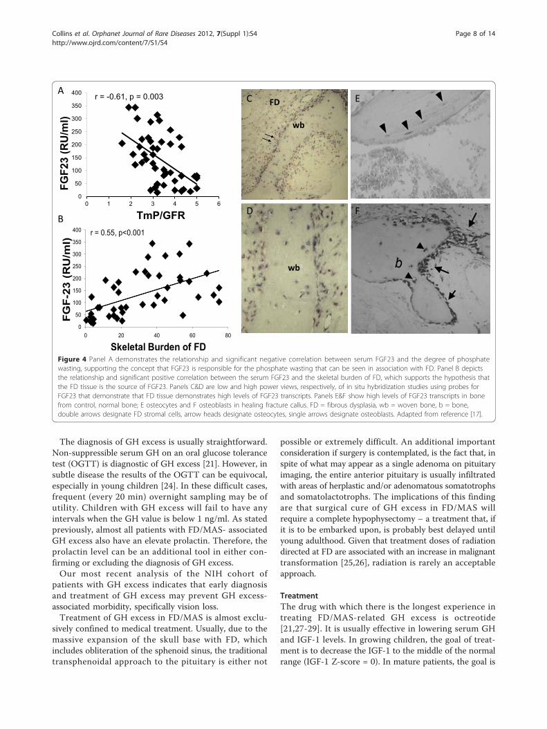
[16]. Overproduction of FGF23 by FD tissue was found tobe the cause [17]. FGF23 is overproduced by FD tissue,such that the greater the disease burden, the higher theFGF23, the greater the degree of renal phosphate wasting,and the lower the serum phosphorus (Fig. 4). Therefore,significant hypophosphatemia is only seen in patientswith a very significant skeletal burden of FD. It has alsobeen observed that, unlike many other extraskeletal man-ifestations aspects, renal phosphate wasting can sponta-neously resolve as patients age. This probably reflectsintrinsic changes that have been observed at the tissuelevel and characterized as “normalization” [18].The clinical sequelae and significance of hypophospha-
temia are an earlier age of first fracture, more fractures,and bone pain [19]. There are no controlled studies tosupport that treating hypophosphatemia decreases frac-tures or improves pain, but observation of treatedpatients suggests that treatment may improve outcomes.Treatment of hypophosphatemia is the same as in otherFGF23-mediated phosphate wasting disorders, andinvolves the use of phosphate and active vitamin D (calci-triol or alfacalcidiol). Details for this treatment regimencan be found elsewhere [8,20].
Growth hormone excessGrowth hormone excess in association with FD is themanifestation of gsp mutation in the anterior pituitary[21]. It is always accompanied by skull base FD, andthe vast majority of patients also have hyperprolactine-mia. The usual presenting sign is increased growthvelocity. However, if GH excess is accompanied by pre-cocious puberty, the clinical sign of increased growthvelocity can be obscured by the increase in growthvelocity that is seen as part of precocious puberty.Likewise, if a patient with precocious puberty achieveshis/her predicted height, this can be a sign of GHexcess, as precocious puberty should have resulted inshort stature. For this reason, it is important to dolaboratory screening for GH excess in patients withFD, as the clinical evaluation can be confounded byconcomitant hormonal excess and make the clinicalexam difficult to interpret.Probably the most important reason to diagnose GH
excess in association with FD is that it is associated withan increase in morbidity, specifically in the craniofacialregion. GH excess in FD is associated with macroce-phaly and vision loss [22,23].
Figure 3 Clinical and ultrasound findings of thyroid involvement in FD/MAS. Panels A & B demonstrate the findings in a 9-year-old girl withMAS and hyperthyroidism. A goiter is clearly seen on inspection (A) and the ultrasound (B) shows the typical cystic (Swiss cheese) appearancethat seen in MAS thyroids. Panels C & D demonstrate the findings of a 30-year-old woman with MAS. While no goiter was evident on inspection,nor was one obvious on palpation, the ultrasound clearly demonstrated the typical findings seen in ultrasounds of patients with MAS andthyroid involvement. Adapted from reference [13.]
Collins et al. Orphanet Journal of Rare Diseases 2012, 7(Suppl 1):S4http://www.ojrd.com/content/7/S1/S4
Page 7 of 14
The diagnosis of GH excess is usually straightforward.Non-suppressible serum GH on an oral glucose tolerancetest (OGTT) is diagnostic of GH excess [21]. However, insubtle disease the results of the OGTT can be equivocal,especially in young children [24]. In these difficult cases,frequent (every 20 min) overnight sampling may be ofutility. Children with GH excess will fail to have anyintervals when the GH value is below 1 ng/ml. As statedpreviously, almost all patients with FD/MAS- associatedGH excess also have an elevate prolactin. Therefore, theprolactin level can be an additional tool in either con-firming or excluding the diagnosis of GH excess.Our most recent analysis of the NIH cohort of
patients with GH excess indicates that early diagnosisand treatment of GH excess may prevent GH excess-associated morbidity, specifically vision loss.Treatment of GH excess in FD/MAS is almost exclu-
sively confined to medical treatment. Usually, due to themassive expansion of the skull base with FD, whichincludes obliteration of the sphenoid sinus, the traditionaltransphenoidal approach to the pituitary is either not
possible or extremely difficult. An additional importantconsideration if surgery is contemplated, is the fact that, inspite of what may appear as a single adenoma on pituitaryimaging, the entire anterior pituitary is usually infiltratedwith areas of herplastic and/or adenomatous somatotrophsand somatolactotrophs. The implications of this findingare that surgical cure of GH excess in FD/MAS willrequire a complete hypophysectomy – a treatment that, ifit is to be embarked upon, is probably best delayed untilyoung adulthood. Given that treatment doses of radiationdirected at FD are associated with an increase in malignanttransformation [25,26], radiation is rarely an acceptableapproach.
TreatmentThe drug with which there is the longest experience intreating FD/MAS-related GH excess is octreotide[21,27-29]. It is usually effective in lowering serum GHand IGF-1 levels. In growing children, the goal of treat-ment is to decrease the IGF-1 to the middle of the normalrange (IGF-1 Z-score = 0). In mature patients, the goal is
0
50
100
150
200
250
300
350
400
0 20 40 60 80
FGF-
23 (
RU
/ml)
Skeletal Burden of FD
r = 0.55, p<0.001
0
50
100
150
200
250
300
350
400
0 1 2 3 4 5 6
FGF2
3 (R
U/m
l)
TmP/GFR
r = -0.61, p = 0.003 FD
wb
wb
A
B
E C
F D
Figure 4 Panel A demonstrates the relationship and significant negative correlation between serum FGF23 and the degree of phosphatewasting, supporting the concept that FGF23 is responsible for the phosphate wasting that can be seen in association with FD. Panel B depictsthe relationship and significant positive correlation between the serum FGF23 and the skeletal burden of FD, which supports the hypothesis thatthe FD tissue is the source of FGF23. Panels C&D are low and high power views, respectively, of in situ hybridization studies using probes forFGF23 that demonstrate that FD tissue demonstrates high levels of FGF23 transcripts. Panels E&F show high levels of FGF23 transcripts in bonefrom control, normal bone; E osteocytes and F osteoblasts in healing fracture callus. FD = fibrous dysplasia, wb = woven bone, b = bone,double arrows designate FD stromal cells, arrow heads designate osteocytes, single arrows designate osteoblasts. Adapted from reference [17].
Collins et al. Orphanet Journal of Rare Diseases 2012, 7(Suppl 1):S4http://www.ojrd.com/content/7/S1/S4
Page 8 of 14

to decrease the serum IGF-1 to as low as possible. The GHreceptor antagonist, pegvisomant, has also been shown tobe effective in treating MAS patients with GH excess[30,31]. Which drug is superior is not known. In somepatients a combination of both octreotide and pegviso-mant is necessary to achieve control, and in a small minor-ity of patients not even the combination is effective. This isthe group that should be considered for surgery and/orradiation. We have attempted to treat the GH excess inMAS with the dopamine agonist, cabergoline as a singleagent in several cases, but had no success (unpublisheddata).The hyperprolactinemia that usually accompanies GH
excess in MAS is not affected by treatment with octreo-tide or pegvisomant, but is almost always effectivelycontrolled with dopamine agonists, such as cabergolineor bromocriptine.
Cushing’s syndromeCushing’s syndrome is the rarest of endocrine abnormal-ities found in MAS [32]. It always occurs in the neonatalperiod, which parallels the involution of the fetal adrenalgland and may suggest a differential effect of the gspmutation on the fetal adrenal, which is supported by thefact that both glands are always involved [33]. Cushing’ssyndrome is one of the few aspects of MAS that is asso-ciated with increased and early mortality. Most of theearly mortality associated with Cushing’s syndrome inMAS is due to opportunistic infections, and highlightsthe importance of prophylactic treatment, notably forPneumocystis species. Cushing’s syndrome usually onlyoccurs in patients with MAS with significant involvementof multiple other tissues. Patients with Cushing’s are alsomore likely to have many of the manifestations men-tioned in Table 2.A review of all the published cases of Cushing’s syn-
drome in MAS [32] listed the following signs and symp-toms: small for gestational age (50%), round facies (67%),failure to thrive (60%), hypertension (33%), nephrocalcino-sis (30%), hirsutism (27%), hyperglycemia (20%), and lineargrowth arrest (10%). While it is clearly documented thatsome cases of Cushing’s syndrome can resolve sponta-neously [34], it is impossible to predict in which patientsthis will occur. Therefore making the diagnosis necessi-tates treatment. This usually involves surgical removal ofdiseased adrenal glands. However, medical treatment issometimes able to lower serum cortisol to normal orlower. Since many children with MAS and Cushing’ssyndrome also have evidence of a cholestatic hepatitis, theoften effective drug ketoconazole is avoided due to it ispotentially hepatotoxicity. Metyrapone is frequently effec-tive. The initial dose is 300 mg/m2/day. It may beincreased to as high as 1200 mg/m2/day, as needed. Inparticularly sick children, medical treatment with
metyrapone may buy time until the child is healthyenough for surgery.Long term sequelae of Cushing’s syndrome in MAS
include a significantly increased prevalence of cognitivedisorders, including specific learning or speech disor-ders, or global developmental delay and speech apraxia.
Other extraskeletal manifestationsThe additional less common extraskeletal manifestationsassociated with MAS are outlined in Table 2. Some willbe discussed below. Hepatitis, when it occurs, is morepronounced after birth, has laboratory manifestationsconsistent with cholestasis, progressively wanes withage, usually persists into adulthood, albeit mild, and isvirtually never associated with a functional defect insynthesis of important hepatic factors [6].
Gastrointestinal refluxGastroesophageal reflux is infrequently seen in MAS, andprimarily in patients with multiple extraskeletal manifes-tations. It usually manifests in childhood and can be asource of significant discomfort to patients. The etiologyis unknown, but is presumed to be incompetence of thelower esophageal sphincter from unknown mechanisms.Hyperacidity does not appear to be the primary issue.Treatment is usually medical and involves the use of his-tamine blockers or proton pump inhibitors. It has notbeen reported to be associated with metaplasia of thelower esophagus (Barrett’s esophagus).
Gastrointestinal polypsGastrointestinal polyps, especially in unusual locations(gastric and duodenal) and of significant size have beenobserved in association with MAS (personal observations,MTC). They can become clinically significant if theyreach a size that can cause obstruction. The long termsignificance and malignant potential is unknown,although reports of a role of activating mutations of Gsahave been seen in association with gastrointestinal malig-nancies [35]. To date, no gastrointestinal malignancieshave been reported in association with FD/MAS.
PancreatitisIdiopathic pancreatitis has been observed in patientswith FD/MAS. The prevalence observed in the NIH(approximately 3%) is greater than would be expected inan unselected population, however a direct associationwith MAS has not been demonstrated and there are noknown associations between gsp mutations and a predis-position to pancreatitis.
CardiacThere are several cardiac abnormalities that have beenreported in association with FD/MAS. These include
Collins et al. Orphanet Journal of Rare Diseases 2012, 7(Suppl 1):S4http://www.ojrd.com/content/7/S1/S4
Page 9 of 14

sudden death, tachycardia, high output heart failure andaortic root dilatation. While much has been made of sud-den death as part of MAS [7,36], and the fact that the gspmutation was found in cardiac tissue of children who hadsudden death, evidence that the cause of death was car-diac, and/or that the gsp mutation played a role is lack-ing. While patients with FD/MAS are clearly at cardiacrisk due to hyperthyroidism and other endocrineabnormalities, the risk for sudden cardiac death is prob-ably minimal if any.
TachycardiaTachycardia can be seen in patients with FD/MAS whoare hyperthyroid [13,15]. Tachycardia in the absence ofhyperthyroidism (unexplained tachycardia) was seen inapproximately 4% of the NIH cohort of patients with FD/MAS. There are at least two possible explanations for this;it could represent the presence of the gsp mutation in theheart, or it could represent the physiologic response toincreased demand placed on the heart due to extensiveFD, which is a very vascular tissue. The two explanationsare not mutually exclusive. In fact, more extensive extra-skeletal involvement is usually seen when there is exten-sive skeletal involvement. Therefore, cardiac gsp, whichhas been demonstrated [7] may be more likely to be seenin patients with extensive disease. All of the patients withunexplained tachycardia in the NIH cohort had extensiveFD. Therefore, it is not clear if the cause was primarycardiac (gsp mutation in the heart), or a secondary, physio-logic response to increased demand placed on the heart byextensive bone disease. How to treat these patients is aconundrum. Untreated pathologic tachycardia, as can beseen in hyperthyroidism, can lead to a cardiomyopathyand heart failure. However, inappropriate suppression ofphysiologically-induced tachycardia that can be seen aspart of increased demand could also lead to heart failure.In one patient with total skeletal involvement with FD aneffort to suppress demand (vascularity) by aggressivebisphosphonate treatment had no effect – at least in partbecause it did not appear to have any effect on vascularity.To date, we have opted not to treat these patients withbeta blockers. However, they are monitored closely withcardiac echocardiogram and cardiac MRI for any earlysigns of decompensation suggestive of impending heartfailure. Any sign of decompensation will ben and indica-tion for treatment. Thus far, with a follow-up of almost 10years there has not been any decompensation.
Aortic root dilatationWe have observed dilatation of the aortic root in severalpatients with GH excess in the NIH cohort. We havemade the assumption that this is the direct effect of GHexcess on the heart, as this has been reported in associa-tion with acromegaly [37]. In one of the patients the
aortic root dilatation is clinically significant. In this sub-ject, who had many years of untreated GH excess andextensive morbidity due to untreated GH excess, thedisease led to aortic root dilatation, marked aortic valveinsufficiency, atrial dilatation and atrial fibrillation [54].In the other subjects who started treatment at an earlierage, there has not been any progression and there is noassociated cardiac morbidity.
Platelet dysfunctionPlatelet dysfunction has also been reported in associa-tion with FD and it has been suggested this may play arole in the extensive bleeding that can be seen duringoperations on FD tissue [38]. However, FD tissue is alsoextremely vascular and it is difficult to determinewhether platelet dysfunction may contribute to bleedingbeyond what is expected from vascularity. Whether ornot all patients should be screened for platelet dysfunc-tion is not clear. However, in subjects with a history ofdifficult to control bleeding, platelet dysfunction shouldbe considered preoperatively.
CancerThe cancers that have been reported in association withFD/MAS, and in which the presumably etiologic gsp muta-tion has been identified in the malignant tissue, includemalignant transformation of FD, thyroid, and breast. Inaddition to these, we have received personal communica-tions of cancers of the testes and lung; however these havenot been checked for gsp mutation. The activating muta-tions that cause FD/MAS were given the designation as anoncogene (gsp) because they were originally found as thecause of benign endocrine adenomas [39]. However thesediseases are almost invariably benign and suggest that formalignant transformation to take place additional muta-tions probably arise in addition to gsp mutations. Thisconcept is supported by the finding that gsp mutations arenot uncommonly seen as part of the genomic landscape ofcommon cancers such as breast and colon, mutations inmany other genes known to be associated with cancerdevelopment are found [35]. This concept is further sup-ported by the detailed chromosomal and genetic analysisof a cell line that was derived from a patient with FD inwhom the FD transformed into a malignant fibrous histio-cytoma [40]. In addition to the expected gsp mutationthere were multiple structural and numerical abnormal-ities of chromosomes with a large number of unidentifi-able chromosomes as well as a p53 mutation in exon 7accompanied by loss of heterozygosity in the counterpartallele.
Bone cancersThere are a number of very good reviews that cataloguethe reports of cases of FD that have transformed to
Collins et al. Orphanet Journal of Rare Diseases 2012, 7(Suppl 1):S4http://www.ojrd.com/content/7/S1/S4
Page 10 of 14
various types of bone cell-related cancers including,among others osteosarcomas, fibrosarcoma, chondrosar-coma, and even a malignant mesenchymoma thatdemonstrated multiple cell types all with malignant fea-tures [25,26,41-45].It is difficult to determine what the risk of malignant
transformation in FD is from the published literature.Series and centers report the number of cases of cancers,but it is difficult to know what the appropriate denomi-nator is to determine the prevalence, and/or it is difficultto judge what the impact of the referral bias is for thatgiven institution and that series. For example, a review ofthe Mayo Clinic data identified 28 cases of malignancyout of 1122 total cases, for a prevalence of about 2% [46].This would be considered by most experts to be a highestimate of the risk of malignant transformation of FDand probably reflects a referral bias of the institution forbone cancers. In the NIH cohort of approximately 140patients with disease on the more severe end of the spec-trum, we have seen only one case in over 20 years ofexperience, for a prevalence of <1%.Malignant transformation is suggested by an expand-
ing, previously stable lesion, new focal pain, with theradiographic hallmark being a breach of the bone cortexwith the extension of a soft tissue mass beyond thecortex.In terms of other factors that may impart additional
risk (or protection), there is little guidance in the litera-ture. Here the problem is that it is not clear to whatextent any individual patient has been studied to identifyadditional risk factors. It is possible that the presence ofGH excess may add additional risk for malignant trans-formation. The two cases of breast cancer and the singlecase of malignant transformation of FD observed in theNIH cohort all occurred in women with GH excess. Inaddition, while not systematically studied, there is a sensefrom the literature in the patients who appear to havebeen thoroughly investigated, that GH excess may impartadditional risk for the malignant transformation of cra-niofacial FD [47-50], as well as for bone cancer in general[51,52].
ThyroidThyroid cancer has been observed in two patients in theNIH cohort (prevalence approximately 1.3%). Support forthe fact that this was a true relationship between the pre-sence of the gsp mutation and thyroid cancer was the factthat in both cases the mutation was found in the neoplas-tic tissue, but not in the adjacent normal tissue [53].Further support is lent by the fact that in both casesthere were unusual features further suggesting an asso-ciation, specifically young age and tumor type (clear cellcarcinoma, which is a rare variant of thyroid cancer thathas been reported in association with hypothyroidism-
associated goiter, in which case there will be increasedTSH/Gsa/cAMP signaling).Diagnosis of cancer within the thyroid of a patient with
MAS is difficult, given that the gland is often diffuselyabnormal and it is difficult to identify malignant changeson this diffusely abnormal background (Fig. 3). Suggestiveclinical findings are an expanding firm nodule, and/or anexpanding solid nodule on ultrasound. If these findingsare present, a fine needle aspiration should be performedwith cytological examination to exclude malignant find-ings. Given that definitive treatment of hyperthyroidism,which includes thyroidectomy, is often recommended, oneshould have a low threshold to perform a thyroidectomyon a fine needle aspiration specimen that is inconclusive.
Breast cancerTwo cases of breast cancer have been reported in associa-tion with FD/MAS [54,55]. In neither case did the inves-tigators examine the malignant tissue for gsp mutations,so it is not possible to determine whether or not thedevelopment of cancer in these women was directlyrelated to the underlying gene defect. In both cases, thewomen had had precocious puberty, and since prolongedestrogen exposure is known to be a risk factor for thedevelopment of breast cancer, it is reasonable to assumethat precocious puberty as part of MAS can be consid-ered a risk factor for breast cancer. We have seen twocases of breast cancer in the NIH cohort; both womenpresented before the age of 30 and both women had hadprecocious puberty and GH excess. (One of thesepatients was the patient reported by Huston et al.,[55])While it is enticing to consider GH excess as an addi-tional risk factor for the development of breast cancer inMAS, it is impossible to say at this point. In fact, whetheror not there is a relationship between breast cancer andsporadic GH excess is not clear [56].
Testicular cancerWhile there are no reported cases of testicular cancer inmen with MAS, one of the authors has encountered onecase (FRS).
HyperparathyroidismWhile there have been a number of reports of FD/MASin association with hyperparathyroidism [57-61], in noneof these cases was there molecular confirmation, and inthe one report where there was a very thorough effort toshow molecular confirmation, there was none [62]. Thisled the authors to conclude that the association of pri-mary hyperparathyroidism with FD/MAS was chance andthat hyperparathyroidism did not represent a molecu-larly-driven association. Furthermore, in reconsideringsome of the cases in light of new information, there is aquestion as to whether the disease described was FD or
Collins et al. Orphanet Journal of Rare Diseases 2012, 7(Suppl 1):S4http://www.ojrd.com/content/7/S1/S4
Page 11 of 14
hyperparathyroidism jaw syndrome (HPT-JT). In HPT-JT, the osseous lesion is a fibroosseous lesion with signifi-cant histopathological similarities to FD, and confusionwith FD is not difficult. In addition, our current under-standing of the molecular regulation of parathyroid func-tion and parathyroid neoplasms does not predict that anactivating mutation in Gsa would lead to hyperparathyr-oidism or a parathyroid adenoma. For these reasons,most investigators today conclude that hyperparathyroid-ism should not be considered to be part of the spectrumof FD/MAS.
NeuropsychiatricWhile there has been passing, ill-defined mention of“mental retardation” in association with FD/MAS [63],the most thorough chronicle of a possible associationbetween FD/MAS and any neuropsychiatric problemswas the evaluation of the NIH cohort by Brown et al [32].In this study a number of findings were seen includinglearning and speech disorders, such as speech apraxia,and global developmental delay. While these findingswere seen in approximately 9% of the cohort as a whole,they were found in 44% of the subjects who had hadCushing’s syndrome, indicating that Cushing’s syndromeis a significant risk factor for neuropsychiatric findings inpatients with FD/MAS. As Cushing’s syndrome is invari-ably found in the neonatal period in MAS, it suggeststhat in utero exposure to high levels of cortisol may bedeleterious to brain development. That said it is also pos-sible that Cushing’s syndrome in MAS may be a markerfor widespread distribution of the gsp mutation and thepresence of neuropsychiatric symptoms is a manifesta-tion of central nervous system involvement. In severalpapers from the Abel laboratory in which the Q227Lactivating mutation of Gsa (Q227L), a mutation that isalso an activating mutation functionally similar to theR201C/H mutations that cause FD/MAS, was targeted tothe central nervous system of mice, the animals devel-oped a spectrum of neuropsychiatric findings includinglearning disorders [64-70]. One of the more striking find-ings seen in these mice was the counterintuitive findingthat treatment with phosphodiesterase inhibitors seemedto reverse the phenotype. Phosphodiesterases breakdowncAMP, and given that the evidence thus far that much ofthe pathophysiology of FD/MAS is the direct effect ofexcess cAMP, one would assume that inhibition ofcAMP breakdown would exacerbate, not treat, symptomsof gsp expression. Clearly there is much more to learn.
SummaryFrom this review, it is clear that the spectrum of extraske-letal manifestations that can be found in MAS is broad –as broad as the tissue distribution of Gsa expression.
While clinicians should consider that almost any findingseen in association with FD/MAS may be the result of tis-sue-specific gsp expression, the majority of the extraskele-tal manifestations of MAS are confined to those listed inTable 1. While effective treatments for FD remain elusive,most of the conditions listed in Table 1 are readily amen-able to treatment. Given that many of these conditionswill worsen the FD if untreated, it is important to suspect,screen for, and treat these extraskeletal manifestations.This information in this review was presented as part of
the Proceedings of the International Meeting on FibrousDysplasia/McCune-Albright Syndrome and Cherubism atthe National Institutes of Health in Bethesda, MarylandOctober 3-5, 2010. MTC, FRS and EE have drafted themanuscript. All authors were involved in the criticalreview of the manuscript. All authors read and approvedthe final manuscript.
AcknowledgmentsThis article was developed as part of the Proceedings of the InternationalMeeting on Fibrous Dysplasia/McCune-Albright Syndrome and Cherubismthat took place at the National Institutes of Health, Bethesda, MD, October3-5, 2010. The meeting was supported by funding from the NationalInstitute of Dental and Craniofacial Research and Office of Rare Diseases,NIH, and the Fibrous Dysplasia Foundation. This manuscript was supportedin part by funding from the Fibrous Dysplasia Foundation and the Divisionof Intramural Research of the National Institute of Dental and CraniofacialResearch, National Institutes of Health. The publication of this manuscriptwas supported by the Fibrous Dysplasia Foundation and an unrestrictedgrant from Zimmer, Inc.This article has been published as part of Orphanet Journal of Rare DiseasesVolume 7 Supplement 1, 2012: International Meeting on Fibrous Dysplasia/McCune-Albright Syndrome and Cherubism. The full contents of thesupplement are available online at http://www.ojrd.com/supplements/7/S1.Publication of the proceedings was funded by the Fibrous DysplasiaFoundation and an unrestricted grant from Zimmer.
Author details1Skeletal Clinical Studies Unit, Craniofacial and Skeletal Diseases Branch,National Institute of Dental and Craniofacial Research, National Institutes ofHealth, Bethesda, MD, USA. 2Director Endocrine and Bone Disease Program,John Wayne Cancer Institute, Santa Monica, CA, USA. 3Section of PediatricEndocrinology, Department of Pediatrics, Indiana University School ofMedicine, Indianapolis, IN, USA.
Competing interestsThe authors declare that they have no competing interests.
Published: 24 May 2012
References1. McCune DJ: Osteitis fibrosa cystica; the case of a nine year old girl who
also exhibits precocious puberty, multiple pigmentation of the skin andhyperthyroidism. Am J Dis Child 1936, 52:743-744.
2. Albright F, Butler AM, Hampton AO, Smith PH: Syndrome characterized byosteitis fibrosa disseminata, areas of pigmentation and endocrinedysfunction, with precocious puberty in females, report of five cases. NEngl J Med 1937, 216:727-746.
3. Cremonini N, Graziano E, Chiarini V, Sforza A, Zampa GA: Atypical McCune-Albright syndrome associated with growth hormone-prolactin pituitaryadenoma: natural history, long-term follow-up, and SMS 201-995–Bromocriptine combined treatment results. J Clin Endocrinol Metab 1992,75:1166-1169.
Collins et al. Orphanet Journal of Rare Diseases 2012, 7(Suppl 1):S4http://www.ojrd.com/content/7/S1/S4
Page 12 of 14
4. Benjamin DR, McRoberts JW: Polyostotic fibrous dysplasia associated withCushing syndrome. Arch Pathol 1973, 96:175-178.
5. Ryan WG, Nibbe AF, Schwartz TB, Ray RD: Fibrous dysplasia of bone withvitamin D resistant rickets: a case study. Metabolism 1968, 17:988-998.
6. Silva ES, Lumbroso S, Medina M, Gillerot Y, Sultan C, Sokal EM:Demonstration of McCune-Albright mutations in the liver of childrenwith high gammaGT progressive cholestasis. J Hepatol 2000,32:154-158.
7. Weinstein LS, Shenker A, Gejman PV, Merino MJ, Friedman E, Spiegel AM:Activating mutations of the stimulatory G protein in the McCune-Albright syndrome. N Engl J Med 1991, 325:1688-1695.
8. Dumitrescu CE, Collins MT: McCune-Albright syndrome. Orphanet J RareDis 2008, 3:12.
9. Weinstein LS, Shenker A, Gejman PV, Merino MJ, Friedman E, Spiegel AM:Activating mutations of the stimulatory G protein in the McCune-Albright syndrome. N Engl J Med 1991, 325:1688-1695.
10. Schwindinger WF, Francomano CA, Levine MA: Identification of amutation in the gene encoding the a subunit of the stimulatory Gprotein of adenylyl cyclase in McCune-Albright syndrome. Proc Natl AcadSci USA 1992, 89:5152-5156.
11. Kim I, Kim ER, Nam HJ, Chin MO, Moon YH, Oh MR, Yeo UC, Song SM,Kim JS, Uhm MR, Beck NS, Jin DK: Activating mutation of Gsa in McCune-Albright syndrome causes skin pigmentation by tyrosinase geneactivation on affected melanocytes. Horm Res 1999, 52:235-240.
12. Ozawa T, Tateishi C, Shirakawa M, Murakami E, Ishii M, Harada T: Long-termfollow-up of a case of cheek hyperpigmentation associated withMcCune-Albright syndrome treated with Q-switched ruby laser. DermatolSurg 2011, 37:263-266.
13. Celi FS, Coppotelli G, Chidakel A, Kelly M, Brillante BA, Shawker T,Cherman N, Feuillan PP, Collins MT: The role of type-1 and type-25’deiodinase in the pathophysiology of the T3 toxicosis of McCune-Albright syndrome. J Clin Endocrinol Metab 2008, 93:2383-2389.
14. Combest WL, Russell DH: Alteration in cyclic AMP-dependent proteinkinases and polyamine biosynthetic enzymes during hypertrophy andhyperplasia of the thyroid in the rat. Mol Pharmacol 1983, 23:641-647.
15. Feuillan PP, Shawker T, Rose SR, Jones J, Jeevanram RK, Nisula BC: Thyroidabnormalities in the McCune-Albright syndrome: ultrasonography andhormone studies. J Clin Endocrinol Metab 1990, 71:1596-1601.
16. Collins MT, Chebli C, Jones J, Kushner H, Consugar M, Rinaldo P,Wientroub S, Bianco P, Robey PG: Renal phosphate wasting in fibrousdysplasia of bone is part of a generalized renal tubular dysfunctionsimilar to that seen in tumor-induced osteomalacia. J Bone Miner Res2001, 16:806-813.
17. Riminucci M, Collins MT, Fedarko NS, Cherman N, Corsi A, White KE,Waguespack S, Gupta A, Hannon T, Econs MJ, Bianco P, Gehron Robey P:FGF-23 in fibrous dysplasia of bone and its relationship to renalphosphate wasting. J Clin Invest 2003, 112:683-692.
18. Kuznetsov SA, Cherman N, Riminucci M, Collins MT, Robey PG, Bianco P:Age-dependent demise of GNAS-mutated skeletal stem cells and“normalization” of fibrous dysplasia of bone. J Bone Miner Res 2008,23:1731-1740.
19. Leet AI, Chebli C, Kushner H, Chen CC, Kelly MH, Brillante BA, Robey PG,Bianco P, Wientroub S, Collins MT: Fracture incidence in polyostoticfibrous dysplasia and the McCune-Albright syndrome. J Bone Miner Res2004, 19:571-577.
20. Carpenter TO, Imel EA, Holm IA, Jan de Beur SM, Insogna KL: A clinician’sguide to X-linked hypophosphatemia. J Bone Miner Res 2011,26:1381-1388, DOI: 10.1002/jbmr.340.
21. Akintoye SO, Chebli C, Booher S, Feuillan P, Kushner H, Leroith D,Cherman N, Bianco P, Wientroub S, Robey PG, Collins MT: Characterizationof gsp-mediated growth hormone excess in the context of McCune-Albright syndrome. J Clin Endocrinol Metab 2002, 87:5104-5112.
22. Lee JS, FitzGibbon E, Butman JA, Dufresne CR, Kushner H, Wientroub S,Robey PG, Collins MT: Normal vision despite narrowing of the optic canalin fibrous dysplasia. N Engl J Med 2002, 347:1670-1676.
23. Cutler CM, Lee JS, Butman JA, FitzGibbon EJ, Kelly MH, Brillante BA,Feuillan P, Robey PG, DuFresne CR, Collins MT: Long-term outcome ofoptic nerve encasement and optic nerve decompression in patientswith fibrous dysplasia: risk factors for blindness and safety ofobservation. Neurosurgery 2006, 59:1011-1017, discussion 1017-1018.
24. Misra M, Cord J, Prabhakaran R, Miller KK, Klibanski A: Growth hormonesuppression after an oral glucose load in children. J Clin Endocrinol Metab2007, 92:4623-4629.
25. Ruggieri P, Sim FH, Bond JR, Unni KK: Malignancies in fibrous dysplasia.Cancer 1994, 73:1411-1424.
26. Liu F, Li W, Yao Y, Li G, Yang Y, Dou W, Zhong D, Wang L, Zhu X, Hu H,Zhang J, Wang R, Chen G: A case of McCune-Albright syndromeassociated with pituitary GH adenoma: therapeutic process and autopsy.J Pediatr Endocrinol Metab 2011, 24:283-287.
27. Geffner ME, Nagel RA, Dietrich RB, Kaplan SA: Treatment of acromegalywith a somatostatin analog in a patient with McCune-Albrightsyndrome. J Pediatr 1987, 111:740-743.
28. Christoforidis A, Maniadaki I, Stanhope R: McCune-Albright syndrome:growth hormone and prolactin hypersecretion. J Pediatr Endocrinol Metab2006, 19(Suppl 2):623-625.
29. Feuillan PP, Jones J, Ross JL: Growth hormone hypersecretion in a girlwith McCune-Albright syndrome: comparison with controls andresponse to a dose of long-acting somatostatin analog. J Clin EndocrinolMetab 1995, 80:1357-1360.
30. Galland F, Kamenicky P, Affres H, Reznik Y, Pontvert D, Le Bouc Y, Young J,Chanson P: McCune-Albright syndrome and acromegaly: effects ofhypothalamopituitary radiotherapy and/or pegvisomant in somatostatinanalog-resistant patients. J Clin Endocrinol Metab 2006, 91:4957-4961, DOI:10.1210/jc.2006-0561.
31. Akintoye SO, Kelly MH, Brillante B, Cherman N, Turner S, Butman JA,Robey PG, Collins MT: Pegvisomant for the treatment of gsp-mediatedgrowth hormone excess in patients with McCune-Albright syndrome. JClin Endocrinol Metab 2006, 91:2960-2966, DOI: 10.1210/jc.2005-2661.
32. Brown RJ, Kelly MH, Collins MT: Cushing syndrome in the McCune-Albright syndrome. J Clin Endocrinol Metab 2010, 95:1508-1515.
33. Carney JA, Young WF, Stratakis CA: Primary bimorphic adrenocorticaldisease: cause of hypercortisolism in McCune-Albright syndrome. Am JSurg Pathol 2011, 35:1311-1326, DOI: 10.1097/PAS.0b013e31821ec4ce.
34. Kirk JM, Brain CE, Carson DJ, Hyde JC, Grant DB: Cushing’s syndromecaused by nodular adrenal hyperplasia in children with McCune-Albrightsyndrome. J Pediatr 1999, 134:789-792.
35. Wood LD, Parsons DW, Jones S, Lin J, Sjoblom T, Leary RJ, Shen D,Boca SM, Barber T, Ptak J, Silliman N, Szabo S, Dezso Z, Ustyanksky V,Nikolskaya T, Nikolsky Y, Karchin R, Wilson PA, Kaminker JS, Zhang Z,Croshaw R, Willis J, Dawson D, Shipitsin M, Willson JK, Sukumar S, Polyak K,Park BH, Pethiyagoda CL, Pant PV, Ballinger DG, Sparks AB, Hartigan J,Smith DR, Suh E, Papadopoulos N, Buckhaults P, Markowitz SD,Parmigiani G, Kinzler KW, Velculescu VE, Vogelstein B: The genomiclandscapes of human breast and colorectal cancers. Science 2007,318:1108-1113.
36. Mauras N, Blizzard RM: The McCune-Albright syndrome. Acta EndocrinolSuppl (Copenh) 1986, 279:207-217.
37. McGuffin WL Jr, Sherman BM, Roth F, Gorden P, Kahn CR, Roberts WC,Frommer PL: Acromegaly and cardiovascular disorders. A prospectivestudy. Ann Intern Med 1974, 81:11-18.
38. Bajpai A, Greenway A, Zacharin M: Platelet dysfunction and increasedbleeding tendency in McCune-Albright syndrome. J Pediatr 2008,153:287-289.
39. Landis CA, Masters SB, Spada A, Pace AM, Bourne HR, Vallar L: GTPaseinhibiting mutations activate the α chain of Gs and stimulate adenylylcyclase in human pituitary tumours. Nature 1989, 340:692-696.
40. Fang Z, Mukai H, Nomura K, Shinomiya K, Matsumoto S, Kawaguchi N,Kitagawa T, Kanda H: Establishment and characterization of a cell linefrom a malignant fibrous histiocytoma of bone developing in a patientwith multiple fibrous dysplasia. J Cancer Res Clin Oncol 2002, 128:45-49.
41. Kyriakos M, McDonald DJ, Sundaram M: Fibrous dysplasia withcartilaginous differentiation ("fibrocartilaginous dysplasia”): a review,with an illustrative case followed for 18 years. Skeletal Radiol 2004,33:51-62.
42. Ozaki T, Lindner N, Blasius S: Dedifferentiated chondrosarcoma in Albrightsyndrome. A case report and review of the literature. J Bone Joint SurgAm 1997, 79:1545-1551.
43. Yalniz E, Er T, Ozyilmaz F: Fibrous dysplasia of the spine with sarcomatoustransformation: a case report and review of the literature. Eur Spine J1995, 4:372-374.
Collins et al. Orphanet Journal of Rare Diseases 2012, 7(Suppl 1):S4http://www.ojrd.com/content/7/S1/S4
Page 13 of 14

44. Beuerlein ME, Schuller DE, DeYoung BR: Maxillary malignantmesenchymoma and massive fibrous dysplasia. Arch Otolaryngol HeadNeck Surg 1997, 123:106-109.
45. Hoshi M, Matsumoto S, Manabe J, Tanizawa T, Shigemitsu T, Izawa N,Takeuchi K, Kawaguchi N: Malignant change secondary to fibrousdysplasia. Int J Clin Oncol 2006, 11:229-235.
46. Ruggieri P, Sim FH, Bond JR, Unni KK: Malignancies in fibrous dysplasia.Cancer 1994, 73:1411-1424.
47. Kanazawa I, Yamauchi M, Yano S, Imanishi Y, Kitazawa R, Nariai Y, Araki A,Kobayashi K, Inaba M, Maruyama R, Yamaguchi T, Sugimoto T:Osteosarcoma in a pregnant patient with McCune-Albright syndrome.Bone 2009, 45:603-608, DOI: 10.1016/j.bone.2009.05.018.
48. Blanco P, Schaeverbeke T, Baillet L, Lequen L, Bannwarth B, Dehais J:Chondrosarcoma in a patient with McCune-Albright syndrome. Reportof a case. Rev Rhum Engl Ed 1999, 66:177-179.
49. Chanson P, Dib A, Visot A, Derome PJ: McCune-Albright syndrome andacromegaly: clinical studies and responses to treatment in five cases. EurJ Endocrinol 1994, 131:229-234.
50. Present D, Bertoni F, Enneking WF: Osteosarcoma of the mandible arisingin fibrous dysplasia. A case report. Clin Orthop Relat Res 1986, 238-244.
51. Lima GA, Gomes EM, Nunes RC, Vieira Neto L, Sieiro AP, Brabo EP,Gadelha MR: Osteosarcoma and acromegaly: a case report and review ofthe literature. J Endocrinol Invest 2006, 29:1006-1011.
52. Baris D, Gridley G, Ron E, Weiderpass E, Mellemkjaer L, Ekbom A, Olsen JH,Baron JA, Fraumeni JF Jr: Acromegaly and cancer risk: a cohort study inSweden and Denmark. Cancer Causes Control 2002, 13:395-400.
53. Collins MT, Sarlis NJ, Merino MJ, Monroe J, Crawford SE, Krakoff JA,Guthrie LC, Bonat S, Robey PG, Shenker A: Thyroid carcinoma in theMcCune-Albright syndrome: contributory role of activating Gs alphamutations. J Clin Endocrinol Metab 2003, 88:4413-4417.
54. Tanabeu Y, Nakahara S, Mitsuyama S, Ono M, Toyoshima S: Breast cancerin a patient with McCune-Albright syndrome. Breast Cancer 1998,25:175-178.
55. Huston TL, Simmons RM: Ductal carcinoma in situ in a 27-year-oldwoman with McCune-Albright syndrome. Breast J 2004, 10:440-442.
56. Jenkins PJ: Cancers associated with acromegaly. Neuroendocrinology 2006,83:218-223.
57. Arik N, Biriken D, Akpolat T, Sungur C, Coskun C, Basoglu T, Keskin M,Sahin M, Toller MO: Severe hyperparathyroidism associated with fibrousdysplasia: a case report. Nephron 1996, 74:481-482.
58. Braccini F, Bacciu A, Bruzzo M, Pech-Gourg F, Thomassin JM: Craniofacialfibrous dysplasia associated with primary hyperparathyroidism. ActaBiomed Ateneo Parmense 1999, 70:5-11.
59. Caudill R, Saltzman D, Gaum S, Granite E: Possible relationship of primaryhyperparathyroidism and fibrous dysplasia: report of case. J Oral Surg1977, 35:483-490.
60. Cavanah SF, Dons RF: McCune-Albright syndrome: how manyendocrinopathies can one patient have? South Med J 1993, 86:364-367.
61. Ehrig U, Wilson DR: Fibrous dysplasia of bone and primaryhyperparathyroidism. Ann Intern Med 1972, 77:234-238.
62. Hammami MM, al-Zahrani A, Butt A, Vencer LJ, Hussain SS: Primaryhyperparathyroidism-associated polyostotic fibrous dysplasia: absence ofMcCune-Albright syndrome mutations. J Endocrinol Invest 1997,20:552-558.
63. Benedict PH: Endocrine features in Albright’s syndrome (fibrous dysplasiaof bone). Metabolism 1962, 11:30-45.
64. Favilla C, Abel T, Kelly MP: Chronic Galphas signaling in the striatumincreases anxiety-related behaviors independent of developmentaleffects. J Neurosci 2008, 28:13952-13956.
65. Kelly MP, Stein JM, Vecsey CG, Favilla C, Yang X, Bizily SF, Esposito MF,Wand G, Kanes SJ, Abel T: Developmental etiology for neuroanatomicaland cognitive deficits in mice overexpressing Galphas, a G-proteinsubunit genetically linked to schizophrenia. Molecular psychiatry 2009,14:398-415, 347.
66. Kelly MP, Cheung YF, Favilla C, Siegel SJ, Kanes SJ, Houslay MD, Abel T:Constitutive activation of the G-protein subunit Galphas within forebrainneurons causes PKA-dependent alterations in fear conditioning andcortical Arc mRNA expression. In Learning & Memory. Volume 15. ColdSpring Harbor, NY; 2008:75-83.
67. Bourtchouladze R, Patterson SL, Kelly MP, Kreibich A, Kandel ER, Abel T:Chronically increased Gsalpha signaling disrupts associative and spatial
learning. In Learning & Memory. Volume 13. Cold Spring Harbor, NY;2006:745-752.
68. Maxwell CR, Liang Y, Kelly MP, Kanes SJ, Abel T, Siegel SJ: Mice expressingconstitutively active Gsalpha exhibit stimulus encoding deficits similar tothose observed in schizophrenia patients. Neuroscience 2006,141:1257-1264.
69. Kelly MP, Isiegas C, Cheung YF, Tokarczyk J, Yang X, Esposito MF,Rapoport DA, Fabian SA, Siegel SJ, Wand G, Houslay MD, Kanes SJ, Abel T:Constitutive activation of Galphas within forebrain neurons causesdeficits in sensorimotor gating because of PKA-dependent decreases incAMP. Neuropsychopharmacology 2007, 32:577-588.
70. Gould TJ, Bizily SP, Tokarczyk J, Kelly MP, Siegel SJ, Kanes SJ, Abel T:Sensorimotor gating deficits in transgenic mice expressing aconstitutively active form of Gs alpha. Neuropsychopharmacology 2004,29:494-501.
doi:10.1186/1750-1172-7-S1-S4Cite this article as: Collins et al.: McCune-Albright syndrome and theextraskeletal manifestations of fibrous dysplasia. Orphanet Journal of RareDiseases 2012 7(Suppl 1):S4.
Submit your next manuscript to BioMed Centraland take full advantage of:
• Convenient online submission
• Thorough peer review
• No space constraints or color figure charges
• Immediate publication on acceptance
• Inclusion in PubMed, CAS, Scopus and Google Scholar
• Research which is freely available for redistribution
Submit your manuscript at www.biomedcentral.com/submit
Collins et al. Orphanet Journal of Rare Diseases 2012, 7(Suppl 1):S4http://www.ojrd.com/content/7/S1/S4
Page 14 of 14