Embed Size (px)
Citation preview

Mayne, R. and Adamatzky, A. (2016) Cellular automata modellingof slime mould actin network signalling. Natural Computing. ISSN1567-7818 Available from: http://eprints.uwe.ac.uk/28903
We recommend you cite the published version.The publisher’s URL is:http://dx.doi.org/10.1007/s11047-016-9559-0
Refereed: Yes
(no note)
Disclaimer
UWE has obtained warranties from all depositors as to their title in the materialdeposited and as to their right to deposit such material.
UWE makes no representation or warranties of commercial utility, title, or fit-ness for a particular purpose or any other warranty, express or implied in respectof any material deposited.
UWE makes no representation that the use of the materials will not infringeany patent, copyright, trademark or other property or proprietary rights.
UWE accepts no liability for any infringement of intellectual property rightsin any material deposited but will remove such material from public view pend-ing investigation in the event of an allegation of any such infringement.
PLEASE SCROLL DOWN FOR TEXT.

Cellular automata modelling of slime mould actin networksignalling
Richard Mayne1 • Andrew Adamatkzy1
� The Author(s) 2016. This article is published with open access at Springerlink.com
Abstract Actin is a cytoskeletal protein which forms
dense, highly interconnected networks within eukaryotic
cells. A growing body of evidence suggests that actin-
mediated intra- and extracellular signalling is instrumental
in facilitating organism-level emergent behaviour patterns
which, crucially, may be characterised as natural expres-
sions of computation. We use excitable cellular automata
modelling to simulate signal transmission through cell
arrays whose topology was extracted from images of
Watershed transformation-derived actin network recon-
structions; the actin networks sampled were from labora-
tory experimental observations of a model organism, slime
mould Physarum polycephalum. Our results indicate that
actin networks support directional transmission of gener-
alised energetic phenomena, the amplification and trans-
network speed of which of which is proportional to net-
work density (whose primary determinant is the anatomical
location of the network sampled). Furthermore, this model
also suggests the ability of such networks for supporting
signal-signal interactions which may be characterised as
Boolean logical operations, thus indicating that a cell’s
actin network may function as a nanoscale data transmis-
sion and processing network. We conclude by discussing
the role of the cytoskeleton in facilitating intracellular
computing, how computation can be implemented in such a
network and practical considerations for designing ‘useful’
actin circuitry.
Keywords Physarum polycephalum � Watershed
transformation � Actin � Cytoskeleton � Cellular automata �Unconventional computing
1 Introduction
The cytoskeleton is a ubiquitous organelle in eukaryotic
cells that comprises a scaffold of proteins whose topology
dynamically rearranges as the cell migrates or deforms
under mechanical pressure. Where it was once thought that
the primary function of the cytoskeleton was to provide
mechanical support to the cell, it is now clear that it also
participates in a multitude of intracellular processes,
including trafficking of organelles through the cytoplasm,
facilitating cell movement via participation in muscular
contraction and transduction of energetic/signalling events
through the cell (Janmey 1998). Although there are a great
many varieties of protein that may be present in a cell’s
cytoskeleton, the predominant two are tubulin, which
forms long tubular structures (microtubules), and actin,
which forms smaller filamentous double helices (microfil-
aments). We here focus on modelling signal transmission
through networks composed of the latter protein, through
the use of excitable cellular automata (CA).
The transmission of mechanical force through actin
microfilaments coupled to cell surface mechanoreceptors is
one of the more thoroughly characterised forms of ener-
getic event which the cytoskeleton is able to conduct
(Janmey 1998), but there is a growing body of evidence to
suggest that further forms of intracellular signal are carried
by the cytoskeleton such as ionic waves, vesicle-bound
Electronic supplementary material The online version of thisarticle (doi:10.1007/s11047-016-9559-0) contains supplementarymaterial, which is available to authorized users.
& Richard Mayne
1 Unconventional Computing Group, University of the West of
England, BristolBS16 1QY, UK
123
Nat Comput
DOI 10.1007/s11047-016-9559-0
signalling molecules and quantum events such as solitons
(Carpenter 2000; Craddock et al. 2012; Davydov 1977;
Forgacs et al. 2004; Maniotis et al. 1997; Schmidt and Hall
1998; Tuszynski et al. 2004; Mayne and Adamatzky 2015).
It has been suggested by several authors that many of the
emergent properties displayed by organisms (e.g. syner-
gistic cooperation between cells in complex organ systems
such as the brain) arise at the sub-cellular level from non-
linear interactions of the myriad signalling processes
mediated by the cytoskeleton. If this is indeed the case,
then by extension any research furthering our knowledge of
cytoskeleton-mediated processes will enhance our under-
standing of poorly-characterised phenomena such as brain
function and hence precipitate a new wave of technologies
and medical therapies (Hameroff 1987; Priel et al. 2010).
From the perspective of computer science, the
advancement of this topic is enthusing as biological sig-
nalling processes can, when interpreted in the language of
computation, represent intracellular ‘data’ that are the
consequences of environmental ‘input’ from cell-surface
reception. Unstructured sensory data is, when transmitted
by the cytoskeleton, transduced into a regular, repeat-
able format that the cell is able to interpret in an unam-
biguous manner.
Whilst we are not the first authors to comment on the
putatively computational nature of energetic cytoskeletal
processes, the majority of the work to date on cytoskeletal
computing focuses on microtubules and typically concerns
modelling the cytoskeleton as a conventional general pur-
pose computer. It has been previously demonstrated, for
example, that microtubules may transmit propagating
waves of protein conformational state transitions and hence
may be thought of as a data bus; Boolean logical operations
have been suggested to occur when these events interact
with the microtubule-associated proteins that link micro-
tubules to other cytoskeletal components (Craddock et al.
2012; Lahoz-Beltra et al. 1993). We emphasise, con-
versely, that the functionality of a cell is, whilst analogous
to a conventional computer in some aspects, so divergent
from silicon-based architectures that any direct compar-
isons between the two are null; consequently, practical
computing devices based on biological substrates must be
built on emphatically unconventional paradigms.
The work presented here concerns using CA to simulate
generalised information transmission events through
experimentally-derived cytoskeletal network conforma-
tions towards indicating the characteristics of the organelle
as a computing resource. We conclude by discussing
practical aspects of cytoskeletal circuit design. The labo-
ratory experimental aspects of this study involved sampling
actin networks from a model organism, slime mould Phy-
sarum polycephalum (Fig. 1); slime mould is an uncon-
ventional computing substrate in its own right (we refer the
reader to Refs. Adamatzky (2010) for an overview of slime
mould computing and Mayne et al. (2015a) for a review of
the concept of cytoskeletal computing within slime mould),
our use of this organism in this context is simply as a
malleable eukaryotic cell capable of expressing a range of
emergent behaviours and possessing an abundant
cytoskeletal network.
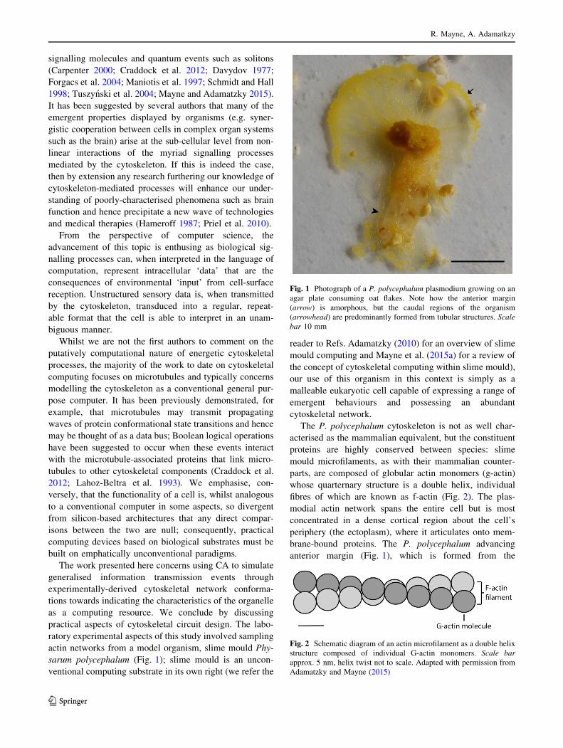
The P. polycephalum cytoskeleton is not as well char-
acterised as the mammalian equivalent, but the constituent
proteins are highly conserved between species: slime
mould microfilaments, as with their mammalian counter-
parts, are composed of globular actin monomers (g-actin)
whose quarternary structure is a double helix, individual
fibres of which are known as f-actin (Fig. 2). The plas-
modial actin network spans the entire cell but is most
concentrated in a dense cortical region about the cell’s
periphery (the ectoplasm), where it articulates onto mem-
brane-bound proteins. The P. polycephalum advancing
anterior margin (Fig. 1), which is formed from the
Fig. 1 Photograph of a P. polycephalum plasmodium growing on an
agar plate consuming oat flakes. Note how the anterior margin
(arrow) is amorphous, but the caudal regions of the organism
(arrowhead) are predominantly formed from tubular structures. Scale
bar 10 mm
Fig. 2 Schematic diagram of an actin microfilament as a double helix
structure composed of individual G-actin monomers. Scale bar
approx. 5 nm, helix twist not to scale. Adapted with permission from
Adamatzky and Mayne (2015)
R. Mayne, A. Adamatkzy
123
confluence of multiple pseudopodia, contains denser actin
networks than its caudal tubular regions (Alexopoulos
1982; Mayne et al. 2015a). Whilst pseudopodium forma-
tion via momentary assembly of actin is a well-observed
phenomenon, this does pose the question of how such
topological dimorphism impacts on intracellular commu-
nicative processes, hence investigation into this was made a
secondary goal of this study.
2 Methods
2.1 Physarum polycephalum culture and microscopy
Stock plasmodia of P. polycephalum (strain HU554 �HU560) were cultivated on 2 % non-nutrient agar (NNA)
plates at room temperature in the absence of light. They
were provided with porridge oats as a nutrient substrate and
were sub-cultured routinely every 3–4 days, as required.
Actin was labelled for confocal microscopy as follows:
two hemispheres of NNA were prepared on large glass
microscope coverslips by dripping 2� 0:5 ml of molten
agar onto them with a pipette, with a gap of approximately
10 mm separating the two. Samples of stock plasmodium
were homogenised with a scalpel blade and transferred to
one NNA hemisphere. An oat flake was placed on the
second hemisphere to encourage the organism to propagate
between them; the cover slip was placed in an air-tight
Petri dish, which was left in the dark at room temperature.
Samples of the anterior margin were taken by chemically
fixing the organism before its advancing regions had fin-
ished propagating across the gap between the hemispheres
and were hence spanning the naked glass, whereas plas-
modial tubes were prepared by waiting until the second
hemisphere had been fully colonised, as this typically
resulted in the gap between the two hemispheres being
linked by a single large tube.
Fixation was achieved by flooding the Petri dish with
2 % paraformaldehyde in pH 7.2 phosphate-buffered saline
solution for 1 h. This was followed by 3 9 5 min rinses in
the same buffer, after which the sample was permeabalised
with 0.1 % Triton X-100. Following further rinsing and
draining, Alexa Fluor 488 Phalloidin (Molecular Probes,
USA) was added for 1 h at a concentration of 5 lg/ml in
methanol. The samples were then washed again before
being stained with DAPI (Abcam, UK).
Confocal microscopy was performed with a Perkin
Elmer UltraView ERS FRET-H spinning disk confocal
laser scanning microscope.1
2.2 Network extraction and CA model
Actin networks were reconstructed using the Watershed
transformation on greyscale confocal micrographs with
Matlab 2015a (Mathworks, USA). To represent the topol-
ogy of actin network reconstructions in 2D cellular
automaton models we assigned cells corresponding to each
black pixel as conductive; other cells are assigned to be
non-conductive. Although any reconstruction of nanoscale
structures using a light microscopic technique necessitates
a certain degree of inaccuracy, the watershed transforma-
tion was chosen for it having previously been demonstrated
as a viable technique for approximating actin network
topologies to a suitable degree of accuracy (Fleischer et al.
2007). The concave hull of extracted networks was also
computed in instances where the cell’s membrane was
present in order to demarcate the cell’s boundaries.
An excitable CA representing the propagation of gen-
eralised energetic events along actin microfilaments was
constructed as follows. In an excitable medium, discretised
spaces (cells) are considered to be in one of three states
(Greenberg and Hastings 1978):
1. Resting A stable equilibrium state.
2. Excited The energy level of the cell is temporarily
higher than the equilibrium level; in context with this
investigation, this could correspond to a breather
propagating through a particular actin molecule or a
local electrochemical disturbance caused by ionic
wave trasmission through a microfilament.
3. Refractory A temporary state directly following exci-
tation wherein the cell is in the process of returning to
equilibrium and cannot be re-excited until it has re-
entered a resting state.
Thus in an excitable CA, the excitable medium is repre-
sented as a 2D array of cells, wherein each cell takes one of
these three states. The array was initialised by importing
2D actin network reconstructions as 500 9 500 pixel bit-
map images wherein each pixel represented a cell: as only
black-coloured cells (corresponding to the presence of
actin in these pixels in the original image) update their
states and other cells remain resting indefinitely, the exci-
tation may only propagate through the reconstructed net-
work. A resting black cell becomes excited (indicated by
turning pink in the simulation, see videos 1 and 2 in
Electronic Supplementary Material) during time iteration
t þ 1 if it has at least one excited neighbouring cell (von
Neumann neighbourhood) at time t, thus ‘signals’—repre-
sented by excitation states—can be observed to propagate
1 Image Processing Photographs were taken with a Samsung I9300
digital camera. Confocal micrographs were digitally post-processed in
Volocity (Improvision, UK), and received colour assignment,
Footnote 1 continued
deconvolution and brightness/colour adjustments. Unprocessed image
files will be made available on request.
Cellular automata modelling of slime mould actin network signalling
123
through the reconstructed networks over time. Cells remain
in an excited state for one time iteration before turning into
a refractory cell (denoted by turning blue in the simulation,
see videos in Electronic Supplementary Material), also for
one time iteration, after which it reverts into a resting cell.
When a wave of excitation, propagating along conductive
cells, ‘collides’ with another excitation wave head-on, both
signals annihilate as their being flanked by refractory cells
disallows their moving ‘through’ or ricocheting from each
other. Excitations were initiated at random locations on the
periphery of reconstructed networks to represent the initi-
ation of an energetic event, e.g. through stimulation of a
cell surface receptor.
Integral excitation dynamics of extracted networks were
measured by computing the number of cells becoming
excited each time iteration. The comparative speed of
transmission through these networks was assessed by
measuring the number of timesteps taken for the signal to
reach a point in the network an arbitrary but fixed distance
away: 450� 1:2 away in all experiments.
All experiments (visualisation of an area, running of CA
and measurement of excitation dynamics) were repeated 5
times for each anatomical region sampled.
3 Results
3.1 Visualisation of actin
In corroboration with previously published findings, the
plasmodial actin network was observed to be concentrated
in the cell’s cortical regions and appeared to form associ-
ations with the surface of the organism. Actin networks
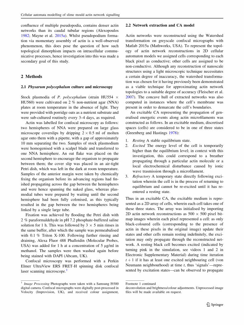
appeared denser in the organism’s anterior margin (Fig. 3a)
than in plasmodial tubes (Fig. 4a).
3.2 Modelling information flow through extracted
actin networks
Exemplar extracted actin networks are shown in Figs. 3b
and 4b; video footage of the CA model being run on both
of these networks is included as supplementary information
(see Electronic Supplementary Material). In both, the ini-
tial perturbation was observed to propagate through the
entire network thanks to the signal becoming amplified
when it met network junctions, as can be observed through
the gradual increase in number of excited actin units over
time in Figs 3c and 4c. Excitation dynamics were found to
be richer in the anterior regions of the plasmodium. Net-
work topologies were found to be structured such that
signal back-propagation rarely occurred; when it did occur,
reversed signals usually collided with signals travelling in
the opposite direction and annihilated.
The speed (in timesteps) for a signal to travel to a net-
work location 450 pixels away was found to be faster in
anterior margin networks in every instance (659 and 829
time steps in the two examples given, respectively).
4 Discussion
4.1 On dynamic actin network transformations
Actin network density was found to be inversely propor-
tional to proximity to active growth areas in the organism’s
anterior regions; this observation was substantiated by a
richer excitation dynamics and greater speed of signal
transduction through reconstructed anterior margin net-
works using excitable CA. Thus, we can speculate that
depending on momentary actual demands of the slime
mould’s physiology, the topology of its communication
network will alter in density and connectivity.
It would seem that a greater degree of signal amplifi-
cation and transduction speed are desirable characteristics
for the protein networks of active growth regions in order
to maximise environmental sensing and the organism’s
ability to consequently react to input data. These charac-
teristics are presumably reduced in caudal regions of the
organism in order to preserve energy where they are not
required, although we concede that other functions of the
actin cytoskeleton that have not been examined here will
also play a role in determining network topology. Biolog-
ical substrates are often quoted as being ‘massively paral-
lel’ in their ability to concurrently sense input from an
almost incalculably large number of sensory data streams
(Stepney et al. 2005); our findings emphasise the efficiency
with which such substrates may optimise these processes in
response to evolutionary selection pressures, thus high-
lighting their value to the field of unconventional com-
puting and bio-inspired computing design.
4.2 Sensoriactuation networks for intracellular
computing
Let us consider how intracellular computation may be
influenced by data network topology. Computing with
algorithms based on planar graphs have been demonstrated
to be viable for implementing general-purpose computa-
tion: furthermore, certain classes of device —Kolmogorov-
Uspensky Machines (Kolmogorov and Uspensky 1958)—
whose physical architecture are based on the retrieval of
data stored in dynamically-rearranging undirected graph
structures, demonstrate the viability of computers built
upon principles of motile components. We may speculate,
therefore, that analogous processes are integrated via actin-
based cellular communications which provoke
R. Mayne, A. Adamatkzy
123
morphological rearrangement. To delineate, consider the
following example: despite lacking a brain or nervous tis-
sue, a slime mould is able to ‘decide’ which of the myriad
chemical gradients one of its advancing margins senses to
follow. The cell’s cortical actin network becomes inner-
vated via the activation of second messenger pathways
incident of chemoreceptor stimulation; by the majority
vote, the sites of the organism with the greatest amount of
chemoreception (corresponding to multiple receptors being
activated) are the sites of consequent directional tip growth
and thus the network rearranges in the process of
migration.
This is an interesting perspective as it implies that the
parallelism inherent in biological computing substrates
occurs as a function of the materials they are composed
from and does not necessitate expending energy on the
coordination or synchronisation of these behaviours. As
such, the complexity of the organism is reduced whilst
enabling the spontaneous generation of complex beha-
viours—in effect, this is a definition of emergence. These
observations are complimentary to recent advances in the
field of morphological computation and entity embodiment
which posit that ‘outsourcing’ a certain amount of com-
putational work to the morphology of data streams is
essential in the design of artificially intelligent entities
(Hauser et al. 2012; Lungarella and Sporns 2006). This
concept is intimately linked to data ‘structuring’—trans-
ducing and transmitting signals in a repeatable and unam-
biguous manner—which is another important concept in
morphological computation and control theory (Fuchslin
Fig. 3 Actin excitation
dynamics in the P.
polycephalum anterior margin.
a Confocal micrograph. The
white box indicates the area
extracted. b Extracted network.
Excitation point indicated by
arrow. c Integral excitation
dynamics of actin network;
trendline is a polynomial fit.
a–b Scale bar 50 lm
Cellular automata modelling of slime mould actin network signalling
123
et al. 2013; Hauser et al. 2012). By logical extension, this
would seem to suggest that the cytoskeleton is a medium
for structuring sensorimotor data streams and hence that
the emergent behaviours displayed by cells may be a pro-
duct of cytoskeletal processes.
In the model presented, back-propagation of signals
appeared to be curtailed via destructive signal–signal
interactions—in effect, this describes collision-based
computation (Adamatzky 2002). Combined with the inci-
dence of signal amplification, we may characterise these
phenomena as the following operations:
– FAN-OUT; signal amplification when a signal reaches a
network bifurcation.
– XOR; when two signals converge following a branch,
they may interact destructively such that the output
configuration of the branch point is hA � �Bþ �A � Bi.
Thus we may speculate that this computation-like process
plays a crucial role in regulating signal propagation
through the organism’s actin network.
To address a pertinent criticism of the work presented
here, the modelled excitation dynamics presented are based
on static, two-dimensional characterisations of dynamic,
three-dimensional structures superimposed onto a two-di-
mensional image: as such, results presented here are purely
illustrative. We note, however, that these techniques may
be applied to three- or even four-dimensional image stacks;
this will be a focus for future research.
4.3 Practical actin computing
Actin, along with its companion motor protein myosin, is
instrumental in the retention and transport of vesicle-bound
Fig. 4 Actin excitation
dynamics in the P.
polycephalum plasmodial tube.
a Confocal micrograph.
b Extracted network. Excitation
point indicated by arrow.
c Integral excitation dynamics
of actin network, which are less
rich than in Fig. 3c; trendline is
a polynomial fit. a–b Scale bar
50 lm
R. Mayne, A. Adamatkzy
123
biomolecules and minerals (DePina and Langford 1999);
we have previously described the movements of calcium-
filled vesicles travelling down P. polycephalum actin fibres
as an example of naturally-occurring collision-based
computing (Mayne and Adamatzky 2015). Following the
results described here, the same may also be said for other
energetic events, such as quantum phenomena or/and
electrical potential propagated in the form of ionic waves.
‘Hijacking’ these processes is a viable route towards the
generation of practical biological computing architectures.
To engineer practical actin computing circuits we are
presented with a few technical considerations. First is to
generate an actin network in a desired topology; this may
be in vitro or in vivo. Although minute manipulation of
actin fibres has been demonstrated in vitro (Lin and Can-
tiello 1993), we suggest that actin growth within model
organisms such as P. polycephalum is also a viable method
to this end as growth patterns are essentially programmable
by manipulation of the organism’s environment and inter-
nal feedback mechanisms.
The second consideration is the initiation and synchro-
nisation of informational events. Again, this issue may best
be addressed by the manipulation of environmental and
internal factors, but may also be induced artificially. In
slime mould models, for example, actin network contrac-
tion can be initiated by tactile stimulation (Mayne et al.
2015). The interaction environment’s topology plays an
important role in synchronisation, especially in collision-
based models as factors such as filament length and delay
elements are necessarily functions of network topology.
4.4 Conclusion
A prime motivation into researching biological computing
substrates such as slime mould is their amorphism—a
property that implies architecture-less massive parallel
processing. Our results indicate that the emergent beha-
vioural capabilities of a live cell may be a product of its
cytoskeletal topology and the information processing
events therein. This implies that emergent behaviour is a
product of the physical properties of the data network; this
has important implications for our understanding of con-
cepts such as human intelligence, which is commonly
regarded as a product (in part, at least) of neural network
morphology and the characteristics of the junctions (sy-
napses) between them. In future works, we will expand this
concept with experimental implementations of actin-based
computing.
Acknowledgments This work was supported by the EU research
Project Physarum Chip: GrowingComputers from Slime Mould’’ (FP7
ICT Ref 316366) The authors extend their thanks to Ben De Lacy
Costello for his supervision and technical support with the experi-
mental work undertaken, and Dr. Tareq Assaf for his critical insights.
Compliance with ethical standards
Conflicts of interest The authors declare no conflict of interest.
Open Access This article is distributed under the terms of the
Creative Commons Attribution 4.0 International License (http://crea
tivecommons.org/licenses/by/4.0/), which permits unrestricted use,
distribution, and reproduction in any medium, provided you give
appropriate credit to the original author(s) and the source, provide a
link to the Creative Commons license, and indicate if changes were
made.
References
Adamatzky A (2002) Collision-based computing. Springer, Berlin
Adamatzky A (2010) Physarum machines: computers from slime
mould. World Scientific Publishing, London
Adamatzky A, Mayne R (2015) Actin automata: phenomenology and
localizations. Int J Bifurc Chaos 25(2):1550030
Alexopoulos C (1982) The biology of physarum and didymium. Cell
biology: a series of monographs, chapter 5: plasmodial structure
and motility. Academic Press, Cambridge
Carpenter C (2000) Actin cytoskeleton and cell signalling. Crit Care
Med 28(4):94–99
Craddock T, Tuszynski J, Hameroff S (2012) Cytoskeletal signalling:
is memory encoded in microtubule lattices by camkii phospho-
rylation? PLoS Comput Biol 8(3):e1002421
Davydov A (1977) Solitons and energy transfer along protein
molecules. J Theor Biol 66:379–387
DePina A, Langford G (1999) Vesicle transport: the role of actin
filaments and myosin motors. Microsc Res Tech 47(2):93–106
Fleischer F, Ananthakrishnan R, Eckel S, Schmidt H, Kas J, Svitkina
T, Schmidt V, Beil M (2007) Actin network architecture and
elasticity in lamellipodia of melanoma cells. New J Phys
9(11):420
Forgacs G, Yook SH, Janmey P, Jeong H, Burd C (2004) Role of the
cytoskeleton in signalling networks. J Cell Sci
117(3):2769–2775
Fuchslin R, Dzyakanchuk A, Flumini D, Hauser H, Hunt K,
Luchsinger R, Reller B, Scheidegger S, Walker R (2013)
Morphological computation and morphological control: steps
towards a formal theory and applications. Artif Life 19:9–34
Greenberg J, Hastings S (1978) Spatial patterns for discrete models of
diffusion in excitable media. SIAM J Appl Math 34(3):515–523
Hameroff S (1987) Ultimate computing: biomolecular consciousness
and nanotechnology. Elsevier, Amsterdam
Hauser H, Ijspeert AJ, Fuchslin RM, Pfeifer R, Maass W (2012)
Towards a theoretic foundation for morphological computation
with compliant bodies. Biol Cybern 105:355–370
Janmey P (1998) The cytoskeleton and cell signalling: component
localization and mechanical coupling. Phys Rev 78(3):763–781
Kolmogorov A, Uspensky V (1958) On the definition of an algorithm.
Uspekhi Matematicheskikh Nauk 13(4):3–28
Lahoz-Beltra R, Hameroff S, Dayhoff J (1993) Cytoskeletal logic: a
model for molecular computation via boolean operations in
microtubules and microtubule-associated proteins. Biosystems
29(1):1–23
Lin EC, Cantiello HF (1993) A novel method to study the
electrodynamic behavior of actin filaments. Evidence for
cable-like properties of actin. Biophysical Journal, 65 (December
1992)
Lungarella M, Sporns M (2006) Mapping information flow in
sensorimotor networks. PLoS Comput Biol 2(10):e144
Cellular automata modelling of slime mould actin network signalling
123

Maniotis A, Chen C, Ingber D (1997) Demonstration of mechanical
connections between integrins, cytoskeletal filaments and nucle-
oplasm that stabilise nuclear structure. PNAS 94(3):849–854
Mayne R, Adamatzky A (2015) On the computing potential of
intracellular vesicles. PLoS One 10(10):e0139617
Mayne R, Adamatzky A, Jones J (2015a) On the role of the
plasmodial cytoskeleton in facilitating intelligent behaviour in
slime mould Physarum polycephalum. Commun Integr Biol
8(4):e1059007
Mayne R, Tsompanas M-A, Sirakoulis GC, Adamatzky A (2015b)
Towards a slime mould-FPGA interface. Biomed Eng Lett
5(1):51–57
Priel A, Tuszynski J, Woolf N (2010) Neural cytoskeleton capabilities
for learning and memory. J Biol Phys 36(1):3–21
Schmidt A, Hall M (1998) Signalling to the actin cytoskeleton. Ann
Rev Cell Dev Biol 14:305–338
Stepney S, Braunstein S, Clark J, Tyrrell A, Adamatzky A, Smith R,
Addis T, Johnson C, Timmis J, Welch P, Milner R, Partridge D
(2005) Journeys in non-classical computation I: a grand
challenge for computing research. Int J Parallel Emerg Distrib
Syst 20(1):5–19
Tuszynski J, Portet S, Dixon J, Luxford C, Cantiello H (2004) Ionic
wave propagation along actin filaments. Biophys J
86:1890–1903
R. Mayne, A. Adamatkzy
123


![Physarum polycephalum arXiv:1403.3973v1 [cs.ET] 16 Mar … · 2 Richard Mayne, Andrew Adamatzky conventional computing substrate as it is easy to culture, tolerant to abuse and has](https://img.pdfslide.us/doc/110x75/5addd3fb7f8b9a9a768d6fd9/physarum-polycephalum-arxiv14033973v1-cset-16-mar-richard-mayne-andrew.jpg)