Embed Size (px)
Citation preview
Pediatrics Grand Rounds 12 October 2012
University of Texas Health Science Center at San Antonio
1
Maybe It’s A Tumor?! – Common Pediatric Brain Tumors
Pediatric Grand Rounds
Emily S. Moses, M.D.
October 12, 2012
Disclosure
• I have no relationships with commercial companies to disclose.
Learning Objectives
• Understand the clinical presentation, diagnostic work-up, and standard treatments of a child with a brain tumor.
• Be aware of some of the common brain tumors found in children.
• Be familiar with the late effects from treatment that can effect a child with a brain tumor.
Case
• 8yo female presents to her PCP with several day history of
– Headache
– Vomiting
– Dizziness
– Subjective Fever
Maybe It’s A Tumor?! Chances It’s A Tumor
• ~3000-3500 new cases of brain tumors diagnosed in the US yearly
• Gastroenteritis is the 2nd most common infection diagnosed in the US
Pediatrics Grand Rounds 12 October 2012
University of Texas Health Science Center at San Antonio
2
Why This Topic Is Important
• Brain tumors are the 2nd most common cancer in children, after leukemia
• Most common solid tumors in children
• Overall 5 year survival = 60-70%
Differences Between Adult and Pediatric Brain Tumors
• See more low grade lesions
• Majority of lesions are infratentorial
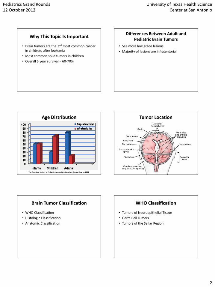
Age Distribution
The American Society of Pediatric Hematology/Oncology Review Course, 2011
Tumor Location
Brain Tumor Classification
• WHO Classification
• Histologic Classification
• Anatomic Classification
WHO Classification
• Tumors of Neuroepithelial Tissue
• Germ Cell Tumors
• Tumors of the Sellar Region
Pediatrics Grand Rounds 12 October 2012
University of Texas Health Science Center at San Antonio
3
Histologic Classification
• Histologic Classification
– Neuroglial Cells
• Astrocytes
• Oligodendrocytes
• Ependyma
• Choroid Plexus
– Neuronal/Embryonal Cells
Anatomic Classification
• Cerebrum
• Cerebellum
• Pineal Region
Clinical Presentation
• Increased intracranial pressure
• Localizing signs/symptoms
• General signs/symptoms
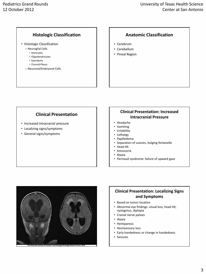
Clinical Presentation: Increased Intracranial Pressure
• Headache • Vomiting • Irritability • Lethargy • Papilledema • Separation of sutures, bulging fontanelle • Head tilt • Anisocoria • Ataxia • Parinaud syndrome: failure of upward gaze
The American Society of Pediatric Hematology/Oncology Review Course, 2011
Clinical Presentation: Localizing Signs and Symptoms
• Based on tumor location
• Abnormal eye findings: visual loss, head tilt, nystagmus, diplopia
• Cranial nerve palsies
• Ataxia
• Hemiparesis
• Hemisensory loss
• Early handedness or change in handedness
• Seizures
Pediatrics Grand Rounds 12 October 2012
University of Texas Health Science Center at San Antonio
4
The American Society of Pediatric Hematology/Oncology Review Course, 2011
Clinical Presentation: General Signs and Symptoms
• Headache
• Vomiting
• Developmental delay
• Weight loss or gain
• Failure to thrive
• Endocrine abnormalities
• Behavioral changes
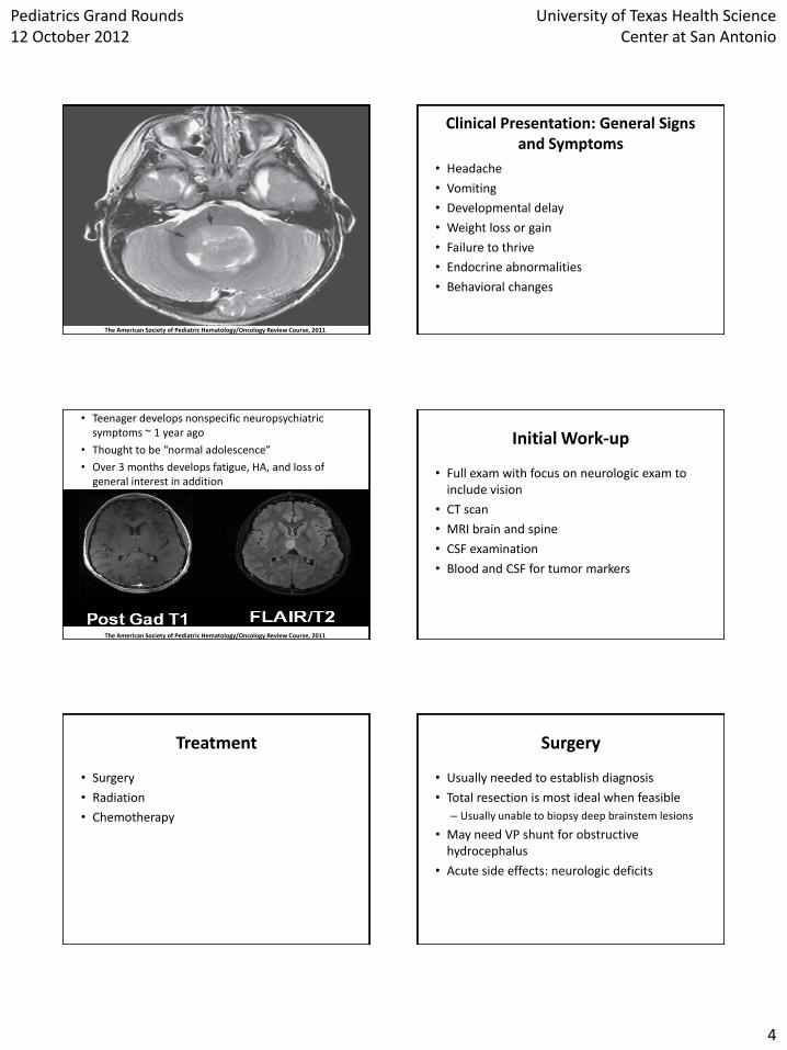
• Teenager develops nonspecific neuropsychiatric symptoms ~ 1 year ago
• Thought to be "normal adolescence”
• Over 3 months develops fatigue, HA, and loss of general interest in addition
The American Society of Pediatric Hematology/Oncology Review Course, 2011
Initial Work-up
• Full exam with focus on neurologic exam to include vision
• CT scan
• MRI brain and spine
• CSF examination
• Blood and CSF for tumor markers
Treatment
• Surgery
• Radiation
• Chemotherapy
Surgery
• Usually needed to establish diagnosis
• Total resection is most ideal when feasible
– Usually unable to biopsy deep brainstem lesions
• May need VP shunt for obstructive hydrocephalus
• Acute side effects: neurologic deficits
Pediatrics Grand Rounds 12 October 2012
University of Texas Health Science Center at San Antonio
5
Radiation
• Can be used for all brain tumors with minimal exceptions
• Full treatment dose: 5400 – 5940 cGy
• Prophylaxis dose: 1800 – 3600 cGy
• Acute side effects: nausea, vomiting, fatigue, skin changes, hair loss
Chemotherapy
• Adjuvant therapy in most cases • In young children ( < 3 yrs), attempt to use as first
line to avoid radiation because of long term neurocognitive sequelae
• Blood brain barrier thought to limit use, however with higher doses, in particular high dose chemotherapy with autologous stem cell rescue, able to overcome this
• Acute side effects: nausea, vomiting, myelosuppression, infection, hair loss, mucositis, neuropathy, changes in taste
Common Pediatric Brain Tumors
• Medulloblastoma
• Ependymomas
• Brain stem tumors
• Astrocytomas
• Optic Glioma
• Intracranial Germ Cell Tumors
• Craniopharyngiomas
• Atypical Teratoid Rhabdoid Tumor (AT/RT)
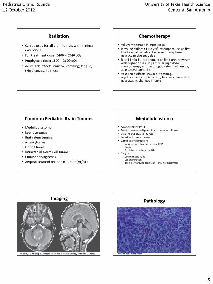
Medulloblastoma
• AKA Cerebellar PNET • Most common malignant brain tumor in children • Small round blue cell tumor • Location: Posterior fossa • Common Presentation:
– Signs and symptoms of increased ICP – Ataxia – Cranial nerve palsies, esp 6th
• Staging: – MRI brain and spine – CSF examination – Bone marrow &/or bone scan – only if symptomatic
Imaging
P.A. Pizzo, D.G. Poplack (eds). Principles and Practice of Pediatric Oncology. 6th Edition. Chapter 26.
Pathology
www.emedicine.medscape.com
Pediatrics Grand Rounds 12 October 2012
University of Texas Health Science Center at San Antonio
6
Medulloblastoma
• Risk Categories:
– Desmoplastic variant
Standard Risk High Risk
Extent of Resection
< 1.5 cm3 residual tumor
> 1.5 cm3 residual tumor
Mets No mets Mets
Histology Anaplasia
Age at Dx > 3yrs < 3yrs
Medulloblastoma
• Treatment for Standard Risk: – Gross total resection (GTR) when possible – Radiation is a must (except < 3 yrs) – Adjuvant chemotherapy with
cisplatin/CCNU/vincristine or cisplatin/cytoxan/vincristine
• High risk: resection, radiation, aggressive chemotherapy to include autologous stem cell rescue
• Prognosis: – Standard risk: 80-85% event free survival (EFS) – High risk: 20-70% EFS
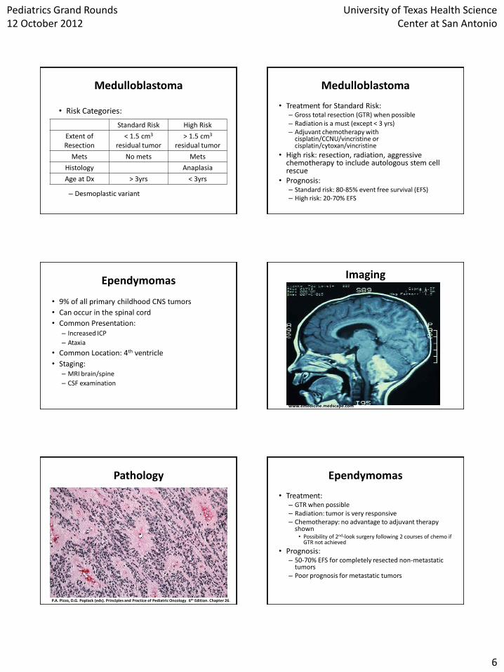
Ependymomas
• 9% of all primary childhood CNS tumors
• Can occur in the spinal cord
• Common Presentation: – Increased ICP
– Ataxia
• Common Location: 4th ventricle
• Staging: – MRI brain/spine
– CSF examination
Imaging
www.emedicine.medscape.com
Pathology
P.A. Pizzo, D.G. Poplack (eds). Principles and Practice of Pediatric Oncology. 6th Edition. Chapter 26.
Ependymomas
• Treatment: – GTR when possible – Radiation: tumor is very responsive – Chemotherapy: no advantage to adjuvant therapy
shown • Possibility of 2nd-look surgery following 2 courses of chemo if
GTR not achieved
• Prognosis: – 50-70% EFS for completely resected non-metastatic
tumors – Poor prognosis for metastatic tumors
Pediatrics Grand Rounds 12 October 2012
University of Texas Health Science Center at San Antonio
7
WHO Classification of Astrocytomas
• Grade I, pilocytic astrocytoma: fibrillary background, rare mitoses, classically Rosenthal fibers, well circumscribed and slow growth, usually behaves in benign fashion
• Grade II, diffuse or fibrillary astrocytoma: more cellular and infiltrative and more likely to undergo anaplastic change
• Grade III, anaplastic astrocytoma (AA): highly cellular with significant cellular atypia, locally invasive and aggressive
• Grade IV, glioblastoma multiforme (GBM): increased nuclear anaplasia, pseudopalisading and multinucleated giant cells
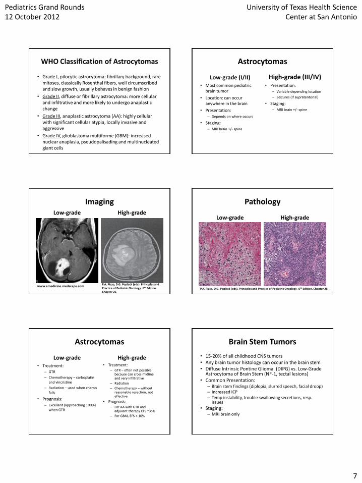
Astrocytomas
Low-grade (I/II) • Most common pediatric
brain tumor
• Location: can occur anywhere in the brain
• Presentation: – Depends on where occurs
• Staging: – MRI brain +/- spine
High-grade (III/IV) • Presentation:
– Variable depending location
– Seizures (if supratentorial)
• Staging: – MRI brain +/- spine
Imaging Low-grade
High-grade
P.A. Pizzo, D.G. Poplack (eds). Principles and Practice of Pediatric Oncology. 6th Edition. Chapter 26.
www.emedicine.medscape.com
Pathology
Low-grade
High-grade
P.A. Pizzo, D.G. Poplack (eds). Principles and Practice of Pediatric Oncology. 6th Edition. Chapter 26.
Astrocytomas
Low-grade • Treatment:
– GTR
– Chemotherapy – carboplatin and vincristine
– Radiation – used when chemo fails
• Prognosis: – Excellent (approaching 100%)
when GTR
High-grade • Treatment:
– GTR – often not possible because can cross midline and very infiltrative
– Radiation
– Chemotherapy – without reasonable resection, not effective
• Prognosis: – For AA with GTR and
adjuvant therapy EFS ~35%
– For GBM, EFS < 10%
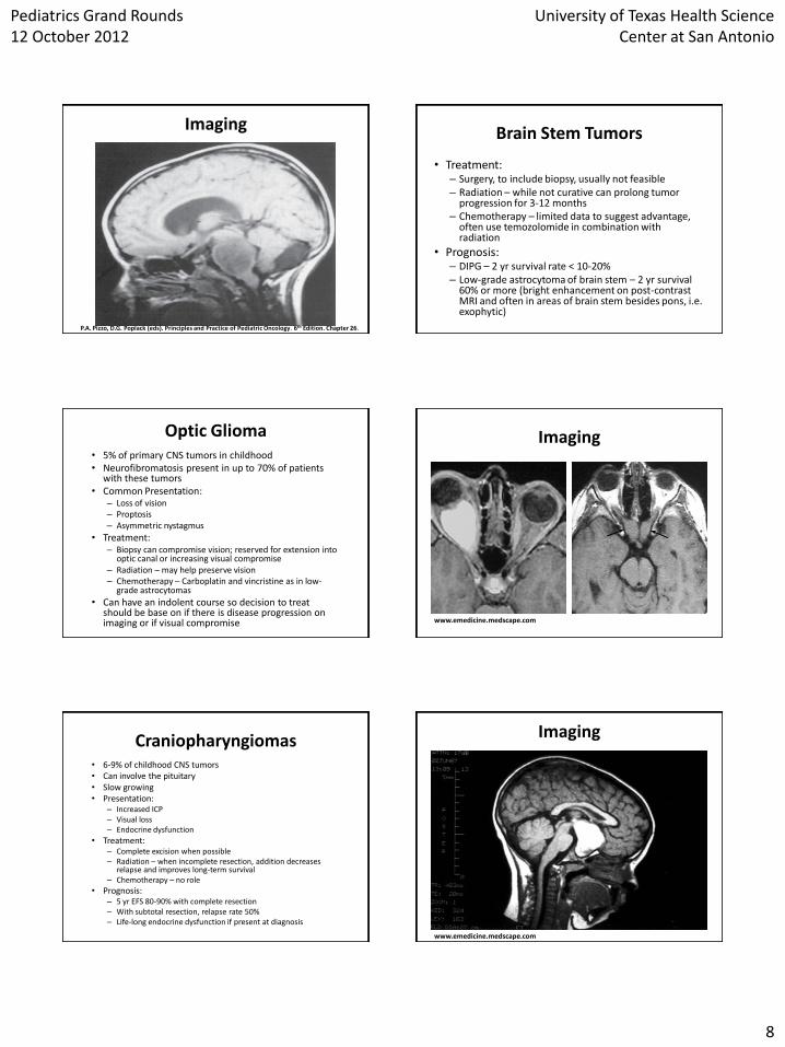
Brain Stem Tumors
• 15-20% of all childhood CNS tumors • Any brain tumor histology can occur in the brain stem • Diffuse Intrinsic Pontine Glioma (DIPG) vs. Low-Grade
Astrocytoma of Brain Stem (NF-1, tectal lesions) • Common Presentation:
– Brain stem findings (diplopia, slurred speech, facial droop) – Increased ICP – Temp instability, trouble swallowing secretions, resp.
issues
• Staging: – MRI brain only
Pediatrics Grand Rounds 12 October 2012
University of Texas Health Science Center at San Antonio
8
Imaging
P.A. Pizzo, D.G. Poplack (eds). Principles and Practice of Pediatric Oncology. 6th Edition. Chapter 26.
Brain Stem Tumors
• Treatment: – Surgery, to include biopsy, usually not feasible – Radiation – while not curative can prolong tumor
progression for 3-12 months – Chemotherapy – limited data to suggest advantage,
often use temozolomide in combination with radiation
• Prognosis: – DIPG – 2 yr survival rate < 10-20% – Low-grade astrocytoma of brain stem – 2 yr survival
60% or more (bright enhancement on post-contrast MRI and often in areas of brain stem besides pons, i.e. exophytic)
Optic Glioma • 5% of primary CNS tumors in childhood • Neurofibromatosis present in up to 70% of patients
with these tumors • Common Presentation:
– Loss of vision – Proptosis – Asymmetric nystagmus
• Treatment: – Biopsy can compromise vision; reserved for extension into
optic canal or increasing visual compromise – Radiation – may help preserve vision – Chemotherapy – Carboplatin and vincristine as in low-
grade astrocytomas
• Can have an indolent course so decision to treat should be base on if there is disease progression on imaging or if visual compromise
Imaging
www.emedicine.medscape.com
Craniopharyngiomas • 6-9% of childhood CNS tumors • Can involve the pituitary • Slow growing • Presentation:
– Increased ICP – Visual loss – Endocrine dysfunction
• Treatment: – Complete excision when possible – Radiation – when incomplete resection, addition decreases
relapse and improves long-term survival – Chemotherapy – no role
• Prognosis: – 5 yr EFS 80-90% with complete resection – With subtotal resection, relapse rate 50% – Life-long endocrine dysfunction if present at diagnosis
Imaging
www.emedicine.medscape.com
Pediatrics Grand Rounds 12 October 2012
University of Texas Health Science Center at San Antonio
9
Intracranial Germ Cell Tumors
• 1-3% of primary pediatric CNS tumors
• Multiple tumor types seen: – Germinomas (~55%)
– Non-germinomatous germ cell tumors (NG-GCT) • Teratomas and mixed germ cell tumors (~33%)
• Others: malignant endodermal sinus tumors, embryonal cell carcinomas, choriocarcinomas, teratocarcinomas (~10%)
• In all but germinomas, serum and CSF alpha-fetoprotein (AFP) and βHCG may be elevated
Intracranial Germ Cell Tumors
Germinoma • Presentation:
– Suprasellar – panhypopituitarism, DI
– Pineal region – raised ICP, Parinauds syndrome
– 10% multifocal
• Staging: – MRI brain/spine
– CSF for tumor markers (AFP, βHCG neg)
NG-GCT • Presentation:
– Precocious puberty
– DI
– Panhypopituitarism
– Raised ICP
• Staging: – MRI brain/spine
– CSF for tumor markers
– No biopsy if markers positive
Imaging
Germinoma
NG-GCT
www.emedicine.medscape.com
Pathology
Germinoma
NG-GCT
P.A. Pizzo, D.G. Poplack (eds). Principles and Practice of Pediatric Oncology. 6th Edition. Chapter 26.
www.emedicine.medscape.com
Intracranial Germ Cell Tumors
Germinoma • Treatment:
– Radiation (whole ventricular, craniospinal)
– +/- Neoadjuvant chemotherapy with radiation dose based on response
• Prognosis: – > 80%
– Endocrine abnormalities will not resolve with successful treatment
NG-GCT • Treatment:
– Neoadjuvant chemotherapy followed by response based radiation (includes craniospinal)
• Prognosis: – 40-70%
– Can follow tumor markers to assess response
Atypical Teratoid Rhabdoid Tumor (AT/RT)
• Very aggressive tumors often seen in children < 3 yrs
• Often mistaken for medulloblastoma
– Pathologically similar except INI-1 negative
• Fatal if treated with medulloblastoma therapy but survival approaches 50% if treated with high-dose chemotherapy with autologous stem-cell rescue
Pediatrics Grand Rounds 12 October 2012
University of Texas Health Science Center at San Antonio
10
INI-1 Staining
www.emedicine.medscape.com
Brain Tumors in Children < 3 yrs
• Worse prognosis
• Higher risk for neurotoxicity:
– Mental retardation
– Growth failure
– Leukoencephalopathy
• Attempt to avoid or delay radiation
Genetic Associations
• Neurofibromatosis-1 (NF-1) – optic pathway gliomas, other gliomas, meningiomas
• Tuberous sclerosis – gliomas, ependymomas • Rentinoblastoma – at risk for trilateral disease with
pineoblastoma • Li-Fraumeni (p53) – choroid plexus tumors,
astrocytomas • Von Hippel-Lindau syndrome – cerebellar
hemangioblastoma • Turcot’s synrome – gliomas • Gorlin’s syndrome – medulloblastoma
Late Effects of Treatment
• Complications related to treatment that persist or arise once cancer therapy is completed
• Can effect quality and quantity of life • Most survivors will experience at least 1 late
effect • Many influencing factors such as age, gender,
tumor type, treatment received, ability to cope and degree of support
• Psychosocial effects such as anxiety, depression, school & employment issues can occur with any treatment
“Late effects represent the price that was paid for cure and the
quality of life that was purchased.” - Jan van Eys
Brain Surgery Late Effects
• Depends on location of tumor and degree of resection
• Impairment of neurological function
Pediatrics Grand Rounds 12 October 2012
University of Texas Health Science Center at San Antonio
11
Cranial Radiation Late Effects
• Neurodevelopmental effects
• Second malignancies (meningiomas, high grade gliomas, melanomas)
• Abnormal growth and maturation
• Endocrine abnormalities – GH Deficiency
– Hypothyroidism
• Vision Problems
• Hearing loss
Brain Tumor Chemotherapy Late Effects
• Neurodevelopmental effects
• Second malignancies
• Hearing loss
• Kidney damage
• Infertility
• Abnormal growth and maturation
• Endocrine abnormalities
Endocrine Late Effects
• There are endocrine abnormalities that the patient may present with or develop during treatment and those that develop much later secondary to treatment – DI on presentation in a patient with a CNS germinoma vs.
hypothyroidism 8-10 years following cranial radiation
• Any damage to the hypothalamic-pituitary axis secondary to the tumor seen on presentation or that develops during treatment is not reversible and the patient will need life-long hormone replacement – Suprasellar tumors
Summary
• Brain tumors are the 2nd most common cancer seen in children
• Overall survival is 60-70% and with newer treatments will continue to improve
• More survivors = more late effects
References
• Lanzkowsky P (ed). Manual of Pediatric Hematology and Oncology. 5th Edition. Chapters 21 & 31.
• Mulhern RK, Merchant TE, Gajjar A, et al. Late neurocognitive sequelae in survivors of brain tumors in childhood. Lancet Oncol 2004;5:399–408.
• Pizzo PA, Poplack DG(eds). Principles and Practice of Pediatric Oncology. 6th Edition. Chapter 26.
• Robinson KE, Futtesch JF, Champion JE, et al. A quantitative meta-analysis of neurocognitive sequelae in survivors of pediatric brain tumors. Pediatr Blood Cancer 2010;55:525–531.
• The American Society of Pediatric Hematology/Oncology Review Course. Brain Tumors. 2011.
• www.emedicine.medscape.com • www.childrensoncologygroup.org • www.survivorshipguidelines.org