Embed Size (px)
Citation preview
Maximum Urine Concentrating Ability in Children WithHb SC Disease: Effects of Hydroxyurea
Rathi Iyer, 1 Radhakrishna Baliga, 1 Ronald L. Nagel, 3 Carlo Brugnara, 4 Kent Kirchner, 2
Shirley Hogan, 1 and Martin H. Steinberg 2*1Department of Pediatrics, G. V. (Sonny) Montgomery VA Medical Center, Jackson, Mississippi
2Department of Medicine, University of Mississippi School of Medicine, and G. V. (Sonny) Montgomery VA Medical Center,Jackson, Mississippi
3Division of Hematology, Albert Einstein College of Medicine, Montefiore Medical Center, Bronx, New York4Department of Laboratory Medicine, Children’s Hospital and Harvard Medical School, Boston, Massachusetts
Studies in adults with Hb SC disease suggested that hydroxyurea reduced hemolysis andincreased red cell hydration. Because increased hydration should diminish the polymer-ization tendency of Hb S we hypothesized that hydroxyurea might repair the urine con-centration defect of HbSC disease. Eight Hb SC disease patients, aged 10 to 17 years,were given hydroxyurea daily. Maximal urine concentrating ability following overnightfasting and after subcutaneous arginine vasopressin (dDAVP), blood counts, and cellvolumes were observed for 12–15 months. All patients had impaired urine concentratingability prior to hydroxyurea treatment and failed to increase their ability to concentrateurine following treatment (maximum urine concentration after an overnight fast anddDAVP, 520–530 mOsm). Mean corpuscular volume (MCV) and reticulocyte MCV in-creased after administration of hydroxyurea, and the reticulocyte count and ratio of redcell hemoglobin to reticulocyte hemoglobin fell but there was little change in PCV. Hb Fincreased substantially in 2 patients but showed little change in the remaining patients.There was no evidence that hydroxyurea was associated with increased urine concen-trating ability in children with Hb SC disease. These results may reflect irreversible renalmedullary damage prior to beginning treatment or insufficient intensity or duration oftreatment. Am. J. Hematol. 64:47–52, 2000. © 2000 Wiley-Liss, Inc.
Key words: hemoglobin SC disease; hemoglobin C; sickle hemoglobin; hydroxyurea;dense cells; hyposthenuria
In Hb SC disease, erythrocytes have reduced cationcontent and increased density because of the presence ofboth Hb S and Hb C [1–4]. Rehydration of Hb SC cellsreduced their oxygen affinity, rate of sickling, deoxygen-ation-induced K+ efflux, and mean cell hemoglobin con-centration (MCHC) [1,2,5], suggesting that decreasingthe density of these cells might alleviate the pathologyassociated with this disease [5].
Hydroxyurea (HU), an approved treatment for sicklecell anemia, may exert its therapeutic benefits by mecha-nisms beyond its effect on fetal hemoglobin (Hb F)[6–13]. In adults with Hb SC disease, HU was associatedwith advantageous cellular changes; hemolysis and“stress” erythropoiesis decreased and cell hydration im-proved [14].
Among the many abnormalities of sickle cell disease isthe effect of sickling on renal concentrating ability [15].
An inability to concentrate urine normally is apparent insickle cell anemia, Hb SC disease, and even in the sicklecell trait. Urine concentrating ability is lost early in sicklecell anemia, late in sickle cell trait, and intermediatebetween these entities in Hb SC disease [16,17]. In sicklecell anemia, an early functional concentrating defect—reversible by transfusion of normal red cells—with timeis converted to anatomic damage to the renal medulla and
Contract grant sponsors: Department of Pediatrics (to R.I.); Depart-ment of Veterans Affairs (to M.H.S.); National Institutes of Health;Contract grant numbers: HL 15157 (to C.B.); HL 38655 (to R.N.).
*Correspondence to: Martin H. Steinberg, M.D., G. V. (Sonny) Mont-gomery VA Medical Center, 1500 E. Woodrow Wilson Drive, Jack-son, MS 39216. E-mail: [email protected]
Received for publication 25 June 1999; Accepted 5 January 2000
American Journal of Hematology 64:47–52 (2000)
© 2000 Wiley-Liss, Inc.
a permanent inability to concentrate urine. Pertinent tothe role of cell density and Hb S polymerization in de-termining the concentrating defect in sickle cell trait, weshowed that maximal urine concentrating ability was de-pendent upon the presence or absence ofa thalassemia,a disorder that affects the density of the red cell [18]. Byreducing the polymerization tendency in sickle cell traitcells, a thalassemia may preserve the medullary hyper-tonicity and countercurrent mechanism required for urineconcentration.
We hypothesized that HU, by reducing the polymer-ization tendency of Hb S, might improve the ability ofyoung patients with Hb SC disease to concentrate urine.Accordingly, we treated children with Hb SC diseasewith HU and followed their ability to maximally concen-trate their urine.
METHODSPatients
Eight children with Hb SC disease, aged 10 to 17 years(mean age 11 years, 3 males, 5 females) were enrolled inthis study which was approved by the Institutional Re-view Board of the University of Mississippi MedicalCenter. They were not transfused, had serum creatinineconcentration less than 175mmol/L, and serum ALT lessthan 45 IU/L.
Hydroxyurea Treatment
HU was given as a single daily dose of 15 mg/kg.
Laboratory Studies
Complete blood counts which were monitored using aCoultert Counter, serum chemistries, Hb F level [19],and red cell cation content [20,21] were done at baselinebefore drug administration and repeated periodically dur-ing treatment.
Bayer H*3™ analysis, also done at baseline and re-peated at intervals during treatment, included standardcell counts and red cell indices [22] and measurements ofreticulocyte volume (MCVr), total reticulocyte hemoglo-bin (rHb), ratio of total hemoglobin to rHb (Hb/rHb), andabsolute reticulocyte count and reticulocyte mean cellhemoglobin concentration (CHCMr). From the absolutereticulocyte count and the reticulocyte hemoglobin con-
centration, rHb, which expresses the hemoglobin contentof all reticulocytes in g/L, is calculated [23]. The ratio oftotal red cell Hb to rHb defines the ratio between thehemoglobin contained in mature red cells and in the re-ticulocytes and is a measure of hemolysis.
Measurement of Maximal UrineConcentrating Ability
Before determining the maximal urine concentratingability, the use of nonsteroidal anti-inflammatory drugsand aspirin were discontinued for at least 1 week. Sub-jects arrived at the clinic following an overnight fast,having been instructed to discard their first morningvoided urine. Following the collection of the next spon-taneously voided urine, 0.08 mcg/kg of DesmopressinAcetate (dDAVP, Rorer Pharmaceuticals) was given sub-cutaneously. Urine voided at 2 and 4 hr after dDAVPadministration was collected. Urine osmolality was mea-sured by freezing point depression using an AdvancedMicro-Osmometer (Model 3MO: Advanced Instruments,Needham Heights, MO).
Statistics
The paired, 2-tailed Wilcoxon signed-rank test, com-paring baseline with post-treatment values, was used totest for significance.
RESULTSUrine Concentrating Ability
Eight patients were treated for a mean of 14 months(12–15 months). At baseline before beginning HU treat-ment and prior to receiving dDAVP, no patient had anovernight urine concentration greater than 545 mOsm(mean 446 mOsm; normal range 800–1000 mOsm). Atthe time of baseline measurements seven of eight patientsincreased their urine concentration after receivingdDAVP although none increased urine concentration tonormal (Table I). In no patient did HU appear to increasethe maximal urine concentration ability beyond that ofthe baseline value. Table I suggests that over the 12–15months of study, there was a deterioration of the maxi-mum urine concentrating ability.
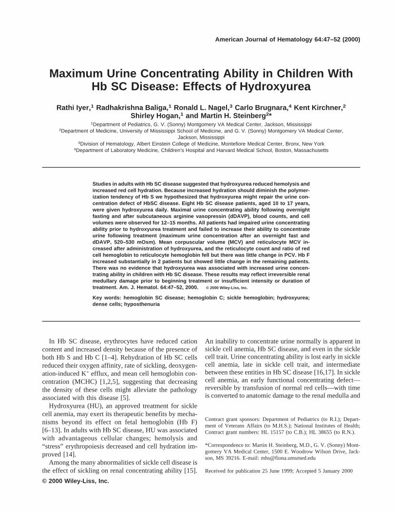
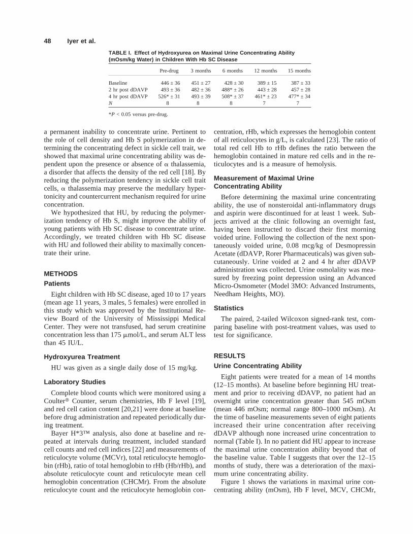
Figure 1 shows the variations in maximal urine con-centrating ability (mOsm), Hb F level, MCV, CHCMr,
TABLE I. Effect of Hydroxyurea on Maximal Urine Concentrating Ability(mOsm/kg Water) in Children With Hb SC Disease
Pre-drug 3 months 6 months 12 months 15 months
Baseline 446 ± 36 451 ± 27 428 ± 30 389 ± 15 387 ± 332 hr post dDAVP 493 ± 36 482 ± 36 488* ± 26 443 ± 28 457 ± 284 hr post dDAVP 526* ± 31 493 ± 39 508* ± 37 461* ± 23 477* ± 34N 8 8 8 7 7
*P < 0.05 versus pre-drug.
48 Iyer et al.
Hb/rHb, and reticulocyte count over time in two patientswith Hb SC disease treated with HU, one (Fig. 1A) withno change in Hb F level and another (Fig. 1B) whose HbF increased from 6.3% to 15% during treatment.
Hb F
Two patients, a 10-year old girl and a 17-year old girl,increased their concentration of Hb F during treatmentwith HU. The first patient had an increase from 6.3% tobetween 14% and 15% Hb F after 2–3 months of treat-ment (Fig. 1B). A second patient increased her Hb F from1% to about 6% after 5–6 months of treatment. No otherpatient had an increase of Hb F beyond 1.5% (Figure 2).
Cell Volumes, Cell Hemoglobin, Cell Counts
MCV increased from 73.5 ± 8.1 fL at baseline to 83 ±12.5 fL at the conclusion of treatment (P 40.018), witha corresponding increased in MCVr from 87.4 ± 6.3 to101 ± 8.7 fL (P 40.028). PCV, 31.9 ± 1.9 at baseline,did not change significantly (32.5 ± 1.3;P 40.21) andMCHC, 36.3 ± 1.1 g/dL at baseline, fell slightly but notsignificantly (35.5 ± 1.4 g/dL;P 40.069).
Absolute reticulocyte count fell from (171.5 ± 49.5) ×109/L pretreatment to (123.3 ± 44.5) × 109/L at the con-clusion of data collection (P 40.017), suggesting eithera reduction of hemolysis or suppression of erythropoie-sis. Under steady-state conditions, erythrocyte survivalmay also be estimated indirectly from the ratio of hemo-globin contained in mature red cells and in reticulocytes[23]. The ratio of Hb/rHb at the baseline (29.5 ± 11.2)and after treatment (37.3 ± 14.2) also suggested someprolongation of red cell survival.
Chemistries and Toxicity
Serum creatinine and ALT were not affected by HUtreatment. There were no complications of HU treatment,and in no patient did toxicity require cessation of treat-ment.
DISCUSSION
Although “milder” than sickle cell anemia, Hb SC dis-ease is associated with considerable morbidity and in-creased mortality underscoring the need for an effectivetreatment [24–29]. Sickle hemoglobin polymerizationwithin Hb SC cells is primarily due to their high hemo-globin concentration. Rehydrating these cells returnedtheir abnormal properties toward normal [1–5]. Weshowed previously that HU was associated with a declinein hemolysis in adults with Hb SC disease and that therewas a fall in the percent of dense red cells [14]. Reducedcell density should diminish the polymerization tendencyof Hb S within Hb SC disease cells. We hypothesizedthat by reducing the polymerization tendency of Hb S,HU might improve and preserve the renal medullary
countercurrent mechanism, thus increasing the ability ofyoung patients with Hb SC disease to concentrate urine.Increased urine concentrating ability might provide aphysiological marker of the clinical effectiveness of thisdrug in Hb SC disease.
Deterioration of renal concentrating ability in Hb SCdisease has not been extensively studied. In 14 patientswith Hb SC disease, ages 6 to 65 years (mean age 30years), the maximum concentrating ability was 537mOsm and appeared to decline with age [16]. The twoyoungest patients in that study, aged 6 and 8 years, hadmaximum urine concentrations of 640 and 698 mOsm,about 60% of normal. In children with sickle cell anemiaunder age 10 years, transfusion with normal red bloodcells can reverse hyposthenuria, suggesting that renalmedullary damage is reversible. Reversibility after trans-fusion is lost between ages 10 and 15 years due to struc-tural damage in the renal medulla [30,31]. Our patients,whose average age was 11 years when treatment began,had pretreatment maximum urine concentrations of 525mOsm—about half the expected normal value—suggesting an earlier than anticipated loss of renal med-ullary function. Within this small group of older children,treatment with HU did not increase the ability to concen-trate urine. Urine concentrating ability failed to increasesignificantly in any patient, even when Hb F concentra-tion increased. During the study period there evenseemed to be a decrease in concentrating ability. Hemo-lysis appeared to be reduced in all patients, as estimatedby the fall in reticulocyte count and the ratio of Hb/rHbyet there were no significant changes in PCV and theMCHC changed little. An increase in PCV is not neces-sarily a desirable goal for the treatment of Hb SC diseasesince much of the pathology of this disorder may resultfrom increased blood viscosity caused by dense cells. Inour previous study of hydroxyurea in adults with Hb SCdisease we speculated that any adverse effect of the smallbut significant increase in PCV might be offset by the fallin red cell density [14].
To effect beneficially renal medullary function—andperhaps to forestall damage to other vital organs—HUmay have to be started in very young children beforethere is irreversible renal medullary or organ damage.Such a study is now being planned. We do not yet knowif HU will prevent organ damage in sickle cell disease orhelp restore function to already injured organs. Splenicregeneration has been reported in two adults with sicklecell anemia who had Hb F values of about 30% duringHU treatment but the spleen may be a special case [32].
Baseline Hb F levels are lower in Hb SC disease thanin sickle cell anemia. Increased cell volume and reducedcell density may occur independent of increases in Hb Fin adults with Hb SC disease treated with HU [14]. Simi-lar observations have been made in sickle cell anemia[11,13,33]. Two patients in the current study, both fe-
Urine Concentrating Ability in Children With Hb SC Disease 49
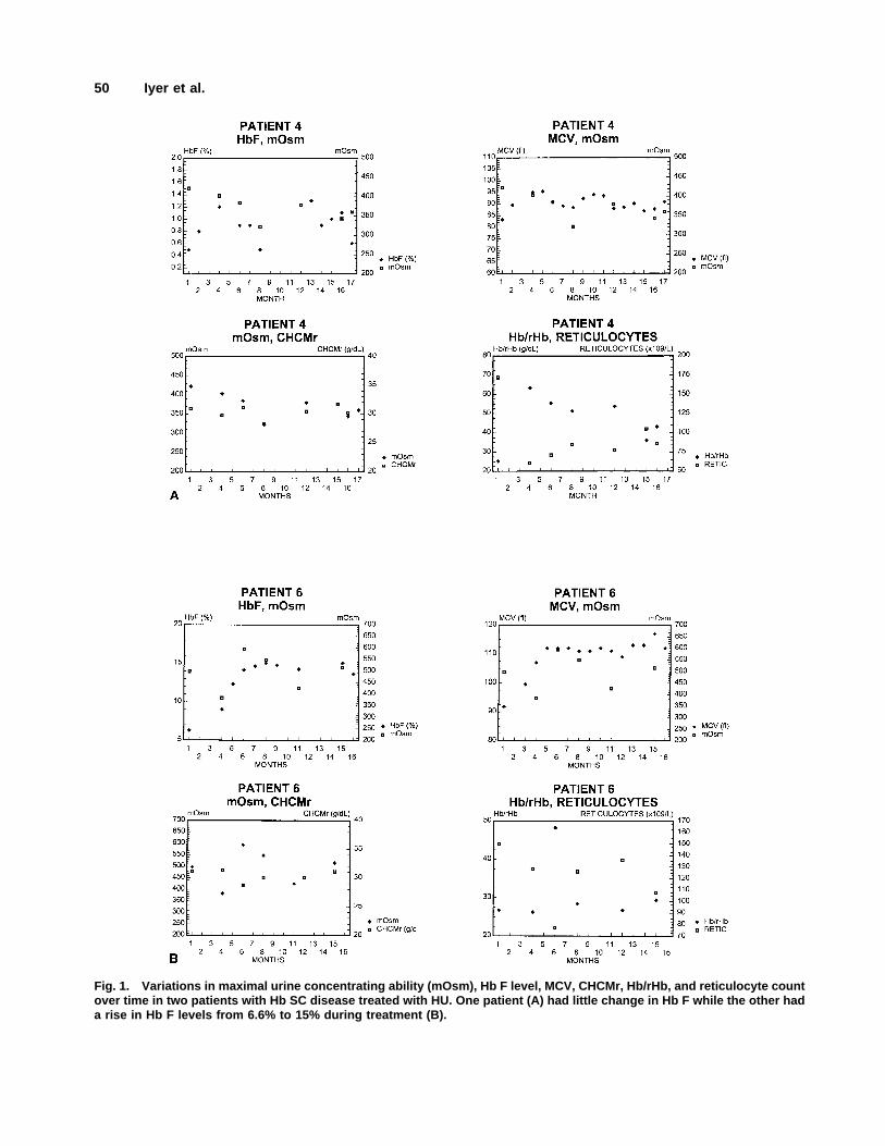
Fig. 1. Variations in maximal urine concentrating ability (mOsm), Hb F level, MCV, CHCMr, Hb/rHb, and reticulocyte countover time in two patients with Hb SC disease treated with HU. One patient (A) had little change in Hb F while the other hada rise in Hb F levels from 6.6% to 15% during treatment (B).
50 Iyer et al.
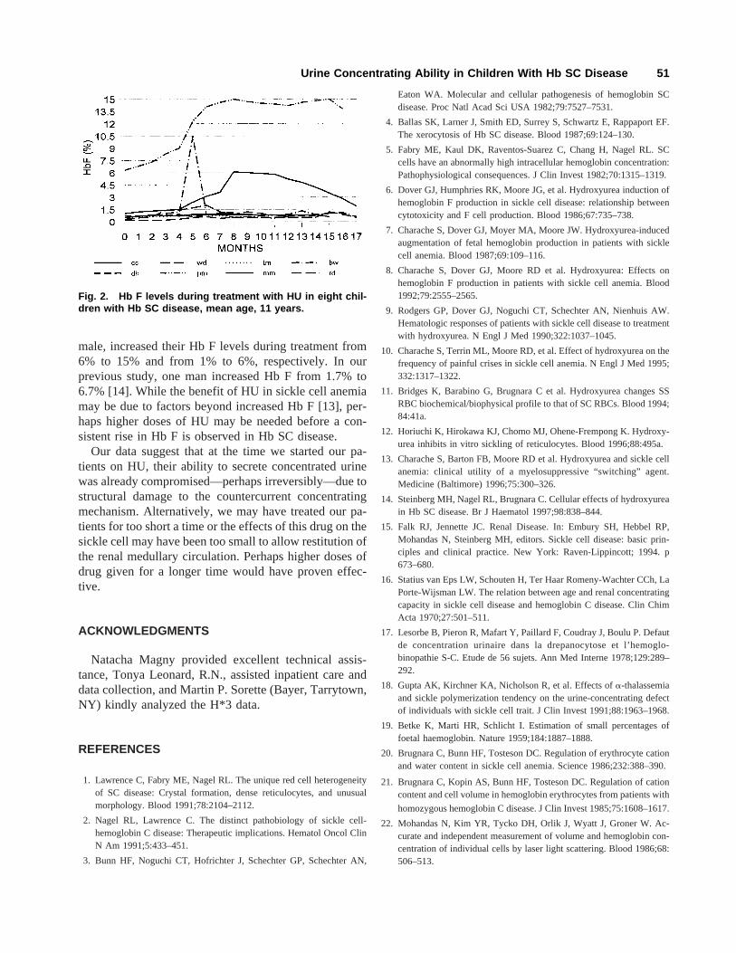
male, increased their Hb F levels during treatment from6% to 15% and from 1% to 6%, respectively. In ourprevious study, one man increased Hb F from 1.7% to6.7% [14]. While the benefit of HU in sickle cell anemiamay be due to factors beyond increased Hb F [13], per-haps higher doses of HU may be needed before a con-sistent rise in Hb F is observed in Hb SC disease.
Our data suggest that at the time we started our pa-tients on HU, their ability to secrete concentrated urinewas already compromised—perhaps irreversibly—due tostructural damage to the countercurrent concentratingmechanism. Alternatively, we may have treated our pa-tients for too short a time or the effects of this drug on thesickle cell may have been too small to allow restitution ofthe renal medullary circulation. Perhaps higher doses ofdrug given for a longer time would have proven effec-tive.
ACKNOWLEDGMENTS
Natacha Magny provided excellent technical assis-tance, Tonya Leonard, R.N., assisted inpatient care anddata collection, and Martin P. Sorette (Bayer, Tarrytown,NY) kindly analyzed the H*3 data.
REFERENCES
1. Lawrence C, Fabry ME, Nagel RL. The unique red cell heterogeneityof SC disease: Crystal formation, dense reticulocytes, and unusualmorphology. Blood 1991;78:2104–2112.
2. Nagel RL, Lawrence C. The distinct pathobiology of sickle cell-hemoglobin C disease: Therapeutic implications. Hematol Oncol ClinN Am 1991;5:433–451.
3. Bunn HF, Noguchi CT, Hofrichter J, Schechter GP, Schechter AN,
Eaton WA. Molecular and cellular pathogenesis of hemoglobin SCdisease. Proc Natl Acad Sci USA 1982;79:7527–7531.
4. Ballas SK, Larner J, Smith ED, Surrey S, Schwartz E, Rappaport EF.The xerocytosis of Hb SC disease. Blood 1987;69:124–130.
5. Fabry ME, Kaul DK, Raventos-Suarez C, Chang H, Nagel RL. SCcells have an abnormally high intracellular hemoglobin concentration:Pathophysiological consequences. J Clin Invest 1982;70:1315–1319.
6. Dover GJ, Humphries RK, Moore JG, et al. Hydroxyurea induction ofhemoglobin F production in sickle cell disease: relationship betweencytotoxicity and F cell production. Blood 1986;67:735–738.
7. Charache S, Dover GJ, Moyer MA, Moore JW. Hydroxyurea-inducedaugmentation of fetal hemoglobin production in patients with sicklecell anemia. Blood 1987;69:109–116.
8. Charache S, Dover GJ, Moore RD et al. Hydroxyurea: Effects onhemoglobin F production in patients with sickle cell anemia. Blood1992;79:2555–2565.
9. Rodgers GP, Dover GJ, Noguchi CT, Schechter AN, Nienhuis AW.Hematologic responses of patients with sickle cell disease to treatmentwith hydroxyurea. N Engl J Med 1990;322:1037–1045.
10. Charache S, Terrin ML, Moore RD, et al. Effect of hydroxyurea on thefrequency of painful crises in sickle cell anemia. N Engl J Med 1995;332:1317–1322.
11. Bridges K, Barabino G, Brugnara C et al. Hydroxyurea changes SSRBC biochemical/biophysical profile to that of SC RBCs. Blood 1994;84:41a.
12. Horiuchi K, Hirokawa KJ, Chomo MJ, Ohene-Frempong K. Hydroxy-urea inhibits in vitro sickling of reticulocytes. Blood 1996;88:495a.
13. Charache S, Barton FB, Moore RD et al. Hydroxyurea and sickle cellanemia: clinical utility of a myelosuppressive “switching” agent.Medicine (Baltimore) 1996;75:300–326.
14. Steinberg MH, Nagel RL, Brugnara C. Cellular effects of hydroxyureain Hb SC disease. Br J Haematol 1997;98:838–844.
15. Falk RJ, Jennette JC. Renal Disease. In: Embury SH, Hebbel RP,Mohandas N, Steinberg MH, editors. Sickle cell disease: basic prin-ciples and clinical practice. New York: Raven-Lippincott; 1994. p673–680.
16. Statius van Eps LW, Schouten H, Ter Haar Romeny-Wachter CCh, LaPorte-Wijsman LW. The relation between age and renal concentratingcapacity in sickle cell disease and hemoglobin C disease. Clin ChimActa 1970;27:501–511.
17. Lesorbe B, Pieron R, Mafart Y, Paillard F, Coudray J, Boulu P. Defautde concentration urinaire dans la drepanocytose et l’hemoglo-binopathie S-C. Etude de 56 sujets. Ann Med Interne 1978;129:289–292.
18. Gupta AK, Kirchner KA, Nicholson R, et al. Effects ofa-thalassemiaand sickle polymerization tendency on the urine-concentrating defectof individuals with sickle cell trait. J Clin Invest 1991;88:1963–1968.
19. Betke K, Marti HR, Schlicht I. Estimation of small percentages offoetal haemoglobin. Nature 1959;184:1887–1888.
20. Brugnara C, Bunn HF, Tosteson DC. Regulation of erythrocyte cationand water content in sickle cell anemia. Science 1986;232:388–390.
21. Brugnara C, Kopin AS, Bunn HF, Tosteson DC. Regulation of cationcontent and cell volume in hemoglobin erythrocytes from patients with
homozygous hemoglobin C disease. J Clin Invest 1985;75:1608–1617.
22. Mohandas N, Kim YR, Tycko DH, Orlik J, Wyatt J, Groner W. Ac-curate and independent measurement of volume and hemoglobin con-centration of individual cells by laser light scattering. Blood 1986;68:506–513.
Fig. 2. Hb F levels during treatment with HU in eight chil-dren with Hb SC disease, mean age, 11 years.
Urine Concentrating Ability in Children With Hb SC Disease 51
23. Brugnara C, Zelmanovic D, Sorette M, Ballas SK, Platt O. Reticulo-cyte hemoglobin: an integrated parameter for evaluation of erythro-poietic activity. Am J Clin Pathol 1997;109:133–142.
24. Kinney TR, Ware RE. Compound heterozygous states. In: Embury SH,Hebbel RP, Mohandas N, Steinberg MH, editors. Sickle cell disease:basic principles and clinical practice. New York: Raven Press; 1994. p437–451.
25. Platt OS, Brambilla DJ, Rosse WF, et al. Mortality in sickle celldisease: life expectancy and risk factors for early death. N Engl J Med1994;330:1639–1644.
26. Lutty GA, Goldberg MF. Ophthalmologic complications. In: EmburySH, Hebbel RP, Mohandas N, Steinberg MH, editors. Sickle cell dis-ease: basic principles and clinical practice. New York: Raven; 1994. p703–724.
27. Milner PF, Kraus AP, Sebes JI, et al. Sickle cell disease as a cause ofosteonecrosis of the femoral head. N Engl J Med 1991;325:1476–1481.
28. Castro O, Brambilla DJ, Thorington B, et al. The acute chest syndromein sickle cell disease: Incidence and risk factors. Blood 1994;84:643–649.
29. Platt OS, Thorington BD, Brambilla DJ, et al. Pain in sickle celldisease: rates and risk factors. N Engl J Med 1991;325:11–16.
30. Keitel HG, Thompson D, Itano HA. Hyposthenuria in sickle cell ane-mia: a reversible renal defect. J Clin Invest 1956;35:998–1007.
31. Statius van Eps LW, Schouten H, La Porte-Wijsman LW, StruykerBoudier AM. The influence of red blood cell transfusions on the hy-posthenuria and renal hemodynamics of sickle cell anemia. Clin ChimActa 1967;17:449–461.
32. Claster S, Vichinsky E. First report of reversal of organ dysfunction insickle cell anemia by the use of hydroxyurea: splenic regeneration.Blood 1996;88:195–203.
33. Bridges KR, Barabino GD, Brugnara C, et al. A multiparameter analy-sis of sickle erythrocytes in patients undergoing hydroxyurea therapy.Blood 1996;88:4701–4710.
52 Iyer et al.