Embed Size (px)
Citation preview

Max Brinsmead MB BS PhDMay 2015
RCOG Green-top Guideline number 27 January 2011
“Placenta praevia, placenta praevia accreta and vasa praevia: diagnosis and management”
Plus a few personal observations

In the 21st century placenta previa is best defined as an ultrasound observation of the placenta in the lower segment of the uterus Major = the placenta covers the internal os Minor (or Partial) = all the others
Problems: The lower segment does not develop until the
last trimester of pregnancy There is no agreed definition of where the
lower segment begins ultrasonically But the internal os is a fixed point that can be
identified with ultrasound So all minor degrees of previa should be
defined in relation to this
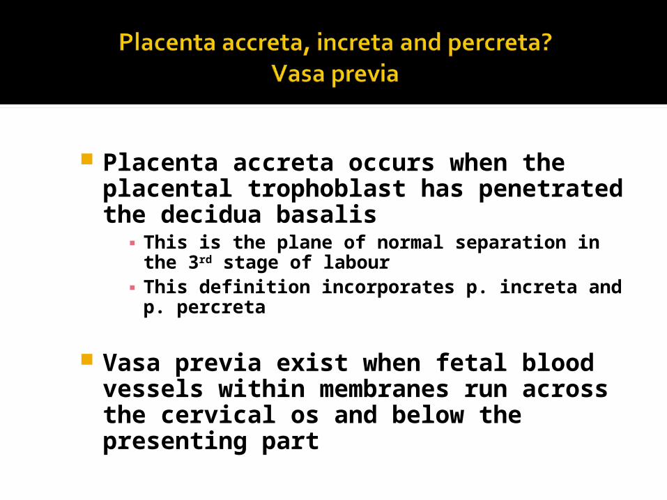
Placenta accreta occurs when the placental trophoblast has penetrated the decidua basalis▪ This is the plane of normal separation in the
3rd stage of labour▪ This definition incorporates p. increta and p.
percreta
Vasa previa exist when fetal blood vessels within membranes run across the cervical os and below the presenting part
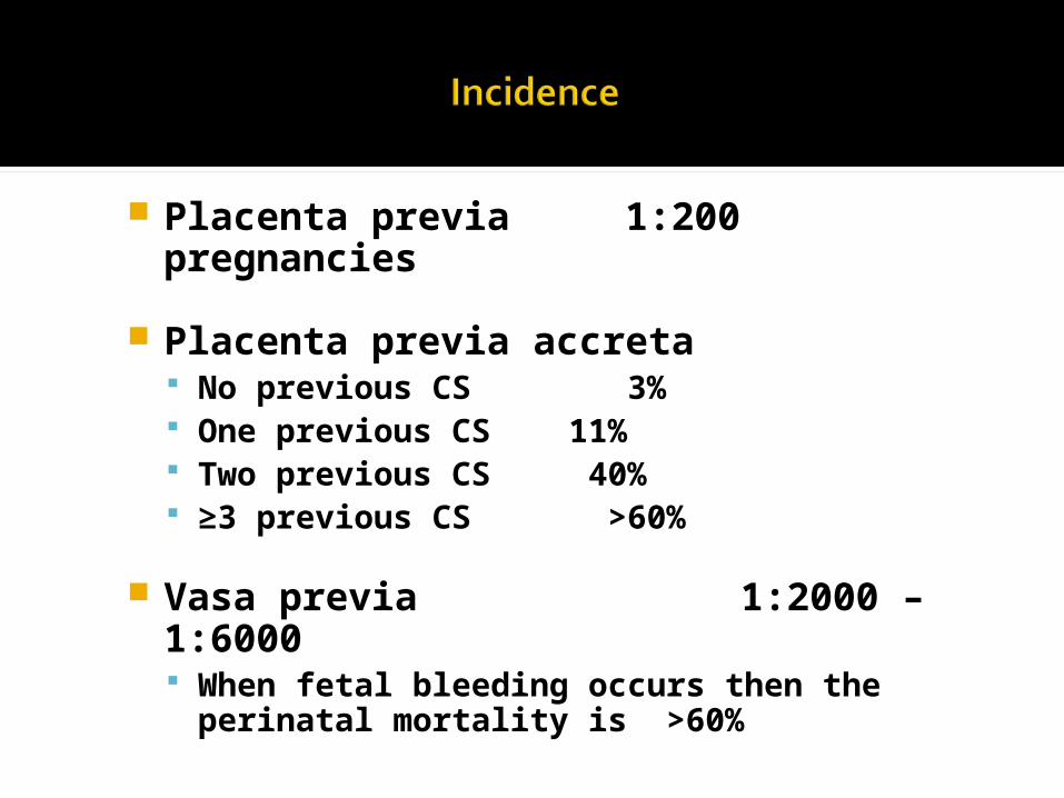
Placenta previa 1:200 pregnancies
Placenta previa accreta No previous CS 3% One previous CS 11% Two previous CS 40% ≥3 previous CS >60%
Vasa previa 1:2000 – 1:6000 When fetal bleeding occurs then the
perinatal mortality is >60%

Clinical suspicion for all women with APH after 20w gestation especially with…
Painless bleeding A high presenting part Irrespective of previous US imaging
Screening for placenta previa occurs with the 18-22w morphology scan When a diagnosis of “low-lying placenta”
may be as high as 1:10
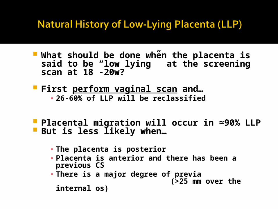
What should be done when the placenta is said to be “low lying” at the screening scan at 18 -20w?
First perform vaginal scan and…▪ 26-60% of LLP will be reclassified
Placental migration will occur in ≈90% LLP
But is less likely when…
▪ The placenta is posterior▪ Placenta is anterior and there has been a
previous CS▪ There is a major degree of previa
(>25 mm over the internal os)

Repeat the scan if there is a clinical need▪ Usually APH
Repeat the scan at 32w when…
▪ The placenta is anterior & there has been a previous CS
▪ Look for evidence of accreta▪ There is a major degree of previa (over the
internal os)
For women with minor PP and no symptoms it is best to defer the scan until 36w

Greyscale ultrasound has 95% sensitivity and 82% positive predictive value.
Look for… Loss of the retroplacental echolucent zone An irregular “ “
" Thinning or disruption of the hyperechoic
serosa/bladder zone Exophytic masses invading bladder Abnormal placental lacunae
Diagnosis can be enhanced using Colour Doppler 3-D Doppler MRI

Prevent and treat anaemia Individualize management when there
is APH or major PP. Home care is possible when…
Immediate transfer to hospital is possible An adult is with the woman at all times Informed and understanding patient Admission occurs if there is any bleeding,
pain or contractions
Tocolysis okay in the absence of severe APH
Group & Save according to local protocols
Beware of thromboembolism with prolonged immobilisation
This decision should be based on clinical judgement supplemented by ultrasound findings
If the placenta is >2 cm from the internal os (and not thick or posterior) then the vaginal delivery rate is >70% If the placenta is <2 cm from the internal os
then the vaginal delivery rate is 12.5%
There is still a role for EUA or amniotomy in theatre at 38+w for minor placenta previa The aim is to bring the head into the lower
segment and rupture the membranes

Patient Information Depends on the clinical scenario For major placenta previa…▪ Risk of major haemorrhage 1:5▪ Risk of hysterectomy 1:10▪ Return to theatre rate 75:1000▪ Bladder injury 23:1000
For previa and previous CS…▪ Risk of hysterectomy is 1:3
For placenta previa accreta…▪ Hysterectomy “very likely”
Consider Place of Delivery▪ ICU and facilities for management massive
haemorrhage Assemble and brief a multidisciplinary team
▪ Anaesthetist, Vascular surgeon, Interventional radiologist etc
▪ Role of prophylactic arterial catheter balloon uncertain

Aim for 38+ weeks for asymptomatic patients and those with minor previa
Use corticosteroids for lung maturation for deliveries that are mandated <38w
Aim for 36 – 37w for those with suspected placenta previa accreta
Consultant obstetrician & anaesthetist available for all Depends on the clinical scenario
Regional block anaesthesia not excluded Consider facilities for cell salvage and
transfusion▪ Especially when a mother refuses transfusion
Surgical tips▪ Use all available techniques for continuing
bleeding after removal of a placenta previa e.g. Oxytocics, direct suture, B-Lynch suture, ut. artery embolisation hysterectomy etc.
▪ Try to avoid section through a placenta accreta
▪ Do not attempt to remove a morbidly adherent placenta▪ Hysterectomy with placenta intact or ▪ Leave the placenta behind when uterine
conservation desired
Provide broad spectrum antibiotics
Methotrexate and or prophylactic arterial embolisation not recommended
Follow with ultrasound and beta-HCG
Risk of haemorrhage is 35%
Risk of infection is 18%
Risk of DIC is 7%

Bi-Lobed or Succinturiate placenta
Low lying placenta in the second trimester
Multiple pregnancy
IVF where the incidence may be as high as
1:300
Always consider this when a (dark) APH occurs Especially if it occurs at the time of spontaneous
or artificial rupture of membranes Rapid test for fetal HB desirable
▪ The best uses 0.14M NaOH▪ But do not delay IMMEDIATE delivery if there is
a strong clinical suspicion or a deteriorating CTG
Diagnosis can sometimes be made by palpation and or amnioscopy
Screening for vasa previa is not recommended because it does not fulfil screening criteria▪ But it can be detected with ≈90% specificity
using colour Doppler ▪ Sensitivity uncertain

Please leave a note on the Welcome Page to this website