Embed Size (px)
Citation preview
7) 3–17www.elsevier.com/locate/gene
Gene 390 (200
Mavericks, a novel class of giant transposable elements widespread ineukaryotes and related to DNA viruses
Ellen J. Pritham ⁎, Tasneem Putliwala, Cédric Feschotte
The University of Texas at Arlington, The Department of Biology, Arlington, TX, United States
Received 14 July 2006; accepted 2 August 2006
Available onlin
Received by I. King Jordan
e 23 August 2006
Abstract
We previously identified a group of atypical mobile elements designated Mavericks from the nematodes Caenorhabditis elegans and C.briggsae and the zebrafish Danio rerio. Here we present the results of comprehensive database searches of the genome sequences available, whichreveal that Mavericks are widespread in invertebrates and non-mammalian vertebrates but show a patchy distribution in non-animal species, beingpresent in the fungi Glomus intraradices and Phakopsora pachyrhizi and in several single-celled eukaryotes such as the ciliate Tetrahymenathermophila, the stramenopile Phytophthora infestans and the trichomonad Trichomonas vaginalis, but not detectable in plants. This distribution,together with comparative and phylogenetic analyses of Maverick-encoded proteins, is suggestive of an ancient origin of these elements ineukaryotes followed by lineage-specific losses and/or recurrent episodes of horizontal transmission. In addition, we report that Maverick elementshave amplified recently to high copy numbers in T. vaginalis where they now occupy as much as 30% of the genome. Sequence analysis confirmsthat most Mavericks encode a retroviral-like integrase, but lack other open reading frames typically found in retroelements. Nevertheless, thelength and conservation of the target site duplication created upon Maverick insertion (5- or 6-bp) is consistent with a role of the integrase-likeprotein in the integration of a double-stranded DNA transposition intermediate. Mavericks also display long terminal-inverted repeats but do notcontain ORFs similar to proteins encoded by DNA transposons. Instead, Mavericks encode a conserved set of 5 to 9 genes (in addition to theintegrase) that are predicted to encode proteins with homology to replication and packaging proteins of some bacteriophages and diverseeukaryotic double-stranded DNA viruses, including a DNA polymerase B homolog and putative capsid proteins. Based on these and otherstructural similarities, we speculate that Mavericks represent an evolutionary missing link between seemingly disparate invasive DNA elementsthat include bacteriophages, adenoviruses and eukaryotic linear plasmids.© 2006 Elsevier B.V. All rights reserved.
Keywords: DNA polymerase B; c-integrase; Transposon; Adenovirus; Polinton; Trichomonas vaginalis
Abbreviations: aa, amino acid(s); bp, base pair(s); CMG, conserved Ma-verick gene; ds, double-strand(ed); hAT, hobo/Activator/Tam3; kb, kilobase(s)or 1000 bp; LTR, long terminal repeat; Mav, Maverick; Mb, megabase(s) or1 million bp; ORF, open reading frame; ss, single-strand(ed); TE, transposableelement; TIR, terminal-inverted repeat; Tlr, Tetrahymena thermophila longrepeat; TSD, target site duplication.⁎ Corresponding author. Department of Biology, Box 19489, The University
of Texas at Arlington, Arlington, TX 76019, United States. Tel.: +1 817 2720981; fax: +1 817 272 2855.
E-mail address: [email protected] (E.J. Pritham).
0378-1119/$ - see front matter © 2006 Elsevier B.V. All rights reserved.doi:10.1016/j.gene.2006.08.008
1. Introduction
Most eukaryotic genomes harbor a vast amount of inter-spersed repetitive DNA, which mostly consists of transposableelements (TEs) or their remnants. The rapidly increasingamount of DNA sequences accumulating in the public databaseand the completion of many genome sequencing projects havecontributed to reveal an extraordinary diversity of TEs, in termsof their structure, survival strategies and dynamics of ampli-fication (e.g. Lander et al., 2001; Feschotte et al., 2002;Kapitonov and Jurka, 2003; Brookfield, 2005; Hua-Van et al.,2005). It has become clear that deciphering the origin andbiology of these elements is an essential facet of genomic
4 E.J. Pritham et al. / Gene 390 (2007) 3–17
research, the value of which is not just limited to the assistanceprovided to sequence assembly and annotation, but is also criticalto understanding how genes and genomes evolve.
Traditionally, transposable elements have been divided into 2classes based on their mechanism of transposition (Finnegan,1989; Capy et al., 1998). Class 1 elements (or retroelements)transpose through reverse-transcription of an RNA intermedi-ate, while class 2 elements (or DNA transposons) move directlythrough a DNA intermediate. Retrotransposition involves rela-tively complex enzymatic machinery and often requires thesequential action of multiple proteins such as reverse transcrip-tase, endo- or ribonucleases and integrase (Craig et al., 2002). Incontrast the transposition of most eukaryotic DNA transposonsrequires a single element-encoded protein called transposase,which catalyzes all the steps of the so-called cut-and-paste re-action (Craig et al., 2002). Retrotransposons are further dividedinto 2 major types based on their structure and transpositionmechanism. Non-long terminal repeat (non-LTR) retroelements(LINEs and SINEs) transpose through a target-primedmechanismwhere integration and reverse-transcription is coupled and ini-tiation occurs via a single-strand nick at the insertion site (Luanet al., 1993). In contrast, reverse-transcription of LTR retro-elements (or retroviral elements) occurs in retroviral particles andprecedes integration (Voytas and Boeke, 2002; Curcio andDerbyshire, 2003).
The integration mechanism of LTR retroelements and retro-viruses is very similar to those of DNA transposons (Haren et al.,1999; Curcio and Derbyshire, 2003). Indeed, the catalyticdomains of many integrases and transposases display significantsimilarities at the primary sequence level and adopt a verysimilar tri-dimensional fold (Capy et al., 1996; Haren et al.,1999; Rice and Baker, 2001; Hickman et al., 2005). Therefore, itis likely that at least a subset of retroviral integrases and trans-posases share a common ancestor (Capy et al., 1996). Thisobservation led several authors to hypothesize that an ancestralLTR retrotransposon evolved from a non-LTR retrotransposonby assimilation of a nested DNA transposon (Capy et al., 1998;Malik and Eickbush, 2001; Eickbush and Malik, 2002). A sub-sequent step in the evolution of LTR retroelements is theiroccasional transition into infectious retroviruses by acquisitionof envelope genes from multiple diverse viral sources (Temin,1980; Malik et al., 2000; Eickbush and Malik, 2002). On theother hand, there is some evidence that retroviruses may alsosubsequently lose their infectious capacities (i.e. envelope gene)and go back to an intracellular lifestyle (Lerat and Capy, 1999;Gifford and Tristem, 2003; Herve et al., 2004; Yano et al., 2005).
The traditional dichotomy of transposable elements (class 1vs. class 2) has been recently challenged by the discovery ofatypical mobile elements, which share no sequence or structuralsimilarities to previously described class 1 or class 2 elements,and thus may define entirely new classes of TEs. For example, agroup of transposons called Helitrons with common structuraland coding capacities were recently identified in the genomes ofplants, animals and fungi (Kapitonov and Jurka, 2001; Poulteret al., 2003; Hood, 2005). Helitrons are apparently related togenetic elements that replicate through a rolling-circle mech-anism, including the circo- and geminiviruses, a group of
single-stranded DNA (ssDNA) viruses that infect plant andanimals. Akin to the reversible switch from retrotransposons toretroviruses, it has been proposed that Helitrons originated fromand/or gave rise to geminiviruses (Feschotte and Wessler, 2001;Kapitonov and Jurka, 2001; Murad et al., 2004).
Together these studies tend to blur the distinction betweentransposable elements and viruses and suggest that there arefrequent exchanges of functional modules and domains betweenTEs and viruses. This could reflect similar evolutionary tra-jectories and, in some cases, the common ancestry of TEs andviruses. Recently we identified a new assemblage of largetransposable elements, calledMavericks (Feschotte and Pritham,2005), that share structural and sequence similarity to Tlr1, anatypical group of mobile element described from the ciliate Te-trahymena thermophila (Wells et al., 1994; Wuitschick et al.,2002). Maverick elements were initially identified in thenematodes Caenorhabditis elegans and C. briggsae and in thezebrafish Danio rerio and we proposed that, together with Tlr1,they define an entirely new class of TEs (Feschotte and Pritham,2005). More recently, Kapitonov and Jurka (2006) described inmore detail some of theseMavericks as well as additional relatedelements that they identified in several eukaryotic genomes.They refer to this group of elements as Polintons collectivelyand proposed a model for the origin and transposition mecha-nism of these unusual transposons (Kapitonov and Jurka, 2006).
In the present study, we further expand the distribution ofMavericks in eukaryotes and offer a detailed characterization oftheir structure and coding capacity. We also provide evidence thatthe parabasalid protozoa Trichomonas vaginalis has recentlyexperienced an explosive amplification ofMavericks, which nowappear to contribute up to one third of its genome. Finally, wehighlight the unique characteristics of Mavericks among endo-geneous mobile elements and their striking similarities to ananciently related group of invasive DNA that includes adeno-viruses, bacteriophages and eukaryotic linear plasmids.
2. Materials and methods
2.1. Computational detection of Mavericks
Candidate Mavericks were identified by homology-basedsearches at NCBI, beginning in April of 2005 until final pre-paration and submission of this manuscript. All sequence datathat have been deposited in the NR, HTGS and WGS databasesat NCBI during this period were subjected to multiple searchesusing the various BLAST tools (Altschul et al., 1990; McGinnisand Madden, 2004). The queries used included the DNA orputative protein sequences derived from previously identifiedMaverick elements (Feschotte and Pritham, 2005).Generally, a hitwas considered significant when the e-valuewas lower than 10−4.Maverick sequences reported in this article are available uponrequest to the authors. Accession numbers from the variousdatabases and the nucleotide coordinates of complete elements arereported in Table 1; Supplemental Table 1. AdditionalMaverickscopies were identified using BLAT (Kent, 2002) against genomesavailable through the UCSC genome browser (http://genome.ucsc.edu/). Due to the relatively large size of completeMaverick
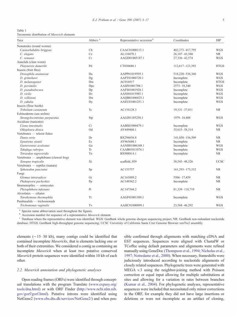
Table 1Taxonomic distribution of Maverick elements
Taxa Abbrev. a Representative accession b Coordinates DBc
Nematodes (round worms)Caenorhabditis briggsae Cb CAAC01000115.1 402,373–417,795 WGSC. elegans Ce AL110478.1 26,107–43,380 NRC. remanei Cr AAGD01005187.1 27,336–42,574 WGS
Annelids (clam worm)Platynereis dumerilii Pd CT030680.1 112,617–123,393 HTGS
Insects (fruit flies)Drosophila ananassae Da AAPP01019595.1 518,220–536,360 WGSD. grimshawi Dg AAPT01000720.1 Incomplete WGSD. melanogaster Dm AC016917 Incomplete HTGSD. persimilis Dpe AAIZ01001788.1 2573–18,340 WGSD. pseudoobscura Dp AAFS01001928.1 Incomplete WGSD. virilis Dv AANI01015985.1 Incomplete WGSD. willistoni Dw AAQB01006825.1 Incomplete WGSD. yakuba Dy AAEU01001251.1 Incomplete WGS
Insects (flour beetle)Tribolium castaneum Tc AC154128.3 19,331–37,011 NR
Echinoderms (sea urchin)Strongylocentrotus purpuratus Stp AAGJ01185250.1 1979–18,488 WGS
Ascidians (tunicates)Ciona intestinalis Ci AABS01000479.1 Incomplete WGSOikopleura dioica Od AY449460.1 35,615–58,314 NR
Vertebrates — teleost fishesDanio rerio Dr BX294656.8 141,050–156,509 NREptatretus stoutii Es AY965680.1 Incomplete NRGasterosteus aculeatus Ga AANH01006348.1 Incomplete WGSTakufugu rubripes Tr CAAB01011074.1 Incomplete WGSTetraodon nigroviridis Tn BX908814.1 Incomplete NR
Vertebrates — amphibians (clawed frog)Xenopus tropicalis Xt scaffold_959 38,543–48,326 UCSC
Vertebrates — reptiles (tuatara)Sphenodon punctatus Sp AC153757 161,293–173,332 NR
FungiGlomus intraradices Gi AC163889.2 5506–17,459 NRPhakopsora pachyrhizi Pp AC149362.2 Incomplete NR
Stramenopiles — oomycetesPhytophthora infestans Pi AC147544.2 81,339–118,719 NR
Alveolates — ciliatesTetrahymena thermophila Tt AAGF01001309.1 Incomplete WGS
Parabasalids — trichomonadsTrichomonas vaginalis Tv AAHC01000099.1 23,568–46,292 WGSa Species name abbreviation used throughout the figures.b Accession number for sequence of a representative Maverick element.c Database where the representative element was identified. WGS: GenBank whole genome shotgun sequencing project; NR: GenBank non-redundant nucleotide
database; HTGS, GenBank high-throughput genome sequencing; UCSC University of California Santa Cruz Genome Browser xenTro2 assembly.
5E.J. Pritham et al. / Gene 390 (2007) 3–17
elements (∼15–30 kb), many contigs could be identified thatcontained incomplete Mavericks, that is elements lacking one orboth of their extremities. We considered a contig as containing anincomplete Maverick when at least two putative conservedMaverick protein sequences were identified within 10 kb of eachother.
2.2. Maverick annotation and phylogenetic analyses
Open reading frames (ORFs) were identified through concept-ual translations with the program Translate (www.expasy.org/tools/dna.html) or with ORF Finder (http://www.ncbi.nlm.nih.gov/gorf/gorf.html). Putative introns were identified usingNetGene2 (www.cbs.dtu.dk/services/NetGene2/) and when pos-
sible confirmed through alignments with matching cDNA andEST sequences. Sequences were aligned with ClustalW orTCoffee using default parameters and alignments were refinedmanually using GeneDoc (Thompson et al., 1994; Nicholas et al.,1997; Notredame et al., 2000). When necessary, frameshifts werejudiciously introduced according to nucleotide alignments ofclosely related sequences. Phylogenetic trees were generated withMEGA v.3 using the neighbor-joining method with Poissoncorrection or equal input allowing for multiple substitutions atsites and allowing for a variation in rates between branches(Kumar et al., 2004). For phylogenetic analyses, representativesequences were included that necessitated only minor correctionsin the ORF, for example they did not have large insertions ordeletions or were not incomplete as an artifact of cloning,
6 E.J. Pritham et al. / Gene 390 (2007) 3–17
sequencing or genome assembly. The function of Maverick-encoded proteins was predicted by homology to proteins ofknown function, by the presence of conserved domains identifiedthrough CDD search (Marchler-Bauer et al., 2005) and by proteinthreading (Kelley et al., 2000) through the analysis of conservedprotein folds using Phyre (http://www.sbg.bio.ic.ac.uk/phyre/).
2.3. Identification of paralogous ‘empty’ sites
To illustrate themobility ofMavericks, paralogous sites (emptysites) devoid of the insertionwere queried. To identify empty sites,searches were completed using the sequences flanking each sideof the insertion as a query using either blastn or BLAT. An emptysite is reported when another region is identified in the samegenome that lacks the insertion yet contains the unduplicatedtarget site.
3. Results and discussion
3.1. Identification and distribution of Mavericks across the treeof life
Recently, we showed that non-mammalian c-integrases fromthe genomes of the nematodes C. elegans and C. briggsae andin the zebrafish are carried by large transposable elements withterminal-inverted repeats, called Mavericks (Feschotte andPritham, 2005). In order to conduct a more comprehensiveanalysis of the structure and distribution of these elements, weused the Mavericks from C. elegans and D. rerio as queriesin BLAST searches of the NR, WGS and HTGS Genbankdatabases. These searches confirmed the presence of Mavericksin C. briggsae and Takufugu rubripes as indicated by the initialstudy of c-integrases (Gao and Voytas, 2005), but alsosuggested that their occurrence was more widely distributedthan previously anticipated (Table 1).
In addition to the elements previously reported fromC. elegans, C. briggsae and D. rerio (Feschotte and Pritham,2005), we could identify complete Mavericks (as defined byelements with TIRs and flanked by a 5- or 6-bp putativeTSD) in twelve additional species from diverse eukaryoticphyla. These include a wide spectrum of invertebrate andvertebrate animals, as well as the mychorrizal fungus Glomusirradices, the oomycete Phytophthora infestans and the para-basalid T. vaginalis (Table 1, Fig. 1). BLAT searches (Kent,2002) using the TIRs of individual Maverick elements againsttheir respective draft genome sequence (when available)allowed the identification of additional copies in all of thesespecies.
With the notable exception of T. vaginalis (see Section 3.5),Maverick elements occur as relatively low copy numberfamilies (3–50 copies per genome) that share between 85 and99% nucleotide similarity between copies of the same family. Inmost species, very distinct families apparently co-exist withinthe same genome. For example, Mav_Pd1.1 and Mav_Pd2.1from the annelid Platynereis dumerilii (see SupplementalTable 1) cannot be confidently aligned at the nucleotide level,but both are clearly Maverick elements sharing conserved
structural features and encoding distantly related genes (data notshown). Members of highly divergent Maverick families arealso apparent in C. elegans, Strongylocentrus purpuratus, Oi-kopleura dioica and D. rerio (see Fig. 1 and Table 1).
It should be noted that elements closely related to thosereported here for T. castaneum, X. tropicalis, S. punctata andG. irradices were also independently identified by Kapitonovand Jurka and recently described as Polintons (Kapitonov andJurka, 2006). However, the elements that we identified inS. purpuratus and T. vaginalis are only distantly related to thosedescribed in these species by Kapitonov and Jurka andpresumably belong to divergent families co-existing withinthe same genome. Maverick elements from C. remanei,P. dumerilii, Drosophila persimilis, D. ananassae, O. dioicaand Phytopthora infestans have not been reported elsewhere.Since we previously coined the name Mavericks for the firstelements discovered in nematodes and zebrafish and predictedin this study that these Mavericks would delineate an entirelynew group of TEs, we refer hereafter to all related elements asMavericks (Feschotte and Pritham, 2005). One exception isTlr1 from T. thermophila, whose composite sequence wasoriginally assembled from several overlapping genomic clones,but yet appears to represent an incomplete copy of a Maverick-like transposon family (see below) (Wells et al., 1994;Wuitschick et al., 2002).
TBLASTN and BLASTX searches of the databases also ledto the identification of protein-coding DNA segments that likelyrepresent internal regions of Maverick elements in othereukaryotic species, but for which it was not possible tounambiguously identify the termini. This was either becauseof gaps in the genome assembly, because the assembled contigsthemselves are smaller than a typicalMaverick (∼15–20 kb) ordue to secondary TE insertions or other recombination eventstruncating and rearranging the elements. Nevertheless, whensignificant hits to multiple conservedMaverick genes (describedbelow) were detected in the same genomic neighborhood (i.e. atleast two genes less than 10 kb of each other), we considered thesegment to be of likely Maverick origin. In this way, it can beestablished that Mavericks have also colonized the genomesof the hagfish Eptatretus stoutii, the three-spined sticklebackGasterosteus aculeatus, the pufferfishes Takufugu rubripesand Tetraodon nigroviridis, the fruit flies D. grimshawi, D.melanogaster, D. pseudobscura, D. virilis, D. willistoni and D.yakuba, the sea squirt Ciona intestinalis, the plant pathogenicfungi Phakopsora pachyrhizi and the ciliate T. thermophila(Table 1).
To complete this overview of Maverick distribution, itshould also be added that sequences clearly related toMaverick-encoded integrases (see Section 3.2) could be identified innumerous other animal species, including the aphid Acyrthosi-phon pisum, the ratite bird Apteryx australis mantelli and thecnidarians Hydractinia echinata and Nematostella vectensis,suggesting that Mavericks are also present in the genomes ofspecies from the phyla Hemiptera, Aves and Cnidaria. However,these sequences were either from very short genomic clones(ratite) or from EST projects (aphids, cnidarians) and thereforecould not be physically associated with other Maverick genes.
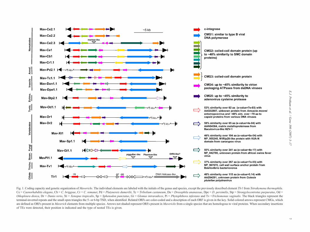
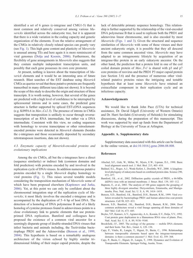
Fig. 1. Coding capacity and genetic organization ofMavericks. The individual elements are labeled with the initials of the genus and species, except the previously described element Tlr1 from Tetrahymena thermophila.Ce = Caenorhabditis elegans, Cb = C. briggsae, Cr = C. remanei, Pd = Platynereis dumerilii, Tc = Tribolium castaneum, Da = Drosophila ananassae, Dpe = D. persimilis, Stp = Strongylocentrotus purpuratus, Od =Oikopleura dioica, Dr = Danio rerio, Xt = Xenopus tropicalis, Sp = Sphenodon punctatus, Gi = Glomus intraradices, Pi = Phytophthora infestans and Tv =Trichomonas vaginalis. The black triangles represent theterminal-inverted repeats and the small open triangles the 5- or 6-bp TSD, when identified. Related ORFs are color-coded and a description of each ORF is given in the key. Solid colored arrows represent CMGs, whichare defined as ORFs present inMaverick elements from multiple species. Arrows not shaded represent ORFs present inMavericks from a single species that are homologous to viral proteins. When secondary insertionsof TEs were detected, their position is indicated and the type of nested TEs is given.
7E.J.
Pritham
etal.
/Gene
390(2007)
3–17
8 E.J. Pritham et al. / Gene 390 (2007) 3–17
Together, these data provide evidence that Mavericks arepresent in a very broad range of eukaryotes, albeit with anintriguingly patchy distribution. For example, no Maverickelements or Maverick-related genes could be identified in anyplant species despite the presence of large amounts of genomicdata for several plant species. Also we could not detect anyMaverick-related sequences in the now complete or nearlycomplete genome sequences of the mosquito Anophelesgambiae, the bee Apis melifera or the silkworm Bombyx mori,althoughMavericks can be readily detected in virtually all other
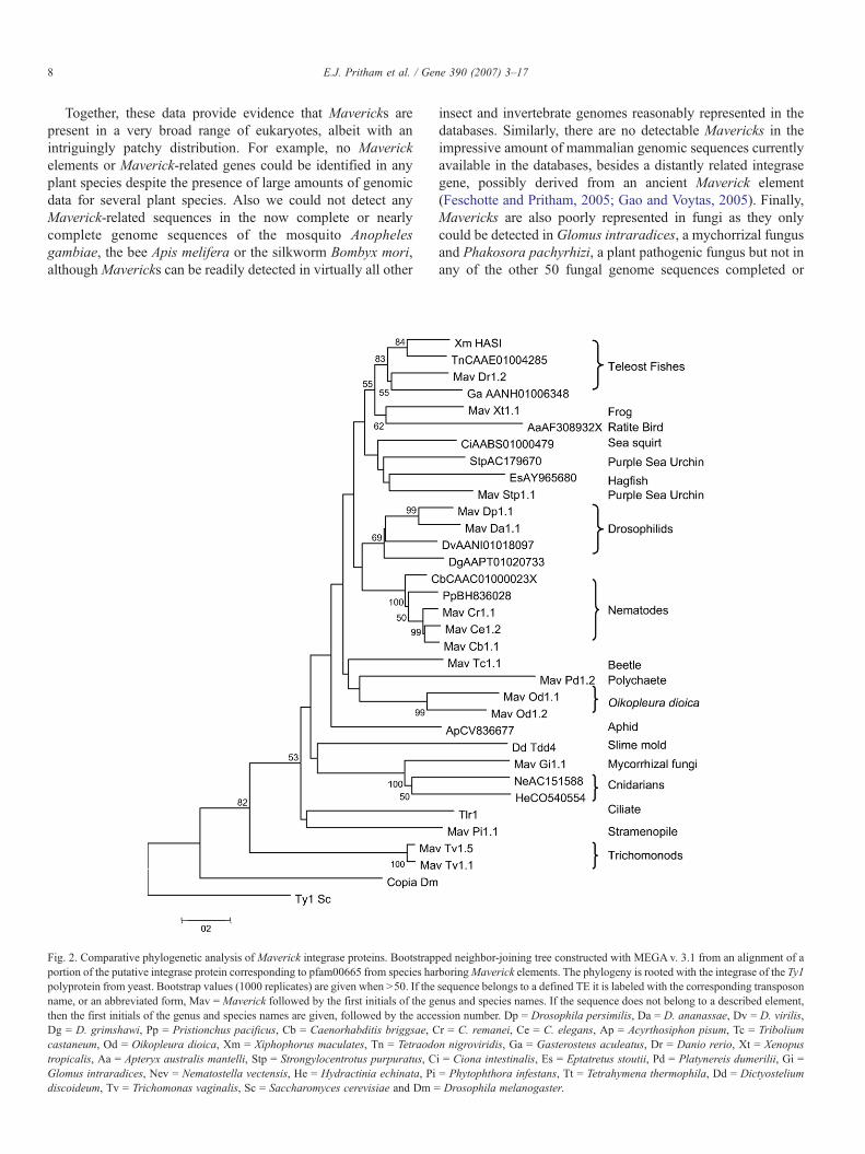
Fig. 2. Comparative phylogenetic analysis of Maverick integrase proteins. Bootstrapportion of the putative integrase protein corresponding to pfam00665 from species hapolyprotein from yeast. Bootstrap values (1000 replicates) are given when N50. If thename, or an abbreviated form, Mav = Maverick followed by the first initials of the gethen the first initials of the genus and species names are given, followed by the acceDg = D. grimshawi, Pp = Pristionchus pacificus, Cb = Caenorhabditis briggsae, Ccastaneum, Od = Oikopleura dioica, Xm = Xiphophorus maculates, Tn = Tetraodotropicalis, Aa = Apteryx australis mantelli, Stp = Strongylocentrotus purpuratus, CGlomus intraradices, Nev = Nematostella vectensis, He = Hydractinia echinata, Pidiscoideum, Tv = Trichomonas vaginalis, Sc = Saccharomyces cerevisiae and Dm =
insect and invertebrate genomes reasonably represented in thedatabases. Similarly, there are no detectable Mavericks in theimpressive amount of mammalian genomic sequences currentlyavailable in the databases, besides a distantly related integrasegene, possibly derived from an ancient Maverick element(Feschotte and Pritham, 2005; Gao and Voytas, 2005). Finally,Mavericks are also poorly represented in fungi as they onlycould be detected in Glomus intraradices, a mychorrizal fungusand Phakosora pachyrhizi, a plant pathogenic fungus but not inany of the other 50 fungal genome sequences completed or
ped neighbor-joining tree constructed with MEGA v. 3.1 from an alignment of arboringMaverick elements. The phylogeny is rooted with the integrase of the Ty1sequence belongs to a defined TE it is labeled with the corresponding transposonnus and species names. If the sequence does not belong to a described element,ssion number. Dp = Drosophila persimilis, Da = D. ananassae, Dv = D. virilis,r = C. remanei, Ce = C. elegans, Ap = Acyrthosiphon pisum, Tc = Triboliumn nigroviridis, Ga = Gasterosteus aculeatus, Dr = Danio rerio, Xt = Xenopusi = Ciona intestinalis, Es = Eptatretus stoutii, Pd = Platynereis dumerilii, Gi == Phytophthora infestans, Tt = Tetrahymena thermophila, Dd = DictyosteliumDrosophila melanogaster.
9E.J. Pritham et al. / Gene 390 (2007) 3–17
nearing completion (mostly ascomycetes and basidiomycetes,see http://www.ncbi.nlm.nih.gov/sutils/genom_table.cgi?organism=fungi). We believe that this broad but erratic dis-tribution reflects an ancient origin of Mavericks, and their dif-ferential success at adapting to different host species and/or apropensity for stochastic loss during evolution. A less parsi-monious yet not mutually exclusive alternative is that the currentdistribution of Mavericks was shaped by repeated horizontalinvasions of only a subset of already differentiated eukaryoticlineages, followed by vertical diversification within these lineages.
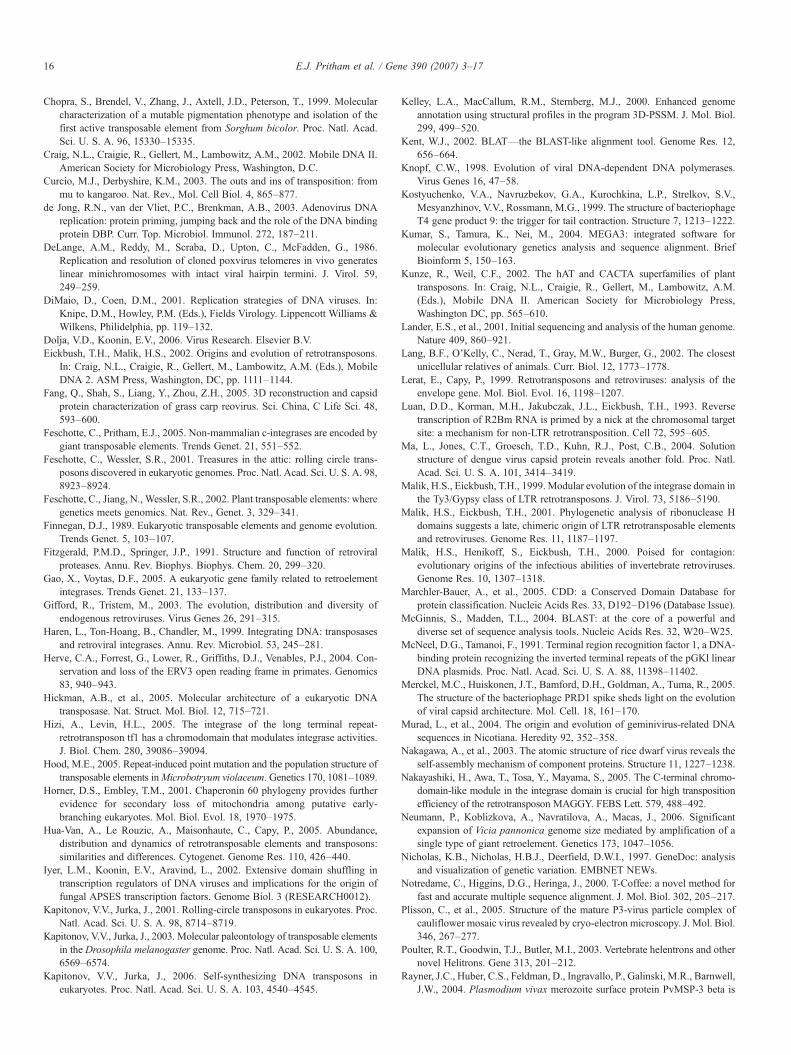
3.2. Phylogenetic analysis of the Maverick c-integrase
The c-integrase gene family, was first reported by Gao andVoytas (2005), as encoding a monophyletic group of cellularintegrases possibly co-opted for host function. They showed thatthe c-integrase proteins from nematodes and fish contain a∼150-aa region with significant similarity to the catalytic coredomain of retroviral integrases (RVE) and harbor a chromodo-main at their C-terminus. The chromodomain is also found at theC-terminus of integrases from the chromoviruses, a subgroup ofTy3/gypsy LTR retrotransposons, where it modulates integraseactivity and might be involved in the targeting of these elementsto particular chromatin environments (Malik and Eickbush,1999; Hizi and Levin, 2005; Nakayashiki et al., 2005). Wesubsequently showed that c-integrases from nematodes andzebrafish are in fact encoded by Maverick elements and likelyinvolved in their chromosomal integration (Feschotte andPritham, 2005). Annotation of the newly identified Mavericksconfirm that almost all of them contain a gene highly similar tothe previously described c-integrase (Fig. 1), which can encodea ∼400-aa protein with both RVE and chromodomains.
To determine the relationship of the Maverick c-integrasesboth to each other, as well as to the integrases encoded by otherelements, the RVE domain from representative Mavericks wasaligned to those present in the ciliate Tlr1, slime mold Tdd4,budding yeast Ty1 and Drosophila copia elements and thealignment was used for phylogenetic reconstruction with theneighbor-joining method (Fig. 2). When the tree is rooted withthe retroviral sequences Ty1 and copia, the Maverick c-integrases group with the integrases of Tlr1 and Tdd-4, forminga monophyletic clade reasonably well supported by bootstrapanalysis (82%). The most basal position in this clade is occupiedby the c-integrase of Mav_Tv1.1 from the parabasalid T.vaginalis. This topology indicates that both Tlr1 and Tdd-4integrase proteins share a more recent common ancestor withMaverick integrases than with the Ty1/copia group and probablyshould be considered part of the c-integrase family, as wassuggested by the initial study of Gao and Voytas.
The most basal position of the T. vaginalis c-integrase in theRVE phylogeny (bootstrap value 53%) is consistent with thedeep-branching position of this species in reconstructions of theeukaryotic tree of life (Baldauf et al., 2000; Horner and Embley,2001) and the hypothetical status of parabasalids as one of themost primitive group of eukaryotes. The detached and similarlybasal position of the c-integrases from the other protozoaneukaryotes P. infestans and T. thermophila relative to a (weakly
supported) monophyletic group of c-integrases from animal,fungi and D. discoideum is also reminiscent of the positionoccupied by these species in phylogenetic analyses of largenuclear and mitochondrial gene sets (Bapteste et al., 2002; Langet al., 2002; Steenkamp et al., 2006). This data provides supportto the idea that Mavericks are very ancient components of theeukaryotic genome that have been vertically inherited anddiversified for over a billion years of evolution.
A more surprising finding in our c-integrase RVE phylogenyis the highly supported grouping (100% bootstrap value) of themycorrhizal fungus with the two cnidarian species as a sisterclade to all other animal sequences. Cnidarians are thought to bethe most primitive extant animals so it is intriguing that theywould group with a fungus rather than with the other animals.To our knowledge, there is no obvious biological link betweencnidarians and mycorrizhal fungi that could indicate a case ofhorizontal transfer between these organisms and explain thisunexpected topology. In light of the well-established mono-phyly of the opisthokonts (animal and fungi), the simplestexplanation for this grouping is that it directly descends from anancient lineage ofMavericks present in the common ancestor offungi and animals.
Phylogenetic resolution is limited among the other animalsequences, although there is a high bootstrap support forgroupings of the four different nematode species and of theteleost fishes and weak support for grouping of the teleosts withthe clawed frog and the ratite bird (Fig. 2). In sum, the topologyof the c-integrase tree appears largely congruent with theestablished relationships of its diverse eukaryotic host species,which we interpret as evidence that Maverick elements are veryancient components of these genomes and probably originatedprior to the divergence of these eukaryotes.
3.3. Other conserved Maverick genes
To gain further insight into the origin and mode oftransposition of Maverick elements, we analyzed the codingcapacity of representative Mavericks using BLAST searches ofthe protein databases, conserved motif scans, and other genediscovery tools available through the NCBI server (Wheeleret al., 2005), as well as tri-dimensional fold prediction byprotein threading (Kelley et al., 2000). These combinedanalyses revealed that most Mavericks harbor multiple ORFs,which constitute a set of four to nine predicted protein-codinggenes (Fig. 1). In addition to c-integrases, 5 distinct groups ofconserved coding sequences were detected in Maverickelements from at least three distant species (Fig. 1). Reiteratedand reciprocal BLAST analyses (Tatusov et al., 2003) showedthat within each group of conservation, the Maverick putativeproteins are closer to each other than to other non-Maverickeukaryotic proteins. We therefore refer to these as clusters of‘conserved Maverick genes’ (CMGs, Fig. 1, SupplementalTable 2) and provide below a description of their codingpotential. Additionally, numerous other putative proteins wereidentified in only one Maverick element or only from Maver-icks from a single species. Interestingly these proteins oftendisplayed homology with known viral proteins (Fig. 1).
10 E.J. Pritham et al. / Gene 390 (2007) 3–17
3.3.1. CMG1 encodes a DNA polymerase B-like proteinCMG1 is predicted to encode a large protein (∼1100–1500-
aa) detected in all Maverick families examined (Fig. 1). CMG1proteins from different Maverick elements have variabledegrees of conservation, which, as for the c-integrases, areroughly congruent with the taxonomic distance of their hostspecies. For example, the CMG1 putative proteins from twodifferent nematode elements Mav_Ce1.1 and Mav_Cb1.2 are70% identical (80% similar) and each is ∼45% identical (62%similar) to the CMG1 protein of the beetle Mav_Tc1 and ∼25%identical (37% similar) to the CMG1 protein from the zebrafishMav_Dr1.2.
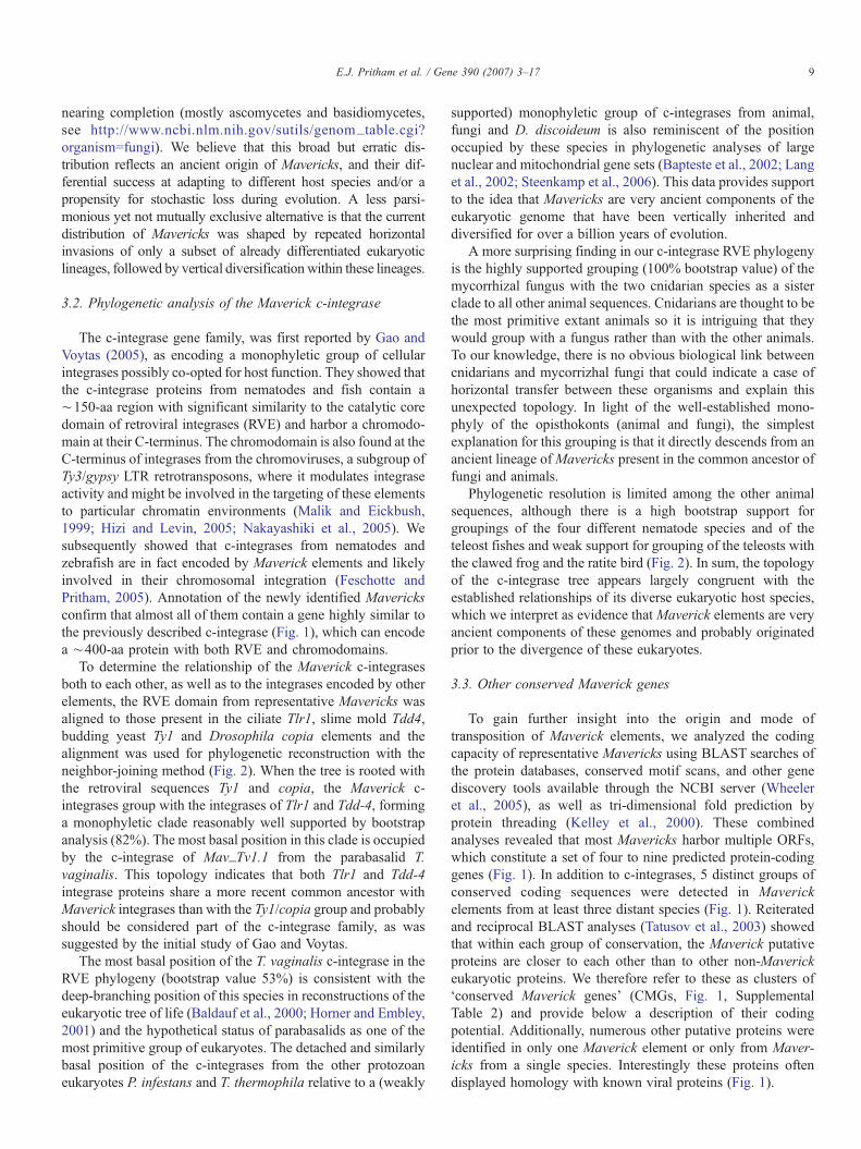
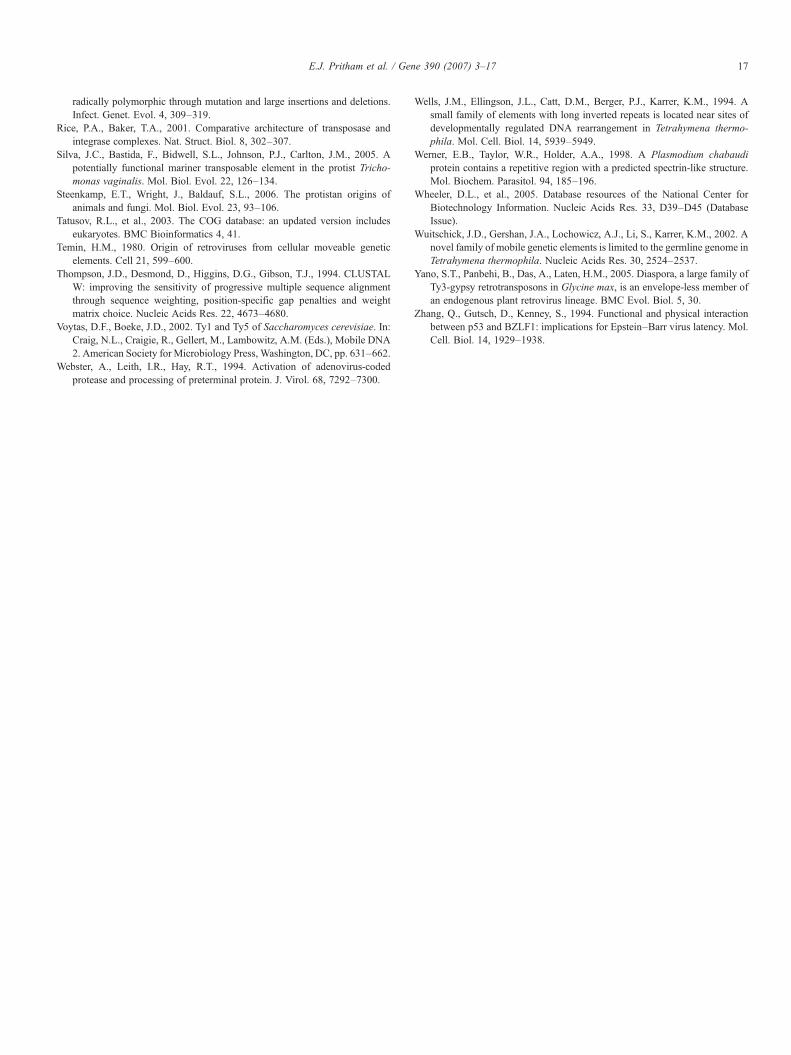
Despite a broad variation in their level of conservation,CMG1 proteins contain a ∼400-aa region that consistentlyaligns to the complete DNA_pol_B_2 domain (PFAMaccession 03175.10) in searches of the conserved domaindatabase (Fig. 3). This domain is characteristic of the type-Bfamily of viral DNA polymerases. Furthermore, BLASTsearches with CMG1 proteins return best hits (other thanother CMG1 proteins) with protein-primed DNA polymerasesencoded by phages from the tectiviridae (e.g. Bam35c) andpodoviridae (e.g. phi29) families, adenoviruses and linearplasmids found in the cytoplasm and mitochondria of someplants, fungi and stramenopiles. For example, the Mav_Gi1.1CMG1 aligns with a protein from a Zea mays mitochondriallinear plasmid (GenBank accession X02451.1; e-value 2e=−04, 40% similarity over 452-aa). In line with the BLASTresults, the DNA_pol_B_2 domain identified in CMG1proteins could be confidently aligned to those of protein-primedpolymerases (Fig. 3), but not to DNA- and RNA-primed DNApolymerases (data not shown). Type-B family viral DNApolymerases typically display DNA-binding, polymerase and3′–5′ exonuclease activities (Knopf, 1998). A partial alignmentof the pol_B_2 domain of Mav_Cr1.1 CMG1 with those of amaize linear plasmid, the PZA bacteriophage and human
Fig. 3. Domain structure and alignment of the DNA polymerase B domain from a linalignment above is from a linear plasmid (mitochondria, Zea mays), the second sequPZA and the last is from human adenovirus 17. DNA polymerase B proteins abacteriophages and in cytoplasmic and mitochondrial linear plasmids from fungi andreplication. The residues known to be necessary for exonuclease activity are markeunwound DNA are underlined and the polymerase catalytic site is boxed.
adenovirus 17 reveals that theMaverick CMG1 protein containsall of the critical residues involved in these functions (Fig. 3).
CMG1 are found in multiple copies within the genomes ofthe species that harbor Maverick elements and are almostalways found in proximity of c-integrase genes (Fig. 1). CMG1and c-integrases generally occupy the most outer regions ofMaverick internal sequences (except in Mav_Tc1), with a tail-to-head orientation in invertebrates and in X. tropicalis and atail-to-tail orientation in all other chordate species (Fig. 1).Despite these variations, the data indicate that a DNApolymerase-like gene is an integral component of Maverickelements and that the resulting protein may be involved in theirpropagation. Interestingly, Maverick DNA polymerase-likegenes were identified in close proximity to genes highly similarto those described in the Tlr elements from T. thermophila,suggesting that complete Tlr elements likely include a DNApolymerase B-like gene and therefore are likely to belong to theMaverick class.
In addition to the pol_B_2 domain, severalMaverick CMG1proteins also contain regions with significant similarity toother domains including VSR, Smc and PolBc (SupplementalTable 2). In the case of the VSR (very short repair endonuclease),this domain is actually embedded within the DNA_pol_B_2domain. The significance and function of a putative protein withendonuclease activity for Maverick transposition is unclear,although it could be involved in initiating replication by single-strand nicking at the Maverick–host DNA junction.
3.3.2. CMG2/3 encode coiled-coil domain proteinsCMG2/3 are predicted to encode proteins that range in size
from ∼200 to ∼900 amino acids. Most products have onlyweak or no significant similarities with eukaryotic proteins ofknown function and therefore they are currently annotated asunknown or hypothetical proteins in the respective sequencedgenomes (if annotated at all). The proteins predicted to be
ear plasmid, Maverick, bacteriophage and adenovirus. The first sequence in theence is from a C. remanei Maverick, the third sequence is from Bacillus phagere known to occur in some eukaryotic double-stranded DNA viruses, someplants. DNA polymerase B proteins utilize a self-encoded protein to primer DNAd by black arrows. The residues involved in DNA-binding and stabilizing the
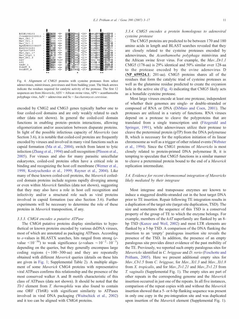
Fig. 4. Alignment of CMG5 proteins with cysteine proteases from selectadenoviruses, mimiviruses, poxviruses and from budding yeast. The black arrowsindicate the residues required for catalytic activity of the protease. The first 12sequences are fromMavericks, ASV = African swine virus, APV = acanthamoebapolyphaga virus, AdV = adenovirus and Sc = Saccharomyces cerevisiae.
11E.J. Pritham et al. / Gene 390 (2007) 3–17
encoded by CMG2 and CMG3 genes typically harbor one tofour coiled-coil domains and are only weakly related to eachother (data not shown). In general the coiled-coil domainfunctions in enabling protein–protein interactions, allowingoligomerization and/or association between disparate proteins.In light of the possible infectious capacity of Mavericks (seeSection 3.6), it is notable that coiled-coil proteins are frequentlyencoded by viruses and involved in many viral functions such ascapsid formation (Ma et al., 2004), switch from latent to lyticinfection (Zhang et al., 1994) and cell recognition (Plisson et al.,2005). For viruses and also for many parasitic unicellulareukaryotes, coiled-coil proteins often have a critical role inbinding and recognizing the host cell membrane (Werner et al.,1998; Kostyuchenko et al., 1999; Rayner et al., 2004). Likemany of these known coiled-coil proteins, the Maverick coiled-coil domain proteins include regions rapidly diverging amongor even within Maverick families (data not shown), suggestingthat they may also have a role in host cell recognition andinfectivity and/or a structural role such as viral proteinsinvolved in capsid formation (see also Section 3.6). Furtherexperiments will be necessary to determine the role of theseproteins in Maverick transposition, if any.
3.3.3. CMG4 encodes a putative ATPaseThe CMG4 putative proteins display similarities to hypo-
thetical or known proteins encoded by various dsDNA viruses,most of which are annotated as packaging ATPases. Accordingto e-values in BLASTX searches, hits ranged from strong (e-value b10−50) to weak significance (e-values ∼10−2–10−3)depending on the queries, but they generally encompass largecoding regions (∼100–300-aa) and they are repeatedlyobtained with different Maverick queries (details on these hitsare given in Fig. 1; Supplemental Table 2). A multiple align-ment of some Maverick CMG4 proteins with representativeviral ATPases confirms this relationship and the presence of themost conserved walker A and B motifs characteristic of thisclass of ATPases (data not shown). It should be noted that theTlr1 element from T. thermophila was also found to containone ORF (Tlr6R) with significant similarity to ATPasesinvolved in viral DNA packaging (Wuitschick et al., 2002)and it too can be aligned with CMG4 proteins.
3.3.4. CMG5 encodes a protein homologous to adenoviralcysteine protease
The CMG5 proteins are predicted to be between 170 and 180amino acids in length and BLAST searches revealed that theyare closely related to the cysteine proteases encoded byadenoviruses, the Acanthamoeba polyphaga mimivirus andthe African swine fever virus. For example, the Mav_Dr1.1CMG5 (178-aa) is 29% identical and 50% similar over 128-aato the protease encoded by the ovine adenovirus D(NP_659524.1; 201-aa). CMG5 proteins shares all of theresidues that form the catalytic triad of cysteine proteases aswell as the glutamine residue predicted to create the oxyanionhole in the active site (Fig. 4) indicating that CMG5 likely actsas a bonafide cysteine protease.
Most large viruses encode at least one protease, independentof whether their genomes are single- or double-stranded orcomposed of RNA or DNA (DiMaio and Coen, 2001). Theproteases are utilized in a variety of functions. RNA virusesdepend on a protease to cleave the polyproteins that aretranslated from a single transcription unit (Fitzgerald andSpringer, 1991), while adenoviruses utilize their protease tocleave the preterminal protein (pTP) from the DNA polymeraseB, which is necessary for the replication initiation of its linearchromosome as well as a trigger of other related events (Websteret al., 1994). Since the CMG1 proteins of Mavericks is mostclosely related to protein-primed DNA polymerase B, it istempting to speculate that CMG5 functions in a similar mannerto cleave a preterminal protein bound to the end of a Maverickreplication intermediate.
3.4. Evidence for recent chromosomal integration of Maverickslikely mediated by their integrase
Most integrase and transposase enzymes are known toinduce a staggered double-stranded cut in the host target DNAprior to TE insertion. Repair following TE integration results ina duplication of the target site (target site duplication, TSD). Thesize and sometimes the sequence of this TSD is a conservedproperty of the group of TE to which the enzyme belongs. Forexample, members of the hAT superfamily are flanked by an 8-bp TSD (Kunze and Weil, 2002) and most LTR elements areflanked by a 5-bp TSD. A comparison of the DNA flanking theinsertion to an ‘empty’ paralogous insertion site reveals thepresence of the TSD. In addition, the presence of an emptyparalogous site provides direct evidence of the past mobility ofthe TE. Previously, we reported such empty paralogous sites forMavericks identified in C. briggsae and D. rerio (Feschotte andPritham, 2005). Here we present additional empty sites forMav_Cb1.5 from C. briggsae, for Mav_Xt1.3 and Mav_Xt1.8from X. tropicalis, and for Mav_Tv1.21 and Mav_Tv1.23 fromT. vaginalis (Supplemental Fig. 1). The empty sites are part ofother repeats in the corresponding genome and the Maverickinsertion occurred in just one of the repeats. In all five instances,comparison of the repeat copies with and without the Maverickinsertion showed that a 5- or 6-bp flanking sequence was presentin only one copy in the pre-integration site and was duplicatedupon insertion of the Maverick element (Supplemental Fig. 1).
12 E.J. Pritham et al. / Gene 390 (2007) 3–17
Together, these data demonstrate that integration of Maverickelements induces a TSD of conserved length, a hallmark of TEintegration.
The size of the Mavericks TSD (5- or 6-bp) is comparable tothe size of the TSD typical of other mobile elements that utilizean integrase for chromosomal insertion (LTR retroelements andretroviruses). For example, murine retroviral elements of theET/MusD family induce a 6-bp TSD and insertion of mostcopia-like LTR retrotransposon provokes a 5-bp TSD. Together,this data strongly suggests that the integration of Maverickelements into the host chromosome is likely mediated by theMaverick-encoded integrase.
3.5. Recent explosive amplification of Maverick elements in theT. vaginalis genome
While most Maverick elements belong to low or moderatecopy number families in their respective genome, several linesof evidence indicate that they have recently proliferated to highcopy numbers in the genome of the parabasalid protozoa T.vaginalis. First, a blastn search using the first 225-bp of the TIRof Mav_Tv1.1 as a query against a database predicted to coverthe complete T. vaginalis genome (177 Mb) yielded 2298 hitswith e-values b10−4 (N84% identical over at least half of thequery). Since this search utilized a region of the element that isexpected to occur twice per element this result indicated thepresence of 1149 closely related elements (2298/2). However,because this search was done at the DNA level using a non-coding region that is expected to evolve faster than Maverickgenes, it is likely to represent an underestimate of total Ma-vericks in the genome of T. vaginalis.
To identify Mavericks that might be more distantly relatedwe utilized tblastn searches with multiple queries representingdifferent well-conserved regions of various T. vaginalis CMGs(see Fig. 5; see Supplemental Table 3 for queries used). Twotblastn searches with ∼150-aa queries representing two non-overlapping well-conserved regions of the DNA polymerase Bprotein domain yielded 1312 and 1304 hits respectively with e-value b10−4. Further intersection and inspection of these hitsconfirmed that they belong to multiple Maverick families co-existing within the T. vaginalis genome and not to other types ofmobile elements (data not shown). This suggests that thegenome of T. vaginalis is likely to include ∼1300 DNApolymerase- encoding Mavericks. Since, only 5 of the 10T. vaginalis complete Mavericks that we identified encode aDNA polymerase, this might also be an under representation ofthe total number of Mavericks in the genome (Fig. 5).
Additional searches were therefore carried out with similarsized queries representing the most conserved region of the c-integrase and the most conserved region of the hypervariableprotein, which are present in more than half of the Mavericksthat we identified in the T. vaginalis genome. These searchesyielded 3362 and 3809 hits, respectively. Based on the size ofthe queries, on the genomic coverage of the database and on thelocation of the corresponding genes in Maverick elements (seeFig. 5) these results indicate that this species might harbor over3000 Mavericks. Considering an average size of Mavericks in
T. vaginalis of 15 to 20 kb (see below), one can infer thatMaverick elements occupy an enormous fraction of the haploidgenome, possibly as much as 60 Mb or one third of the genome.To our knowledge, this makes Mavericks the most prominentcomponent of the T. vaginalis genome and the most prevalentTE family ever described in a single-celled eukaryote.
Additional evidence for the recent activity of Mavericks inT. vaginalis includes the discovery of a Maverick insertionembedded within a Mar1 Mariner element (SupplementalFig. 1). This family of Mariner elements was reported todisplay all the hallmarks of an active transposable element andto have recently amplified to hundreds of copies in the genomeof T. vaginalis (Silva et al., 2005). Discovery of a Maverickelement within a Mar1 suggests that the Maverick element wasactive more recently or at least around the same time as theMar1 family.
In order to characterize the Mav_Tv1.1 family in more detailall the complete closely related elements were identified usingthe Mav_Tv1.1 copy as a query. To our surprise, we could onlyidentify 11 elements closely related to Mav_Tv1.1 with twoclear termini (e.g. contained within the same contig and flankedby a 5-bp TSD), despite the equivalent in sequence to a 7.2times coverage of the genome is in the database. The isolatedMav_Tv1 elements range in size from 8.2 to 22.2 kb (average16.2 kb) and share over 94% nucleotide similarity in pairwisecomparisons (Fig. 5). All of the elements except Mav_Tv1.1contain multiple sequence gaps of unknown length, so theirsizes may be underestimated. In addition, Mav_Tv1.1, whichcontained no gaps and is the largest element, did not contain allthe proteins predicted to be important for transposition (Section3.6). Thus, full-length and autonomous Maverick_Tv1 copieswould likely be over 22 kb long. This may in part explain whyelements with two ends are difficult to identify in the currentassembly of the T. vaginalis genome. Indeed 70% of the contigsare less than 10 kb in length (data not shown), and are thereforeprobably much shorter than the elements themselves. Due totheir highly repetitive nature, high level of sequence similarityand large size, it is likely that Maverick elements represent amajor burden for the correct assembly of the T. vaginalisgenome sequence. Indeed, we observed that a very largenumber of contig ends and gaps coincide within Maverick-related sequences.
3.6. Mavericks from T. vaginalis potentially encode manyadditional proteins
To determine the complete set of possible proteins encoded bythis family of Trichomonas Maverick elements, every ORF largerthan 200 residues (startingwith the firstmethionine)was identifiedfor each element (see Fig. 5). For simplicity, we consider eachunique ORF as potentially encoding a different protein. However,it remains possible that some ORFs could be transcribed andtranslated as part of the same polypeptide. Each putative proteinwas assigned a functional classification based on comparisons topredicted proteins from otherMavericks, to searches of the proteindatabases and based on structural homology of predicted proteinfolds (see Section 2). In total, we were able to assign 9 of the 11
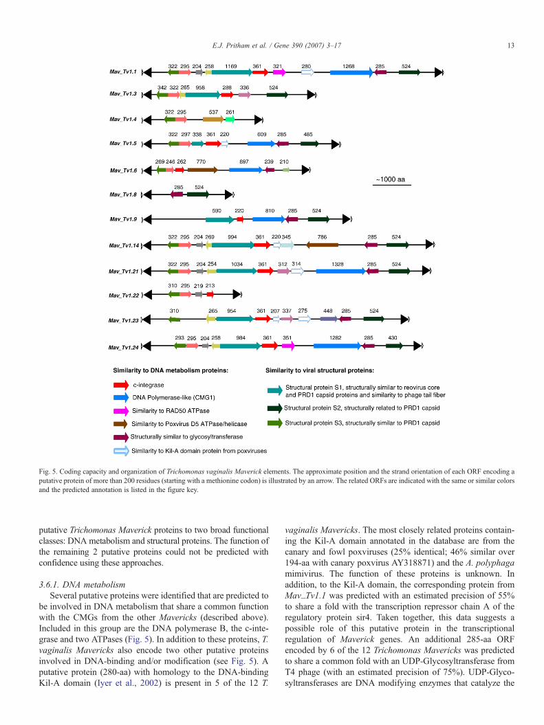
Fig. 5. Coding capacity and organization of Trichomonas vaginalis Maverick elements. The approximate position and the strand orientation of each ORF encoding aputative protein of more than 200 residues (starting with a methionine codon) is illustrated by an arrow. The related ORFs are indicated with the same or similar colorsand the predicted annotation is listed in the figure key.
13E.J. Pritham et al. / Gene 390 (2007) 3–17
putative Trichomonas Maverick proteins to two broad functionalclasses: DNA metabolism and structural proteins. The function ofthe remaining 2 putative proteins could not be predicted withconfidence using these approaches.
3.6.1. DNA metabolismSeveral putative proteins were identified that are predicted to
be involved in DNA metabolism that share a common functionwith the CMGs from the other Mavericks (described above).Included in this group are the DNA polymerase B, the c-inte-grase and two ATPases (Fig. 5). In addition to these proteins, T.vaginalis Mavericks also encode two other putative proteinsinvolved in DNA-binding and/or modification (see Fig. 5). Aputative protein (280-aa) with homology to the DNA-bindingKil-A domain (Iyer et al., 2002) is present in 5 of the 12 T.
vaginalis Mavericks. The most closely related proteins contain-ing the Kil-A domain annotated in the database are from thecanary and fowl poxviruses (25% identical; 46% similar over194-aa with canary poxvirus AY318871) and the A. polyphagamimivirus. The function of these proteins is unknown. Inaddition, to the Kil-A domain, the corresponding protein fromMav_Tv1.1 was predicted with an estimated precision of 55%to share a fold with the transcription repressor chain A of theregulatory protein sir4. Taken together, this data suggests apossible role of this putative protein in the transcriptionalregulation of Maverick genes. An additional 285-aa ORFencoded by 6 of the 12 Trichomonas Mavericks was predictedto share a common fold with an UDP-Glycosyltransferase fromT4 phage (with an estimated precision of 75%). UDP-Glyco-syltransferases are DNA modifying enzymes that catalyze the
14 E.J. Pritham et al. / Gene 390 (2007) 3–17
transfer of a glucose from uridine diphosphoglucose (UDP-Glc)to 5-hydroxymethylcytosine (5-HMC) in double-strandedDNA. They are thought to function in shielding the T4 phageDNA from attack by cellular nucleases.
3.6.2. Structural proteinsThree putative proteins (S1–S3) ranging in size from 285-aa
to 1169-aa were each predicted to share a common fold withbacteriophage capsid or core proteins (Fig. 5). The largest ofthese predicted proteins, structural protein S1, ranges in sizebetween 994- and 1169-aa and is encoded by 7 of the 12complete elements (see Fig. 5). An alignment of these putativeproteins reveals several well-conserved regions located near theN- and C-termini flanking a central hypervariable region(Supplemental Fig. 2). Given the variability of the centralregion, structural fold prediction tools were more informativeabout the possible function of these proteins and relationships toother proteins than those based on primary sequence homology(e.g. PSI-BLAST). Nevertheless, both approaches converged inrevealing an intriguing recurrent relationship to proteinsinvolved in the assembly of the phage core and capsid as wellas proteins involved in cell adhesion. For example, one region ofthe Mav_Tv1.1 protein S1 spanning the entire hypervariableregion (from ∼100- to 730-aa) is predicted using Phyre, withconfidence levels between 75% and 90% to share a common foldwith the reovirus core proteins p3 and p7 (Nakagawa et al., 2003;Fang et al., 2005) and the major capsid protein of bacteriophagePRD1 (Bamford et al., 2002). This fold extends completelythrough the hypervariable region of structural protein S1 (Fig. 5;Supplemental Fig. 2). The reovirus core p3, p7 proteins and thePRD1 major capsid proteins are the building blocks of the viralcore and capsid structures of the respective viruses. In addition,PSI-BLAST searches revealed significant similarity in the C-terminal region with the gp10 baseplate wedge subunit and tailpin proteins of the bacteriophage RB49 (AAQ15390; e-value=2e−04, 25% identity; 40% similar over 252-aa). Thebaseplate wedge and tail pin proteins are involved in host cellrecognition and adsorption of the phage to host cells.Additionally, two other putative proteins were identified thatare also predicted to share a common fold with the PRD1 majorcapsid protein, suggesting either that these proteins are encodedby members of a multi gene family or that they converged to thesame structure (Fig. 5). It has been previously suggested thatviral genes encoding proteins involved in capsid formation havearisen via gene duplication events (Boyko et al., 1992; Merckelet al., 2005), thus supporting the idea that the putativeMaverickstructural genes are related by descent.
4. Conclusions and relationship ofMavericks to other formsof invasive DNA
The results presented in this study confirm and expand ourinitial prediction that Mavericks represent an entirely new classof mobile elements widespread in eukaryotic genomes. Belowwe summarize several of the unique features of these elementsin an attempt to shed some light on their evolutionary origin andtheir replication mechanism.
4.1. Structural features
One of the most striking characteristics of Mavericks in allspecies examined is their large size. In the absence of any geneticevidence of the sequence and biochemical activities required forMaverick mobility, we can only speculate based on the geneticorganization and coding capacity of a canonical full-length,autonomous Maverick element. Most elements that are devoidof obvious truncation or internal deletion reach an average sizeof ∼15–20 kb. Very few transposable elements have beenpreviously shown to reach such a size. To our knowledge,elements larger than 15 kb have been seldom described and theyare often limited to one subtype or family of elements within asuperfamily of generally smaller TEs. For example, Ogreelements are a group of plant gypsy-like LTR retrotransposonsthat are ∼25 kb (Neumann et al., 2006). The Candystripe1element from sorghum is a 23-kb DNA transposon of theCACTA superfamily that can still be excised at high frequency(Chopra et al., 1999). In contrast, gigantism appears to be thenorm for Maverick elements and it does not hinder theirevolutionary success or their proliferation (e.g.∼3000 copies inthe relatively compact genome of T. vaginalis).
Another distinctive structural feature of Mavericks is thepresence of large (typically 400–700 bp) and often perfectTIRs. DNA transposons are also characterized by TIRs, butthese are usually much shorter (typically 10–200 bp). Giventheir large size and long TIRs, the genome ofMavericks is morereminiscent of the linear genome of other types of mobile andinvasive DNA such as linear plasmids, bacteriophages andmany eukaryotic DNA viruses. In these elements, the TIRs aredirectly involved in their replication mechanism (De Jong et al.,2003) and/or are thought to play a role in the protection of theends from degradation by cellular enzymes (DeLange et al.,1986). In addition, all identified Maverick TIRs began with ashort simple repeat motif (AGTAGT, TGTGTG, AGAGAGAG),which is seldom observed in DNA transposons, but resembled theshort terminal repeat motifs at or near the end of the TIRs ofadenoviruses (e.g. CATCAT in FADV-9 and CADV-1) and linearplasmids (AAAAATACTAC, NC_004946; CCCGTGTGTGT,NC_004935) (Supplemental Fig. 3). These short repeats aregenerated through a so-called sliding-back mechanism typical ofthe protein-primed replication of their genome (McNeel andTamanoi, 1991; De Jong et al., 2003). We also observed that theMaverick TIRs are generally more similar to each other than toany other TIRs detectable within the same genome (data notshown). This suggests that either one TIR is replicated using theother TIR as a template (akin to the LTRs of retroelements) duringMaverick replication or that some other mechanisms areresponsible for homogenization of the TIRs after Maverickintegration (e.g. gene conversion). The structural similaritiesbetweenMavericks and some DNAviruses suggest an importantrole of the TIRs in the replication ofMavericks.
4.2. Genetic organization
We have shown thatMavericks typically contain a set of 4–9genes and occasionally many more (e.g. Section 3.6). We
15E.J. Pritham et al. / Gene 390 (2007) 3–17
identified a set of 6 genes (c-integrase and CMG1-5) that ismost common and relatively conserved among various Ma-vericks identified across the eukaryotic tree, but it is apparentthat there is a wide variation in the coding capacity and geneticorganization of the elements. Even the relative arrangement ofthe CMGs in relatively closely related species can greatly vary(see Fig. 1). This high gene content and plasticity of Mavericksis unusual among TEs and here again it is more reminiscent ofviral genomes (Dolja and Koonin, 2006). Furthermore, theflexibility of gene arrangements inMavericks also suggests thatthey contain multiple independent transcription units, andpossibly that each gene possesses its own core promoter.
At present, nothing is known about the transcription of Ma-verick elements and it would be an interesting area of futureresearch. Blast searches of the EST database using MaverickCMGs as queries reveal that these putative proteins are frequentlytranscribed in many different taxa (data not shown). It is beyondthe scope of this study to describe the origin and structure of thesetranscripts. It is worth noting, however, that several of the genesare predicted with a high level of confidence to be interrupted byspliceosomal introns and in some cases, the predicted genestructure is further supported by spliced EST/cDNA sequences(e.g. K09F6.6 inMav_Ce2.3). The presence of introns in CMGssuggests that transposition is unlikely to occur through reverse-transcription of an RNA intermediate, but rather via a DNAintermediate. Consistent with this view, no coding sequenceswith homology to reverse transcriptase or other retroelement-encoded proteins were detected in Maverick elements (besidesthe c-integrases and those occasionally deposited by secondaryretrotransposon insertions, data not shown).
4.3. Enzymatic capacity of Maverick-encoded proteins andevolutionary implications
Among the six CMGs, all but the c-integrases have a direct(sequence similarity) or indirect link (common domains andfold prediction) with proteins encoded by and involved in thereplication cycle of DNAviruses. In addition numerous putativeproteins encoded by a single Maverick display homology toviral proteins (Fig. 1). This raises several testable modelsconsidering the transposition mechanism of Mavericks some ofwhich have been proposed elsewhere (Kapitonov and Jurka,2006). Yet, at this point we can only be confident about thechromosomal integration step of the replication cycle of Ma-vericks; it most likely involves the encoded c-integrase and isaccompanied by the duplication of 5–6 bp of host DNA. Thedetection of a homolog of DNA polymerase B and of a likelyhomolog of cysteine proteases found in adenoviruses points to aclose evolutionary link to genetic elements that use protein-primed DNA replication. Bamford and colleagues haveproposed the existence of a common viral ancestor for aseemingly disparate group of double-stranded DNAviruses thatinfect bacteria and animals including, the Tectiviridae bacte-riophage PRD1 and the Adenoviridae (Benson et al., 1999,2004). This hypothesis is based on a remarkably analogousarchitecture of the virion echoed by highly similar tri-dimensional folding of their major capsid proteins, despite the
lack of detectable primary sequence homology. This relation-ship is further supported by the relationship of the viral-encodedDNA polymerase B that is used to replicate both the PRD1 andadenovirus linear chromosome, and is also encoded by mostMavericks (Figs. 1 and 3). Given the structural and sequencesimilarities of Mavericks with some of these viruses and theirancient eukaryotic origin, it is possible that they all descendfrom the same common ancestral virus. Mavericks may haveadapted to an intragenomic lifestyle by acquisition of anintegrase-like protein in an early eukaryote ancestor. On theother hand, the prediction that a protein fold in one of the coilcoiled domain protein of Mav_Tv1 is shared with p3 and p7core proteins of reovirus and the PRD1 P3 major capsid protein(see Section 3.6) and the presence of numerous other viral-related putative proteins raises the intriguing and testablehypothesis that at least some Mavericks have retained anextracellular component in their replication cycle and aninfectious capacity.
Acknowledgements
We would like to thank John Pace (UTA) for technicalassistance, Dr. David Edgell (University of Western Ontario)and Dr. Harri Savilahti (University of Helsinki) for stimulatingdiscussions, during the preparation of this manuscript. Thiswork was supported by start up funds from the Department ofBiology at the University of Texas at Arlington.
Appendix A. Supplementary data
Supplementary data associated with this article can be found,in the online version, at doi:10.1016/j.gene.2006.08.008.
References
Altschul, S.F., Gish, W., Miller, W., Myers, E.W., Lipman, D.J., 1990. Basiclocal alignment search tool. J. Mol. Biol. 215, 403–410.
Baldauf, S.L., Roger, A.J., Wenk-Siefert, I., Doolittle, W.F., 2000. A kingdom-level phylogeny of eukaryotes based on combined protein data. Science 290,972–977.
Bamford, J.K., et al., 2002. Diffraction quality crystals of PRD1, a 66-MDadsDNA virus with an internal membrane. J. Struct. Biol. 139, 103–112.
Bapteste, E., et al., 2002. The analysis of 100 genes supports the grouping ofthree highly divergent amoebae: Dictyostelium, Entamoeba, and Mastiga-moeba. Proc. Natl. Acad. Sci. U. S. A. 99, 1414–1419.
Benson, S.D., Bamford, J.K., Bamford, D.H., Burnett, R.M., 1999. Viral evo-lution revealed by bacteriophage PRD1 and human adenovirus coat proteinstructures. Cell 98, 825–833.
Benson, S.D., Bamford, J.K., Bamford, D.H., Burnett, R.M., 2004. Doescommon architecture reveal a viral lineage spanning all three domains oflife? Mol. Cell 16, 673–685.
Boyko, V.P., Karasev, A.V., Agranovsky, A.A., Koonin, E.V., Dolja, V.V., 1992.Coat protein gene duplication in a filamentous RNA virus of plants. Proc.Natl. Acad. Sci. U. S. A. 89, 9156–9160.
Brookfield, J.F., 2005. The ecology of the genome — mobile DNA elementsand their hosts. Nat. Rev., Genet. 6, 128–136.
Capy, P., Vitalis, R., Langin, T., Higuet, D., Bazin, C., 1996. Relationshipsbetween transposable elements based upon the integrase-transposasedomains: is there a common ancestor? J. Mol. Evol. 42, 359–368.
Capy, P., Bazin, C., Higuet, D., Langin, T., 1998. Dynamics and Evolution ofTransposable Elements. Springer-Verlag, Austin, Texas.
16 E.J. Pritham et al. / Gene 390 (2007) 3–17
Chopra, S., Brendel, V., Zhang, J., Axtell, J.D., Peterson, T., 1999. Molecularcharacterization of a mutable pigmentation phenotype and isolation of thefirst active transposable element from Sorghum bicolor. Proc. Natl. Acad.Sci. U. S. A. 96, 15330–15335.
Craig, N.L., Craigie, R., Gellert, M., Lambowitz, A.M., 2002. Mobile DNA II.American Society for Microbiology Press, Washington, D.C.
Curcio, M.J., Derbyshire, K.M., 2003. The outs and ins of transposition: frommu to kangaroo. Nat. Rev., Mol. Cell Biol. 4, 865–877.
de Jong, R.N., van der Vliet, P.C., Brenkman, A.B., 2003. Adenovirus DNAreplication: protein priming, jumping back and the role of the DNA bindingprotein DBP. Curr. Top. Microbiol. Immunol. 272, 187–211.
DeLange, A.M., Reddy, M., Scraba, D., Upton, C., McFadden, G., 1986.Replication and resolution of cloned poxvirus telomeres in vivo generateslinear minichromosomes with intact viral hairpin termini. J. Virol. 59,249–259.
DiMaio, D., Coen, D.M., 2001. Replication strategies of DNA viruses. In:Knipe, D.M., Howley, P.M. (Eds.), Fields Virology. Lippencott Williams &Wilkens, Philidelphia, pp. 119–132.
Dolja, V.D., Koonin, E.V., 2006. Virus Research. Elsevier B.V.Eickbush, T.H., Malik, H.S., 2002. Origins and evolution of retrotransposons.
In: Craig, N.L., Craigie, R., Gellert, M., Lambowitz, A.M. (Eds.), MobileDNA 2. ASM Press, Washington, DC, pp. 1111–1144.
Fang, Q., Shah, S., Liang, Y., Zhou, Z.H., 2005. 3D reconstruction and capsidprotein characterization of grass carp reovirus. Sci. China, C Life Sci. 48,593–600.
Feschotte, C., Pritham, E.J., 2005. Non-mammalian c-integrases are encoded bygiant transposable elements. Trends Genet. 21, 551–552.
Feschotte, C., Wessler, S.R., 2001. Treasures in the attic: rolling circle trans-posons discovered in eukaryotic genomes. Proc. Natl. Acad. Sci. U. S. A. 98,8923–8924.
Feschotte, C., Jiang, N., Wessler, S.R., 2002. Plant transposable elements: wheregenetics meets genomics. Nat. Rev., Genet. 3, 329–341.
Finnegan, D.J., 1989. Eukaryotic transposable elements and genome evolution.Trends Genet. 5, 103–107.
Fitzgerald, P.M.D., Springer, J.P., 1991. Structure and function of retroviralproteases. Annu. Rev. Biophys. Biophys. Chem. 20, 299–320.
Gao, X., Voytas, D.F., 2005. A eukaryotic gene family related to retroelementintegrases. Trends Genet. 21, 133–137.
Gifford, R., Tristem, M., 2003. The evolution, distribution and diversity ofendogenous retroviruses. Virus Genes 26, 291–315.
Haren, L., Ton-Hoang, B., Chandler, M., 1999. Integrating DNA: transposasesand retroviral integrases. Annu. Rev. Microbiol. 53, 245–281.
Herve, C.A., Forrest, G., Lower, R., Griffiths, D.J., Venables, P.J., 2004. Con-servation and loss of the ERV3 open reading frame in primates. Genomics83, 940–943.
Hickman, A.B., et al., 2005. Molecular architecture of a eukaryotic DNAtransposase. Nat. Struct. Mol. Biol. 12, 715–721.
Hizi, A., Levin, H.L., 2005. The integrase of the long terminal repeat-retrotransposon tf1 has a chromodomain that modulates integrase activities.J. Biol. Chem. 280, 39086–39094.
Hood, M.E., 2005. Repeat-induced point mutation and the population structure oftransposable elements inMicrobotryum violaceum. Genetics 170, 1081–1089.
Horner, D.S., Embley, T.M., 2001. Chaperonin 60 phylogeny provides furtherevidence for secondary loss of mitochondria among putative early-branching eukaryotes. Mol. Biol. Evol. 18, 1970–1975.
Hua-Van, A., Le Rouzic, A., Maisonhaute, C., Capy, P., 2005. Abundance,distribution and dynamics of retrotransposable elements and transposons:similarities and differences. Cytogenet. Genome Res. 110, 426–440.
Iyer, L.M., Koonin, E.V., Aravind, L., 2002. Extensive domain shuffling intranscription regulators of DNA viruses and implications for the origin offungal APSES transcription factors. Genome Biol. 3 (RESEARCH0012).
Kapitonov, V.V., Jurka, J., 2001. Rolling-circle transposons in eukaryotes. Proc.Natl. Acad. Sci. U. S. A. 98, 8714–8719.
Kapitonov, V.V., Jurka, J., 2003. Molecular paleontology of transposable elementsin the Drosophila melanogaster genome. Proc. Natl. Acad. Sci. U. S. A. 100,6569–6574.
Kapitonov, V.V., Jurka, J., 2006. Self-synthesizing DNA transposons ineukaryotes. Proc. Natl. Acad. Sci. U. S. A. 103, 4540–4545.
Kelley, L.A., MacCallum, R.M., Sternberg, M.J., 2000. Enhanced genomeannotation using structural profiles in the program 3D-PSSM. J. Mol. Biol.299, 499–520.
Kent, W.J., 2002. BLAT—the BLAST-like alignment tool. Genome Res. 12,656–664.
Knopf, C.W., 1998. Evolution of viral DNA-dependent DNA polymerases.Virus Genes 16, 47–58.
Kostyuchenko, V.A., Navruzbekov, G.A., Kurochkina, L.P., Strelkov, S.V.,Mesyanzhinov, V.V., Rossmann, M.G., 1999. The structure of bacteriophageT4 gene product 9: the trigger for tail contraction. Structure 7, 1213–1222.
Kumar, S., Tamura, K., Nei, M., 2004. MEGA3: integrated software formolecular evolutionary genetics analysis and sequence alignment. BriefBioinform 5, 150–163.
Kunze, R., Weil, C.F., 2002. The hAT and CACTA superfamilies of planttransposons. In: Craig, N.L., Craigie, R., Gellert, M., Lambowitz, A.M.(Eds.), Mobile DNA II. American Society for Microbiology Press,Washington DC, pp. 565–610.
Lander, E.S., et al., 2001. Initial sequencing and analysis of the human genome.Nature 409, 860–921.
Lang, B.F., O'Kelly, C., Nerad, T., Gray, M.W., Burger, G., 2002. The closestunicellular relatives of animals. Curr. Biol. 12, 1773–1778.
Lerat, E., Capy, P., 1999. Retrotransposons and retroviruses: analysis of theenvelope gene. Mol. Biol. Evol. 16, 1198–1207.
Luan, D.D., Korman, M.H., Jakubczak, J.L., Eickbush, T.H., 1993. Reversetranscription of R2Bm RNA is primed by a nick at the chromosomal targetsite: a mechanism for non-LTR retrotransposition. Cell 72, 595–605.
Ma, L., Jones, C.T., Groesch, T.D., Kuhn, R.J., Post, C.B., 2004. Solutionstructure of dengue virus capsid protein reveals another fold. Proc. Natl.Acad. Sci. U. S. A. 101, 3414–3419.
Malik, H.S., Eickbush, T.H., 1999. Modular evolution of the integrase domain inthe Ty3/Gypsy class of LTR retrotransposons. J. Virol. 73, 5186–5190.
Malik, H.S., Eickbush, T.H., 2001. Phylogenetic analysis of ribonuclease Hdomains suggests a late, chimeric origin of LTR retrotransposable elementsand retroviruses. Genome Res. 11, 1187–1197.
Malik, H.S., Henikoff, S., Eickbush, T.H., 2000. Poised for contagion:evolutionary origins of the infectious abilities of invertebrate retroviruses.Genome Res. 10, 1307–1318.
Marchler-Bauer, A., et al., 2005. CDD: a Conserved Domain Database forprotein classification. Nucleic Acids Res. 33, D192–D196 (Database Issue).
McGinnis, S., Madden, T.L., 2004. BLAST: at the core of a powerful anddiverse set of sequence analysis tools. Nucleic Acids Res. 32, W20–W25.
McNeel, D.G., Tamanoi, F., 1991. Terminal region recognition factor 1, a DNA-binding protein recognizing the inverted terminal repeats of the pGKl linearDNA plasmids. Proc. Natl. Acad. Sci. U. S. A. 88, 11398–11402.
Merckel, M.C., Huiskonen, J.T., Bamford, D.H., Goldman, A., Tuma, R., 2005.The structure of the bacteriophage PRD1 spike sheds light on the evolutionof viral capsid architecture. Mol. Cell. 18, 161–170.
Murad, L., et al., 2004. The origin and evolution of geminivirus-related DNAsequences in Nicotiana. Heredity 92, 352–358.
Nakagawa, A., et al., 2003. The atomic structure of rice dwarf virus reveals theself-assembly mechanism of component proteins. Structure 11, 1227–1238.
Nakayashiki, H., Awa, T., Tosa, Y., Mayama, S., 2005. The C-terminal chromo-domain-like module in the integrase domain is crucial for high transpositionefficiency of the retrotransposon MAGGY. FEBS Lett. 579, 488–492.
Neumann, P., Koblizkova, A., Navratilova, A., Macas, J., 2006. Significantexpansion of Vicia pannonica genome size mediated by amplification of asingle type of giant retroelement. Genetics 173, 1047–1056.
Nicholas, K.B., Nicholas, H.B.J., Deerfield, D.W.I., 1997. GeneDoc: analysisand visualization of genetic variation. EMBNET NEWs.
Notredame, C., Higgins, D.G., Heringa, J., 2000. T-Coffee: a novel method forfast and accurate multiple sequence alignment. J. Mol. Biol. 302, 205–217.
Plisson, C., et al., 2005. Structure of the mature P3-virus particle complex ofcauliflower mosaic virus revealed by cryo-electron microscopy. J. Mol. Biol.346, 267–277.
Poulter, R.T., Goodwin, T.J., Butler, M.I., 2003. Vertebrate helentrons and othernovel Helitrons. Gene 313, 201–212.
Rayner, J.C., Huber, C.S., Feldman, D., Ingravallo, P., Galinski, M.R., Barnwell,J.W., 2004. Plasmodium vivax merozoite surface protein PvMSP-3 beta is
17E.J. Pritham et al. / Gene 390 (2007) 3–17
radically polymorphic through mutation and large insertions and deletions.Infect. Genet. Evol. 4, 309–319.
Rice, P.A., Baker, T.A., 2001. Comparative architecture of transposase andintegrase complexes. Nat. Struct. Biol. 8, 302–307.
Silva, J.C., Bastida, F., Bidwell, S.L., Johnson, P.J., Carlton, J.M., 2005. Apotentially functional mariner transposable element in the protist Tricho-monas vaginalis. Mol. Biol. Evol. 22, 126–134.
Steenkamp, E.T., Wright, J., Baldauf, S.L., 2006. The protistan origins ofanimals and fungi. Mol. Biol. Evol. 23, 93–106.
Tatusov, R.L., et al., 2003. The COG database: an updated version includeseukaryotes. BMC Bioinformatics 4, 41.
Temin, H.M., 1980. Origin of retroviruses from cellular moveable geneticelements. Cell 21, 599–600.
Thompson, J.D., Desmond, D., Higgins, D.G., Gibson, T.J., 1994. CLUSTALW: improving the sensitivity of progressive multiple sequence alignmentthrough sequence weighting, position-specific gap penalties and weightmatrix choice. Nucleic Acids Res. 22, 4673–4680.
Voytas, D.F., Boeke, J.D., 2002. Ty1 and Ty5 of Saccharomyces cerevisiae. In:Craig, N.L., Craigie, R., Gellert, M., Lambowitz, A.M. (Eds.), Mobile DNA2. American Society for Microbiology Press, Washington, DC, pp. 631–662.
Webster, A., Leith, I.R., Hay, R.T., 1994. Activation of adenovirus-codedprotease and processing of preterminal protein. J. Virol. 68, 7292–7300.
Wells, J.M., Ellingson, J.L., Catt, D.M., Berger, P.J., Karrer, K.M., 1994. Asmall family of elements with long inverted repeats is located near sites ofdevelopmentally regulated DNA rearrangement in Tetrahymena thermo-phila. Mol. Cell. Biol. 14, 5939–5949.
Werner, E.B., Taylor, W.R., Holder, A.A., 1998. A Plasmodium chabaudiprotein contains a repetitive region with a predicted spectrin-like structure.Mol. Biochem. Parasitol. 94, 185–196.
Wheeler, D.L., et al., 2005. Database resources of the National Center forBiotechnology Information. Nucleic Acids Res. 33, D39–D45 (DatabaseIssue).
Wuitschick, J.D., Gershan, J.A., Lochowicz, A.J., Li, S., Karrer, K.M., 2002. Anovel family of mobile genetic elements is limited to the germline genome inTetrahymena thermophila. Nucleic Acids Res. 30, 2524–2537.
Yano, S.T., Panbehi, B., Das, A., Laten, H.M., 2005. Diaspora, a large family ofTy3-gypsy retrotransposons in Glycine max, is an envelope-less member ofan endogenous plant retrovirus lineage. BMC Evol. Biol. 5, 30.
Zhang, Q., Gutsch, D., Kenney, S., 1994. Functional and physical interactionbetween p53 and BZLF1: implications for Epstein–Barr virus latency. Mol.Cell. Biol. 14, 1929–1938.