Embed Size (px)
Citation preview
This is an electronic reprint of the original article.This reprint may differ from the original in pagination and typographic detail.
Powered by TCPDF (www.tcpdf.org)
This material is protected by copyright and other intellectual property rights, and duplication or sale of all or part of any of the repository collections is not permitted, except that material may be duplicated by you for your research use or educational purposes in electronic or print form. You must obtain permission for any other use. Electronic or print copies may not be offered, whether for sale or otherwise to anyone who is not an authorised user.
Mattinen, Maija Liisa; Riviere, Guillaume; Henn, Alexander; Nugroho, Robertus Wahyu N.;Leskinen, Timo; Nivala, Outi; Valle-Delgado, Juan José; Kostiainen, Mauri A.; Österberg,MonikaColloidal lignin particles as adhesives for soft materials
Published in:Nanomaterials
DOI:10.3390/nano8121001
Published: 01/12/2018
Document VersionPublisher's PDF, also known as Version of record
Please cite the original version:Mattinen, M. L., Riviere, G., Henn, A., Nugroho, R. W. N., Leskinen, T., Nivala, O., ... Österberg, M. (2018).Colloidal lignin particles as adhesives for soft materials. Nanomaterials, 8(12), [1001].https://doi.org/10.3390/nano8121001
nanomaterials
Article
Colloidal Lignin Particles as Adhesivesfor Soft Materials
Maija-Liisa Mattinen 1,* , Guillaume Riviere 1, Alexander Henn 1,Robertus Wahyu N. Nugroho 1, Timo Leskinen 1, Outi Nivala 2,Juan José Valle-Delgado 1, Mauri A. Kostiainen 3 and Monika Österberg 1
1 Bioproduct Chemistry, Department of Bioproducts and Biosystems, School of Chemical Engineering,Aalto University, P.O. Box 16300, FI-00076 Aalto, Espoo, Finland; [email protected] (G.R.);[email protected] (A.H.); [email protected] (R.W.N.N.); [email protected] (T.L.);[email protected] (J.J.V.-D.); [email protected] (M.Ö.)
2 VTT Technical Research Centre of Finland Ltd., P.O. Box 1000, FI-02044 VTT Espoo, Finland;[email protected]
3 Biohybrid Materials, Department of Bioproducts and Biosystems, School of Chemical Engineering,Aalto University, P.O. Box 16100, FI-00076 Aalto, Espoo, Finland; [email protected]
* Correspondence: [email protected] or [email protected]; Tel.: +358-50-302-3511
Received: 4 November 2018; Accepted: 29 November 2018; Published: 3 December 2018�����������������
Abstract: Lignin has interesting functionalities to be exploited in adhesives for medicine, foods andtextiles. Nanoparticles (NPs) < 100 nm coated with poly (L-lysine), PL and poly(L-glutamic acid)PGA were prepared from the laccase treated lignin to coat nanocellulose fibrils (CNF) with heat.NPs ca. 300 nm were prepared, β-casein coated and cross-linked with transglutaminase (Tgase) toagglutinate chamois. Size exclusion chromatography (SEC) and Fourier-transform infrared (FTIR)spectroscopy were used to characterize polymerized lignin, while zeta potential and dynamic lightscattering (DLS) to ensure coating of colloidal lignin particles (CLPs). Protein adsorption on ligninwas studied by quartz crystal microbalance (QCM). Atomic force microscopy (AFM) was exploitedto examine interactions between different polymers and to image NPs with transmission electronmicroscopy (TEM). Tensile testing showed, when using CLPs for the adhesion, the stress improved ca.10 and strain ca. 6 times compared to unmodified Kraft. For the β-casein NPs, the values were 20 and8, respectively, and for the β-casein coated CLPs between these two cases. When NPs were dispersedin adhesive formulation, the increased Young’s moduli confirmed significant improvement in thestiffness of the joints over the adhesive alone. Exploitation of lignin in nanoparticulate morphology isa potential method to prepare bionanomaterials for advanced applications.
Keywords: lignin; nanoparticle; protein; nanocellulose; fibril; enzyme; heat; self-assembly; cross-link
1. Introduction
Technologies focusing on the preparation of adhesives utilizing natural polymers such as proteinsand cellulosics for medical, textile and food applications are emerging research fields [1–3] However,biorefinery industry produces also lignin by-product, which is still underutilized, even though thisaromatic, antioxidative and antimicrobial biopolymer could be an interesting raw material for manyvalue-added applications [4].
Nanocellulose can be produced by several methods [5–8]. It is a lightweight, transparent andbiodegradable polymer. Hence, nanocellulose fibrils (CNF) as well as bacterial nanocellulose (BC)are excellent raw materials for tissue regeneration and replacement [9–12]. Major challenges for theexploitation of CNF include the ability to disperse colloidal material with different formulations.
Nanomaterials 2018, 8, 1001; doi:10.3390/nano8121001 www.mdpi.com/journal/nanomaterials
Nanomaterials 2018, 8, 1001 2 of 20
Surface functionalization of the fibrils could be an attractive method to improve stability, functionalityand compatibility of the nanomaterial with selected matrices. For example, poly (L-lysine, PL) coatedwax particles assembled on CNF surface yield hydrophobic fibrils [13]. Furthermore, capability toobtain tight bonding between tissue edges to prevent bleeding with excellent gas barrier properties andto achieve strong mechanical strength for the sealant, are crucial properties for the medical adhesivesto support wound healing and tissue deformation during the recovery [14–16].
Due to excellent solubility and biocompatibility, regenerated silk proteins have been used inmedical applications such as textiles, implants and materials for controlled drug release. Depositionof silk fibrin on polymeric surfaces is a remarkable challenge [2]. Enzymatic cross-linking withtransglutaminase (Tgase, EC 2.3.1.13) catalyzing cross-links between glutamines and lysines has beenused to stabilize proteins against chemicals and proteases [17]. Furthermore, Tgase has been usedto graft silk proteins onto damaged wool fibers to improve strength of the surfaces and to degreasefelting shrinkage during washing [18].
Foods such as edible coatings are nearby medical applications. Food packages based onpetroleum-based raw materials are not biodegradable. They have poor oxygen barrier propertiespossibly leaching harmful compounds into foods. Water-soluble edible coatings based on dairy proteinscould be excellent alternatives for these packages. Protein coatings have good gas barrier propertiesand no bad flavor or taste [19–21]. For example, nanospheres prepared from caseins form opaquefilms and could be used to coat foods as well as biological tissues [22]. However, poor water resistanceand mechanical strength of the casein coatings needs to be improved to meet full applicability of thenanomaterial in above applications [19].
Silkworm adhesive is an excellent biomimetic model for the preparation coatings fornanoparticulate bonding agents [23] since many technologies for tissue engineering and surgery relyon nanoparticle (NP) based adhesion [24]. Strong and rapid adhesion between hydrogels is feasible atroom temperature by spraying hard NPs on the surfaces and bringing them into contact. Tight adhesionbetween the soft materials is based on the NPs’ ability to adsorb tightly onto surfaces, where they actas connectors between polymer chains dissipating energy under stress [24]. Thus, tailored colloidallignin particles (CLPs) prepared from technical lignin could be interesting nanomaterials to be usedas additives in adhesives and coatings. Different CLPs could be produced in the laboratory andsemi-industrial scale [25–36]. Enzymatic cross-linking could be an attractive method to increaseporosity of NPs in addition to stability improvement against organic solvents [37–39]. Including smallmolecules in the hydrophobic core of CLPs antioxidant and antimicrobial property of the particles couldbe enhanced [40,41]. Specificity of the particles could be tailored via surface modification [34,42–44].
In this contribution, tiny CLPs including bilayer polypeptide modifications were prepared fromKraft using self-assembling to tailor CNF surfaces with heat treatment. Furthermore, larger proteincoated CLPs were prepared and enzymatically cross-linked for adhering skin tissue (chamois). Finally,water-soluble adhesive formulation was used to demonstrate effect of various NPs for the adhesion ofsoft chamois specimens. It was concluded that nanoparticle architecture could be an interesting generalplatform for the preparation technical lignin-based nanobiomaterials for advanced applications.
2. Materials and Methods
2.1. Chemicals
Reagent grade chemicals and solvents for the CLP preparation and modifications were purchasedfrom Sigma-Aldrich (Steinheim, Germany). Water soluble Pritt adhesive (Henkel AG & Co, Düsseldorf,Germany) was purchased from a department store in Finland. Throughout the study, Milli-Q waterwas used in the aqueous solutions.
Nanomaterials 2018, 8, 1001 3 of 20
2.2. Proteins
Mixture of serum proteins (pI 5.2–7.8), casein from bovine milk (mixture of α-, β-, λ- andκ-subunits), gelatin (MW 47 kDa, pI 7.0–9.0), bovine serum albumin (BSA, 66 kDa, pI 4.8–5.6)and purified β-casein (MW 24 kDa, pI 4.6–5.1) were purchased from Sigma-Aldrich (Germany).For analyses, gelatin was dialyzed (cut-off 21 kDa) and freeze-dried. Due to low solubility, β-caseinwas first dissolved in H2O and vortexed at room temperature following dilution with H2O (1 mg mL−1)and pH adjustment (pH 3.0). After 2 h, the solution was vortexed, ultrasonicated and filtrated. CollagenIV (Col IV) from human placenta (Sigma Aldrich, USA) was treated according to Goffin et al. [45]PL peptide (0.1 m-% in H2O w/v, MW 150–300 kDa, pI 9.0) and sodium salts of poly(L-glutamicacid, PGA) peptides (MW 50–100 kDa and 15–50 kDa) diluted with water (1 mg mL−1) were orderedfrom Sigma-Aldrich (Germany). Chamois for the adhesion experiments was purchased from Biltema(Espoo, Finland).
2.3. Enzymes
Low redox Melanocarpus albomyces laccase (MaL, pH-range 5.0–7.5) was overproduced inTrichoderma reesei. High redox Trametes hirsuta laccase (ThL, pH-range 4.5–5.0) was produced inits native host following chromatographic purification [46,47]. The reactivities of the enzymepreparations were determined against 2.2-azinobis-(3-ethylbenzothiazoline)-6-sulfonate (ABTS) at pH4.5 in 25 mM Na-succinate buffer [46] using Perkin Elmer Lambda 45 spectrophotometer (USA) at436 nm (ε = 29.300 M−1 cm−1). For ThL (3.5 mg mL−1), the activity was 5270 nkat mL−1 and, for MaL(8.1 mg mL−1), it was 2050 nkat mL−1. Tgase (pH-range 4–9) [17] was purchased from Activa MPAjinomoto (Japan). After further purification, the enzyme activity (8764 nkat mL−1) was determinedas previously described [48].
2.4. Nanocellulose
The preparation of CNF exploited in this study was described by Valle-Delgado et al. [49] CNF wasproduced using mechanical fibrillation of never-dried, bleached Kraft hardwood birch pulps obtainedfrom Finnish pulp mills using a high-pressure fluidizer (Microfluidics M-110Y) from Microfluidics Int.Co. (Westwood, MA, USA). No pre-treatments were used prior to fibrillation. The number of passesthrough the microfluidizer was 12 and the final dry matter content was 1.35 wt-%. The operatingpressure was 2000 bar. The average width of the fibrils was 8–9 nm, length several micrometers and azeta potential ca. −3 mV. CNF thin films for the CLP coatings were prepared on the silica plates asrecently described [49].
2.5. Preparation of CLPs
Lignin nanoparticles were prepared from LignoboostTM purchased from Domtar plant (Plymouth,NC, USA) with minor changes in the procedure [25]. First, lignin (2 g) was dissolved in the mixtureof THF and H2O (3:1, v/v). Then, H2O was added in the filtrated solution, filtrated again and finallydialyzed (Spectra/Por® 1, RC dry dialysis tube, 6–8 kDa) for removal of THF. Concentration of theCLP dispersion was ca. 1.5 mg mL−1, average particle size ca. 300 nm and zeta potential ca. −33 mV.
For the preparation of tiny CLPs, lignin was enzymatically oxidized using low and high redoxpotential laccases. Powdered lignin (1 g) was dissolved in 0.1 M NaOH (700 mL) under constantmagnetic stirring at pH 12.5. Then HCl (1 M) was slowly added to adjust the pH 6.0 and 8.0 for theThL and MaL treatments, respectively. Then, the solution was transferred into a 1 L measuring flask.Due to low reactivity of ThL in alkaline reaction conditions, pH 8.0 was omitted for this enzyme. Then,lignin solutions (330 mL) were oxidized with laccases (500 nkat g−1) and magnetically stirred (20 h).The enzymatic reactions were terminated using acid precipitation (1 M HCl). The supernatant (pH2) was removed using ultracentrifugation (OptimaTM L Series, rotor type 70 Ti, Beckman Coulter,

Nanomaterials 2018, 8, 1001 4 of 20
Bromma, Sweden) at 6000 rpm (G-force 1000) for 20 min at 25 ◦C. The precipitate was collected withH2O (pH 5.5) and dried (80 ◦C) prior preparation of tiny CLPs.
After lignin oxidation, the method modified from Lievonen et al. [25] was used to prepare CLPsbelow 100 nm. Enzymatically treated lignin (0.5–1 mg mL−1) and the references (2.1 mg mL−1) weresolubilized in THF:H2O (3:7, v/v) and the mixture was stirred (3 h) following filtration with 0.7 µmWhatman GF/F (Sigma-Aldrich, Germany). Then, H2O was fast poured into the solution underconstant stirring following vigorous mixing (15 min). THF was removed the solution using dialysis(cut-off: 6–8 kDa) under constant flux for 3 days. The aqueous CLP dispersion was filtrated andcharacterized as previously described [25].
2.6. Adsorption of Proteins on Lignin
Adsorption of model proteins on lignin surface was studied by quartz crystal microbalancewith dissipation (QCM-D, Q-Sense E4, Sweden) at different pHs [42]. For the analysis, golden plateswere oxidized with UV-light (10 min), spin-coated with polystyrene (PS) and lignin (WS 650, LaurellTechnologies Corp., North Wales, PA, USA) [50]. PS was dissolved in toluene (0.5 mg mL−1) appliedtwice (50 µL) and dried at 80 ◦C (30 min). Lignin was coated from the dioxane-H2O mixture (85:15v/v, 0.5 mg mL−1) and applied four times on a plate and dried as above. Spin-coating sequence was300 rpm (3 s), 1000 rpm (5 s) and finally 2000 rpm (30 s). Protein samples were dissolved in water (10 µgmL−1) at 40 ◦C and filtrated. For the pH optimization of the β-casein adsorption, it was dissolved inthe buffers (50 mM, 0.1 mg mL−1): pH 3.0 and 5.0 (citrate), 6.5 (phosphate), 7.4 (PBS) and 8.5 (Tris-HCl).Then, lignin films were stabilized with the buffers (1 h) and exposed to β-casein adsorption (100 µLmin−1, 25 ◦C) until stable baseline was detected following rinsing with the buffer (30 min). Massesof the adsorbed β-casein were calculated from the frequencies according to Johannsmann et al. [51].After β-casein adsorption on lignin film at the optimized pH, the protein coating was cross-linkedwith Tgase using enzyme dosages 5, 25 and 50 nkat in the measuring cell (40 µL).
2.7. Coating CLPs with Proteins
Surfaces of CLPs (1 mg mL−1) were coated with β-casein at pH 3.0 using β-casein to CLP massratio of 0.00001 to 1. Extent of surface charge modifications and changes in the average particle size ofCLPs were analyzed after stabilization of the samples at room temperature overnight. Bilayer proteincoated CLPs were prepared by modifying only slightly negatively charged particles first with PL andthen with acidic of sodium salts of PGA. In the end of the experiment excess of PGA was added inthe solution to ensure maximal coverage of single PL coated CLPs and presence of large amounts ofcarboxylic acid groups for the esterification reaction, crucial for CNF coating.
2.8. Stabilization of Protein Coated CLPs
Surfaces of β-casein coated CLPs were enzymatically stabilized using Tgase. To avoidcross-linking and aggregation of the particles, the enzyme dosage was optimized. In the reactions,Tgase activities varied 5–40 nkat g−1. After overnight incubation at room temperature, the enzymeactivity was terminated using ultracentrifugation (5000 rpm, 30 min). Supernatant was removed andthe precipitate, cross-linked β-casein coated CLPs, were redispersed in H2O at pH 3 and pH 7.5 for thestability studies.
2.9. Physicochemical Characterization of CLPs
Average particle sizes and zeta potential values of CLP dispersions were analyzed using aZetasizer (Malvern, Nano-ZS90 instrument, Malvern, UK). The zeta potential values were calculatedfrom the electrophoretic mobility data using Smoluchowski model. Three scans were collected forzeta potential and five scans for the average particle size measurement using dynamic light scattering(DLS) to evaluate the reproducibility of the measurements.
Nanomaterials 2018, 8, 1001 5 of 20
2.10. SEC
Polymerization of lignin by laccases was studied by aqueous high-performance gel permeationsize exclusion chromatography (HP-GPC/SEC). For the analyses, enzymatically polymerized andcross-linked lignins including molecular weight standards (194 Da to 0.1 kDa) were dissolved in NaOH(0.1 M) in two concentrations (0.1 and 0.5 mg mL−1). Weight-average molar mass (MW) of the sampleswere analyzed by Agilent 1260 Infinity (Agilent Technologies, Espoo, Finland) equipped with a UVdetector operating at 280 nm as previously [39,52].
2.11. FTIR
Fourier-transform infrared (FTIR) spectra of the lignin samples were recorded using ThermoNicolet iS50 FTIR spectrometer with iS50 ATR-crystal (Thermo Fisher Scientific, Vantaa, Finland).Analysis of the spectral area (3800–600 cm−1) was carried out as duplicate measurements with 32 scansfrom each sample and averaged prior normalization, which was based on peak area using Excel(Microsoft, Espoo, Finland).
2.12. AFM
Atomic force microscopy (AFM) was used to characterize spherical morphology and roughnessof CLP surfaces before and after protein coating and enzymatic cross-linking to evaluate aggregationbetween NPs after the treatments. For the imaging, 10 µL of CLP dispersion was pipetted on a freshlycleaved mica sheet and dried overnight at ambient temperature. All samples were imaged in tappingmode in ambient air using a Multimode 8 AFM equipped with a Nanoscope V controller from BrukerCorporation, Santa Barbara, CA, USA). NCHV-A probes with a fundamental resonance frequency of320–370 kHz, a nominal spring constant of 40 N m−1, and a tip radius below 10 nm were used forimaging. At least three sample areas were imaged from the same mica sheet without further processingof the images except flattening using Nanoscope Analysis 8.15 software from Bruker (USA).
Furthermore, AFM was used to measure adhesion energies between Col IV and lignin, Col IVand gelatin as well as Col IV and casein. For the force measurements the tip less silicon cantilever(CSC38/No Al coating, MicroMasch, Tallinn, Estonia) with a normal spring constant of 40 N m−1
was used to study the interactions. Prior to force measurements, the nominal spring constant wasdetermined analyzing the thermal vibration spectra using Sader method [53]. The biomaterial-coatedprobe was prepared with same method as previously [54]. Adsorption of Col IV onto the colloidalprobe was performed in several steps. First, the collagen solution (1 mg mL−1) was placed in ice-filledbeaker and thawed by sonication (2 × 10 min). Then, the glass probe was surface modified with 5 vol-%3-aminopropyl triethoxysilane (APTES) dissolved in ethanol to improve physical adsorption of proteinon the glass probe (45 min). Unreacted APTES was rinsed with ethanol and dried. The APTES-modifiedprobe was glued with an optical adhesive (Norland Products, Inc., Cranbury, New Jersey, USA) on thefree-end of the cantilever with 3D micromanipulator following UV curing (15 min) at the wavelengthof 365 nm. After gluing, the colloidal probes modified with APTES were mounted on metallic discfacilitated with double-side tape and few drops of collagen solution were spin-coated (40 s) at 1000 rpm.The collagen-coated probes were dried overnight and rinsed with Milli-Q water before use. The neatglass probe was used a reference.
AFM force measurements were performed using a Multimode 8 AFM NanoScope V controllercoupled with a Pico Force (PF) scanner from Bruker (USA) in a liquid mode. The colloidal probe wasmounted on the liquid cell and subsequently inserted into the AFM head. Few drops of PBS buffer(pH 7.4) were injected onto sample film and equilibrated (10 min) before the force measurements.The rate of the approach and retraction of the colloidal probe towards the surface was 2 µm s−1. At leastthree random locations were probed to ensure the homogeneity of the film surface. The deflectionsensitivity was determined from a freshly cleaved mica surface. The recorded data were converted tothe profiles of normalized force and the separation distances, where D = 0 was adjusted to be at the
Nanomaterials 2018, 8, 1001 6 of 20
maximum applied load [55]. The measured force profiles were compared to the DLVO theory [56,57].and the adhesion energy was calculated through the integration over the adhesion area. For theproteins (Col IV), the Hamaker constant for calculation of van der Waals forces was 7.5 × 10⁻²¹ J [58].
2.13. TEM
FEI Tecnai 12 (Hope, CA, USA) operating at 120 kV was used to obtain transmission electronmicroscopy (TEM) images from the CLP dispersions. For the imaging, 3 µL of the sample was appliedon a carbon film supported grid and incubated (2 min). The excess of the solvent was removed byblotting the side of the grid onto paper. Imaging was performed in the brightfield mode with slightunder focus.
2.14. Sample Preparation
Chamois specimens washed with acetone and dried with filter paper were cut to narrow strips(3.5 cm × 1.0 cm) following stabilization in the standard conditions (25 ◦C, 50% humidity). The areaused for adhesion was 1 cm−2. In addition to aqueous NP dispersions (CLP, β-casein and CLP coatedwith β-casein) in 1 mg mL−1 concentration, Tgase (100 nkat cm−2) was used for curing β-caseincoated CLPs joints. Furthermore, NPs (1 mg mL−1) were dispersed in diluted water-soluble adhesive(10 mg mL−1) to study the effect of the NPs on the adhesion in the agglutinative formulation. Lignindissolved in THF (1 mg mL−1) and diluted adhesive formulation (H2O:THF, 99:1, v:v) in 10 mg mL−1
concentration were used as references. After sticking the specimens with NP dispersions (50 µL and100 µL), the samples where kept under a metal plate (ca. 200 g) in the standard conditions for 3 daysprior to tensile testing (MTS400, MTS Systems Corporation, Eden Prairie, MN, USA).
Tiny and bilayer protein coated CLPs (mass ratio 1 g g−1 lignin) were linked on the CNF surfacesusing esterification reaction between the carboxylic acid groups with hydroxyls of CNF [49,59]. For theanalysis, two drops of modified CLP dispersions were coated (4000 rpm, 1 min) on the CNF surfaceusing a spin-coater from Laurell Technologies Corp., (North Wales, PA, USA). Heated up to 105 ◦C(10 min) following 5 min treatment at 155 ◦C. To remove unbound particles, CNF surfaces were rinsedwith H2O and dried under nitrogen flow.
3. Results and Discussion
3.1. Tailoring CLP Surfaces with Proteins
Proteins adsorb on lignin surface. The extent of the interactions depends on the physicochemicalproperties of the biomolecule resulting from the three-dimensional (3D) structure and amino acidcomposition of the protein [42,50]. To show potential to exploit actual by-product from industry,purified β-casein, previously used in wood [60] and food [21] adhesives was used a model protein forthe surface functionalization of CLPs.
3.1.1. β-Casein
Adsorption ofβ-casein on lignin surface was studied at pH 7.4 using QCM-D (Figure S1A). Gelatin,serum proteins and PL, commonly used to coat tissues to improve cell adhesion [61], were studiedfor comparison. Positively charged gelatin (47 kDa, pI 7.0–9.0) at pH 7.4 adsorbed better on ligninsurface than smaller negatively charged β-casein (24 kDa, pI 4.6–5.1). Adsorption of serum proteinsand polypeptide (PL) were weaker. The increase in dissipation was considerably higher at similarfrequency values for the β-casein coating compared to other proteins, indicating that the coating issofter and contains more water (Figure S1B).
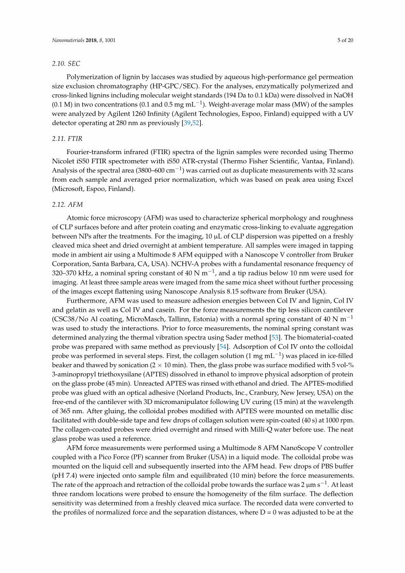
For the coating of individual CLPs with β-casein, protein adsorption on lignin surface wasexamined at pH range 3.0–8.5 (Figure 1). It was the highest at pH 3.0 due to positive charge of β-caseinin the acidic reaction conditions. A similar adsorbed mass was observed at pH 8.5, but in this casenegatively charged β-casein formed particles (ca. 300 nm) that adsorbed on the lignin surface together
Nanomaterials 2018, 8, 1001 7 of 20
with the polymeric protein. This is further confirmed when comparing the increase in dissipation(Figure S1C). The dissipation is much higher for layers adsorbed at alkaline pH compared to pH 3.5,indicating that these layers are more loosely bound and contain more water due to the nanoparticulatemorphology. The formation of β-casein NPs depends on the pH, time, mixing, protein and saltconcentration [20].
Nanomaterials 2018, 8, x FOR PEER REVIEW 7 of 21
value when β-casein concentration increased. During the coating, CLPs aggregated when the surface
charge of the particles was close to zero (CLP—protein ratio ca. 0.01), as shown in Figure 2b. On the
other hand, once clearly positively charged, the protein-coated CLP dispersions were stable for weeks.
Large-scale all atom MD simulations [62] have shown that aromatic residues contribute significantly
to the protein adsorption on hydrophobic surfaces via strong π–π stacking interactions between p2-
carbons. The basic residues such as arginine and lysine play equally strong role for the adsorption.
The effect of proline residues has been demonstrated recently [42].
Figure 1. Adsorption of β-casein on lignin thin film at different pH observed using QCM-D.
(a) (b)
Figure 1. Adsorption of β-casein on lignin thin film at different pH observed using QCM-D.
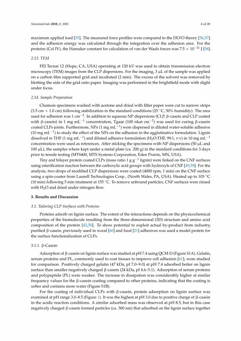
Hence, pH 3.0 was selected for the coating CLPs with β-casein for further experiments. Figure 2ashows the zeta potential of the CLPs varying from negative (ca. −25 mV) to positive (ca. 25 mV)value when β-casein concentration increased. During the coating, CLPs aggregated when the surfacecharge of the particles was close to zero (CLP—protein ratio ca. 0.01), as shown in Figure 2b. On theother hand, once clearly positively charged, the protein-coated CLP dispersions were stable for weeks.Large-scale all atom MD simulations [62] have shown that aromatic residues contribute significantlyto the protein adsorption on hydrophobic surfaces via strong π–π stacking interactions betweenp2-carbons. The basic residues such as arginine and lysine play equally strong role for the adsorption.The effect of proline residues has been demonstrated recently [42].
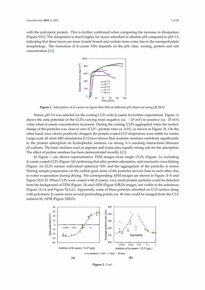
In Figure 2 are shown representative TEM images from single CLPs (Figure 2c) includingβ-casein coated CLPs (Figure 2d) confirming that after protein adsorption, and enzymatic cross-linking(Figure 2e) CLPs remain individual spherical NPs and the aggregation of the particles is minor.During sample preparation on the carbon grid, some of the particles moved close to each other dueto water evaporation during drying. The corresponding AFM images are shown in Figure 2f–h andFigure S2A–D. When CLPs were coated with β-casein, very small protein particles could be detectedfrom the background of TEM (Figure 2d) and AFM (Figure S2B,D) images, not visible in the references(Figure 2f,i–k and Figure S2A,C). Apparently, some of these particles adsorbed on CLP surface alongwith polymeric β-casein since several protruding points (ca. 40 nm) could be imaged from the CLPsurfaces by AFM (Figure S2B,D).
Nanomaterials 2018, 8, x FOR PEER REVIEW 7 of 21
value when β-casein concentration increased. During the coating, CLPs aggregated when the surface
charge of the particles was close to zero (CLP—protein ratio ca. 0.01), as shown in Figure 2b. On the
other hand, once clearly positively charged, the protein-coated CLP dispersions were stable for weeks.
Large-scale all atom MD simulations [62] have shown that aromatic residues contribute significantly
to the protein adsorption on hydrophobic surfaces via strong π–π stacking interactions between p2-
carbons. The basic residues such as arginine and lysine play equally strong role for the adsorption.
The effect of proline residues has been demonstrated recently [42].
Figure 1. Adsorption of β-casein on lignin thin film at different pH observed using QCM-D.
(a) (b)
Figure 2. Cont.
Nanomaterials 2018, 8, 1001 8 of 20
Nanomaterials 2018, 8, x FOR PEER REVIEW 8 of 21
Figure 2. Coating CLPs with β-casein (pH 3.0) evidenced using zeta potential (a) and DLC (b)
measurements as a function of time. TEM images measured from unmodified CLPs (c), β-casein
coated CLPs (d) and β-casein coated CLPs cross-linked with Tgase (e). In (f,g,h) are shown
representative AFM images from enzymatically stabilized CLPs and in (i,j,k) are presented the
corresponding references.
In Figure 2 are shown representative TEM images from single CLPs (Figure 2c) including β-
casein coated CLPs (Figure 2d) confirming that after protein adsorption, and enzymatic cross-linking
(Figure 2e) CLPs remain individual spherical NPs and the aggregation of the particles is minor.
During sample preparation on the carbon grid, some of the particles moved close to each other due
to water evaporation during drying. The corresponding AFM images are shown in Figure 2f–h and
Figure S2A–D. When CLPs were coated with β-casein, very small protein particles could be detected
from the background of TEM (Figure 2d) and AFM (Figure S2B,D) images, not visible in the
references (Figure 2f,i–k and Figure S2A,C). Apparently, some of these particles adsorbed on CLP
surface along with polymeric β-casein since several protruding points (ca. 40 nm) could be imaged
from the CLP surfaces by AFM (Figure S2B,D).
3.1.2. Poly(L-glutamic acid)
Feasibility to coat CLP surfaces using selected proteins to maximize specific interactions with
the substrate such as CNF surface was evaluated. Thus, negatively charged CLPs were first coated
with positively charged PL following modification with PGA containing large number of carboxylic
acid groups for the esterification reaction with hydroxyls on nanocellulose surface via fast heat
treatment. It was hypothesized that tiny CLPs below 100 nm in size coat single CNF fibrils better than
larger particles since the typical width for the nanocellulose fibrils is ca. 5–20 nm and the length
several micrometers. The average molecular masses of enzymatically polymerized and cross-linked
lignin used for tiny CLP preparation are shown in Figure 3 and the characterization using FTIR
spectroscopy in Tables S1–S3. In general, the changes in the FTIR spectra between the references and
laccase treated samples were minor due to heterogeneous cross-linking reactions and residual
moisture in the samples slightly interfering the interpretation of the spectra.
Figure 2. Coating CLPs with β-casein (pH 3.0) evidenced using zeta potential (a) and DLC(b) measurements as a function of time. TEM images measured from unmodified CLPs (c), β-caseincoated CLPs (d) and β-casein coated CLPs cross-linked with Tgase (e). In (f,g,h) are shownrepresentative AFM images from enzymatically stabilized CLPs and in (i,j,k) are presented thecorresponding references.
3.1.2. Poly(L-glutamic acid)
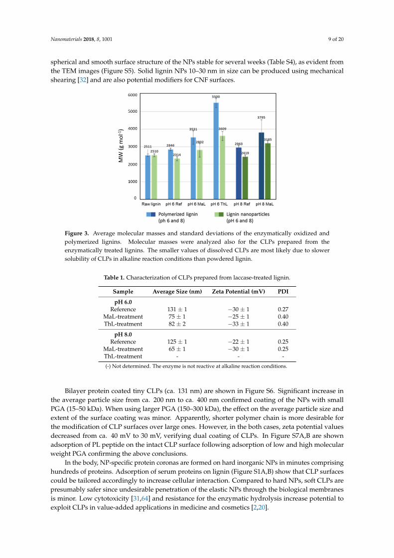
Feasibility to coat CLP surfaces using selected proteins to maximize specific interactions with thesubstrate such as CNF surface was evaluated. Thus, negatively charged CLPs were first coated withpositively charged PL following modification with PGA containing large number of carboxylic acidgroups for the esterification reaction with hydroxyls on nanocellulose surface via fast heat treatment.It was hypothesized that tiny CLPs below 100 nm in size coat single CNF fibrils better than largerparticles since the typical width for the nanocellulose fibrils is ca. 5–20 nm and the length severalmicrometers. The average molecular masses of enzymatically polymerized and cross-linked ligninused for tiny CLP preparation are shown in Figure 3 and the characterization using FTIR spectroscopyin Tables S1–S3. In general, the changes in the FTIR spectra between the references and laccase treatedsamples were minor due to heterogeneous cross-linking reactions and residual moisture in the samplesslightly interfering the interpretation of the spectra.
The appearance of the CLP dispersions at pH 6.0 are shown in Figure S3. The average particlesizes for CLPs prepared from enzymatically oxidized lignin were below 100 nm (Table 1). For thereferences, the particle size was half of that obtained according to the method of Lievonen at al. [25].In both cases, the zeta potentials were on the same order of magnitude as previously described [25].The increased molecular weight, higher hydrophobicity of the polymerized and cross-linked ligninas well as lower concentration enabled tight packing of enzymatically oxidized lignin fast mixingpromoting tiny NP CLP formation. In the laccase-catalyzed reactions, the cross-links are formedin lignin via different radical reactions. Enzymatic initiation of the radicalization starts from thephenolic hydroxyl groups of lignin following condensation of the free radicals to covalent chemicalbonds [37,38,46,47,63]. The representative AFM images of the different CLPs (Figure S4) verify the
Nanomaterials 2018, 8, 1001 9 of 20
spherical and smooth surface structure of the NPs stable for several weeks (Table S4), as evident fromthe TEM images (Figure S5). Solid lignin NPs 10–30 nm in size can be produced using mechanicalshearing [32] and are also potential modifiers for CNF surfaces.Nanomaterials 2018, 8, x FOR PEER REVIEW 9 of 21
Figure 3. Average molecular masses and standard deviations of the enzymatically oxidized and
polymerized lignins. Molecular masses were analyzed also for the CLPs prepared from the
enzymatically treated lignins. The smaller values of dissolved CLPs are most likely due to slower
solubility of CLPs in alkaline reaction conditions than powdered lignin.
The appearance of the CLP dispersions at pH 6.0 are shown in Figure S3. The average particle
sizes for CLPs prepared from enzymatically oxidized lignin were below 100 nm (Table 1). For the
references, the particle size was half of that obtained according to the method of Lievonen at al. [25].
In both cases, the zeta potentials were on the same order of magnitude as previously described [25].
The increased molecular weight, higher hydrophobicity of the polymerized and cross-linked lignin
as well as lower concentration enabled tight packing of enzymatically oxidized lignin fast mixing
promoting tiny NP CLP formation. In the laccase-catalyzed reactions, the cross-links are formed in
lignin via different radical reactions. Enzymatic initiation of the radicalization starts from the
phenolic hydroxyl groups of lignin following condensation of the free radicals to covalent chemical
bonds [37,38,46,47,63]. The representative AFM images of the different CLPs (Figure S4) verify the
spherical and smooth surface structure of the NPs stable for several weeks (Table S4), as evident from
the TEM images (Figure S5). Solid lignin NPs 10–30 nm in size can be produced using mechanical
shearing [32] and are also potential modifiers for CNF surfaces.
Table 1. Characterization of CLPs prepared from laccase-treated lignin.
Sample Average Size (nm) Zeta Potential (mV) PDI
pH 6.0
Reference 131 ± 1 −30 ± 1 0.27
MaL-treatment 75 ± 1 −25 ± 1 0.40
ThL-treatment 82 ± 2 −33 ± 1 0.40
pH 8.0
Reference 125 ± 1 −22 ± 1 0.25
MaL-treatment 65 ± 1 −30 ± 1 0.25
ThL-treatment - - -
(-) Not determined. The enzyme is not reactive at alkaline reaction conditions.
Bilayer protein coated tiny CLPs (ca. 131 nm) are shown in Figure S6. Significant increase in the
average particle size from ca. 200 nm to ca. 400 nm confirmed coating of the NPs with small PGA (15–
50 kDa). When using larger PGA (150–300 kDa), the effect on the average particle size and extent of
the surface coating was minor. Apparently, shorter polymer chain is more desirable for the
modification of CLP surfaces over large ones. However, in the both cases, zeta potential values
decreased from ca. 40 mV to 30 mV, verifying dual coating of CLPs. In Figure S7A,B are shown
Figure 3. Average molecular masses and standard deviations of the enzymatically oxidized andpolymerized lignins. Molecular masses were analyzed also for the CLPs prepared from theenzymatically treated lignins. The smaller values of dissolved CLPs are most likely due to slowersolubility of CLPs in alkaline reaction conditions than powdered lignin.
Table 1. Characterization of CLPs prepared from laccase-treated lignin.
Sample Average Size (nm) Zeta Potential (mV) PDI
pH 6.0Reference 131 ± 1 −30 ± 1 0.27
MaL-treatment 75 ± 1 −25 ± 1 0.40ThL-treatment 82 ± 2 −33 ± 1 0.40
pH 8.0Reference 125 ± 1 −22 ± 1 0.25
MaL-treatment 65 ± 1 −30 ± 1 0.25ThL-treatment - - -
(-) Not determined. The enzyme is not reactive at alkaline reaction conditions.
Bilayer protein coated tiny CLPs (ca. 131 nm) are shown in Figure S6. Significant increase inthe average particle size from ca. 200 nm to ca. 400 nm confirmed coating of the NPs with smallPGA (15–50 kDa). When using larger PGA (150–300 kDa), the effect on the average particle size andextent of the surface coating was minor. Apparently, shorter polymer chain is more desirable forthe modification of CLP surfaces over large ones. However, in the both cases, zeta potential valuesdecreased from ca. 40 mV to 30 mV, verifying dual coating of CLPs. In Figure S7A,B are shownadsorption of PL peptide on the intact CLP surface following adsorption of low and high molecularweight PGA confirming the above conclusions.
In the body, NP-specific protein coronas are formed on hard inorganic NPs in minutes comprisinghundreds of proteins. Adsorption of serum proteins on lignin (Figure S1A,B) show that CLP surfacescould be tailored accordingly to increase cellular interaction. Compared to hard NPs, soft CLPs arepresumably safer since undesirable penetration of the elastic NPs through the biological membranesis minor. Low cytotoxicity [31,64] and resistance for the enzymatic hydrolysis increase potential toexploit CLPs in value-added applications in medicine and cosmetics [2,20].
Nanomaterials 2018, 8, 1001 10 of 20
3.2. Stability of β-Casein Coated CLP
3.2.1. Effect of Enzymatic Cross-Linking
Tailoring CLP surfaces with β-casein allows further modification of the particles with cross-linkingenzymes [65]. In vitro studies have shown that Tgase can cross-link proteins in a gel in minutes [17,48].Hence, to improve stability of β-casein coated CLPs at physiological pHs, for example in stomach(pH 1.0–3.0), duodenum (pH 4.8–8.2) and blood (pH 7.4), Tgase was used to cross-link the β-caseincoating. Figure S8A shows the increase in the average particle size of CLPs as a function of enzymedosage. Particles cross-linked with high enzyme dosage above 20 nkat g−1 aggregated immediatelybecause covalent bonds were also formed between the individual particles. The zeta potential values(Figure S8B) decreased after 24-h incubation and, after seven-day treatment, extensive cross-linking ofthe NPs was detected by eye. Reasonable enzyme dosage for the cross-linking only β-casein coatingwas found to be 15 nkat g−1. Prior to pH stability studies of the enzymatically cross-linked particles,Tgase activity was removed from the dispersions using ultra-centrifugation (Figure S9). After thetreatment, some of the unbound β-casein adsorbed on CLP surfaces increasing the average particlesize by ca. 40 nm. Then, the particles were stable for several weeks.
Elasticity of the enzymatically cross-linked β-casein coating was studied by QCM-D(Figure S10A,B). After adsorption of β-casein on lignin film until a stable baseline was observed(Figure S1), Tgase (2 min) was injected into the chamber following cross-linking (10 min) of theprotein film. When using the low enzyme dosage (5 nkat), loosely bound β-casein was washed away.However, at the same time, the cross-linking reaction proceeded to a certain extent since a sharpdecrease in dissipation was detected due to the formation of elastic networked protein coating. Instead,when using higher enzyme dosages in the measuring cell (25 and 50 nkat), first a small amount ofunbound β-casein was removed (tiny increase in the frequency signal). After that, a sharp drop in thefrequency was detected due to enzyme adsorption on the β-casein surface. The cross-linking reactionproceeded as in the case of low enzyme dosage. During the enzymatic reaction (10 min), the frequencyincreased slightly, verifying an accompanied loss of small molecule reaction product (-NH3
+) [66] aswell as decreased water binding capacity of the cross-linked protein coating. Tgase injection followingstabilization of the cross-linking reaction was repeated three times until stable baseline was observed.
Figure 2c–h shows representative TEM and AFM images ofβ-casein coated CLPs cross-linked withTgase. Compared to CLPs coated with β-casein (Figure S2B,D) the surfaces of cross-linked particleswere smoother. From the corresponding TEM images (Figure 2d,c), it is evident that also small proteinparticles in the background were cross-linked to larger particles. Regarding the applicability of theenzymatically stabilized protein coated CLP dispersions, polymerization of free β-casein to nanosizedparticles [65] improves homogeneity and stability of the dispersion.
3.2.2. Effect of pH
Understanding dispersion properties is essential for the exploitation CLPs in adhesiveformulations. CLPs are stable in wide pH range [25]. However, due to better electrostatic stability inalkaline conditions, the reactivity of CLPs is much higher than in acidic conditions [39,63]. For medical,cosmetic and food applications, it is pivotal that tailored CLPs are stable in physiological conditions.
Figure 4a,b shows the average particle sizes and zeta potential values for β-casein coated CLPsat pH 3.0 and 7.4 as a function of time. In acidic dispersion, NPs started to aggregate after four-dayincubation, which was evident also from the more positive zeta potential values. After 25 days,protein-coated particles precipitated. At pH 3.0, β-casein was positively charged, but CLP surface wasnearly neutral, promoting aggregation of the coated CLPs. At slightly alkaline dispersion (pH 7.4),the stability of β-casein coated CLPs was excellent. The average particle sizes and zeta potential valuesremained nearly unchanged for 25 days.

Nanomaterials 2018, 8, 1001 11 of 20
Nanomaterials 2018, 8, x FOR PEER REVIEW 11 of 21
Figure 4a,b shows the average particle sizes and zeta potential values for β-casein coated CLPs
at pH 3.0 and 7.4 as a function of time. In acidic dispersion, NPs started to aggregate after four-day
incubation, which was evident also from the more positive zeta potential values. After 25 days,
protein-coated particles precipitated. At pH 3.0, β-casein was positively charged, but CLP surface
was nearly neutral, promoting aggregation of the coated CLPs. At slightly alkaline dispersion (pH
7.4), the stability of β-casein coated CLPs was excellent. The average particle sizes and zeta potential
values remained nearly unchanged for 25 days.
(a) (b)
Figure 4. Stability of β-casein coated and enzymatically cross-linked CLPs at (a) pH 3.0 and (b) pH
7.4 evidenced using average particle size and zeta potential measurements. In both cases, the protein
coating following enzymatic cross-linking was performed at 3.0. Enzyme activity was removed using
ultracentrifugation following redispersion of the particles at above pH.
To improve stability of the β-casein coated CLPs at pH 3.0, the optimized Tgase dosage (15 nkat
g−1) was used to cross-link the surfaces of the particles. The stability of cross-linked protein coated
CLPs was excellent compared to the non-cross-linked particles (Figure 4a). The average particle sizes
remained nearly unchanged for 25 days. Instead, the zeta potential values became slightly more
positive due to some instability of CLPs, as explained above. Instead, at pH 7.4 (Figure 4b), the zeta
potential values were stable for weeks, but the average particle sizes increased during the first day
after enzyme inhibition and solvent exchange. Then, the cross-linked protein coated CLPs were stable
for weeks.
3.3. Adhesive Interactions
AFM force measurement was used to compare strength of the interactions between lignin and
model proteins (Figure 5). Col IV is the main component of the skin and, therefore, understanding
the interactions of Col IV with other proteins and lignin could be exploited in wound healing and
other biomedical applications.
Figure 4. Stability of β-casein coated and enzymatically cross-linked CLPs at (a) pH 3.0 and (b) pH7.4 evidenced using average particle size and zeta potential measurements. In both cases, the proteincoating following enzymatic cross-linking was performed at 3.0. Enzyme activity was removed usingultracentrifugation following redispersion of the particles at above pH.
To improve stability of the β-casein coated CLPs at pH 3.0, the optimized Tgase dosage(15 nkat g−1) was used to cross-link the surfaces of the particles. The stability of cross-linked proteincoated CLPs was excellent compared to the non-cross-linked particles (Figure 4a). The average particlesizes remained nearly unchanged for 25 days. Instead, the zeta potential values became slightly morepositive due to some instability of CLPs, as explained above. Instead, at pH 7.4 (Figure 4b), the zetapotential values were stable for weeks, but the average particle sizes increased during the first dayafter enzyme inhibition and solvent exchange. Then, the cross-linked protein coated CLPs were stablefor weeks.
3.3. Adhesive Interactions
AFM force measurement was used to compare strength of the interactions between lignin andmodel proteins (Figure 5). Col IV is the main component of the skin and, therefore, understanding theinteractions of Col IV with other proteins and lignin could be exploited in wound healing and otherbiomedical applications.
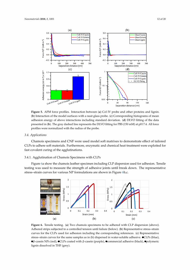
Long-range attractive interactions were detected when Col IV surface was brought into contactwith casein and gelatin (Figure 5a), while no meaningful adhesions were detected when the neat glasssurface was used (Figure 5b). The force interactions between Col IV and lignin were three times largerthan the ones between Col IV and model proteins (Figure 5c) with adhesion energy being 0.0102 ±0.0006 nJ m−1. Interestingly, the adhesion energy between Col IV and casein as well as Col IV andgelatin was nearly identical at pH 7.4, evident also from the overlapping retraction force profiles(Figure 5a). At high salt concentration, the repulsions detected from the approach force profiles wasmuch longer-ranged than predicted by DLVO theory for pure electrostatic double-layer repulsion(Figure 5d). This reveals that steric repulsions dominate [67]. Based on the approach force profilesseparation distance between Col IV and gelatin pair is nearly twofold compared to that of Col IV andcasein, while Col IV and lignin was placed in between. Even though casein show lower adhesionenergy with Col IV, it is an attractive protein to coat CLPs compared to poorly soluble gelatin havinghigh tendency to aggregate, as recently demonstrated [42]. Thus, purified β-casein containing manyreactive sites for enzymatic cross-linking and stabilization is an excellent coat protein for CLPs enablingalso fast curing enzymatic means [65].
Nanomaterials 2018, 8, 1001 12 of 20
Nanomaterials 2018, 8, x FOR PEER REVIEW 12 of 21
Figure 5. AFM force profiles. Interaction between (a) Col IV probe and other proteins and lignin. (b)
Interaction of the model surfaces with a neat glass probe. (c) Corresponding histograms of mean
adhesion energy of above interactions including standard deviation. (d) DLVO fitting of the data
presented in (b). The gray dashed line represents the DLVO fitting for PBS (150 mM) at pH 7.4. All
force profiles were normalized with the radius of the probe.
Long-range attractive interactions were detected when Col IV surface was brought into contact
with casein and gelatin (Figure 5a), while no meaningful adhesions were detected when the neat glass
surface was used (Figure 5b). The force interactions between Col IV and lignin were three times larger
than the ones between Col IV and model proteins (Figure 5c) with adhesion energy being 0.0102 ±
0.0006 nJ m−1. Interestingly, the adhesion energy between Col IV and casein as well as Col IV and
gelatin was nearly identical at pH 7.4, evident also from the overlapping retraction force profiles
(Figure 5a). At high salt concentration, the repulsions detected from the approach force profiles was
much longer-ranged than predicted by DLVO theory for pure electrostatic double-layer repulsion
(Figure 5d). This reveals that steric repulsions dominate [67]. Based on the approach force profiles
separation distance between Col IV and gelatin pair is nearly twofold compared to that of Col IV and
casein, while Col IV and lignin was placed in between. Even though casein show lower adhesion
energy with Col IV, it is an attractive protein to coat CLPs compared to poorly soluble gelatin having
high tendency to aggregate, as recently demonstrated [42]. Thus, purified β-casein containing many
reactive sites for enzymatic cross-linking and stabilization is an excellent coat protein for CLPs
enabling also fast curing enzymatic means [65].
3.4. Applications
Chamois specimens and CNF were used model soft matrixes to demonstrate effect of tailored
CLPs to adhere soft materials. Furthermore, enzymatic and chemical heat treatment were exploited
for fast covalent curing of the agglutinations.
3.4.1. Agglutination of Chamois Specimens with CLPs
Figure 6a show the chamois leather specimen including CLP dispersion used for adhesion.
Tensile testing was used to measure the strength of adhesive joints until break down. The
representative stress–strain curves for various NP formulations are shown in Figure 6b,c.
Figure 5. AFM force profiles. Interaction between (a) Col IV probe and other proteins and lignin.(b) Interaction of the model surfaces with a neat glass probe. (c) Corresponding histograms of meanadhesion energy of above interactions including standard deviation. (d) DLVO fitting of the datapresented in (b). The gray dashed line represents the DLVO fitting for PBS (150 mM) at pH 7.4. All forceprofiles were normalized with the radius of the probe.
3.4. Applications
Chamois specimens and CNF were used model soft matrixes to demonstrate effect of tailoredCLPs to adhere soft materials. Furthermore, enzymatic and chemical heat treatment were exploited forfast covalent curing of the agglutinations.
3.4.1. Agglutination of Chamois Specimens with CLPs
Figure 6a show the chamois leather specimen including CLP dispersion used for adhesion. Tensiletesting was used to measure the strength of adhesive joints until break down. The representativestress–strain curves for various NP formulations are shown in Figure 6b,c.
Nanomaterials 2018, 8, x FOR PEER REVIEW 13 of 21
(a) (b) (c)
Figure 6. Tensile testing. (a) Two chamois specimens to be adhered with CLP dispersion (above).
Adhered strips subjected to a controlled tension until failure (below). (b) Representative stress–strain
curves for the CLPs used for adhesion including the corresponding references. (c) Representative stress–
strain curves for the same samples as in (b) dispersed in water-soluble adhesive. ● CLPs (blue), ● β-
casein NPs (red), ● CLPs coated with β-casein (purple), ● commercial adhesive (black), ● polymeric
lignin dissolved in THF (grey).
Figure 7a shows the effect of different NPs for adhering protein matrix that was significantly
better than that of lignin and commercial water-soluble adhesive. When the number of NPs doubled,
the agglutination of the specimens was more than twofold stronger, which was evident from the
measured stress values. The differences between the type of the soft material, i.e., lignin or protein,
used for the NP preparation was apparent when high NP concentration was used. Adhesive property
of β-casein NPs on protein matrix was the highest and for CLPs only half of that. Effect of β-casein
coated CLPs on the adhesion was between these two cases showing potential as a method to prepare
functional low-cost β-casein NPs from lignin via self-assembly. The tensile strain (Figure 7b)
measured from the corresponding samples followed the same order as the stress values. However,
the differences between the samples were smaller. When β-casein coated CLPs were used for
adhering, the strain values were rather similar between two different quantities. In the case of large
number of NPs, the repeatability was poor, presumably due to instability of the β-casein coating at
pH 3.0, as explained above. Regarding to the references, lignin powder dissolved in THF and diluted
adhesive, the agglutination of the specimens was minor compared to that of NPs. Due to rough, hairy
surface of chamois, the NPs spread, adsorbed and penetrated on the soft material better than the thick
adhesive formulation, efficiently dissipating energy and retarding fracture of the joints under stress.
Figure 6. Tensile testing. (a) Two chamois specimens to be adhered with CLP dispersion (above).Adhered strips subjected to a controlled tension until failure (below). (b) Representative stress–straincurves for the CLPs used for adhesion including the corresponding references. (c) Representativestress–strain curves for the same samples as in (b) dispersed in water-soluble adhesive. CLPs (blue),β-casein NPs (red), CLPs coated with β-casein (purple), commercial adhesive (black), polymeric
lignin dissolved in THF (grey).

Nanomaterials 2018, 8, 1001 13 of 20
Figure 7a shows the effect of different NPs for adhering protein matrix that was significantlybetter than that of lignin and commercial water-soluble adhesive. When the number of NPs doubled,the agglutination of the specimens was more than twofold stronger, which was evident from themeasured stress values. The differences between the type of the soft material, i.e., lignin or protein,used for the NP preparation was apparent when high NP concentration was used. Adhesive property ofβ-casein NPs on protein matrix was the highest and for CLPs only half of that. Effect of β-casein coatedCLPs on the adhesion was between these two cases showing potential as a method to prepare functionallow-cost β-casein NPs from lignin via self-assembly. The tensile strain (Figure 7b) measured from thecorresponding samples followed the same order as the stress values. However, the differences betweenthe samples were smaller. When β-casein coated CLPs were used for adhering, the strain values wererather similar between two different quantities. In the case of large number of NPs, the repeatability waspoor, presumably due to instability of the β-casein coating at pH 3.0, as explained above. Regardingto the references, lignin powder dissolved in THF and diluted adhesive, the agglutination of thespecimens was minor compared to that of NPs. Due to rough, hairy surface of chamois, the NPsspread, adsorbed and penetrated on the soft material better than the thick adhesive formulation,efficiently dissipating energy and retarding fracture of the joints under stress.Nanomaterials 2018, 8, x FOR PEER REVIEW 14 of 21
Figure 7. Comparison between tensile stress–strain histograms. (a,b) Different CLPs including the
references at two quantities. (c,d) Effect of Tgase catalyzed cross-linking on the adhesion. (e,f) Various
NPs including the references dispersed in diluted water-soluble adhesive.
Figure 7c,d shows the increased stress and strain values when β-casein coated CLPs were
covalently linked to chamois specimens using low Tgase dosage (100 nkat). Increasing the enzyme
activity, the strength of the adhesion could be increased [47,62]. These results suggest that protein
coated CLPs could also be linked to biological matrixes using enzymes enabling fast curing in moist
environments essential for the medical applications. In such seals, CLPs remain single nanoparticles
retaining their physicochemical properties since the covalent linkages are formed via protein coating.
It is also plausible that cross-links were formed between amine groups in lysine side-chains and the
acyl group derived from the carboxylic acid groups present in CLPs due to side reactions of Tgase
[17]. If CLPs are coated with tissue specific proteins, the potential rejection reactions could be
diminished during the wound healing, yielding small scars important for cosmetic applications.
Accordingly, it is presupposed that CLP formulations could be exploited for the preparation of edible
coatings for foods.
Furthermore, to study potential to use CLPs as additive in adhesive formulations, NPs and the
references were dispersed in the water-soluble adhesive (Figure 7e,f). Unexpectedly, the stress value
was the largest (ca. 5%) for the dispersion containing lignin powder compared to that of diluted
adhesive. Mixing protein NPs in the adhesive formulation decreased stress value ca. 3%, and for the
unmodified CLPs ca. 7%. In the case of tensile strain measurement, the results were opposite. For
CLPs, β-casein NPs and β-casein coated CLPs, the tensile strain improved ca. 5%, 12% and 10%,
respectively. For lignin powder, the elasticity of the adhesive joint degreased ca. 5% compared to the
diluted adhesive.
Figure 8 shows histograms of Young’s modulus for various NPs. The values doubled for the
specimens adhered with high number of NPs regardless of the type of the polymer used for the
adhesion. Instead, when the NPs were dispersed in the adhesive formulation, the differences between
Figure 7. Comparison between tensile stress–strain histograms. (a,b) Different CLPs including thereferences at two quantities. (c,d) Effect of Tgase catalyzed cross-linking on the adhesion. (e,f) VariousNPs including the references dispersed in diluted water-soluble adhesive.
Figure 7c,d shows the increased stress and strain values when β-casein coated CLPs werecovalently linked to chamois specimens using low Tgase dosage (100 nkat). Increasing the enzymeactivity, the strength of the adhesion could be increased [47,62]. These results suggest that proteincoated CLPs could also be linked to biological matrixes using enzymes enabling fast curing in moistenvironments essential for the medical applications. In such seals, CLPs remain single nanoparticles
Nanomaterials 2018, 8, 1001 14 of 20
retaining their physicochemical properties since the covalent linkages are formed via protein coating.It is also plausible that cross-links were formed between amine groups in lysine side-chains and theacyl group derived from the carboxylic acid groups present in CLPs due to side reactions of Tgase [17].If CLPs are coated with tissue specific proteins, the potential rejection reactions could be diminishedduring the wound healing, yielding small scars important for cosmetic applications. Accordingly, it ispresupposed that CLP formulations could be exploited for the preparation of edible coatings for foods.
Furthermore, to study potential to use CLPs as additive in adhesive formulations, NPs and thereferences were dispersed in the water-soluble adhesive (Figure 7e,f). Unexpectedly, the stress valuewas the largest (ca. 5%) for the dispersion containing lignin powder compared to that of dilutedadhesive. Mixing protein NPs in the adhesive formulation decreased stress value ca. 3%, and forthe unmodified CLPs ca. 7%. In the case of tensile strain measurement, the results were opposite.For CLPs, β-casein NPs and β-casein coated CLPs, the tensile strain improved ca. 5%, 12% and 10%,respectively. For lignin powder, the elasticity of the adhesive joint degreased ca. 5% compared to thediluted adhesive.
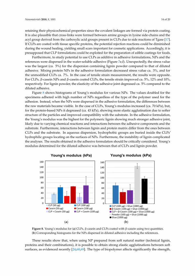
Figure 8 shows histograms of Young’s modulus for various NPs. The values doubled for thespecimens adhered with high number of NPs regardless of the type of the polymer used for theadhesion. Instead, when the NPs were dispersed in the adhesive formulation, the differences betweenthe raw materials became visible. In the case of CLPs, Young’s modulus increased (ca. 70 kPa), but,for the protein-based NPs it degreased (ca. 43 kPa), showing more elastic agglutination due to softerstructure of the particles and improved compatibility with the substrate. In the adhesive formulation,the Young’s modulus was the highest for the polymeric lignin showing much stronger adhesive joint,likely due to varying chemical reactions and interactions between the adhesive components and thesubstrate. Furthermore, interactions between lignin and protein matrix differ from the ones betweenCLPs and the substrate. In aqueous dispersion, hydrophobic groups are buried inside the CLPs’hydrophilic groups locating on the surfaces of NPs. Furthermore, the instability of lignin complicatedthe analyses. The results obtained in the adhesive formulation should be critically considered. Young’smodulus determined for the diluted adhesive was between that of CLPs and lignin powder.
Nanomaterials 2018, 8, x FOR PEER REVIEW 15 of 21
the raw materials became visible. In the case of CLPs, Young’s modulus increased (ca. 70 kPa), but,
for the protein-based NPs it degreased (ca. 43 kPa), showing more elastic agglutination due to softer
structure of the particles and improved compatibility with the substrate. In the adhesive formulation,
the Young’s modulus was the highest for the polymeric lignin showing much stronger adhesive joint,
likely due to varying chemical reactions and interactions between the adhesive components and the
substrate. Furthermore, interactions between lignin and protein matrix differ from the ones between
CLPs and the substrate. In aqueous dispersion, hydrophobic groups are buried inside the CLPs’
hydrophilic groups locating on the surfaces of NPs. Furthermore, the instability of lignin complicated
the analyses. The results obtained in the adhesive formulation should be critically considered.
Young’s modulus determined for the diluted adhesive was between that of CLPs and lignin powder.
(a) (b)
Figure 8. Young’s modulus for (a) CLPs, β-casein and CLPs coated with β-casein using two quantities.
(b) Corresponding histograms for the NPs dispersed in diluted adhesive including the references.
These results show that, when using NP prepared from soft natural matter (technical lignin,
proteins and their combinations), it is possible to obtain strong elastic agglutinations between soft
surfaces, as evidenced recently [24,68,69]. The type of biopolymer affects significantly the strength,
flexibility and elongation of the joint. Studies with soybean-based adhesives containing polymeric
lignin pointed out that protein–lignin ratio is the most critical parameter affecting the adhesive
interactions [70,71]. In textiles, enzymatic cross-linking has been used to strengthen lignin-containing
adhesives [72]. Apparently, exploitation of technical lignin in nanoparticulate morphology in stable
dispersion for adhering is an interesting approach for many lignin applications reviewed [73].
3.4.2. Grafting CLPs on CNF Surfaces
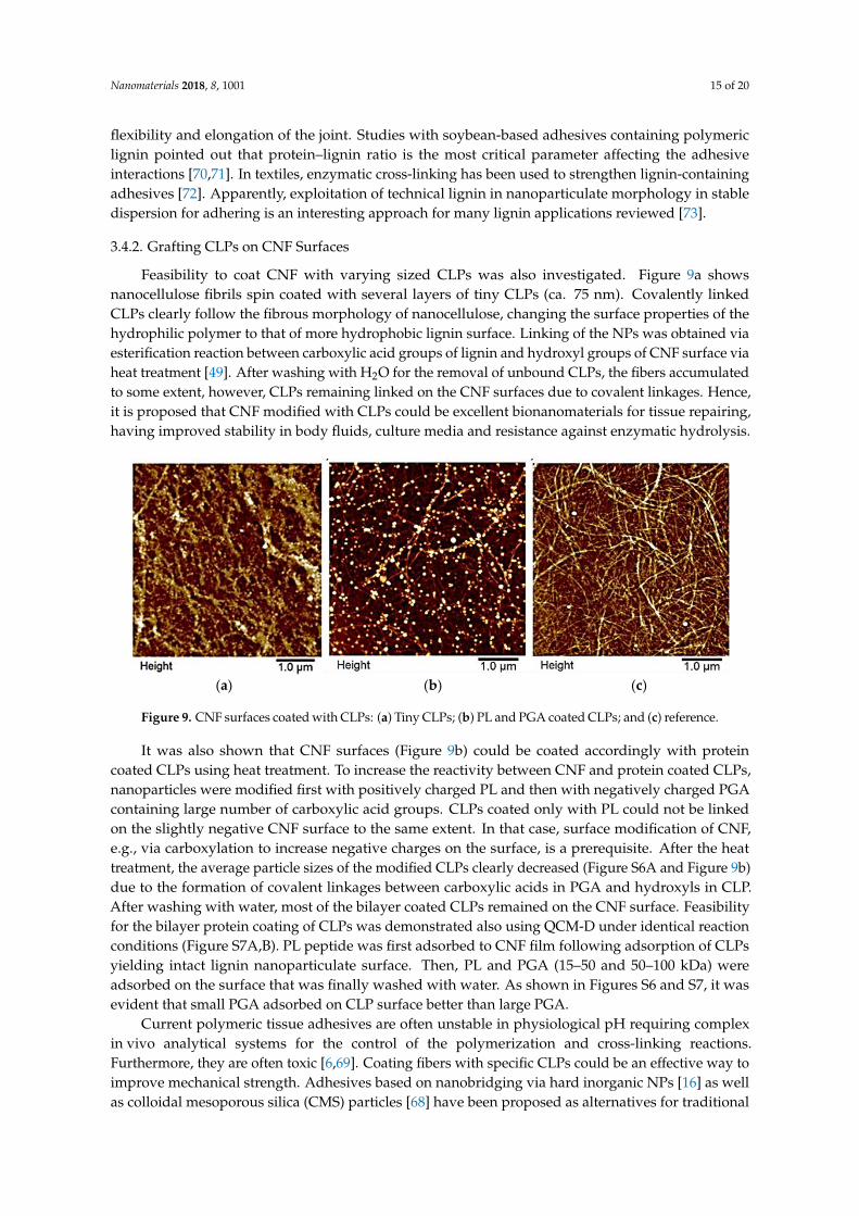
Feasibility to coat CNF with varying sized CLPs was also investigated. Figure 9a shows
nanocellulose fibrils spin coated with several layers of tiny CLPs (ca. 75 nm). Covalently linked CLPs
clearly follow the fibrous morphology of nanocellulose, changing the surface properties of the
hydrophilic polymer to that of more hydrophobic lignin surface. Linking of the NPs was obtained
via esterification reaction between carboxylic acid groups of lignin and hydroxyl groups of CNF
surface via heat treatment [49]. After washing with H2O for the removal of unbound CLPs, the fibers
accumulated to some extent, however, CLPs remaining linked on the CNF surfaces due to covalent
linkages. Hence, it is proposed that CNF modified with CLPs could be excellent bionanomaterials for
tissue repairing, having improved stability in body fluids, culture media and resistance against
enzymatic hydrolysis.
It was also shown that CNF surfaces (Figure 9b) could be coated accordingly with protein coated
CLPs using heat treatment. To increase the reactivity between CNF and protein coated CLPs,
Figure 8. Young’s modulus for (a) CLPs, β-casein and CLPs coated with β-casein using two quantities.(b) Corresponding histograms for the NPs dispersed in diluted adhesive including the references.
These results show that, when using NP prepared from soft natural matter (technical lignin,proteins and their combinations), it is possible to obtain strong elastic agglutinations between softsurfaces, as evidenced recently [24,68,69]. The type of biopolymer affects significantly the strength,
Nanomaterials 2018, 8, 1001 15 of 20
flexibility and elongation of the joint. Studies with soybean-based adhesives containing polymericlignin pointed out that protein–lignin ratio is the most critical parameter affecting the adhesiveinteractions [70,71]. In textiles, enzymatic cross-linking has been used to strengthen lignin-containingadhesives [72]. Apparently, exploitation of technical lignin in nanoparticulate morphology in stabledispersion for adhering is an interesting approach for many lignin applications reviewed [73].
3.4.2. Grafting CLPs on CNF Surfaces
Feasibility to coat CNF with varying sized CLPs was also investigated. Figure 9a showsnanocellulose fibrils spin coated with several layers of tiny CLPs (ca. 75 nm). Covalently linkedCLPs clearly follow the fibrous morphology of nanocellulose, changing the surface properties of thehydrophilic polymer to that of more hydrophobic lignin surface. Linking of the NPs was obtained viaesterification reaction between carboxylic acid groups of lignin and hydroxyl groups of CNF surface viaheat treatment [49]. After washing with H2O for the removal of unbound CLPs, the fibers accumulatedto some extent, however, CLPs remaining linked on the CNF surfaces due to covalent linkages. Hence,it is proposed that CNF modified with CLPs could be excellent bionanomaterials for tissue repairing,having improved stability in body fluids, culture media and resistance against enzymatic hydrolysis.
Nanomaterials 2018, 8, x FOR PEER REVIEW 16 of 21
nanoparticles were modified first with positively charged PL and then with negatively charged PGA
containing large number of carboxylic acid groups. CLPs coated only with PL could not be linked on
the slightly negative CNF surface to the same extent. In that case, surface modification of CNF, e.g.,
via carboxylation to increase negative charges on the surface, is a prerequisite. After the heat
treatment, the average particle sizes of the modified CLPs clearly decreased (Figure S6A and Figure
9b) due to the formation of covalent linkages between carboxylic acids in PGA and hydroxyls in CLP.
After washing with water, most of the bilayer coated CLPs remained on the CNF surface. Feasibility
for the bilayer protein coating of CLPs was demonstrated also using QCM-D under identical reaction
conditions (Figure S7A,B). PL peptide was first adsorbed to CNF film following adsorption of CLPs
yielding intact lignin nanoparticulate surface. Then, PL and PGA (15–50 and 50–100 kDa) were
adsorbed on the surface that was finally washed with water. As shown in Figures S6 and S7, it was
evident that small PGA adsorbed on CLP surface better than large PGA.
(a) (b) (c)
Figure 9. CNF surfaces coated with CLPs: (a) Tiny CLPs; (b) PL and PGA coated CLPs; and (c)
reference.
Current polymeric tissue adhesives are often unstable in physiological pH requiring complex in
vivo analytical systems for the control of the polymerization and cross-linking reactions. Furthermore,
they are often toxic [6,69]. Coating fibers with specific CLPs could be an effective way to improve
mechanical strength. Adhesives based on nanobridging via hard inorganic NPs [16] as well as
colloidal mesoporous silica (CMS) particles [68] have been proposed as alternatives for traditional
medical adhesives. Since the adhesion energy is proportional to the surface area of NPs,
enzymatically cross-linked CLPs [39] with tailored functionalities could be potential additives for
medical adhesives following enzymatic or thermal treatment for fast curing shown above.
Additionally, porous structure of CLPs enables quicker decomposition in biological media than
inorganic NPs preventing undesirable accumulation in the body. Due to strong autofluorescence of
lignin, it is an attractive raw material enabling sensitive real time detection crucial for the
development of image-guided procedures for clinical applications.
4. Conclusions
Development of green technologies [74,75] for the preparation of bio(nano)materials [76] from
the forest process side-streams such as adhesives and coatings [77,78] is increasing constantly.
Different CLPs prepared and modified to adhere chamois and to modify CNF surface could be
potential additives for various formulations to be exploited for wound sealing, edible coatings and
fiber modification for textiles to improve adhesion, hydrophobicity, antimicrobial and antioxidative
properties of the coatings. Since the cross-linking methods are fast and feasible in the moist
environment, clinical fluorescence imaging of aromatic CLPs is possible. Furthermore, it was
concluded that, when using tissue specific proteins, e.g., hydrolyzed from collagen, sericins extracted
from silk and caseins fractionated from the dairy side-streams, compatibility of the NPs with the
Figure 9. CNF surfaces coated with CLPs: (a) Tiny CLPs; (b) PL and PGA coated CLPs; and (c) reference.
It was also shown that CNF surfaces (Figure 9b) could be coated accordingly with proteincoated CLPs using heat treatment. To increase the reactivity between CNF and protein coated CLPs,nanoparticles were modified first with positively charged PL and then with negatively charged PGAcontaining large number of carboxylic acid groups. CLPs coated only with PL could not be linkedon the slightly negative CNF surface to the same extent. In that case, surface modification of CNF,e.g., via carboxylation to increase negative charges on the surface, is a prerequisite. After the heattreatment, the average particle sizes of the modified CLPs clearly decreased (Figure S6A and Figure 9b)due to the formation of covalent linkages between carboxylic acids in PGA and hydroxyls in CLP.After washing with water, most of the bilayer coated CLPs remained on the CNF surface. Feasibilityfor the bilayer protein coating of CLPs was demonstrated also using QCM-D under identical reactionconditions (Figure S7A,B). PL peptide was first adsorbed to CNF film following adsorption of CLPsyielding intact lignin nanoparticulate surface. Then, PL and PGA (15–50 and 50–100 kDa) wereadsorbed on the surface that was finally washed with water. As shown in Figures S6 and S7, it wasevident that small PGA adsorbed on CLP surface better than large PGA.
Current polymeric tissue adhesives are often unstable in physiological pH requiring complexin vivo analytical systems for the control of the polymerization and cross-linking reactions.Furthermore, they are often toxic [6,69]. Coating fibers with specific CLPs could be an effective way toimprove mechanical strength. Adhesives based on nanobridging via hard inorganic NPs [16] as wellas colloidal mesoporous silica (CMS) particles [68] have been proposed as alternatives for traditional
Nanomaterials 2018, 8, 1001 16 of 20
medical adhesives. Since the adhesion energy is proportional to the surface area of NPs, enzymaticallycross-linked CLPs [39] with tailored functionalities could be potential additives for medical adhesivesfollowing enzymatic or thermal treatment for fast curing shown above. Additionally, porous structureof CLPs enables quicker decomposition in biological media than inorganic NPs preventing undesirableaccumulation in the body. Due to strong autofluorescence of lignin, it is an attractive raw materialenabling sensitive real time detection crucial for the development of image-guided procedures forclinical applications.
4. Conclusions
Development of green technologies [74,75] for the preparation of bio(nano)materials [76] from theforest process side-streams such as adhesives and coatings [77,78] is increasing constantly. DifferentCLPs prepared and modified to adhere chamois and to modify CNF surface could be potential additivesfor various formulations to be exploited for wound sealing, edible coatings and fiber modification fortextiles to improve adhesion, hydrophobicity, antimicrobial and antioxidative properties of the coatings.Since the cross-linking methods are fast and feasible in the moist environment, clinical fluorescenceimaging of aromatic CLPs is possible. Furthermore, it was concluded that, when using tissue specificproteins, e.g., hydrolyzed from collagen, sericins extracted from silk and caseins fractionated fromthe dairy side-streams, compatibility of the NPs with the substrates could be enhanced. Compared toNPs prepared solely from proteins, the costs of the raw materials are remarkably lower. Apparently,these results pave the way for the exploitation of technical lignin in multiple forms.
Supplementary Materials: The electronic supplementary data associated with this article is available onlineat http://www.mdpi.com/2079-4991/8/12/1001/s1. Figure S1: Adsorption of proteins on lignin thin filmsanalyzed by QCM-D, Figure S2: AFM images of β-casein coated CLPs, Figure S3: CLP dispersions prepared fromlaccases treated lignins, Figure S4: AFM height images from CLPs prepared from laccase treated lignin at pH 6,Figure S5: TEM images from CLPs prepared from enzymatically treated lignin one month after preparation, FigureS6: Average particle size and zeta potential of CLPs as a function of PL−CLP mass ratio, Figure S7: Adsorption ofPL, PGA and CLPs on slightly negatively charged CNF analyzed by QCM-D, Figure S8: β-Casein coating andenzymatic stabilization of the particles with Tgase, Figure S9: Variation of stabilized CLPs in size after removal ofenzyme activity using ultracentrifugation, Figure S10: Elasticity of enzymatically cross-linked β-casein coating,Tables S1–S3: Characterization of laccase treated lignins by FTIR, Table S4: Effect of time on the average particlesize, zeta potential and polydispersity (PDI) of CLPs prepared from different lignins at starting concentration0.5 g L−1.
Author Contributions: M.-L.M. supervised the work including writing of the final version of the manuscript.Furthermore, she was responsible for the adsorption, coating and stabilization studies, TEM imaging as wellas planning of the application studies. G.R. prepared and characterized CLP dispersions using differentphysicochemical methods such as AFM, SEC and FTIR spectroscopy. R.W.N.N. and T.L. carried out the AFMretraction force measurements. A.H. carried out the adhesion experiments with various NPs and chamoisspecimens using tensile testing. O.N. purified and characterized enzymes used in the study at VTT. J.J.V.-D.conducted the AFM imaging. M.A.K. and M.Ö. provided facilities, scientific discussion and guidance for thestudy. All authors contributed to the production of the manuscript.
Funding: This research was funded by the Academy of Finland (TaBioMat, Tailored biomass derivedself-assembling building blocks for bionanomaterial applications, grant number 276696). Furthermore, this work hasreceived funding from the Bio Based Industries Joint Undertaking under the European Union’s Horizon 2020research and innovation programme under grant agreement No 720303 (Zelcor project).
Acknowledgments: MSc students Anni Pyysing, Helena Båtsman, Teemu Kemppainen, Ari Ruotsalainen,Mustafa Çan, Andrey Vinogradov (Aalto University, JOIN-E3000 Life science technologies project course, coatedlignin nanoparticles for biomaterial applications, Finland) and MSc student Zhenxing Yan (Aalto University,Finland) are acknowledged for the laboratory assistance.
Conflicts of Interest: There are no conflicts to declare.
Nanomaterials 2018, 8, 1001 17 of 20
References
1. Fu, J.; Su, J.; Wang, P.; Yu, Y.; Wang, Q.; Cavaco-Paulo, A. Enzymatic processing of protein-based fibers.Appl. Microbiol. Biotechnol. 2015, 99, 10387–10397. [CrossRef] [PubMed]
2. Ngo, H.-T.; Bechtold, T. Surface modification of textile material through deposition of regenerated silk fibroin.J. Appl. Polym. Sci. 2017, 134, 45098–45109. [CrossRef]
3. Hemmilä, V.; Adamopoulos, S.; Karlsson, O.; Kumar, A. Development of sustainable bio-adhesives forengineered wood panels—A Review. RSC Adv. 2017, 7, 38604–38630. [CrossRef]
4. Esposito, D.; Antonietti, M. Redefining biorefinery: The search for unconventional building blocks formaterials. Chem. Soc. Rev. 2015, 44, 5821–5835. [CrossRef] [PubMed]
5. Klemm, D.; Kramer, F.; Moritz, S.; Lindström, T.; Ankerfors, M.; Gray, D.; Dorris, A. Nanocelluloses: A newfamily of nature-based materials. Angew. Chem. Int. Ed. 2011, 50, 5438–5466. [CrossRef] [PubMed]
6. Eichhorn, S.J.; Dufresne, A.; Aranguren, M.; Marcovich, N.E.; Capadona, J.R.; Rowan, S.J.; Weder, C.;Thielemans, W.; Roman, M.; Renneckar, S.; et al. Review: Current international research into cellulosenanofibers and nanocomposites. J. Mater. Sci. 2010, 45, 1–33. [CrossRef]
7. Xu, X.; Liu, F.; Jiang, L.; Zhu, J.Y.; Haagenson, D.; Wiesenborn, D.P. Cellulose nanocrystals vs. Cellulosenanofibrils: A comparative study on their microstructures and effects as polymer reinforcing agents.ACS Appl. Mater. Interfaces 2013, 5, 2999–3009. [CrossRef]
8. Abe, K.; Iwamoto, S.; Yano, H. Obtaining cellulose nanofibers with a uniform width of 15 nm from wood.Biomacromolecules 2007, 8, 3276–3278. [CrossRef]
9. Bhattacharya, M.; Malinen, M.M.; Lauren, P.; Lou, Y.-R.; Kuisma, S.W.; Kanninen, L.; Lille, M.; Corlu, A.;Guen-Guillouzo, C.; Ikkala, O.; et al. Nanofibrillar cellulose hydrogel promotes three-dimensional liver cellculture. J. Control. Release 2012, 164, 291–298. [CrossRef]
10. Lou, Y.-R.; Kanninen, L.; Kuisma, T.; Niklander, J.; Noon, L.A.; Burks, D.; Urtti, A.; Yliperttula, M. The use ofnanofibrillar cellulose hydrogel as a flexible three-dimensional model to culture human pluripotent stemcells. Stem Cells Dev. 2014, 23, 380–392. [CrossRef]
11. Petersen, N.; Gatenholm, P. Bacterial cellulose-based materials and medical devices: Current state andperspectives. Appl. Microbiol. Biotechnol. 2011, 91, 1277–1286. [CrossRef] [PubMed]
12. Lin, N.; Dufresne, A. Nanocellulose in biomedicine: Current status and future prospect. Eur. Polym. J. 2014,59, 302–325. [CrossRef]
13. Forsman, N.; Lozhechnikova, A.; Khakalo, A.; Johansson, L.-S.; Vartiainen, J.; Österberg, M. Hydrophobicsimple and sustainable coating of CNF films and cellulosic textiles based on layer-by-layer deposition ofpoly-L-lysine and natural wax particles. Carbohydr. Polym. 2017, 173, 392–402. [CrossRef]
14. Oliveira, C.; Santos, C.; Bezerra, F.; Bezerra, M.; Rodrigues, L. Utilization of Cyanoacrylates Adhesives inSkin suture. Rev. Bras. Cir. Plást. 2010, 25, 573–576. [CrossRef]
15. Peng, H.; Shek, P. Novel wound sealants: Biomaterials and applications. Expert Rev. Med. Devices 2010, 7,639–659. [CrossRef] [PubMed]
16. Meddahi-Pelle, A.; Legrand, A.; Marcellan, A.; Louedec, L.; Letourneur, D.; Leibler, L. Organ Repair,Hemostasis, and In Vivo Bonding of Medical Devices by Aqueous Solutions of Nanoparticles. Angew. Chem.Int. Ed. 2014, 53, 6369–6373. [CrossRef]
17. Yokoyama, K.; Nio, N.; Kikuch, Y. Properties and applications of microbial transglutaminase.Appl. Microbiol. Biot. 2004, 64, 447–454. [CrossRef]
18. Cortez, J.; Anghieri, A.; Bonner, P.; Griffin, M.; Freddi, G. Transglutaminase mediated grafting of silk proteinsonto wool fabrics leading to improved physical and mechanical properties. Enzyme Microb. Technol. 2007, 40,1698–1704. [CrossRef]
19. Arvanitoyannis, I.S.; Dionisopoulou, N.K. Irradiation of Food Commodities. Techniques, Applications, Detection,Legislation, Safety and Consumer Opinion, 1st ed.; Arvanitoyannis, I.S., Ed.; Academic Press: London, UK, 2010;pp. 609–634, ISBN 13 978-0128101919.
20. Bonnaillie, L.; Aburto, L.; Tunick, M.; Mulherin, J.; Du, M.; Kwoczak, R.; Akkurt, S.; Tomasula, P. Advancesin food packaging films from milk proteins. In Proceedings of the 252nd National Meeting & Expositionof ACS, AGFD: Division of Agricultural and Food Chemistry, Philadelphia, PA, USA, 21–25 August 2016;p. 327816.
Nanomaterials 2018, 8, 1001 18 of 20
21. Bonnaillie, M.L.; Tomasula, P.M. Application of Humidity-Controlled Dynamic Mechanical Analysis(DMA-RH) to Moisture-Sensitive Edible Casein Films for Use in Food Packaging. Polymers 2015, 7, 91–114.[CrossRef]
22. Zhang, F.; Ma, J.; Xu, Q.; Zhou, J.; Simion, D.; Carmen, G.; Wang, J.; Li, Y. Hollow Casein-Based PolymericNanospheres for Opaque Coatings. ACS Appl. Mater. Interfaces 2016, 8, 11739–11748. [CrossRef]
23. Stewart, R.J.; Wang, C.S.; Shao, H. Complex coacervates as a foundation for synthetic underwater adhesives.Adv. Colloid Interface Sci. 2011, 167, 85–93. [CrossRef]
24. Rose, S.; Prevoteau, A.; Elziére, P.; Hourdet, D.; Marcellan, A.; Leibler, L. Nanoparticle solutions as adhesivesfor gels and biological tissues. Nature 2014, 505, 382–385. [CrossRef]
25. Lievonen, M.; Valle-Delgado, J.J.; Mattinen, M.-L.; Hult, E.-L.; Lintinen, K.; Kostiainen, M.A.; Paananen, A.;Szilvay, G.R.; Setälä, H.; Österberg, M. A simple process for lignin nanoparticle preparation. Green Chem.2016, 18, 1416–1422. [CrossRef]
26. Frangville, C.; Rutkevicius, M.; Richter, A.P.; Velev, O.D.; Stoyanov, S.D.; Paunov, V.N. Fabrication ofenvironmentally biodegradable lignin nanoparticles. ChemPhysChem 2012, 13, 4235–4243. [CrossRef][PubMed]
27. Qian, Y.; Deng, Y.; Qiu, X.; Li, H.; Yang, D. Formation of uniform colloidal spheres from lignin, a renewableresource recovered from pulping spent liquor. Green Chem. 2014, 16, 2156–2163. [CrossRef]
28. Qian, Y.; Zhang, Q.; Qiu, X.; Zhu, S. CO2-responsive diethylaminoethyl-modified lignin nanoparticles andtheir application as surfactants for CO2/N2-switchable Pickering emulsions. Green Chem. 2014, 16, 4963–4968.[CrossRef]
29. Yiamsawas, D.; Baier, G.; Thines, E.; Landfester, K.; Wurm, F.R. Biodegradable lignin nanocontainers.RSC Adv. 2014, 4, 11661–11663. [CrossRef]
30. Gilca, I.A.; Popa, V.I.; Crestini, C. Obtaining lignin nanoparticles by sonication. Ultrason. Sonochem. 2015, 23,369–375. [CrossRef]
31. Tortora, M.; Cavalieri, F.; Mosesso, P.; Ciaffardini, F.; Melone, F.; Crestini, C. Ultrasound driven assembly oflignin into microcapsules for storage and delivery of hydrophobic molecules. Biomacromolecules 2014, 15,1634–1643. [CrossRef]
32. Nair, S.S.; Sharma, S.; Pu, Y.; Sun, Q.; Pan, S.; Zhu, J.Y.; Deng, Y.; Ragauskas, A.J. High shear homogenizationof lignin to nanolignin and thermal stability of nanolignin-polyvinyl alcohol blends. ChemSusChem 2014, 7,3513–3520. [CrossRef]
33. Yearla, S.R.; Padmasree, K. Preparation and characterisation of lignin nanoparticles: Evaluation of theirpotential as antioxidants and UV protectants. J. Exp. Nanosci. 2016, 11, 289–302. [CrossRef]
34. Richter, A.P.; Bharti, B.; Armstrong, H.B.; Brown, J.S.; Plemmons, D.A.; Paunov, V.N.; Stoyanov, S.D.;Velev, O.D. Synthesis and characterization of biodegradable lignin nanoparticles with tunable surfaceproperties. Langmuir 2016, 32, 6468–6477. [CrossRef] [PubMed]
35. Ago, M.; Huan, S.; Borghei, M.; Raula, J.; Kauppinen, E.; Rojas, O. High-throughput synthesis of ligninparticles (∼30 nm to ∼2 µm) via aerosol flow reactor: Size fractionation and utilization in Pickering emulsions.ACS Appl. Mater. Interfaces 2016, 8, 23302–23310. [CrossRef]
36. Beisl, S.; Miltner, A.; Friedl, A. Lignin from Micro- to Nanosize: Production Methods. Int. J. Mol. Sci. 2017,18, 1244. [CrossRef] [PubMed]
37. Roth, S.; Spiess, A.C. Laccases for biorefinery applications: A critical review on challenges and perspectives.Bioproc. Biosyst. Eng. 2015, 38, 2285–2313. [CrossRef] [PubMed]
38. Munk, L.; Sitarz, A.K.; Kalyani, D.C.; Mikkelsen, J.D.; Meyer, A.S. Can laccases catalyze bond cleavage inlignin? Biotechnol. Adv. 2015, 33, 13–24. [CrossRef] [PubMed]
39. Mattinen, M.-L.; Valle-Delgado, J.J.; Leskinen, T.; Anttila, T.; Riviere, G.; Sipponen, M.; Paananen, A.;Lintinen, K.; Kostiainen, K.; Österberg, M. Enzymatically and chemically oxidized lignin nanoparticles forbiomaterial applications. Enzyme Microb. Technol. 2018, 111, 48–56. [CrossRef]
40. Mohammadinejad, R.; Karimi, S.; Iravani, S.; Varma, R.S. Plant-derived nanostructures: Types andapplications. Green Chem. 2016, 18, 20–52. [CrossRef]
41. Pan, K.; Zhong, Q. Organic Nanoparticles in Foods: Fabrication, Characterization and Utilization. Annu. Rev.Food Sci. Technol. 2016, 7, 245–266. [CrossRef] [PubMed]

Nanomaterials 2018, 8, 1001 19 of 20
42. Leskinen, T.; Witos, J.; Valle-Delgado, J.J.; Lintinen, K.; Kostiainen, M.A.; Wiedmer, S.K.; Österberg, M.;Mattinen, M.-L. Adsorption of Proteins on Colloidal Lignin Particles for Advanced Biomaterials.Biomacromolecules 2017, 18, 2767–2776. [CrossRef] [PubMed]
43. Richter, A.P.; Brown, J.S.; Bharti, B.; Wang, A.; Gangwal, S.; Houck, K.; Cohen, E.; Hubal, A.; Paunov, V.N.;Stoyanov, S.D.; et al. An environmentally benign antimicrobial nanoparticle based on a silver-infused lignincore. Nat. Nanotechnol. 2015, 10, 817–823. [CrossRef]
44. Goffin, A.J.; Rajadas, J.; Fuller, G.G. Interfacial Flow Processing of Collagen. Langmuir 2010, 26, 3514–3521.[CrossRef]
45. Tenzer, S.; Docter, D.; Kuharev, J.; Musyanovych, A.; Fetz, V.; Hecht, R.; Schlenk, F.; Fischer, D.; Kiouptsi, K.;Reinhardt, C.; et al. Rapid formation of plasma protein corona critically affects nanoparticle pathophysiology.Nat. Nanotechnol. 2013, 8, 772–781. [CrossRef]
46. Niku-Paavola, M.; Karhunen, E.; Salola, P.; Raunio, V. Ligninolytic enzymes of the white-rot-fungus Phlebiaradiate. Biochem. J. 1988, 254, 877–884. [CrossRef]
47. Rittstieg, K.; Suurnäkki, A.; Suortti, T.; Kruus, K.; Guebitz, G.; Buchert, J. Investigations on thelaccase-catalyzed polymerization of lignin model compounds using size-exclusion HPLC. Enzyme Microb.Technol. 2002, 31, 403–410. [CrossRef]
48. Lantto, R.; Puolanne, E.; Kalkkinen, N.; Buchert, J.; Autio, K. Enzyme-aided modification of chicken-breastmyofibril proteins: Effect of laccase and transglutaminase on gelation and thermal stability. J. Agric.Food Chem. 2005, 53, 9231–9237. [CrossRef] [PubMed]
49. Valle-Delgado, J.J.; Johansson, L.-S.; Österberg, M. Bioinspired lubricating films of cellulose nanofibrils andhyaluronic acid. Colloids Surf. B Biointerfaces 2016, 138, 86–93. [CrossRef]
50. Salas, C.; Rojas, O.J.; Lucia, L.A.; Hubbe, M.A.; Genzer, J. On the surface interactions of proteins with lignin.ACS Appl. Mater. Interfaces 2013, 5, 199–206. [CrossRef] [PubMed]
51. Johannsmann, D.; Mathauer, K.; Wegner, G.; Knoll, W. Viscoelastic properties of thin films probed with aquartz-crystal resonator. Phys. Rev. B 1992, 46, 7808–7815. [CrossRef]
52. Sipponen, M.H.; Pihlajaniemi, V.; Sipponen, S.; Pastinen, O.; Laakso, S. Autohydrolysis and aqueousammonia extraction of wheat straw: Effect of treatment severity on yield and structure of hemicellulose andlignin. RSC Adv. 2014, 4, 23177–23184. [CrossRef]
53. Sader, J.E.; Chon, J.W.; Mulvaney, P. Calibration of rectangular atomic force microscope cantilevers. Rev. Sci.Instrum. 1999, 70, 3967–3969. [CrossRef]
54. Nugroho, R.W.N.; Harjumäki, R.; Zhang, X.; Lou, Y.-R.; Yliperttula, M.; Valle-Delgado, J.J.; Österberg, M.Quantifying the interactions between biomimetic biomaterials—Collagen I, collagen IV, laminin 521 andcellulose nanofibrils—By colloidal probe microscopy. Colloids Surf. B Biointerfaces 2019, 173, 571–580.[CrossRef]
55. Butt, H.; Cappella, B.; Kappl, M. Force measurements with the atomic force microscope: Technique,interpretation and applications. Surf. Sci. Rep. 2005, 59, 1–152. [CrossRef]
56. Verwey, E. Theory of the stability of lyophobic colloids. J. Phys. Chem. 1947, 51, 631–636. [CrossRef]57. Assemi, S.; Nalaskowski, J.; Johnson, W.P. Direct force measurements between carboxylate-modified latex
microspheres and glass using atomic force microscopy. Colloid Surf. A Physicochem. Eng. Asp. 2006, 286,70–77. [CrossRef]
58. Bowen, W.R.; Hilal, N.; Lovitt, R.W.; Wright, C.J. Direct measurement of interactions between adsorbedprotein layers using an atomic force microscope. J. Colloid Interface Sci. 1998, 197, 348–352. [CrossRef]
59. Schmidt, S.; Madaboosi, N.; Uhlig, K.; Köhler, D.; Skirtach, A.; Duschl, C.; Möhwald, H.; Volodkin, D.V.Control of Cell Adhesion by Mechanical Reinforcement of Soft Polyelectrolyte Films with Nanoparticles.Langmuir 2012, 28, 7249–7257. [CrossRef]
60. Bye, C.N. Casein and mixed protein adhesives. In Handbook of Adhesives; Skeist, I., Ed.; Van Nostrand Reinold:New York, NY, USA, 1990; pp. 135–152. ISBN 978-1-4613-0671-9.
61. Mazia, D. Adhesion of cells to surfaces coated with polylysine. Applications to electron microscopy.J. Cell Biol. 1975, 66, 198–200.
62. Gu, Z.; Yang, Z.; Wang, L.; Zhou, H.; Jimenez-Cruz, C.A.; Zhou, R. The role of basic residues in the adsorptionof blood proteins onto the graphene surface. Sci. Rep. 2015, 5, 10873–10884. [CrossRef]
63. Van de Pas, D.; Hickson, A.; Donaldson, L.; Lloyd-Jones, G.; Tamminen, T.; Fernyhough, A.; Mattinen, M.-L.Characterization of fractionated lignins polymerized by fungal laccases. BioResources 2011, 6, 1105–1121.

Nanomaterials 2018, 8, 1001 20 of 20
64. Figueiredo, P.; Lintinen, K.; Kiriazis, A.; Hynninen, V.; Liu, V.; Ramos, T.B.; Rahikkala, A.; Correia, A.;Kohout, T.; Sarmento, B.; et al. In vitro evaluation of biodegradable lignin-based nanoparticles for drugdelivery and enhanced antiproliferation effect in cancer cells. Biomaterials 2017, 121, 97–108. [CrossRef]
65. Monogioudi, E. Enzymatic Cross-Linking of β-Casein and Its Impact on Digestibility and Allergenicity; Universityof Helsinki: Helsinki, Finland, 18 January 2011; ISBN 978-951-38-7421-6.
66. Berglin, M.; Delage, L.; Potin, P.; Vilter, H.; Elwing, H. Enzymatic Cross-Linking of a Phenolic PolymerExtracted from the Marine Alga Fucus serratus. Biomacromolecules 2004, 5, 2376–2383. [CrossRef]
67. Boström, M.; Williams, D.; Ninham, B. Specific ion effects: Why DLVO theory fails for biology and colloidsystems. Phys. Rev. Lett. 2001, 87, 168103–168107. [CrossRef]
68. Kim, J.-H.; Kim, H.; Choi, Y.; Lee, D.S.; Kim, J.; Yi, G.-R. Colloidal Mesoporous Silica Nanoparticles as StrongAdhesives for Hydrogels and Biological Tissues. ACS Appl. Mater. Interfaces 2017, 9, 31469–31477. [CrossRef]
69. Shin, K.; Choi, J.W.; Ko, G.; Baik, S.; Kim, D.; Park, O.K.; Lee, K.; Cho, H.R.; Han, S.I.; Lee, S.H.; et al.Multifunctional nanoparticles as a tissue adhesive and an injectable marker for image-guided procedures.Nat. Commun. 2017, 8, 15807–15819. [CrossRef]
70. Pradyawong, S.; Qi, G.; Li, N.; Sun, X.S.; Wang, D. Adhesion properties of soy protein adhesives enhancedby biomass lignin. Int. J. Adhes. Adhes. 2017, 75, 66–73. [CrossRef]
71. Luo, J.; Yuan, C.; Zhang, W.; Li, J.; Gao, Q.; Chen, H. An eco-friendly wood adhesive from soy protein andlignin: Performance properties. RSC Adv. 2015, 5, 100849–100855. [CrossRef]
72. Aracri, E.; Blanco, C.D.; Tzanov, T. An enzymatic approach to develop a lignin-based adhesive for wool floorcoverings. Green Chem. 2014, 16, 2597–2603. [CrossRef]
73. Kai, D.; Tan, M.J.; Chee, P.L.; Chua, Y.K.; Yap, Y.L.; Loh, X.J. Towards lignin-based functional materials in asustainable world. Green Chem. 2016, 18, 1175–1200. [CrossRef]
74. Didaskalou, C.; Buyuktiryaki, S.; Kecili, R.; Fonte, C.P.; Szekely, G. Valorisation of agricultural waste with anadsorption/nanofiltration hybrid process: From materials to sustainable process design. Green Chem. 2017,19, 3116–3125. [CrossRef]
75. Ashok, R.P.B.; Oinas, P.; Lintinen, K.; Sarwar, G.; Kostiainen, M.A.; Österberg, M. Techno-economicassessment for the large-scale production of colloidal lignin particles. Green Chem. 2018, 20, 4911–4919.[CrossRef]
76. Tian, D.; Hu, J.; Bao, J.; Chandra, R.P.; Saddler, J.N.; Lu, C. Lignin valorization: Lignin nanoparticles ashigh-value bio-additive for multifunctional nanocomposites. Biotechnol. Biofuels 2017, 10, 192–203. [CrossRef]
77. García, J.L.; Pans, G.; Phanopoulos, C. Use of lignin in polyurethane-based structural wood adhesives.J. Adhes. 2018, 94, 814–828. [CrossRef]
78. Kalami, S.; Arefmanesh, M.; Master, E.; Nejad, M. Replacing 100% of phenol in phenolic adhesiveformulations with lignin. J. Appl. Polym. Sci. 2017, 134, 45124–45133. [CrossRef]
© 2018 by the authors. Licensee MDPI, Basel, Switzerland. This article is an open accessarticle distributed under the terms and conditions of the Creative Commons Attribution(CC BY) license (http://creativecommons.org/licenses/by/4.0/).