Embed Size (px)
Citation preview

CELLULAR EFFECTS OF ENAMEL MATRIXDERIVATIVE ARE ASSOCIATED WITH
SPECIFIC PROTEIN COMPONENTS
A THESIS
Presented to the Faculty ofThe University of Texas Graduate School of Biomedical Sciences
at San Antonioin Partial Fulfillmentof the Requirements
for the Degree of
MASTER OF SCIENCE
ByDwight Layne Johnson, B.S., B.A., D.M.D.
San Antonio, Texas
May 2005
20050712 140

CELLULAR EFFECTS OF ENAMEL MATRIXDERIVATIVE ARE ASSOCIATED WITH
SPECIFIC PROTEIN COMPONENTS
Dwight Layne Johnson, M.S.
The University of Texas Graduate School of Biomedical Sciences at San Antonio
Supervising Professor: David L. Cochran, D.D.S., Ph.D.
Background: Emdogain®, or enamel matrix derivative (EMD), is a preparation of matrix
proteins derived from developing porcine teeth. Although EMD has been shown to enhance
both soft tissue healing and regeneration of the periodontium, the mechanism of this action is
still unknown. It is assumed, but not yet proven, that amelogenin, the most abundant protein
in EMD, is the protein primarily responsible for the effects of EMD. The purpose of this
study was to fractionate EMD and associate specific cellular effects with specific protein
components.
v

DEDICATION
I would like to dedicate this to my dear wife and best friend,,'i!j. Her support, love,
encouragement, and understanding have truly been the "wind beneath my wings." I am so
grateful for her dedication, patience, and commitment, as well as the many sacrifices she has
made during the last three very busy and demanding years. Words cannot even begin to
describe the love and appreciation I feel for her. Much of the time she has had to be both
mom and dad to our six sons; To each of my wonderful boys,(.X >,. '' .:.. .. ,. <
.. for sacrificing special time with dad and trying to understand
the many hours of study and writing required to complete this project and many others
associated with the residency; To my parents and siblings, for their encouraging calls, visits,
support and prayers. Finally, to my Lord and Savior, Jesus Christ, who has sustained,
comforted, and given me hope in my most difficult and trying hours. Without Him none of
this would even have been possible.
iii

ACKNOWLEDGEMENTS
I would like to thank my research committee: Drs. David Cochran, my supervising
professor, David Carnes, Thomas Oates, Victor Sylvia, and Howard McDonnell for all their
insights and suggestions in completing this work. They have guided me through this whole
process with encouragement and understanding. A special thanks is due Dr. Carnes who
helped me really grasp the concepts of this project, dedicated extra time and energy to
interpret the data and prepare the graphs and tables for presentation. His commitment and
understanding of this project have been crucial for completion of this thesis. I would also
like to thank Helen Hoffer, Dr. Carnes lab assistant, for her help in running the many assays
required for this research as well as Dr. Bjom Steffensen and Dr. Yao Wang for doing the
zymograms.
iv

THE VIEWS EXPRESSED IN THIS ARTICLE ARETHOSE OF THE AUTHOR AND DO NOT REFLECTTHE OFFICIAL POLICY OR POSITION OF THEUNITED STATES AIR FORCE, DEPARTMENT OFDEFENSE, OR THE U.S. GOVERNMENT.

Methods and Materials: Thirty milligrams of EMD were fractionated over a Sephadex G-
100 column. Forty-five 7ml fractions were collected, desalted, lyophilized, and resuspended
in 10mM acetic acid. The amount of protein was determined and each fraction adjusted to
25ýtg protein/ml with cell culture media. Cell proliferation was examined in osteoprogenitor
(C2C12) and human microvascular endothelial cells (HMVEC). Noggin-sensitive alkaline
phosphatase activity (C2C12) and angiogenesis (HMVEC) were evaluated as markers for
differentiation. Polyacrylamide gels containing protein substrate (denatured type I collagen)
were used to determine collagenolytic activity in each fraction.
Results: EMD fractionated into three major protein peaks associated with the column void
volume, the 50 kDa and the 10 kDa molecular weight proteins. The void volume peak
represents proteins greater than 100 kDa molecular weight while the 50 kDa and 10 kDa
peaks most likely represent amelogenin and fragments, as well as other minor proteins in the
mixture. Proliferation activity was associated with the 50 kDa and 10 kDa proteins peaks for
both osteoprogenitor and microvascular cells. Differentiation of osteoprogenitor cells, as
indicated by alkaline phosphatase activity, was stimulated by protein fractions between the
void volume and 50 kDa peaks. This activity was inhibited by prior treatment of the
fractions with noggin, indicative of bone morphogenetic protein (BMP) activity rather than
amelogenin activity. Angiogenic activity (HMVEC) was associated with the same fractions
that stimulated proliferation. Collagenolytic activity was associated with the void volume, as
well as with proteins in the 68 kDa and 25-40 kDa molecular weight regions.
Conclusions: EMD stimulated multiple activities in more than one cell type, important
because of the multiple cell types involved in the regenerative process. These cellular
vi
activities are apparently not associated with a single protein species, i.e. amelogenin, because
the activities are associated with proteins of differing molecular weights. Differentiation of
osteoprogenitor cells is probably the result of BMP(s) present in the EMD mixture and not
the result of amelogenin activity because it fractionated in a different molecular weight
region from amelogenin, and was inhibited by noggin, a known antagonist of BMP-2, -4, and
-7. This suggests that cooperation of multiple growth factors may be important for
successful periodontal regeneration. EMD also stimulates proliferation and differentiation of
microvascular endothelial cells, activities not previously reported, and these activities are
associated with the fractions containing amelogenin. Finally, EMD was shown to contain
collagenolytic activity, also not previously reported, that is capable of degrading type I
collagen. This proteolytic activity was manifest in both the 68 kDa and 25-40 kDa molecular
weight regions, consistent with MMP-2 and/or MMP-20, both known to digest amelogenin.
This is important for both matrix degradation and cell migration during regeneration of the
periodontal tissues. Although the multiple cellular effects of EMD cannot be ascribed to any
single protein species, this study suggests that combined activities may be necessary for
successful periodontal regeneration.
vii

TABLE OF CONTENTS
T itle ......................................................................................................... i
A pproval ................................................................................................... ii
D edication ................................................................................................ iii
Acknowledgements ................................................................................... iv
A b stract .................................................................................................... v
T able of C ontents ...................................................................................... viii
List of Tables .......................................................................................... x
List of Figures ........................................................................................ xi
I. INTRODUCTION AND LITERATURE REVIEW ........................................ 1
A . Introduction ................................................................................... 1
B. Literature Review ......................................................................... 5
1. Periodontal Disease .................................................................. 5
2. Periodontal Treatment ................................................................... 7
3. Periodontal Regeneration ........................................................... 8
4. Enamel Matrix Derivative ........................................................... 9
5. A m elogenin ............................................................................. 14
6. The Problem-Mechanism of Action Poorly Understood .................... 15
II. INVESTIGATION PURPOSE AND AIMS ................................................ 17
III. MATERIALS AND METHODS ......................................................... 18
A. Materials ................................................................................. 18
viii

B. Methods ................................................................................... 20
1. EMD Fractionation ................................................................ 20
2. Determination of Alkaline Phosphatase Activity .............................. 21
3. Determination of Cell Proliferation ............................................. 21
4. In Vitro Angiogenesis Assay .................................................... 22
5. Polyacrylamide Gel Electrophoresis ............................................. 23
C. Data Analysis ............................................................................ 23
IV . R E SU LT S ....................................................................................... 24
A. EMD and Amelogenin data ........................................................... 24
1. Alkaline Phosphatase Activity .................................................... 24
2. Proliferation Activity .................................................................. 26
3. Angiogenesis ......................................................................... 31
4. SDS Page Gel of EMD and Amelogenin ........................................ 34
B. Sephadex G-100 Column Calibration ............................................... 40
C. EMD Protein Separation .................................................................. 44
D. Fractionated Alkaline Phosphatase Activity ........................................ 44
E. Fractionated Proliferation Activity ...................................................... 48
F. Fractionated Angiogenic Activity .................................................... 53
G. Fractionated Collagenolytic Activity ................................................. 56
H. SDS Page Gels of Fractionated EMD .................................................... 56
V. DISCUSSION AND SUMMARY ............................................................. 61
Literature C ited ........................................................................................... 67
V ita ...................................................................................................... 74
ix

LIST OF TABLES
Table 1. The Media Contents of HMVEC Growth Media ............................... 33
Table 2. In Vitro Angiogenesis Assay Scoring System ................................. 35

LIST OF FIGURES
Figure 1. Effect of EMD on Alkaline Phosphatase Activity of C2C12 Cells ............. 25
Figure 2. Effect of Amelogenin on Alkaline Phosphatase Activity of C2C12 Cells.....27
Figure 3. Effect of EMD on Proliferation of C2C 12 Cells ............................... 28
Figure 4. Effect of Amelogenin on Proliferation of C2C12 Cells ....................... 29
Figure 5. Effect of EMD on Proliferation Activity of HMVEC Cells ..................... 30
Figure 6. Effect of Amelogenin on Proliferation Activity of HMVEC Cells ............. 32
Figure 7. Representative Samples of Angiogenic Scores ................................. 36
Figure 8. Effect of EMD and Amelogenin on Angiogenesis of HMVEC Cells at1 H our ................................................................................... 37
Figure 9. Effect of EMD and Amelogenin on Angiogenesis of HMVEC Cells at4 H ours ............................................................................... 38
Figure 10. SDS-PAGE of Amelogenin and Enamel Matrix Derivative ..................... 39
Figure 11. Sephadex G-100 Column Calibration Molecular Weight Markers ............. 41
Figure 12. Molecular Weight Standard Curve ............................................... 43
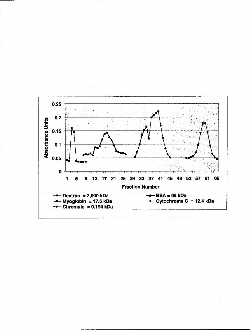
Figure 13. EMD Protein Separation Profile ..................................................... 45
Figure 14. Fractionation of EMD Over Sephadex G-100 Column with MolecularW eight M arkers ........................................................................ 46
Figure 15. Molecular Weight of EMD Protein Peaks ...................................... 47
Figure 16. Alkaline Phosphatase Activity of EMD Fractions on C2C12 Cells ............ 49
Figure 17. Noggin Inhibition of Alkaline Phosphatase Activity of EMD Fractions onC 2C 12 C ells ............................................................................ 50
Figure 18. Proliferation Activity of EMD Fractions on C2C 12 Cells .................... 51
Figure 19. Proliferation Activity of EMD Fractions on HMVEC Cells ..................... 52
xi

Figure 20. Angiogenic Activity of EMD Fractions on HMVEC Cells at 1 Hour ..... 54
Figure 21. Angiogenic Activity of EMD Fractions on HMVEC Cells at 4 Hours ......... 55
Figure 22. Zymograms of EMD Column Fractions ........................................ 57
Figure 23. SDS-PAGE Gel of EMD Column Fractions ........................................ 60
xii
I. INTRODUCTION AND LITERATURE REVIEW
A. INTRODUCTION
Periodontitis, one of the most common reasons for tooth loss in the United States
today, occurs due to the loss of the supporting structures of the periodontium, specifically,
cementum, periodontal ligament (PDL) and alveolar bone. This disease has a prevalence of
between 35-70% of the U.S. population, and even though the disease may not be curable, it is
treatable. For years, the goal of therapy was to eliminate the etiologic factors in order to stop
the continued loss of the supporting structures. In the last decade, the advances in
periodontal therapy have led to the rationale of not only trying to stop progression of the
disease and maintain what is left of the attachment apparatus, but also to trying to regenerate
what has been lost.
Regeneration has been approached and attempted in many ways with varying degrees
of success and predictability. This is because successful periodontal regeneration requires
the integration of multiple tissue interactions in order to build or rebuild the entire
periodontal attachment apparatus. Materials such as autogenous and allograft bone have
been used to create a scaffold on which resident cells can grow and provide protein factors to
help stimulate host cell growth into the defect. There are, however, varying theories as to
what target cells should be stimulated in the host. Melcher initially suggested that cells from
the periodontal ligament hold the potential for regenerating the attachment apparatus
(Melcher, 1976), but a few years later modified his theory to include bone cells along with
cells from the periodontal ligament. Meyer states that pluripotential periodontal ligament
cells differentiate into cells of cementogenic, osteogenic and fibroblastic lineage (Meyer,
I
2
1986). More recent studies on the other hand suggest that the formation of acellular
cementum is the key tissue in the development of a functional periodontium (Hammarstr6m
et al., 1997) and that periodontal ligament and alveolar bone development is associated with
the formation of acellular cementum. In either case, these theories led to the use of barrier
membranes in an attempt to exclude epithelial and gingival fibroblasts from entering wound
sites, thus giving these precursor cells the opportunity to "regenerate" the attachment
apparatus. This treatment modality of regeneration is commonly known as guided tissue
regeneration (GTR).
GTR has been proven successful both clinically and histologically and is an important
part of periodontal therapy. The concept of GTR has also progressed over the years from
using materials such as non-resorbable membranes, which require a second-stage surgical
procedure for removal, to using resorbable porcine and bovine collagen membranes, which
degrade over time. Even though these modalities of treatment were successful, complete
regeneration was not always predictable or consistent. To try and increase the predictability
of regeneration, GTR was used in combination with osseous autografts and allografts, as well
as with xenografts and alloplasts. Even though these combination approaches improved
regeneration in certain situations, the goal of predictable and complete regeneration has yet to
be achieved.
Current research on regeneration has focused on attempting to regenerate the
attachment apparatus by mimicking the events that occur during the initial embryogenesis of
the periodontal tissues (Gestrelius, 2000). Root formation is primarily the result of the down
growth of the apical extension of the enamel organ, known as Hertwig's epithelial root
sheath (HERS). It is thought that Hertwig's epithelial root sheath is responsible for

3
depositing enamel matrix proteins on the root surface (Slavkin and Boyde, 1975; Slavkin,
1976) that may be the stimulus initiating cementum formation. In vitro studies show that
acellular cementum is formed when either endogenous or exogenous enamel matrix is
exposed to dental follicle cells (Hammarstr~m, 1997b).
Because of the complexity of the interactions of the many cell types, growth factors
and proteins associated with nascent tooth formation, there has been much research into how
these elements function individually and in concert to potentially regenerate a diseased
periodontium. With the discovery that HERS secretes enamel matrix proteins in association
with root and acellular cementum formation, more of today's focus has been on using enamel
matrix proteins to induce host cells to regenerate new cementum, periodontal ligament and
alveolar bone. These enamel matrix proteins are available today in a material known as
enamel matrix derivative (EMD).
Enamel matrix derivative, commercially known as Emdogain® was originally
produced and distributed by BioraTm AB, Malmo, Sweden. The company has been purchased
and now operates as Straumann Biologics Division, Waltham, MA. Emdogain® is now
marketed by Straumann as a surgical adjunct for use in regeneration. According to the
company, indications for use include such areas as intrabony defects and gingival recession,
as well as shallow class II furcation defects. All situations involve attempts to regenerate
periodontal tissues that have been lost due to disease. The effectiveness of EMD has been
proven both clinically and histologically in both animal and human models. Clinically, EMD
has been used in many situations with promising results. When EMD is used in conjunction
with open flap debridement compared to open flap debridement alone the outcome is better;
When GTR is compared to EMD the results have also been comparable. The combined use

4
of EMD with particulate grafting materials, whether allograft or xenograft, has also received
increasing amounts of attention in the quest for complete and predictable regeneration. The
results have not been decisive, but an obvious trend appears to be evident favoring better
results when the two materials are combined.
Even though there is mounting evidence for the successful regenerative potential of
EMD, the mechanism of action by which EMD exerts its effects on the tissues involved in
the regenerative process continues to elude investigators. It is truly a case in which the
preliminary research necessary to understand the mechanism of action leading to the desired
results was not accomplished prior to FDA approval for its clinical use in the field of
periodontics. The observed clinical success of the product has prompted investigators to
search for the reason(s) behind the clinical success. Much of that research has focused on
amelogenin, the major protein component of EMD. The underlying question still to be
answered is: are the observed beneficial effects of EMD a result of amelogenin or some
other component(s) in the mixture?
The ideal way to answer this question would be to separate the proteins (i.e.
amelogenin) contained in the commercially available mixture of EMD, apply them to
multiple cell types, and then assay for activities, such as proliferation and differentiation,
known to be associated with EMD. Performing such experiments and then associating the
activities with the various protein components of the mixture would help determine and
clarify whether or not the observed activities and clinical success are the result of a single
protein component (i.e. amelogenin).

5
The goal of this research was to separate the various proteins contained in EMD and
associate the known cellular activities with the proteins contained in EMD in order to
determine if amelogenin is the protein responsible for its regenerative properties.
B. LITERATURE REVIEW
1. PERIODONTAL DISEASE
Periodontics is the specialty of dentistry involved in dealing with the tissues near,
around, or investing (peri) the teeth (odontics). Those tissues, collectively known by the
term "periodontium", primarily involve the gingiva, alveolar mucosa, cementum, periodontal
ligament, and alveolar and basal bone (AAP, 2001). When these tissues become diseased in
any way, the condition is known by the broad term of periodontal disease. This disease and
many of its forms have been classified and reclassified over the years in an attempt to
categorize and describe the nature, extent, and severity of the disease process that is
occurring (Annals of Periodontology, 1999).
Periodontal disease currently is divided into two broad categories based upon the
amount of tissue destruction that occurs and the reversibility of the diseased state. Those
terms are gingivitis and periodontitis. Gingivitis is the less severe of the two disease states
and as the term implies, is an inflammatory condition affecting primarily the gingiva and
occurs without any corresponding loss of attachment. It occurs over time as a result of the
primary etiologic factor, bacterial plaque (Lbe et al., 1965; Socransky and Haffajee, 1991),
and progresses through various histologic stages (Page and Schroeder, 1976). This disease
process is considered reversible, and once the bacterial plaque is removed, a normal state of
gingival health can be achieved as long as no attachment loss has occurred. Once the
6
inflammatory process progresses to the point of losing underlying supporting tissue, i.e.
bone, periodontal ligament, and cementum, the irreversible diseased state known as
periodontitis exists. However, the progression of gingivitis to periodontitis and the
accompanying loss of attachment do not occur universally in every person (L6e et al., 1986;
Page and Schroeder, 1981). In fact, according to Brown et al. (1988), the prevalence of
gingivitis in the United States was approximately 50% while that of periodontitis was 36%.
The variability in an individual's progression from gingivitis to periodontitis requires
more than the mere presence of bacterial plaque. Periodontitis is truly multifactorial in
nature. Host susceptibility (Greenstein and Lamster, 2000; Kornman et al., 1997; Seymour,
1991), including an individual's immunologic state, the presence of "favorable or non-
pathologic bacteria", calculus, smoking, occlusion, anatomic anomalies, age and genetics,
have all been shown, to serve as possible modifying or contributing factors. Each affects the
detrimental shift from gingivitis to periodontitis and a corresponding loss of attachment to
one degree or another.
The dentogingival attachment apparatus around healthy teeth and its corresponding
dimensions was originally described by Gargiulo et al. (1961) in which they found a mean
sulcus depth of 0.69mm, and mean junctional epithelium and connective tissue attachments
of 0.97mm and 1.07mm respectively. In periodontitis, the underlying connective tissue,
bone, and PDL are destroyed, resulting in an apical migration of the junctional epithelium,
with accompanying clinical attachment loss (CAL) and increased probing depths (PD). Deep
probing depths also provide a conducive environment that potentiates a bacterial shift from
the predominantly gram-positive flora associated with health to the predominantly gram-
negative flora necessary to cause disease. If left untreated, the likelihood of continued CAL

7
and tooth loss is extremely high (L6e et al., 1986; Becker et al., 1979). The destructive
nature of the disease may produce osseous defects around teeth, some of which may be
amenable to regenerative therapy.
2. PERIODONTAL TREATMENT
For many years, the goal of periodontics was to halt CAL leading to tooth loss by the
removal of the primary etiologic factor, bacterial plaque. In doing this, as well as in
attempting to eliminate or at least decrease the number of secondary etiologic factors, it was
hoped that an individual could surpass a certain threshold in which an equilibrium between
continued destruction and a reduced but healthy periodontium would exist. At best, healing
by repair with a long junctional epithelium was all that could be hoped for. This objective
was realized by using both non-surgical and surgical means of therapy to eliminate both the
etiologic factors and decrease the pocket depths.
A non-surgical approach with scaling and root planning is routinely provided first and
is a proven method of eliminating bacterial plaque, and decreasing probing depths,
inflammation and tooth loss (Hujoel et al., 2000; Morrison et al., 1980; Lindhe et al., 1982;
Hung and Douglass, 2002), however, the inability to completely remove accretions or reduce
the probing depths to levels that can be adequately maintained by both the patient and dental
professionals, many times requires a surgical approach. For many years the only surgical
approach available was one that was resective in nature. In the early years it was primarily
accomplished through soft tissue resection or gingivectomy (Orban, 1942; Goldman 1951;
Glickman, 1956; Waite, 1975), but this only partially solved the problem of deep probing
depths. Many times the deeper probing depths were not only due to soft tissue but to

8
underlying bony defects, therefore osseous resection was applied (Schluger, 1949). This was
an effective means in reducing pockets, and eliminating etiologic factors as well as
facilitating maintenance by both the patient and dental professional but not without the
drawbacks of increased attachment loss from recession and increased root sensitivity. This
treatment modality offering the compromise of a "reduced but stable periodontium" was
acceptable for many years in periodontics and is an acceptable means of treatment even today
in the 2 1 st century. However, with the discovery that the periodontium can indeed be
regenerated under certain circumstances, the once held belief that periodontitis was an
irreversible process is fast becoming a part of historical periodontics.
3. PERIODONTAL REGENERATION
In 1970, a landmark article by Schallhorn et al. (1970) demonstrated clinical
supracrestal bone growth in "no wall", one-wall, and two-wall defects using fresh frozen
bone from the iliac crest. From this point on, the quest of treating periodontitis by means of
regeneration, an additive technique, instead of a resective technique, became the ultimate
goal. The fact that the magnitude of supracrestal bone growth in this landmark study has
never been reproduced is indicative of one of the greatest challenges inherent in regeneration,
that of predictability. Even though there is histologic proof of principle (Dragoo and
Sullivan, 1973; Bowers et al., 1985, 1989), that both autogenous bone and allograft may
achieve true regeneration of bone, PDL and cementum, the fact remains that there are
inconsistencies and dissenting opinions (Becker et al., 1995).
Attempts at increasing the predictability of regeneration have led to newer treatment
modalities such as that initially described by Gottlow et al. (1986) as guided tissue

9
regeneration (GTR), in which barrier membranes are used in an attempt to exclude the
epithelium in order to give precursor cells from bone and PDL time to initiate the
regenerative process. GTR has proven to be successful using both non-resorbable and
resorbable barrier membranes (Cortellini et al., 2004; Gottlow et al. 1986; Nyman et al.,
1982). In some instances (furcation defects), the combined use of both GTR and allograft
has proven to be more conducive to regeneration while at other times (intrabony defects), the
combination therapy has made no difference (Murphy and Gunsolley, 2003). The fact
remains that a method of achieving predictable and complete regeneration of bone, PLD and
cementum, 100% of the time has yet to be found. Much of today's search for such a material
or adjunct has turned to the use of biologics such as growth factors and enamel matrix
proteins.
4. ENAMEL MATRIX DERIVATIVE
A current and increasingly popular adjunct used in today's treatment of periodontal
disease is Enamel Matrix Derivative (EMD), commercially known as Emdogain®. This
section will discuss its development, indications for use, composition, and some of the
controversies surrounding the nature of attachment, possible mechanisms of action, and
current research to date.
EMD is a mixture of freeze-dried enamel matrix proteins derived from the tooth
germs of fetal porcine teeth. It was approved for clinical use by the FDA in 1997 and was
originally produced and distributed by BioraTM, a Swedish company. In 2004, however, it
was purchased by and began to be marketed commercially by StraumannTM, a company
widely known for their implant system. As part of its introduction for use in periodontics, a
10
whole issue of The Journal of Clinical Periodontology, (Vol 24, Number 9, 1997) was
devoted to research articles demonstrating its effectiveness both in vitro and in vivo. The
producers, past and present, claim the following may be indications for its clinical use:
intrabony defects, gingival recession, and shallow class two furcation defects. Furthermore,
it has also been used clinically in treating avulsed teeth, endodontic surgery (failed RCT),
and in bony defects from third molar extraction sites.
EMD has demonstrated the ability to enhance regeneration when applied in
conjunction with periodontal surgery showing the formation of new acellular cementum,
bone and periodontal ligament (PDL) in one human subject (Heijl, 1997) and in monkeys
with dehiscences (Hammarstr6m et al., 1997). Another study showed that the use of EMD in
periodontal defects has a superior defect fill compared to surgical debridement alone (Froum
et al., 2001). It also selectively enhances the proliferation of PDL cells but not of epithelial
cells (Gestrelius, 1997). This last effect is due primarily to its cytostatic effect against
epithelial cells in suppressing the down growth of junctional epithelium onto the root
surfaces (Kawase et al., 2000). Contrasting research suggests that when the enamel proteins
are secreted by the HERS, they are only secreted in the apical portion of the root and are not
present in the region of acellular cementum growth during root development (Fong and
Hammerstr~m, 2000). Other studies show that the regeneration of acellular cementum is not
a consistent finding (Sculean et al., 1999; Yukna et al., 2000). In spite of these studies
reporting clear histological differences, the number of successful clinical studies evaluating
different applications and/or combinations of uses continues to grow.
The promising clinical results of using EMD have only grown since it was first shown
in 1997 that the clinical parameters after surgery were better using EMD than for open flap

11
debridement alone (Heijl et al., 1997; Okuda et al., 2000; Francetti et al., 2004). When
comparing EMD to GTR using resorbable membranes, no superiority or significant
differences of one treatment over the other could be demonstrated (Sculean et al., 2004,
Silvestri et al., 2000), but the use of EMD did appear to decrease the percentage of post-
operative complications (Sanz et al., 2004). This finding gives credence to the idea that there
is an accelerated soft tissue healing response associated with its use. One study attempting to
explain the mechanism by which the better soft tissue healing that occurred when using EMD
stated that it was due to its ability to resolve the post-operative inflammation more quickly
(Wennstr6m and Lindhe, 2002). Once again, however, a contrarian study implied that there
is no difference in soft-tissue wound healing following periodontal surgery with the use of
EMD (Hagenaars et al., 2004). One meta-analysis evaluating EMD's use in regenerating
intrabony defects found that even though there was a wide range of variability in the
outcome, the average clinical attachment level gain of 3.2mm and corresponding average
probing depth reduction of 4mm (Kalpidis and Ruben, 2002) was still quite significant.
Another important meta-analysis was conducted by the 2003 Workshop on Contemporary
Science in Clinical Periodontics in which the evidence for using EMD for repair and
regeneration was evaluated. T authors concluded that the evidence does support its use for
improving CAL's and reducing PD's (Giannobile et al., 2003). The fact that EMD is non-
space maintaining has also led some researchers to try combining various particulate grafting
materials with EMD. One in vivo study, which combined DFDBA with EMD, found that it
had an increased osteogenic effect in the mouse calf muscle (Boyan et al., 2000). A recent
clinical trial combining these same two materials found that the greatest advantage of using
EMD + DFDBA versus DFDBA alone was a greater percentage of bone fill, 74.9% and

12
55.3%, respectively (Gurinsky et al., 2004). When combining xenografts, specifically Bio-
Oss, with EMD, the results have been mixed with some authors finding an additional benefit
(Zucchelli et al., 2003; Valasques-Plata et al., 2002; Lekovic et al., 2001), while other
authors have not (Scheyer et al., 2002; Sculean et al., 2002). Even though EMD continues to
be used clinically by many periodontists with encouraging results, controversy still exists and
there are many questions left unanswered.
Many in vitro studies attempt to answer the questions that continue to arise such as
what are the cellular effects of EMD and are they cell dependent. One such study shows that
EMD has the effect of both stimulating proliferation and/or differentiation depending on the
cell type (Schwartz et al., 2000). Another suggests that EMD inhibits the growth of gram-
negative periodontal pathogens (Spahr et al., 2002), while another states, that the
antibacterial properties on the specific periodontal pathogen, Porphyromonas gingivalis, is
more due to the carrier, propylene glycol alginate, than the EMD itself (Newman et al.,
2003).
Other effects of EMD appear to include stimulating the attachment and spread of
periodontal ligament cells as well as the release of transforming growth factor [3 (TGF- 0)
(Van der Pauw et al., 2000). Several studies focus on the major protein in EMD,
amelogenin, as a cellular adhesion protein potentially responsible for its therapeutic effects
(Huang et al, 2002) and as the molecule that contains integrin binding sites for cellular
attachment (Lyngstadaas et al., 2001; Suzuki et al., 2001). Some investigators have shown
that it is not the amelogenin in the EMD that is causing the effects; rather, it is some type of
bone morphogenetic protein (BMP) (Iwata et al., 2002) or transforming growth factor
(Kawase et al., 2001).

13
Even with the known and well-documented activities of EMD, there still seems to be
conflicting reports in the literature. Multiple markers such as alkaline phosphatase activity
and osteocalcin are used to identify cells with osteoblast phenotypes as well-differentiated
osteoblasts (Aubin et al., 1995 and 1998). Several studies have found that these markers are
present and show that EMD does promote cellular differentiation (Gestrelius 1997; Schwartz
et al., 2000; Ohyama et al., 2002), whereas another study states that osteocalcin is not present
(Hakki et al., 2001) and does not promote differentiation in either osteogenic or
myofibroblastic cell lines (Chano et al., 2003). There are many explanations for these
differences but one is the difference in cell lines used. Of particular interest to this research
project are the studies by Ohyama (2002) which uses the pluripotential C2C12 mesenchymal
cell lines from mouse calf muscle and arrives at the same conclusion: that EMD inhibits the
differentiation of the C2C 12 cells into myoblasts and promotes differentiation into osteoblast
and chondroblast cells. Also of particular interest to this research is a study in which a ST2
mouse bone marrow stromal cell line was used in conjunction with porcine enamel extracts
(not Emdogain®) and then fractionated in order to isolate the osteoinductive portion. The
osteoinductive fraction containing the principal enamel protein amelogenin, however, did not
stimulate any alkaline phosphatase activity in the ST2 cells. Using the recently discovered
protein noggin, found to antagonize the functions of BMP's, the authors showed that the
osteogenic activity of the enamel extracts was attributed to BMP instead of amelogenin
(Iwata et al., 2002).
The fact that EMD stimulates cellular proliferation and differentiation seems to be
well established and accepted by most researchers today. However, the nature of the

14
regeneration that occurs and its mechanism of action whether, associated with amelogenin,
BMP or some other protein, are still largely unknown and very controversial.
5. AMELOGENIN
Amelogenin is the major protein component of EMD making up 90% of the enamel
matrix produced by the epithelial-derived ameloblasts (Gestrelius et al., 2000). Other
proteins such as amelin (also called ameloblastin or sheathlin), enamelin, and tuftelin, along
with proteases and albumin complete the known enamel matrix. Amelogenin is a 20-25 kDa
hydrophobic molecule in the enamel matrix whose main functions during enamel formation
include mineral ion binding as crystal precursors, control and support of growing crystals,
and protection of the mineral phase (Robinson et al., 1998). This protein is nearly
completely homologous among all higher vertebrates with only 4% of the gene transcript
bases differing between humans and pigs (Gestrelius et al., 2000). The high similarity
between porcine and human amelogenin nearly eliminates the chance for the amelogenin in
EMD triggering any type of allergic reaction, even in patients with a propensity for allergies
(Zetterstr6m et al., 1997).
Many believe that the cellular effects associated with EMD are due primarily to
amelogenin, but again, there are conflicting studies. As previously stated, one of the more
accepted affects of EMD is its ability to increase the proliferation of the cells necessary for
regeneration. The idea that amelogenin is the protein responsible for this effect seems
plausible since it is the main protein component in EMD. However, Viswanathan et al.
(2003) in studying amelogenin's ability to regulate cementum-associated genes, found that
the proliferative activity observed when cells were exposed to EMD was not due to

15
amelogenin, but some other component in the mixture. Another recent article looking at
gene expression profiles of periodontal ligament cells treated with EMD, found that
something (possibly amelogenin) in EMD was causing some genes (mostly inflammatory
genes) to be down regulated while other genes coding for growth factors and receptors were
up-regulated (Parkar and Tonetti, 2004). One in vitro study showed that amelogenin
promotes both cell attachment and spreading and, therefore, as a cellular adhesion protein,
partially explains EMD's similar effects (Huang et al., 2002). Another controversial theory
is that amelogenin has some kind of cell signaling ability, which causes immature
mesenchymal cells to change their phenotypic and maturation pathways (Veis et al., 2000).
This theory has received further validation in a recent in vitro study which found that two
low molecular weight isoforms of amelogenin had distinct activities in the differentiation of
the mouse tooth germ (Tompkins et al., 2005). In contrast, a study using mouse molars
showed that neither amelogenin nor enamelin directly participate in the formation of dentin
or cementum and were not observed anywhere along the developing roots (Hu et al., 2001).
In view of this information, it is clearly evident that the mechanism of action of EMD
and the role of amelogenin as its major component is not well understood. More research is
needed in order to fully understand its mechanism and if amelogenin is the protein
component associated with the cellular effects or if it is BMP or some other minor protein.
6. THE PROBLEM - MECHANISM OF ACTION POORLY UNDERSTOOD
EMD has been proven to cause or at least enhance regeneration both histologically
and clinically but what is making it work as of yet remains unknown. Amelogenin, as the
major protein component of EMD, is most likely to be the component of EMD responsible

16
for the observed cellular effects and resulting regeneration, but this has not been definitively
proven. In fact, from the studies in the literature to date, the proteins in the commercially
available product known as Emdogain® have not yet been separated out and the cellular
effects of the individual components compared to the known cellular effects of the parent
compound. It is unknown if the individual components of EMD would stimulate the same
cellular effects alone, or if there is a process requiring the orchestration of multiple elements
in the EMD mixture. By answering these questions it could be shown whether or not
amelogenin truly is the protein component responsible for the desired effects. Knowing this
would be beneficial in several ways. If it were known that amelogenin really is the
component responsible for the cellular effects, this protein could be manufactured
recombinantly in its pure form in unlimited supplies, thus decreasing potential
immunogenicity and increasing patient acceptance. On the other hand, if it were known that
amelogenin is not responsible for the cellular effects which occur, research could move in
other directions looking at more of the minor proteins such as amelin, enamelin, tuftelin or
even growth factors such as BMP's or TGF-P3.
The fact remains that basic science research to date has provided little evidence in
understanding the mechanism by which EMD works. The most important question yet to be
answered is what component of EMD is responsible for its beneficial effects.

II. INVESTIGATION PURPOSE AND AIMS
Overall Objective: To fractionate enamel matrix derivative in order to determine the extent
to which proliferation, differentiation, angiogenesis, and proteolytic activities are the result of
a single protein component in the mixture.
Hypothesis: Amelogenin is the protein component of enamel matrix derivative responsible
for the cellular effects of differentiation and proliferation in an osteoprogenitor cell line, and
proliferation and angiogenesis in human microvascular endothelial cells (HMVEC).
SPECIFIC AIM 1. To evaluate the proliferation and differentiation of C2C12 cells when
stimulated by enamel matrix derivative and the potential of Noggin to inhibit the reaction.
SPECIFIC AIM 2. To evaluate the proliferation and differentiation of C2C12 cells when
stimulated by amelogenin. If differentiation occurs, noggin will be used to attempt to block
the reaction.
SPECIFIC AIM 3. To evaluate proliferation and angiogenesis in human microvascular
endothelial cells (HMVEC) when stimulated by both EMD and amelogenin.
SPECIFIC AIM 4. To fractionate enamel matrix derivative in order to determine which
component is responsible for proliferation, differentiation, angiogenesis and proteolytic
activity in the various cell types. Once determined, noggin will be used on the active
fractions in order to rule out the possibility of BMP being responsible for the effect.
17

III. MATERIALS AND METHODS
A. MATERIALS
Enamel matrix derivative (EMD) used in this study was obtained from Biora AB,
Malmo, Sweden (now Straumann Biologics Division, Waltham, MA). The material, known
as Emdogain® commercially, was supplied as a lyophilized material in 30mg vials. All
material used was from the same lot number, 094093 2002-09. A stock solution was
prepared by dissolving the lyophilized material in 10mM acetic acid. Freshly dissolved
material was allowed to stand at 4"C for at least lhr prior to use in order to solubilize the
material. The stock solution was kept at 4°C for 7 to 10 days before discarding. Working
solutions of EMD for cell culture studies were prepared by diluting the stock solution with
cell culture media appropriate for the cell type studied. Control solutions were prepared
similarly from 10mM acetic acid vehicle. For column separations, EMD was dissolved in
5mls of 0.05M sodium bicarbonate buffer, pH 10.8. Freshly dissolved material was allowed
to stand at 4°C for at least lhr prior to column application in order to solublize the material.
Porcine amelogenin, prepared as described by Ryu et al. (1999), was the generous gift
of Dr. James Simmer, University of Michigan School of Dentistry (Ann Arbor, MI). Briefly,
recombinant porcine amelogenin (rP 172) was expressed from the pET 1 expression vector in
E. coli BL21(DE3) cells (Stratagene, La Jolla, CA, USA) and purified from E. coli extracts
by selective precipitation in ammonium sulfate (20% saturation), followed by ion exchange
chromatography, followed by separation on a C4 reversed-phase column. While EMD
18

19
(Emdogain®) is a mixture of many proteins, the predominant species present corresponds to
the processed form of amelogenin (Fukae, 1999). Working solutions of amelogenin for cell
culture studies were prepared by diluting a stock solution of 15mg/ml in 10mM acetic acid
with cell culture media appropriate for the cell type studied. Control solutions were prepared
similarly from 10mM acetic acid vehicle.
C2C12 cells (CRL-1772) were obtained from the American Type Culture Collection
(ATCC Manassas, VA). This cell line is a subclone established by D. Yaffe and 0. Saxel
(1977), of a mouse myoblast cell line previously established by Blau (1985).
The C2C12 cell line differentiates rapidly, forming contractile myotubes and producing
characteristic muscle proteins. Treatment with bone morphogenetic protein 2 (BMP-2)
causes a shift in the differentiation pathway from myoblastic to osteoblastic (Katagiri, 1994).
Thus, this cell line served as an in vitro model cell system for evaluating the differentiation
potential of EMD on osteoprogenitor cells. BMP-2 served as the control factor for
stimulating differentiation of the cell line. The C2C12 cells were maintained in Dulbecco's
modified Eagle's medium (DMEM) with 4 mM L-glutamine adjusted to contain 1.5 g/L
sodium bicarbonate, 4.5 g/L glucose, and 10% fetal bovine serum (FBS).
Human microvascular endothelial cells (HMVEC) were obtained from Cambrex Bio
Science Walkersville, Inc. (Walkersville, MD). The cells used were human microvascular
endothelial cells, dermal neonatal pooled. Three different lot numbers were used over the
course of the project: 3F1066, 3F1489, and 4F1613. The cells were maintained in EGM2
MV medium. This medium was prepared from EBM2 medium by adding a growth factor
supplement kit (see table 1) obtained from Cambrex. Cells were only used as long as they

20
maintained the same endothelial cell morphology evident in the parent culture, usually 4 to 5
passages.
B. METHODS
1. EMD Fractionation
EMD was fractionated using Sephadex G-100, medium (S-6147, Sigma, St. Louis,
MO). A 2.5 X 100cm column was prepared and equilibrated with 0.05M sodium bicarbonate
buffer, pH 10.8 (column buffer). The column flow rate was 21ml/hr. Prior to use the elution
pattern of the column was determined using a mixture of blue dextran, bovine serum albumin
(BSA), myoglobin, cytochrome C, and potassium dichromate. A molecular weight
calibration curve was plotted from the resulting data. EMD (30mg) dissolved in 5ml of
column buffer as described above was applied to the column and eluted. The eluate was
collected in 20 minute (7ml) fractions. An aliquot of each fraction was assayed for protein
content using the BCA protein method from Pierce Biotechnology, Inc. (Rockford, IL). The
protein elution pattern was determined by plotting the protein content of each fraction aliquot
versus the fraction number. A 5ml aliquot of each fraction was desalted over a PD-10
column (Amersham Biosciences, Newark, NJ) using 0.5M acetic acid and then lyophilized.
The lyophilized fractions were resuspended in 0.7ml 10mM acetic acid. Prior to freezing at
-20°C, the protein content of each fraction was again determined using the BCA procedure.
At the time of assay the sample fractions were thawed and the protein concentration of each
fraction adjusted to 501tg/ml using tissue culture medium. Some fractions did not contain
detectable protein and were not tested.

21
2. Determination of Alkaline Phosphatase Activity
C2C12 cells were plated into 96 well plates at 2 x 10⁴ cells / well in growth medium.
After 18 hours, the medium was removed and replaced with fresh growth medium containing
5% FBS and EMD or amelogenin at concentrations of 0, 6.25, 12.5, 25, 50, and 100gig/ml.
Fractionated EMD samples were tested at a final protein concentration of 25gig/ml.
Fractionated EMD samples were also preincubated with 100 ng/ml noggin (R & D Systems,
Minneapolis, MN) for 1 hour prior to addition to the cells. At the appropriate time (0, 2, 5,
and 9 days), the medium was removed and the cell layer washed twice with phosphate
buffered saline (PBS). Next, 100jl of a solution containing 50gl PBS, 16.5gl 1.5M 2-
amino2-methyl-l-propanol, pH 10.25, 16.5Iil 20mM p-nitro-phenyl phosphate, and 16.5[tl
10mM MgC12 was added and the cells incubated for up to 1 hour at 37°C. The reaction was
stopped by the addition of 100ýdl of 1N sodium hydroxide and the absorbance read at 405nm.
A standard curve was prepared from known concentrations of p-nitrophenol. The alkaline
phosphatase activity was expressed as the amount of p-nitrophenol formed/well/40minutes.
3. Determination of Cell Proliferation
C2C12 cells or HMVEC cells (5 x 10³ cells / well) were plated into 96 well plates
using growth media appropriate for each cell type. After 18 hours, the media was removed
and replaced with low serum media (5% serum C2C12, 2% serum HMVEC) containing
EMD or amelogenin at concentrations of 0, 12.5, 25, 50, and 100lgg/ml. Fractionated EMD
samples were tested at a final protein concentration of 25jig/ml. After incubation for 72
hours at 370C (5% C0 2: 95 % air), the medium was removed and replaced with fresh serum
free culture medium containing 10% (vol : vol) WST-1 reagent (Roche, Indianapolis, IN).
Incubation was continued for 2 hours at 37*C prior to determining the absorbance of the

22
reaction products at 450nm. This assay is based on the cleavage of the tetrazolium salt WST-
1 by mitochondrial dehydrogenases in viable cells to a soluble formazan product that is
readily detectable spectrophotometrically at 450nm. The absorbance is proportional to the
amount of cells in the reaction volume such that cell number can be determined by
establishing a standard curve of optical density vs. known cell numbers for the cell type in
question, either C2C12 or HMVEC.
4. In Vitro Angiogenesis Assay
The BD Biocoat® 96 well angiogenesis system was used (BD Discovery Labware,
Bedford, MA) to determine the angiogenic activity of EMD, amelogenin, and the
fractionated EMD samples in vitro. This system consists of a BD FalconTM 96-well black
plate with a clear bottom that is uniformly coated with BD matrigel extracellular matrix
preparation. Prior to use, the package containing the plate was removed from the -20'C
freezer and allowed to thaw at a 4'C for 4 hours. Then, under a laminar flow hood, the plate
was removed from the package and the flexible cover sealing the wells removed and
discarded. The plate was covered with the plastic lid and placed into the cell culture
incubator to allow the matrigel matrix to polymerize for 30 minutes prior to use. Twenty
thousand HMVEC cells in 50gl of EBM-2 containing 5% serum but no endothelial cell
growth factors were plated into the wells. An additional 50gl of medium containing EMD or
amelogenin at 50tg/ml, or EMD fractions containing 50gg protein/ml, was added to the
wells. Control wells contained the same amount of cells in EBM-2 medium containing 5%
serum and the endothelial cell growth factors (Table 1). The plates were incubated at 37°C
in humidified air containing 5% CO2 and 95% air. At various time periods from 0 to 24 hrs,
the plates were removed from the incubator and digital images obtained using brightfield

23
microscopy at 1 OOX. The images were scored blindly for the extent of the angiogenic
process using a scoring system developed by Chemicon International, Inc (Temecula,
California) (Table 2).
5. Polyacrylamide Gel Electrophoresis
EMD and amelogenin were resolved on 15% polyacrylamide gels using the SDS-
PAGE procedure described by Laemmli (1970). Aliquots of 5 or 2.5gg of unfractionated
EMD or amelogenin, or 2j.g of fractionated EMD were applied to the gel. Resolved bands
were stained with Coomassie Brilliant Blue R-250 and photographed. For determination of
enzyme activity, the gels contained denatured type I collagen. When stained, areas in which
the collagen was degraded appeared clear (unstained). All electrophoresis reagents and
apparatus were obtained from BioRad Laboratories (Hercules, CA).
C. DATA ANALYSIS
All data was analyzed by ANOVA with Tukey's method as the post hoc test using the
GraphPad Prism statistical software program v 2.01 (Graphpad Software, Inc, San Diego,
CA).

IV. RESULTS
A. EMD AND AMELOGENIN DATA
1. Alkaline Phosphatase Activity
Before separating EMD into fractions and testing for the various activities,
differentiation and proliferation were evaluated using commercially obtained EMD and
purified amelogenin on both C2C 12 and HMVEC cell lines. Differentiation of C2C 12 was
evaluated on days 2, 5, and 9 by determining alkaline phosphatase activity following
stimulation by EMD or amelogenin at concentrations of 6.25gtg/ml, 12.5jig/ml, 251gg/ml,
5OAg/ml, and lOO1g/ml. When EMD was applied to the C2C12 cells and differentiation
evaluated after 2 days, no differentiation was noted at any of the concentrations (See Fig. 1).
At 50tjg/ml, and 1 00tg/ml, differentiation appeared to be inhibited and was less than with no
EMD present, although the decrease in activity was not statistically significant. At day 5,
differentiation appeared to be concentration dependent, with differentiation increasing with
increasing EMD concentrations. In fact, the increase in alkaline phosphatase activity was
statistically significant at each concentration of EMD tested when compared to unstimulated
control cells. The greatest amount of differentiation at any time period occurred at day 5 at
an EMD concentration of 100Itg/ml (See Fig. 1). At day 9, there was again no increase in
alkaline phosphatase activity at any of the EMD concentrations tested compared to the
unstimulated control. Thus, EMD stimulated differentiation of C2C12 cells, as indicated by
alkaline phosphatase activity, in both a time and dose dependent manner on day 5 (See Fig.
24

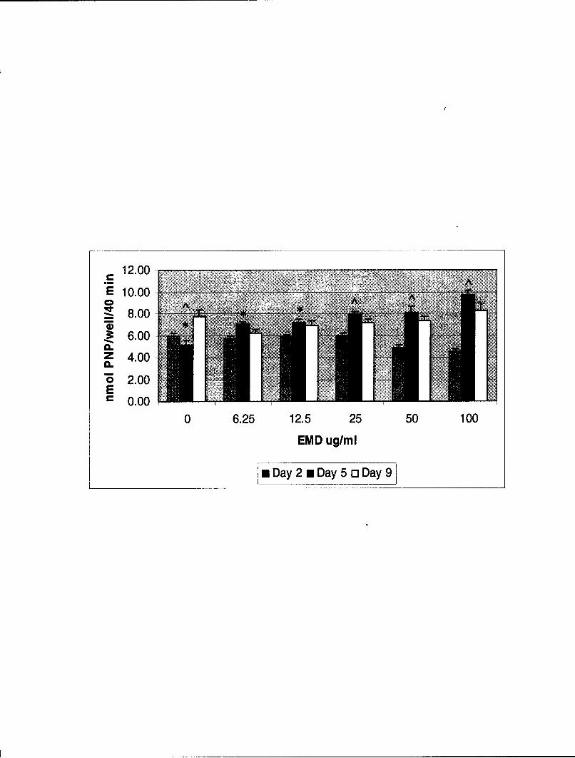
25
Figure 1. Effect of EMD on Alkaline Phosphatase Activity of C2C12 Cells. C2C12 cellswere plated into 96 well plates at 2 x 10⁴ cells / well in growth medium. After 18 hours themedium was removed and replaced with fresh growth medium containing 5% FBS and EMDat concentrations of 0, 6.25, 12.5, 25, 50, and 100jg/ml. At the appropriate time (0, 2, 5, and9 days), the medium was removed and the cell layer assayed for alkaline phosphataseactivity. Note that on day 2 no difference in activity was noted at any of the concentrationstested, and that at 50[tg/ml, and 100gtg/ml, activity appeared to be inhibited, although thedecrease in activity was not statistically significant. At day 5, activity increased withincreasing EMD concentrations, the increase being statistically significant at eachconcentration of EMD tested when compared to unstimulated control cells. At day 9, therewas again no increase in alkaline phosphatase activity at any of the EMD concentrationstested compared to the unstimulated control. Alkaline phosphatase activity is expressed asthe amount of p-nitrophenol (PNP) formed/well/40minutes. The data are the mean ± s.e.m.(n = 8). *p < 0.05, p < 0.001.
12.00-
_•8.00
6.00-z 4.00-
"0 2.00E" 0.00
0 6.25 12.5 25 50 100
EMD ug/rl
mDay 2 m Day 5 o Day 9

26
1). When amelogenin was applied to C2C12 cells, once again, as with EMD at day 2,
differentiation was not evident and even appeared to be inhibited at all concentrations. The
decreases in activity at 50 and IOOjig/ml were significant at p < 0.001 (See Fig. 2). At days 5
and 9, there were no significant increases in alkaline phosphatase activity at any of the
amelogenin concentrations tested, indicating that differentiation did not occur in response to
stimulation by this factor (See Fig. 2).
2. Proliferation Activity
The effect of EMD and amelogenin on proliferation of C2C12 cells was examined in
5% serum containing 0, 12.5gtg/ml, 25jtg/ml, 50tig/ml, and 100gig/ml of the factors. Culture
medium containing 10% serum and no factor served as a positive control. EMD, when
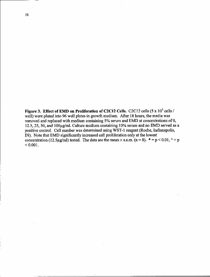
applied to C2C12 cells, significantly increased cell proliferation (p < 0.01) only at the lowest
concentration (12.5gig/ml) tested (See Fig. 3). By contrast, amelogenin had no effect on
C2C12 proliferation at any of the concentrations tested. (See Fig. 4).
The effect of EMD and amelogenin on proliferation of HMVEC cells was examined
both in 5% and 2% serum containing 0, 12.5lig/ml, 25jtg/ml, 50[ig/ml, and 100Ig/ml of the
factors. In the absence of EMD or amelogenin, HMVEC cell proliferation was significantly
greater (p< 0.001) in medium containing 5% serum compared to medium containing 2%
serum. When stimulated with EMD in medium containing 5% serum, HMVEC cell
proliferation was not increased at any of the concentrations tested compared to the 5% serum
control (See Fig. 5). By contrast, when stimulated with amelogenin in 5% serum containing
medium, HMVEC cell proliferation was significantly increased by 6.25 .g/ml amelogenin
compared to the 5% serum control. Higher concentrations of amelogenin did not stimulate a

27
Figure 2. Effect of Amelogenin on Alkaline Phosphatase Activity of C2C12 Cells.C2C12 cells were plated into 96 well plates at 2 x 10⁴ cells / well in growth medium. After18 hours the medium was removed and replaced with fresh growth medium containing 5%FBS and amelogenin at concentrations of 0, 6.25, 12.5, 25, 50, and 100gg/ml. At theappropriate time (0, 2, 5, and 9 days), the medium was removed and the cell layer assayed foralkaline phosphatase activity. Note that on day 2, activity was not increased at any of theconcentrations tested, and even appeared to be inhibited at all concentrations, the decreasesin activity at 50 and 100ýtg/ml being significant. At days 5 and 9, there were no significantincreases in alkaline phosphatase activity at any of the amelogenin concentrations tested.Alkaline phosphatase activity is expressed as the amount of p-nitrophenol (PNP)formed/well/40minutes. The data are the mean ± s.e.m. (n = 8). * = p < 0.001.

8.00€7.00
6.00
4.00-
mz 3.00-_2.000 1 .0 0 -E
"0 000
0 6.25 12.5 25 50 100
Amelogenin ug/ml
Loa u3y Da~y9Iy Dy5ý
28
Figure 3. Effect of EMD on Proliferation of C2C12 Cells. C2C12 cells (5 x 10³ cells /well) were plated into 96 well plates in growth medium. After 18 hours, the media wasremoved and replaced with medium containing 5% serum and EMD at concentrations of 0,12.5, 25, 50, and 100[tg/ml. Culture medium containing 10% serum and no EMD served as apositive control. Cell number was determined using WST-1 reagent (Roche, Indianapolis,IN). Note that EMD significantly increased cell proliferation only at the lowestconcentration (12.5pgg/ml) tested. The data are the mean ± s.e.m. (n = 8). * = p < 0.01, A = p< 0.001.

30,000
25,000 -
20,000E
S10,0005,000
05% 10% 12.5 25 50 100
serum serum
*EMD ug/ml

29
Figure 4. Effect of Amelogenin on Proliferation of C2C12 Cells. C2C12 cells (5 x 10³
cells / well) were plated into 96 well plates in growth medium. After 18 hours, the mediawas removed and replaced with medium containing 5% serum and amelogenin atconcentrations of 0, 12.5, 25, 50, and 100igg/ml. Culture medium containing 10% serum andno amelogenin served as a positive control. Cell number was determined using WST-1reagent (Roche, Indianapolis, IN). Note that amelogenin had no effect on C2C12proliferation at any of the concentrations tested. The data are the mean ± s.e.m. (n = 8).*=p < 0.00 1.
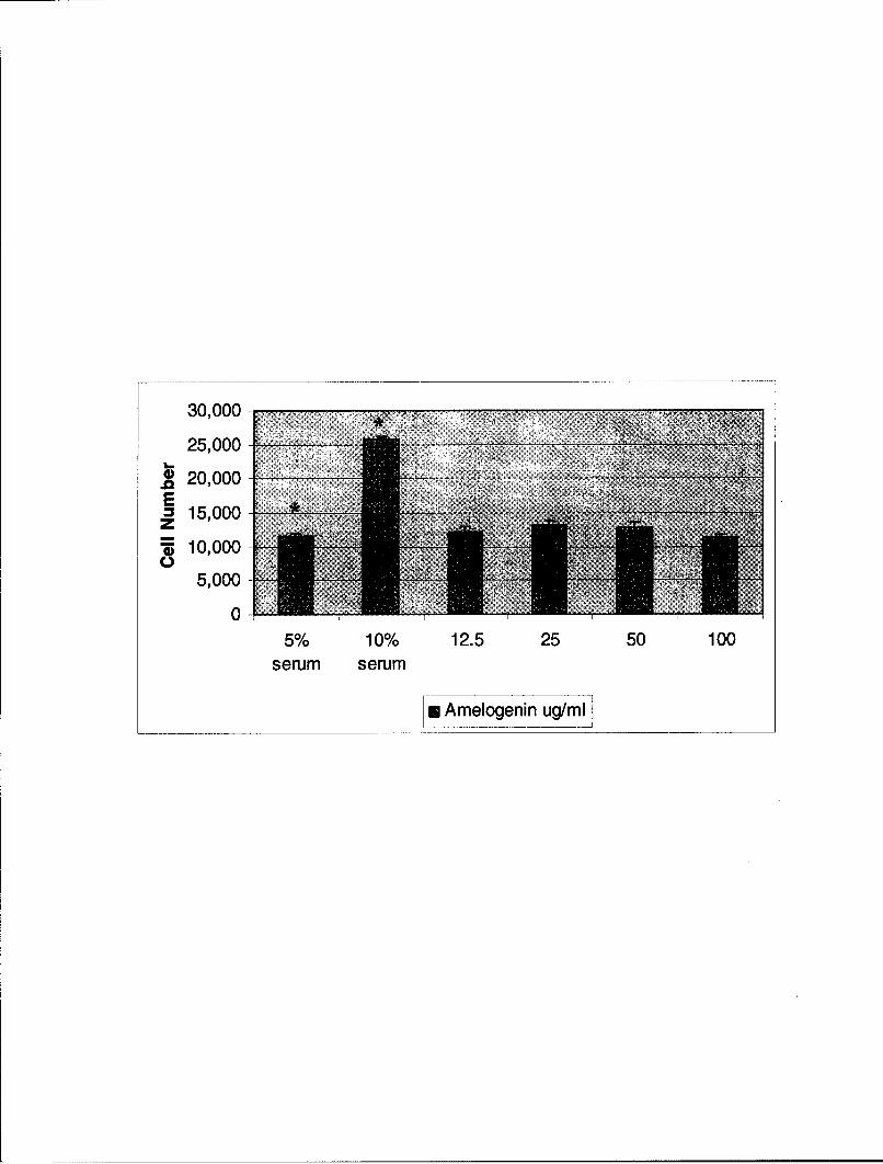
30,000
25,000
S20,00015,000
10,0005,000
05% 10% 12.5 25 50 100
serum serum
mn Amelogenin ug/mi

30
Figure 5. Effect of EMD on HMVEC Cell Proliferation. HMVEC cells (5 x 10³ cells /well) were plated into 96 well plates in growth medium. After 18 hours, the media wasremoved and replaced with medium containing 2% serum and EMD at concentrations of 0,12.5, 25, 50, and I100jtg/ml. Culture medium containing 5% serum and similarconcentrations of EMD served as a control. Cell number was determined using WST-1reagent (Roche, Indianapolis, IN). Note that in the absence of EMD, HMVEC cellproliferation was significantly greater in medium containing 5% serum compared to mediumalone. Note also that when stimulated with EMD in medium containing 5% serum, HMVECcell proliferation was not increased at any of the concentrations tested compared to the 5%serum control. By contrast, EMD in medium containing 2% serum stimulated significantincreases in HMVEC cell proliferation at all concentrations tested compared to the 2% serumcontrol. Note that the greatest increase in EMD stimulated HMVEC cell proliferationoccurred at 25gtg/ml. The data are the mean ± s.e.m. (n = 8). * and A = p < 0.001, + = p <
0.01.

30,000
25,000 -
.• 20,000EA; 15,000
5,000
0-
0 6.25 12.5 25 50 100
EMD uglml
*5% serum o 2% serum

31
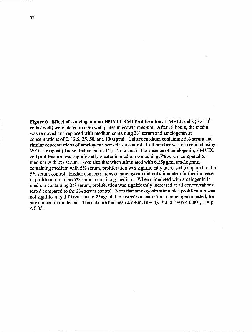
further increase in proliferation in the 5% serum containing medium (See Fig. 6). When
stimulated with either EMD or amelogenin in 2% serum containing medium, HMVEC cell
proliferation was significantly increased at all concentrations tested compared to the 2%
serum control. The greatest increase in EMD stimulated HMVEC cell proliferation occurred
at 25jtg/ml (See Fig. 5). By contrast, amelogenin stimulated proliferation was maximal at
6.25gtg/ml, the lowest concentration of amelogenin tested (See Fig. 6). All concentrations
tested that were greater than 6.25gtg/ml, were not significantly different than the proliferation
stimulated at this dose.
3. Angiogenesis
Angiogenesis, defined as new blood vessel formation arising from the presence of
existing vasculature, was examined in vitro as an indicator of differentiation by
microvascular cells. HMVEC cells, when cultured in EGM2 MV medium undergo rapid
angiogenesis in vitro. This occurs because the medium contains stimulatory growth factors,
particularly VEGF (See Table 1). HMVEC cells cultured in this medium served as the
positive control for the angiogenesis assay. To examine the effects of EMD or amelogenin
on angiogenesis, these materials were dissolved in EBM2 basal medium which is identical to
EGM2 MV medium, except that it does not contain all of the growth factors listed in Table 1.
Angiogenesis was scored by taking photos of the HMVEC cells at each time period
for each media type and then assigning a score of 0-5 based on the maturity of the new
vascular growth. Complete and mature vessel formation, as evidenced by a complete mesh
configuration of all endothelial cells, would receive a score of 5 whereas a separation only of
individual cells would be scored as zero. A complete description of the scoring system can
32
Figure 6. Effect of Amelogenin on HMVEC Cell Proliferation. HMVEC cells (5 x 10³
cells / well) were plated into 96 well plates in growth medium. After 18 hours, the mediawas removed and replaced with medium containing 2% serum and amelogenin atconcentrations of 0, 12.5, 25, 50, and 1OOjig/ml. Culture medium containing 5% serum andsimilar concentrations of amelogenin served as a control. Cell number was determined usingWST-1 reagent (Roche, Indianapolis, IN). Note that in the absence of amelogenin, HMVECcell proliferation was significantly greater in medium containing 5% serum compared tomedium with 2% serum. Note also that when stimulated with 6.25gg/ml amelogenin,containing medium with 5% serum, proliferation was significantly increased compared to the5% serum control. Higher concentrations of amelogenin did not stimulate a further increasein proliferation in the 5% serum containing medium. When stimulated with amelogenin inmedium containing 2% serum, proliferation was significantly increased at all concentrationstested compared to the 2% serum control. Note that amelogenin stimulated proliferation wasnot significantly different than 6.25gg/ml, the lowest concentration of amelogenin tested, forany concentration tested. The data are the mean ± s.e.m. (n = 8). * and A = p < 0.001, + = p< 0.05.

25,000
20,000
"E 15,000--
z- 10,000'di
5,000
00 6.25 12.5 25 50 100
Amelogenin ug/ml
*Lo5% serum u2% serum

33
Table 1. The Media Contents of HMVEC Growth Media. Endothelial cell growthmedium, EGM-2MV is prepared by adding the supplements listed in the table to endothelialcell basal medium 2 (EBM2). Both the basal medium and the supplement kit are obtainedfrom Cambrex. EGM-2MV is the recommended medium for maintenance of the HMVECcells supplied by the company.

GROWTH FACTOR SUPPLEMENT KIT
Gentamicin Sulfate Amphotericin-B
Ascorbic Acid
Long R Insulin-Like Growth Factor-I
Hydrocortisone
Human Fibroblast Growth Factor-B
Human Recombinant Vascular Endothelial Growth Factor
Human Recombinant Epidermal Growth Factor
Fetal Bovine Serum

34
be seen in table 2 and a sample, representative of each of the angiogenesis scores can be seen
in figure 7.
At one hour following stimulation by EMD, there was a significant increase ( p <
0.05) in the angiogenesis score to 0.8 ± 0.2 (mean ± s.e.m.), indicating that the HMVEC cells
had begun to migrate and align themselves. This stimulation by EMD was similar to the
positive control. By contrast, the effect of amelogenin on angiogenesis at this time was not
significantly different from the negative control medium that did not contain amelogenin
(See Fig. 8). At four hours following stimulation by EMD, there was a significant increase
(p < 0.05) in the angiogenesis score to 2.5 ± 0.3 (mean ± s.e.m.), compared to the negative
control (0-f), indicating that capillary tubes were now visible. Although it was not
statistically significant, this stimulation by EMD was greater than the positive control (0+f).
The effect of amelogenin on angiogenesis at this time was not significantly different from
either the positive (0+f) or negative (0-f) control medium (See Fig. 9).
4. SDS Page Gel of EMD and Amelogenin
Before performing the column separation of EMD, an SDS-Page gel with standard
molecular weight markers was run in order to compare the protein components of the EMD
mixture with the purified amelogenin (See Fig. 10). Several things are apparent. EMD
contains a mixture of proteins, the majority of which are relatively low molecular weight.
The major EMD proteins have a molecular weight of less than 30 kDa, with the exception of
one prominent band in the area of 52.2 kDa. By contrast, the major band of the amelogenin
preparation occurs at a molecular weight of 28.9 kDa. Although there are a few weak bands

35
Table 2. In Vitro Angiogenesis Assay Scoring System. The progression of angiogenesis invitro is scored using the criteria displayed in the table. The scoring system was developed byChemicon International, Inc. (Temecula, California).
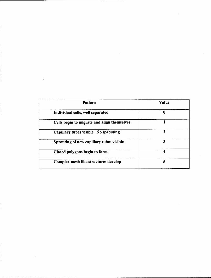
Pattern Value
Individual cells, well separated 0
Cells begin to migrate and align themselves 1
Capillary tubes visible. No sprouting 2
Sprouting of new capillary tubes visible 3
Closed polygons begin to form. 4
Complex mesh like structures develop 5

36
Figure 7. Representative Samples of Angiogenic Scores.


37
Figure 8. Effect of EMD and Amelogenin on HMVEC Cell Angiogenesis after OneHour. To examine the effects of EMD or amelogenin on angiogenesis, these materials weredissolved in EBM2 basal medium (0 - f) and applied to 20,000 HMVEC cells previouslyplated in the BD Biocoat® 96 well angiogenesis system. Digital images obtained usingbrightfield microscopy at IOX were scored blindly for the extent of the angiogenesis. Notethat at one hour following stimulation by EMD (EMD - f), there was a significant increase inthe angiogenesis score to 0.8 + 0.2 indicating that the HMVEC cells had begun to migrateand align themselves. This stimulation by EMD was similar to the positive control (0 + f),which contained endothelial cell growth factors. By contrast, the effect of amelogenin(AMEL - f) on angiogenesis at this time was not significantly different from the negativecontrol medium that did not contain amelogenin. Data are expressed as the mean ± s.e.m.(n = 5). * and A = p < 0.05.

5.00
S4.00o
0 S3.00-
2.00
<1.00-r
0.00
0 + f 0- f EMD - f AMEL- f

38
Figure 9. Effect of EMD and Amelogenin on HMVEC Cell Angiogenesis after FourHours. To examine the effects of EMD or amelogenin on angiogenesis, these materials weredissolved in EBM2 basal medium (0 - f) and applied to 20,000 HMVEC cells previouslyplated in the BD Biocoat® 96 well angiogenesis system. Digital images obtained usingbrightfield microscopy at IOOX were scored blindly for the extent of the angiogenesis. Notethat at four hours following stimulation by EMD (EMD - f), there was a significant increasein the angiogenesis score to 2.5 ± 0.3 indicating that capillary tubes were now visible.Although it was not statistically significant, this stimulation by EMD was greater than thepositive control (0 + f). By contrast, the effect of amelogenin (AMEL - f) on angiogenesis atthis time was not significantly different from either the positive (0+f) or negative (0-f)control media. Data are expressed as the mean + s.e.m. (n = 5). * = p < 0.05.

5
w3
0 f 0
0+f 0 -f EMD -f AMEL -f

39
Figure 10. SDS-PAGE of Amelogenin and Enamel Matrix Derivative. EMD andamelogenin were resolved on 15% polyacrylamide gels using the SDS-PAGE proceduredescribed by Laemmli (1970). Aliquots of 5 (lanes 3, 5) or 2.5gtg (lanes 2, 4) of EMD oramelogenin were applied to the gel. Standard molecular weight markers were run in lanes 1and 6. Resolved bands were stained with Coomassie Brilliant Blue R-250 and photographed.Note that EMD contains a mixture of proteins, the majority of which are relatively lowmolecular weight, less than 30 kDa, with the exception of one prominent band in the area of52.2 kDa. By contrast, the major band of the amelogenin preparation occurs at a molecularweight of 28.9 kDa. Although there are a few weak bands present at higher molecularweights greater than 50 kDa, there are no protein bands present at molecular weights below28.9 kDa.

S AMEL EMD S
52,200 52,20035,700 35,70028,900 28•i:• ,90
20,800 20,800
6,800
1 2 3 4 5 6
40
present at higher molecular weights greater than 50 kDa, there are no protein bands present at
molecular weights below 28.9 kDa (See Fig. 10). As a result of this data, a Sephadex G-100
column was employed to fractionate the EMD protein mixture. This column separates
proteins that are in the range of 10 to 100 kDa from one another.
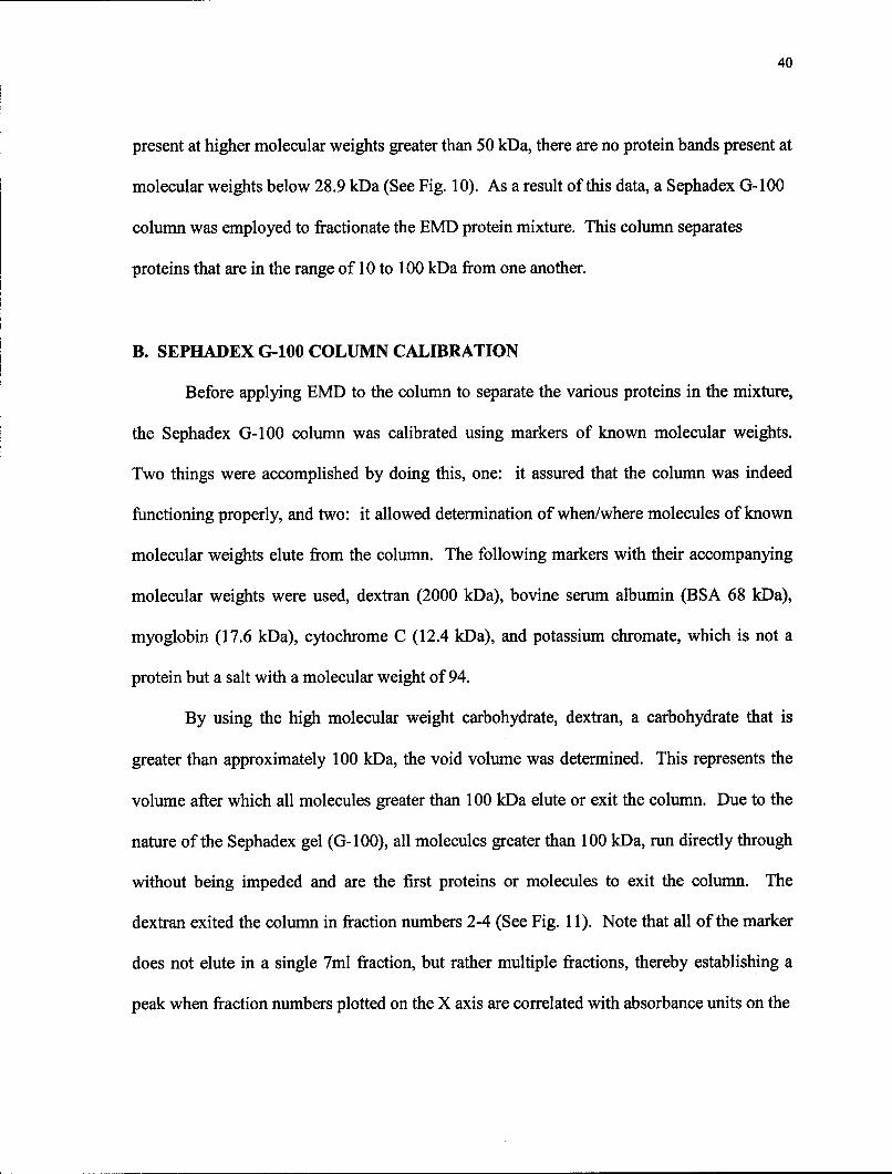
B. SEPHADEX G-100 COLUMN CALIBRATION
Before applying EMD to the column to separate the various proteins in the mixture,
the Sephadex G-100 column was calibrated using markers of known molecular weights.
Two things were accomplished by doing this, one: it assured that the column was indeed
functioning properly, and two: it allowed determination of when/where molecules of known
molecular weights elute from the column. The following markers with their accompanying
molecular weights were used, dextran (2000 kDa), bovine serum albumin (BSA 68 kDa),
myoglobin (17.6 kDa), cytochrome C (12.4 kDa), and potassium chromate, which is not a
protein but a salt with a molecular weight of 94.
By using the high molecular weight carbohydrate, dextran, a carbohydrate that is
greater than approximately 100 kDa, the void volume was determined. This represents the
volume after which all molecules greater than 100 kDa elute or exit the column. Due to the
nature of the Sephadex gel (G-100), all molecules greater than 100 kDa, run directly through
without being impeded and are the first proteins or molecules to exit the column. The
dextran exited the column in fraction numbers 2-4 (See Fig. 11). Note that all of the marker
does not elute in a single 7ml fraction, but rather multiple fractions, thereby establishing a
peak when fraction numbers plotted on the X axis are correlated with absorbance units on the

41
Figure 11. Sephadex G-100 Column Calibration Molecular Weight Markers. EMD wasfractionated using 2.5 X 100cm column of Sephadex G-100 equilibrated with 0.05M sodiumbicarbonate buffer, pH 10.8 (column buffer). The column flow rate was 21ml/hr. Note theelution positions of the markers: blue dextran, bovine serum albumin (BSA), myoglobin,cytochrome C, and potassium dichromate.
0.25
S0.2
a 0.15
-E 0.1 -0
S0.05 ..
0
1 5 9 13 17 21 25 29 33 37 41 45 49 53 57 61 65
Fraction Number
Dextran = 2,000 kDa --- BSA = 68 kDaMyoglobin = 17.6 kDa - Cytochrome C = 12.4 kDaChromate = 0.194 kDa

42
Y axis. It is important to note that the height of the peak is not correlated to molecular
weight, but to the amount of actual protein in the individual fraction.
Potassium chromate was used to determine when the last molecules elute from the
column. This low molecular weight salt is retarded more than any protein as it passes
through the G-100 column. The complete elution of this molecule from the column signals
that everything applied to the column for separation has exited in prior fractions and that the
column is now clear of protein and small molecular weight molecules. Potassium chromate
eluted from the column in fraction numbers 54-64 (See Fig. 11). Proteins with molecular
weights between those of potassium chromate and 100 kDa, the upper limit of the columns
ability to separate protein, elute in different fraction numbers depending on their molecular
weights. Proteins with higher molecular weights elute first while smaller proteins elute in
later fraction numbers. This is apparent from the elution positions of BSA, myoglobin, and
cytochrome C, which elute in fraction numbers, 13-23, 30-36, and 37-44 respectively. Figure
eleven displays the established protein elution profile of the column used in the present
study. By plotting the log of the known molecular weights of BSA, myoglobin, and
cytochrome C, versus the fraction number corresponding to the peak in which they eluted
from the column, a standard curve relating fraction number to molecular weight was
established (See Fig. 12). This calibration curve was used to estimate the molecular weights
of unknown protein(s) that eluted from the column and were associated with distinct
biological activities.

43
Figure 12. Molecular Weight Standard Curve. A molecular weight calibration curve ofthe log of the molecular weight versus the elution fraction number was plotted from theelution positions of the marker proteins, bovine serum albumin (BSA), myoglobin, andcytochrome C.

4.9 6 o S4.8
._m 4.7
• 4.6 30 1Q00.9 4.5 -
S4.40 4.3 Myog1CM 4.2
"4.1 -4 1000
0 5 10 15 20 25 30 35 40 45
Fraction Number
r-.- log molecular weight

44
C. EMD PROTEIN SEPARATION
Thirty milligrams of EMD was fractionated during each column run. The protein
concentration of each fraction was determined spectrophotometricly and plotted as a function
of fraction number in order to establish the protein separation profile for EMD (See Fig. 13).
The proteins in EMD separate into 3 distinct peaks. The first peak represents high molecular
weight proteins associated with the void volume that go directly through the column without
being retarded. The second protein peak elutes between the BSA and myoglobin calibration
proteins, while the 3rd protein peak elutes slightly after the cytochrome c marker. A
comparison of the major protein peaks in the EMD protein separation profile with the
proteins of known molecular weights is displayed in figure 14. The standard curve of the
proteins of known molecular weights (See Fig. 12) was used to approximate the molecular
weights of each of the major protein peaks in the EMD protein separation profile (See Fig.
15). The second protein peak is approximately 50 kDa or 50000 MW while the 3rd peak was
extrapolated to be about 10 kDa, or 10000 MW. The first peak, associated with the void
volume, contains those proteins that are 100 kDa or 100000 MW or greater. It is interesting
to note, that proteins known to be a part of the EMD mixture such as amelogenin, amelin,
and tuftelin, if pure, would fall along the second protein peak of the chromatogram while
enamelin would fall within the void volume peak (See Fig. 15).
D. FRACTIONATED ALKALINE PHOSPHATASE ACTIVITY
The amount of protein in each fraction was determined using the BCA protein assay.
Cell culture media was used to adjust the protein concentration in each fraction to 501tg/ml
prior to determining the biological activity of the fractions. Biological activities examined
45
Figure 13. EMD Protein Separation Profile. EMD was fractionated over a 2.5 X 100cmSephadex G-100 column equilibrated with 0.05M sodium bicarbonate buffer, pH 10.8(column buffer). The column flow rate was 21ml/hr. EMD (30mg) dissolved in 5ml ofcolumn buffer was applied to the column and eluate collected in 20 minute (7ml) fractions.An aliquot of each fraction was assayed for protein content using the BCA protein assay andthe protein elution pattern determined by plotting the protein content of each fraction versusthe fraction number.

0.3
0.25
0.2S0.15-
u 0.1
0.05A
0
1 5 9 13 17 21 25 29 33 37 41 45 49 53 57
Fraction Number
-.- EMD

46
Figure 14. Fractionation of EMD Over Sephadex G-100 Column with MolecularWeight Markers. The figure displays the elution position of the marker proteinssuperimposed over the elution profile of fractionated EMD.

0.3-S0.25-0.-\
00.15 • ""0
*~0.1-
1 5 9 13 17 21 25 29 33 37 41 45 49 53 57 61 65
Fraction Number
EMD -Dextran = 2,000 kDaSBSA = 68 kDa -Myoglobin = 17.6 kDa
-Cytohrnome C = 12.4 kDa- - CXrhrormate = 0.1q4__kDa

47
Figure 15. Molecular Weight of EMD Protein Peaks. The figure displays the elutionpattern of fractionated EMD. The molecular weights of the major peaks are indicated by thearrows at 50 kDa and 10 kDa. Also, displayed are the expected elution positions of proteinsknown to be present in EMD based on their molecular weights.

0.3
0.25S0 ,C, 0.15-
0 0.1
0.05-
1 5 9 13 17 21 25 29 33 37 41 45 49 53 57
Fraction Number
-.- EMD

48
included differentiation, proliferation, and angiogenesis, as well as collagenolytic activity.
The ability of C2C12 cells to differentiate into osteoprogenitor, or bone-forming cells is well
known and is characterized by an increase in the expression of alkaline phosphatase activity.
When the alkaline phosphatase activity of each fraction was determined and
superimposed on the EMD protein separation profile (See Fig. 16), it was evident that the
strongest alkaline phosphatase activity occurred in fractions 12-17, which does not
correspond to any of the major protein peaks found in EMD. In fact, there appears to be little
activity in the fractions containing the major EMD protein peaks. When the fractions were
preincubated with 100 ng/ml of noggin, a decoy receptor known to bind to BMP 2, 4, and 7,
thus blocking their activity, the alkaline phosphatase activity was decreased, especially in the
area where the highest activity occurred prior to noggin treatment (See Fig. 17). These data
are consistent with the suggestion that the osteoinductive activity associated with EMD is not
related to amelogenin, but more likely with a member of the BMP family of proteins, and
that only a small amount of protein accounts for this activity.
E. FRACTIONATED PROLIFERATION ACTIVITY
The proliferation of C2C12 and human microvascular endothelial cells (HMVEC)
was determined and superimposed on the protein separation profile for EMD. Unlike
alkaline phosphatase activity, the proliferative activity for C2C12 cells (See Fig. 18) and
HMVEC cells (See Fig. 19) mirrored the EMD protein separation profile, with the greatest
proliferative activity occurring in the fractions coincident with the major protein peaks. This
data is consistent with the suggestion that the proliferative activity for both the
osteoprogenitor and human microvascular endothelial cells is associated with the major

49
Figure 16. Alkaline Phosphatase Activity of EMD Fractions on C2C12 Cells. Theprotein concentration of each fraction was adjusted to 50gtg/ml using tissue culture medium.Some fractions did not contain detectable protein and were not tested. Note that when thealkaline phosphatase activity of each fraction was determined and superimposed on the EMDprotein separation profile, the strongest activity occurred in fractions 12-17, which does notcorrespond to any of the major EMD protein peaks.

14.00 40.00
12.00 35.00
30.00o10.0025.00
S8.0020.00
o .0 t 15.00CL 4.00 10.00
2.00 5.00
%Ln~lv0.00
0 10 20 30 40 50
Fraction Number
* EMD E AlkPhos

50
Figure 17. Noggin Inhibition of Alkaline Phosphatase Activity of EMD Fractions onC2C12 Cells. The protein concentration of each fraction was adjusted to 50jg/ml usingtissue culture medium. Some fractions did not contain detectable protein and were not tested.Prior to application on the cells, the fractions were preincubated for 1 hour with 1 OOng/mlnoggin, a decoy receptor known to bind to BMP 2, 4, and 7, and block their activity. Notethat alkaline phosphatase activity was decreased, especially in the area where the highestactivity occurred prior to noggin treatment. These data are consistent with the suggestionthat the osteoinductive activity associated with EMD is not related to amelogenin, but morelikely with a member of the BMP family of proteins.

14.00 40.00
12.00 J 35.00
S10.00 - 30.000 .25.00
8.00-
000
" 4.00 - "10.00
2.00 - 5.00
0.00 - 0.00
0 10 20 30 40 50
Fraction Number
.--•- EMD -m Alk Phos +_Noggin]
51
Figure 18. Proliferation Activity of EMD Fractions on C2C12 Cells. Proliferation ofC2C12 cells was determined and superimposed on the protein separation profile for EMD.Note that the proliferative activity for C2C 12 cells mirrored the EMD protein separationprofile, with the greatest proliferative activity occurring in the fractions coincident with themajor protein peaks. The protein concentration of each fraction was adjusted to 50gtg/mlusing tissue culture medium prior to assay. Some fractions did not contain detectable proteinand were not tested.

14.00 2.0012.0- 1.8012.00 - -- _ __--____ ____ 1.80
1.60S10.00 - 1.40 E01.20
8.00 1.20 -
Z 60- 0.8
0.40°•.0-" 0.20
0.00, 0.00
0 10 20 30 40 50
Fraction Number
I-. EMD ---- C2C12 Cells
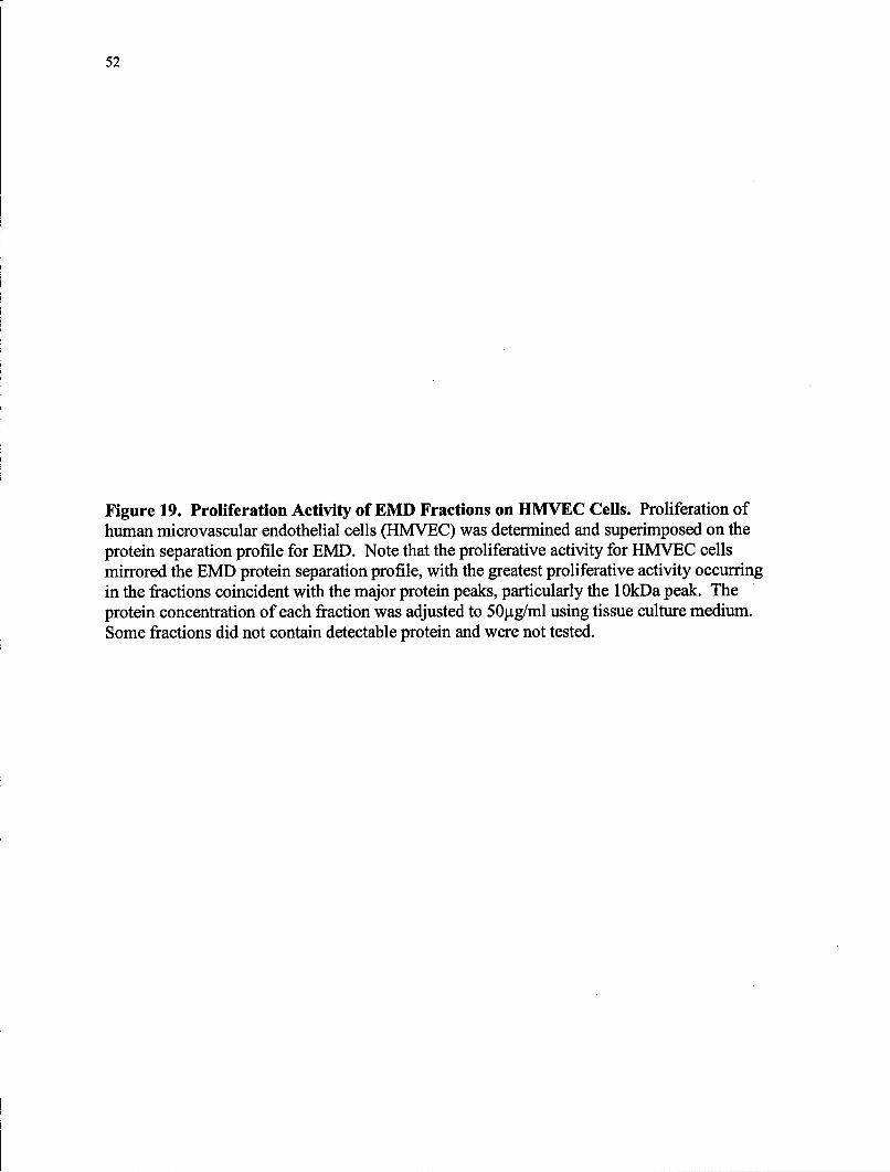
52
Figure 19. Proliferation Activity of EMD Fractions on HMVEC Cells. Proliferation ofhuman microvascular endothelial cells (HMVEC) was determined and superimposed on theprotein separation profile for EMD. Note that the proliferative activity for HMVEC cellsmirrored the EMD protein separation profile, with the greatest proliferative activity occurringin the fractions coincident with the major protein peaks, particularly the 1OkDa peak. Theprotein concentration of each fraction was adjusted to 50tg/ml using tissue culture medium.Some fractions did not contain detectable protein and were not tested.

14.00 0.60
12.00 Sio.oo 0.5010.00 0.50
O E0)8.00 -- l 0______
0.40 u"6.00
0 0. 4.00 ML 0.30
2.00-
0.00 -0.20
0 10 20 30 40 50
Fraction Number
-.- EMD - HMVEC Cells
53
protein components of the EMD mixture, but is distinct from the proteins associated with the
induction of alkaline phosphatase activity.
F. FRACTIONATED ANGIOGENIC ACTIVITY
EMD was fractionated and applied to the HMVEC cells at a concentration of
25gjg/ml, and angiogenesis scored at 1 and 4 hours. When the scores were plotted against
fraction number and superimposed on the EMD protein separation profile, angiogenic
activity at one hour was present in the same fractions as the 2nd and 3rd EMD protein peaks
(See Fig. 20). The angiogenesis score of 1.0 for each of the peaks may initially appear
insignificant, but when compared to the angiogenic activity of the unfractionated EMD at 1
hour (angiogenesis score of 0.8), it is evident that there is a slight increase in angiogenic
activity in the fractions. It is also interesting to note that at one hour, there is no activity
associated with the void volume peak. At 4 hours an angiogenesis score of 3.0 corresponded
to the void volume peak (See Fig. 21). Furthermore, even though the angiogenic activity
peaks at 4 hours, once again corresponding to the 2 nd and 3rd EMD peaks, there were several
important differences. The angiogenic activity corresponding to the second peak was
increased significantly (angiogenesis score of 4.0), signifying almost complete vascular
maturity and organization, while activity associated with the 3rd protein peak, although
present, remained the same as at 1 hour (angiogenesis score of one). These data are
consistent with the interpretation that angiogenesis, like proliferation, is associated with the
major protein peaks found in EMD, particularly peak 2 that likely contains amelogenin. The
fact that purified amelogenin exhibited little if any angiogenic activity (See Figures 8 and 9),

54
Figure 20. Angiogenic Activity of EMD Fractions on HMVEC Cells at 1 Hour. EMDwas fractionated, applied to the HMVEC cells at a concentration of 25gtg/ml, angiogenesisscored and plotted against fraction number, and superimposed on the EMD protein separationprofile. Note that angiogenic activity at one hour was coincident with the 2n, and 3rd EMDprotein peaks. Note also that at one hour, there is no activity associated with the void volumepeak.

8.00
7.00 4.56.00 Iw-
S5.00S4.00 2.5 "
S3.0 0=o. 2.001.5 0_
2 2.000 0.50.00
-1.00 9 -0.5
fraction number
-.- EMD = HMVEC

55
Figure 21. Angiogenic Activity of EMD Fractions on HMVEC Cells at 4 Hours. EMDwas fractionated, applied to the HMVEC cells at a concentration of 25gtg/ml, angiogenesisscored and plotted against fraction number, and superimposed on the EMD protein separationprofile. Note that angiogenic activity at four hours was coincident with the 2 nd and 3"d EMDprotein peaks. Note also that an angiogenesis score of 3.0 is associated with the void volumepeak. Note that the angiogenic activity corresponding to the second peak was increased overthat present at one hour, while activity associated with the 3P protein peak, although present,remained the same as at 1 hour (See Fig. 20).

8.007.00 -- 4.5
6.00O5.00
S 4.00 2.5"1." 3.001.
3.00
2 .00 ,F"S0.5
0.00
-1.00 b -0.5
fraction number
L--EMD - HMVEC _

56
suggests that the angiogenic activity present in this region may be associated with proteins
other than amelogenin.
G. FRACTIONATED COLLAGENOLYTIC ACTIVITY
To our knowledge, the association of enzymatic or collagenolytic activity with EMD
has not been previously reported in the literature. Therefore, the ability of the EMD fractions
to degrade denatured type I collagen was evaluated by utilizing polyacrylamide gel
zymograms. In each of the gels, lane 1 was a standard preparation derived from the rat
osteosarcoma cell line ROS 17/2.8, and used to verify that the gels functioned properly.
Lane two contained unfractionated EMD. After electrophoreses, collagenolytic activity was
evident in two areas, fractions 3-8 and 17-21 (See Fig. 22a). Thus, the collagenolytic activity
of EMD was associated with proteins in peak 1, as well as with proteins eluting in the same
molecular weight region as BSA (See Fig. 22b), prior to the elution of amelogenin. There
was no further collagenolytic activity apparent in any of the later eluting fractions. These
data are the first to demonstrate that EMD contains enzymes that are capable of degrading
type I collagen, and suggest that a more detailed analysis of the protein degrading activity of
EMD is warranted.
H. SDS PAGE GELS OF FRACTIONATED EMD
Finally, for comparison purposes with unfractionated EMD, each of the fractions was
run on an SDS page gel with molecular weight markers. On the gel, there were no protein
bands evident in any of the fractions from 0-18. In fraction nineteen, a very strong band

57
Figure 22. Zymograms of EMD Column Fractions. EMD fractions were resolved on15% polyacrylamide gels containing denatured type I collagen. Lane 1 was a standard fromthe rat osteosarcoma cell line (ROS) used to verify that the gels functioned properly, whilelane two contained unfractionated EMD. The resulting gels were stained with CoomassieBrilliant Blue R-250 and photographed. Note that areas in which the collagen was degradedappear clear (unstained). Note that collagenolytic activity was evident in two areas, fractions3-8 and 17-21(Fig. 22a). Thus, the collagenolytic activity of EMD was associated withproteins in peak 1 (Fig. 22b), as well as with proteins eluting at the same molecular weightregion as BSA (68 kDa), prior to the elution position of amelogenin. There was no furthercollagenolytic activity apparent in any of the later eluting fractions.

22a
Fractions I-11 - Fractions 12- 22
22b
14.00_12.00
10.00A8.00
S6.00-
4.00 -2.00-
0.00
0 10 20 30 40 50
Fraction Number
58
became evident between the 20.8 kDa and 28.9 kDa markers, consistent with where we
would expect amelogenin to appear (See Fig. 23a). This band remained prominent through
fraction 29 when it began to disappear. In fraction 24 an additional group of light bands
began appearing and became more prominent with each following fraction number in the
molecular weight range of 6.8-20.8 kDa (See Fig. 23a). The appearance and increase of
these protein bands corresponded with the decrease and disappearance of the higher
molecular weight band (i.e. amelogenin). From fraction 32-45 there were only 2 light bands
evident around the 6.8 kDa marker. These bands appeared to merge into one band in the
latter fractions at slightly less than the 6.8 kDa marker (See Fig. 23b). The protein pattern
evident in the fractions on the SDS page gel corresponds extremely well to the fractions from
the column separation in which the second and third protein peaks appear (See Fig. 23c). It
is interesting to note that whereas prefractionated EMD (See Fig. 10) contained a higher
molecular weight band, the fractionated EMD contained no such band (See Fig. 23a and
23b).
One potential reason for this to occur is that no protein was detected from the BCA
assay on fractions 5-18 in the column used to collect fractions for analysis on the SDS page
gels (See Fig. 23c). Therefore, samples from these same fractions (5-18) were not included
on the SDS gels (See Fig. 23a). It is possible however, that proteins in this molecular weight
region are present in small amounts, but only detectable with a more sensitive assay. The
proteins in this molecular weight region would be greater than a molecular weight of 52,200
(See Fig. 10), which would place these proteins between the first and 2 nd major protein peaks
on the EMD protein separation profile (See Fig. 15), and in the region of alkaline

59
phosphatase activity. Interestingly enough, this would also be the molecular weight region
where BMP's would be expected to fall.
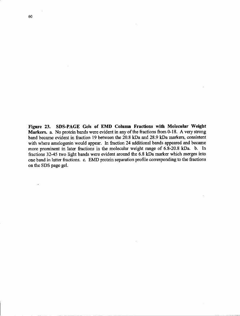
60
Figure 23. SDS-PAGE Gels of EMD Column Fractions with Molecular WeightMarkers. a. No protein bands were evident in any of the fractions from 0-18. A very strongband became evident in fraction 19 between the 20.8 kDa and 28.9 kDa markers, consistentwith where amelogenin would appear. In fraction 24 additional bands appeared and becamemore prominent in later fractions in the molecular weight range of 6.8-20.8 kDa. b. Infractions 32-45 two light bands were evident around the 6.8 kDa marker which merges intoone band in latter fractions. c. EMD protein separation profile corresponding to the fractionson the SDS page gel.

S 3 4 19 20 21 22 23 24 MW S 25 26 27 28 29 30 31 32
35,700
28,900 %
-~020,800 *k
6,800
a.
S 33 34 35, 36 37 38 39 40 MW S 41 42 43 44 45 46
52,20035,700
___ 28,900
20,8004
6,800
6.00
5.004.00
0.00
-1.00
fraction numb~er
C. L-7!- EMD]

V. DISCUSSION AND SUMMARY
With the approval of EMD for clinical use in 1997, a new era began in periodontics,
one in which the goal of achieving more predictable and complete regeneration has come one
step closer to being realized. The rationale for using enamel matrix proteins is that
regeneration could be accomplished by mimicking what occurs during normal tooth
development. Unfortunately, the mechanism by which this occurs is still unknown, thus this
is quest of much of today's research. Since amelogenin is the main protein in EMD, making
up 90% of the composition, it is widely believed that the effects of EMD must be due to
amelogenin, but this has not been proven. The goal of this study, to fractionate enamel
matrix derivative and determine the extent to which the known activities of proliferation and
differentiation, are the result of a single protein component in the mixture (i.e. amelogenin),
has been accomplished. In the process, this research contributes two new, important, and
exciting pieces of information to the literature. First, the study shows that EMD stimulates
angiogenesis in microvascular endothelial cells, and second, that EMD also contains
proteolytic activity, information that, to our knowledge, has not been previously reported.
In this study, 30mg of commercially available EMD (Emdogain8) was fractionated
using a Sephadex G-100 column, into 45 seven ml fractions containing protein that eluted in
three distinct peaks. This is in agreement with Iwata et al. (2002), who fractionated their
own preparation of enamel matrix proteins obtained from the tooth germs of 6 month old
61

62
pigs and by using the same method for fractionation also found three distinct protein peaks.
The protein separation profile was very similar to that found in the study reported here.
With regard to alkaline phosphatase activity, a marker for cellular differentiation by
osteoblasts and osteoprogenitor cells, this study found that when applying both EMD and the
column fractions to C2C12 cells that differentiation did occur. This is in agreement with
other studies stating that EMD does stimulate cellular differentiation (Schwartz et al., 2000;
Ohyama et al., 2002), but opposed to other findings, in which markers for differentiation
were not found (Hakki et al., 2001; Chano et al., 2003). In this study, when the alkaline
phosphatase activity of the column fractions applied to C2C12 cells was measured and
superimposed on the EMD protein separation profile, it was evident that it did not correspond
to any of the major protein peaks, but rather, the strongest activity fell between the first and
second protein peaks. The same pattern was observed when using ST2 cells as well (data not
shown). This partially agrees with the study reported by Iwata et al. (2002), who found that
the strongest alkaline phosphatase activity of EMD fractions also occurred between the first
and second major protein peaks. However, in contrast to our data, they also found alkaline
phosphatase activity to be associated with the first protein peak or void volume. The
difference could possibly be explained by the difference between the commercial EMD used
in our research compared to their enamel matrix protein formulation. It may also be due to
the fact that the Iwata study used a mouse bone marrow stromal cell line, ST2 as the test cell.
The present study also found that by pre-incubating the column fractions with the decoy
receptor noggin, prior to treating C2C12 cells, that alkaline phosphatase activity was
inhibited. Again, this is consistent with the Iwata et al. (2002) study using ST2 cells. In
another recent study by Takayama et al. (2005) using C2C12 cells, which normally do not

63
express core binding factor al/Runt-related transcription factor-2 (Cbfal/Runx2) unless
stimulated by BMP-2 (Nishimura et al., 2002), found that EMD stimulated Cbfal/Runx2
expression in the cells that could also be inhibited by noggin. These studies, together with
the results of this study, suggest that the ability of EMD to stimulate cellular differentiation is
due to a BMP family member. This is because the activity is both inhibited by noggin (Iwata
et al., 2002), and does not correspond to the molecular weight region of the chromatogram
associated with amelogenin or any of its fragments. It is interesting to note that one recently
hypothesized mechanism of action for EMD is that proposed by Parkar et al. (2004), in
which gene arrays from PDL cells treated with EMD appeared to up regulate genes coding
for growth factors and growth factor receptors while down regulating inflammatory genes.
One of the specific genes that is upregulated, is BMP-4, one of three BMP's known to be
inhibited by noggin. Because alkaline phosphatase activity stimulated by EMD is inhibited
by noggin, this potentially strengthens the argument that the differentiation that occurs is due
to a BMP family member, possibly BMP-4. Parkar's et al., (2004) idea of EMD
upregulating genes goes a long way towards explaining not only many of the observed
effects involving growth factors and/or their receptors (Hoang et al., 2002 ; Lyngstadaas et
al., 2001), but also the marked improvement in wound healing that has been observed by
many when using EMD.
Another of the genes that appears to be upregulated by EMD is vascular endothelial
growth factor precursor (Parkar et al., 2004). With vascularity so essential to wound healing,
it follows that an upregulation of this growth factor may stimulate the necessary cellular
activities leading to improved wound healing. The results of the present study appear to
confirm this finding because when HMVEC cells were stimulated with EMD, an ensuing

64
increase in proliferation was observed, consistent with similar results reported by Yuan et al.
(2003) both in vitro and in a murine model. A similar increase in proliferative activity was
also found to occur in the individual column fractions that corresponded to the major protein
peaks, suggesting that the proliferative activity of EMD may be due to amelogenin. This
pattern of proliferation was not unique to HMVEC cells. It also occurred in the C2C12 cell
line.
Another important event in wound healing besides that of endothelial cell
proliferation is angiogenesis. To our knowledge, the angiogenic activity of EMD has not
been reported previously in the literature, and like proliferation, corresponded to the major
protein peaks, suggestive, once again, of being associated with amelogenin or one of its
fragments. Yuan et al. (2003), although stating that they looked at angiogenesis, really only
reported data on endothelial cell proliferation and chemotaxis, and not angiogenesis. One of
the important factors in the formation of new vessels and granulation tissue is vascular
endothelial growth factor (VEGF) (Howdieshell et al., 2001). In a study by Mirastschijski et
al. (2004), they found that VEGF production by fibroblasts was increased five fold and 5.5-
fold as compared to control-treated fibroblasts at 3 and 6 days. This appeared to not only
increase the formation of granulation tissue, but also made it more resistant to trauma.
Several other significant findings related to healing in this same paper, were that wounds
appeared to be smaller, epithelialization greater, and healing times decreased when EMD was
used compared to controls. If EMD is upregulating precursors to vascular endothelial growth
factors as suggested by Parkar et al. (2004), it would help explain not only the healing results
by Mirastschijski et al. (2004), but it also helps explain the link between the proliferative and

65
angiogenic activities of the HMVEC cells observed in this study, both of which were found
to correspond to the major EMD protein peaks.
Another important part of both wound healing and regeneration is the breakdown of
type I collagen. In this study proteolytic enzyme activity, or more specifically collagenolytic
activity, was found to occur in two regions, in fractions 3-8 and 17-21. The first region was
associated with a high molecular weight region in the void volume of the column, possibly
consistent with the presence of MMP-2, a matrix metalloproteinase falling into the broad
category of gelatinase A, and known to digest amelogenin (Caron et al., 2001; Cotrim et al.,
2002). A wound-healing study by Mirastschjski et al. (2004) gives further credence to the
findings of this study that the collagenolytic activity observed, may be due to MMP-2. They
found increased levels and activation of MMP-2 by both endothelial and fibroblast cells
when treated with EMD. Although endothelial cells are known to express MMP-2 during
wound healing (Mirastschijski et al., 2002), the amount was significantly higher when treated
with EMD, suggesting that either MMP-2 is either present in EMD or up-regulated by it.
The collagenolytic activity in this higher molecular weight region could also be masking
potential alkaline phosphatase or proliferative activities that may be associated with this
protein peak by degrading necessary substrates before being activated. To evaluate the
possibility of this occurring one would need to rerun the alkaline phosphatase and
proliferation assays utilizing an enzyme inhibitor that would allow the activity to be manifest
if present.
The second region of collagenolytic activity was associated with a lower molecular
weight region consistent with the presence of MMP-20, or enamelysin, another matrix
66
metalloproteinase known to digest amelogenin (Wang and Moradian-Oldak, 2002). Further
research is necessary to specifically characterize the newly discovered metalloproteinase
activities and is beyond the scope of this research.
Although individual activities have been separated out and discussed in conjunction
with the protein(s) or peaks potentially responsible for the activity, it is important to realize
that individual activities do not occur in a vacuum exclusive to one another. Rather, the
activities more likely act in concert with each other, and the various cell types and/or
receptors in a dynamic environment to stimulate and/or accelerate the regenerative process.
Further research to identify the specific BMP's, proteins, and/or amelogenin fragments which
are responsible for these activities is necessary before one can hope to understand the
interactions that occur which lead to predictable regeneration.
In conclusion, the results of this research have shown that EMD has the ability to
stimulate multiple activities in more than one cell type, which is extremely important in the
multicellular process of regeneration. The differing and specific biological effects of EMD
were also clearly associated with proteins of differing molecular weights, and, therefore,
unlikely to be associated with a single protein species (Amelogenin =- 23 kDa). The
proliferative and angiogenic activities may be associated with amelogenin and/or its
fragments, while differentiation of osteoprogenitor cells was most likely the result of
BMP(s). Finally, we found that EMD contains proteolytic activity, an activity that has not
been previously reported in the literature and which merits further investigation.
LITERATURE CITED
Araujo M, Lindhe J (1998). GTR treatment of degree III furcation defects followingapplication of enamel matrix proteins: An experimental study in dogs. J Clin Periodontol25:524-530.
Aubin J, Liu F, Malaval L, Gupta A (1995). Osteoblast and chondroblast differentiation.Bone 17:77-83.
Aubin J (1998). Advances in the osteoblast lineage. Biochem Cell Biol 76:899-910.
Becker W, Berg L, Becker B (1979). Untreated periodontal disease: a longitudinal study. JPeriodontol 50:234-244.
Becker W, Urist M, Tucker I, Becker B, Ochsenbein C (1995). Human demineralizedfreeze-dried bone: Inadequate induced bone formation in athymic mice. A preliminaryreport. JPeriodontol 66:822-828.
Blau H (1985). Plasticity of the differentiated state. Science 230: 758-766.
Boyan B, Weesner T, Lohmann C, Andreaccio D, Carnes D, Dean D, Cochran D, Schwartz Z(2000). Porcine fetal enamel matrix derivative enhances bone formation induced bydemineralized freeze dried bone allograft. JPeriodontol 71:1278-1286.
Brown L, Oliver R, L6e H (1988). Periodontal disease in the U.S. in 1981: prevalence,severity, extent and role in tooth mortality. JPeriodontol 59:7-11.
Caron C, Xue J, Sun X, Simmer J, Bartlett J (2001). Gelatinase A (MMP-2) in developingtooth tissues and amelogenin hydrolysis. JDent Research. 80:1660-1664.
Chano L, Tenenbaum H, Lekic P, Sodek J, McCulloch C (2003). Emdogain® regulation ofcellular differentiation in wounded rat periodontium. JPeriodont Res 38: 164-174.
Cortellini P, Tonetti M (2004). Long-term tooth survival following regenerative treatment ofintrabony defects. JPeriodontol 75:672-678.
Cotrim P, Andrade C, Line S, Almeida 0, Coletta R (2002). Expression and activity ofmatrix metalloproteinase-2 (mmp-2) in the development of the rat first molar tooth germ.Braz Dent J 13:97-102.
Dragoo M, Sullivan H (1973). A clinical and histological evaluation of autogenous iliacbone grafts in human: Part I. Wound healing 2 to 8 months. JPeriodontol 44:614-625.
67

68
Fong C, Hammarstr6m L (2000). Expression of amelin and amelogenin in epithelial rootsheath remnants of fully formed rat molars. Oral Surg Oral Med Oral Pathol 90:218-223.
Francetti L, Del Fabbro M, Basso M, Testori T, Weinstein R (2004). Enamel matrix proteinsin the treatment of intra-bony defects: a prospective 24-month clinical trial. J ClinPeriodontol 31:52-59.
Froum S, Weinberg M, Rosenberg E, Tamow D (2001). A Comparative Study UtilizingOpen Flap Debridement With and Without Enamel Matrix Derivative in the Treatment ofPeriodontal Intrabony Defects. A 12-month Re-Entry Study. JPeriodontol 72:25-34.
Fukae M (1999). Molecular components of Emdogain [Japanese]. Dent Outlook 94:184-192.
Gargiulo A, Wentz F, Orban B (1961). Dimensions and relations of the dentogingivaljunction in humans. JPeriodontol 32:261-267.
Gestrelius S, Andersson C, Lidstrom D, Hammarstr6m L, Somerman M (1997). In vitrostudies on periodontal ligament cells and enamel matrix derivative. J Clin Periodontol24:685-692.
Gestrelius S, Lyngstadaas S, Hammarstr6m L (2000). Emdogain-periodontal regenerationbased on biomimicry. Clin Oral Invest 4:120-125.
Giannobile W, Martha J, Somerman M (2003). Growth and Amelogenin-Like Factors inPeriodontal Wound Healing. A Systematic Review. Annals of Periodontology 8:193-204.
Glickman I (1956). Results obtained with unembellished gingivectomy technique in aclinical study in humans. JPeriodontol 27:247-255.
Goldman H (1951). Gingivectomy. Oral Surg Oral Med Oral Pathol 4:1136-1157.
Gottlow J, Nyman S, Karring T, Wennstrom J (1986). New attachment formation in thehuman periodontium by guided tissue regeneration. Case reports. J Clin Periodontol13:604-613.
Greenstein G, Lamster 1 (2001). Changing periodontal paradigms: therapeutic implications.Int J Periodontics Restorative Dent 20:336-357.
Gurinsky B, Mills M, Mellonig J (2004). Clinical evaluation of demineralized freeze-driedbone allograft and enamel matrix derivative versus enamel matrix derivative alone for thetreatment of periodontal osseous defects in humans. JPeriodontol 75:1309-1318.
Haase H, Bartold P (2001). Enamel Matrix Derivative Induces Matrix Synthesis by CulturedHuman Periodontal Fibroblast Cells. JPeriodontol 72:341-348.
Hagenaars S, Louwerse P, Timmerman M, Van der Velden U, Vander der Weijden G (2004).Soft-tissue wound healing following periodontal surgery and Emdogain® application. J ClinPeriodontol 31:850-856.
Hakki S, Berry J, Somerman M (2001). The Effect of Enamel Matrix Protein Derivative onFollicle Cells in Vitro. JPeriodontol 72:679-687.
69
Hammarstrim L (1997). Enamel matrix, cementum development and regeneration. J ClinPeriodontol 24:658-668.
Hammarstr6m L, Heijl L, Gestrelius S. (1997). Periodontal regeneration in a buccaldehiscence model in monkeys after application of enamel matrix proteins. J ClinPeriodontol 24:669-677.
Heijl L (1997). Periodontal regeneration with enamel matrix derivative in one humanexperimental defect. J Clin Periodontol 24:693-696.
Heijl L, Heden G, Svdrdstrim G, (Ostgren A (1997). Enamel matrix derivative (Emdogain®)in the treatment of intrabony periodontal defects. J Clin Periodontol 24:705-714.
Hoang A, Oates T, Cochran D (2000). In vitro wound healing responses to enamel matrixderivative. JPeriodonto1 71:1270-1277.
Hoang A, Klebe R, Ryu 0, Steffensen B, Simmer J, Cochran D (2002). Amelogenin is aCell Adhesion Protein. JDent Res 81:497-500.
Howdieshell T, Callaway D, Webb W, Gaines M, Procter C, Sathyanarayana P, Brock T,McNeil P (2001). Antibody neutralization of vascular endothelial growth factor inhibitswound granulation tissue formation. JSurgRes 96:173-182.
Hu JC-C, Sun X, Shang C, Simmer J (2001). A comparison of enamelin and amelogeninexpression in developing mouse molars. Eur J Oral Sci 109:125-132.
Hujoel P, Leroux B, Selipsky H, White B (2000). Non-surgical periodontal therapy andtooth loss. A cohort study. JPeriodonto1 75:736-742.
Hung H, Douglass C (2002). Meta-analysis of the effect of scaling and root planning,surgical treatment and antibiotic therapies on periodontal probing depth and attachment loss.J Clin Periodonto1 29:975-986.
Iwata T, Morotome Y, Tanabe T, Fukae M, Ishikawa I, Oida S (2002). Noggin BlocksOsteoinductive Activity of Porcine Enamel Extracts. JDent Res 82:387-391.
Katagiri T (1994). Bone morphogenetic protein-2 converts the differentiation pathway ofC2C12 myoblasts into the osteoblast lineage [published erratum appears in J Cell Biol 1995Feb; 128(4):following 713]. J. Cell Biol 127:1755-1766.
Kawase T, Okuda K, Yoshie H, Bums D (2000). Cytostatic action of enamel matrixderivative (EMDOGAIN) on human oral squamous cell carcinoma-derived SCC25 epithelialcells. J Periodontol Res 35:291-300.
Kawase T, Okuda K, Momose M, Kato Y, Yoshie H, Bums D (2001). Enamel Matrixderivative (Emdogain®) rapidly stimulates phosphorylation of the MAP kinase family andnuclear accumulation of smad2 in both oral epithelial and fibroblastic human cells. JPeriodontol Res 36:367-376.
Kornman K, Page R, Tonetti M (1997). The host response to the microbial challenge inperiodontitis; assembling the players. Periodontology 2000 14:33-35.

70
Laemmli U (1970). Cleavage of structural proteins during the assembly of the head ofbacteriophage T-4. Nature 227: 680-688.
Lindhe J, Nyman S, Karring T (1982). Scaling and root planning in shallow pockets. J ClinPeriodontol 9:415-418.
LUe H, Theilade E, Jensen S (1965). Experimental gingivitis in man. JPeriodontol 36:177-187.
LUe H, Anerud A, Boysen H, Morrison E (1986). Natural history of periodontal disease inman. Rapid, moderate and no loss of attachment in Sri Lankan laborers 14 to 46 years ofage. J Clin Peridontol 13: 431-440.
Lyngstadaas S, Lundberg E, Ekdahl H, Andersson C, Gestrelius S (2001). Autocrine growthfactors in human periodontal ligament cells cultured on enamel matrix derivative. J ClinPeriodontol 28:181-188.
Melcher A (1976). On the repair potential of periodontal tissues. JPeriodonto/ 47:256-2650.
Meyer J (1986). The regenerative potential of the periodontal ligament. J Prosthet Dent55:260-265.
Mirastschijski U, Impola U, Karlsmark T, Agren M, Saarialho-Kere U (2002). Ectopiclocalization of matrix metalloproteinase-9 in chronic cutaneous wounds. Hum Pathol33:355-364.
Mirastschijski U, Konrad D, Lundberg E, Lyngstadaas S, Jorgensen L, Agren M (2004).Effect of a topical enamel matrix derivative on skin wound healing. Wound Rep Reg 12:100-108.
Morrison E, Ramfjord S, Hill R (1980). Short-term effects of initial nonsurgical periodontaltreatment (hygiene phase). J Clin Periodontol 7:199-211.
Murphy K, Gunsolley J (2003). Guided tissue regeneration for the treatment of periodontalintrabony and furcation defects. A systematic review. Annals of Periodontol 8:266-302.
Newman S, Coscia S, Jutwani R, Iacono V, Cutler C (2003). Effects of enamel matrixderivative on Porphyromonas gingivalis. JPeriodonto/ 74: 1191-1195.
Nishimura R, Hata K, Harris S, Ikeda F, Yoneda T (2002). Core-binding factor al (Cbfal)induced osteoblastic differentiation of C2C12 cells without interactions with Smadl andSmad5. Bone 31:303-312.
Nyman S, Lindhe J, Karring T, Rylander H (1982). New attachment following surgicaltreatment of human periodontal disease. J Clin Periodontol 9:290-296.
Ohyama M, Suzuki N, Yamaguchi Y, Maeno M, Otsuka K (2002). Effect of Enamel MatrixDerivative on the Differentiation of C2C12 Cells. JPeriodonto/ 73:543-550.
Orban B (1942). New methods in periodontal treatment. Bur 42:116.
71
Page R, Schroeder H (1976). Pathogenesis of inflammatory periodontal disease: a summaryof current work. Lab Invest 33:235-249.
Parkar H, Tonetti M (2004). Gene expression profiles of periodontal ligament cells treatedwith enamel matrix proteins in vitro: analysis using cDNA arrays. J Periodontol 75: 1539-1546.
Ryu 0, Fincham A, Hu C, Zhang C, Qian Q, Bartlett J (1999). Characterization ofrecombinant pig enamelysin activity and cleavage of recombinant pig and mouseamelogenins. JDent Res 78:743-750.
Sanz M, Tonetti M, Zabalegui I, Sicilia A, Blanco J, Rebleo H, Rasperini G, Merli M (2004).Treatment of intrabony defects with enamel matrix proteins or barrier membranes: resultsfrom a multicenter practice-based clinical trial. JPeriodontol 75:726-733.
Schallhorn R, Hiatt W, Boyce W (1970). Iliac transplants in periodontal therapy. JPeriodontol 41:556-580.
Schluger S (1949). Osseous resection - a basic principle in periodontal surgery. Oral SurgOral Med Oral Path 2:316-325.
Schwartz Z, Carnes D, Pulliam R, Lohmann C, Sylvia V, Liu Y, Dean D, Cochran D, BoyanB (2000). Porcine fetal enamel matrix derivative stimulates proliferation but notdifferentiation of pre-osteoblastic 2T9 cells, inhibits proliferation and stimulatesdifferentiation of osteoblast-like MG63 cells, and increases proliferation and differentiationof normal human osteoblast NHOst cells. JPeriodontol 71:1287-1296.
Sculean A, Donos N, Schwarz F, Becker J, Brecx M, Arweiler N (2004). Five-year resultsfollowing treatment of intrabony defects with enamel matrix proteins and guided tissueregeneration. J Clin Periodontol 31:545-549.
Sculean A, Donos N, Windisch P, Brecx M, Gera I, Reich E, Karring T (1999). Healing ofhuman intrabony defects following treatment with enamel matrix proteins or guided tissueregeneration. J Periodontol Res 34:310-322.
Seymour G (1991). Importance of the host response in the periodontium. J Clin Periodontol18:421-426.
Silvestri M, Rasperini G, Ricci G, Rota C, Cattaneo V (2003). Comparison of intrabonydefects treated with enamel matrix derivative versus guided tissue regeneration with anonrestorable membrane. A multicenter clinical trial. J Clin Periodontol 30:386-393.
Slavkin H (1976). Towards a cellular and molecular understanding of periodontics.Cementogenesis revisited. JPeriodontol 47:249-255.
Slavkin H, Boyde A (1975). Cementum: An epithelial secretory product? J Dent Res53:157 (abstract #409).
Socransky S, Haffajee A (1991). Microbial mechanisms in the pathogenesis of periodontaldiseases: a critical assessment. J Clin Periodontol 26:195-212.

72
Spahr A, Lyngstadaas S, Boeckh C, Andersson C, Podbielski A, Hailer B (2002). Effect ofthe enamel matrix derivative Emdogain® on the growth of periodontal pathogens in vitro. JClin Periodontol 29:62-72.
Suzuki N, Ohyama M, Maeno M, Ito K, Otsuka K (2001). Attachment of HumanPeriodontal Ligament Cells to Enamel Matrix-Derived Protein Is Mediated Via InteractionBetween BSP-Like Molecules and Integrin av03. JPeriodontol 72:1520-1526.
Takayama T, Suzuki N, Narukawa M, Tokunaga T, Otsuka K (2005). Enamel MatrixDerivative Stimulates Core Binding Factor al/Runt-Related Transcription Factor-2Expression via Activation of Smadl in C2C12 Cells. JPeriodontol 76:244-249.
Tompkins K, Alvares K, George A, Veis A (2005). Two related low molecular masspolypeptide isoforms of amelogenin have distinct activities in mouse tooth germdifferentiation in vitro. JBone Miner Res 20:341-349.
Van der Pauw M, Van den Bos T, Everts V, Beersten W (2000). Enamel Matrix-DerivedProtein Stimulates Attachment of Periodontal Ligament Fibroblasts and Enhances AlkalinePhosphatase Activity and Transforming Growth Factor P, Release of Periodontal Ligamentand Gingival Fibroblasts. JPeriodontol 71:31-43.
Veis A, Tompkins K, Alvares K, Wei K, Wang L, Wang X, Brownell A, Jengh S, Healy K(2000). Specific Amelogenin Gene Splice Products Have Signaling Effects on Cells inCulture and in Implants in Vivo. Journal of Biological Chemistry 275:41263-41272.
Viswanathan H, Berry J, Foster B, Gibson C, Li Y (2003). Amelogenin: a potential regulatorof cementum-associated genes. J Periodontol 74:1423-1431.
Waite I (1975). The present status of the gingivectomy procedure. J Clin Periodontol 2:241-249.
Wang L, Moradian-Oldak J (2002). Assessment of Enamelysin (MMP-20) Selectivity toThree Peptide Bonds on Amelogenin Sequence. J Dent Res 81(10): 664-667.
Wennstrbrm J, Lindhe J (2002). Some effects of enamel matrix proteins on wound healing inthe dento-gingival region. J Clin Periodontol 29:9-14.
Yaffe D, Saxel 0 (1977). Serial passaging and differentiation of myogenic cells isolated fromdystrophic mouse muscle. Nature 270: 725-727.
Yuan K, Chen C, Lin M (2003). Enamel matrix derivative exhibits angiogenic effect in vitroand in a murine model. J Clin Periodontol 30:732-738.
Yukna R, Mellonig J (2000). Histologic evaluation of periodontal healing in humansfollowing regenerative therapy with enamel matrix derivative. A 10-case series. JPeriodontol 71:752-759.
Zetterstrfm 0, Andersson C, Eriksson L, Fredriksson A, Friskopp J, Heden G, Jansson B,Lundgren T, Nilveus R, Olsson A, Renvert S, Salonen L, Sj6str6m L, Inell A, Ostgren A,Gestrelius S (1997). Clinical safety of enamel matrix derivative (Emdogain®) in thetreatment of periodontal defects. J Clin Periodontol 24:697-704.

73
Zucchelli G, Amore C, Montebugnoli L, DeSanctis M (2003). Enamel matrix proteins andbovine porous bone mineral in the treatment of intrabony defects: a comparative controlledclinical trial. JPeriodontol 74:1725-1735.

VITA
/7Dwight L. Johnson was born, 1Ž~''y (in Colorado Springs, Colorado to-'
- He graduated as Salutatorian from Clearfield High School in 1986 and
attended Brigham Young University in Provo, Utah where he graduated with a Bachelor of
Science degree in Chemical Engineering, a Bachelor of Arts degree in Portuguese, and a
minor in Chemistry in 1994. Dr. Johnson attended Southern Illinois University School of
Dental Medicine where he earned his Doctor of Dental Medicine Degree with honors on June
5, 1999. During his second year of dental school Dr. Johnson received a Health Professions
Scholarship from the Air Force, and following graduation, entered active duty military
service as a Captain in the United States Air Force (USAF). He completed a 1 year
Advanced Education in General Dentistry (AEGD) program at Wright-Patterson Air Force
Base in Dayton, Ohio in July of 2000 and then served as a general dentistry officer in the Air
Force for two years while stationed at Lajes Field, Azores, Portugal. In June 2002, Dr.
Johnson entered the three-year post-doctoral Periodontics program at Wilford Hall Medical
Center and the University of Texas Health Science Center at San Antonio, Texas. Upon
graduation, he will be assigned as a staff Periodontist and advanced education in general
dentistry (AEGD) instructor in Periodontics at Langley AFB in Virginia.
74

REPORT DOCUMENTATION PAGE Fonrm Approved
IT OMB No. 0704-0188
Public reporting burden for this collection of information is estimated to average 1 hour per response, including the time for reviewing instructions, searching existing data sources,gathering and maintaining the data needed, and completing and reviewing the collection of information. Send comments regarding this burden estimate or any other aspect of thiscollection of information, including suggestions for reducing this burden, to Washington Headquarters Services, Directorate for Information Operations and Reports, 1215 JeffersonDavis Highway, Suite 1204, Arlington, VA 22202-4302, and to the Office of Management and Budget, Paperwork Reduction Project (0704-0188), Washington, DC 20503.
1. AGENCY USE ONLY (Leave blank) 2. REPORT DATE 3. REPORT TYPE AND DATES COVERED
I 5.Jul.05 THESIS4. TITLE AND SUBTITLE 5. FUNDING NUMBERS
CELLULAR EFFECTS OF ENAMEL MATRIX DERIVATIVE ARE ASSOCIATEDWITH SPECIFIC PROTEIN COMPONENTS.
6. AUTHOR(S)
CAPT JOHNSON DWIGHT L
7. PERFORMING ORGANIZATION NAME(S) AND ADDRESS(ES) 8. PERFORMING ORGANIZATION
UNIVERSITY OF TEXAS HSC AT SAN ANTONIO REPORT NUMBER
C104-1123
9. SPONSORING/MONITORING AGENCY NAME(S) AND ADDRESS(ES) 10. SPONSORING/MONITORING
THE DEPARTMENT OF THE AIR FORCE AGENCY REPORT NUMBER
AFIT/CIA, BLDG 1252950 P STREETWPAFB OH 45433
11. SUPPLEMENTARY NOTES
12a. DISTRIBUTION AVAILABILITY STATEMENT 12b. DISTRIBUTION CODE
Unlimited distributionIn Accordance With AFI 35-205/AFIT Sup 1
13. ABSTRACT (Maximum 200 words)
DISTRIBUTION STATEMENTAApproved for Public Release
Distribution Unlimited
14. SUBJECT TERMS 15. NUMBER OF PAGES
11216. PRICE CODE
17. SECURITY CLASSIFICATION 18. SECURITY CLASSIFICATION 19. SECURITY CLASSIFICATION 20. LIMITATION OF ABSTRACTOF REPORT OF THIS PAGE OF ABSTRACT
Standard Form 298 (Rev. 2-89) (EG)
Prescribed by ANSI Std. 239.18Designed using Perform Pro, WHS/DIOR, Oct 94