Embed Size (px)
Citation preview
This article was downloaded by: [68.98.91.199]On: 15 January 2015, At: 19:49Publisher: RoutledgeInforma Ltd Registered in England and Wales Registered Number: 1072954 Registeredoffice: Mortimer House, 37-41 Mortimer Street, London W1T 3JH, UK
Click for updates
American Journal of Clinical HypnosisPublication details, including instructions for authors andsubscription information:http://www.tandfonline.com/loi/ujhy20
Hypnosis and Imaging of the LivingHuman BrainMathieu Landrya & Amir Razab
a McGill University, Montreal, Quebec, Canadab Jewish General Hospital, Montreal, Quebec, CanadaPublished online: 13 Jan 2015.
To cite this article: Mathieu Landry & Amir Raz (2015) Hypnosis and Imaging of the Living HumanBrain, American Journal of Clinical Hypnosis, 57:3, 285-313, DOI: 10.1080/00029157.2014.978496
To link to this article: http://dx.doi.org/10.1080/00029157.2014.978496
PLEASE SCROLL DOWN FOR ARTICLE
Taylor & Francis makes every effort to ensure the accuracy of all the information (the“Content”) contained in the publications on our platform. However, Taylor & Francis,our agents, and our licensors make no representations or warranties whatsoever as tothe accuracy, completeness, or suitability for any purpose of the Content. Any opinionsand views expressed in this publication are the opinions and views of the authors,and are not the views of or endorsed by Taylor & Francis. The accuracy of the Contentshould not be relied upon and should be independently verified with primary sourcesof information. Taylor and Francis shall not be liable for any losses, actions, claims,proceedings, demands, costs, expenses, damages, and other liabilities whatsoever orhowsoever caused arising directly or indirectly in connection with, in relation to or arisingout of the use of the Content.
This article may be used for research, teaching, and private study purposes. Anysubstantial or systematic reproduction, redistribution, reselling, loan, sub-licensing,systematic supply, or distribution in any form to anyone is expressly forbidden. Terms &

Conditions of access and use can be found at http://www.tandfonline.com/page/terms-and-conditions
Dow
nloa
ded
by [
68.9
8.91
.199
] at
19:
49 1
5 Ja
nuar
y 20
15
American Journal of Clinical Hypnosis, 57: 285–313, 2015Copyright © American Society of Clinical HypnosisISSN: 0002-9157 print / 2160-0562 onlineDOI: 10.1080/00029157.2014.978496
Hypnosis and Imaging of the Living Human Brain
Mathieu LandryMcGill University, Montreal, Quebec, Canada
Amir RazMcGill University, Montreal, Quebec, Canada
Jewish General Hospital, Montreal, Quebec, Canada
Over more than two decades, studies using imaging techniques of the living human brain have begunto explore the neural correlates of hypnosis. The collective findings provide a gripping, albeit pre-liminary, account of the underlying neurobiological mechanisms involved in hypnotic phenomena.While substantial advances lend support to different hypotheses pertaining to hypnotic modulationof attention, control, and monitoring processes, the complex interactions among the many mediatingvariables largely hinder our ability to isolate robust commonalities across studies. The present accountpresents a critical integrative synthesis of neuroimaging studies targeting hypnosis as a function ofsuggestion. Specifically, hypnotic induction without task-specific suggestion is examined, as well assuggestions concerning sensation and perception, memory, and ideomotor response. The importanceof carefully designed experiments is highlighted to better tease apart the neural correlates that sub-serve hypnotic phenomena. Moreover, converging findings intimate that hypnotic suggestions seemto induce specific neural patterns. These observations propose that suggestions may have the abil-ity to target focal brain networks. Drawing on evidence spanning several technological modalities,neuroimaging studies of hypnosis pave the road to a more scientific understanding of a dramatic, yetlargely evasive, domain of human behavior.
Keywords: functional magnetic resonance imaging, hypnosis, neuroimaging, positron emissiontomography
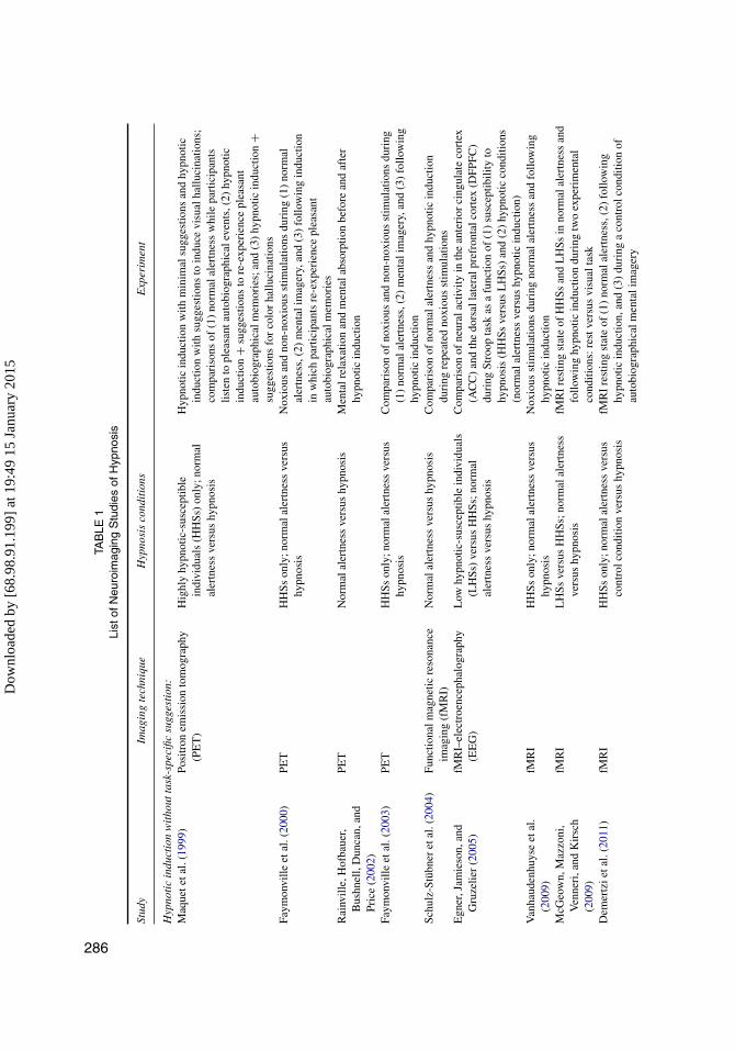
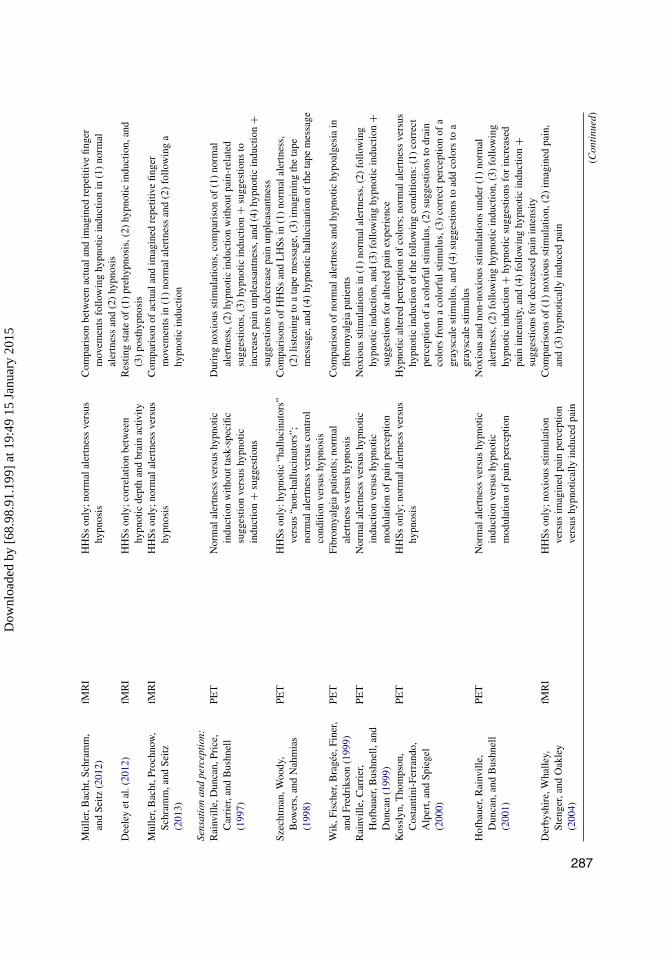
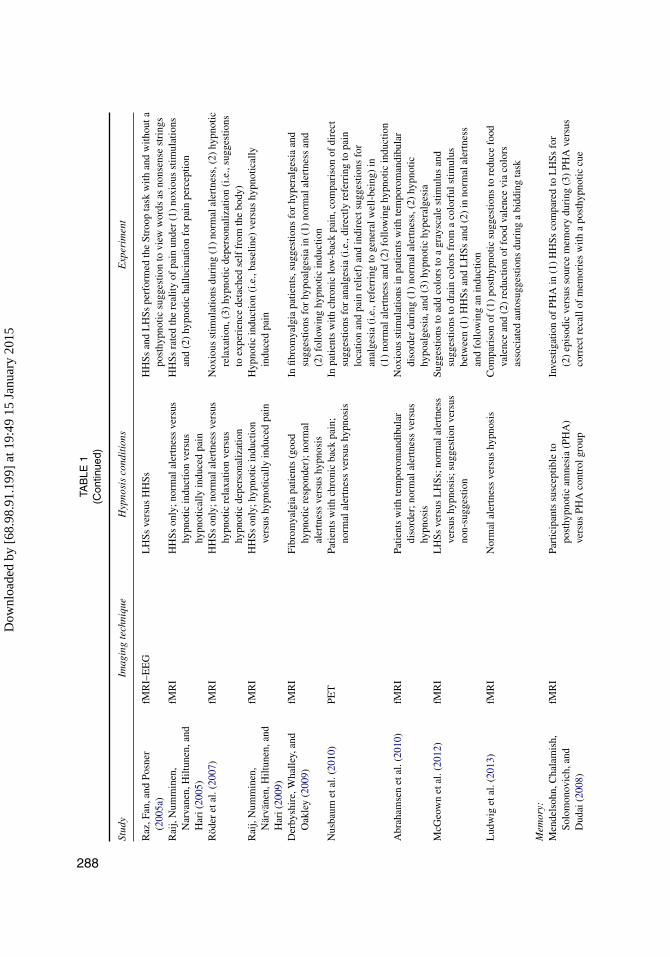
While developments in neuroimaging techniques continue to flourish, imaging studiesof hypnosis have yet to deliver convincing evidence that would inform a reliable neu-robiological theory of hypnosis (Halligan & Oakley, 2013; Jamieson, 2007; Kihlstrom,2013; Oakley & Halligan, 2009, 2013; Raz & Shapiro, 2002). Here we review rele-vant neuroimaging studies to appraise current opinions concerning the neurobiologicalunderpinnings of hypnosis. Our goal is twofold: (1) to identify the relative merits anddrawbacks of neuroimaging studies concerning hypnosis and (2) to offer an integrativesynthesis of neuroimaging findings and how they relate to theoretical models of hypnosis(see Table 1).
Address correspondence to Amir Raz, 4333 Cote-Sainte-Catherine Road, Montreal, Quebec H3T1E4, Canada.E-mail: [email protected]
Dow
nloa
ded
by [
68.9
8.91
.199
] at
19:
49 1
5 Ja
nuar
y 20
15
TAB
LE1
List
ofN
euro
imag
ing
Stu
dies
ofH
ypno
sis
Stud
yIm
agin
gte
chni
que
Hyp
nosi
sco
ndit
ions
Exp
erim
ent
Hyp
noti
cin
duct
ion
wit
hout
task
-spe
cific
sugg
esti
on:
Maq
uete
tal.
(199
9)Po
sitr
onem
issi
onto
mog
raph
y(P
ET
)H
ighl
yhy
pnot
ic-s
usce
ptib
lein
divi
dual
s(H
HSs
)on
ly;n
orm
alal
ertn
ess
vers
ushy
pnos
is
Hyp
notic
indu
ctio
nw
ithm
inim
alsu
gges
tions
and
hypn
otic
indu
ctio
nw
ithsu
gges
tions
toin
duce
visu
alha
lluci
natio
ns;
com
pari
sons
of(1
)no
rmal
aler
tnes
sw
hile
part
icip
ants
liste
nto
plea
sant
auto
biog
raph
ical
even
ts,(
2)hy
pnot
icin
duct
ion
+su
gges
tions
tore
-exp
erie
nce
plea
sant
auto
biog
raph
ical
mem
orie
s;an
d(3
)hy
pnot
icin
duct
ion
+su
gges
tions
for
colo
rha
lluci
natio
nsFa
ymon
ville
etal
.(20
00)
PET
HH
Sson
ly;n
orm
alal
ertn
ess
vers
ushy
pnos
isN
oxio
usan
dno
n-no
xiou
sst
imul
atio
nsdu
ring
(1)
norm
alal
ertn
ess,
(2)
men
tali
mag
ery,
and
(3)
follo
win
gin
duct
ion
inw
hich
part
icip
ants
re-e
xper
ienc
epl
easa
ntau
tobi
ogra
phic
alm
emor
ies
Rai
nvill
e,H
ofba
uer,
Bus
hnel
l,D
unca
n,an
dPr
ice
(200
2)
PET
Nor
mal
aler
tnes
sve
rsus
hypn
osis
Men
talr
elax
atio
nan
dm
enta
labs
orpt
ion
befo
rean
daf
ter
hypn
otic
indu
ctio
n
Faym
onvi
lleet
al.(
2003
)PE
TH
HSs
only
;nor
mal
aler
tnes
sve
rsus
hypn
osis
Com
pari
son
ofno
xiou
san
dno
n-no
xiou
sst
imul
atio
nsdu
ring
(1)
norm
alal
ertn
ess,
(2)
men
tali
mag
ery,
and
(3)
follo
win
ghy
pnot
icin
duct
ion
Schu
lz-S
tübn
eret
al.(
2004
)Fu
nctio
nalm
agne
ticre
sona
nce
imag
ing
(fM
RI)
Nor
mal
aler
tnes
sve
rsus
hypn
osis
Com
pari
son
ofno
rmal
aler
tnes
san
dhy
pnot
icin
duct
ion
duri
ngre
peat
edno
xiou
sst
imul
atio
nsE
gner
,Jam
ieso
n,an
dG
ruze
lier
(200
5)fM
RI–
elec
troe
ncep
halo
grap
hy(E
EG
)L
owhy
pnot
ic-s
usce
ptib
lein
divi
dual
s(L
HSs
)ve
rsus
HH
Ss;n
orm
alal
ertn
ess
vers
ushy
pnos
is
Com
pari
son
ofne
ural
activ
ityin
the
ante
rior
cing
ulat
eco
rtex
(AC
C)
and
the
dors
alla
tera
lpre
fron
talc
orte
x(D
FPFC
)du
ring
Stro
opta
skas
afu
nctio
nof
(1)
susc
eptib
ility
tohy
pnos
is(H
HSs
vers
usL
HSs
)an
d(2
)hy
pnot
icco
nditi
ons
(nor
mal
aler
tnes
sve
rsus
hypn
otic
indu
ctio
n)V
anha
uden
huys
eet
al.
(200
9)fM
RI
HH
Sson
ly;n
orm
alal
ertn
ess
vers
ushy
pnos
isN
oxio
usst
imul
atio
nsdu
ring
norm
alal
ertn
ess
and
follo
win
ghy
pnot
icin
duct
ion
McG
eow
n,M
azzo
ni,
Ven
neri
,and
Kir
sch
(200
9)
fMR
IL
HSs
vers
usH
HSs
;nor
mal
aler
tnes
sve
rsus
hypn
osis
fMR
Ire
stin
gst
ate
ofH
HSs
and
LH
Ssin
norm
alal
ertn
ess
and
follo
win
ghy
pnot
icin
duct
ion
duri
ngtw
oex
peri
men
tal
cond
ition
s:re
stve
rsus
visu
alta
skD
emer
tzie
tal.
(201
1)fM
RI
HH
Sson
ly;n
orm
alal
ertn
ess
vers
usco
ntro
lcon
ditio
nve
rsus
hypn
osis
fMR
Ire
stin
gst
ate
of(1
)no
rmal
aler
tnes
s,(2
)fo
llow
ing
hypn
otic
indu
ctio
n,an
d(3
)du
ring
aco
ntro
lcon
ditio
nof
auto
biog
raph
ical
men
tali
mag
ery
286
Dow
nloa
ded
by [
68.9
8.91
.199
] at
19:
49 1
5 Ja
nuar
y 20
15
Mül
ler,
Bac
ht,S
chra
mm
,an
dSe
itz(2
012)
fMR
IH
HSs
only
;nor
mal
aler
tnes
sve
rsus
hypn
osis
Com
pari
son
betw
een
actu
alan
dim
agin
edre
petit
ive
finge
rm
ovem
ents
follo
win
ghy
pnot
icin
duct
ion
in(1
)no
rmal
aler
tnes
san
d(2
)hy
pnos
isD
eele
yet
al.(
2012
)fM
RI
HH
Sson
ly;c
orre
latio
nbe
twee
nhy
pnot
icde
pth
and
brai
nac
tivity
Res
ting
stat
eof
(1)
preh
ypno
sis,
(2)
hypn
otic
indu
ctio
n,an
d(3
)po
sthy
pnos
isM
ülle
r,B
acht
,Pro
chno
w,
Schr
amm
,and
Seitz
(201
3)
fMR
IH
HSs
only
;nor
mal
aler
tnes
sve
rsus
hypn
osis
Com
pari
son
ofac
tual
and
imag
ined
repe
titiv
efin
ger
mov
emen
tsin
(1)
norm
alal
ertn
ess
and
(2)
follo
win
ga
hypn
otic
indu
ctio
n
Sens
atio
nan
dpe
rcep
tion
:R
ainv
ille,
Dun
can,
Pric
e,C
arri
er,a
ndB
ushn
ell
(199
7)
PET
Nor
mal
aler
tnes
sve
rsus
hypn
otic
indu
ctio
nw
ithou
ttas
k-sp
ecifi
csu
gges
tion
vers
ushy
pnot
icin
duct
ion
+su
gges
tions
Dur
ing
noxi
ous
stim
ulat
ions
,com
pari
son
of(1
)no
rmal
aler
tnes
s,(2
)hy
pnot
icin
duct
ion
with
outp
ain-
rela
ted
sugg
estio
ns,(
3)hy
pnot
icin
duct
ion
+su
gges
tions
toin
crea
sepa
inun
plea
sant
ness
,and
(4)
hypn
otic
indu
ctio
n+
sugg
estio
nsto
decr
ease
pain
unpl
easa
ntne
ssSz
echt
man
,Woo
dy,
Bow
ers,
and
Nah
mia
s(1
998)
PET
HH
Sson
ly:h
ypno
tic“h
allu
cina
tors
”ve
rsus
“non
-hal
luci
nato
rs”;
norm
alal
ertn
ess
vers
usco
ntro
lco
nditi
onve
rsus
hypn
osis
Com
pari
sons
ofH
HSs
and
LH
Ssin
(1)
norm
alal
ertn
ess,
(2)
liste
ning
toa
tape
mes
sage
,(3)
imag
inin
gth
eta
pem
essa
ge,a
nd(4
)hy
pnot
icha
lluci
natio
nof
the
tape
mes
sage
Wik
,Fis
cher
,Bra
gée,
Fine
r,an
dFr
edri
kson
(199
9)PE
TFi
brom
yalg
iapa
tient
s;no
rmal
aler
tnes
sve
rsus
hypn
osis
Com
pari
son
ofno
rmal
aler
tnes
san
dhy
pnot
ichy
poal
gesi
ain
fibro
mya
lgia
patie
nts
Rai
nvill
e,C
arri
er,
Hof
baue
r,B
ushn
ell,
and
Dun
can
(199
9)
PET
Nor
mal
aler
tnes
sve
rsus
hypn
otic
indu
ctio
nve
rsus
hypn
otic
mod
ulat
ion
ofpa
inpe
rcep
tion
Nox
ious
stim
ulat
ions
in(1
)no
rmal
aler
tnes
s,(2
)fo
llow
ing
hypn
otic
indu
ctio
n,an
d(3
)fo
llow
ing
hypn
otic
indu
ctio
n+
sugg
estio
nsfo
ral
tere
dpa
inex
peri
ence
Kos
slyn
,Tho
mps
on,
Cos
tant
ini-
Ferr
ando
,A
lper
t,an
dSp
iege
l(2
000)
PET
HH
Sson
ly;n
orm
alal
ertn
ess
vers
ushy
pnos
isH
ypno
tical
tere
dpe
rcep
tion
ofco
lors
;nor
mal
aler
tnes
sve
rsus
hypn
otic
indu
ctio
nof
the
follo
win
gco
nditi
ons:
(1)
corr
ect
perc
eptio
nof
aco
lorf
ulst
imul
us,(
2)su
gges
tions
todr
ain
colo
rsfr
oma
colo
rful
stim
ulus
,(3)
corr
ectp
erce
ptio
nof
agr
aysc
ale
stim
ulus
,and
(4)
sugg
estio
nsto
add
colo
rsto
agr
aysc
ale
stim
ulus
Hof
baue
r,R
ainv
ille,
Dun
can,
and
Bus
hnel
l(2
001)
PET
Nor
mal
aler
tnes
sve
rsus
hypn
otic
indu
ctio
nve
rsus
hypn
otic
mod
ulat
ion
ofpa
inpe
rcep
tion
Nox
ious
and
non-
noxi
ous
stim
ulat
ions
unde
r(1
)no
rmal
aler
tnes
s,(2
)fo
llow
ing
hypn
otic
indu
ctio
n,(3
)fo
llow
ing
hypn
otic
indu
ctio
n+
hypn
otic
sugg
estio
nsfo
rin
crea
sed
pain
inte
nsity
,and
(4)
follo
win
ghy
pnot
icin
duct
ion
+su
gges
tions
for
decr
ease
dpa
inin
tens
ityD
erby
shir
e,W
halle
y,St
enge
r,an
dO
akle
y(2
004)
fMR
IH
HSs
only
;nox
ious
stim
ulat
ion
vers
usim
agin
edpa
inpe
rcep
tion
vers
ushy
pnot
ical
lyin
duce
dpa
in
Com
pari
sons
of(1
)no
xiou
sst
imul
atio
n,(2
)im
agin
edpa
in,
and
(3)
hypn
otic
ally
indu
ced
pain
(Con
tinu
ed)
287
Dow
nloa
ded
by [
68.9
8.91
.199
] at
19:
49 1
5 Ja
nuar
y 20
15
TAB
LE1
(Con
tinue
d)
Stud
yIm
agin
gte
chni
que
Hyp
nosi
sco
ndit
ions
Exp
erim
ent
Raz
,Fan
,and
Posn
er(2
005a
)fM
RI–
EE
GL
HSs
vers
usH
HSs
HH
Ssan
dL
HSs
perf
orm
edth
eSt
roop
task
with
and
with
outa
post
hypn
otic
sugg
estio
nto
view
wor
dsas
nons
ense
stri
ngs
Rai
j,N
umm
inen
,N
arva
nen,
Hilt
unen
,and
Har
i(20
05)
fMR
IH
HSs
only
;nor
mal
aler
tnes
sve
rsus
hypn
otic
indu
ctio
nve
rsus
hypn
otic
ally
indu
ced
pain
HH
Ssra
ted
the
real
ityof
pain
unde
r(1
)no
xiou
sst
imul
atio
nsan
d(2
)hy
pnot
icha
lluci
natio
nfo
rpa
inpe
rcep
tion
Röd
eret
al.(
2007
)fM
RI
HH
Sson
ly;n
orm
alal
ertn
ess
vers
ushy
pnot
icre
laxa
tion
vers
ushy
pnot
icde
pers
onal
izat
ion
Nox
ious
stim
ulat
ions
duri
ng(1
)no
rmal
aler
tnes
s,(2
)hy
pnot
icre
laxa
tion,
(3)
hypn
otic
depe
rson
aliz
atio
n(i
.e.,
sugg
estio
nsto
expe
rien
cede
tach
edse
lffr
omth
ebo
dy)
Rai
j,N
umm
inen
,N
ärvä
nen,
Hilt
unen
,and
Har
i(20
09)
fMR
IH
HSs
only
;hyp
notic
indu
ctio
nve
rsus
hypn
otic
ally
indu
ced
pain
Hyp
notic
indu
ctio
n(i
.e.,
base
line)
vers
ushy
pnot
ical
lyin
duce
dpa
in
Der
bysh
ire,
Wha
lley,
and
Oak
ley
(200
9)fM
RI
Fibr
omya
lgia
patie
nts
(goo
dhy
pnot
icre
spon
der)
;nor
mal
aler
tnes
sve
rsus
hypn
osis
Infib
rom
yalg
iapa
tient
s,su
gges
tions
for
hype
ralg
esia
and
sugg
estio
nsfo
rhy
poal
gesi
ain
(1)
norm
alal
ertn
ess
and
(2)
follo
win
ghy
pnot
icin
duct
ion
Nus
baum
etal
.(20
10)
PET
Patie
nts
with
chro
nic
back
pain
;no
rmal
aler
tnes
sve
rsus
hypn
osis
Inpa
tient
sw
ithch
roni
clo
w-b
ack
pain
,com
pari
son
ofdi
rect
sugg
estio
nsfo
ran
alge
sia
(i.e
.,di
rect
lyre
ferr
ing
topa
inlo
catio
nan
dpa
inre
lief)
and
indi
rect
sugg
estio
nsfo
ran
alge
sia
(i.e
.,re
ferr
ing
toge
nera
lwel
l-be
ing)
in(1
)no
rmal
aler
tnes
san
d(2
)fo
llow
ing
hypn
otic
indu
ctio
nA
brah
amse
net
al.(
2010
)fM
RI
Patie
nts
with
tem
poro
man
dibu
lar
diso
rder
;nor
mal
aler
tnes
sve
rsus
hypn
osis
Nox
ious
stim
ulat
ions
inpa
tient
sw
ithte
mpo
rom
andi
bula
rdi
sord
erdu
ring
(1)
norm
alal
ertn
ess,
(2)
hypn
otic
hypo
alge
sia,
and
(3)
hypn
otic
hype
ralg
esia
McG
eow
net
al.(
2012
)fM
RI
LH
Ssve
rsus
LH
Ss;n
orm
alal
ertn
ess
vers
ushy
pnos
is;s
ugge
stio
nve
rsus
non-
sugg
estio
n
Sugg
estio
nsto
add
colo
rsto
agr
aysc
ale
stim
ulus
and
sugg
estio
nsto
drai
nco
lors
from
aco
lorf
ulst
imul
usbe
twee
n(1
)H
HSs
and
LH
Ssan
d(2
)in
norm
alal
ertn
ess
and
follo
win
gan
indu
ctio
nL
udw
iget
al.(
2013
)fM
RI
Nor
mal
aler
tnes
sve
rsus
hypn
osis
Com
pari
son
of(1
)po
sthy
pnot
icsu
gges
tions
tore
duce
food
vale
nce
and
(2)
redu
ctio
nof
food
vale
nce
via
colo
rsas
soci
ated
auto
sugg
estio
nsdu
ring
abi
ddin
gta
sk
Mem
ory:
Men
dels
ohn,
Cha
lam
ish,
Solo
mon
ovic
h,an
dD
udai
(200
8)
fMR
IPa
rtic
ipan
tssu
scep
tible
topo
sthy
pnot
icam
nesi
a(P
HA
)ve
rsus
PHA
cont
rolg
roup
Inve
stig
atio
nof
PHA
in(1
)H
HSs
com
pare
dto
LH
Ssfo
r(2
)ep
isod
icve
rsus
sour
cem
emor
ydu
ring
(3)
PHA
vers
usco
rrec
trec
allo
fm
emor
ies
with
apo
sthy
pnot
iccu
e
288
Dow
nloa
ded
by [
68.9
8.91
.199
] at
19:
49 1
5 Ja
nuar
y 20
15
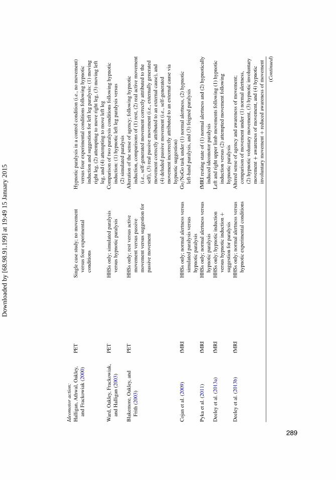
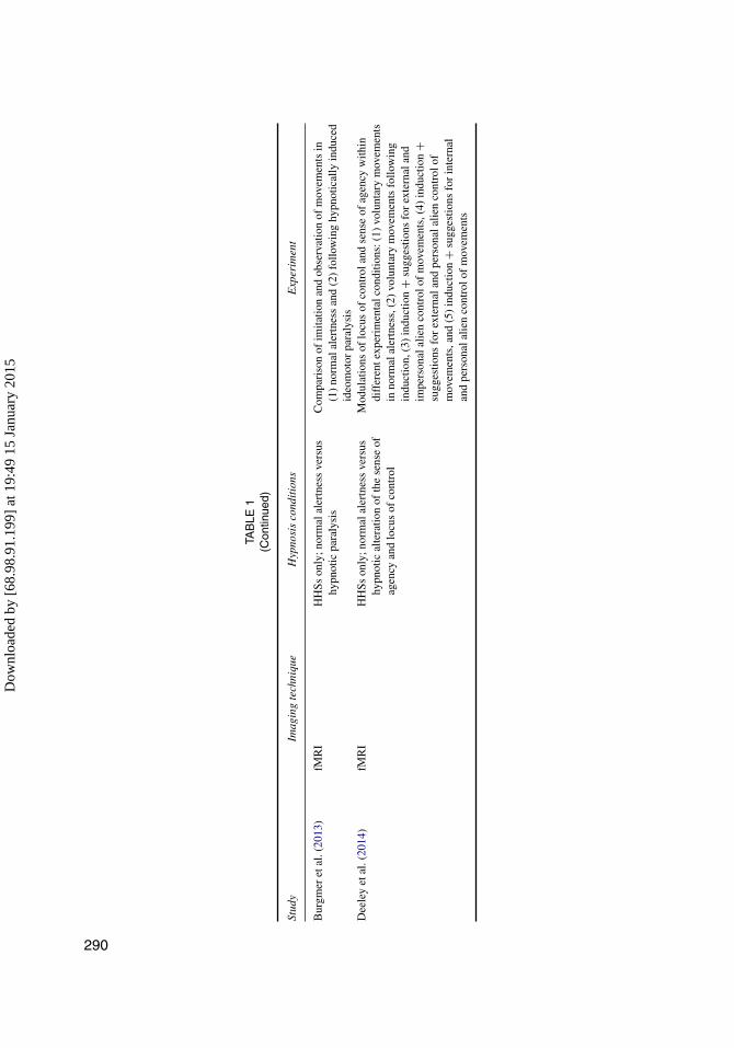
Ideo
mot
orac
tion
:H
allig
an,A
thw
al,O
akle
y,an
dFr
acko
wia
k(2
000)
PET
Sing
leca
sest
udy;
nom
ovem
ent
vers
usfo
urex
peri
men
tal
cond
ition
s
Hyp
notic
para
lysi
sin
aco
ntro
lcon
ditio
n(i
.e.,
nom
ovem
ent)
vers
usfo
urex
peri
men
talc
ondi
tions
follo
win
ghy
pnot
icin
duct
ion
and
sugg
estio
nfo
rle
ftle
gpa
raly
sis:
(1)
mov
ing
righ
tleg
,(2)
atte
mpt
ing
tom
ove
righ
tleg
,(3)
mov
ing
left
leg,
and
(4)
atte
mpt
ing
tom
ove
left
leg
War
d,O
akle
y,Fr
acko
wia
k,an
dH
allig
an(2
003)
PET
HH
Sson
ly;s
imul
ated
para
lysi
sve
rsus
hypn
otic
para
lysi
sC
ompa
riso
nof
two
para
lysi
sco
nditi
ons
follo
win
ghy
pnot
icin
duct
ion:
(1)
hypn
otic
left
leg
para
lysi
sve
rsus
(2)
sim
ulat
edpa
raly
sis
Bla
kem
ore,
Oak
ley,
and
Frith
(200
3)PE
TH
HSs
only
;res
tver
sus
activ
em
ovem
entv
ersu
spa
ssiv
em
ovem
entv
ersu
ssu
gges
tion
for
pass
ive
mov
emen
t
Alte
ratio
nof
the
sens
eof
agen
cy;f
ollo
win
ghy
pnot
icin
duct
ion,
com
pari
sons
of(1
)re
st,(
2)re
alac
tive
mov
emen
t(i
.e.,
self
-gen
erat
edm
ovem
entc
orre
ctly
attr
ibut
edto
the
self
),(3
)re
alpa
ssiv
em
ovem
ent(
i.e.,
exte
rnal
lyge
nera
ted
mov
emen
tcor
rect
lyat
trib
uted
toan
exte
rnal
caus
e),a
nd(4
)de
lude
dpa
ssiv
em
ovem
ent(
i.e.,
self
-gen
erat
edm
ovem
enti
ncor
rect
lyat
trib
uted
toan
exte
rnal
caus
evi
ahy
pnot
icsu
gges
tion)
Coj
anet
al.(
2009
)fM
RI
HH
Sson
ly;n
orm
alal
ertn
ess
vers
ussi
mul
ated
para
lysi
sve
rsus
hypn
otic
para
lysi
s
Go/
NoG
ota
skun
der
(1)
norm
alal
ertn
ess,
(2)
hypn
otic
left
-han
dpa
raly
sis,
and
(3)
feig
ned
para
lysi
s
Pyka
etal
.(20
11)
fMR
IH
HSs
only
;nor
mal
aler
tnes
sve
rsus
hypn
otic
para
lysi
sfM
RI
rest
ing
stat
eof
(1)
norm
alal
ertn
ess
and
(2)
hypn
otic
ally
indu
ced
ideo
mot
orpa
raly
sis
Dee
ley
etal
.(20
13a)
fMR
IH
HSs
only
;hyp
notic
indu
ctio
nve
rsus
hypn
otic
indu
ctio
n+
sugg
estio
nfo
rpa
raly
sis
Lef
tand
righ
tupp
erlim
bm
ovem
ents
follo
win
g(1
)hy
pnot
icin
duct
ion
vers
us(2
)at
tem
pted
mov
emen
tfol
low
ing
hypn
otic
para
lysi
sD
eele
yet
al.(
2013
b)fM
RI
HH
Sson
ly;n
orm
alal
ertn
ess
vers
ushy
pnot
icex
peri
men
talc
ondi
tions
Alte
red
sens
eof
agen
cyan
daw
aren
ess
ofm
ovem
ent;
com
pari
son
ofm
ovem
entu
nder
(1)
norm
alal
ertn
ess,
(2)
hypn
otic
volu
ntar
ym
ovem
ent,
(3)
hypn
otic
invo
lunt
ary
mov
emen
t+aw
aren
ess
ofm
ovem
ent,
and
(4)
hypn
otic
invo
lunt
ary
mov
emen
t+re
duce
daw
aren
ess
ofm
ovem
ent
(Con
tinu
ed)
289
Dow
nloa
ded
by [
68.9
8.91
.199
] at
19:
49 1
5 Ja
nuar
y 20
15
TAB
LE1
(Con
tinue
d)
Stud
yIm
agin
gte
chni
que
Hyp
nosi
sco
ndit
ions
Exp
erim
ent
Bur
gmer
etal
.(20
13)
fMR
IH
HSs
only
;nor
mal
aler
tnes
sve
rsus
hypn
otic
para
lysi
sC
ompa
riso
nof
imita
tion
and
obse
rvat
ion
ofm
ovem
ents
in(1
)no
rmal
aler
tnes
san
d(2
)fo
llow
ing
hypn
otic
ally
indu
ced
ideo
mot
orpa
raly
sis
Dee
ley
etal
.(20
14)
fMR
IH
HSs
only
;nor
mal
aler
tnes
sve
rsus
hypn
otic
alte
ratio
nof
the
sens
eof
agen
cyan
dlo
cus
ofco
ntro
l
Mod
ulat
ions
oflo
cus
ofco
ntro
land
sens
eof
agen
cyw
ithin
diff
eren
texp
erim
enta
lcon
ditio
ns:(
1)vo
lunt
ary
mov
emen
tsin
norm
alal
ertn
ess,
(2)
volu
ntar
ym
ovem
ents
follo
win
gin
duct
ion,
(3)
indu
ctio
n+
sugg
estio
nsfo
rex
tern
alan
dim
pers
onal
alie
nco
ntro
lof
mov
emen
ts,(
4)in
duct
ion
+su
gges
tions
for
exte
rnal
and
pers
onal
alie
nco
ntro
lof
mov
emen
ts,a
nd(5
)in
duct
ion
+su
gges
tions
for
inte
rnal
and
pers
onal
alie
nco
ntro
lof
mov
emen
ts
290
Dow
nloa
ded
by [
68.9
8.91
.199
] at
19:
49 1
5 Ja
nuar
y 20
15
HYPNOSIS AND IMAGING OF THE LIVING HUMAN BRAIN 291
Neuroimaging studies often operationalize hypnosis through induction and sugges-tions procedures. This approach raises concerns as to whether experimental findingsgeneralize to clinical contexts. However, researchers train participants prior to exper-iments and confirm the effects of induction via self-report, thereby supporting the“ecological validity” of hypnosis in the laboratory (Oakley, Deeley, & Halligan, 2007).
The Challenge of Neuroimaging Hypnotic Phenomena
The advent of brain-imaging technology captures the imagination of the masses (Ali,Lifzhitz, & Raz, 2014; Choudhury & Slaby, 2011; Jones & Mendell, 1999). Thesetechnological advances generate excitement and expectation in the cognitive sciences,including psychology and psychiatry (Aue, Lavelle, & Cacioppo, 2009; Axmacher,Elger, & Fell, 2009; Choudhury & Slaby, 2011; Dolan, 2008; Jones & Mendell,1999; Kirmayer & Crafa, 2014; Malhi & Lagopoulos, 2008; Nathan, Phan, Harmer,Mehta, & Bullmore, 2014; Poldrack, 2012). This enthusiasm also applies to hypnosisresearch (Halligan & Oakley, 2013; Jamieson & Woody, 2007; Kihlstrom, 2013; Oakley& Halligan, 2009, 2013). However, after nearly two decades of imaging hypnotizedbrains, the pursuit of a neurobiological model based on neuroimaging remains incon-clusive (Kirmayer & Crafa, 2014; Macdonald & Raz, 2014; Raz & Macdonald, 2014,2015).
Imaging the brain imposes a wealth of practical and theoretical restrictions thatconfine the scope of investigation. These limitations include, for example, burdeningparticipants with unnatural testing environment and ecologically invalid postures (Razet al., 2005b; Thibault, Lifshitz, Jones, & Raz, 2014), the temporal and spatial resolutionof (e.g., fMRI) scanners (Axmacher et al., 2009), and the capacity to detect meaning-ful signals amid background noise (Filippi, 2009). However, hypnotic effects seem tolargely transcend these caveats and ecological barriers (Oakley et al., 2007).
Neuroimaging entails specific challenges to the study of hypnosis. For example, hyp-nosis typically encompasses an induction procedure, designed to increase the hypnoticresponse, followed by direct suggestions to modify perception, cognition, or behavior(Kihlstrom, 2008). Despite this induction–suggestion distinction, neuroimaging pro-tocols provide little means to differentiate between the effects of hypnotic inductionand the effects of hypnotic suggestions (Cardeña, Jönsson, Terhune, & Marcusson-Clavertz, 2013; Mazzoni, Venneri, McGeown, & Kirsch, 2013). The instructions duringthe induction procedure already represent some form of suggestion (Gandhi & Oakley,2005). Some researchers attempt to resolve this concern by inducing a so-called neu-tral plane of hypnosis using an induction with minimal suggestions (Cardeña et al.,2013; Kihlstrom & Edmonston, 1971; Mazzoni et al., 2013). Yet, the use of sugges-tions appears inevitable, thereby undermining the notion of a suggestion-free induction.Studies may partially circumvent this central issue by keeping the task and suggestionsconstant across hypnotic conditions, thereby allowing participants to receive identical
Dow
nloa
ded
by [
68.9
8.91
.199
] at
19:
49 1
5 Ja
nuar
y 20
15
292 LANDRY AND RAZ
suggestions while performing the same task under hypnosis and normal alertness.However, isolating effects pertaining to the induction procedure from those pertain-ing to suggestions represents a constant challenge for neuroimaging studies addressinghypnosis.
A high level of inter-individual variability in susceptibility to suggestions impactsboth practical and theoretical aspects of hypnosis research (Carli, Manzoni, &Santarcangelo, 2008; Heap, Brown, & Oakley, 2004; Laurence, Beaulieu-Prévost, &Du Chéné, 2008; Piccione, Hilgard, & Zimbardo, 1989; Terhune, Cardeña, & Lindgren,2011). To account for this variability, studies often compare the effects elicited by HHSswith those elicited by LHSs (Nash & Barnier, 2008). As it turns out, HHSs use variouscognitive strategies to comply with hypnotic suggestions (Barnier, Cox, & McConkey,2014; McConkey & Barnier, 2004; McConkey, Glisky, & Kihlstrom, 1989; Nash &Barnier, 2008). The principle of equifinality (i.e., this principle states that different meanscan lead to the same end state) therefore applies to hypnotic response, whereby similarhypnotic responses may rely on different cognitive routes (Cardeña, 2014a). This situ-ation further contributes to the inherent heterogeneity of hypnotic phenomena. The factthat numerous hypnotic effects surface only in subjective reports also exacerbates thisconcern (Kihlstrom, 2008). Thus, individual differences reduce our ability to preciselysequester commonalities among disparate studies.
Discerning hypnotic-related from task-related effects represents another importantchallenge. For example, both the Stroop task and hypnosis relate to modulations ofthe ACC, making it difficult to separate hypnotic from Stroop effects (Egner et al.,2005; Raz, Fan, & Posner, 2005a). Subsequently, a meaningful study that includes anexperimental task requires a control condition to differentiate hypnotic from task effects.
Most neuroimaging studies involving hypnosis either focus on hypnotic phenom-ena or use hypnosis as an experimental instrument to investigate (a)typical cognition(Bortolotti, Cox, & Barnier, 2012; Cox & Barnier, 2010; Oakley & Halligan, 2009,2013). Due to their different aims, intrinsic and instrumental hypnosis seldom combinein the same study (Barnier, 2002; Oakley & Halligan, 2009). Together, all of the above-mentioned concerns constrain our ability to build a reliable neurobiological theory ofhypnosis.
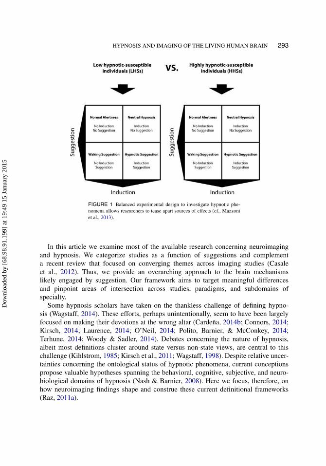
Careful experimentation represents the primary solution to collect reliableneuroimaging data (Filippi, 2009). Moreover, combining evidence from different meth-ods makes for a good strategy to test and support experimental hypotheses (Henson,2005, 2006). In the specific context of hypnosis, adopting a two-by-two design cross-ing hypnosis and suggestion as a function of hypnotic susceptibility embodies the primestrategy for teasing apart the effects of hypnosis (see Figure 1; Mazzoni et al., 2013;Oakley & Halligan, 2010). This matrix ascertains the effects of key variables that influ-ence hypnotic phenomena. Alas, in light of practical and financial limitations, mostresearch groups rarely follow this template. Instead, they rely on imaging of specifichypnotic phenomena, often with a single control condition.
Dow
nloa
ded
by [
68.9
8.91
.199
] at
19:
49 1
5 Ja
nuar
y 20
15
HYPNOSIS AND IMAGING OF THE LIVING HUMAN BRAIN 293
FIGURE 1 Balanced experimental design to investigate hypnotic phe-nomena allows researchers to tease apart sources of effects (cf., Mazzoniet al., 2013).
In this article we examine most of the available research concerning neuroimagingand hypnosis. We categorize studies as a function of suggestions and complementa recent review that focused on converging themes across imaging studies (Casaleet al., 2012). Thus, we provide an overarching approach to the brain mechanismslikely engaged by suggestion. Our framework aims to target meaningful differencesand pinpoint areas of intersection across studies, paradigms, and subdomains ofspecialty.
Some hypnosis scholars have taken on the thankless challenge of defining hypno-sis (Wagstaff, 2014). These efforts, perhaps unintentionally, seem to have been largelyfocused on making their devotions at the wrong altar (Cardeña, 2014b; Connors, 2014;Kirsch, 2014; Laurence, 2014; O’Neil, 2014; Polito, Barnier, & McConkey, 2014;Terhune, 2014; Woody & Sadler, 2014). Debates concerning the nature of hypnosis,albeit most definitions cluster around state versus non-state views, are central to thischallenge (Kihlstrom, 1985; Kirsch et al., 2011; Wagstaff, 1998). Despite relative uncer-tainties concerning the ontological status of hypnotic phenomena, current conceptionspropose valuable hypotheses spanning the behavioral, cognitive, subjective, and neuro-biological domains of hypnosis (Nash & Barnier, 2008). Here we focus, therefore, onhow neuroimaging findings shape and construe these current definitional frameworks(Raz, 2011a).
Dow
nloa
ded
by [
68.9
8.91
.199
] at
19:
49 1
5 Ja
nuar
y 20
15
294 LANDRY AND RAZ
Culling of Neuroimaging Findings
Method
We searched for neuroimaging studies of hypnosis using combinations of the followingkey words in Google Scholar, PubMed, and PsycINFO: hypnosis, neuroimaging, fMRI,and PET. Because we aim to localize and frame the underlying neural mechanismsof hypnosis across very similar methodologies, our review solely includes fMRI andPET, therefore excluding EEG, magnetoencephalography, near-infrared spectroscopy,and structural or volumetric imaging studies. We included 37 neuroimaging studies.We excluded studies that did not use imaging of at least one experimental conditionof hypnotic induction or hypnotic response. We also excluded three studies that did notprovide or provided only vague indications concerning the induction procedure. Table 1provides a brief summary of the method and results for each study. In the first sectionof this article, we describe the studies that used hypnotic induction but did not usesubsequent task-specific hypnotic suggestions. Suggestions in these studies are eitherindirect or unspecified in connection with the task. We describe studies that investigatedhypnotic suggestions that focus on sensation and perception, memory, and ideomotoraction. These studies highlight how hypnotic suggestions can elicit changes in focal brainareas. Overall, the findings support the idea that hypnosis engages brain areas related toattention, cognitive control, and monitoring.
Hypnotic Induction Without Task-Specific Suggestion
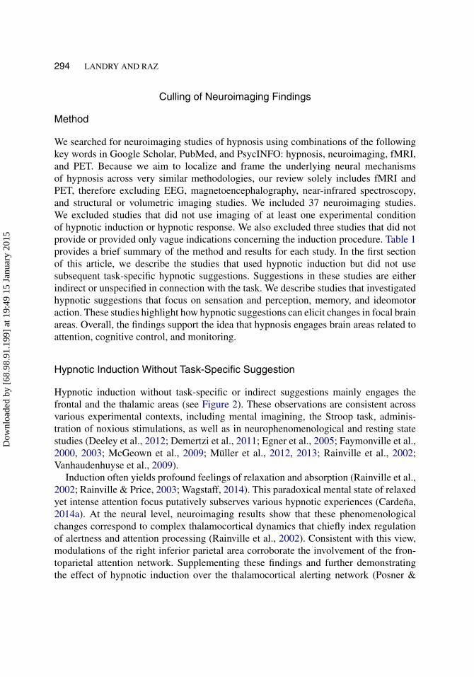
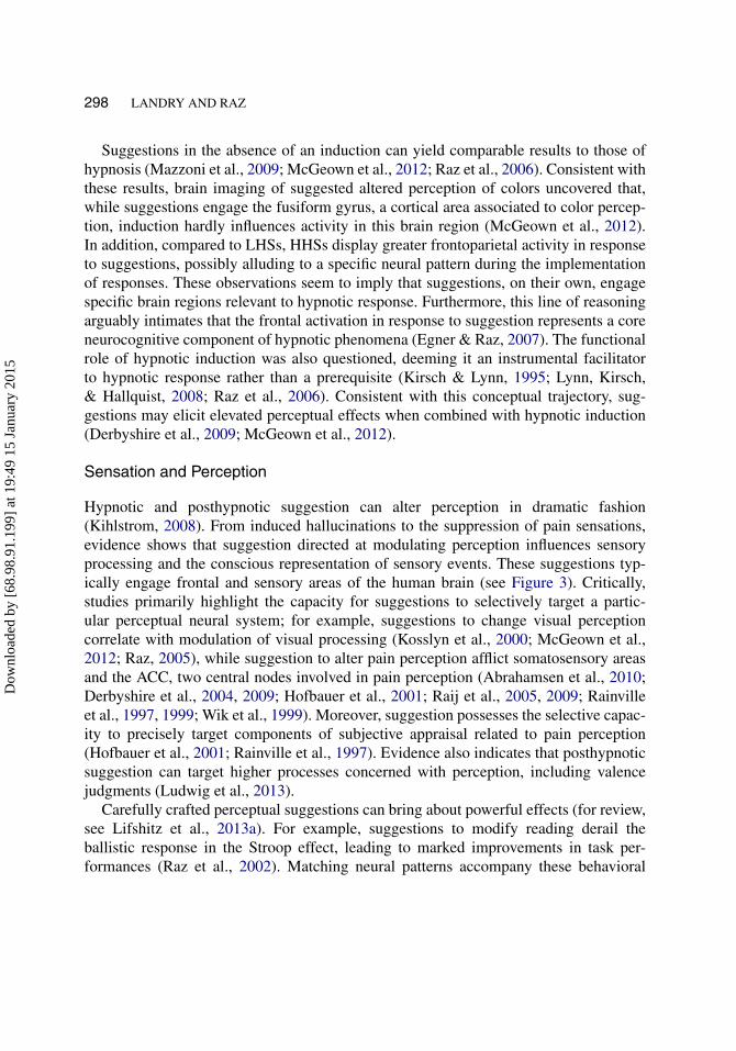
Hypnotic induction without task-specific or indirect suggestions mainly engages thefrontal and the thalamic areas (see Figure 2). These observations are consistent acrossvarious experimental contexts, including mental imagining, the Stroop task, adminis-tration of noxious stimulations, as well as in neurophenomenological and resting statestudies (Deeley et al., 2012; Demertzi et al., 2011; Egner et al., 2005; Faymonville et al.,2000, 2003; McGeown et al., 2009; Müller et al., 2012, 2013; Rainville et al., 2002;Vanhaudenhuyse et al., 2009).
Induction often yields profound feelings of relaxation and absorption (Rainville et al.,2002; Rainville & Price, 2003; Wagstaff, 2014). This paradoxical mental state of relaxedyet intense attention focus putatively subserves various hypnotic experiences (Cardeña,2014a). At the neural level, neuroimaging results show that these phenomenologicalchanges correspond to complex thalamocortical dynamics that chiefly index regulationof alertness and attention processing (Rainville et al., 2002). Consistent with this view,modulations of the right inferior parietal area corroborate the involvement of the fron-toparietal attention network. Supplementing these findings and further demonstratingthe effect of hypnotic induction over the thalamocortical alerting network (Posner &
Dow
nloa
ded
by [
68.9
8.91
.199
] at
19:
49 1
5 Ja
nuar
y 20
15
HYPNOSIS AND IMAGING OF THE LIVING HUMAN BRAIN 295
FIGURE 2 Brain regions related to hypnotic induction without task-specific suggestions (SFG: superior frontal gyrus; MFG: middle frontalgyrus; IFG: inferior frontal gyrus; MeFG: medial frontal gyrus; laterality:“r” for right, “l” for left, and “b” for bilateral).
Petersen, 1990; Raz & Buhle, 2006), additional studies report hypnotic modulationsof the thalamic area (Deeley et al., 2012; Faymonville et al., 2003; McGeown et al.,2009; Müller et al., 2012, 2013; Rainville et al., 2002; Vanhaudenhuyse et al., 2009).These collective results reveal that hypnotized individuals conform to the directives ofan induction by engaging mental relaxation and absorption. Moreover, they highlight thetop-down, versus bottom-up, nature of hypnosis (Raz, 2011b).
Evidence from resting-state studies of hypnosis also reveals the engagement of atten-tion (Deeley et al., 2012; McGeown et al., 2009). Resting states represent recordings
Dow
nloa
ded
by [
68.9
8.91
.199
] at
19:
49 1
5 Ja
nuar
y 20
15
296 LANDRY AND RAZ
of spontaneous cerebral interactions between various regions in the absence of task-directed activities (Fox & Raichle, 2007). Using this approach, two independent researchgroups established that (1) hypnotic inductions link to a substantial activity decrease inthe default-mode network (DMN), a brain network typically related to the spontaneousgeneration of cognition (Buckner, Andrews-Hanna, & Schacter, 2008), and (2) increasedactivity in the frontoparietal attention network (Deeley et al., 2012; McGeown et al.,2009). These opposing neural patterns between DMN and the attention network likelyaccount for negatively correlated interactions (Fox et al., 2005). These findings proposethat induction procedures instigate a marked reduction in the production of cognition, asindexed by the disengagement of the DMN as well as a discernible increase in absorp-tion, manifest by the activation of the frontoparietal attention network (cf., Demertziet al., 2011).
Beyond changes in processing of attention, dissociation theorists argue that hypno-sis decouples control and monitoring processes (Woody & Farvolden, 1998; Woody &Sadler, 2008). A pivotal combined fMRI–EEG study tested this idea by investigatingneural activity in the Stroop task following hypnotic induction without a task-specificsuggestion (Egner et al., 2005). Considered one of the gold standards to measure exec-utive attention during cognitive conflict (MacLeod, 1991), the Stroop task providesthe means to appropriately examine control and monitoring processes. Supporting thedissociation hypothesis, results revealed distinct profiles of activation for the dorsolat-eral prefrontal cortex (DLPFC), a brain region associated with attention and cognitivecontrol (Corbetta, Patel, & Shulman, 2008; Nee et al., 2013) and the ACC—a key nodein cognitive monitoring (Botvinick, 2007; Botvinick, Cohen, & Carter, 2004; Shenhav,Botvinick, & Cohen, 2013). These distinct activation profiles emerged as a functionof hypnotic trait (HHSs versus LHSs) and hypnotic conditions (hypnosis versus base-line). Specifically, for HHSs under hypnosis, elevated ACC activity reflected the onlinedetection of a cognitive conflict, whereas the DLPFC showed little, if any, modula-tion. This subpar detection of cognitive control supports the idea that hypnotic inductionmay decouple conflict from monitoring. Poor behavioral performance on task followinginduction further supports this interpretation (Egner & Raz, 2007).
Certain investigations concerning the perception of pain report hypnoanalgesiadespite the absence of direct analgesic suggestions. In these experiments, researchersask participants to focus on autobiographical memories, thereby orienting their atten-tion away from sensory inputs (Faymonville et al., 2000, 2003; Maquet et al., 1999;Vanhaudenhuyse et al., 2009). Hence, inattention to noxious stimulus seems centralto this hypnotic phenomenon (Landry, Appourchaux, & Raz, 2014). According to thisview, hypnoanalgesia is analogous to inattentional blindness in that a secondary atten-tion demanding task (e.g., focusing on autobiographical memories) taxes cognitiveresources, directs attention away from noxious sensations, and reduces conscious accessto nociception (Kuhn & Tatler, 2011; Memmert, 2010; Moran & Brady, 2010; Most,2010).
Dow
nloa
ded
by [
68.9
8.91
.199
] at
19:
49 1
5 Ja
nuar
y 20
15
HYPNOSIS AND IMAGING OF THE LIVING HUMAN BRAIN 297
Current findings render unlikely the proposition that altered pain perception comesabout via exclusive modulations of selection mechanisms (Faymonville, Boly, &Laureys, 2006; Pekala & Kumar, 2007). Instead, several studies report that noxious stim-ulation during induction without direct analgesic suggestions leads to modulation of theACC, a central component of the pain neuromatrix (Faymonville et al., 2000; Maquetet al., 1999; Schulz-Stübner et al., 2004). One such study reports a marked reduction ofACC, thalamic, and striatal activity for matched-intensity stimulation (Vanhaudenhuyseet al., 2009). Such findings intimate that hypnosis and top-down regulation alter theconscious representation of nociception. Hypnotic modulations of the ACC, whichplays a central role in the integration of negative affect, pain, and cognitive control(Shackman et al., 2011), may therefore reflect perception of nociceptive signals underaltered conscious perception. Moreover, the ACC shares many neural pathways with var-ious brain regions. Unlocking the effect of hypnosis on these various pathways, fMRIresults show that hypnotic induction and indirect suggestions modify the connectivitybetween the ACC and several brain regions (Faymonville et al., 2003). Likewise, induc-tion and noxious stimulation links to altered connectivity between the frontal area andthe primary somatosensory cortex, a fundamental area for nociception (Vanhaudenhuyseet al., 2009). Collectively, therefore, these hypnotic changes in connectivity putativelycorrespond to perception under altered consciousness and reinterpretation of sensoryevents.
Hypnotic and Posthypnotic Suggestions
Following the exploration of studies that scrutinized neural correlates of hypnotic induc-tion in the absence of task-specific suggestion, we now turn to studies that consideredhypnotic and posthypnotic suggestions. Suggestions are communicable representationscapable of transforming thoughts and actions (Halligan & Oakley, 2014). From targetedalterations of sensory processing (e.g., Raz, Kirsch, Pollard, & Nitkin-Kaner, 2006)to placebo responses (e.g., Flaten, Simonsen, & Olsen, 1999), various reports docu-ment their effects on cognition and behaviors (Michael, Garry, & Kirsch, 2012). Thesingularity of hypnotic and posthypnotic suggestions lies in their usage following aninduction procedure. Thus, following the induction, operators provide participants withdirectives designed to prompt certain mental and behavioral changes (Hammond, 1990).These suggestions convey imaginative instructions to modify perception, memory, orbehavioral action and compel individuals to adopt cognitive and behavioral strategiescompatible with the suggested idea (Kihlstrom, 2008). Generally, suggestions promotethe establishment of strategies that lead to the implementation of hypnotic responses(Halligan & Oakley, 2014; Kihlstrom, 2008; Lifshitz, Aubert Bonn, Fischer, Kashem,& Raz, 2013a; Raz et al., 2006; Raz, Shapiro, Fan, & Posner, 2002). Importantly, theseresponses largely feel involuntary and effortless (Spanos, Rivers, & Ross, 1977), one ofthe hallmarks of the hypnotic experience (Kirsch & Lynn, 1998).
Dow
nloa
ded
by [
68.9
8.91
.199
] at
19:
49 1
5 Ja
nuar
y 20
15
298 LANDRY AND RAZ
Suggestions in the absence of an induction can yield comparable results to those ofhypnosis (Mazzoni et al., 2009; McGeown et al., 2012; Raz et al., 2006). Consistent withthese results, brain imaging of suggested altered perception of colors uncovered that,while suggestions engage the fusiform gyrus, a cortical area associated to color percep-tion, induction hardly influences activity in this brain region (McGeown et al., 2012).In addition, compared to LHSs, HHSs display greater frontoparietal activity in responseto suggestions, possibly alluding to a specific neural pattern during the implementationof responses. These observations seem to imply that suggestions, on their own, engagespecific brain regions relevant to hypnotic response. Furthermore, this line of reasoningarguably intimates that the frontal activation in response to suggestion represents a coreneurocognitive component of hypnotic phenomena (Egner & Raz, 2007). The functionalrole of hypnotic induction was also questioned, deeming it an instrumental facilitatorto hypnotic response rather than a prerequisite (Kirsch & Lynn, 1995; Lynn, Kirsch,& Hallquist, 2008; Raz et al., 2006). Consistent with this conceptual trajectory, sug-gestions may elicit elevated perceptual effects when combined with hypnotic induction(Derbyshire et al., 2009; McGeown et al., 2012).
Sensation and Perception
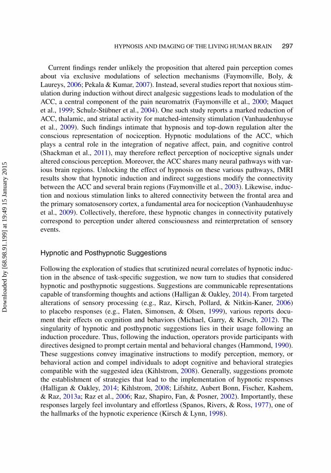
Hypnotic and posthypnotic suggestion can alter perception in dramatic fashion(Kihlstrom, 2008). From induced hallucinations to the suppression of pain sensations,evidence shows that suggestion directed at modulating perception influences sensoryprocessing and the conscious representation of sensory events. These suggestions typ-ically engage frontal and sensory areas of the human brain (see Figure 3). Critically,studies primarily highlight the capacity for suggestions to selectively target a partic-ular perceptual neural system; for example, suggestions to change visual perceptioncorrelate with modulation of visual processing (Kosslyn et al., 2000; McGeown et al.,2012; Raz, 2005), while suggestion to alter pain perception afflict somatosensory areasand the ACC, two central nodes involved in pain perception (Abrahamsen et al., 2010;Derbyshire et al., 2004, 2009; Hofbauer et al., 2001; Raij et al., 2005, 2009; Rainvilleet al., 1997, 1999; Wik et al., 1999). Moreover, suggestion possesses the selective capac-ity to precisely target components of subjective appraisal related to pain perception(Hofbauer et al., 2001; Rainville et al., 1997). Evidence also indicates that posthypnoticsuggestion can target higher processes concerned with perception, including valencejudgments (Ludwig et al., 2013).
Carefully crafted perceptual suggestions can bring about powerful effects (for review,see Lifshitz et al., 2013a). For example, suggestions to modify reading derail theballistic response in the Stroop effect, leading to marked improvements in task per-formances (Raz et al., 2002). Matching neural patterns accompany these behavioral
Dow
nloa
ded
by [
68.9
8.91
.199
] at
19:
49 1
5 Ja
nuar
y 20
15
HYPNOSIS AND IMAGING OF THE LIVING HUMAN BRAIN 299
FIGURE 3 Brain regions related to sensation and perceptual suggestions(laterality: “r” for right, “l” for left, and “b” for bilateral).
results, wherein a specific posthypnotic suggestion to obviate reading induces modu-lations of the visual areas and reduces ACC activity (Raz et al., 2005a). This neuralpattern proposes that suggestion likely mediates early sensory processing, subsequentlyaltering orthographic and semantic processing, which results in reduced cognitive con-flict between the reading and color-naming responses in the Stroop task. The contrastbetween the completion of the Stroop task with and without posthypnotic suggestionsaffords an examination of how suggestion may lead HHSs to adopt a potential heuristicstratagem.
Hypnotic hallucinations are astonishing perceptual phenomena, which have been thor-oughly documented in the laboratory (Bryant & Mallard, 2003; Kallio & Koivisto, 2013;Koivisto, Kirjanen, Revonsuo, & Kallio, 2013; Kosslyn et al., 2000; Mazzoni et al., 2009;McGeown et al., 2012; Spiegel, 2003; Szechtman et al., 1998; Woody & Szechtman,
Dow
nloa
ded
by [
68.9
8.91
.199
] at
19:
49 1
5 Ja
nuar
y 20
15
300 LANDRY AND RAZ
2000). Most imaging studies of hypnotic hallucinations report corresponding sensoryactivation; activation of perceptual representations is likely at the core (Derbyshire et al.,2004; Kosslyn et al., 2000; McGeown et al., 2012; Raij et al., 2005). Consistent withother forms of hallucinations (Allen, Larøi, McGuire, & Aleman, 2008), one hypothesispostulates that these particular experiences stem from distorted monitoring of reality,leading to misapprehension of mental representation and perceptual experience (Bryant& Mallard, 2003, 2005). In support of this view, hypnotic alterations of the ACC havebeen reliably reported during various forms of hypnotic hallucinations (Derbyshire et al.,2004; McGeown et al., 2012; Raij et al., 2005; Szechtman et al., 1998). Other findingspropose that the involvement of the right DLPFC correlates with the intensity of the hal-lucination (Raij et al., 2009). These neuroimaging reports are reminiscent of dissociationtheories wherein the breakdown in communication between control and monitoringprocesses may explain lapses in the proper evaluation and interpretation of perceptualrepresentations.
Memory
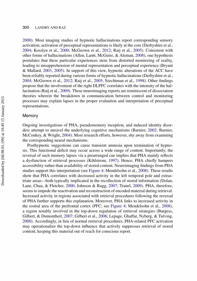
Ongoing investigations of PHA, pseudomemory inception, and induced identity disor-ders attempt to unravel the underlying cognitive mechanisms (Barnier, 2002; Barnier,McConkey, & Wright, 2004). Most research efforts, however, shy away from examiningthe corresponding neural mechanisms.
Posthypnotic suggestions can cause transient amnesia upon termination of hypno-sis. This functional deficit may occur across a wide range of content. Importantly, thereversal of such memory lapses via a prearranged cue implies that PHA mainly reflectsa dysfunction of retrieval processes (Kihlstrom, 1997). Hence, PHA chiefly hampersaccessibility rather than availability of stored content. Neuroimaging findings from PHAstudies support this interpretation (see Figure 4; Mendelsohn et al., 2008). These resultsshow that PHA correlates with decreased activity in the left temporal pole and extras-triate areas—both typically implicated in the recollection of stored information (Dolan,Lane, Chua, & Fletcher, 2000; Johnson & Rugg, 2007; Tranel, 2009). PHA, therefore,seems to impede the reactivation and reconstruction of encoded material during retrieval.Increased activity in regions associated with retrieval procedures following the reversalof PHA further supports this explanation. Moreover, PHA links to increased activity inthe rostral area of the prefrontal cortex (PFC; see Figure 4; Mendelsohn et al., 2008),a region notably involved in the top-down regulation of retrieval strategies (Burgess,Gilbert, & Dumontheil, 2007; Gilbert et al., 2006; Lepage, Ghaffar, Nyberg, & Tulving,2000). Accordingly, in lieu of normal retrieval procedures, PHA-related PFC activationmay operationalize the top-down influence that actively suppresses retrieval of storedcontent, keeping this material out of reach for conscious report.
Dow
nloa
ded
by [
68.9
8.91
.199
] at
19:
49 1
5 Ja
nuar
y 20
15
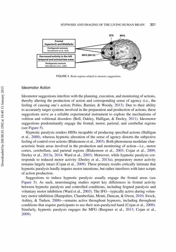
HYPNOSIS AND IMAGING OF THE LIVING HUMAN BRAIN 301
FIGURE 4 Brain regions related to memory suggestions.
Ideomotor Action
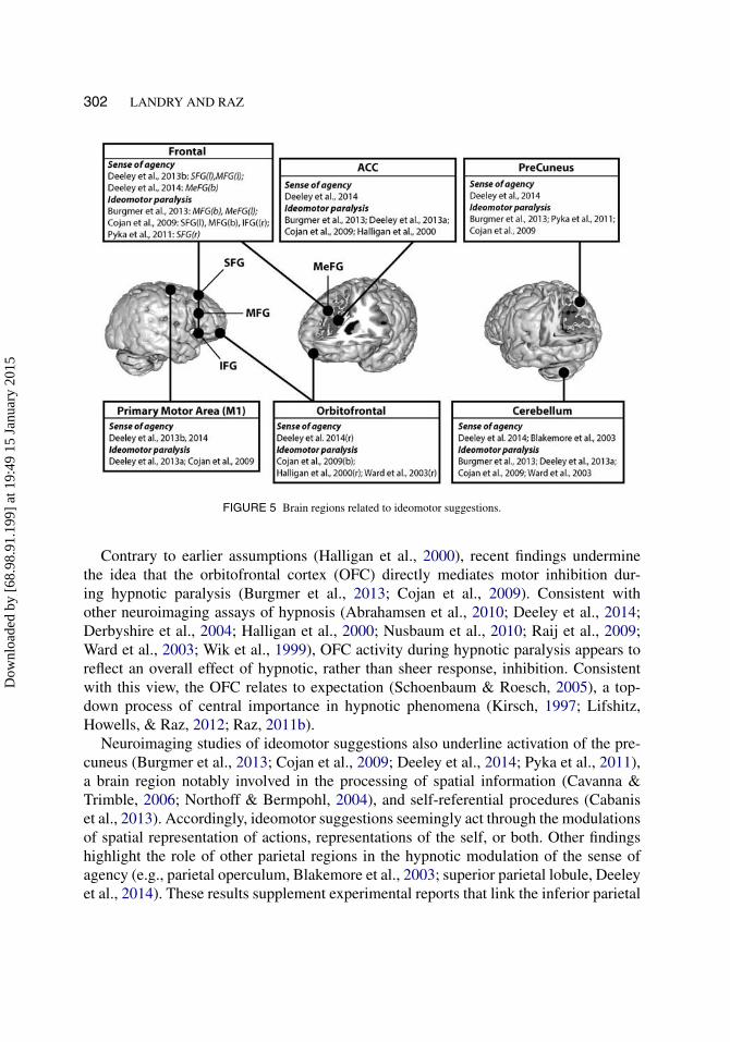
Ideomotor suggestions interfere with the planning, execution, and monitoring of actions,thereby altering the production of action and corresponding sense of agency (i.e., thefeeling of causing one’s action; Polito, Barnier, & Woody, 2013). Due to their abilityto accurately target systems involved in the preparation and production of actions, thesesuggestions serve as a reliable experimental instrument to explore the mechanisms ofvolition and volitional disorders (Bell, Oakley, Halligan, & Deeley, 2011). Ideomotorsuggestions predominantly engage the frontal, motor, parietal, and cerebellar regions(see Figure 5).
Hypnotic paralysis renders HHSs incapable of producing specified actions (Halliganet al., 2000), whereas hypnotic alteration of the sense of agency distorts the subjectivefeeling of control over actions (Blakemore et al., 2003). Both phenomena modulate char-acteristic brain areas involved in the production and monitoring of action—i.e., motorcortex, cerebellum, and parietal regions (Blakemore et al., 2003; Cojan et al., 2009;Deeley et al., 2013a, 2014; Ward et al., 2003). Moreover, while hypnotic paralysis cor-responds to reduced motor activity (Deeley et al., 2013a), preparatory motor activityremains largely intact (Cojan et al., 2009). These primary results critically intimate thathypnotic paralysis hardly impairs motor intentions, but rather interferes with later scriptsof action production.
Suggestions to induce hypnotic paralysis usually engage the frontal areas (seeFigure 5). As such, neuroimaging studies report key differences in frontal activitybetween hypnotic paralysis and controlled conditions, including feigned paralysis andvoluntary motor inhibition (Ward et al., 2003). The IFG—typically active during volun-tary motor inhibition (Hampshire, Chamberlain, Monti, Duncan, & Owen, 2010; Swick,Ashley, & Turken, 2008)—remains active throughout hypnosis, including throughoutconditions that require participants to use their non-paralyzed hand (Cojan et al., 2009).Similarly, hypnotic paralysis engages the MFG (Burgmer et al., 2013; Cojan et al.,2009).
Dow
nloa
ded
by [
68.9
8.91
.199
] at
19:
49 1
5 Ja
nuar
y 20
15
302 LANDRY AND RAZ
FIGURE 5 Brain regions related to ideomotor suggestions.
Contrary to earlier assumptions (Halligan et al., 2000), recent findings underminethe idea that the orbitofrontal cortex (OFC) directly mediates motor inhibition dur-ing hypnotic paralysis (Burgmer et al., 2013; Cojan et al., 2009). Consistent withother neuroimaging assays of hypnosis (Abrahamsen et al., 2010; Deeley et al., 2014;Derbyshire et al., 2004; Halligan et al., 2000; Nusbaum et al., 2010; Raij et al., 2009;Ward et al., 2003; Wik et al., 1999), OFC activity during hypnotic paralysis appears toreflect an overall effect of hypnotic, rather than sheer response, inhibition. Consistentwith this view, the OFC relates to expectation (Schoenbaum & Roesch, 2005), a top-down process of central importance in hypnotic phenomena (Kirsch, 1997; Lifshitz,Howells, & Raz, 2012; Raz, 2011b).
Neuroimaging studies of ideomotor suggestions also underline activation of the pre-cuneus (Burgmer et al., 2013; Cojan et al., 2009; Deeley et al., 2014; Pyka et al., 2011),a brain region notably involved in the processing of spatial information (Cavanna &Trimble, 2006; Northoff & Bermpohl, 2004), and self-referential procedures (Cabaniset al., 2013). Accordingly, ideomotor suggestions seemingly act through the modulationsof spatial representation of actions, representations of the self, or both. Other findingshighlight the role of other parietal regions in the hypnotic modulation of the sense ofagency (e.g., parietal operculum, Blakemore et al., 2003; superior parietal lobule, Deeleyet al., 2014). These results supplement experimental reports that link the inferior parietal
Dow
nloa
ded
by [
68.9
8.91
.199
] at
19:
49 1
5 Ja
nuar
y 20
15
HYPNOSIS AND IMAGING OF THE LIVING HUMAN BRAIN 303
region to action monitoring and the subjective experience of agency (Chambon, Wenke,Fleming, Prinz, & Haggard, 2013). In addition, ideomotor suggestion alters connectiv-ity between several brain regions, including the prefrontal, supplementary, and primarymotor area, and the parietal region, which leads to loss of perceived control during armmovement and decreased awareness of involuntary movements (Deeley et al., 2013b,2014). Collectively, these results bolster the notion that motor preparation and the plan-ning of action in the frontal and parietal sites relate to fundamental dimensions of thephenomenology of action and agency. They also highlight how ideomotor suggestionscan modify the coupling between various brain regions, altering production of actionand sense of agency.
Finally, ideomotor suggestions also encompass activation of the ACC (see Figure 5).During hypnotic paralysis, the ACC activity may potentially arise from the incongruencebetween the desired and actual outcome (Burgmer et al., 2013; Deeley et al., 2013a).According to this view, ACC activity indexes the online detection of a conflict betweenthe explicit intent and the observed result. Attempted movements during paralysis cor-respond to altered connectivity between the ACC and the motor cortex (Burgmer et al.,2013), as well as between the ACC and supplementary motor area (Deeley et al., 2013a).However, the prevalence of ACC activation in the neurocognitive literature of hypnosissuggests that this brain region likely indexes a main effect of hypnosis rather than task-specific effects (Casale et al., 2012). Supporting this latter interpretation, a neuroimagingstudy of hypnotic paralysis reports ACC activity during each hypnotic experimental con-dition, including situations that did not require attempts to move the paralyzed limb(Cojan et al., 2009). This result strongly supports the idea that ACC activity reflects aglobal effect related to hypnotic phenomena.
Conclusions
The wide spectrum of results from neuroimaging studies of hypnosis emphasizes theneed to follow tightly controlled experimental designs to better constrain the neuralcorrelates subserving specific hypnotic phenomena (Mazzoni et al., 2013). Moreover,the fact that few studies compare HHSs and LHSs diminishes the general validity ofresults. Focusing on HHSs alone impedes generalizability and overlooks inter-individualvariability in hypnotic responses (Heap et al., 2004; Laurence et al., 2008). Centrally,the distinction between induction and suggestion remains imprecise (Kihlstrom, 2008).Moreover, because most studies investigate hypnotic effects within a task, it remainsdifficult to experimentally isolate hypnotic-specific from task-specific effects. Beyondemploying experimental designs that account for individual differences, evidence fromvarious methodological approaches must be combined to inform and triangulate findings.For example, a recent study reports that temporary disruption of the left DLPFC with
Dow
nloa
ded
by [
68.9
8.91
.199
] at
19:
49 1
5 Ja
nuar
y 20
15
304 LANDRY AND RAZ
repetitive transcranial magnetic stimulation leads to increased sensibility to hypnoticsuggestion (Dienes & Hutton, 2013). In this regard, neuroimaging permits researchersto further appreciate the central role of specific brain regions in hypnotic response.Likewise, the combination of first-person approaches with cutting-edge brain-imagingtechniques provides a reliable paradigm to explore the neuroscience of subjective expe-riences (Lutz & Thompson, 2003). The neurophenomenology of hypnosis thereforeaffords researchers with unique insights concerning the phenomenological propertiesof hypnosis and their underlying neural correlates (Lifshitz, Cusumano, & Raz, 2013b).
The difference between hypnotic induction with and without task-specific sugges-tions underscores the importance of suggestions in causing hypnotic phenomena (Egner& Raz, 2007). Suggestion can induce potent effects; however, while the absence of task-specific suggestions relates to poor performance, posthypnotic suggestions may provideHHSs with reliable cognitive strategies that lead to substantial increases in performance.Neuroimaging findings substantiate this contrast. Hypnotic induction in the absenceof task-specific suggestions corresponds to fronto-thalamic modulations—presumablyreflecting engagement of attention—accompanied by deeper relaxation (Rainville et al.,2002). Neural responses to suggestion showcase the potential of hypnosis to selectivelytarget brain processes (Oakley & Halligan, 2009, 2013). However, because findingscut across various types of suggestion, each engaging a specific system, commonalitiesbetween categories of suggestions remain difficult to pinpoint. The prospect of a reliableneurobiological model of hypnosis, therefore, requires a finer appreciation of the scienceof suggestion (Halligan & Oakley, 2014; Michael et al., 2012). Overall, the interactionsbetween inter-individual variability, hypnotic responses, and hypnotic induction paint acomplex picture (Mazzoni et al., 2013). Most modern views attempt to account for thiscomplexity in terms of alterations of attention, expectation, cognitive control, and moni-toring (Kihlstrom, 2014; Kirsch, 1997; Maldonado & Spiegel, 2008; Raz, 2004; Woody& Sadler, 2008).
Neuroimaging studies of hypnosis reveal a primary role of top-down modulationsindexed by PFC and ACC activity. Dissociation theories argue that most hypnoticphenomena stem from a decoupling of control and monitoring processes (Woody &Farvolden, 1998; Woody & Szechtman, 2000). According to this model, PFC activityreflects the selection and implementation of hypnotic responses, while modulations ofthe ACC index changes in monitoring. This decoupling between control and monitor-ing procedures seems to enable suggestions to bypass evaluative procedures and directlyact upon control processes. This dissociation subsequently leads to misrepresentationsof hypnotic responses in consciousness (Kihlstrom, 2008). Specifically, without propermonitoring feedback, the implementation of the hypnotic response is less attributable tothe self and remains beyond subjective feelings of control. In sum, hypnosis yields sub-stantial changes in attention, control, and monitoring processing. It is likely that patternsof neural activity in the frontal areas operationalize these changes.
Dow
nloa
ded
by [
68.9
8.91
.199
] at
19:
49 1
5 Ja
nuar
y 20
15
HYPNOSIS AND IMAGING OF THE LIVING HUMAN BRAIN 305
Here we argue that the hypnotic experience alters connectivity between numerousbrain regions. However, neuroscientists are gradually mapping out the spatial loca-tion and time-course of neural events pertaining to hypnotic phenomena (Halligan &Oakley, 2013; Kihlstrom, 2013). Overall, neuroimaging studies not only deliver a bet-ter framework to understand hypnotic phenomena, but they supplement the importantphenomenological accounts and subjective impressions of participants with objectivemeasures of direct and indirect physiological indexes (Cusumano & Raz, 2014; Lifshitzet al., 2013b; Raz & Lifshitz, 2015). This juxtaposition paves the road to a more scientificunderstanding of hypnosis.
References
Abrahamsen, R., Dietz, M., Lodahl, S., Roepstorff, A., Zachariae, R., Østergaard, L., & Svensson, P. (2010).Effect of hypnotic pain modulation on brain activity in patients with temporomandibular disorder pain.Pain, 151, 825–833. doi:10.1016/j.pain.2010.09.020
Ali, S. S., Lifshitz, M., & Raz, A. (2014). Empirical neuroenchantment: From reading minds to thinkingcritically. Frontiers in Human Neuroscience, 8, 357. doi:10.3389/fnhum.2014.00357
Allen, P., Larøi, F., McGuire, P. K., & Aleman, A. (2008). The hallucinating brain: A review of structural andfunctional neuroimaging studies of hallucinations. Neuroscience & Biobehavioral Reviews, 32, 175–191.doi:10.1016/j.neubiorev.2007.07.012
Aue, T., Lavelle, L. A., & Cacioppo, J. T. (2009). Great expectations: What can fMRI researchtell us about psychological phenomena?. International Journal of Psychophysiology, 73, 10–16.doi:10.1016/j.ijpsycho.2008.12.017
Axmacher, N., Elger, C. E., & Fell, J. (2009). The specific contribution of neuroimaging ver-sus neurophysiological data to understanding cognition. Behavioural Brain Research, 200, 1–6.doi:10.1016/j.bbr.2009.01.028
Barnier, A. J. (2002). Remembering and forgetting autobiographical events: Instrumental uses of hypnosis.Contemporary Hypnosis, 19, 51–61. doi:10.1002/ch.242
Barnier, A. J., Cox, R. E., & McConkey, K. M. (2014). The province of “highs”: The high hypnotizableperson in the science of hypnosis and in psychological science. Psychology of Consciousness: Theory,Research, and Practice, 1, 168–183.
Barnier, A. J., McConkey, K. M., & Wright, J. (2004). Posthypnotic amnesia for autobiographical episodes:Influencing memory accessibility and quality. International Journal of Clinical & ExperimentalHypnosis, 52, 260–279. doi:10.1080/0020714049052351
Bell, V., Oakley, D. A., Halligan, P. W., & Deeley, Q. (2011). Dissociation in hysteria and hypnosis:Evidence from cognitive neuroscience. Journal of Neurology, Neurosurgery & Psychiatry, 82, 332–339.doi:10.1136/jnnp.2009.199158
Blakemore, S.-J., Oakley, D. A., & Frith, C. D. (2003). Delusions of alien control in the normal brain.Neuropsychologia, 41, 1058–1067. doi:10.1016/S0028-3932(02)00313-5
Bortolotti, L., Cox, R. E., & Barnier, A. J. (2012). Can we recreate delusions in the laboratory?.Philosophical Psychology, 25, 109–131. doi:10.1080/09515089.2011.569909
Botvinick, M. M. (2007). Conflict monitoring and decision making: Reconciling two perspectiveson anterior cingulate function. Cognitive, Affective, & Behavioral Neuroscience, 7, 356–366.doi:10.3758/CABN.7.4.356
Botvinick, M. M., Cohen, J. D., & Carter, C. S. (2004). Conflict monitoring and anterior cingulate cortex:An update. Trends in Cognitive Sciences, 8, 539–546. doi:10.1016/j.tics.2004.10.003
Dow
nloa
ded
by [
68.9
8.91
.199
] at
19:
49 1
5 Ja
nuar
y 20
15
306 LANDRY AND RAZ
Bryant, R. A., & Mallard, D. (2003). Seeing is believing: The reality of hypnotic hallucinations.Consciousness and Cognition, 12, 219–230. doi:10.1016/S1053-8100(03)00003-5
Bryant, R. A., & Mallard, D. (2005). Reality monitoring in hypnosis: A real-simulating anal-ysis. International Journal of Clinical and Experimental Hypnosis, 53, 13–25. doi:10.1080/00207140490914216
Buckner, R. L., Andrews-Hanna, J. R., & Schacter, D. L. (2008). The brain’s default network: Anatomy,function, and relevance to disease. Annals of the New York Academy of Sciences, 1124, 1–38.doi:10.1196/annals.1440.011
Burgess, P. W., Gilbert, S. J., & Dumontheil, I. (2007). Function and localization within rostral prefrontalcortex (area 10). Philosophical Transactions of the Royal Society B: Biological Sciences, 362, 887–899.doi:10.1098/rstb.2007.2095
Burgmer, M., Kugel, H., Pfleiderer, B., Ewert, A., Lenzen, T., Pioch, R., & Heuft, G. (2013). The mirrorneuron system under hypnosis–Brain substrates of voluntary and involuntary motor activation in hypnoticparalysis. Cortex, 49, 437–445. doi:10.1016/j.cortex.2012.05.023
Cabanis, M., Pyka, M., Mehl, S., Müller, B. W., Loos-Jankowiak, S., Winterer, G., & Rapp, A. M.(2013). The precuneus and the insula in self-attributional processes. Cognitive, Affective, & BehavioralNeuroscience, 13, 330–345. doi:10.3758/s13415-012-0143-5
Cardeña, E. (2014a). Hypnos and psyche: How hypnosis has contributed to the study of consciousness.Psychology of Consciousness: Theory, Research, and Practice, 1, 123.
Cardeña, E. (2014b). Spinning in circles. The Journal of Mind–Body Regulation, 2, 121–123.Cardeña, E., Jönsson, P., Terhune, D. B., & Marcusson-Clavertz, D. (2013). The neurophenomenology of
neutral hypnosis. Cortex, 49, 375–385. doi:10.1016/j.cortex.2012.04.001Carli, G., Manzoni, D., & Santarcangelo, E. L. (2008). Hypnotizability-related integration of perception and
action. Cognitive Neuropsychology, 25, 1065–1076. doi:10.1080/02643290801913712Casale, A. D., Ferracuti, S., Rapinesi, C., Serata, D., Sani, G., Savoja, V., & Girardi, P. (2012).
Neurocognition under hypnosis: Findings from recent functional neuroimaging studies. InternationalJournal of Clinical and Experimental Hypnosis, 60, 286–317. doi:10.1080/00207144.2012.675295
Cavanna, A. E., & Trimble, M. R. (2006). The precuneus: A review of its functional anatomy andbehavioural correlates. Brain, 129, 564–583. doi:10.1093/brain/awl004
Chambon, V., Wenke, D., Fleming, S. M., Prinz, W., & Haggard, P. (2013). An online neural substrate forsense of agency. Cerebral Cortex, 23, 1031–1037. doi:10.1093/cercor/bhs059
Choudhury, S., & Slaby, J. (2011). Critical neuroscience: A handbook of the social and cultural contexts ofneuroscience. London, UK: Wiley-Blackwell.
Cojan, Y., Waber, L., Schwartz, S., Rossier, L., Forster, A., & Vuilleumier, P. (2009). The brain under self-control: Modulation of inhibitory and monitoring cortical networks during hypnotic paralysis. Neuron,62, 862–875.
Connors, M. H. (2014). Defining hypnosis: Altered states and the need for parsimony. The Journal of Mind–Body Regulation, 2, 126–128.
Corbetta, M., Patel, G., & Shulman, G. L. (2008). The reorienting system of the human brain: Fromenvironment to theory of mind. Neuron, 58, 306–324. doi:10.1016/j.neuron.2008.04.017
Cox, R. E., & Barnier, A. J. (2010). Hypnotic illusions and clinical delusions: Hypnosis as a researchmethod. Cognitive Neuropsychiatry, 15, 202–232. doi:10.1080/13546800903319884
Cusumano, E. P., & Raz, A. (2014). Harnessing psychoanalytical methods for a phenomenologicalneuroscience. Frontiers in Psychology, 5, 334. doi:10.3389/fpsyg.2014.00334
Deeley, Q., Oakley, D. A., Toone, B., Bell, V., Walsh, E., Marquand, A. F., & Mehta, M. A. (2013a). Thefunctional anatomy of suggested limb paralysis. Cortex, 49, 411–422. doi:10.1016/j.cortex.2012.09.016
Deeley, Q., Oakley, D. A., Toone, B., Giampietro, V., Brammer, M. J., Williams, S. C. R., & Halligan, P.W. (2012). Modulating the default mode network using hypnosis. International Journal of Clinical andExperimental Hypnosis, 60, 206–228. doi:10.1080/00207144.2012.648070
Dow
nloa
ded
by [
68.9
8.91
.199
] at
19:
49 1
5 Ja
nuar
y 20
15
HYPNOSIS AND IMAGING OF THE LIVING HUMAN BRAIN 307
Deeley, Q., Oakley, D. A., Walsh, E., Bell, V., Mehta, M. A., & Halligan, P. W. (2014). Modellingpsychiatric and cultural possession phenomena with suggestion and fMRI. Cortex, 53, 107–119.doi:10.1016/j.cortex.2014.01.004
Deeley, Q., Walsh, E., Oakley, D. A., Bell, V., Koppel, C., Mehta, M. A., & Halligan, P. W. (2013b). Usinghypnotic suggestion to model loss of control and awareness of movements: An exploratory fMRI study.Plos One, 8, e78324. doi:10.1371/journal.pone.0078324
Demertzi, A., Soddu, A., Faymonville, M.-E., Bahri, M. A., Gosseries, O., Vanhaudenhuyse, A., & Laureys,S. (2011). Hypnotic modulation of resting state fMRI default mode and extrinsic network connectivity.Progress in Brain Research, 193, 309–322. doi:10.1016/B978-0-444-53839-0.00020-X
Derbyshire, S. W. G., Whalley, M. G., & Oakley, D. A. (2009). Fibromyalgia pain and its modulationby hypnotic and non-hypnotic suggestion: An fMRI analysis. European Journal of Pain, 13, 542–550.doi:10.1016/j.ejpain.2008.06.010
Derbyshire, S. W. G., Whalley, M. G., Stenger, V. A., & Oakley, D. A. (2004). Cerebralactivation during hypnotically induced and imagined pain. NeuroImage, 23, 392–401.doi:10.1016/j.neuroimage.2004.04.033
Dienes, Z., & Hutton, S. (2013). Understanding hypnosis metacognitively: rTMS applied to left DLPFCincreases hypnotic suggestibility. Cortex, 49, 386–392. doi:10.1016/j.cortex.2012.07.009
Dolan, R. J. (2008). Neuroimaging of cognition: Past, present, and future. Neuron, 60, 496–502.doi:10.1016/j.neuron.2008.10.038
Dolan, R. J., Lane, R., Chua, P., & Fletcher, P. (2000). Dissociable temporal lobe activations duringemotional episodic memory retrieval. Neuroimage, 11, 203–209. doi:10.1006/nimg.2000.0538
Egner, T., Jamieson, G. A., & Gruzelier, J. (2005). Hypnosis decouples cognitive control from conflict mon-itoring processes of the frontal lobe. NeuroImage, 27, 969–978. doi:10.1016/j.neuroimage.2005.05.002
Egner, T., & Raz, A. (2007). Cognitive control processes and hypnosis. In G. A. Jamieson (Ed.), Hypnosisand conscious states: The cognitive neuroscience perspective (pp. 29–50). New York, NY: OxfordUniversity Press.
Faymonville, M.-E., Boly, M., & Laureys, S. (2006). Functional neuroanatomy of the hypnotic state. Journalof Physiology-Paris, 99, 463–469. doi:10.1016/j.jphysparis.2006.03.018
Faymonville, M.-E., Laureys, S., Degueldre, C., DelFiore, G., Luxen, A., Franck, G., & Maquet, P.(2000). Neural mechanisms of antinociceptive effects of hypnosis. Anesthesiology, 92, 1257–1267.doi:10.1097/00000542-200005000-00013
Faymonville, M.-E., Roediger, L., Del Fiore, G., Delgueldre, C., Phillips, C., Lamy, M., & Laureys, S.(2003). Increased cerebral functional connectivity underlying the antinociceptive effects of hypnosis.Cognitive Brain Research, 17, 255–262. doi:10.1016/S0926-6410(03)00113-7
Filippi, M. Ed. (2009). fMRI techniques and protocols. New York, NY: Humana Press.Flaten, M. A., Simonsen, T., & Olsen, H. (1999). Drug-related information generates placebo
and nocebo responses that modify the drug response. Psychosomatic Medicine, 61, 250–255.doi:10.1097/00006842-199903000-00018
Fox, M. D., & Raichle, M. E. (2007). Spontaneous fluctuations in brain activity observed with functionalmagnetic resonance imaging. Nature Reviews Neuroscience, 8, 700–711. doi:10.1038/nrn2201
Fox, M. D., Snyder, A. Z., Vincent, J. L., Corbetta, M., Van Essen, D. C., & Raichle, M. E. (2005). Fromthe cover: The human brain is intrinsically organized into dynamic, anticorrelated functional networks.Proceedings of the National Academy of Sciences USA, 102, 9673–9678. doi:10.1073/pnas.0504136102
Gandhi, B., & Oakley, D. A. (2005). Does ‘hypnosis’ by any other name smell as sweet? The efficacyof ‘hypnotic’inductions depends on the label ‘hypnosis’. Consciousness and Cognition, 14, 304–315.doi:10.1016/j.concog.2004.12.004
Gilbert, S., Spengler, S., Simons, J., Steele, J., Lawrie, S., Frith, C., & Burgess, P. (2006). Functional special-ization within rostral prefrontal cortex (area 10): A meta-analysis. Journal of Cognitive Neuroscience,18, 932–948. doi:10.1162/jocn.2006.18.6.932
Dow
nloa
ded
by [
68.9
8.91
.199
] at
19:
49 1
5 Ja
nuar
y 20
15
308 LANDRY AND RAZ
Halligan, P. W., Athwal, B. S., Oakley, D. A., & Frackowiak, R. S. (2000). Imaging hypnotic paralysis:Implications for conversion hysteria. The Lancet, 355, 986–987. doi:10.1016/S0140-6736(00)99019-6
Halligan, P. W., & Oakley, D. A. (2013). Hypnosis and cognitive neuroscience: Bridging the gap. Cortex,49, 359–364. doi:10.1016/j.cortex.2012.12.002
Halligan, P. W., & Oakley, D. A. (2014). Hypnosis and beyond: Exploring the broader domain of suggestion.Psychology of Consciousness: Theory, Research, and Practice, 1, 105.
Hammond, D. C. (1990). Handbook of hypnotic suggestions and metaphors. New York, NY: W.W. Norton& Company.
Hampshire, A., Chamberlain, S. R., Monti, M. M., Duncan, J., & Owen, A. M. (2010). The roleof the right inferior frontal gyrus: Inhibition and attentional control. Neuroimage, 50, 1313–1319.doi:10.1016/j.neuroimage.2009.12.109
Heap, M., Brown, R. J., & Oakley, D. A. (2004). The highly hypnotizable person: Theoretical, experimentaland clinical issues. London: Routledge.
Henson, R. (2005). What can functional neuroimaging tell the experimental psychologist?. The QuarterlyJournal of Experimental Psychology Section A, 58, 193–233. doi:10.1080/02724980443000502
Henson, R. (2006). Forward inference using functional neuroimaging: Dissociations versus associations.Trends in Cognitive Sciences, 10, 64–69. doi:10.1016/j.tics.2005.12.005
Hofbauer, R. K., Rainville, P., Duncan, G. H., & Bushnell, M. C. (2001). Cortical representation of thesensory dimension of pain. Journal of Neurophysiology, 86, 402–411.
Jamieson, G. A. (2007). Hypnosis and conscious states: The cognitive neuroscience perspective. New York,NY: Oxford University Press.
Jamieson, G. A., & Woody, E. Z. (2007). Dissociated control as a paradigm for cognitive neuroscienceresearch and theorizing in hypnosis. In G. A. Jamieson (Ed.), Hypnosis and conscious states: Thecognitive neuroscience perspective (pp. 111–132). New York, NY: Oxford University Press.
Johnson, J. D., & Rugg, M. D. (2007). Recollection and the reinstatement of encoding-related corticalactivity. Cerebral Cortex, 17, 2507–2515. doi:10.1093/cercor/bhl156
Jones, E. G., & Mendell, L. M. (1999). Assessing the decade of the brain. Science, 284, 739–739.doi:10.1126/science.284.5415.739
Kallio, S., & Koivisto, M. (2013). Posthypnotic suggestion alters conscious color perception inan automatic manner. International Journal of Clinical & Experimental Hypnosis, 61, 371–387.doi:10.1080/00207144.2013.810446
Kihlstrom, J. F. (1985). Hypnosis. Annual Review of Psychology, 36, 385–418.Kihlstrom, J. F. (1997). Hypnosis, memory and amnesia. Philosophical Transactions of the Royal Society
B-Biological Sciences, 352, 1727–1732. doi:10.1098/rstb.1997.0155Kihlstrom, J. F. (2008). The domain of hypnosis, revisited. In M. R. Nash & A. J. Barnier (Eds.), The Oxford
handbook of hypnosis: Theory, research and practice. Oxford, UK: Oxford University Press.Kihlstrom, J. F. (2013). Neuro-hypnotism: Prospects for hypnosis and neuroscience. Cortex, 49, 365–374.
doi:10.1016/j.cortex.2012.05.016Kihlstrom, J. F. (2014). Hypnosis and cognition. Psychology of Consciousness: Theory, Research, and
Practice, 1, 139–152.Kihlstrom, J. F., & Edmonston, J. W. E. (1971). Alterations in consciousness in neutral hyp-
nosis: Distortions in semantic space. American Journal of Clinical Hypnosis, 13, 243–248.doi:10.1080/00029157.1971.10402120
Kirmayer, L. J., & Crafa, D. (2014). What kind of science for psychiatry?. Frontiers in Human Neuroscience,8, 435. doi:10.3389/fnhum.2014.00435
Kirsch, I. (1997). Response expectancy theory and application: A decennial review. Applied and PreventivePsychology, 6, 69–79. doi:10.1016/S0962-1849(05)80012-5
Kirsch, I. (2014). Wagstaff’s definition of hypnosis. The Journal of Mind–Body Regulation, 2, 124–125.
Dow
nloa
ded
by [
68.9
8.91
.199
] at
19:
49 1
5 Ja
nuar
y 20
15
HYPNOSIS AND IMAGING OF THE LIVING HUMAN BRAIN 309
Kirsch, I., Cardena, E., Derbyshire, S. W., Dienes, Z., Heap, M., Kallio, S., & Potter, C. (2011). Definitionsof hypnosis and hypnotizability and their relation to suggestion and suggestibility: A consensusstatement. Contemporary Hypnosis, 28, 107–115.
Kirsch, I., & Lynn, S. J. (1995). The altered state of hypnosis—changes in the theoretical landscape.American Psychologist, 50, 846–858. doi:10.1037/0003-066X.50.10.846
Kirsch, I., & Lynn, S. J. (1998). Dissociation theories of hypnosis. Psychological Bulletin, 123, 100.Koivisto, M., Kirjanen, S., Revonsuo, A., & Kallio, S. (2013). A preconscious neural mechanism of hypnot-
ically altered colors: A double case study. Plos One, 8, e70900. doi:10.1371/journal.pone.0070900Kosslyn, S. M., Thompson, W. L., Costantini-Ferrando, M. F., Alpert, N. M., & Spiegel, D. (2000). Hypnotic
visual illusion alters color processing in the brain. The American Journal of Psychiatry, 157, 1279–1284.doi:10.1176/appi.ajp.157.8.1279
Kuhn, G., & Tatler, B. W. (2011). Misdirected by the gap: The relationship between inat-tentional blindness and attentional misdirection. Consciousness and Cognition, 20, 432–436.doi:10.1016/j.concog.2010.09.013
Landry, M., Appourchaux, K., & Raz, A. (2014). Elucidating unconscious processing with instrumentalHypnosis. Frontiers in Psychology, 5, 785. doi:10.3389/fpsyg.2014.00785
Laurence, J.-R. (2014). When in doubt, forbear! The Journal of Mind–Body Regulation, 2, 109–111.Laurence, J.-R., Beaulieu-Prévost, D., & Du Chéné, T. (2008). Measuring and understanding individual
differences in hypnotizability. In A. J. Barnier & M. R. Nash (Eds.), The Oxford handbook of hypnosis:Theory, research, and practice. New York, NY: Oxford University Press.
Lepage, M., Ghaffar, O., Nyberg, L., & Tulving, E. (2000). Prefrontal cortex and episodic memory retrievalmode. Proceedings of the National Academy of Sciences USA, 97, 506–511. doi:10.1073/pnas.97.1.506
Lifshitz, M., Aubert Bonn, N., Fischer, A., Kashem, I. F., & Raz, A. (2013a). Using suggestion to mod-ulate automatic processes: From Stroop to McGurk and beyond. Cortex, 49, 463–473. doi:10.1016/j.cortex.2012.08.007
Lifshitz, M., Cusumano, E. P., & Raz, A. (2013b). Hypnosis as neurophenomenology. Frontiers in HumanNeuroscience, 7, 469. doi:10.3389/fnhum.2013.00469
Lifshitz, M., Howells, C., & Raz, A. (2012). Can expectation enhance response to sugges-tion? De-automatization illuminates a conundrum. Consciousness and Cognition, 21, 1001–1008.doi:10.1016/j.concog.2012.02.002
Ludwig, V. U., Stelzel, C., Krutiak, H., Magrabi, A., Steimke, R., Paschke, L. M., & Walter, H. (2013). Thesuggestible brain: Posthypnotic effects on value-based decision-making. Social Cognitive and AffectiveNeuroscience. doi:10.1093/scan/nst110
Lutz, A., & Thompson, E. (2003). Neurophenomenology: Integrating subjective experience and braindynamics in the neuroscience of consciousness. Journal of Consciousness Studies, 10, 31–52.
Lynn, S. J., Kirsch, I., & Hallquist, M. N. (2008). Social cognitive theories of hypnosis. In M. R. Nash &A. J. Barnier (Eds.), The Oxford handbook of hypnosis: Theory, research and practice (pp. 111–139).Oxford, UK: Oxford University Press.
Macdonald, E. B., & Raz, A. (2014). The marginalization of phenomenological consciousness. Frontiers inHuman Neuroscience, 8, 306. doi:10.3389/fnhum.2014.00306
MacLeod, C. M. (1991). Half a century of research on the Stroop effect: An integrative review.Psychological Bulletin, 109, 163–203. doi:10.1037/0033-2909.109.2.163
Maldonado, J. R., & Spiegel, D. (2008). Hypnosis psychiatry (pp. 1982–2026). Chichester, UK: John Wiley& Sons, Ltd.
Malhi, G. S., & Lagopoulos, J. (2008). Making sense of neuroimaging in psychiatry. Acta PsychiatricaScandinavica, 117, 100–117.
Maquet, P., Faymonville, M.-E., Degueldre, C., Delfiore, G., Franck, G., Luxen, A., & Lamy, M.(1999). Functional neuroanatomy of hypnotic state. Biological Psychiatry, 45, 327–333. doi:10.1016/S0006-3223(97)00546-5
Dow
nloa
ded
by [
68.9
8.91
.199
] at
19:
49 1
5 Ja
nuar
y 20
15
310 LANDRY AND RAZ
Mazzoni, G., Rotriquenz, E., Carvalho, C., Vannucci, M., Roberts, K., & Kirsch, I. (2009).Suggested visual hallucinations in and out of hypnosis. Consciousness and Cognition, 18, 494–499.doi:10.1016/j.concog.2009.02.002
Mazzoni, G., Venneri, A., McGeown, W. J., & Kirsch, I. (2013). Neuroimaging resolution of the alteredstate hypothesis. Cortex, 49, 400–410. doi:10.1016/j.cortex.2012.08.005
McConkey, K. M., & Barnier, A. J. (2004). High hypnotisability: Unity and diversity in behaviour and expe-rience. In M. Heap, R. J. Brown, & D. A. Oakley (Eds.), The highly hypnotizable person: Theoretical,experimental and clinical issues (pp. 61–84). New York, NY: Routledge.
McConkey, K. M., Glisky, M. L., & Kihlstrom, J. F. (1989). Individual differences among hypnoticvirtuosos: A case comparison. Australian Journal of Clinical & Experimental Hypnosis, 17, 131–140.
McGeown, W. J., Mazzoni, G., Venneri, A., & Kirsch, I. (2009). Hypnotic induction decreases anteriordefault mode activity. Consciousness and Cognition, 18, 848–855. doi:10.1016/j.concog.2009.09.001
McGeown, W. J., Venneri, A., Kirsch, I., Nocetti, L., Roberts, K., Foan, L., & Mazzoni, G. (2012). Suggestedvisual hallucination without hypnosis enhances activity in visual areas of the brain. Consciousness andCognition, 21, 100–116. doi:10.1016/j.concog.2011.10.015
Memmert, D. (2010). The gap between inattentional blindness and attentional misdirection. Consciousnessand Cognition, 19, 1097–1101. doi:10.1016/j.concog.2010.01.001
Mendelsohn, A., Chalamish, Y., Solomonovich, A., & Dudai, Y. (2008). Mesmerizing memories:Brain substrates of episodic memory suppression in posthypnotic amnesia. Neuron, 57, 159–170.doi:10.1016/j.neuron.2007.11.022
Michael, R. B., Garry, M., & Kirsch, I. (2012). Suggestion, cognition, and behavior. Current Directions inPsychological Science, 21, 151–156. doi:10.1177/0963721412446369
Moran, A., & Brady, N. (2010). Mind the gap: Misdirection, inattentional blindness and therelationship between overt and covert attention. Consciousness and Cognition, 19, 1105–1106.doi:10.1016/j.concog.2010.03.011
Most, S. B. (2010). What’s “inattentional” about inattentional blindness?. Consciousness and Cognition, 19,1102–1104. doi:10.1016/j.concog.2010.01.011
Müller, K., Bacht, K., Prochnow, D., Schramm, S., & Seitz, R. J. (2013). Activation of thala-mus in motor imagery results from gating by hypnosis. Neuroimage, 66, 361–367. doi:10.1016/j.neuroimage.2012.10.073
Müller, K., Bacht, K., Schramm, S., & Seitz, R. J. (2012). The facilitating effect of clinicalhypnosis on motor imagery: An fMRI study. Behavioural Brain Research, 231, 164–169. doi:10.1016/j.bbr.2012.03.013
Nash, M. R., & Barnier, A. J. (2008). The Oxford handbook of hypnosis: Theory, research and practice.Oxford, UK: Oxford University Press.
Nathan, P. J., Phan, K. L., Harmer, C. J., Mehta, M. A., & Bullmore, E. T. (2014). Increasingpharmacological knowledge about human neurological and psychiatric disorders through functionalneuroimaging and its application in drug discovery. Current Opinion in Pharmacology, 14, 54–61.doi:10.1016/j.coph.2013.11.009
Nee, D. E., Brown, J. W., Askren, M. K., Berman, M. G., Demiralp, E., Krawitz, A., & Jonides, J.(2013). A meta-analysis of executive components of working memory. Cerebral Cortex, 23, 264–282.doi:10.1093/cercor/bhs007
Northoff, G., & Bermpohl, F. (2004). Cortical midline structures and the self. Trends in Cognitive Sciences,8, 102–107. doi:10.1016/j.tics.2004.01.004
Nusbaum, F., Redouté, J., Le Bars, D., Volckmann, P., Simon, F., Hannoun, S., & Sappey-Marinier, D.(2010). Chronic low-back pain modulation is enhanced by hypnotic analgesic suggestion by recruiting anemotional network: A PET imaging study. International Journal of Clinical and Experimental Hypnosis,59, 27–44. doi:10.1080/00207144.2011.522874
O’Neil, J. A. (2014). Hazards of ‘final’ definition. The Journal of Mind–Body Regulation, 2, 129–131.
Dow
nloa
ded
by [
68.9
8.91
.199
] at
19:
49 1
5 Ja
nuar
y 20
15
HYPNOSIS AND IMAGING OF THE LIVING HUMAN BRAIN 311
Oakley, D. A., Deeley, Q., & Halligan, P. W. (2007). Hypnotic depth and response to suggestion understandardized conditions and during fMRI scanning. International Journal of Clinical and ExperimentalHypnosis, 55, 32–58. doi:10.1080/00207140600995844
Oakley, D. A., & Halligan, P. W. (2009). Hypnotic suggestion and cognitive neuroscience. Trends inCognitive Sciences, 13, 264–270. doi:10.1016/j.tics.2009.03.004
Oakley, D. A., & Halligan, P. W. (2010). Psychophysiological foundations of hypnosis and suggestion. In J.W. Rhue, S. J. Lynn, & I. Kirsch (Eds.), Handbook of clinical hypnosis (pp. 79–177). Washington, DC:American Psychological Association.
Oakley, D. A., & Halligan, P. W. (2013). Hypnotic suggestion: Opportunities for cognitive neuroscience.Nature Reviews Neuroscience, 14, 565–576. doi:10.1038/nrn3538
Pekala, R. J., & Kumar, V. K. (2007). An empirical—phenomenological approach to quantifying conscious-ness. In G. Jamieson (Ed.), Hypnosis and conscious states: The cognitive neuroscience perspective (pp.167–194). New York, NY: Oxford University Press.
Piccione, C., Hilgard, E. R., & Zimbardo, P. G. (1989). On the degree of stability of measuredhypnotizability over a 25-year period. Journal of Personality and Social Psychology, 56, 289–295.doi:10.1037/0022-3514.56.2.289
Poldrack, R. A. (2012). The future of fMRI in cognitive neuroscience. Neuroimage, 62, 1216–1220.doi:10.1016/j.neuroimage.2011.08.007
Polito, V., Barnier, A. J., & McConkey, K. M. (2014). Defining hypnosis: Process, product, and the value oftolerating ambiguity. The Journal of Mind–Body Regulation, 2, 118–120.
Polito, V., Barnier, A. J., & Woody, E. Z. (2013). Developing the sense of agency rating scale (SOARS):An empirical measure of agency disruption in hypnosis. Consciousness and Cognition, 22, 684–696.doi:10.1016/j.concog.2013.04.003
Posner, M. I., & Petersen, S. E. (1990). The attention system of the human brain. Annual Review ofNeuroscience, 13, 25–42. doi:10.1146/annurev.ne.13.030190.000325
Pyka, M., Burgmer, M., Lenzen, T., Pioch, R., Dannlowski, U., Pfleiderer, B., & Konrad, C. (2011).Brain correlates of hypnotic paralysis—a resting-state fMRI study. NeuroImage, 56, 2173–2182.doi:10.1016/j.neuroimage.2011.03.078
Raij, T. T., Numminen, J., Narvanen, S., Hiltunen, J., & Hari, R. (2005). Brain correlates of subjective realityof physically and psychologically induced pain. Proceedings of the National Academy of Sciences USA,102, 2147–2151. doi:10.1073/pnas.0409542102
Raij, T. T., Numminen, J., Närvänen, S., Hiltunen, J., & Hari, R. (2009). Strength of prefrontalactivation predicts intensity of suggestion-induced pain. Human Brain Mapping, 30, 2890–2897.doi:10.1002/hbm.20716
Rainville, P., Carrier, B., Hofbauer, R. K., Bushnell, M. C., & Duncan, G. H. (1999). Dissociationof sensory and affective dimensions of pain using hypnotic modulation. Pain, 82, 159–171.doi:10.1016/S0304-3959(99)00048-2
Rainville, P., Duncan, G. H., Price, D. D., Carrier, B., & Bushnell, M. C. (1997). Pain affectencoded in human anterior cingulate but not somatosensory cortex. Science, 277, 968–971.doi:10.1126/science.277.5328.968
Rainville, P., Hofbauer, R. K., Bushnell, M. C., Duncan, G. H., & Price, D. D. (2002). Hypnosis mod-ulates activity in brain structures involved in the regulation of consciousness. Journal of CognitiveNeuroscience, 14, 887–901. doi:10.1162/089892902760191117
Rainville, P., & Price, D. D. (2003). Hypnosis phenomenology and the neurobiology of con-sciousness. International Journal of Clinical and Experimental Hypnosis, 51, 105–129. doi:10.1076/iceh.51.2.105.14613
Raz, A. (2004). Atypical attention: Hypnosis and conflict reduction. In M. I. Posner (Ed.), Cognitiveneuroscience of attention. New York, NY: Guilford Press.
Dow
nloa
ded
by [
68.9
8.91
.199
] at
19:
49 1
5 Ja
nuar
y 20
15
312 LANDRY AND RAZ
Raz, A. (2005). Attention and hypnosis: Neural substrates and genetic associations of two con-verging processes. International Journal of Clinical and Experimental Hypnosis, 53, 237–258.doi:10.1080/00207140590961295
Raz, A. (2011a). Does neuroimaging of suggestion elucidate hypnotic trance? International Journal ofClinical and Experimental Hypnosis, 59, 363–377. doi:10.1080/00207144.2011.570682
Raz, A. (2011b). Hypnosis: A twilight zone of the top-down variety Few have never heard of hypnosis butmost know little about the potential of this mind-body regulation technique for advancing science. Trendsin Cognitive Sciences, 15, 555–557. doi:10.1016/j.tics.2011.10.002
Raz, A., & Buhle, J. (2006). Typologies of attentional networks. Nature Reviews Neuroscience, 7, 367–379.doi:10.1038/nrn1903
Raz, A., Fan, J., & Posner, M. I. (2005a). Hypnotic suggestion reduces conflict in the human brain.Proceedings of the National Academy of Sciences USA, 102, 9978–9983. doi:10.1073/pnas.0503064102
Raz, A., Kirsch, I., Pollard, J., & Nitkin-Kaner, Y. (2006). Suggestion reduces the stroop effect.Psychological Science, 17, 91–95. doi:10.1111/j.1467-9280.2006.01669.x
Raz, A., Lieber, B., Soliman, F., Buhle, J., Posner, J., Peterson, B. S., & Posner, M. I. (2005b). Ecologicalnuances in functional magnetic resonance imaging (fMRI): Psychological stressors, posture, andhydrostatics. Neuroimage, 25, 1–7. doi:10.1016/j.neuroimage.2004.11.015
Raz, A., & Lifshitz, M. (2015). Hypnosis and meditation: Towards an integrative science of consciousplanes. Oxford, UK: Oxford University Press.
Raz, A., & Macdonald, E. B. (2014). Narrow perspectives on consciousness. PsycCRITIQUES, 59, 18.doi:10.1037/a0036440
Raz, A., & Macdonald, E. B. (2015). Paying attention to a field in crisis. In L. J. Kirmayer, M.-F. Chesselet,K. Shinobu, & C. Worthman (Eds.), Revisioning psychiatry. Cambridge, MA: Cambridge UniversityPress.
Raz, A., & Shapiro, T. (2002). Hypnosis and neuroscience: A cross talk between clinical and cognitiveresearch. Archives of General Psychiatry, 59, 85–90. doi:10.1001/archpsyc.59.1.85
Raz, A., Shapiro, T., Fan, J., & Posner, M. I. (2002). Hypnotic suggestion and the modulation of Stroopinterference. Archives of General Psychiatry, 59, 1155–1161. doi:10.1001/archpsyc.59.12.1155
Röder, C. H., Michal, M., Overbeck, G., van de Ven, V. G., & Linden, D. E. J. (2007). Pain response indepersonalization: A functional imaging study using hypnosis in healthy subjects. Psychotherapy andPsychosomatics, 76(2), 115–121.
Schoenbaum, G., & Roesch, M. (2005). Orbitofrontal cortex, associative learning, and expectancies. Neuron,47, 633–636. doi:10.1016/j.neuron.2005.07.018
Schulz-Stübner, S., Krings, T., Meister, I. G., Rex, S., Thron, A., & Rossaint, R. (2004). Clinicalhypnosis modulates functional magnetic resonance imaging signal intensities and pain percep-tion in a thermal stimulation paradigm. Regional Anesthesia and Pain Medicine, 29, 549–556.doi:10.1097/00115550-200411000-00008
Shackman, A. J., Salomons, T. V., Slagter, H. A., Fox, A. S., Winter, J. J., & Davidson, R. J. (2011).The integration of negative affect, pain and cognitive control in the cingulate cortex. Nature ReviewsNeuroscience, 12, 154–167. doi:10.1038/nrn2994
Shenhav, A., Botvinick, M. M., & Cohen, J. D. (2013). The expected value of control: An integrative theoryof anterior cingulate cortex function. Neuron, 79, 217–240. doi:10.1016/j.neuron.2013.07.007
Spanos, N. P., Rivers, S. M., & Ross, S. (1977). Experienced involuntariness and response to hyp-notic suggestions. Annals of the New York Academy of Sciences, 296, 208–221. doi:10.1111/j.1749-6632.1977.tb38173.x
Spiegel, D. (2003). Negative and positive visual hypnotic hallucinations: Attending inside andout. International Journal of Clinical & Experimental Hypnosis, 51, 130–146. doi:10.1076/iceh.51.2.130.14612
Dow
nloa
ded
by [
68.9
8.91
.199
] at
19:
49 1
5 Ja
nuar
y 20
15
HYPNOSIS AND IMAGING OF THE LIVING HUMAN BRAIN 313
Swick, D., Ashley, V., & Turken, U. (2008). Left inferior frontal gyrus is critical for response inhibition.BMC Neuroscience, 9, 102. doi:10.1186/1471-2202-9-102
Szechtman, H., Woody, E. Z., Bowers, K. S., & Nahmias, C. (1998). Where the imaginal appears real:A positron emission tomography study of auditory hallucinations. The Proceedings of the NationalAcademy of Sciences of the United States of America, 95, 1956–1960. doi:10.1073/pnas.95.4.1956
Terhune, D. B. (2014). Defining hypnosis: The pitfalls of prioritizing spontaneous experience over responseto suggestion. The Journal of Mind–Body Regulation, 2, 115–117.
Terhune, D. B., Cardeña, E., & Lindgren, M. (2011). Dissociated control as a signature of typo-logical variability in high hypnotic suggestibility. Consciousness and Cognition, 20, 727–736.doi:10.1016/j.concog.2010.11.005
Thibault, R. T., Lifshitz, M., Jones, J. M., & Raz, A. (2014). Posture alters human resting-state. Cortex, 58,199–205. doi:10.1016/j.cortex.2014.06.014
Tranel, D. (2009). The left temporal pole is important for retrieving words for unique concrete entities.Aphasiology, 23, 867–884. doi:10.1080/02687030802586498
Vanhaudenhuyse, A., Boly, M., Balteau, E., Schnakers, C., Moonen, G., Luxen, A., & Maquet, P. (2009).Pain and non-pain processing during hypnosis: A thulium-YAG event-related fMRI study. Neuroimage,47, 1047–1054. doi:10.1016/j.neuroimage.2009.05.031
Wagstaff, G. F. (1998). The semantics and physiology of hypnosis as an altered state: Towards a definitionof hypnosis. Contemporary Hypnosis, 15, 149–165. doi:10.1002/ch.125
Wagstaff, G. F. (2014). On the centrality of the concept of an altered state to definitions of hypnosis. TheJournal of Mind–Body Regulation, 2, 90–108.
Ward, N. S., Oakley, D. A., Frackowiak, R. S. J., & Halligan, P. W. (2003). Differential brain activationsduring intentionally simulated and subjectively experienced paralysis. Cognitive Neuropsychiatry, 8,295–312. doi:10.1080/13546800344000200
Wik, G., Fischer, H., Bragée, B., Finer, B., & Fredrikson, M. (1999). Functional anatomy of hyp-notic analgesia: A PET study of patients with fibromyalgia. European Journal of Pain, 3, 7–12.doi:10.1016/S1090-3801(99)90183-0
Woody, E. Z., & Farvolden, P. (1998). Dissociation in hypnosis and frontal executive function. AmericanJournal of Clinical Hypnosis, 40, 206–216. doi:10.1080/00029157.1998.10403427
Woody, E. Z., & Sadler, P. (2008). Dissociation theories of hypnosis. In M. R. Nash & A. J. Barnier (Eds.),The Oxford handbook of hypnosis (pp. 81–110). Oxford, UK: Oxford University Press.
Woody, E. Z., & Sadler, P. (2014). A somewhat altered debate about the hypnotic state. The Journal ofMind–Body Regulation, 2, 112–114.
Woody, E. Z., & Szechtman, H. (2000). Hypnotic hallucinations: Towards a biology of epistemology.Contemporary Hypnosis, 17, 4–14. doi:10.1002/ch.186D
ownl
oade
d by
[68
.98.
91.1
99]
at 1
9:49
15
Janu
ary
2015