Embed Size (px)
Citation preview

ABCDEFG
UNIVERS ITY OF OULU P.O.B . 7500 F I -90014 UNIVERS ITY OF OULU F INLAND
A C T A U N I V E R S I T A T I S O U L U E N S I S
S E R I E S E D I T O R S
SCIENTIAE RERUM NATURALIUM
HUMANIORA
TECHNICA
MEDICA
SCIENTIAE RERUM SOCIALIUM
SCRIPTA ACADEMICA
OECONOMICA
EDITOR IN CHIEF
PUBLICATIONS EDITOR
Senior Assistant Jorma Arhippainen
Lecturer Santeri Palviainen
Professor Hannu Heusala
Professor Olli Vuolteenaho
Senior Researcher Eila Estola
Director Leila Risteli
Professor Jari Juga
Professor Olli Vuolteenaho
Publications Editor Kirsti Nurkkala
ISBN 978-951-42-9402-0 (Paperback)ISBN 978-951-42-9403-7 (PDF)ISSN 0355-3221 (Print)ISSN 1796-2234 (Online)
U N I V E R S I TAT I S O U L U E N S I S
MEDICA
ACTAD
D 1092
ACTA
Tuija Männistö
OULU 2011
D 1092
Tuija Männistö
MATERNAL THYROID FUNCTION DURING PREGNANCYEFFECTS ON PREGNANCY, PERI- AND NEONATAL OUTCOME AND ON LATER MATERNAL HEALTH
UNIVERSITY OF OULU, FACULTY OF MEDICINE,INSTITUTE OF CLINICAL MEDICINE, DEPARTMENT OF OBSTETRICS AND GYNECOLOGY,INSTITUTE OF HEALTH SCIENCES,INSTITUTE OF DIAGNOSTICS, DEPARTMENT OF CLINICAL CHEMISTRY;NATIONAL INSTITUTE FOR HEALTH AND WELFARE,DEPARTMENT OF CHILDREN, YOUNG PEOPLE AND FAMILIES


A C T A U N I V E R S I T A T I S O U L U E N S I SD M e d i c a 1 0 9 2
TUIJA MÄNNISTÖ
MATERNAL THYROID FUNCTION DURING PREGNANCYEffects on pregnancy, peri- and neonatal outcome and on later maternal health
Academic Dissertation to be presented with the assent ofthe Faculty of Medicine of the University of Oulu forpublic defence in Auditorium 4 of Oulu UniversityHospital, on 15 April 2011, at 12 noon
UNIVERSITY OF OULU, OULU 2011

Copyright © 2011Acta Univ. Oul. D 1092, 2011
Supervised byDoctor Eila SuvantoDocent Anneli PoutaProfessor Marjo-Riitta Järvelin
Reviewed byDocent Camilla Schalin-JänttiDocent Esa Hämäläinen
ISBN 978-951-42-9402-0 (Paperback)ISBN 978-951-42-9403-7 (PDF)http://herkules.oulu.fi/isbn9789514294037/ISSN 0355-3221 (Printed)ISSN 1796-2234 (Online)http://herkules.oulu.fi/issn03553221/
Cover DesignRaimo Ahonen
JUVENES PRINTTAMPERE 2011

Männistö, Tuija, Maternal thyroid function during pregnancy. Effects onpregnancy, peri- and neonatal outcome and on later maternal healthUniversity of Oulu, Faculty of Medicine, Institute of Clinical Medicine, Department ofObstetrics and Gynecology, Institute of Health Sciences, Institute of Diagnostics, Department ofClinical Chemistry, P.O. Box 5000, FI-90014 University of Oulu, Finland; National Institute forHealth and Welfare, Department of Children, Young People and Families, P.O. Box 310, FI-90101 Oulu, FinlandActa Univ. Oul. D 1092, 2011Oulu, Finland
AbstractMaternal thyroid dysfunction and/or antibodies are present in 5-10% of pregnancies and may beassociated with increased risks of adverse pregnancy and perinatal outcomes. In the present studymaternal thyroid function and antibody status in the Northern Finland Birth Cohort 1986 wasanalyzed using early pregnancy serum samples.
The impact of long-term storage on the stability of thyroid hormones and antibodies wasstudied and while TSH and thyroid hormone levels were not affected by storage time theconcentrations of thyroid antibodies appeared to be significantly increased after 10 years ofstorage. Normal maternal thyroid function was evaluated by calculating thyroid hormonereference intervals in the thyroid antibody-negative population using a biobank of stored serumsamples. Thyrotropin, free thyroxine and triiodothyronine reference intervals in the first andsecond trimester were 0.07–3.1 mU/L and 0.10–3.5 mU/L, 11.4–22.4 pmol/L and 11–18.9 pmol/L; and 3.4–7.0 pmol/L and 3.5–7.3 pmol/L, respectively, in this population (Abbott Architectmethod).
Compared with thyroid antibody-negative mothers, antibody-positive mothers hadsignificantly higher TSH and lower fT4 concentrations and an increased risk of experiencing deathof an infant in the perinatal period with odds ratios (ORs) of 3.1 (95% confidence interval 1.4–7.1)for thyroid-peroxidase and OR 2.6 (1.1–6.2) for thyroglobulin antibody positivity. These infantswere more often born very preterm, which could possibly explain these increased risks. Positivethyroid antibody status was not associated with preterm birth in this study. No other majorpregnancy or perinatal complications were observed among mothers or newborns of mothers withthyroid dysfunction/antibodies. Mothers, who had hypothyroidism or thyroid antibodies duringpregnancy, had a very high risk of subsequent thyroid disease: hazard ratio (HR) 17.7 (7.8–40.6)for overt hypothyroidism, 4.2 (2.3–7.4) for thyroid-peroxidase and 3.3 (1.9–6.0) for thyroglobulinantibody positivity. Mothers with hypothyroidism during pregnancy had increased risk ofsubsequent diabetes, (HR 6.0 [2.2–16.4]).
Women at risk of thyroid dysfunction should be recognized and their prepregnancy counseling,blood sampling and treatment is probably beneficial. Whether universal screening of all pregnantwomen is justified is still under debate.
Keywords: autoimmune, infant, morbidity, newborn, pregnancy, reference values,Thyroid - diseases, Thyroid gland, Thyroid hormones, Thyroiditis


Männistö, Tuija, Äidin kilpirauhasen toiminta raskauden aikana. Vaikutuksetraskauden kulkuun, vastasyntyneisyyskauteen ja äidin myöhempään terveyteenOulun yliopisto, Lääketieteellinen tiedekunta, Kliinisen lääketieteen laitos, Synnytys- janaistentautien klinikka, Terveystieteiden laitos, Diagnostiikan laitos, Kliininen kemia, PL 5000,90014 Oulun yliopisto; Terveyden ja hyvinvoinnin laitos, Lapset, nuoret ja perheet osasto, PL310, 90101 OuluActa Univ. Oul. D 1092, 2011Oulu
TiivistelmäKilpirauhasen toimintahäiriö tai ainoastaan kilpirauhasvasta-aineita (tyreoideaperoksidaasi- taityreoglobuliinivasta-aineita) esiintyy 5-10 % raskaana olevista naisista ja ne mahdollisesti lisää-vät riskiä raskausajan ja vastasyntyneisyyskauden ongelmiin. Tässä väitöskirjatyössä tutkittiinPohjois-Suomen syntymäkohorttia vuodelta 1985–1986. Äitien kilpirauhasen toimintaa tutkit-tiin alkuraskauden verinäytteiden avulla. Selvitimme pitkäaikaisen (20 vuotta) pakkassäilytyk-sen vaikutusta kilpirauhaslaboratoriokokeisiin. Tutkimuksessamme pakkassäilytyksellä ei ollutvaikutusta kilpirauhashormonien pitoisuuksiin, mutta kilpirauhasvasta-aineiden pitoisuudet oli-vat merkittävästi lähtötasoa korkeampia 10 säilytysvuoden jälkeen. Äitien normaali kilpirauha-sen toiminta arvioitiin laskemalla aineistosta kilpirauhashormonien viitevälit kilpirauhasvasta-ainenegatiivisille naisille raskauden ensimmäiselle ja toiselle kolmannekselle käyttäen AbbottArchitect metodia. Viitearvot olivat: tyreotropiinille 0.07–3.1 mU/l ja 0.10–3.5 mU/l, vapaalletyroksiinille 11.4–22.4 ja 11–18.9 pmol/l sekä vapaalle trijodotyroniinille 3.4–7.0 ja 3.5–7.3pmol/l.
Äidin kilpirauhasen toimintahäiriöt eivät liittyneet vaikeisiin raskausajan tai vastasyntynei-syyskauden ongelmien, kuten ennenaikaisuuden ja kohtukuolemien esiintymiseen. Äidin kilpi-rauhasvasta-aineiden esiintyminen, mikä osoittaa kroonista autoimmuunityreoidiittia, lisäsi ris-kiä lapsen kohtukuolemaan ja ensimmäisen elinviikon kuolemaan; riski oli jopa kolminkertai-nen tyreoideaperoksidaasivasta-ainepositiivisten äitien vastasyntyneillä. Nämä vastasyntyneetolivat usein syntyneet hyvin ennenaikaisina (ennen 28. raskausviikkoa), mikä voi selittää tätäriskiä. Äidin kilpirauhasvasta-aineet eivät kuitenkaan lisänneet ennenaikaisten synnytysten riskiätässä tutkimuksessa. Äideillä, joilla oli todettu kilpirauhasen vajaatoiminta tai kilpirauhasvasta-aineita, itsellään oli korkea, jopa 17–kertainen, riski sairastua myöhempiin kilpirauhasen saira-uksiin, ja kilpirauhasen vajaatoiminta kuusinkertaisti sokeritautiin sairastumisriskin.
Olisi tärkeää tunnistaa jo ennen raskautta ne naiset, joilla on riski sairastua kilpirauhasenvajaatoimintaan. Raskauden aikaisesta yleisestä seulonnasta ei vielä ole yksimielisyyttä.
Asiasanat: kilpirauhanen - sairaudet, kilpirauhanen - toiminta, raskaus, sairastavuus,vastasyntynyt, viitearvot


“And now for something completely different.”
Monty Python
To my family

8

9
Acknowledgements
The present work was carried out in collaboration with the Department of
Obstetrics and Gynecology, the Institute of Health Sciences and the Department
of Clinical Chemistry, University of Oulu, and the National Institute for Health
and Welfare. I express my sincerest gratitude to all of the people behind the
Northern Finland Birth Cohort 1986. I have been privileged to study this unique
cohort. Special thanks go to the cohort initiators and developers, professors Anna-
Liisa Hartikainen and Marjo-Riitta Järvelin for all their knowledge and advice
regarding the study population.
I’d like to thank the supervisors of this thesis, Eila Suvanto, M.D., Ph.D.,
docent Anneli Pouta and professor Marjo-Riitta Järvelin for their constant advice
and encouragement. Special thanks are owed to Eila, who has truly been a great
teacher, mentor and friend.
I owe my gratitude to the whole of my research team. Professor Anna-Liisa
Hartikainen has always given me great advice and comments. Without her effort
this work would not be the same. I also thank Marja Vääräsmäki, M.D., Ph.D., for
her constructive comments and help. Thanks go to Aini Bloigu, B.Sc., for her
advice and teaching in the field of statistics, and to Heljä-Marja Surcel, Ph.D., for
her help regarding the laboratory analyses. Finally, thanks go to professor Aimo
Ruokonen for his advice regarding clinical chemistry and endocrinology and for
evoking my interest in the field of laboratory medicine.
I thank the reviewers of this thesis, docent Esa Hämäläinen and docent
Camilla Schalin-Jäntti for their constructive comments and suggestions. Special
thanks go to the members of my support group, docent Päivi Tapanainen and
docent Laure Morin-Papunen, for great discussions on research, life and
everything.
Thanks also go to the staff of the laboratory of the National Institute for
Health and Welfare for conducting the laboratory analyses necessary to perform
this study. Special thanks to Mr. Jouni Sallinen and Mr. Frank Quinn for
providing laboratory reagents. Thanks are owed to Mr. Markku Koiranen, Ms.
Tuula Ylitalo and Ms. Sarianna Vaara for their work with the Northern Finland
Birth Cohort 1986.
Thanks go to my colleagues and friends at the National Graduate School of
Clinical Investigation for good courses and talks. Special thanks go to my
colleagues at Oulu University Hospital Laboratory for providing the means to
finish this project.

10
To my friends Sallariina, Merja, Johanna, Satu, Jenni, Elina, Eveliina, Raisa,
Tuija and others – thank you for all your support and help along the way!
Finally, I’d like to thank my parents, Jaakko and Maire Männistö, for their
love and support. The same gratitude goes to my siblings Ismo, Oili, Pirjo and
Tarja. To my spouse Mika I express my sincerest love and gratitude.
This study was financially supported by the National Graduate School of
Clinical Investigation, the Alma and K.A. Snellman Foundation (Oulu, Finland),
the Jalmari and Rauha Ahokas Foundation (Finland), the Lilly Foundation
(Finland), Oulu University Scholarship Foundation (Oulu, Finland), the Finnish
Medical Association of Clinical Chemistry, the Foundation of the Northern
Ostrobothnia Hospital District (Finland), the Finnish Medical Foundation
(Finland), the Finnish Medical Society Duodecim (Finland), the National
Graduate School of Clinical Investigation (Finland) and the Academy of Finland,
who are gratefully acknowledged.
11
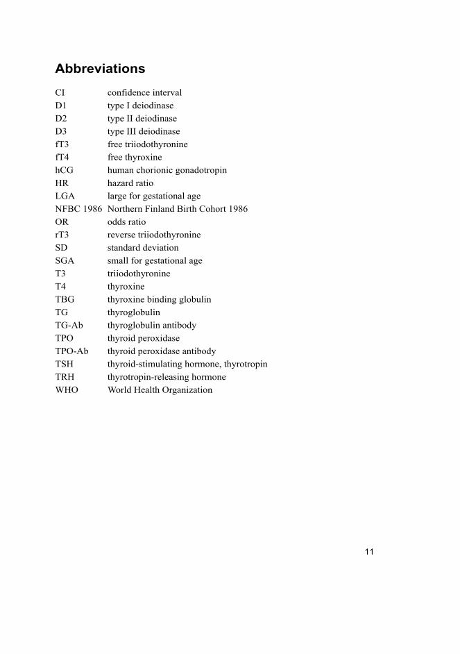
Abbreviations
CI confidence interval
D1 type I deiodinase
D2 type II deiodinase
D3 type III deiodinase
fT3 free triiodothyronine
fT4 free thyroxine
hCG human chorionic gonadotropin
HR hazard ratio
LGA large for gestational age
NFBC 1986 Northern Finland Birth Cohort 1986
OR odds ratio
rT3 reverse triiodothyronine
SD standard deviation
SGA small for gestational age
T3 triiodothyronine
T4 thyroxine
TBG thyroxine binding globulin
TG thyroglobulin
TG-Ab thyroglobulin antibody
TPO thyroid peroxidase
TPO-Ab thyroid peroxidase antibody
TSH thyroid-stimulating hormone, thyrotropin
TRH thyrotropin-releasing hormone
WHO World Health Organization

12

13
List of original articles
This thesis is based on the following articles, which are referred to in the text by
their Roman numerals:
I Männistö T, Surcel H-M, Bloigu A, Ruokonen A, Hartikainen A-L, Järvelin M-R, Pouta A, Vääräsmäki M & Suvanto-Luukkonen E (2007) The effect of freezing, thawing, and short- and long-term storage on serum thyrotropin, thyroid hormones, and thyroid autoantibodies: implications for analyzing samples stored in serum banks. Clin Chem 53: 1986–1987.
II Männistö T, Vääräsmäki M, Pouta A, Hartikainen A-L, Ruokonen A, Surcel H-M, Bloigu A, Järvelin M-R & Suvanto-Luukkonen E (2009) Perinatal outcome of children born to mothers with thyroid dysfunction or antibodies: A prospective population-based cohort study. J Clin Endocrinol Metab 94(3): 772–779.
III Männistö T, Vääräsmäki M, Pouta A, Hartikainen A-L, Ruokonen A, Surcel H-M, Bloigu A, Järvelin M-R & Suvanto E (2010) Thyroid dysfunction and autoantibodies during pregnancy as predictive factors of pregnancy complications and maternal morbidity in later life. J Clin Endocrinol Metab 95(3): 1084–1094.
IV Männistö T, Surcel H-M, Ruokonen A, Vääräsmäki M, Pouta A, Bloigu A, Järvelin M-R, Hartikainen A-L & Suvanto E (2011) Early pregnancy reference intervals of thyroid hormone concentrations in a thyroid antibody-negative pregnant population. Thyroid (published ahead of print January 2011).

14

15
Contents
Abstract
Tiivistelmä
Acknowledgements 9 Abbreviations 11 List of original articles 13 Contents 15 1 Introduction 19 2 Review of the literature 21
2.1 Thyroid hormone production and metabolism ........................................ 21 2.1.1 Effects of thyroid hormones ......................................................... 22
2.2 Thyroid diseases ...................................................................................... 23 2.2.1 Hyperthyroidism ........................................................................... 23 2.2.2 Hypothyroidism ............................................................................ 24 2.2.3 Chronic autoimmune thyroiditis ................................................... 25 2.2.4 Hypothyroxinemia ........................................................................ 26
2.3 Iodine prophylaxis in Finland ................................................................. 26 2.4 Thyroid function during pregnancy ........................................................ 27
2.4.1 Gestational age-specific reference intervals of serum fT4
and TSH ........................................................................................ 29 2.5 Hypothyroidism, hyperthyroidism, chronic autoimmune
thyroiditis and reproductive health .......................................................... 34 2.6 The risk associated with thyroid dysfunction and autoimmune
thyroiditis during pregnancy ................................................................... 35 2.6.1 Peri- and neonatal risk factors ...................................................... 35 2.6.2 Risk of pregnancy complications ................................................. 42
2.7 Development of the thyroid gland and fetal thyroid hormone
supply ...................................................................................................... 43 2.7.1 Risk of adverse outcome during further follow-up of
children ......................................................................................... 43 2.8 Screening of thyroid dysfunction and antibodies during
pregnancy ................................................................................................ 44 2.9 Maternal morbidity and mortality associated with thyroid
dysfunction or antibodies ........................................................................ 45 2.9.1 Autoimmune thyroiditis and hypothyroidism ............................... 45 2.9.2 Diabetes ........................................................................................ 46

16
2.9.3 Arterial hypertension .................................................................... 46 2.9.4 Mortality ....................................................................................... 47
2.10 Preanalytical considerations – TSH, free thyroid hormones,
TPO-Ab and TG-Ab laboratory analyses ................................................ 47 3 Purpose of the present study 49 4 Subjects and methods 51
4.1 Subjects ................................................................................................... 51 4.1.1 The Northern Finland Birth Cohort 1986 ..................................... 51 4.1.2 Special considerations concerning the NFBC 1986 ..................... 52 4.1.3 Finnish Maternity Cohort ............................................................. 53
4.2 Methods ................................................................................................... 53 4.2.1 Thyroid hormone and antibody assays (I, II, III, IV) ................... 53 4.2.2 Laboratory methods (I) ................................................................. 54 4.2.3 Sampling and analyses (II, III, IV) ............................................... 55 4.2.4 Representativeness analysis .......................................................... 55 4.2.5 Categorization of the study population according to TSH
and fT4 levels (II, III) ................................................................... 56 4.2.6 Categorization of the study population according to levels
of thyroid antibodies (II, III, IV) .................................................. 59 4.2.7 Register-based data (III) ............................................................... 59 4.2.8 Power analysis .............................................................................. 60 4.2.9 Statistical methods (I, II, III, IV) .................................................. 60
5 Results and comments 63 5.1 Thyroid hormone and antibody analyses after freezing, thawing
and long-term frozen storage (I) .............................................................. 63 5.1.1 Comments ..................................................................................... 63 5.1.2 Implications regarding the use of stored serum samples .............. 64
5.2 Maternal characteristics (II, III) .............................................................. 68 5.3 Perinatal outcome (II) and pregnancy outcome (III) ............................... 71
5.3.1 Perinatal outcome ......................................................................... 71 5.3.2 Analyses of sensitivity .................................................................. 74 5.3.3 Pregnancy outcome ...................................................................... 75 5.3.4 Comments ..................................................................................... 77
5.4 Maternal morbidity and mortality (III) .................................................... 83 5.4.1 Subsequent maternal thyroid disease morbidity ........................... 83 5.4.2 Later diabetes morbidity of the mothers ....................................... 84 5.4.3 Subsequent maternal arterial hypertension ................................... 84

17
5.4.4 Maternal mortality ........................................................................ 84 5.4.5 Comments ..................................................................................... 85
5.5 Gestational age-specific reference intervals of thyroid hormone
levels (IV) ............................................................................................... 86 5.5.1 BMI and thyroid hormones .......................................................... 89 5.5.2 Comments ..................................................................................... 89
6 Conclusions 95 7 General discussion 97
7.1 Strengths and limitations of the present study ........................................ 97 7.2 Confounding factors ................................................................................ 98 7.3 Recommendations ................................................................................... 99
References 105 Original publications 119

18

19
1 Introduction
The thyroid is a small endocrine gland located in front of the trachea. It utilizes
iodine to produce thyroid hormones, which are essential for normal growth,
development, maturation and regulation of metabolism. This is evident from
history: in iodine deficiency and thus thyroid hormone deprivation, children are
affected by cretinism, characterized by intellectual deficiency, mutism, spastic
diplegia, squint, thyroid dysfunction and short stature. (Ganong 2005.)
Fortunately, in many countries iodine deficiency is no longer a problem, as a
result of iodine supplementation. Finland has had a successful iodine prophylaxis
regime since the 1940s, and the whole of the Finnish population has an adequate
iodine intake (Lamberg et al. 1981).
Up to 5–10% of women of childbearing age may suffer from thyroid
dysfunction or the presence of thyroid antibodies. Thyroid diseases are known to
affect the reproductive health of women, who thus have trouble in conceiving or
have more miscarriages (Benson & Dailey 1955, Scott & Mussey 1964, Stagnaro-
Green et al. 1990, Abalovich et al. 2002, Anselmo et al. 2004). Thyroid
dysfunction also has a relatively high prevalence during pregnancy, affecting up
to 5% of all pregnant women (Glinoer et al. 1990). The effects of thyroid
dysfunction on pregnancy outcome and on the developing fetus are currently of
interest, with the most devastating observation in the literature being decreased
intelligence quotient of the offspring (Haddow et al. 1999, Liu et al. 2010). It is
known that the fetus is totally dependent on maternal thyroid hormone supply
during the first trimester of pregnancy, which is the crucial time in organogenesis
(Calvo et al. 2002).
The aim of this study was to evaluate, in a large population-based pregnant
population, how maternal thyroid dysfunction and antibodies affect pregnancy,
peri- and neonatal outcomes and subsequent health of the mothers. Other aims
were to find out if freezing and long-term frozen storage change TSH, thyroid
hormone and thyroid autoantibody levels and to create a framework for
gestational age-specific reference intervals for TSH and thyroid hormone
concentrations.

20

21
2 Review of the literature
2.1 Thyroid hormone production and metabolism
The thyroid gland secretes two main hormones, thyroxine (T4) and
triiodothyronine (T3). Thyroxine is produced in greater quantity than T3 (at a rate
10:1), but T3 is the major biologically active thyroid hormone and is mostly
derived from T4 in the peripheral tissues. The thyroid gland utilizes and conserves
iodine to produce thyroid hormones. Iodine is obtained from the diet, converted to
iodide, actively transported to the thyroid, and incorporated into thyroglobulin
(TG) by way of the enzyme thyroid peroxidase (TPO). This leads to production of
monoiodotyrosine and diiodotyrosine, which are then coupled to form T4, T3 and
reverse T3 (rT3). Reverse T3 has no biological activity. The thyroid hormones are
part of the TG stored in the colloid of thyroid follicles until excreted into the
circulation. In iodine sufficiency the thyroid utilizes approximately 20% of daily
ingested iodine, and the rest is excreted in the urine. (Ganong 2005, Hadley &
Levine 2007.)
Thyroid hormone production is regulated by the pituitary through the action
of thyrotropin (thyroid-stimulating hormone, TSH). TSH comprises two subunits
and it has one alpha-subunit in common with luteinizing hormone, follicle-
stimulating hormone and human chorionic gonadotropin (hCG), and one specific
beta-subunit. TSH shows circadian and pulsatory secretion – its secretion peaks at
around midnight and declines during the day. The function of the pituitary is
controlled by the hypothalamus, which excretes thyrotropin-releasing hormone
(TRH). It accelerates the production of TSH, whereas dopamine and somatostatin
hinder it. The thyroid hormones have a negative feedback effect on the pituitary
and hypothalamus, which is modified by the T4 concentration in the serum and
the conversion of T4 to T3 locally in the brain. Therefore, if T4 concentration in
the serum drops, the inhibitory stimulus is decreased due to a diminished local
effect of T3 in the pituitary and TSH levels rise to stimulate the thyroid gland.
(Ganong 2005, Hadley & Levine 2007.)
The thyroid hormones are protein-bound in the serum, and only 0.02% of T4
and 0.2% of T3 are free, biologically active hormones. 45–70% of thyroid
hormones are bound to thyroxine-binding globulin (TBG), and the rest to
transthyretin and albumin. Familial conditions, estrogen treatment and pregnancy

22
may have effects on the concentrations of the binding proteins, leading to changes
in thyroid hormone fractions until a new equilibrium is reached. (Ganong 2005.)
Free T4 (fT4) is metabolized in the tissues to the active form free T3 (fT3) by
three deiodinase enzymes. The tissues have different rates of fT3 production and
uptake according to the presence of the deiodinase enzymes. Type I deiodinase
(D1) is located in the liver, kidneys, thyroid and pituitary and is primarily
responsible for fT3 formation. Its activity is low in the fetus. Type II deiodinase
(D2) is located in the central nervous system and pituitary and it produces a
supply of fT3 to the brain. Type III deiodinase (D3) is located in the brain and in
reproductive tissues and it inactivates both fT4 and fT3, maintaining an
equilibrium in the fT3 concentration. Only D2 and D3 have been detected in
human placental tissue, the former providing the placenta with a supply of fT3,
and the latter maintaining its equilibrium. (Ganong 2005.)
2.1.1 Effects of thyroid hormones
The thyroid hormones stimulate oxygen consumption and increase the metabolic
rate. They have an effect on the heart and connective tissues and affect growth
and development. Thyroid hormones have a marked effect on brain development,
especially on the cerebral cortex and the basal ganglia. Lack of thyroid hormones
during development due to iodine deficiency leads to cretinism, a condition which
can be fully prevented with iodine prophylaxis. In addition, congenital
hypothyroidism, if unnoticed and untreated, leads to intellectual deficiency, which
fortunately can be prevented by screening programmes and thyroxine treatment
started in infancy. (Hadley & Levine 2007.)
High concentrations of thyroid hormones can elevate the body temperature as
they increase the metabolic rate. A rise in body temperature activates the
cardiovascular system to dissipate heat. Also, peripheral vascular resistance
decreases due to vasodilatation, and blood volume and cardiac output increases
through the direct actions of thyroid hormones. Thyroid hormones also lead to
protein catabolism from muscles and increase carbohydrate absorption. Therefore
they have an effect on glucose metabolism. They also lower circulating
cholesterol levels by increasing the hepatic removal of cholesterol. (Ganong 2005.)

23
2.2 Thyroid diseases
2.2.1 Hyperthyroidism
Hyperthyroidism, thyroid hormone overproduction, has a prevalence of
approximately 1% in the population. Thyrotoxicosis refers to increased amounts
of thyroid hormones in the circulation, the cause of which can be also other than
thyroid hormone overproduction. The symptoms of hyperthyroidism include
anxiousness, tachycardia or even atrial fibrillation, increased heat sensitivity,
perspiration, skin changes, loss of weight, insulin resistance, fatigue and shivering.
The symptoms may vary between patients. Approximately 70% cases of
hyperthyroidism are caused by Graves’ (Basedow’s) disease. Other common
reasons are toxic multinodular goitre, the prevalence of which is, however,
diminishing as a result of iodine prophylaxis, and toxic adenoma. Graves’ disease
is 8–10 times more common in women than men. Usually it affects people aged
30–40. It is characterized by the presence of symptoms related to hyperthyroidism
and extrathyroidal symptoms such as Graves’ ophthalmopathy. The disease is of
autoimmune origin, and hyperthyroidism is mediated by thyroid-stimulating
antibodies. They bind to TSH receptors, mimicking the actions of TSH.
Hyperthyroidism is diagnosed using laboratory analyses: it presents as raised
concentrations of serum thyroid hormones with low or much suppressed
concentrations of TSH. Subclinical hyperthyroidism is diagnosed when serum
TSH levels are low but thyroid hormone concentrations are within reference
intervals. Hyperthyroidism is treated with antithyroid drugs or medical treatment,
radioiodine therapy or with surgery. In pregnancy antithyroid drugs pass through
the placenta, but they can be used when monitoring maternal fT4 levels carefully
to prevent fetal hypothyroidism. (Välimäki & Schalin-Jäntti 2009.)
Overt and subclinical hyperthyroidism during pregnancy
Overt hyperthyroidism complicates approximately 0.05–0.2% of pregnancies
(Burrow 1993). Hyperthyroidism is associated with ovulatory dysfunction,
miscarriages and difficulties conceiving (Krassas et al. 1994, Anselmo et al. 2004)
unless treated. Subclinical hyperthyroidism has a prevalence of approximately
1.7% in pregnant women (Casey et al. 2006). It is often associated with the rise
observed in hCG concentrations during early pregnancy and considered as a fairly

24
normal phenomenon. Hyperemesis gravidarum seems to be associated with
subclinical hyperthyroidism (Valentine et al. 1980).
2.2.2 Hypothyroidism
Hypothyroidism is a deficiency of T4. It is present in approximately 2% of
women and 0.1–0.2% of men (Tunbridge et al. 1977, Vanderpump et al. 1995).
The prevalence of congenital hypothyroidism is 1/4000. Hypothyroidism can
present with different symptoms such as fatigue, dry and coarse skin, puffiness,
weight gain, diminished perspiration and poor cold endurance, as well as
menstrual abnormalities and infertility in women. Hypothyroidism is often caused
by factors associated with the thyroid itself (primary hypothyroidism). Only 5%
of hypothyroidism are the result of central hypothyroidism, i.e. due to lack of
TSH or its effects. The most common causes of hypothyroidism are autoimmune
thyroiditis, radioiodine therapy and thyroid surgery, but also iodine, medicines or
rare genetic disorders may cause hypothyroidism. Overt hypothyroidism is
diagnosed by laboratory testing and it presents as low concentrations of
circulating thyroid hormones with raised concentrations of TSH. Subclinical
hypothyroidism is present when TSH concentrations are raised but thyroid
hormone concentrations are still normal. The presence of thyroid antibodies
reveals if hypothyroidism is caused by chronic autoimmune thyroiditis.
Hypothyroidism is treated with thyroxine. (Välimäki & Schalin-Jäntti 2009.)
Overt and subclinical hypothyroidism during pregnancy
Overt hypothyroidism is often detected and treated before the onset of pregnancy,
since it causes infertility and recurrent miscarriages (Krassas et al. 1999).
However, 0.2–0.5% of all pregnant women have overt hypothyroidism (Allan et al. 2000, Casey et al. 2005), representing either new undiagnosed cases or
inadequate treatment of previously detected disease. Subclinical hypothyroidism
has a prevalence of approximately 2.2–2.5% in pregnant women (Allan et al. 2000, Casey et al. 2005).
Nearly all levothyroxine-treated mothers need an approximately 30–50%
increase in their dosage to maintain euthyroidism during pregnancy (Mandel et al. 1993, Alexander et al. 2004). The dosage should be raised to 2.0–2.4 μg/kg when
trying to conceive or at least when pregnancy tests are positive. It is noteworthy
that women without residual thyroid function (after radioiodine treatment or

25
thyroidectomy) require a greater increase in their daily dose of levothyroxine than
women with chronic autoimmune thyroiditis. Hypothyroid mothers should be
followed by using thyroid function tests (serum TSH and fT4) throughout
pregnancy (4 weeks apart if test results are abnormal and 6–8 weeks apart if
results are within reference intervals) and their thyroxine dosage altered when
necessary. This can be based on the degree of TSH elevation: for women with
serum TSH levels of 5–10 mIU/L the increment in thyroxine is 25–50 μg/day; for
those with serum TSH levels of 10–20 mIU/L the increment is 50–75μg/day and
for those with serum TSH levels of > 20 mIU/L the increment is 75–100 μg/day.
A newly diagnosed case of hypothyroidism during pregnancy should be
vigorously treated. Thyroxine treatment should be initiated with a dose of 100–
150 μg thyroxine/day or a dose calculated according to body weight. In severe
hypothyroidism, therapy may be initated with a double dose of the estimated final
daily dose for the first few days. The treatment of hypothyroidism during
pregnancy should be adjusted so that TSH and fT4 levels remain within the
established reference ranges for pregnant women. (Abalovich et al. 2007.)
2.2.3 Chronic autoimmune thyroiditis
Chronic autoimmune thyroiditis is the most common cause of primary
hypothyroidism. A high affinity marker of the disease is thyroid peroxidase
antibody (TPO-Ab), and high concentrations of TPO-Ab can be measured in most
cases of autoimmune thyroiditis. A similar marker of autoimmune thyroiditis is
thyroglobulin antibody (TG-Ab). Patients with autoimmune thyroiditis often have
other autoimmune diseases, such as diabetes, Addison’s disease and rheumatoid
arthritis. Autoimmune thyroiditis is detected in 3.5/1000 women and 0.8/1000
men in the United States, but 5–10% of the normal population have detectable
thyroid antibodies. (Tunbridge et al. 1977, Gordin & Lamberg 1981, Välimäki &
Schalin-Jäntti 2009.) Autoimmune thyroiditis is a silent disease process in which
the thyroid may be enlarged or atrophied and the production of thyroid hormones
may decrease resulting in hypothyroidism. The prevalence of positive thyroid
autoantibodies increases with age, with the highest frequency observed in women
aged 40–60 years (Vanderpump et al. 1995, Hollowell et al. 2002). The annual
incidence of overt hypothyroidism in cases of symptomless autoimmune
thyroiditis in Finland is approximately 7%, but the incidence of subclinical,
asymptomatic hypothyroidism is not known. The annual incidence of overt
hypothyroidism is 26% among cases in which autoimmune thyroiditis is

26
accompanied by diminished thyroid function, indicated by elevated TSH values,
at early assessment. (Gordin & Lamberg 1981.)
Chronic autoimmune thyroiditis during pregnancy
Positive TPO-Ab and/or TG-Ab test resuls are found in approximately 5% of
euthyroid pregnant women. However, a thyroid autoantibody prevalence of up to
15% has been found in pregnant populations (Cleary-Goldman et al. 2008). At
parturition, 56% of thyroid antibody-positive mothers have been reported to have
high TSH values. During gestation, a significant amount of TPO-Ab- and/or TG-
Ab-positive women are at risk of developing hypothyroidism, since they have
lower thyroid function reserve. (Glinoer et al. 1994.) Chronic autoimmune
thyroiditis is the most important cause of hypothyroidism in pregnant women, and
up to 90% of women with hypothyroidism during pregnancy test positive for
thyroid antibodies (Klein et al. 1991).
2.2.4 Hypothyroxinemia
Hypothyroxinemia is defined as a low serum fT4 concentration with normal TSH
concentrations. Its prevalence during pregnancy is approximately 1.3–2.1%
(Casey et al. 2007, Cleary-Goldman et al. 2008). The clinical significance of
hypothyroxinemia is still largely unknown. It is noteworthy that central
hypothyroidism shows similar features. Hypothyroxinemia has been particularly
associated with iodine deficiency and it has a high prevalence in iodine-deficient
populations. Due to lack of raw materials (iodine), the thyroid produces more T3
instead of T4, thus leading to hypothyroxinemia. (Glinoer 1997.)
2.3 Iodine prophylaxis in Finland
Iodine deficiency is still a common condition worldwide and low iodine intakes
lead to iodine deficiency disorders (WHO 2004). The most severe consequences
of iodine deficiency are associated with adverse brain development, leading to
cretinism. Milder degrees of brain damage as well as goitre formation and
pregnancy complications are also associated with iodine deficiency. (Hetzel 1983.)
Through the 1920s to the 1940s Finland, especially Eastern and Southern
Finland, were areas with iodine deficiency (Tuovinen 1933, Virtanen & Virtanen
1940, Hiilesmaa 1948, Lamberg 2003b). Although Finland was iodine-deficient,

27
no intellectual deficiency due to lack of iodine was found (Lamberg 2003a).
Iodine prophylaxis was started in Finland as early as the 1920s by fortifying
animal fodder. From 1949 onward voluntary iodine prophylaxis was started with
fortifying of table salt. (Lamberg 2003b, Suojanen 2003.)
World Health Organization (WHO) recommendations indicate that women of
childbearing age should have an average iodine intake of 150 µg/day. During
pregnancy and breastfeeding the intake should be 250 µg/day (Andersson et al. 2007). Long-term follow-up studies have shown that the Finnish population
reached iodine sufficiency in the 1970s (Lamberg et al. 1981, Varo et al. 1982,
Nissinen et al. 1983). Further studies have shown that the whole Finnish
population receives sufficient amounts of iodine from the diet (Erkkola et al. 1998, Paturi et al. 2008, Flynn et al. 2009).
2.4 Thyroid function during pregnancy
During normal pregnancy, the maternal thyroid produces up to 50% more thyroid
hormones. The rise in thyroid hormones results from physiological changes in
pregnancy. Firstly, following conception, estrogen concentrations increase
markedly (Lindberg et al. 1974) leading to increased synthesis of TBG from the
liver and increased binding of T4 to TBG (Robbins & Nelson 1958). TBG is also
more sialylated during pregnancy and has a reduced clearance rate (Ain et al. 1987). The increase in TBG causes an increased need of thyroid hormones in
order to maintain euthyroidism during pregnancy. Also, the developing fetus
utilizes the maternal thyroid hormone supply, maternal plasma volume is
increased and the distribution volume of T4 is higher during pregnancy,
contributing to the increased need of thyroid hormones. Secondly, hCG has direct
thyrotropic activity due to its structural similarity with TSH (Pekonen et al. 1988).
Levels of hCG rise markedly in early pregnancy, peaking at around the 10th
gestational week (Hay 1988), and lead to an increase in thyroid hormone
production. Concentrations of TSH mirror those of hCG, as a result of a feedback
mechanism, and serum TSH levels are at a transient nadir in the first trimester
(Glinoer 1997).
Levels of serum total T4 increase markedly during the first trimester of
pregnancy, measuring up to 50% more in pregnant than non-pregnant women,
both by immunoassay and mass spectrometry (Glinoer et al. 1990, Soldin et al. 2004, Kahric-Janicic et al. 2007). Total T4 concentrations reach a peak at the 20th
gestational week and high levels of total T4 are maintained through the second

28
and third trimesters. Thyroxine has a high affinity to TBG and therefore the levels
of total T4 follow the changes in TBG concentrations. The concentration of TBG
doubles during early pregnancy and reaches a plateau at approximately 20th
gestational week. The rise in total T3 is more progressive throughout pregnancy.
(Glinoer et al. 1990, Glinoer 1997.)
Levels of serum fT4 have a similar, but less pronounced increase in early
pregnancy following stimulation of the thyroid gland by hCG. Reports as to how
high this increase is vary markedly in different studies, since changes in serum
protein fractions have an effect on assays of free thyroid hormones, especially as
regards fT4 immunoassays (Roti et al. 1991, Keffer 1996, Lee et al. 2009).
However, serum fT4 levels are on average higher in the first trimester of
pregnancy than in nonpregnant women (Glinoer et al. 1990, Soldin et al. 2004,
Kahric-Janicic et al. 2007), although in some studies this has not been observed
(Lee et al. 2009, Anckaert et al. 2010). In the second and third trimester, serum
fT4 levels are 20–30% decreased compared with first trimester concentrations, a
fact observed using all current methods (Glinoer et al. 1990, Kahric-Janicic et al. 2007, Lee et al. 2009, Anckaert et al. 2010). At delivery serum fT4 levels are
approximately 10–15% lower than in nonpregnant women, but usually within the
reference range of the general population (Glinoer et al. 1990, Glinoer 1997).
Serum TSH concentrations are lower in pregnant than nonpregnant women
and may even be subnormal compared with the lower reference limit of the
general population. This is especially evident in the first trimester due to the
thyrotropic effects of hCG. Mean serum TSH levels increase as levels of hCG
decrease after the 10th gestational week, and mean second and third trimester TSH
levels are higher than those in the first trimester. (Glinoer et al. 1990, Glinoer
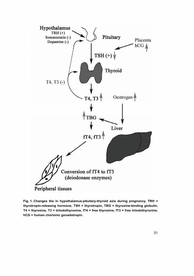
1997.) The changes observed in the hypothalamus-pituitary-thyroid axis during
pregnancy are summarized in Figure 1.
The peripheral metabolism of thyroid hormones is also altered during normal
pregnancy. D1 is thought to function as in nonpregnant subjects. D2 and D3 are
present in the placental tissue, the former preferring T4 and rT3 as substrates and
the latter converts T4 and T3 to their inactive forms. D2 is present in the
chorionic and decidual membranes of the placenta and D3 in the trophoblasts.
The activity of D2 increases when the availability of T4 decreases, and therefore
the placental tissues are able to maintain T3 production in the placenta even when
T4 values are reduced. (Glinoer 1997.)

29
2.4.1 Gestational age-specific reference intervals of serum fT4 and TSH
Concentrations fT4 and TSH, due to the physiological changes in pregnancy, are
not comparable in pregnant and nonpregnant women. The reference intervals of
both TSH and fT4 are calculated from data from the general population and
cannot be directly implemented in relation to pregnant women. For instance, the
reference interval for serum TSH is 0.4–4.0 mU/L in the general population
(www.huslab.fi) and 0.35–4.94 mU/L as reported by the manufacturer of the
assay used in this study (Abbott Diagnostics). Most recent evaluations in the
general population have shown that TSH reference intervals are tighter, and with
the Abbott assay set to 0.5–3.6 mU/L (personal communication with Camilla
Schalin-Jäntti). Similar results have been published previously (Spencer et al. 2007). The reference interval for serum fT4 is 9–19 pmol/L as calculated from the
general population data (www.huslab.fi) and reported by the assay manufacturer.
If these same reference intervals were to be used in the pregnant population, one
would underestimate the prevalence of hypothyroidism, since serum TSH levels
are generally lower and fT4 levels higher in pregnant women than in the general
population.
Serum fT4 concentrations are commonly analyzed by using immunoassays,
which rather than measuring fT4 directly, give an estimate of the concentration.
Immunoassays are known to be sensitive to alterations in binding proteins (Roti et al. 1991, Lee et al. 2009, Anckaert et al. 2010). For instance in one study, the
Roche Elecsys assay failed to show the first trimester increase in fT4 levels (Lee
et al. 2009). A recent study revealed that the Abbott Architect method for fT4 was
more sensitive to binding protein alterations during pregnancy than the Roche
Cobas or Siemens Immulite assays and showed a smaller decrease in the second
and third trimester levels of serum fT4 than the other assays (Roti et al. 1991, Lee
et al. 2009, Anckaert et al. 2010). Some recent studies have therefore involved
evaluation of serum fT4 levels in pregnant women by using equilibrium dialysis
and mass spectrometry. With equilibrium dialysis/mass spectrometry fT4
reference intervals decrease from 13.90–23.42 pmol/L at gestational week 14 to
11.07–19.69 pmol/L at gestational week 20, showing the typical rise in early
pregnancy and a decrese in the second trimester. Uniformly, values were higher in
pregnant than in nonpregnant women, using this method. (Yue et al. 2008.)
Immunoassays have shown a similar pattern, but with reference intervals varying
greatly between methods and populations (Panesar et al. 2001, Price et al. 2001,

30
Haddow et al. 2004, Dashe et al. 2005, Spencer et al. 2005, Dhatt et al. 2006,
La'ulu & Roberts 2007, Stricker et al. 2007, Gilbert et al. 2008, Gong & Hoffman
2008, Marwaha et al. 2008, Yue et al. 2008, Bocos-Terraz et al. 2009, Shan et al. 2009, Silvio et al. 2009, Fister et al. 2010, Klajnbard et al. 2010, Yan et al. 2011).
For instance, with the Abbott Architect method, the lower reference limit of fT4
in the first trimester ranges from 8.9 to 10.68 pmol/L as measured in pregnant
women from the United Arab Emirates and Spain, respectively. Similar variability
is seen with the upper reference limits. This would suggest that despite the same
method, some population characteristics or genetic traits may influence the results.
These results are summarized in Table 1.
With serum TSH a wide variation is seen in results from different studies
(Table 1 and more specifically in Table 11). The lower reference limit of TSH
varies from 0.02 to 0.09 mU/L in the first trimester and is significantly lower than
in the general population. Many women would be mistakenly classified as
hyperthyroid if the reference intervals of the general population were to be used.
The upper reference limit of serum TSH is generally lower in pregnant women
than in the nonpregnant population, although wide variation is seen between
studies. If those with chronic autoimmune thyroiditis are not excluded the upper
reference limit of TSH concentrations is skewed and does not represent that of the
healthy population (Spencer et al. 2007, Pearce et al. 2008). This might explain
the differences seen between different studies. Studies with suffiecient population
sizes to create gestational age-specific reference intervals (over 100 samples per
gestational week), with exclusion of all TPO-Ab- and/or TG-Ab-positive women
show that the first trimester upper reference limit of TSH ranges between 2.2–3.6
mU/L (Table 11) (Haddow et al. 2004, Gilbert et al. 2008, Lambert-Messerlian et al. 2008, Bocos-Terraz et al. 2009, Shan et al. 2009, Springer et al. 2009).
Previously it has been established that ethnicity has some effect on thyroid
function and prevalence of thyroid antibodies (Hollowell et al. 2002, La'ulu &
Roberts 2007). In addition, populations may have differences in iodine intake,
which naturally affects thyroid function. Iodine-deficient pregnant women lack
the typical rise in fT4 levels during early pregnancy and have a higher prevalence
of subclinical hypothyroidism than iodine-sufficient populations (Costeira et al. 2010).
31
Fig. 1. Changes the in hypothalamus-pituitary-thyroid axis during pregnancy. TRH =
thyrotropin-releasing hormone, TSH = thyrotropin, TBG = thyroxine-binding globulin,
T4 = thyroxine, T3 = triiodothyronine, fT4 = free thyroxine, fT3 = free triiodothyronine,
hCG = human chorionic gonadotropin.

Ta
ble
1. S
um
ma
ry o
f stu
die
s c
on
ce
rnin
g t
he
re
fere
nce
in
terv
als
of
TS
H,
fT4
an
d f
T3 i
n p
reg
na
nt
wo
men
.
Firs
t aut
hor
Yea
r C
ount
ry
n M
etho
d A
naly
te
Ref
eren
ce in
terv
als
Trim
este
r I
Trim
este
r II
Trim
este
r III
Yan
20
11
Chi
na
505
IA, A
dvia
Cen
taur
TS
H m
IU/L
fT4
pmol
/L
0.03
-4.5
1
11.8
-21.
0
0.05
-4.5
1
10.6
-17.
6
0.47
-4.5
4
9.2-
16.7
Kup
pens
20
10
Hol
land
10
58
IA, I
mm
ulite
TS
H m
IU/L
0.
51-2
.89
Fist
er
2010
S
love
nia
116
IA, A
dvia
Cen
taur
TS
H m
U/L
fT4
pmol
/L
fT3
pmol
/L
0.61
-3.9
6
7.91
-14.
15
3.30
-4.5
3
Kla
jnba
rd
2010
N
ordi
c co
untri
es
801
IA, I
mm
ulite
TS
H m
IU/L
fT4
pmol
/L
0.
3-4.
1
11.9
-18.
7
0.4-
3.7
9.4-
17.8
Sha
n 20
09
Chi
na
4800
IA
, Dia
gnos
tics
Pro
duct
s TS
H m
IU/L
fT4
pmol
/L
0.09
-2.9
6
11.4
1-21
.70
0.35
-3.8
8
11.6
0-21
.69
Silv
io
2009
U
.S.
3102
IA
, Ele
csys
E17
0 TS
H m
IU/L
fT4
pmol
/L
0.
18-4
.07
9.5-
15.8
Boc
os-T
erra
z 20
09
Spa
in
1198
IA
, Abb
ott A
rchi
tect
TS
H m
IU/L
fT4
pmol
/L
0.10
-2.6
5
10.6
8-17
.76
0.12
-2.6
4
9.0-
14.6
7
Gon
g 20
08
Can
ada
715
IA, R
oche
Mod
ular
E17
0 fT
4 pm
ol/L
11
.0-1
9.0
9.7-
17.5
8.
1-15
.3
Mar
wah
a 20
08
Indi
a 54
1 IA
, Ele
csys
101
0 TS
H
fT4
fT3
0.6-
5.0
12.0
0-19
.45
1.92
-5.8
6
0.44
-5.7
8
9.48
-19.
58
3.2-
5.7
0.74
-5.7
11.3
-17.
71
3.3-
5.18
Lars
son
2008
S
wed
en
52
IA, A
bbot
t Arc
hite
ct
TSH
mU
/L
0.09
-3.3
9 0.
37-3
.40
0.40
-3.8
8
Gilb
ert
2008
A
ustra
lia
2159
IA
, Abb
ott A
rchi
tect
TS
H m
U/L
fT4
pmol
/L
fT3
pmol
/L
0.02
-2.1
5
10.4
-17.
8
3.3-
5.7
Yue
20
08
U.S
.
Equ
ilibr
ium
dia
lysi
s-m
ass
spec
trom
etry
fT4
pmol
/L
13.9
0-23
.42
11.0
7-19
.69
32

Firs
t aut
hor
Yea
r C
ount
ry
n M
etho
d A
naly
te
Ref
eren
ce in
terv
als
Trim
este
r I
Trim
este
r II
Trim
este
r III
Stri
cker
20
07
Sw
itzer
land
22
72
IA, A
bbot
t Arc
hite
ct
TSH
mIU
/L
fT4
pmol
/L
fT3
pmol
/l
0.09
-2.8
3
10.5
3-18
.28
3.52
-6.2
2
1.02
-2.7
9
9.53
-15.
68
3.41
-5.7
8
1.14
-2.9
0
8.63
-13.
61
3.33
-5.5
9
La’u
lu
2007
U
.S.
3064
IA
, Abb
ott A
rchi
tect
TS
H m
IU/L
fT4
pmol
/L
0.
15-3
.11
9.3-
15.2
Dha
tt 20
06
Uni
ted
Ara
b
Em
irate
s
1140
IA
, Abb
ott A
rchi
tect
TS
H m
IU/L
fT4
pmol
/L
0.06
-8.3
8.9-
24.6
0.17
-5.9
8.4-
19.3
0.21
-6.9
8.0-
18.0
Das
he
2005
U
.S.
1359
9 IA
, Sie
men
s Im
mul
ite
TSH
mU
/L
0.01
-5.0
9 0.
02-4
.09
0.20
-6.4
0
Spe
ncer
20
05
U.S
.
IA, R
oche
Ele
csys
and
Toso
h
TSH
mIU
/L
0.03
-2.4
Had
dow
20
04
U.S
. 11
26
IA, S
iem
ens
Imm
ulite
TS
H m
IU/L
0.
08-3
.61
0.39
-3.7
1
Pric
e 20
01
UK
, Asi
a 70
IA
, AC
S 1
80
TSH
mU
/L
fT4
pmol
/L
0.09
-3.0
10.1
-16.
0
Pan
esar
20
01
Chi
na
343
IA, A
CS
180
TS
H m
IU/L
fT4
pmol
/L
fT3
pmol
/L
0.03
-2.3
11.1
-22.
9
3.0-
5.7
0.03
-3.7
8.1-
16.7
2.8-
4.2
0.13
-3.4
8.5-
14.4
2.4-
4.1
IA=
imm
unoa
ssay
33

34
2.5 Hypothyroidism, hyperthyroidism, chronic autoimmune
thyroiditis and reproductive health
Thyroid hormones are important in maintaining normal ovarian function. Up to
24–56% and 22–58% of women with hypothyroidism and hyperthyroidism,
respectively, have ovulatory dysfunction (Benson & Dailey 1955, Scott & Mussey
1964, Krassas et al. 1994, Krassas et al. 1999) and may be infertile. Infertility is
usually defined as difficulty of conceiving after one year of intercourse without
contraception. Several changes are observed in reproductive endocrinology as a
result of hypo- and hyperthyroidism. In hypothyroidism the concentrations of sex-
hormone-binding globulin decrease, while they increase in hyperthyroidism. This
in turn leads to changes in estrogen and testosterone concentrations, which tend to
be lower in hypothyroidism and higher in hyperthyroidism. (Koutras 1997.)
Treatment of thyroid disease usually normalizes the ovarian function and enables
conception.
In addition to an increased risk of ovarian dysfunction, miscarriage rates
seem to be higher in nontreated and undertreated hypo- or hyperthyroid women
(Abalovich et al. 2002, Anselmo et al. 2004). Hypothyroid women in particular
have high miscarriage rates: in hospital material 67% of inadequately treated
hypothyroid women miscarried (Abalovich et al. 2002). In one study in which
hospital ambulatory patients were evaluated, even women with mild subclinical
hypothyroidism (TSH 2.5–5 mIU/L in TPO-Ab-negative women) had higher,
almost double, rates of pregnancy loss compared with euthyroid women (Negro et al. 2010a). In women with miscarriage or fetal death, serum TSH concentrations
have been found to be higher and those of serum fT3 and fT4 lower than in
control women, and the authors of the study speculated that in 5% of miscarriages
undiagnosed hypothyroidism might be a contributing factor (Ashoor et al. 2010a).
In contrast, in a prospective population-based study, no increase in miscarriage
rate was seen among those with clear-cut subclinical hypothyroidism (TSH > 4.29
mU/L) (Cleary-Goldman et al. 2008), although the cut-off point used to signify
hypothyroidism in this study were very high for TSH, and therefore the euthyroid
population may have included those with subclinical disease, thus affecting the
results. Women with mild subclinical hypothyroidism (TSH ≥ 2.5 mU/L) and
pregnancy achieved by way of in-vitro fertilization showed no increase in
miscarriage rate (13% vs. 13% in euthyroid women) or difference in pregnancy
rate compared with euthyroid women (Reh et al. 2010). Differences between

35
studies may be connected to the populations involved: the first studies were on
patient material from hospitals/community hospitals (Abalovich et al. 2002,
Negro et al. 2010a, Ashoor et al. 2010a); the study by Cleary-Goldman et al. involved predominantly white women from the private sector (Cleary-Goldman et al. 2008), and the study by Reh et al. was among women undergoing assisted
reproductive technologies (Reh et al. 2010). Therefore, the studies are of highly
different characters and those based in hospital settings probably represent
populations more susceptible to adverse outcomes.
Positivity of TPO-Ab and/or TG-Ab as markers of autoimmune thyroiditis in
euthyroid women seems to be associated with miscarriages (Stagnaro-Green et al. 1990, Lejeune et al. 1993) and lower rates of success in assisted reproductive
technologies (Monteleone et al. 2011). The risk of miscarriage in euthyroid
women with autoimmune thyroiditis has been two-fold compared with controls in
recent meta-analyses (Prummel & Wiersinga 2004, Toulis et al. 2010). The
association between autoimmune thyroiditis, ovarian dysfunction, difficulty in
conceiving and miscarriage is thought to represent either a generalized
autoimmune disorder or subclinical hypothyroidism. The latter is supported the
results of by several studies: aggressive levothyroxine treatment reduced the
miscarriage rate in TPO-Ab-positive ambulatory hospital patients compared with
nontreated TPO-Ab-positive women (Negro et al. 2006). The clinical pregnancy
rate did not improve, but fewer miscarriages were reported in TPO-Ab-positive
women under levothyroxine treatment among those who achieved pregnancy by
way of assisted reproductive technology (Negro et al. 2005). Recently, in a case-
control study of women of over 38 years of age, and pregnancy achieved by in vitro fertilization, no increased rate of miscarriage was found among women with
autoimmune thyroiditis (Reh et al. 2011), but the women who miscarried and had
autoimmune thyroiditis had higher serum TSH concentrations than the control
women.
2.6 The risk associated with thyroid dysfunction and autoimmune
thyroiditis during pregnancy
2.6.1 Peri- and neonatal risk factors
Untreated overt hypothyroidism and hyperthyroidism are known to be associated
with maternal and fetal complications. Common fetal complications of untreated

36
maternal thyroid disease are low birth weight and a high frequency of fetal death
(Davis et al. 1988, Kriplani et al. 1994). On the other hand, mothers with treated
hypothyroidism may have an increased risk of large-for-gestational age (LGA)
infants (Wikner et al. 2008) and also low-birth weight infants (Blazer et al. 2003),
infants with malformations (Wikner et al. 2008) and, especially, preterm delivery
(birth at or before the 37th gestational week) (Antolic et al. 2006, Wikner et al. 2008). However, Matalon et al. demonstrated that rates of perinatal adverse
events were not increased in hypothyroid mothers who had adequate thyroxine
replacement therapy (Matalon et al. 2006). It is noteworthy that in the other
studies the women may not have been adequately treated, since these studies were
register-based or retrospective.
In a previous study, mothers with subclinical hypothyroidism (TSH 6–10
mU/L or TSH > 10 mU/L) had increased rates of fetal death (Allan et al. 2000).
However, based on data on reference intervals of serum TSH during pregnancy,
the cases in this study all represented clear-cut hypothyroidism and the data are in
accordance with those in studies showing a high miscarriage rate in inadequately
treated hypothyroidism (Abalovich et al. 2002). Casey et al. defined subclinical
hypothyroidism as TSH ≥ 2.74 mU/L with normal fT4 levels and found no
increase in fetal or neonatal deaths (Casey et al. 2005), but miscarriages were not
evaluated. In another study, with subclinical hypothyroidism defined as serum
TSH > 4.29 mU/L and fT4 9.27–18.79 pmol/L, no increase was found in any
adverse outcome studied (Cleary-Goldman et al. 2008). Again, the cut-off values
used to signify sublinical hypothyroidism in that study may have led to inclusion
of women with subclinical diseases in the euthyroid group. Finally, a recent study
revealed that women with mild hypothyroidism (serum TSH 2.5–5 mIU/L and
negative for TPO-Ab) had an increased risk of pregnancy loss (miscarriage and
fetal death) (Negro et al. 2010a), as discussed earlier. It has been reported that
there is a 60% increase in the incidence of child loss with every doubling of TSH
concentration (Benhadi et al. 2009) and it has been estimated that a total of 5% of
miscarriages might be attributed to maternal hypothyroidism (Ashoor et al. 2010a).
Similar results were found in a study in which the effects of universal
screening and treatment of women with subclinical hypothyroidism were
evaluated (TSH > 2.5 mIU/L in TPO-Ab-positive women). In that study, the
population was divided into two arms and further divided into high- and low-risk
women as regards thyroid dysfunction. In the screening group all women were
evaluated during pregnancy and all those with detected hypothyroidism were

37
treated in order to maintain serum TSH levels at < 2.5 mIU/L and at TSH < 3
mIU/L in the first and second trimester, respectively. In the second arm, only
those with a high risk of thyroid dysfunction were studied and treated for
hypothyroidism during pregnancy and low-risk women were evaluated
retrospectively, thus being the only group with undetected and untreated
hypothyroidism. In that study women with treated subclinical hypothyroidism had
fewer adverse outcomes than untreated women (34.9 vs. 91.2%). In particular
rates of miscarriage were lower among treated women (4.7 vs. 20.6%). It should
be noted that the overall rate of adverse outcomes was very high in this study and
the population studied represented ambulatory patients from community hospitals.
Also, because of the study design, the treated and untreated mothers with
subclinical hypothyroidism were not directly compared, and therefore the effect
of confounding factors was not evaluated. (Negro et al. 2010b.)
Preterm birth has been associated with autoimmune thyroiditis and
subclinical hypothyroidism in several studies. The first studies reported a doubled
rate of preterm deliveries in connection with autoimmune thyroiditis and
declining thyroid function during pregnancy (Glinoer et al. 1994). Since then,
subclinical hypothyroidism has been associated with preterm birth at or before 34
gestational weeks, with infants having to be committed to neonatal intensive care
(Casey et al. 2005). A retrospective case-control study showed that rates of
preterm delivery are higher in women with inadequately treated hypothyroidism
(Abalovich et al. 2002). In another study, the incidence of subclinical
hypothyroidism (TSH ≥ 3 mIU/L) was very high in women who had a very
preterm delivery (before the 32nd gestational week) (Stagnaro-Green et al. 2005).
In contrast to the above-mentioned results, in a large population-based cohort
study, no association was found between severe subclinical hypothyroidism and
peri- or neonatal complications (Cleary-Goldman et al. 2008). Interestingly, no
increase in preterm births was found in a recent study in which mild subclinical
hypothyroidism in a TPO-Ab-negative population was evaluated (Negro et al. 2010a). In a prospective trial of concerning the effect of levothyroxine treatment
in TPO-Ab-positive women, the rate of preterm birth was markedly lower in
aggressively treated (7.0 vs. 22.4%) than in nontreated TPO-Ab-positive mothers.
The nontreated mothers also had higher serum TSH and lower serum fT4 levels,
suggesting that treatment of underlying hypothyroidism reduces this risk. (Negro
et al. 2006.) However, the number evaluated was relatively small in this study,
and the population was based on hospital patients, which might explain the

38
overall high rate of preterm births observed. Similar results have not been
reported in any other study.
Hypothyroxinemia in early pregnancy has also been associated with preterm
birth as well as gestational diabetes, elevated birth weight of the infant (Cleary-
Goldman et al. 2008) and breech presentation (Pop et al. 2004). These
associations have not been proven in other studies (Casey et al. 2007, Hamm et al. 2009). In the study by Cleary-Goldman et al., the definition of hypothyroxinemia
in the first trimester was a serum fT4level of < 9.14 pmol/L with normal TSH
concentrations. As mentioned earlier, because of these cut-offs, their euthyroid
group probably included women with thyroid dysfunction (Cleary-Goldman et al. 2008). Hypothyroidism in the third trimester has been associated with
malpresentation and increased rates of assisted delivery (Wijnen et al. 2009,
Kuppens et al. 2010)
Subclinical hyperthyroidism has been reported not to be associated with any
perinatal complications in a large cohort (Casey et al. 2006). Overt
hyperthyroidism has been associated with low birth weight of the infant (Kriplani
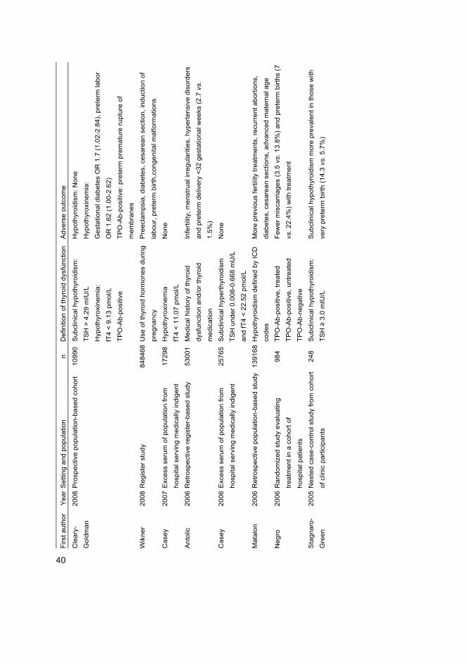
et al. 1994). The literature is summarized in Table 2.

Ta
ble
2. L
arg
e s
tud
ies
in
wh
ich
th
e e
ffe
cts
of
thy
roid
dy
sfu
ncti
on
/th
yro
id a
uto
an
tib
od
ies
du
rin
g p
reg
nan
cy
ha
ve
be
en
ev
alu
ate
d.
Firs
t aut
hor
Yea
rS
ettin
g an
d po
pula
tion
n D
efin
ition
of t
hyro
id d
ysfu
nctio
n A
dver
se o
utco
me
Had
dow
20
11P
rosp
ectiv
e po
pula
tion-
base
d co
hort
1006
2 TP
O-A
b-po
sitiv
e P
lace
ntal
abr
uptio
n O
R 1
.83
(0-9
9-3.
37) a
nd O
R 2
.2
(1.2
1-3.
99) i
n fir
st a
nd s
econ
d tri
mes
ter
Ash
oor
2011
Cas
e-co
ntro
l stu
dy o
n ho
spita
l
patie
nts
4420
TP
O-A
b- o
r TG
-Ab-
posi
tive
Sim
ilar r
ates
of p
ositi
ve T
PO
-Ab
and/
or T
G-A
b in
term
and
pret
erm
birt
hs
Abb
assi
-
Gha
nava
ti
2010
Exc
ess
seru
m o
f pop
ulat
ion
from
hosp
ital s
ervi
ng m
edic
ally
indi
gent
1729
8 TP
O-A
b-po
sitiv
e P
lace
ntal
abr
uptio
n O
R 3
.4 (1
.7-6
.7)
Ash
oor
2010
Cas
e-co
ntro
l stu
dy
4520
Cas
es w
ith m
isca
rria
ge h
ad h
ighe
r TS
H a
nd lo
wer
fT4
Ash
oor
2010
Cas
e-co
ntro
l stu
dy
4420
Cas
es w
ith la
te o
nset
pre
ecla
mps
ia h
ad h
ighe
r TS
H a
nd
low
er fT
4
Had
dow
20
10P
rosp
ectiv
e po
pula
tion-
base
d co
hort
1006
2 TP
O-A
b- a
nd/o
r TG
-Ab-
posi
tive
Pre
term
pre
mat
ure
rupt
ure
of m
embr
anes
OR
1.6
7
(1.0
5–2.
44)
Kup
pens
20
10P
rosp
ectiv
e po
pula
tion-
base
d co
hort
1058
S
ubcl
inic
al h
ypot
hyro
idis
m:
TSH
> 2
.5 m
U/L
(in
III tr
imes
ter)
Bre
ech
pres
enta
tion
OR
2.2
3 (1
.14-
4.39
)
Neg
ro
2010
Coh
ort o
f am
bula
tory
pat
ient
s fro
m
com
mun
ity h
ospi
tals
4125
S
ubcl
inic
al h
ypot
hyro
idis
m:
TSH
2.5
-5 m
U/L
, TP
O-A
b-
nega
tive
Pre
gnan
cy lo
ss 6
.1 v
s. 3
.6%
in s
ubcl
inic
al h
ypot
hyro
id
vs. e
uthy
roid
mot
hers
Neg
ro
2010
Ran
dom
ized
stu
dy: s
cree
ning
and
treat
men
t of a
mbu
lato
ry p
atie
nts
from
com
mun
ity h
ospi
tals
4562
S
ubcl
inic
al h
ypot
hyro
idis
m:
TSH
> 2
.5 m
U/L
, TP
O-A
b-
posi
tive
Less
adv
erse
out
com
es if
trea
ted
Ham
m
2009
Pop
ulat
ion-
base
d co
hort
879
Hyp
othy
roxi
nem
ia:
fT4
< 8.
5 pm
ol/L
Non
e
39
Firs
t aut
hor
Yea
rS
ettin
g an
d po
pula
tion
n D
efin
ition
of t
hyro
id d
ysfu
nctio
n A
dver
se o
utco
me
Cle
ary-
Gol
dman
2008
Pro
spec
tive
popu
latio
n-ba
sed
coho
rt 10
990
Sub
clin
ical
hyp
othy
roid
ism
:
TSH
> 4
.29
mIU
/L
Hyp
othy
roxi
nem
ia:
fT4
< 9.
13 p
mol
/L
TPO
-Ab-
posi
tive
Hyp
othy
roid
ism
: Non
e
Hyp
othy
roxi
nem
ia:
Ges
tatio
nal d
iabe
tes
OR
1.7
(1.0
2-2.
84),
pret
erm
labo
r
OR
1.6
2 (1
.00-
2.62
)
TPO
-Ab-
posi
tive:
pre
term
pre
mat
ure
rupt
ure
of
mem
bran
es
Wik
ner
2008
Reg
iste
r stu
dy
8484
68U
se o
f thy
roid
hor
mon
es d
urin
g
preg
nanc
y
Pre
ecla
mps
ia, d
iabe
tes,
ces
area
n se
ctio
n, in
duct
ion
of
labo
ur, p
rete
rm b
irth,
cong
enita
l mal
form
atio
ns
Cas
ey
2007
Exc
ess
seru
m o
f pop
ulat
ion
from
hosp
ital s
ervi
ng m
edic
ally
indi
gent
1729
8 H
ypot
hyro
xine
mia
fT4
< 11
.07
pmol
/L
Non
e
Ant
olic
20
06R
etro
spec
tive
regi
ster
-bas
ed s
tudy
53
001
Med
ical
his
tory
of t
hyro
id
dysf
unct
ion
and/
or th
yroi
d
med
icat
ion
Infe
rtilit
y, m
enst
rual
irre
gula
ritie
s, h
yper
tens
ive
diso
rder
s
and
pret
erm
del
iver
y <3
2 ge
stat
iona
l wee
ks (2
.7 v
s.
1.5%
)
Cas
ey
2006
Exc
ess
seru
m o
f pop
ulat
ion
from
hosp
ital s
ervi
ng m
edic
ally
indi
gent
2576
5 S
ubcl
inic
al h
yper
thyr
oidi
sm
TSH
und
er 0
.008
-0.6
68 m
U/L
and
fT4
< 22
.52
pmol
/L
Non
e
Mat
alon
20
06R
etro
spec
tive
popu
latio
n-ba
sed
stud
y 13
9168
Hyp
othy
roid
ism
def
ined
by
ICD
code
s
Mor
e pr
evio
us fe
rtilit
y tre
atm
ents
, rec
urre
nt a
borti
ons,
diab
etes
, ces
area
n se
ctio
ns, a
dvan
ced
mat
erna
l age
Neg
ro
2006
Ran
dom
ized
stu
dy e
valu
atin
g
treat
men
t in
a co
hort
of
hosp
ital p
atie
nts
984
TPO
-Ab-
posi
tive,
trea
ted
TPO
-Ab-
posi
tive,
unt
reat
ed
TPO
-Ab-
nega
tive
Few
er m
isca
rria
ges
(3.5
vs.
13.
8%) a
nd p
rete
rm b
irths
(7
vs. 2
2.4%
) with
trea
tmen
t
Sta
gnar
o-
Gre
en
2005
Nes
ted
case
-con
trol s
tudy
from
coh
ort
of c
linic
par
ticip
ants
248
Sub
clin
ical
hyp
othy
roid
ism
:
TSH
≥ 3
.0 m
IU/L
Sub
clin
ical
hyp
othy
roid
ism
mor
e pr
eval
ent i
n th
ose
with
very
pre
term
birt
h (1
4.3
vs. 5
.7%
)
40

Firs
t aut
hor
Yea
rS
ettin
g an
d po
pula
tion
n D
efin
ition
of t
hyro
id d
ysfu
nctio
n A
dver
se o
utco
me
Cas
ey
2005
Exc
ess
seru
m o
f pop
ulat
ion
from
hosp
ital s
ervi
ng m
edic
ally
indi
gent
1729
8 S
ubcl
inic
al h
ypot
hyro
idis
m:
TSH
2.7
4 m
IU/L
Pre
term
birt
h <3
4 ge
stat
iona
l wee
k R
R 1
.8 (1
.1-2
.9)
Adm
issi
on to
inte
nsiv
e ca
re
Pla
cent
al a
brup
tion
RR
3.0
(1.1
-8.2
)
Pop
20
04P
rosp
ectiv
e co
mm
unity
-bas
ed s
tudy
27
0 H
ypot
hyro
xine
mia
:
fT4
< 12
.4 p
mol
/L
Bre
ech
pres
enta
tion
OR
4.7
(1.1
-19)
Bla
zer
2003
Ret
rosp
ectiv
e ca
se-c
ontro
l stu
dy
385
Trea
ted
hypo
thyr
oid
mot
hers
Lo
wer
birt
h w
eigh
t and
hea
d ci
rcum
fere
nce
Aba
lovi
ch
2002
Des
crip
tive
stud
y on
hos
pita
l pat
ient
s
with
trea
ted
hypo
thyr
oidi
sm
114
Ove
rt hy
poth
yroi
dism
:
mea
n TS
H 3
3.4
mU
/L
Sub
clin
ical
hyp
othy
roid
ism
:
mea
n TS
H 1
2.87
mU
/L
Abo
rtion
rate
60%
, 71%
vs.
4%
and
pre
term
del
iver
y
20%
, 7.2
% v
s. 1
1.1%
in o
vert
hypo
thyr
oid,
sub
clin
cal
hypo
thyr
oid
and
euth
yroi
d m
othe
rs.
Alla
n 20
00P
rosp
ectiv
e po
pula
tion-
base
d co
hort
9403
H
ypot
hyro
idis
m:
TSH
6-9
.99
mIU
/L
or T
SH
>10
mIU
/L
Feta
l dea
th O
R 4
.4 (1
.9-9
.5)
Glin
oer
1994
Pro
spec
tive
coho
rt on
hos
pita
l
patie
nts
87
TPO
-pos
itive
M
isca
rria
ge 7
% v
s. 3
.3%
, pre
term
del
iver
y 16
% v
s. 8
%
in T
PO
-Ab-
posi
tive
vs. h
ealth
y co
ntro
ls
Sta
gnar
o-
Gre
en
1990
Uns
elec
ted
coho
rt fro
m o
bste
tric
prac
tice
552
TG-A
-b a
nd T
PO
-Ab-
posi
tive
H
ighe
r mis
carr
iage
rate
(17
vs. 8
.4%
) in
antib
ody
posi
tive
vs. n
egat
ive
41

42
2.6.2 Risk of pregnancy complications
Untreated mothers with hypothyroidism more often have complications such as
preeclampsia, placental abruption, postpartum hemorrhage and cardiac
dysfunction (Davis et al. 1988). Mothers with treated hypothyroidism more often
seem to have preexisting hypertension and preeclampsia compared with healthy
women (Antolic et al. 2006) but this was not seen in another study (Matalon et al. 2006). Cesarean sections (Matalon et al. 2006, Wikner et al. 2008) and induction
of labour (Wikner et al. 2008) are also more prevalent among those with treated
hypothyroidism. The use of thyroid hormones has also been strongly associated
with gestational diabetes or preexisting diabetes (Wikner et al. 2008). A recent
study also showed that those with late-onset preeclampsia more often had
subclinical hypothyroidism in early pregnancy (Ashoor et al. 2010b).
In large cohort studies, mothers with antibodies have been found to have a
higher risk of preterm premature rupture of membranes (Cleary-Goldman et al. 2008, Haddow et al. 2010). Thyroid antibodies have been associated with
increased risk of gestational diabetes (Olivieri et al. 2000), but with conflicting
results (Montaner et al. 2008). Hypothyroxinemia has not been found to be
associated with pregnancy complications (Casey et al. 2007).
Placental abruption has been found to be three times more common in women
with subclinical hypothyroidism (Casey et al. 2005), but this association has not
been confirmed in other studies (Allan et al. 2000, Cleary-Goldman et al. 2008).
However, mothers with TPO-Ab positivity have shown a 3-fold increase in
placental abruption, most cases occurring in euthyroid mothers (Abbassi-
Ghanavati et al. 2010), and a slightly smaller association with autoimmune
thyroiditis and placental abruption was observed in another large study (Haddow
et al. 2011). Subclinical hypothyroidism does not seem to be associated with
preeclampsia, gestational hypertension or pregnancy-induced hypertension (Allan
et al. 2000, Casey et al. 2005), or increased rates of cesarean sections (Allan et al. 2000). However, pregnancy-induced hypertension has been found to be associated
with thyroid echographic abnormalities and slightly lowered serum fT4
concentrations (Lejeune et al. 1993).
Overt hyperthyroidism might be associated with an increased prevalence of
preeclampsia and pregnancy-induced hypertension (Kriplani et al. 1994, Mestman
et al. 1995). No association has been found between subclinical hyperthyroidism
and pregnancy complications (Casey et al. 2006).

43
2.7 Development of the thyroid gland and fetal thyroid hormone supply
During early pregnancy, the fetus is totally dependent on maternal thyroid
hormone supply. Thyroid hormones are needed for normal development of the
fetus, especially brain, lung and bone tissue, and thyroid hormone receptors can
be found in fetuses in very early pregnancy. The thyroid gland is derived from the
endoderm of the alimentary canal during the embryonic period. During the fetal
period the thyroid gland does not become metabolically active until the 9th to 12th
weeks of pregnancy. The fetal thyroid gland is able to produce TG, the precursor
of thyroid hormones, at the 4th week of pregnancy and to concentrate iodine from
the 11th week onward. At approximately 18 weeks of pregnancy the fetal
hypothalamus-pituitary-thyroid axis is fully developed, the thyroid gland has
developed its typical follicular structure and it can start producing thyroid
hormones of its own. (Burrow et al. 1994.)
In the first trimester, total T4 concentrations rise in the fetal compartment and
are directly related to the concentrations observed in the mother. However, T3
concentrations tend to be lower in the fetus than in the mother, but rT3
concentrations are higher in the fetus. This is due to high activity of D3 in the
placenta, which acts as a barrier for the transfer of maternal thyroid hormones to
the fetus and protects the fetus from thyroid hormone excess. (Calvo et al. 2002.)
Concentrations of fT4 are high in the fetus and similar to those observed in adults.
The fetus has lower amounts of binding proteins and therefore has high free
thyroid hormone levels. Fetal tissues have D2 and D3 activity, which ensures the
them a supply of fT3. (Burrow et al. 1994, Calvo et al. 2002.)
2.7.1 Risk of adverse outcome during further follow-up of children
Children born to mothers with overt untreated hypothyroidism have been found to
have a lower average intelligence quotient at 7–9 years of age than children born
to euthyroid mothers (Haddow et al. 1999). Low maternal fT4 levels have also
been associated with poor psychomotor development of the offspring (Pop et al. 1999) and delay in neurodevelopment (Pop et al. 2003). This poor
neuropsychological development is thought to be due to low T4 concentrations
derived from the mother during early stages of development, during the stage
when the fetus is unable to produce thyroid hormones itself, which could affect
neuronal maturation (de Escobar et al. 2008, Liu et al. 2010). A total lack of
44
thyroid hormones during development leads to severe mental retardation, as seen
in iodine-deficient areas (Hetzel 1983).
The effects of low maternal fT4 levels (hypothyroxinemia) were recently
evaluated in a large cohort and the offspring of mothers with mild or severe
hypothyroxinemia more often had expressive language delay. The offspring of
severely hypothyroxinemic mothers also had a higher risk of nonverbal cognitive
delay (Henrichs et al. 2010). There are also conflicting results, with maternal total
T4 levels not being associated with cognitive function of the offspring (Oken et al. 2009). The weakness of the study is the lack of measurement of free,
metabolically active T4. Preliminary results from the CATS (Controlled Antenatal
Thyroid Screening) trial showed no difference in the cognitive function at the age
of 3 years when comparing the offspring of mothers with untreated
hypothyroidism during pregnancy to those with levothyroxine treatment. In this
trial women were randomized to either screening or control groups at the 16th
gestational week and mothers in the screening group were given 150 mcg
levothyroxine per day if they had serum fT4 level below percentile point 2.5
and/or serum TSH level above percentile point 97.5. Some increase in the
proportion of offspring with IQ < 85 was seen among those with inadequate dose
of levothyroxine, but there is some doubt about the finding. (Sullivan 2010,
Negro et al. 2011) Preterm births have been discussed as being one possible
explanation for the poorer neuropsychological outcome of children born to
hypothyroid mothers, since hypothyroidism might be associated with preterm
birth (Abalovich et al. 2002, Casey et al. 2005, Stagnaro-Green et al. 2005,
Cleary-Goldman et al. 2008). Preterm infants with low fT4 levels in cord blood
have been found to have significantly decreased cognitive function (Delahunty et al. 2010).
2.8 Screening of thyroid dysfunction and antibodies during pregnancy
Whether or not there is a need for universal screening for thyroid dysfunction and
antibodies during pregnancy is currently being debated. However, since maternal
thyroid dysfunction, especially hypothyroidism, is associated with adverse
outcomes, recognizing those at risk of thyroid dysfunction might be beneficial
(Abalovich et al. 2007). This kind of targeted high-risk case finding, however,
would fail to identify up to 30–55% of women with elevated TSH levels, low fT4
levels and/or TPO-Ab positivity during pregnancy (Vaidya et al. 2007, Horacek et

45
al. 2010). Targeted high-risk case finding has also been directly compared with a
universal screening program. In a study by Negro et al. approximately 2% of
pregnant women with subclinical hypothyroidism (defined as TSH > 2.5 mU/L in
TPO-Ab-positive women) and up to 16% of elevated TSH levels in antibody-
negative subjects would have been missed by high-risk case finding (Negro et al. 2010b). That study also showed that treatment of subclinical hypothyroidism
during pregnancy with levothyroxine is beneficial considering adverse pregnancy
and perinatal outcomes (Negro et al. 2010b). This has evoked discussion that
universal screening and prompt treatment of TPO-Ab-positive women with
mildly elevated TSH levels should be recommended (Alexander 2010).
Although treatment of thyroid dysfunction has been found to be beneficial
considering adverse outcomes of pregnancy (Negro et al. 2010b), there have not
yet been any cost-effectiveness analyses concerning prevention of adverse
outcomes with levothyroxine. However, in the study by Negro et al. 36 women
were needed for screening in order to identify one requiring treatment and 40
women were needed to prevent a single adverse outcome. This, and the fact that
levothyroxine treatment is cheap, indicates that treatment is cost-effective.
Universal screening for hypothyroidism during pregnancy is considered cost-
effective if treatment can prevent the possible neuropsychological damage that
untreated hypothyroidism can inflict on the child. (Thung et al. 2009)
2.9 Maternal morbidity and mortality associated with thyroid dysfunction or antibodies
2.9.1 Autoimmune thyroiditis and hypothyroidism
The natural history of subclinical hypothyroidism and autoimmune thyroiditis
during long-term follow-up has been evaluated in a large cohort: If a nonpregnant
woman has an elevated TSH level (> 6 mU/L) or tests positive for thyroid
antibodies, her risk of developing hypothyroidism is 8-fold compared with
euthyroid women. If she has both elevated TSH and thyroid antibody positivity,
her risk of hypothyroidism is 32-fold in comparison with euthyroid women.
(Vanderpump et al. 1995) Similarly, untreated hypothyroidism during pregnancy
was associated with later thyroid disease morbidity in a nested case-control study.
In that study, 64% of women with hypothyroidism during pregnancy had

46
subsequent hypothyroidism. The great majority of the hypothyroid women in that
study were thyroid antibody-positive. (Haddow et al. 1999)
Postpartum thyroiditis, i.e. autoimmune thyroiditis defined as transient or
permanent thyroid dysfunction occurring during the first year after delivery, has a
prevalence of approximately 5–7% in iodine sufficient areas. TPO-Ab-positive
women have a 30–52% chance of developing postpartum thyroiditis. (Muller et al. 2001.) After an episode of postpartum thyroiditis with hypothyroidism, up to 30%
of women develop subsequent permanent hypothyroidism during long-term
follow-up (Lucas et al. 2005).
2.9.2 Diabetes
Autoimmune thyroiditis has long been associated with other autoimmune diseases,
especially in females. Patients with type I diabetes have a high prevalence of
thyroid antibodies and thyroid dysfunction. Up to 27% of patients with type I
diabetes have autoimmune thyroiditis, and up to 60% of them are hypothyroid
(McCanlies et al. 1998). Thyroid dysfunction and thyroid autoimmunity also
seem to be more prevalent in patients with type II diabetes than in non-diabetic
subjects. Especially in patients with latent autoimmune diabetes thyroid
antibodies are as prevalent as in type I diabetics. (Akbar et al. 2006, Schroner et al. 2008.)
Both overt hypo- and hyperthyroidism are associated with insulin resistance
(Dimitriadis et al. 2008, Maratou et al. 2009). Women with subclinical
hypothyroidism seem to have a similar risk of insulin resistance as have women
with overt hypothyroidism (Maratou et al. 2009). Women with gestational
diabetes have a risk of developing diabetes and hypertension later in life,
especially if they are overweight (Pirkola et al. 2010).
2.9.3 Arterial hypertension
Hypertension is significantly more prevalent in subjects with hypothyroidism than
in euthyroid subjects. Up to 30% of women with hypothyroidism older than 50
years have hypertension. Adequate treatment of hypothyroidism has a lowering
effect on blood pressure (Saito et al. 1983).
Preeclampsia and gestational hypertension are risk factors of subsequent
hypertension (Pouta et al. 2004). Women who have had preeclampsia seem to

47
have higher cardiovascular disease mortality compared with healthy women
(Irgens et al. 2001).
2.9.4 Mortality
Overt hyperthyroidism is associated with increased all-cause and cardiovascular
mortality in older women (Bauer et al. 2007). Older subjects with subclinical
hyperthyroidism have a 2 to 5-fold risk of atrial fibrillation compared with
euthyroid subjects (Auer et al. 2001, Cappola et al. 2006), which could explain
why cardiovascular mortality risk is higher in those with hyperthyroidism (Parle
et al. 2001). Hypothyroidism does not seem to be associated with cardiovascular
diseases, such as coronary heart disease, in subjects older than 65 years (Cappola
et al. 2006, Razvi et al. 2008). However, in subjects younger than 65 years,
subclinical hypothyroidism seems to increase the risks of atherosclerosis (Hak et al. 2000), ischemic heart disease and cardiovascular mortality, especially in
women (Razvi et al. 2008). This could possibly be explained by the higher
incidence of elevated serum serum total cholesterol and low-density-lipoprotein
cholesterol levels and insulin resistance in subjects with hypothyroidism (Chubb
et al. 2005, Maratou et al. 2009). In addition, hypertension in more prevalent in
hypothyroid subjects (Saito et al. 1983).
In cases of cardiovascular death, thyroid antibodies have been found to be
more prevalent (Aho et al. 1984), but other studies have failed to show a
connection between thyroid autoimmunity and cardiovascular diseases
(Vanderpump et al. 1996, Hak et al. 2000). However, women with both
subclinical hypothyroidism and thyroid antibodies have a high risk of
atherosclerosis and myocardial infarction (Hak et al. 2000).
2.10 Preanalytical considerations – TSH, free thyroid hormones, TPO-Ab and TG-Ab laboratory analyses
Large epidemiological studies are often conducted by using of serum banks,
which can hold vast quantities of samples collected at various time points and
from different populations. Analyses of different laboratory parameters may take
place several years after the initial sample collection. Therefore, there are several
interfering preanalytical factors to be considered when analyzing such samples,
and often the effects of long-term storage on the parameters to be analyzed are
unknown.
48
Different laboratory parameters have variable stability during storage
(Jauhiainen et al. 1991, Leinonen & Stenman 2000, Seifarth et al. 2004). TSH
and thyroid hormones seem to be relatively stable during short- and long-term
frozen storage (Walte et al. 1987, Koliakos et al. 1999), although in cord-blood
serum the levels of T4, T3, TSH and rT3 have been found to be slightly decreased
during a frozen storage time of five years (Oddie et al. 1979). The stability of
TSH, thyroid hormones and thyroid autoantibodies during long-term storage have
not been evaluated by means of the current sensitive methods and data concerning
the effects of bench-lag time, freezing and thawing on these analytes are scarce or
non-existent (Livesey et al. 1980, Reimers et al. 1982).
Some preanalytical factors are known to affect thyroid hormone analyses.
TSH levels have a circadian rhythm; they peak during the night and reach a nadir
in the afternoon. The diurnal variation may be up to 50%. (Fisher 1996.) Thyroid
hormone and TSH concentrations show some seasonal variation, with slightly
higher concentrations of thyroid hormones observed in the winter (Keffer 1996).
It is recommended that samples for thyroid function analyses should be obtained
in the morning to reduce the effect of circadian variation in TSH. Posture,
immobilization, smoking, stress, exercise, sampling technique and drug
treatments can have some effect on the results of thyroid laboratory analyses.
(Keffer 1996, Wang et al. 2006). Interestingly, obese or morbidly obese patients
have higher TSH, fT4 and fT3 levels than lean subjects (Michalaki et al. 2006,
Bastemir et al. 2007).

49
3 Purpose of the present study
Maternal thyroid function is known to alter during pregnancy and an increased
need of thyroid hormones in gestation may predispose those with underlying
thyroid disturbances (e.g. chronic autoimmune thyroiditis) to hypothyroidism.
Thyroid dysfunction may be associated with several adverse peri-, neonatal and
pregnancy outcomes, and possibly affect later maternal health as well. These
associations can only be confirmed or discarded by means of high-quality
population-based studies with sufficient pregnancy and follow-up data. The
obtainable knowledge is crucial when deciding whether or not pregnant women
should be screened for thyroid dysfunction and antibodies.
The specific aims of the study were:
1. To find out if freezing and long-term storage has an effect on the
concentrations of TSH, fT4, fT3, TPO-Ab and TG-Ab.
2. To find out if TSH and thyroid hormone concentrations, and/or TPO-Ab
and/or TG-Ab positivity as markers of autoimmune thyroiditis are associated
with adverse peri- and neonatal outcomes or with pregnancy complications.
3. To find out if women with hypothyroidism or hyperthyroidism and/or TPO-
Ab and/or TG-Ab positivity during pregnancy have higher risks of
subsequent thyroid diseases, diabetes or hypertension.
4. To create a framework for gestational age-specific reference intervals of
serum TSH, fT4 and fT3 concentrations in a Finnish pregnant population and
to find out if TSH, thyroid hormones and BMI are associated in pregnant
women.

50
51
4 Subjects and methods
4.1 Subjects
4.1.1 The Northern Finland Birth Cohort 1986
The Northern Finland Birth Cohort 1986 (NFBC 1986) is a prospectively
collected cohort involving 9362 mothers with 9479 births, the expected date of
birth falling between 1.7.1985 and 30.6.1986, and drawn from the two
northernmost provinces of Finland, Oulu and Lapland. The cohort has been
followed since the 12th gestational week and the data was collected during routine
visits to community maternity welfare clinics, using questionnaires. The
questionnaires were distributed during the first trimester and at the end of
pregnancy. The participation rate concerning maternity welfare clinic visits was
99.8%. Only singleton pregnancies were included (n = 9247) in the present study,
and a small proportion of mothers/children refused the use of their data (n = 251).
(Järvelin et al. 1993, Järvelin et al. 1997)
The first cohort questionnaire covered the period up to 24 gestational weeks,
when the mothers were recruited to the study if still pregnant. Coverage of the
questionnaire was 99%. The first questionnaire contained items about the
demographic, biologic and socioeconomic characteristics of the participants and
their families. It also included a question regarding the mothers’ previous thyroid
diseases (any thyroid disease requiring medical attention), and 128 mothers
reported having had a previous thyroid disease. The hospital records of these
mothers were examined by investigators Tuija Männistö and Eila Suvanto to
confirm the diagnoses of thyroid disease.
The second cohort questionnaire contained items about the course of
pregnancy and the mother’s health during pregnancy. The nurses helped the
mothers to fill in the questionnaire and ensured that all questions were answered.
The third questionnaire was completed in the maternity hospital by the attending
midwifes and contained items about the pregnancy, hospital care, delivery and
neonatal outcome. All births took place in hospitals and no private hospitals exist
in the area. The cohort children were followed extensively up to 16 years of age
using data from child welfare clinic records, further cohort questionnaires and
clinical examinations at the age of 16 years.

52
Maternal data obtained from the three cohort questionnaires included age at
delivery, parity, body mass index (BMI), alcohol usage, smoking, previous
obstetric complications (i.e. previous abortions, preterm births, fetal or neonatal
deaths, number of previous low-birth-weight infants [i.e. birth weight < 2500 g],
infertility treatments) and previous maternal diseases (i.e. thyroid or autoimmune
diseases, hypertension or diabetes). Gestational age was defined by calculating
from the date of the last menstrual period and/or confirmed by ultrasonographic
measurement (over 70% of cases). (Järvelin et al. 1993, Järvelin et al. 1997.)
The second and third cohort questionnaires were used to obtain data
regarding pregnancy outcome. The following were evaluated regarding perinatal
outcome: gestational age at birth, preterm births at <37th and <34th gestational
weeks, birth measurements, birth weight, number of small-for-gestational age
(SGA) and LGA infants, number of low-birth-weight infants (birth weight
< 2500g), ponderal index (birth weight/birth length3), head circumference, Apgar
scores, perinatal mortality, i.e. stillborns and early neonatal deaths (≤ 7 days after
birth); neonatal deaths, malformations, presentation at birth, mode of delivery,
absolute and relative placental weight (placental weight/birth weight x100), and
umbilical cord length. Data regarding pregnancy complications included
preeclampsia (defined as blood pressure ≥ 140/90 mmHg and proteinuria ≥ 0.3
g/L, those with chronic hypertension excluded), gestational hypertension (defined
as blood pressure ≥ 140/90 mmHg without proteinuria, those with chronic
hypertension excluded), placental abruption, gestational diabetes (defined as one
or more abnormal values in oral glucose tolerance tests) and excessive weight
gain (≥ 20 kg) during pregnancy (calculated from prepregnancy weight to weight
at last antenatal visit).
4.1.2 Special considerations concerning the NFBC 1986
The NFBC 1986 is a birth cohort, and mothers were recruited into the cohort at
the 24th gestational week if still pregnant. The cohort database does not include
any information on miscarriages, since by definition a miscarriage is a birth
before the 22nd gestational week or birth of a fetus weighing less than 500 grams.
The NFBC 1986 is very representative of the pregnant population, since it
covers over 99% of all births in the area during a one-year period. The subcohort
with laboratory analyses is representative of the whole NFBC 1986, as is
discussed further on. However, again by definition, the birth cohort does not
represent all fertile-aged women, as only those without major health issues
53
conceive and can maintain pregnancy to term. For instance, women with
untreated overt hypothyroidism more often have mentstrual disturbances,
miscarriages and inferlitily. Although up to 30% of them could conceive without
levothyroxine treatment (Abalovich et al. 2002), it is probable that a birth cohort
setting such as ours underestimates the real prevalence of, for example, overt
hypothyroidism in the population. We consider the mothers of the NFBC 1986 to
be generally healthy and as regards thyroid dysfunction and chronic autoimmune
thyroiditis to represent mostly only mild, subclinical cases.
4.1.3 Finnish Maternity Cohort
All Finnish pregnant women give routine serum samples during the first trimester
to screen for infectious diseases such as hepatitis B, HIV and syphilis. The
screening is based on Finnish law, it is organized by the National Institute for
Health and Welfare and has been active since 1983. Sampling covers over 98% of
all pregnant women. (Koskela et al. 2000, Lehtinen et al. 2003.) Whole blood
samples from the Finnish Maternity Cohort were drawn without preservative
during the first trimester of pregnancy at local Maternity Care Units and were sent
thereafter to the prenatal serology laboratory of the National Institute for Health
and Welfare in Oulu. There, sera were separated by centrifugation, screening
analyses were performed and the remainder of each sample was stored in a single
serum aliquot in a polypropylene cryo-vial (Nunc GmbH & Co., Wiesbaden,
Germany) at -25 °C. The mean bench-lag time, i.e. time between sampling and
freezing, was 3.7 (standard deviation [SD] 2.2) days (range 1–12). According to
Finnish law (327/2001), these left-over samples can be utilized without further
consent of the subject in studies promoting public health.
4.2 Methods
4.2.1 Thyroid hormone and antibody assays (I, II, III, IV)
All samples used in the studies reported in this dissertation were analyzed in the
prenatal serology laboratory of the National Institute of Health and Welfare in
Oulu. The laboratory provides early pregnancy infectious diseases screen and also
functions as a research laboratory. The personnel are experienced and well-trained.
Quantitative analyses of thyroid hormones (TSH, fT3 and fT4) and antibodies

54
(TPO-Ab and TG-Ab) were performed by means of chemiluminescent
microparticle immunoassays (Abbott Diagnostics), using an Architect i2000
automatic analyzer. The reagents for the assays were provided by Abbott
Laboratories.
The lower limits of detection, and intra- and interassay coefficients of
variation were 0.0025 mU/L, 1.7 and 5.3% for TSH, 5.1 pmol/L, 3.6 and 7.8% for
fT4, 1.53 pmol/L, 2.3 and 5.0% for fT3, 1.0 IU/mL, 2.5 and 9.8% for TPO-Ab
and 1.0 IU/mL, 2.7 and 8.2% for TG-Ab. As an internal quality control three-level
control samples of TSH, fT4 and fT3 and positive and negative control samples of
TPO-Ab and TG-Ab provided by the manufacturer were analyzed daily before
each run of research samples. This research laboratory does not participate in
external quality surveys.
The assay manufacturer’s reference intervals for the methods are 0.35–4.94
mU/L for TSH, 9–19 pmol/L for fT4 and 2.62–5.70 pmol/L for fT3 as calculated
by using serum specimens from individuals with normal thyroid function.
4.2.2 Laboratory methods (I)
In order to evaluate the effects of freezing, thawing and long-term storage on
serum concentrations of TSH, fT4, fT3, TPO-Ab and TG-Ab, subsets of the
Finnish Maternity Cohort were used. Serum TSH, fT4, fT3, TPO-Ab and TG-Ab
concentrations were assayed in 50 fresh samples which arrived at the laboratory
on the day of analysis. Subject characteristics were as follows: maternal mean age
26.5 years, range 22–30, gestational age 6–18 weeks. The results from these
samples were compared with those from frozen serum samples stored for six
months in the FMC bank, to examine the effect of freezing and thawing.
The effect of storage time on serum TSH, fT3, fT4, TPO-Ab and TG-Ab
concentrations was studied by comparing frozen serum samples from the Finnish
Maternity Cohort with storage times of 6 months versus 2–23 years (50 samples
collected at every time point; total n = 645; 5 could not be processed). Strong
hemolysis of samples was used as an exclusion criterion. There was no visible
sign of freeze-drying in the vials. The samples were randomly selected among
those from women aged 22–30 years, with gestational age between 6–18 weeks
and less than five previous deliveries. All samples were drawn annually between
March and April during office hours (8.00 a.m.–4.00 p.m.). The samples were
thawed for the first time for our current study and were analyzed immediately
after thawing from a volume of 300 μL.

55
In order to evaluate the possible effect of bench-lag time on thyroid hormone
parameters, serum samples were drawn from eight non-pregnant female
volunteers (mean age 29; range 26–39), who had thyroid function parameter
values within reference intervals (TSH 0.4–4.5 mU/L). Concentrations of serum
TSH, fT4, fT3, TPO-Ab and TG-Ab were analyzed immediately after sampling
and thereafter 1, 3 and 6 days after storage at +4 °C. Possible disturbance caused
by turbidity was excluded by reanalyzing a selected number of autoantibody-
positive samples with a storage time of 23 years (n = 10), after centrifugation of
80 000 r/min for 10 minutes.
4.2.3 Sampling and analyses (II, III, IV)
The subjects of NFBC 1986 provided serum samples during routine screening for
infectious diseases during pregnancy. After the initial analyses in 1986, the
samples were stored in Finnish Maternity Cohort premises. The subjects of NFBC
1986 were linked to databases of the Finnish Maternity Cohort using personal
social security numbers, which are given to all Finns at birth. This was carried out
by personnel uninvolved in the study and the researchers had no access to data
that could be used to identify the subjects. After identifying the samples, they
were hand-picked by the personnel of the National Institute for Health and
Welfare and analyzed thereafter in batches of 50–200 over the course of two
months. Only samples drawn before the 20th gestational week and of sufficient
volume (≥ 1 mL) were accepted for analyses. Altogether, 5805 serum samples
(61.2% of the NFBC 1986) were available for analyses of TSH, fT4, fT3, TPO-
Ab and TG-Ab. All samples were similarly processed and thawed for the first
time for the analyses performed for these studies.
4.2.4 Representativeness analysis
To rule out the possible bias in the results due to the lack of laboratory data in part
of the study population, we carried out a representativeness analysis comparing
the women with and without laboratory data. The results are presented in Table 3.
There were minor but statistically significant differences in maternal
characteristics and in some of the outcomes. Maternal BMI was somewhat lower
among those with laboratory data. They also smoked less often and had visited
maternity welfare clinics earlier. There were no differences in rates of previous or
current pregnancy complications. Gestational week at birth and birth weight were

56
slightly higher and the rate of malformations was lower among those with
laboratory data. Based on this analysis we consider that the selection bias
resulting from obtaining laboratory data from a subcohort to be minor and the
subcohort with laboratory data to be representative of the whole NFBC 1986.
Table 3. Representativeness analysis of the cohort
Variable studied No laboratory data Laboratory data available
n 3280 5805
Maternal age (yr) 27.9 (5.8) 27.7 (5.4)
Maternal BMI (kg/m2) 22.4 (3.6) 22.2 (3.4)*
Nulliparous 1151 (35.1%) 1933 (33.4%)
Gestational week at birth 39.3 (1.9) 39.4 (1.7)*
Gestational week at first visit to
maternity welfare clinic
11.2 (3.9) 10.6 (2.9)*
Birth weight (g) 3530 (582) 3569 (550)*
SGA 80 (2.4%) 114 (2.0%)
LGA 93 (2.8%) 144 (2.5%)
Gestational hypertension 162 (5.3%) 285 (5.0%)
Preeclampsia 97 (3.2%) 179 (3.1%)
Stillborn 14 (0.4%) 30 (0.5%)
Malformations 142 (4.3%) 174 (3.0%)*
Smokers 771 (23.7%) 1218 (21.1%)*
Alcohol users 208 (6.9%) 328 (6.0%)
Obstetric history
One or more miscarriages
One or more preterm births
One or more fetal deaths
Infertility treatments
653 (19.9%)
150 (4.6%)
53 (1.7%)
148 (4.7%)
1133 (19.6%)
316 (5.5%)
71 (1.3%)
309 (5.5%)
All values are mean (SD) or n (%). *p<0.05 when comparing the groups with and without laboratory data
(Student’s t test or Fisher’s exact test).
4.2.5 Categorization of the study population according to TSH and
fT4 levels (II, III)
The study population of NFBC 1986 was categorized according to early
pregnancy analyses of serum TSH and fT4. The mothers were divided into six
groups according to percentile categories of thyroid hormone concentrations.
Such percentile grouping was carried out because reference values given by the
assay manufacturer apply to a nonpregnant population. The grouping was as
follows:

57
1. Reference group, with TSH and fT4 levels between the 5th and 95th
percentiles (n = 4719).
2. Overt hypothyroidism, with TSH levels above the 95th percentile and fT4
levels below the 5th percentile (n = 54).
3. Subclinical hypothyroidism, with TSH levels above the 95th percentile and
fT4 levels between the 5th and 95th percentiles (n = 224).
4. Hypothyroxinemia, with TSH levels between the 5th and 95th percentiles and
fT4 levels under the 5th percentile (n = 227).
5. Overt hyperthyroidism, with TSH levels below the 5th percentile and fT4
levels over the 95th percentile (n = 77).
6. Subclinical hyperthyroidism, with TSH levels below the 5th percentile and
fT4 levels between the 5th and 95th percentiles (n = 204).
The geometric means, and 5th and 95th percentiles of TSH, fT3 and fT4 levels in
our study population are presented in Table 4 and the study population presented
in detail in Figure 2.
Table 4. Biochemical characteristics of the study population (Männistö et al. 2009,
published with permission of The Endocrine Society)
Laboratory
parameter
n Geometric mean 95% confidence
interval
5th percentile 95th percentile
TSH (mU/l) 5779 1.06 1.03–1.08 0.19 3.60
fT4 (pmol/l) 5726 15.30 15.30–15.40 11.96 20.50
fT3 (pmol/l) 5737 5.13 5.10–5.15 3.85 6.63
TPO-Ab (IU/ml) 5763 6.23 6.03–-6.44 2.00 167.70
TG-Ab (IU/ml) 5705 12.50 12.30–12.70 6.40 47.70

Fig
. 2. F
low
ch
art
o
f th
e
stu
dy
p
op
ula
tio
n
(Män
nis
tö
et
al.
2
01
0,
pu
bli
sh
ed
w
ith
p
erm
iss
ion
o
f T
he
E
nd
oc
rin
e
So
cie
ty).
58
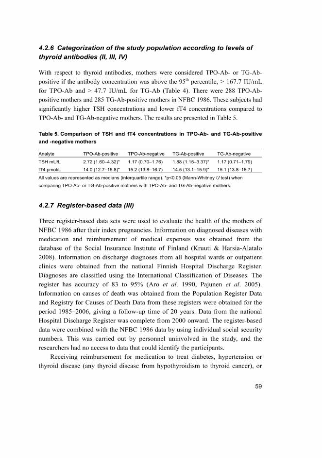
59
4.2.6 Categorization of the study population according to levels of thyroid antibodies (II, III, IV)
With respect to thyroid antibodies, mothers were considered TPO-Ab- or TG-Ab-
positive if the antibody concentration was above the 95th percentile, > 167.7 IU/mL
for TPO-Ab and > 47.7 IU/mL for TG-Ab (Table 4). There were 288 TPO-Ab-
positive mothers and 285 TG-Ab-positive mothers in NFBC 1986. These subjects had
significantly higher TSH concentrations and lower fT4 concentrations compared to
TPO-Ab- and TG-Ab-negative mothers. The results are presented in Table 5.
Table 5. Comparison of TSH and fT4 concentrations in TPO-Ab- and TG-Ab-positive
and -negative mothers
Analyte TPO-Ab-positive TPO-Ab-negative TG-Ab-positive TG-Ab-negative
TSH mU/L 2.72 (1.60–4.32)* 1.17 (0.70–1.76) 1.88 (1.15–3.37)* 1.17 (0.71–1.79)
fT4 pmol/L 14.0 (12.7–15.8)* 15.2 (13.8–16.7) 14.5 (13.1–15.9)* 15.1 (13.8–16.7)
All values are represented as medians (interquartile range). *p<0.05 (Mann-Whitney U test) when
comparing TPO-Ab- or TG-Ab-positive mothers with TPO-Ab- and TG-Ab-negative mothers.
4.2.7 Register-based data (III)
Three register-based data sets were used to evaluate the health of the mothers of
NFBC 1986 after their index pregnancies. Information on diagnosed diseases with
medication and reimbursement of medical expenses was obtained from the
database of the Social Insurance Institute of Finland (Kruuti & Harsia-Alatalo
2008). Information on discharge diagnoses from all hospital wards or outpatient
clinics were obtained from the national Finnish Hospital Discharge Register.
Diagnoses are classified using the International Classification of Diseases. The
register has accuracy of 83 to 95% (Aro et al. 1990, Pajunen et al. 2005).
Information on causes of death was obtained from the Population Register Data
and Registry for Causes of Death Data from these registers were obtained for the
period 1985–2006, giving a follow-up time of 20 years. Data from the national
Hospital Discharge Register was complete from 2000 onward. The register-based
data were combined with the NFBC 1986 data by using individual social security
numbers. This was carried out by personnel uninvolved in the study, and the
researchers had no access to data that could identify the participants.
Receiving reimbursement for medication to treat diabetes, hypertension or
thyroid disease (any thyroid disease from hypothyroidism to thyroid cancer), or

60
having discharge diagnoses of the diseases was considered verification of these
diseases. Mothers with diagnoses of these diseases before pregnancy were
excluded from analyses of the disease in question but not from analyses of other
diseases. (For instance, mothers with a diagnosis of diabetes before pregnancy
were excluded from diabetes analyses, but not from thyroid disease or
hypertension analyses.)
4.2.8 Power analysis
Since this is a prospective population-based study, no a priori power analysis was
performed. However, we evaluated our study population after the laboratory analyses
and we grouped the data according to serum levels of TSH, fT4, TPO-Ab and TG-Ab.
In power analysis we used two-tailed tests with a 5% significance level. Preterm
births was used as an example variable (preterm birth before the 37th and 34th
gestational week). With a power of 80% our study population was of sufficient size to
show 4% and 2.6% differences in the rates of preterm birth before the 37th and 34th
gestational week, respectively, when comparing TPO-Ab-positive and -negative
mothers. This was calculated with rates of preterm birth of 4.3% before the 37th and
1.3% before the 34th gestational week in TPO-Ab-negative mothers. Similarly with a
power of 80%, our study group could show 10.5% and 7.5% differences in the rates
of preterm birth before the 37th and before the 34th gestational week, respectively,
when comparing mothers with overt hypothyroidism with euthyroid mothers (who
had had rates of preterm birth of 4.3% and 1.4% in the calculations).
4.2.9 Statistical methods (I, II, III, IV)
All statistical analyses were performed using SPSS software for Windows (SPSS
Inc., Chicago, Illinois, USA), and versions 13.0, 14.0, 15.0 and 18.0 were used.
Normally distributed data was presented as mean and SD and skewed data was
presented as geometric mean with a 95% confidence interval (CI). All skewed
data was logarithmically transformed before statistical analysis to achieve
normality. Categorical data was represented as n and percentage (%). Laboratory
data was presented as medians and percentile categories (2.5th, 5th, 25th, 75th, 95th,
97.5th percentiles). A p-value of <0.05 was deemed significant in all studies.
To evaluate the influence of storage years on laboratory parameters (Study I),
the variables were analyzed using linear regression analysis. Analysis of variance
followed by Dunnett’s post hoc test was used to reveal significant differences in

61
concentrations when comparing a storage time of 6 months against all other
storage periods. Categorical variables were compared using Pearson’s χ2 test or
Fisher’s exact test, as appropriate. Student’s t test for independent samples was
used to compare continuous variables in two groups.
Comparisons between the thyroid dysfunction groups and the reference group
and between TPO-Ab/TG-Ab-positive and -negative women were evaluated by
using ANOVA, Student’s t test and Fisher’s exact test. To estimate the risk for
adverse perinatal and pregnancy outcome in different thyroid study groups
(Studies II and III), a univariate logistic regression analysis was applied.
Significant associations were represented as odds ratios (ORs) with 95% CIs and
further adjusted for maternal age and parity using multiple logistic regression
analyses. Cox’s regression analysis was applied to assess the effects of maternal
thyroid parameters during pregnancy on later morbidity (Study III) and was
adjusted further for covariates (maternal age, parity, BMI, and presence of
gestational diabetes or gestational hypertension/preeclampsia). The findings were
presented as hazard ratios (HRs) with 95% CIs.
Comparisons of thyroid hormone percentiles in different BMI categories
(Study IV) were evaluated by using (nonparametric) Kruskal-Wallis and Mann–
Whitney U tests, since the data was skewed. BMI categories were defined as
follows: underweight, BMI less than 20 kg/m2, normal weight, BMI of 20.0–24.9
kg/m2, overweight, BMI of 25.0–29.9 kg/m2 and obese, BMI ≥ 30 kg/m2. To test
for thyroid hormone concentration trends in different BMI categories the
Kruskal–Wallis test was used and the Mann–Whitney U test was used for
pairwise comparisons of thyroid hormone concentrations when the population
belonging in the normal BMI category was compared with all other categories.

62

63
5 Results and comments
5.1 Thyroid hormone and antibody analyses after freezing, thawing
and long-term frozen storage (I)
Freezing and thawing had no effect on serum parameters of TSH, fT4, TPO-Ab
and TG-Ab, but fT3 concentrations were higher in frozen samples compared with
fresh samples. Bench-lag time of up to six days at +4 °C did not have any effect
on serum concentrations of TSH, fT4, fT3, TPO-Ab and TG-Ab.
The randomly selected serum samples from the Finnish Maternity Cohort
with storage times of 6 months to 23 years at -25 °C were initially drawn from
pregnant women of mean age 27.2 years and mean gestational age at sampling of
11.9 weeks. The medians and distributions of TSH and fT3 concentrations were
similar in all samples analyzed after different storage times; long-term storage
times had no effect on their concentrations. Concentrations of fT4 were different
from those at baseline after 10, 12 and 20–23 years of storage and storage time
explained 5.5% of the total variation in fT4 concentration.
The concentrations of both TPO-Ab and TG-Ab appeared to increase steadily
with extended storage time. Statistically significant differences from baseline
were seen after two and six years of storage of TPO-Ab and TG-Ab, respectively.
Marked increases in both TPO-Ab and TG-Ab concentrations were seen after 14
years of storage. Storage time explained 19.7% and 19.4% of the total variation
seen in TPO-Ab and TG-Ab concentrations, respectively. Ultracentrifugation had
no effect on antibody concentrations in a subset of samples.
5.1.1 Comments
The stability of TSH, thyroid hormone and antibody analyses after long-term
storage was evaluated for the first time in this study. The samples were selected
from a large biobank, the Finnish Maternity Cohort. Biologic variation between
subjects was minimized by good laboratory procedure ensuring that preanalytical
conditions (for instance time at sampling) were similar in all samples. Also,
subjects whose samples were obtained from the cohort were of certain
characteristics and all samples represented the same months (March and April).
These procedures ensured that seasonal variation seen with thyroid hormones

64
(Oddie et al. 1979) and circadian variation of TSH (Pekonen et al. 1988) were
minimized.
Although fT4 concentrations showed a difference from baseline after long-
term storage, the total variation seen due to storage time was smaller than the
interassay variation of the method. Therefore, the changes in fT4 concentrations
were not clinically significant. Thyroid antibodies, TPO-Ab and TG-Ab, can
fairly reliably be analyzed from samples stored for less than 14 years at -25 °C.
When using older samples, only cross-sectional analyses are permissible. Thyroid
hormones, i.e. fT4 and fT3, and TSH seem to be stable during long-term storage
of up to 23 years at -25 °C. Their concentrations showed somewhat increased
variation with extended storage time, but this was clinically insignificant and
probably due to slight biologic variation.
The concentrations of TPO-Ab and TG-Ab showed very marked variation in
samples with storage times of more than 14 years. Higher thyroid autoantibody
concentrations are likely to be due to storage time, because preanalytical
conditions (sampling, tubes, transportation, sample processing, freezing, thawing,
storage conditions and processing of the samples) did not differ in our study. The
causes of changes in thyroid autoantibody stability during long-term storage are
unknown and probably of a complex character. One can speculate that the
antibodies have degraded in storage and react differently, using our methodology,
but to find out why is beyond the scope of this study.
Freezing and thawing had no effect on TSH, fT4 or thyroid antibody
concentrations. TSH concentrations are stable even after repeated freezing and
thawing (Livesey et al. 1980). Concentrations of fT3 seem to be slightly higher
after freezing and thawing, rendering fT4 analyses more reliable in clinical and
scientific work. Bench-lag time had no effect on any thyroid function parameters
studied, which was a new finding as regards thyroid antibodies. Concentrations of
fT4 and TSH have previously been found to be stable for up to 8 days at +4 °C
(Livesey et al. 1980, Diver et al. 1996).
5.1.2 Implications regarding the use of stored serum samples
The concentrations of TSH and thyroid hormones (fT4 and fT3) were highly
comparable after long-term frozen storage compared with levels in freshly drawn
serum samples. Although fT4 concentrations showed a slight increase with
storage time, analyses of both fT4 and TSH will give reliable results concerning
thyroid function and allow classification of thyroid dysfunction and euthyroidism.

65
Thyroid hormones and TSH analyzed in stored samples can be fairly reliably used
for both cross-sectional and longitudinal analyses and comparison of different
populations.
Thyroid antibodies, TPO-Ab and TG-Ab, were not as stable during long-term
storage. For both TPO-Ab and TG-Ab the results were shifted towards higher
values. For TPO-Ab, the reference value given by the assay manufacturer (> 5.61
IU/mL) to signify antibody positivity was exceeded in nearly 30% of the samples.
For TG-Ab this shift was even greater, as nearly all of the samples gave results
higher than the reference value (> 4.11 IU/mL). Since all of the samples were
stored and handled in exactly the same way, we presume that storage affected all
samples similarly. Thus, we presume that those samples which would have
produced low values signifying antibody negativity if they had been analysed
primarily in 1986 provided low values in our current analyses.
Our theory has been backed up to some degree by a small number of results
from TG-Ab and antimicrosomal antibody analyses done in the 1980s, obtained
from the hospital records of mothers with self-informed thyroid disease. Of these
123 mothers, 15 had had TG-Ab and/or antimicrosomal antibody testing during
the index pregnancy. Current analyses were done on 8 of these samples.
Comparison of the results of the analyses in the 1980s and the current data, is
presented in Table 6. All the negative results were also negative in the current
analyses and all the positive results were clearly positive. The only discrepancy in
the results concerns patient 4, who had a measurable titer of TG-Ab during
pregnancy (at gestational week 33). The current analysis gave a negative result,
but the sample was from early pregnancy (gestational week 15), which might
explain it. Also, according to the definition of TPO-Ab positivity (see Methods,
and below), patient 5 was deemed to be TPO-Ab-negative in our current data, but
the result is near the cut-off value of TPO-Ab positivity. Despite these results, we
cannot rule out the possibility that samples have given high TPO-Ab and TG-Ab
values as a result of storage and would have primarily given negative results.
Looking at the distribution of TPO-Ab and TG-Ab, 90% of the samples
showed results lower than 30 IU/mL and 23.75 IU/mL, respectively. In the
literature, up to 5–10% of pregnant women are considered to be TPO-Ab- and/or
TG-Ab-positive. We decided on a cut-off value of the 95th percentile to define
TPO-Ab and TG-Ab positivity; this resulted in absolute values of 167.70 and
47.70 IU/mL for TPO-Ab and Tg-Ab, as described earlier. Upon deciding on this
cut-off point, we may be underestimating or overestimating the total number of
TPO-Ab- and TG-Ab-positive mothers.

66
Table 6. Comparison of patient results from the 1980s with our current analyses.
Patients
with thyroid
disease
Results 1980s Results 2006
Microsomal Ab/TPO-Ab TG-Ab TPO-Ab IU/mL
(negative/positive)
TG-Ab IU/mL
(negative/positive)
Patient 1 Negative Negative 4.53 (negative) 12.28 (negative)
Patient 2 Positive 277.6 (positive)
Patient 3 Positive 1000 (positive)
Patient 4 Negative 1:400 5.17 (negative) 14.15 (negative)
Patient 5 Weak positive 118.53 (negative)
Patient 6 Negative Negative 3.92 (negative) 11.94 (negative)
Patient 7 Positive 1:25600 1000 (positive) 574.02 (positive)
Patient 8 Negative Negative 17.34 (negative) 8.90 (negative)
As described earlier, TPO-Ab- and TG-Ab-positive mothers have higher TSH
concentrations and lower fT4 concentrations than TPO-Ab- and TG-Ab-negative
mothers (Table 5). This indicates that the used cut-off is suitable. We have also
looked at the distributions of TPO-Ab and TG-Ab in euthyroid mothers and
mothers with thyroid dysfunction. The distributions are very similar to those seen
in other studies: a high prevalence of both TPO-Ab and TG-Ab in mothers with
overt and subclinical hypothyroidism as markers of chronic autoimmune
thyroiditis and lower prevalence in other groups (Figure 3).
In conclusion, the definition of TPO-Ab and TG-Ab positivity in this study is
based on data from stored serum samples with changes during storage.
Interpretation of the results based solely on our TPO-Ab and TG-Ab
measurements requires some consideration, since the definition of antibody
positivity may over- or underestimate the real prevalence, and storage time has
had an effect on the analytes, with no information available as to whether or not
the change is linear or random.

67
Fig. 3. Percentages of TPO-Ab- (upper graph) and TG-Ab-positive (lower graph)
mothers in NFBC 1986. The p-values were obtained by comparing the group in
guestion with the euthyroid group by (Chi2 test).
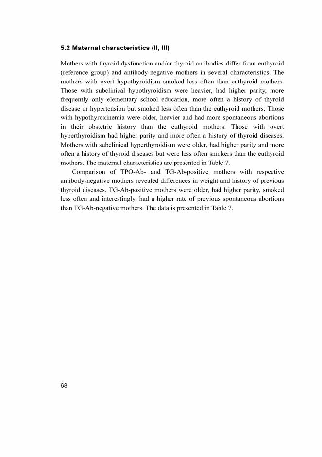
68
5.2 Maternal characteristics (II, III)
Mothers with thyroid dysfunction and/or thyroid antibodies differ from euthyroid
(reference group) and antibody-negative mothers in several characteristics. The
mothers with overt hypothyroidism smoked less often than euthyroid mothers.
Those with subclinical hypothyroidism were heavier, had higher parity, more
frequently only elementary school education, more often a history of thyroid
disease or hypertension but smoked less often than the euthyroid mothers. Those
with hypothyroxinemia were older, heavier and had more spontaneous abortions
in their obstetric history than the euthyroid mothers. Those with overt
hyperthyroidism had higher parity and more often a history of thyroid diseases.
Mothers with subclinical hyperthyroidism were older, had higher parity and more
often a history of thyroid diseases but were less often smokers than the euthyroid
mothers. The maternal characteristics are presented in Table 7.
Comparison of TPO-Ab- and TG-Ab-positive mothers with respective
antibody-negative mothers revealed differences in weight and history of previous
thyroid diseases. TG-Ab-positive mothers were older, had higher parity, smoked
less often and interestingly, had a higher rate of previous spontaneous abortions
than TG-Ab-negative mothers. The data is presented in Table 7.

Ta
ble
7. D
em
og
rap
hic
ch
ara
cte
ris
tic
s o
f N
FB
C 1
98
6 m
oth
ers
.
Dem
ogra
phic
cha
ract
eris
tics
Ref
eren
ce g
roup
O
vert
hypo
thyr
oidi
sm
Sub
clin
ical
hypo
thyr
oidi
sm
Hyp
othy
roxi
nem
iaO
vert
hype
rthyr
oidi
sm
Sub
clin
ical
hype
rthyr
oidi
sm
Age
(yea
rs)
28.0
(5.3
) 28
.7 (5
.1)
28.6
(5.8
) 29
.4 (6
.0)*
28
.9 (5
.8)
29.5
(5.5
)*
BM
I (kg
/m2 )
22.1
(3.4
) 22
.5 (3
.8)
22.6
(3.7
)*
23.0
(3.8
)*
22.0
(2.9
) 22
.5 (3
.5)
Nul
lipar
ous
1628
(34.
5%)
12 (2
2.2%
) 69
(30.
8%)
69 (3
0.7%
) 17
(22.
1%)*
45
(22.
1%)*
Par
ity (m
in-m
ax)
1.3
(0-1
5)
1.5
(0-7
) 1.
6 (0
-13)
* 2.
0 (0
-14)
2.
1 (0
-12)
* 1.
9 (0
-12)
*
Sm
oker
s 13
07 (2
7.7%
) 3
(5.6
%)*
40
(17.
9%)*
74
(32.
6%)
17 (2
1.1%
) 31
(15.
2%)*
Alc
ohol
use
dur
ing
preg
nanc
y 27
1 (5
.7%
) 3
(5.6
%)
11 (4
.9%
) 18
(7.9
%)
3 (3
.9%
) 10
(4.9
%)
Ele
men
tary
sch
ool e
duca
tion
1058
(22.
4%)
13 (2
4.1%
) 66
(29.
5%)*
52
(22.
9%)
20 (2
6.0%
) 53
(26.
0%)
Voc
atio
nal s
choo
l edu
catio
n 25
05 (5
3.1%
) 22
(40.
7%)
95 (4
2.4%
)*
123
(54.
2%)
39 (5
0.6%
) 10
5 (5
1.3%
)
Upp
er s
econ
dary
sch
ool e
duca
tion
244
(5.2
%)
5 (9
.3%
) 14
(6.3
%)
12 (5
.3%
) 3
(3.9
%)
9 (4
.4%
)
Uni
vers
ity e
duca
tion
344
(7.3
%)
7 (1
3.0%
) 17
(7.6
%)
10 (4
.4%
) 5
(6.5
%)
19 (9
.3%
)
Obs
tetri
c an
d di
seas
e hi
stor
y
Spo
ntan
eous
abo
rtion
s 91
9 (1
9.5%
) 12
(22.
2%)
36 (1
6.1%
) 53
(23.
5%) *
12
(15.
6%)
45 (2
2.1%
)
Low
-birt
h-w
eigh
t inf
ants
21
2 (4
.5%
) 1
(1.9
%)
8 (3
.6%
) 12
(5.3
%)
5 (6
.5%
) 9
(4.4
%)
Pre
term
birt
hs
262
(5.5
%)
0 11
(5.3
%)
14 (6
.2%
) 7
(9.1
%)
9 (4
.4%
)
Feta
l dea
ths
65 (1
.4%
) 0
1 (0
.4%
) 1
(0.4
%)
1 (1
.3%
) 1
(0.5
%)
Neo
nata
l dea
ths
56 (1
.2%
) 0
3 (1
.3%
) 3
(1.3
%)
2 (2
.6%
) 1
(0.5
%)
Infe
rtilit
y tre
atm
ents
25
3 (5
.4%
) 3
(5.6
%)
10 (4
.5%
) 9
(4.1
%)
4 (5
.2%
) 14
(6.9
%)
Thyr
oid
dise
ases
41
(0.9
%)
2 (3
.7%
) 14
(6.2
%)*
4
(1.8
%)
3 (3
.9%
)*
5 (2
.5%
)*
Aut
oim
mun
e di
seas
es
10 (0
.2%
) 0
2 (0
.9%
) 1
(0.4
%)
0 0
Dia
bete
s 6
(0.1
%)
0 0
0 0
0
Hyp
erte
nsio
n 87
(1.9
%)
2 (3
.7%
) 11
(4.9
%)*
8
(3.5
%)
2 (2
.6%
) 2
(1.0
%)
Val
ues
are
n (%
) or m
ean
(SD
). *p
<0.0
5 w
hen
com
parin
g th
yroi
d dy
sfun
ctio
n gr
oup
with
refe
renc
e gr
oup
69

Ta
ble
7 (
co
nti
nu
ed
). D
em
og
rap
hic
ch
ara
cte
ris
tic
s o
f N
FB
C 1
98
6 m
oth
ers
.
Dem
ogra
phic
cha
ract
eris
tics
TPO
-Ab-
posi
tive
TPO
-Ab-
nega
tive
TG-A
b-po
sitiv
e TG
-Ab-
nega
tive
Age
(yea
rs)
28.6
(5.1
) 28
.2 (5
.4)
29.1
(5.4
)*
28.1
(5.4
)
BM
I (kg
/m2 )
22.7
(4.0
)*
22.2
(3.4
) 22
.7 (4
.0)*
22
.2 (3
.4)
Nul
lipar
ous
81 (2
8.1%
) 18
42 (3
3.6%
) 73
(25.
6%)*
18
28 (3
3.7%
)
Par
ity (m
in-m
ax)
1.3
(0-1
3)
1.4
(0-1
5)
1.7
(0-1
3)*
1.4
(0-1
5)
Sm
oker
s 74
(25.
7%)
1486
(27.
1%)
45 (1
5.8%
)*
1491
(27.
5%)
Alc
ohol
use
dur
ing
preg
nanc
y 16
(5.6
%)
310
(5.7
%)
15 (5
.3%
) 31
0 (5
.7%
)
Ele
men
tary
sch
ool e
duca
tion
71 (2
4.7%
) 12
57 (2
3.0%
) 70
(24.
6%)
1246
(23.
0%)
Voc
atio
nal s
choo
l edu
catio
n 15
5 (5
3.8%
) 28
70 (5
2.4%
) 14
0 (4
9.1%
) 28
54 (5
2.7%
)
Upp
er s
econ
dary
sch
ool e
duca
tion
12 (4
.2%
) 28
9 (5
.3%
) 15
(5.3
%)
282
(5.2
%)
Uni
vers
ity e
duca
tion
20 (6
.9%
) 35
8 (6
.5%
) 22
(7.7
%)
379
(7.0
%)
Obs
tetri
c an
d di
seas
e hi
stor
y
Spo
ntan
eous
abo
rtion
s 59
(20.
5%)
1063
(19.
4%)
67 (2
3.5%
)*
1045
(19.
3%)
Low
-birt
h-w
eigh
t inf
ants
13
(4.5
%)
246
(4.5
%)
15 (5
.3%
) 24
2 (4
.5%
)
Pre
term
birt
hs
19 (6
.6%
) 29
3 (5
.3%
) 16
(5.3
%)
294
(5.4
%)
Feta
l dea
ths
4 (1
.3%
) 6
7 (1
.3%
) 6
(2.2
%)
64 (1
.2%
)
Neo
nata
l dea
ths
3 (1
.0%
) 63
(1.1
%)
5 (1
.8%
) 60
(1.1
%)
Infe
rtilit
y tre
atm
ents
18
(6.3
%)
290
(5.3
%)
17 (6
.0%
) 28
7 (5
.3%
)
Thyr
oid
dise
ases
12
(4.2
%)*
42
(0.8
%)
7 (2
.5%
)*
47 (0
.9%
)
Aut
oim
mun
e di
seas
es
1 (0
.3%
) 11
(0.2
%)
0 12
(0.2
%)
Dia
bete
s 1
(0.3
%)
5 (0
.1%
) 1
(0.4
%)
5 (0
.1%
)
Hyp
erte
nsio
n 8
(2.8
%)
113
(2.1
%)
8 (2
.8%
) 11
2 (2
.1%
)
Val
ues
are
n (%
) or m
ean
(SD
). *p
<0.0
5 w
hen
com
parin
g an
tibod
y-po
sitiv
e m
othe
rs w
ith a
ntib
ody-
nega
tive
mot
hers
70

71
5.3 Perinatal outcome (II) and pregnancy outcome (III)
5.3.1 Perinatal outcome
The peri- and neonatal characteristics of infants born to the studied NFBC 1986
mothers are presented in Table 8. The peri- and neonatal outcomes of infants born
to mothers with hypothyroxinemia were evaluated upon finalizing this thesis.
The newborns of overtly hypothyroid mothers had higher ponderal indexes
and their mothers had higher absolute and also higher relative placental weights
than reference group mothers. Similar placental findings were found in mothers
with overt hyperthyroidism. Also, their children were more often LGA compared
with those in the reference group; the calculated OR was 2.7 (95% CI 1.1–6.9).
The significant association between LGA and overt hyperthyroidism did not
persist after adjusting for maternal age and parity.
No adverse outcomes were associated with subclinical hypothyroidism or
subclinical hyperthyroidism. In fact, fewer low Apgar scores (< 7 at 5 minutes)
were seen among children born to mothers with subclinical hyperthyroidism (OR
0.4, 95% CI 0.2–0.95) compared with children of euthyroid mothers. The
association was no longer significant after adjusting for maternal age and parity.
The TPO-Ab-positive mothers gave birth more often to LGA infants, the OR
being 2.1 (95% CI 1.2–3.7). Adjustment for maternal characteristics did not
change this association. Interestingly, TPO-Ab-positive mothers also had higher
rates of low-birth-weight infants (birth weight < 2500 g) and higher placental
weights.
Perinatal mortality was higher among infants born to TPO-Ab-positive
mothers compared with TPO-Ab-negative mothers. Also, the newborns of TG-
Ab-positive mothers had a higher perinatal mortality rate and more of them were
born in non-cephalic presentation. The risk of perinatal death was elevated in
TPO-Ab- and TG-Ab-positive mothers (OR 3.1 [95% CI 1.4–7.1] and OR 2.6
[95% CI 1.1–6.2]) in TPO-Ab and TG-Ab positive mothers, respectively, and
persisted even after adjusting for maternal age and parity.

Ta
ble
8. P
eri
- a
nd
ne
on
ata
l o
utc
om
e o
f s
tud
y p
op
ula
tio
n.
Out
com
e R
efer
ence
gro
up
Ove
rt
hypo
thyr
oidi
sm
Sub
clin
ical
hypo
thyr
oidi
sm
Hyp
othy
roxi
nem
ia
Ove
rt
hype
rthyr
oidi
sm
Sub
clin
ical
hype
rthyr
oidi
sm
Birt
h w
eigh
t (g)
35
68 (5
48)
3676
(657
)35
14(5
42)
3569
(612
)37
03 (5
68)*
3564
(524
)Lo
w b
irth
wei
ght
149
(3.2
%)
2 (3
.7%
) 8
(3.6
%)
8 (3
.5%
) 2
(2.6
%)
7 (3
.4%
) S
GA
93
(2.0
%)
0 5
(2.3
%)
6 (2
.7%
) 1
(1.4
%)
2 (1
.0%
) LG
A
116
(2.5
%)
3 (5
.6%
) 4
(1.8
%)
7 (3
.2%
) 5
(6.6
%)*
3
(1.5
%)
Birt
h le
ngth
(cm
) 50
.5 (2
.4)
50.6
(2.7
) 50
.2 (2
.5)
50.5
(2.2
) 50
.9 (2
.3)
50.5
(2.2
) P
onde
ral i
ndex
(kg/
m3 )
27.6
(2.4
) 28
.4 (2
.8)*
27
.5 (2
.3)
27.6
(2.7
) 27
.9 (2
.2)
27.5
(2.1
) H
ead
circ
umfe
renc
e (c
m)
35.2
(1.5
) 35
.4 (2
.4)
35.0
(1.6
) 35
.3 (1
.4)
35.2
(1.2
) 35
.1 (1
.6)
Ges
tatio
nal w
eeks
at b
irth
39.4
(1.7
) 39
.4 (2
.3)
39.3
(1.8
) 39
.3 (1
.9)
39.4
(1.5
) 39
.4 (1
.5)
Pre
term
birt
h
<
37 g
esta
tiona
l wee
ks
204
(4.3
%)
4 (7
.4%
) 7
(3.1
%)
13 (5
.7%
) 2
(2.6
%)
9 (4
.4%
) <
34 g
esta
tiona
l wee
ks
64 (1
.4%
) 1
(1.9
%)
5 (2
.2%
) 4
(1.8
%)
1 (1
.3%
) 3
(1.5
%)
Non
ceph
alic
pre
sent
atio
n 17
5 (3
.7%
) 1
(1.9
%)
7 (3
.2%
) 6
(2.7
%)
4 (5
.2%
) 6
(3.0
%)
Apg
ar s
core
7 o
r les
s at
5 m
in
317
(6.7
%)
6 (1
1.1%
) 14
(6.3
%)
16 (7
.0%
) 4
(5.2
%)
6 (2
.9%
)*
Apg
ar s
core
7 o
r les
s at
10
min
10
2 (2
.7%
) 1
(2.6
%)
5 (2
.9%
) 5
(2.7
%)
2 (3
.4%
) 4
(2.5
%)
Apg
ar s
core
7 o
r les
s at
15
min
42
(1.3
%)
1 (2
.3%
) 3
(2.0
%)
3 (1
.8%
) 0
2 (1
.3%
) P
erin
atal
mor
talit
y 39
(0.8
%)
1 (1
.9%
) 4
(1.8
%)
4 (1
.8%
) 0
0 Fe
tal d
eath
24
(0.5
%)
1 (1
.9%
) 1
(0.4
%)
2 (0
.9%
) 0
0 N
eona
tal d
eath
15
(0.3
%)
0 3
(1.4
%)
2 (0
.9%
) 0
0 M
alfo
rmat
ions
14
1 (3
.0%
) 1
(1.9
%)
8 (3
.6%
) 7
(3.1
%)
2 (2
.6%
) 3
(1.5
%)
Pla
cent
al w
eigh
t (g)
64
2 (1
32)
688
(142
)*
636
(133
) 64
6 (1
43)
696
(140
)*
644
(139
) R
elat
ive
plac
enta
l wei
ght (
%)
18.1
(3.3
) 19
.1 (4
.3)*
18
.2 (3
.2)
18.3
(3.7
) 18
.9 (2
.9)*
18
.1 (3
.3)
Um
bilic
al c
ord
leng
th (c
m)
56.1
(12.
0)
56.0
(9.9
) 56
.6 (1
2.0)
56
.9 (1
2.3)
55
.6 (1
1.5)
56
.5 (1
3.6)
C
esar
ean
sect
ions
62
0 (1
4.1%
) 8
(15.
7%)
29 (1
4.0%
) 27
(12.
6%)
7 (9
.5%
) 27
(13.
8%)
Val
ues
are
n (%
) or m
ean
(SD
). *p
<0.0
5 w
hen
com
parin
g th
yroi
d dy
sfun
ctio
n gr
oup
vs. r
efer
ence
gro
up
72

Ta
ble
8 (
co
nti
nu
ed
). P
eri
- a
nd
ne
on
ata
l o
utc
om
e o
f s
tud
y p
op
ula
tio
n.
Out
com
e TP
O-A
b-po
sitiv
e TP
O-A
b-ne
gativ
e TG
-Ab-
posi
tive
TG-A
b-ne
gativ
e
Birt
h w
eigh
t (g)
35
51 (6
58)
3570
(543
)36
06 (5
96)
3567
(548
)Lo
w b
irth
wei
ght
15 (5
.2%
) 16
8 (3
.1%
) 7
(2.5
%)
174
(3.2
%)
SG
A
8 (2
.9%
) 10
6 (2
.0%
) 7
(2.6
%)
107
(2.0
%)
LGA
14
(5.0
%)*
13
0 (2
.4%
) 11
(4.0
%)
132
(2.5
%)
Birt
h le
ngth
(cm
) 50
.3 (2
.8)
50.5
(2.3
) 50
.5 (3
.0)
50.5
(2.3
) P
onde
ral i
ndex
(kg/
m3 )
27.6
(2.5
) 27
.6 (2
.4)
27.8
(2.6
) 27
.6 (2
.4)
Hea
d ci
rcum
fere
nce
(cm
) 35
.1 (2
.1)
35.2
(1.5
) 35
.2 (1
.9)
35.2
(1.5
) G
esta
tiona
l wee
ks a
t birt
h 39
.2 (2
.3)
39.4
(1.7
) 39
.3 (1
.9)
39.4
(1.7
) P
rete
rm b
irth
< 37
ges
tatio
nal w
eeks
17
(5.9
%)
237
(4.3
%)
12 (4
.2%
) 23
8 (4
.4%
) <
34 g
esta
tiona
l wee
ks
8 (2
.8%
) 70
(1.3
%)
5 (1
.8%
) 73
(1.3
%)
Non
ceph
alic
pre
sent
atio
n 11
(3.9
%)
197
(3.6
%)
18 (6
.3%
)*
186
(3.5
%)
Apg
ar s
core
7 o
r les
s at
5 m
in
20 (7
.0%
) 35
4 (6
.5%
) 17
(6.0
%)
352
(6.5
%)
Apg
ar s
core
7 o
r les
s at
10
min
8
(3.6
%)
117
(2.7
%)
8 (3
.6%
) 11
6 (2
.7%
) A
pgar
sco
re 7
or l
ess
at 1
5 m
in
5 (2
.3%
) 49
(1.3
%)
6 (2
.7%
) 48
(1.3
%)
Per
inat
al m
orta
lity
7 (2
.4%
)*
43 (0
.8%
) 6
(2.1
%)*
44
(0.8
%)
Feta
l dea
th
4 (1
.4%
) 26
(0.5
%)
4 (1
.4%
) 26
(0.5
%)
Neo
nata
l dea
th
3 (1
.1%
) 17
(0.3
%)
2 (0
.7%
) 18
(0.3
%)
Mal
form
atio
ns
8 (2
.8%
) 16
5 (3
.0%
) 7
(2.5
%)
163
(3.0
%)
Pla
cent
al w
eigh
t (g)
65
4 (1
46)
642
(131
) 64
1 (1
28)
643
(133
) R
elat
ive
plac
enta
l wei
ght (
%)
18.7
(3.9
)*
18.1
(3.3
) 18
.1 (4
.0)
18.1
(3.3
) U
mbi
lical
cor
d le
ngth
(cm
) 56
.9 (1
1.1)
56
.1 (1
2.0)
56
.4 (1
1.0)
56
.1 (1
2.0)
C
esar
ean
sect
ions
40
(15.
1%)
708
(13.
8%)
36 (1
3.5%
) 70
3 (1
3.9%
)
Val
ues
are
n (%
) or m
ean
(SD
). *p
<0.0
5 w
hen
com
parin
g an
tibod
y-po
sitiv
e m
othe
rs v
s. a
ntib
ody-
nega
tive
mot
hers
.
73

74
5.3.2 Analyses of sensitivity
In the light of recent literature, we carried out sensitivity analyses, in which we
evaluated separately the effect of mild subclinical hypothyroidism and all cases of
hypothyroidism (pooled analysis) on adverse outcomes. This was done to
evaluate if the TSH cut-off used to initially determine subclinical hypothyroidism
skewed the results by including those with subclinical disease in the reference
group. Subclinical hypothyroidism in TPO-Ab- and TG-Ab-negative subjects was
defined on the basis of our calculated reference intervals in the first trimester as
follows: TSH between 2.7–4.94 mU/L and fT4 between 11.5–22.4 pmol/L. The
high cut-off limit of TSH was based on the reference interval given by the assay
manufacturer. Similarly in the pooled analysis, hypothyroidism was defined as
TSH > 2.7 mU/L with normal or low fT4 concentrations with/without TPO-Ab
and/or TG-Ab positivity. Euthyroidism was defined as TSH 0.1–2.69 mU/L and
fT4 11.5–22.4 pmol/L with negative TPO-Ab and TG-Ab. All those with self-
informed thyroid disease were excluded.
Upon implementing this definition, 329 TPO-Ab- and TG-Ab-negative
mothers were found to have subclinical hypothyroidism. Mothers with subclinical
hypothyroidism were heavier than the euthyroid mothers, but no other difference
in maternal characteristics was seen. Mothers with subclinical hypothyroidism
more often had LGA infants. The OR for having an LGA infant was 1.91 (1.06–
3.44) unadjusted and 1.91 (1.05–3.45) adjusted for maternal age and parity. None
of the 13 mothers with subclinical hypothyroidism and an LGA infant was obese
or had type I diabetes or gestational diabetes. Rates of other adverse outcomes
were similar in euthyroid mothers and mothers with subclinical hypothyroidism.
In the pooled analyses hypothyroidism was detected in 561 women. These
women were somewhat older and heavier and smoked less often than the
euthyroid mothers. They were found to have increased risks of LGA infants (OR
1.7 [1.1–2.8] both unadjusted and adjusted for maternal age and parity) and
neonatal deaths (OR 3.7 [1.4–9.7] unadjusted, and adjusted OR 3.6 [1.4–9.5]). Of
the six mothers whose neonates died, all had TSH concentrations higher than 3
mU/L and normal fT4 levels, and three were TPO-Ab-positive. The mothers with
hypothyroidism and TPO-Ab and/or TG-Ab positivity (n = 175) were also heavier,
older and less often smokers than the euthyroid mothers. They had an increased
risk of neonatal mortality. These results are presented in Table 9.

75
Table 9. Adverse perinatal outcome among mothers with mild subclinical
hypothyroidism, and all cases of hypothyroidism, compared with euthyroid mothers.
Outcome Euthyroidism
(n=4600)
Subclinical
hypothyroidism
(n=329)
Overt and
subclinical
hypothyroidism
(n=561)
Overt and subclinical
hypothyroidism and
TPO-Ab and/or TG-Ab
positivity (n=175)
Perinatal deaths 33 (0.7%) 3 (0.9%) 8 (1.4%) 4 (2.3%)
Fetal deaths 22 (0.5%) 0 2 (0.4%) 1 (0.6%)
Neonatal deaths 13 (0.3%) 3 (0.9%) 6 (1.1%)* 3 (1.7%)*
Preterm births
< 37 gestational week 196 (4.4%) 13 (4.0%) 24 (4.3%) 8 (4.6%)
< 34 gestational week 57 (1.3%) 6 (1.8%) 10 (1.8%) 3 (1.7%)
Non-cephalic presentation 164 (3.7%) 6 (1.9%) 13 (2.3%) 6 (3.5%)
LGA 97 (2.2%) 13 (4.2%)* 21 (3.8%)* 8 (4.7%)
SGA 84 (1.9%) 10 (3.2%) 13 (2.4%) 3 (1.8%)
Malformations 135 (3.0%) 10 (3.1%) 19 (3.4%) 7 (4.0%)
Apgar scores ≤ 7at 5 min 288 (6.5%) 20 (6.2%) 41 (7.3%) 15 (8.6%)
Maternal characteristics
Diabetes 9 (0.2%) 1 (0.3%) 2 (0.4%) 1 (0.5%)
Smokers 1238 (29.0%) 79 (25.2%) 122 (22.7%)* 34 (20.9%)*
Preeclampsia 136 (3.1%) 14 (4.3%) 21 (3.8%) 5 (2.9%)
Gestational hypertension 203 (4.6%) 23 (7.1%) 35 (6.3%) 9 (5.2%)
BMI (kg/m2) 22.1 (3.3) 22.5 (3.3)* 22.5 (3.5)* 22.7 (4.0)*
Age (yr) 28.0 (5.3) 28.4 (5.7) 28.5 (5.6)* 28.8 (5.3)*
Gestational diabetes 43 (1.0%) 4 (1.3%) 4 (0.7%) 0
All values are n (%) or mean (SD). *p<0.05
5.3.3 Pregnancy outcome
The studied pregnancy complications of the NFBC 1986 mothers are presented in
Table 10. Mothers with thyroid dysfunction during pregnancy had similar rates of
pregnancy complications as the reference group mothers. TG-Ab-positive
mothers more often had gestational hypertension, but no other pregnancy
complications were seen more often among antibody-positive mothers compared
with antibody-negative mothers.

Ta
ble
10
. a
) P
reg
na
nc
y c
om
pli
ca
tio
ns
ob
se
rve
d in
mo
the
rs w
ith
th
yro
id d
ysfu
nc
tio
n.
Ta
ble
10
. b
) P
reg
nan
cy
co
mp
lic
ati
on
s o
bs
erv
ed
in
an
tib
od
y-p
os
itiv
e a
nd
-n
eg
ati
ve m
oth
ers
.
Pre
gnan
cy o
utco
me
TPO
-Ab-
posi
tive
TPO
-Ab-
nega
tive
TG-A
b-po
sitiv
e TG
-Ab-
nega
tive
Ges
tatio
nal h
yper
tens
ion
7 (2
.5%
) 16
1 (3
.0%
) 14
(5.1
%)*
15
3 (2
.9%
)
Pre
ecla
mps
ia
6 (2
.2%
) 10
8 (2
.0%
) 3
(1.1
%)
111
(2.1
%)
Ges
tatio
nal d
iabe
tes
mel
litus
0
50 (1
.0%
) 2
(0.7
%)
48 (0
.9%
)
Pla
cent
al a
brup
tion
1 (0
.4%
) 28
(0.5
%)
3 (1
.1%
) 26
(0.5
%)
Mat
erna
l wei
ght g
ain
> 20
kg
durin
g
preg
nanc
y
31 (1
1.4%
) 47
7 (9
.3%
) 30
(11.
3%)
477
(9.4
%)
Val
ues
are
n (%
). *p < 0
.05
whe
n co
mpa
ring
antib
ody-
posi
tive
mot
hers
vs.
ant
ibod
y-ne
gativ
e m
othe
rs.
Pre
gnan
cy o
utco
me
Ref
eren
ce
grou
p
Ove
rt
hypo
thyr
oidi
sm
Sub
clin
ical
hypo
thyr
oidi
sm
Hyp
othy
roxi
nem
ia
Ove
rt
hype
rthyr
oidi
sm
Sub
clin
ical
hype
rthyr
oidi
sm
Ges
tatio
nal h
yper
tens
ion
139
(3.0
%)
2 (3
.8%
) 7
(3.3
%)
7 (3
.3%
) 2
(2.7
%)
7 (3
.5%
)
Pre
ecla
mps
ia
89 (1
.9%
) 1
(1.9
%)
8 (3
.8%
) 3
(1.4
%)
2 (2
.7%
) 7
(3.5
%)
Ges
tatio
nal d
iabe
tes
mel
litus
47
(1.0
%)
0 0
0 1
(1.3
%)
1 (0
.5%
)
Pla
cent
al a
brup
tion
24 (0
.5%
) 0
1 (0
.5%
) 1
(0.5
%)
0 2
(1.0
%)
Mat
erna
l wei
ght g
ain
> 20
kg
durin
g
preg
nanc
y
430
(9.8
%)
5 (1
0.0%
) 22
(10.
5%)
18 (8
.7%
) 5
(6.7
%)
13 (6
.7%
)
Val
ues
are
n (%
). *p < 0
.05
whe
n co
mpa
ring
thyr
oid
dysf
unct
ion
grou
ps v
s. re
fere
nce
grou
p
76

77
5.3.4 Comments
The present study is one of the few large prospective studies evaluating the effect
of overt and subclinical hypothyroidism or hyperthyroidism and positive TPO-Ab
or TG-Ab status during pregnancy on the perinatal, neonatal and pregnancy
outcome. This study is based on a large prospective birth cohort, with extensive
data collected throughout pregnancy and a vast amount of laboratory data. Our
results are, however, somewhat contradictory to previous findings.
Preterm births
Previously, both overt and subclinical maternal hypothyroidism have been
associated with an increased risk of preterm birth (Abalovich et al. 2002, Casey et al. 2005, Stagnaro-Green et al. 2005, Antolic et al. 2006, Wikner et al. 2008). The
retrospective study carried out by Abalovich et al. showed a very high incidence
of preterm births in a small population based on hospital patients: 20%, 7.2% and
11.1% among mothers with overt hypothyroidism, subclinical hypothyroidism
and euthyroidism under levothyroxine treatment, respectively (Abalovich et al. 2002). The mothers in this study represented clear-cut hypothyroidism, as
indicated by the high TSH levels (> 4 mIU/L) observed in the hypothyroidism
groups. Two studies have involved retrospective evaluation of a large number of
women with hypothyroidism (but with no evaluation of the adequacy of
treatment), and increases in the rate of preterm birth were found (Antolic et al. 2006, Wikner et al. 2008). In a large population-based study concerning
adequately treated hypothyroidism, no increase in preterm birth was seen
(Matalon et al. 2006). It would therefore seem that although preterm births are
usually of multifactorial origin, adequate treatment of maternal hypothyroidism is
of importance to prevent some cases of preterm births. In our data, those with
clear-cut overt hypothyroidism had a (nonsignificantly) higher rate of preterm
birth (7.4%) compared with euthyroid mothers (4.3%), which would support this
association.
The effects of subclinical hypothyroidism on preterm birth have also been
evaluated. In a large study by Casey et al. 4% of mothers with subclinical
hypothyroidism were found to have preterm birth before the 34th gestational week,
compared with 2.5% of euthyroid mothers (Casey et al. 2005). The population in
this study represented a mostly Hispanic, medically indigent population from a
tax-supported hospital, and as such may represent a population with more social

78
problems and other confounding factors. Stagnaro-Green et al. reported that of
those with very preterm birth (before the 32nd gestational week), up to 14.3% had
mildly elevated TSH levels, as opposed to 5.7% of controls (Stagnaro-Green et al. 2005). However, in one large population-based study in which subclinical
hypothyrodism was defined with a high cut-off level of TSH, no increase in
preterm births was seen (Cleary-Goldman et al. 2008). However, this population
may be positively skewed as it represented mostly white Caucasian women from
the private sector and the high TSH cut-off level used may have resulted in some
women with subclinical hypothyroidism being classified as healthy, thus diluting
the possible differences. In a recent study in which very mild subclinical
hypothyroidism in TPO-Ab-negative mothers was evaluated, no increase in rates
of preterm births was seen (Negro et al. 2010a). In our study, among those with
subclinical hypothyroidism (initial definition TSH > 3.61 mU/L and normal fT4,
thyroid autoantibody-positive mothers included), no increase in preterm births
was seen. These results were further confirmed by evaluating only those with
mild TSH elevation (TSH 2.7–4.92 mU/L) and TPO-Ab and TG-Ab negativity
and those with hypothyroidism (TSH ≥2.7 mU/L and normal or low fT4)
with/without TPO-Ab and TG-Ab positivity to study if our initially high TSH cut-
off ameliorated these results. In this respect, our results are in accordance with
those in study by Negro et al. (Negro et al. 2010a), suggesting that among
mothers with mild subclinical hypothyroidism (TSH < 5 mU/L) and TPO-Ab/TG-
Ab negativity, the rates of preterm birth are similar to those in euthyroid mothers.
Also, in our data, those with subclinical or overt hypothyroidism and TPO-Ab
and/or TG-Ab positivity did not have an increased risk of preterm births.
Autoimmune thyroiditis has been shown to increase the risk of preterm births
in some studies (Glinoer et al. 1994, Negro et al. 2006), and this risk increase is
thought to result from underlying hypothyroidism (Negro et al. 2006). The
incidence of preterm birth is reported to be up to 16.1–26.8% among thyroid
autoantibody-positive mothers in a hospital setting (Glinoer et al. 1994, Ghafoor
et al. 2006, Negro et al. 2006), although some contradictory results have also
been presented. In a large population-based study, the rates of preterm birth were
7.2% and 9.0% in euthyroid and hypothyroid TPO-Ab- and/or TG-Ab-positive
women and 6.4% and 5.9% in hypothyroid and euthyroid antibody-negative
women. These differences did not reach statistical significance, probably due to
small sample size after grouping. (Haddow et al. 2010.) In another study, 6% of
both TPO-Ab-positive and -negative mothers had preterm birth, and no increase
in preterm births was seen in euthyroid TPO-Ab-positive mothers (Abbassi-

79
Ghanavati et al. 2010). A recent case-control study showed that women with
preterm birth (< 34 gestational weeks) had a similar prevalence of TPO-Ab or
TG-Ab positivity as women with term pregnancies, but mothers with preterm
births had slightly lower fT4 levels (Ashoor et al. 2011). In our population-based
study, only 5.9% vs. 4.3% of TPO-Ab-positive vs. -negative mothers and 4.2% vs. 4.4% of TG-Ab-positive vs. -negative mothers experienced preterm birth. Similar
rates were seen in euthyroid TPO-Ab- and TG-Ab-positive mothers, with
nonsignificant differences vs. antibody-negative euthyroid mothers. Although our
numbers of TPO-Ab- and/or TG-Ab-positive mothers were relatively small, they
were of sufficient size to show at least marked differences between groups. The
great differences between studies may arise from different study populations,
study designs and definitions of hypothyroidism. Studies showing minor to no
risk of an increase in preterm births in TPO-Ab- and/or TG-Ab-positive mothers
are large population-based studies and studies showing marked differences are
based on hospital data. As preterm births are rare events of multifactorial origin,
the risk factors for preterm birth can be studied only in large population-based
studies with good control of confounding factors.
Placental abruption
A few recent studies have shown increased rates of placental abruption and
preterm premature rupture of membranes among mothers with TPO-Ab and/or
TG-Ab positivity (Cleary-Goldman et al. 2008, Abbassi-Ghanavati et al. 2010,
Haddow et al. 2010). The association with preterm premature rupture of
membranes was even higher among those with hypothyroidism and TPO-Ab
and/or TG-Ab positivity (Haddow et al. 2010). Placental abruption has previously
been associated with subclinical hypothyroidism (Casey et al. 2005), but later the
same group published results showing that placental abruption occurred more
often among euthyroid TPO-Ab-positive mothers (Abbassi-Ghanavati et al. 2010),
suggesting that the increased risk in the subclinical hypothyroidism group might
have been the result of an autoimmune disorder. This association was further
evaluated by Haddow et al. who showed a two-fold increased risk of placental
abruption in TPO-Ab-positive women (Haddow et al. 2011). The association had
previously been evaluated in the same population without significant findings,
probably because of a lower cut-off value as regards TPO-Ab positivity and hence
a smaller study group (Cleary-Goldman et al. 2008).

80
Although the increased risk of placental abruption seems high, absolute
differences between TPO-Ab-positive and -negative mothers as regards placental
abruption have been small: only 1.51 vs. 0.83% in the study by Haddow et al. and
1.0 vs. 0.3% in the study by Abbassi-Ghanavati et al. (Abbassi-Ghanavati et al. 2010, Haddow et al. 2011). In our study, mothers with overt or subclinical
hypothyroidism or TPO-Ab or TG-Ab positivity did not have an increased risk of
placental abruption. Placental abruption is a very rare pregnancy complication,
and therefore our study population may not have been large enough to show a
small absolute increase in its rate. Preterm rupture of membranes was not studied
by us. The authors of such studies hypothesize that the increased rates of placental
abruption and preterm premature rupture of membranes among TPO-Ab-positive
mothers might contribute to the increased rate of preterm birth observed in this
group. Again, it is a matter of speculation as to whether or not hypothyroidism or
a generalized autoimmune disorder is the underlying cause of these pregnancy
complications, or if they can be prevented by levothyroxine treatment (Abbassi-
Ghanavati et al. 2010, Haddow et al. 2010). Currently, as there is no way of
predicting placental abruption or preterm rupture of membranes, these adverse
outcomes do not warrant screening, but they do warrant recognition as possible
risks.
Miscarriages, fetal, perinatal and neonatal deaths
Mothers with overt or subclinical hypothyroidism, even those with mild
subclinical disease, might have an increased risk of miscarriages, at least in
hospital setting (Abalovich et al. 2002, Negro et al. 2010a). One study has also
shown increases in fetal death rates among mothers with severe hypothyroidism
(Allan et al. 2000), but no association has been seen in other studies concerning
subclinical hypothyroidism and fetal deaths (Casey et al. 2005, Cleary-Goldman
et al. 2008). In our data, none of the mothers with a serum TSH concentration > 6
mU/L had a fetal death. Some investigators have evaluated pregnancy loss, i.e.
miscarriages and fetal deaths together (Ashoor et al. 2010a, Negro et al. 2010a),
although early pregnancy miscarriage and late fetal death are of totally different
aetiology (Fretts 2005). In our study, we were not able to evaluate miscarriages,
because of our study design.
Interestingly, an increased risk of perinatal deaths in our study was observed
in TPO-Ab- and TG-Ab-positive mothers. The perinatally deceased infants of
TPO-Ab- and TG-Ab-positive mothers were more often born very preterm than

81
those of antibody-negative mothers. Importantly, TPO-Ab- and TG-Ab-positive
mothers were characterized by significantly higher TSH and lower free T4
concentrations compared with antibody-negative mothers (Table 5). This indicates
that the differences seen between these two groups indeed may be mediated
through hypothyroidism among mothers with autoimmune thyroiditis. This is
supported by our additional analyses with stricter definition of hypothyroidism,
which showed that neonatal mortality was higher among those with
hypothyroidism and TPO-Ab and/or TG-Ab positivity. However, upon evaluating
TPO-Ab and TG-Ab negative mothers with elevated TSH levels, no increased
risks of fetal, perinatal or neonatal deathss were observed. Also, although four of
the seven TPO-Ab-positive mothers with a perinatally deceased infant had TSH
levels of > 3 mU/L, only one of the six TG-Ab-positive mothers with perinatal
death had a high TSH. Taken together, these results would suggest that underlying
hypothyroidism could be one possible explanation for the observed increased risk
of perinatal and neonatal mortality, but the lack of women with hypothyroidism
among TG-Ab-positive mothers who experienced perinatal death does not support
this. Partly the risk increase may be due to an autoimmune disorder per se
(Borchers et al. 2010). Autoimmune thyroiditis has been previously associated
with miscarriage (Stagnaro-Green et al. 1990, Lejeune et al. 1993), and this
association is thought to be mediated by subclinical hypothyroidism (Negro et al. 2006). Whether or not perinatal deaths have a similar association and can be
prevented by levothyroxine treatment cannot be resolved in our study.
Other perinatal and pregnancy complications
A new finding in our study was that mothers with overt hypothyroidism, overt
hyperthyroidism and TPO-Ab positivity had higher relative placental weights and
gave birth to larger infants. The risk of having an LGA infant was almost 3-fold if
the mother had overt hyperthyroidism and 2-fold if she was TPO-Ab-positive,
which is a novel finding. Also, in sensitivity analysis, we found that mild
hypothyroidism was associated with an increased risk of having an LGA infant,
irrespective of maternal diabetes. The mothers with overt hypothyroidism smoked
less often, which could possibly have had some effect on the higher placental
weights and ponderal indexes observed, as smoking is known to affect both fetal
and placental growth (Einarson & Riordan 2009). The mothers with subclinical
hypothyroidism also smoked less often and were slightly heavier, possibly
explaining the association with an increased rate of LGA. The mothers with overt

82
hyperthyroidism were more often multiparous, which might have affected the rate
of LGA infants seen in our study as multiparity is a risk factor of LGA (Juntunen
et al. 1997) The association between LGA and overt hyperthyroidism was no
longer significant after adjusting for maternal age and parity, which would
support this hypothesis. TPO-Ab-positive mothers were slightly heavier, which
might have had some effect on the higher rate of LGA observed. (Raatikainen et al. 2006b, Voigt et al. 2010) Similar association of an increased risk of LGA
infants in hypothyroid mothers was seen in a large register-based study (Wikner et al. 2008). Further studies, however, are needed to confirm this association, and to
see if the risk can be reduced by treatment.
An interesting finding was that both hypothyroidism and hyperthyroidism,
and the presence of thyroid antibodies appeared to have an effect on placental
growth, as higher relative placental weights were observed in these groups. We
can only speculate if these findings are due to maternal characteristics or are truly
a marker of the effect of lack and excess of thyroid hormones in the placenta. The
placenta plays an active part in thyroid hormone metabolism during pregnancy
(Glinoer 1997), acting as a barrier between maternal and fetal compartments, and
metabolism of thyroid hormones in the placenta might partly explain this
association.
Subclinical hyperthyroidism and hypothyroxinemia were not associated with
any adverse peri- or neonatal outcomes in our study. Subclinical hyperthyroidism
is considered to be a somewhat physiological condition during pregnancy. In line
with this, adverse outcomes have not been associated with subclinical
hypothyroidism previously (Casey et al. 2006). Our results are in accordance with
this.
Hypothyroxinemia, on the other hand, has been considered to be a risk factor
for preterm births: they have been observed in 8.4% of hypothyroxinemic mothers
compared with 6.1% euthyroid mothers (Cleary-Goldman et al. 2008). In our
analyses, only 5.7% of hypothyroxinemic mothers experienced preterm birth, a
result not differing significantly from that in the reference group. In the study by
Cleary-Goldman et al., a very low cut-off value of fT4 was used to define
hypothyroxinemia, and they probably had cases of hypothyroidism in this group,
which could explain the discrepancy as regards our results. Previously,
hypothyroxinemia has also been associated with macrosomia (Cleary-Goldman et al. 2008) and breech presentation (Pop et al. 2004). In our study mothers with
hypothyroxinemia did not have higher proportion of LGA infants or infants born
in a non-cephalic presentation in comparison with euthyroid mothers. Therefore,

83
our results are in accordance with those of Casey et al. (Casey et al. 2007), and
show that hypothyroxinemia during pregnancy in an iodine-sufficient population
is probably not associated with adverse pregnancy or perinatal outcomes.
However, as discussed earlier, the infants of hypothyroxinemic mothers may have
poorer neuropsychological outcomes (Pop et al. 2003, Henrichs et al. 2010).
We found no association between thyroid dysfunction and preeclampsia or
gestational hypertension, a finding which has also been established in other
studies (Allan et al. 2000, Casey et al. 2005), but which has been questioned
recently regarding late-onset preeclampsia (Ashoor et al. 2010b). We carried out
an additional analysis, dividing our cases of preeclampsia into early-onset and
late-onset, and found no increased risk of late-onset preeclampsia in mothers with
overt or subclinical hypothyroidism. The discrepancy between our results and
those reported previously may be a result of definition, as Ashoor et al. included
cases of preeclampsia superimposed on chronic hypertension in their analyses,
whereas we excluded such cases. The association between excessive maternal
weight gain and thyroid dysfunction or antibodies has not been studied earlier and
we found no association between this outcome and thyroid dysfunction or
antibodies during pregnancy. In our study TG-Ab-positive mothers more often
had gestational hypertension, which is a new finding. This may be a chance
finding, and thyroid dysfunction and maternal characteristics are intervening
factors, since the association was no longer significant when those with thyroid
dysfunction were excluded. Further studies are needed to show if TG-Ab
positivity truly is a risk factor of gestational hypertension.
5.4 Maternal morbidity and mortality (III)
5.4.1 Subsequent maternal thyroid disease morbidity
During follow-up of 20 years, thyroid diseases (hypothyroidism, hyperthyroidism
or thyroid cancer) were observed in 192 mothers (3.3%), of whom 107 had had
normal thyroid function during pregnancy. The mothers were approximately 48
years old at the end of the follow-up period.
Hypothyroidism during pregnancy was found to be a remarkable risk factor
of subsequent thyroid disease. In particular mothers with overt hypothyroidism
during pregnancy had a high risk of subsequent thyroid disease (HR 17.7, 95% CI
7.8–40.6). Mothers with subclinical hypothyroidism had a lower risk of
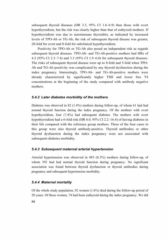
84
subsequent thyroid diseases (HR 3.3, 95% CI 1.6–6.9) than those with overt
hypothyroidism, but the risk was clearly higher than that of euthyroid mothers. If
hypothyroidism was due to autoimmune thyroiditis, as indicated by increased
levels of TPO-Ab or TG-Ab, the risk of subsequent thyroid disease was greater,
20-fold for overt and 8-fold for subclinical hypothyroidism.
Positivity for TPO-Ab or TG-Ab also posed an independent risk as regards
subsequent thyroid diseases. TPO-Ab- and TG-Ab-positive mothers had HRs of
4.2 (95% CI 2.3–7.4) and 3.3 (95% CI 1.9–6.0) for subsequent thyroid diseases.
The risks of subsequent thyroid disease were up to 8-fold and 5-fold when TPO-
Ab and TG-Ab positivity was complicated by any thyroid dysfunction during the
index pregnancy. Interestingly, TPO-Ab- and TG-Ab-positive mothers were
already characterized by significantly higher TSH and lower free T4
concentrations at the beginning of the study compared with antibody negative
mothers.
5.4.2 Later diabetes morbidity of the mothers
Diabetes was observed in 82 (1.4%) mothers during follow-up, of whom 61 had had
normal thyroid function during the index pregnancy. Of the mothers with overt
hypothyroidism, four (7.4%) had subsequent diabetes. The mothers with overt
hypothyroidism had a 6-fold risk (HR 6.0, 95% CI 2.2–16.4) of having diabetes in
their life compared with the reference group mothers. Three of the four cases in
this group were also thyroid antibody-positive. Thyroid antibodies or other
thyroid dysfunction during the index pregnancy were not associated with
subsequent diabetes morbidity.
5.4.3 Subsequent maternal arterial hypertension
Arterial hypertension was observed in 483 (8.3%) mothers during follow-up, of
whom 392 had had normal thyroid function during pregnancy. No significant
association was found between thyroid dysfunction or thyroid antibodies during
pregnancy and subsequent hypertension morbidity.
5.4.4 Maternal mortality
Of the whole study population, 92 women (1.6%) died during the follow-up period of
20 years. Of these women, 74 had been euthyroid during the index pregnancy. We did

85
not find an association between later mortality and overt or subclinical
hypothyroidism or hyperthyroidism and/or TPO-Ab or TG-Ab positivity during
pregnancy. The study population was relatively young (mean age 48 years) at the end
of the follow-up period, which probably explains this outcome.
5.4.5 Comments
This study showed for the first time in a large pregnant population sample the
magnitude of risk of subsequent thyroid disease in cases of hypothyroidism
and/or TPO-Ab or TG-Ab positivity during pregnancy. The risk of later thyroid
disease was marked, up to 20-fold in women with overt hypothyroidism and
thyroid antibodies, and three- to eight-fold in women with subclinical
hypothyroidism and/or thyroid antibodies during pregnancy. Previously, Haddow
et al. (Haddow et al. 1999) have shown in a nested case-control setting that
hypothyroidism during pregnancy is associated with subsequent thyroid disease,
as 64% (29 of 45 cases) of women with undiagnosed and untreated
hypothyroidism during pregnancy had confirmed hypothyroidism during follow-
up. Interestingly, 4% of the control women (total n = 124) in the study by
Haddow et al. also had subsequent hypothyroidism (Haddow et al. 1999). In our
study population, only 2% of the mothers who were euthyroid during pregnancy
had subsequent thyroid disease. Our results are based on a very large population
and we have been able to analyze the effect of thyroid antibodies on the later
thyroid disease risk separately and together with thyroid dysfunction. Therefore,
our results can be considered to be reliable.
Positivity for TPO-Ab, as a marker of autoimmune thyroiditis, and
diminished thyroid function at first assessment is associated with a 32-fold risk of
hypothyroidism in non-pregnant populations. (Gordin & Lamberg 1981,
Vanderpump et al. 1995) As our population is based on a birth cohort, this has
probably led to some selection bias. Only those with mild thyroid dysfunction
have been able to conceive and hence be included in our study population. This
may have led to some underestimation of the risk of later thyroid disease at a
population level, but the present results are reliable for women with the capacity
to conceive. All in all, our results are in accordance with those of previous studies
and show that thyroid dysfunction and/or antibodies detected during pregnancy
are good predictors of subsequent thyroid disease.
A new finding was that mothers with overt hypothyroidism with or without
thyroid antibodies showed increased subsequent diabetes morbidity, although the

86
number of women evaluated was relatively small. Hypothyroidism has been
associated with insulin resistance (Maratou et al. 2009), and it is known to slow
down the metabolic rate, which could lead to excessive weight gain (Ganong
2005) and therefore to an increased risk of diabetes. In addition, autoimmune
thyroiditis and thyroid dysfunction have been found to be more prevalent among
patients with both type I and type II diabetes compared with nondiabetic subjects
(McCanlies et al. 1998 Akbar et al. 2006, Schroner et al. 2008). This finding was
also evident in our study, as three of the four hypothyroid women who had
subsequent diabetes were positive for thyroid antibodies. Thyroid antibodies by
themselves were not risk factors for subsequent diabetes in our study. The
mechanism underlying the association between hypothyroidism during pregnancy
and subsequent diabetes cannot be resolved in the present study – the disease
mechanism may possibly be related to other autoimmune factors or to increased
insulin resistance.
Interestingly, no association was seen between arterial hypertension and
thyroid dysfunction or antibodies during pregnancy. Hypertension has been
associated with both hypo- and hyperthyroidism (Saito et al. 1983). This finding
is in contrast to our results, possibly because our study population was relatively
young (mean age 48 at the end of the follow-up period). Also, we were not able to
study non-medically treated hypertension because of our study setting. These
factors could explain why no association was found. The young age of the women
may also explain why no association was seen between mortality and thyroid
dysfunction or antibodies during pregnancy. Several other studies have shown
that both hypothyroidism and hyperthyroidism are associated with cardiovascular
mortality in subjects younger than 65 years (Bauer et al. 2007, Razvi et al. 2008).
However, none of the previous studies were conducted in a pregnant population.
5.5 Gestational age-specific reference intervals of thyroid hormone levels (IV)
Gestational age-specific reference intervals could be reliably calculated for
gestational weeks 5 to 19, as for these gestational weeks we had an acceptable
amount of thyroid antibody-negative serum samples available for thyroid
hormone measurements. Percentile points 2.5, 5, 25, 75, 95 and 97.5 and medians
of TSH, fT4 and fT3 concentrations categorized using different gestational weeks
are presented in Figure 4. Concentrations of fT4 and fT3 were relatively uniform
throughout pregnancy, although fT4 concentrations tended to be lower as

87
pregnancy progressed. TSH concentrations decreased up to the 11th gestational
week, and rose thereafter. In high TSH categories marked variation is seen in
different gestational weeks.
The upper reference limit of TSH concentrations was calculated to be
approximately 2.5 multiples of the median during gestational weeks 2–20, with
absolute TSH concentrations ranging from 2.7 to 3.9 mU/L at percentile points 95
and 97.5. The lower reference limit of TSH (or percentile point 2.5) ranged from
as low as 0.07 mU/L to 0.35 mU/l during gestational weeks 2–20. Concentrations
of fT4 showed a small rise during early pregnancy and decreased thereafter. The
95% reference intervals of fT4 ranged from 11 to 22 pmol/L, with the highest
values observed during gestational weeks 8 to 10. Concentrations of fT3, on the
other hand, were uniform throughout gestation and the 95% reference intervals
ranged between 3.5 and 7.0 pmol/L.

88
Fig. 4. Percentile points of TSH, fT4 and fT3 according to gestational week (Männistö
et al. 2011 published with permission of The American Thyroid Association).

89
Fig. 4 (continued) Percentile points of TSH, fT4 and fT3 according to gestational week (Männistö et al. 2011 published with permission of The American Thyroid Association).
5.5.1 BMI and thyroid hormones
The associations between BMI and thyroid hormones were also evaluated in this
study. Women were defined as underweight, normal weight, overweight or obese
according to their BMI. Obese women had higher TSH concentrations than
normal-weight women. Similarly, overweight and obese women had higher fT3
concentrations. Concentrations of fT4 were, however, lower in overweight and
obese women compared with normal weight women.
5.5.2 Comments
This study presents for the first time gestational age-specific reference intervals of
serum TSH, fT4 and fT3 concentrations in a Finnish pregnant population. Our
reference intervals are derived from an iodine-sufficient and antibody-negative
population. The results differ somewhat from those reported earlier. Table 11
presents TSH reference intervals in different populations. In particular, the upper
reference limit varies widely in different studies, from 2.13 mU/L to 4.70 mU/L.
The presented reference values emphasize the importance of creating reference

90
intervals for each laboratory. These results show fairly uniformly that reference
intervals derived from a nonpregnant population are not suitable for evaluating
the thyroid hormone status of pregnant women. For instance, the current upper
limit for TSH in Finland is 4.0 mU/L and the one provided by the manufacturer of
the method used in our study even higher, 4.94 mU/L. In our study, we
established that the upper reference limit for the pregnant population should be
approximately 2.5 multiples of the median, which covers percentile points 95 and
97.5. Since no cases of hypothyroidism should be overlooked during pregnancy,
we selected the 95th percentile point as the upper reference limit. This translates to
2.7–3.1 mU/L in the first trimester and to 3.1–3.5 mU/L in the second trimester in
absolute TSH concentrations, a result lower than the upper reference limit for the
general population. This upper reference limit agrees very well with the results of
other studies with large sample sizes and the possibility to exclude TPO-Ab- and
TG-Ab-positive mothers (Haddow et al. 2004, Gilbert et al. 2008, Lambert-
Messerlian et al. 2008, Bocos-Terraz et al. 2009, Shan et al. 2009, Springer et al. 2009). Previously it has been shown that TSH assays agree with 10% variation
when measuring serum TSH within the normal range, but some assays show
wider variation at low and high serum TSH concentrations (Thienpont et al. 2010). A similar conclusion can be drawn when comparing gestational age-
specific reference intervals of TSH: median values agree well, but there is wider
variation as regards the upper reference limit.
The lower reference limit of TSH ranges from 0.02 to 0.10 mU/L as observed
in other studies (Table 11). Uniformly, all studies have shown that percentile point
2.5 is lower in the pregnant than in the nonpregnant population. In our study, the
lowest concentration was 0.07 mU/L, observed at gestational week 11, a value
much lower than the lower reference limit calculated for the general population
(0.35 mU/L as presented by the assay manufacturer, Abbott Diagnostics).
The much lower TSH values observed in a pregnant population are probably
explained by the thyreotropic effect of hCG, which is greatest during early
pregnancy (Pekonen et al. 1988). hCG along with TBG, affects fT4
concentrations, which rise during early pregnancy and decrease thereafter. This
has been established in several studies and is also present in our data. The fT4
reference interval of 11–22 pmol/L in our pregnant population is higher than that
of a nonpregnant population (9–19 pmol/L as defined in the general population).
Interestingly, two previous studies (Cotzias et al. 2008, Gilbert et al. 2008) have
shown lower reference intervals for fT4 during early pregnancy (although the
investigators used the same method) and one study has shown results comparable

91
to ours (Panesar et al. 2001). Assays of fT4 show relatively great variation, and
different methods show different sensitivity to binding protein alterations during
pregnancy (Anckaert et al. 2010). It also seems that ethnicity may have some
effect, since when using the same method, the upper reference limit of fT4 varies
from 21.9 pmol/L (present study) to 17.4 pmol/L (Gilbert et al. 2008).
The 95% reference interval for fT3 was set at 3.4–7.0 pmol/L, which is
higher than that seen in the nonpregnant population (2.62–5.70 pmol/L as given
by the assay manufacturer). Our first study (I) showed that fT3 concentrations
tend to be somewhat higher after freezing and thawing of samples, which
probably affected our results. However, our results have shown that fT3
concentrations are relatively uniform throughout gestation. Similar results have
been reported earlier in one study (Gilbert et al. 2008), but in another study fT3
concentrations declined over the course of pregnancy (Panesar et al. 2001).
It is noteworthy that different immunoassays give slightly different results
even when evaluating the same samples (Steele et al. 2005). The effect of various
methods on reference intervals of fT4 and fT3 can be observed in Table 1.
Therefore the most accurate reference intervals for research purposes may be
obtained by using the same immunoassay method and research population for
calculations of reference intervals, as was done in this work. As an example, three
populations from China have been evaluated by means of three different assays
for fT4: with the Siemens Advia Centaur method the second trimester reference
interval is 10.6–17.6 pmol/L, with the immunoassay from Diagnostic Products it
is 11.6–21.7 pmol/L and with the ACS180 method it is 8.1–16.7 pmol/L (Panesar
et al. 2001, Shan et al. 2009, Yan et al. 2011). Different immunoassay methods
may explain most of the discrepancies between numerical results of different
studies in which gestational age-specific reference intervals have been evaluated.
Altogether, the reference intervals of TSH and fT4 based on the Abbott
Architect method and obtained in this study can be implemented in clinical
practice to better detect thyroid dysfunction in pregnant women. Concentrations
of fT3 are uniform during gestation, and fT3 is usually only measured when
suspecting hyperthyroidism. Therefore we suggest that nonpregnant reference
intervals can be used for fT3 concentrations, but gestational age-specific
reference intervals need to be implemented in clinical practice concerning TSH
and fT4.
We also evaluated the association between BMI and thyroid hormones in a
TPO-Ab- and TG-Ab-negative population. Obesity seems to be associated with
higher TSH, and overweight and obesity with higher fT3 and lower fT4
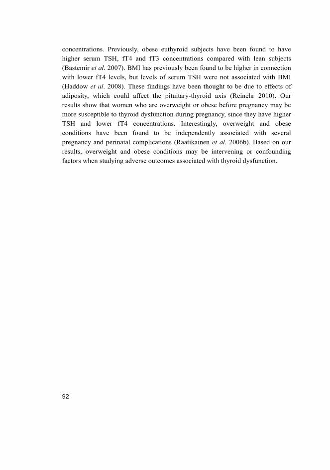
92
concentrations. Previously, obese euthyroid subjects have been found to have
higher serum TSH, fT4 and fT3 concentrations compared with lean subjects
(Bastemir et al. 2007). BMI has previously been found to be higher in connection
with lower fT4 levels, but levels of serum TSH were not associated with BMI
(Haddow et al. 2008). These findings have been thought to be due to effects of
adiposity, which could affect the pituitary-thyroid axis (Reinehr 2010). Our
results show that women who are overweight or obese before pregnancy may be
more susceptible to thyroid dysfunction during pregnancy, since they have higher
TSH and lower fT4 concentrations. Interestingly, overweight and obese
conditions have been found to be independently associated with several
pregnancy and perinatal complications (Raatikainen et al. 2006b). Based on our
results, overweight and obese conditions may be intervening or confounding
factors when studying adverse outcomes associated with thyroid dysfunction.

Ta
ble
11
. C
om
pa
riso
n o
f T
SH
re
fere
nc
e i
nte
rva
ls o
bta
ine
d f
rom
dif
fere
nt
po
pu
lati
on
s d
uri
ng
ea
rly
pre
gn
an
cy.
Firs
t aut
hor
Yea
r C
ount
ry
Pop
ulat
ion
Met
hod
TPO
-Ab/
TG-A
b
mea
sure
d
n G
esta
tiona
l
wee
k
TSH
(mU
/L) p
erce
ntile
poi
nts
2.5
5 50
95
97
.5 o
r 98
Män
nist
ö 20
11
Finl
and
Cau
casi
an
Abb
ott A
rchi
tect
Y
es
772
9 0.
070.
121.
072.
763.
20
721
10
0.07
0.15
1.05
3.09
3.44
627
11
0.09
0.17
1.04
2.79
3.34
Boc
os-T
erra
z 20
09
Spa
in
Cau
casi
an
Abb
ott A
rchi
tect
Y
es
330
<11
0.10
1.00
2.65
Spr
inge
r 20
09
Cze
ch R
epub
lic
Cau
casi
an
Adv
ia C
enta
ur
Yes
43
37
9-11
0.
060.
151.
213.
143.
67
Sha
n 20
08
Chi
na
Chi
nese
D
iagn
ostic
Pro
duct
s Y
es
120
12
0.09
0.8
2.96
Lam
berg
-
Mes
serli
an
2008
U
.S.
Cau
casi
an
Imm
ulite
200
0 Y
es
8351
I t
rimes
ter
0.12
1.00
2.68
3.37
Gilb
ert
2008
A
ustra
lia
Cau
casi
an
Abb
ott A
rchi
tect
Y
es
101
9 0.
052.
20
679
10
0.02
2.13
515
11
0.02
2.16
Cot
zias
20
07
U.K
. C
auca
sian
,
Asi
an
Adv
ia C
enta
ur
No
335
(tota
l) 9
0.00
1.30
4.90
10
0.10
1.30
4.70
11
0.10
1.30
4.40
Das
he
2005
U
.S.
His
pani
c,
Bla
ck
Imm
ulite
200
0 N
o 10
37
9 0.
030.
844.
04
982
10
0.02
0.74
3.12
888
11
0.01
0.76
3.65
Had
dow
20
04
U.S
. C
auca
sian
Im
mul
ite 2
000
No
312
9 0.
071.
034.
196.
60
247
10
0.10
0.93
3.06
4.10
Yes
178
11
0.11
1.00
3.05
4.55
1005
I t
rimes
ter
0.08
0.94
2.73
3.61
Pan
esar
20
01
Chi
na
Asi
an
AC
S 1
80
No
37
7 0.
051.
222.
30
55
11
0.03
0.80
2.30
93

94

95
6 Conclusions
The following conclusions can be drawn on the basis of our studies (I, II, III, IV):
1. Long-term frozen storage does not affect the concentrations of serum TSH,
fT3 and fT4, but concentrations of thyroid antibodies (TPO-Ab and TG-Ab)
appear to have a tendency to rise with extended storage time. Similarly, fT3
levels are slightly higher after freezing and thawing. Therefore TSH and fT4
levels can be reliably evaluated and compared using archived samples, but
cut-off values of frozen-stored TPO-Ab, TG-Ab and fT3 need to be
established for different serum banks.
2. Thyroid antibodies are associated with an increased risk of perinatal mortality,
and these infants are more often born very preterm. TPO-Ab- and TG-Ab-
positive mothers presented more often with subtle thyroid dysfunction, as
these mothers had significantly higher TSH and lower free T4 concentrations
compared with antibody-negative mothers. However, TPO-Ab or TG-Ab
positivity or hypothyroidism was not associated with preterm birth. Mild
subclinical hypothyroidism in TPO-Ab- and TG-Ab-negative mothers was
not associated with adverse perinatal or pregnancy outcomes; neither were
hypothyroxinemia or subclinical hyperthyroidism.
3. Thyroid dysfunction and/or antibodies increase the maternal risk of
subsequent thyroid disease. Their detection of during pregnancy would help
to identify women at future risk of thyroid diseases. Also, in women with
overt hypothyroidism the risk of subsequent diabetes was increased.
4. Gestational age-specific reference intervals of thyroid hormone
concentrations can help clinicians to identify thyroid dysfunction during
pregnancy and help in the monitoring of pregnant women on thyroxine
substitution. There is need to create reference intervals for pregnant women,
as their thyroid hormone concentrations differ clearly from those in the
nonpregnant population. Overweight and obese women may be at an
increased risk of subclinical hypothyroidism during pregnancy.

96

97
7 General discussion
7.1 Strengths and limitations of the present study
The current study is one of the few concerning a large prospective population-
based cohort, in which the effects of hypothyroidism and hyperthyroidism and
TPO-Ab and/or TG-Ab positivity during pregnancy on peri-, neonatal and
pregnancy outcome have been evaluated. The cohort represents over 99% of the
pregnant population collected in a one-year time period, and although we had to
omit some cohort data because of a lack of laboratory samples, the subcohort is
still very representative of the whole cohort. This is an unquestionable strength in
our study, and at the population level such cohorts are the only way to reliably
evaluate the true risks of adverse outcomes. Also, the vast amount of data
collected during pregnancy enables identification and elimination of several
confounding factors, which otherwise could have affected the results
This study has some limitations. Firstly, maternal serum samples were
obtained after long-term frozen storage. However, storage time did not have a
significant effect on the concentrations of TSH and fT4 and analyses based on
them are fairly reliable, especially when they are evaluated together. Although
TPO-Ab and TG-Ab concentrations showed increasing variation with prolonged
storage, the cut-off values selected for this study population gives sensible results
as regards TSH and fT4 concentrations. However, as discussed earlier, we cannot
be certain that all samples behaved similarly during storage time and it is possible
that our results regarding TPO-Ab and TG-Ab positivity underestimate or
overestimate the real prevalence in this population.
Secondly, where peri- and neonatal complications are concerned, the rarity of
some of these made them difficult to evaluate even in a large population like ours.
After grouping the population according to levels of TSH and fT4, some of the
rendered study groups were small which may decrease the power to detect all
significant differences. Also, although several confounding factors were evaluated,
some might still have had an effect on the results seen, and the small number or
cases in some groups has limited the use of adjustable variables.
Thirdly, subsequent maternal morbidity was assessed using national registers
on hospital discharges and reimbursement of medication expences. The hospital
discharge register has an accuracy of 83 to 95% (Aro et al. 1990, Pajunen et al. 2005) and information on all bought reimbursable medication can be obtained

98
from the register of the Social Insurance Institution. Of course we may be lacking
some information, especially on non-medically treated hypertension and type II
diabetes, and therefore maybe underestimating the number of diseases during
follow-up. Also, those with defined diagnoses but who were not compliant with
medication are probably underrepresented in our analyses.
7.2 Confounding factors
Several confounding factors need to be assessed upon evaluating the results of
different studies regarding adverse pregnancy and perinatal outcome. It is also
important to recognize that differences in the definition of certain aspects,
especially subclinical hypothyroidism, in different studies may lead to
discrepancy in the results. Firstly, maternal characteristics such as ethnicity, age,
parity and weight are important confounding factors. Different ethnic groups may
have differences in susceptibility to hypothyroidism (Hollowell et al. 2002). Also,
mothers of different ethnicity may as a whole have increased risks in regard to
some complications. For example, black women are known to have a significantly
higher rate of stillbirths than white women (Fretts 2005). Advanced maternal age
is a known risk factor for miscarriages and stillbirths and for instance preterm
births, hypertension, diabetes, previous abortions and placental abruptions are
associated with advanced maternal age (Aldous & Edmonson 1993, Jacobsson et al. 2004). Smoking during pregnancy leads to increased risks of SGA infants,
perinatal death and preterm birth (Raatikainen et al. 2007).
A maternal overweight condition and obesity are other major confounding
factors, and many of the aforementioned studies have failed to report maternal
weight or have not discussed it. Overweight and obese women more often have
had previous miscarriages and they have a significantly increased risk of fetal and
perinatal death (Raatikainen et al. 2006b). To the best of our knowledge, only one
study on miscarriages in hypothyroid mothers has included maternal weight as a
factor (Ashoor et al. 2010a). In the study, mothers with subclinical
hypothyroidism were more often overweight and the authors concluded that in
only 5% of pregnancies resulting in miscarriage subclinical hypothyroidism may
have been a contributing factor. In our study, we have reported maternal weight
(mean BMI 22 kg/m2) and we have been able to study the effects of maternal
weight on the outcomes separately. Staggering maternal weights have been seen
in some studies, with a mean BMI of over 30 kg/m2 (Casey et al. 2005, Abbassi-

99
Ghanavati et al. 2010) underlining the fact that these populations were of a highly
complex character and did not represent the general pregnant population.
Maternal social status is also a major confounding factor, as seen with a
recent Finnish study: married women had 20% fewer adverse outcomes
(Raatikainen et al. 2005). In Finland, unemployment is not a significant factor in
increasing adverse outcomes in pregnant women, as all mothers attend free-of-
charge maternity welfare clinics (Raatikainen et al. 2006a), but this may be a
problem in many countries.
Iodine deficiency is a serious problem in large areas of the world, and mild
iodine deficiency affects many European countries. Finland is one of the few
countries with sufficient iodine intake. Iodine deficiency during pregnancy leads
to failure in thyroxine production, which presents most often as
hypothyroxinemia. In a recent Cochrane review it was speculated that in iodine-
sufficient populations the effects of levothyroxine in preventing adverse outcomes
such as miscarriage and preterm birth might be less than in intervention trials
based on populations with mild iodine deficiency (Reid et al. 2010).
Finally, as regards miscarriages and preterm births, another major
confounding factor is maternal infection (Fretts 2005), which has not been
reported in any of the aforementioned studies. We have been able to carry out
subanalyses from, for instance, TPO-Ab-positive mothers who experienced
perinatal death to show that these mothers did not have infections during
pregnancy.
7.3 Recommendations
On the basis of the results of our study, combined with those reported in the
literature, some recommendations can be drawn, and some findings indicate
further studies. Firstly, it is of importance to establish gestational age-specific
reference intervals of TSH and fT4 concentrations in regard to different methods
in Finland. Our large amount of data on reference intervals obtained by using the
Abbott Architect method can be a framework for this work. Gestational age-
specific reference intervals are of utmost importance, so that clinicians can
reliably evaluate thyroid function and monitor thyroxine replacement therapy in
pregnant women.
Secondly, overt inadequately treated hypothyroidism is a risk factor of
miscarriage and possibly preterm birth and fetal death (Allan et al. 2000,
Abalovich et al. 2002). Our study showed a trend toward preterm birth in those

100
with overt hypothyroidism. These findings indicate that adequate treatment of
those with known hypothyroidism and recognition of those at risk of progressing
to overt hypothyroidism during pregnancy is to be recommended – preferably
before pregnancy. These at-risk women are those with elevated TSH levels before
pregnancy, especially if they are TPO-Ab-positive. If possible, these at-risk
women and women with known hypothyroidism should be treated to normalize
their TSH levels even before conception (the goal is to maintain TSH within the
reference intervals of pregnant women in those planning pregnancy), to minimize
the risk of adverse outcomes even from the very beginning of gestation. The
treatment of overt hypothyroidism before and during pregnancy is discussed in
Section 2.2.2.
Thirdly, subclinical hyperthyroidism and overt hyperthyroidism, by our
definitions, were not associated with severe adverse outcomes during pregnancy.
These conditions are probably physiological to some degree and associated with
the increased thyroidal stimulation brought about by high hCG levels in the first
trimester. It is recommended that those with overt hyperthyroidism, as defined by
the new trimester-specific reference intervals, are treated and closely monitored,
as the need for antithyroid therapy typically decreases as pregnancy progresses.
Overt hyperthyroidism during pregnancy is usually due to Graves’ disease.
However, there are several questions we cannot answer in the present study.
Our results do not warrant universal screening of all pregnant women for TSH
and TPO-Ab, since the increased risks observed were of outcomes of rare
incidence and multifactorial origin and therefore further studies are needed to
prove the associations. Miscarriages and later neuropsychological outcome of the
offspring are beyond the scope of this thesis. Two recent studies have shown
fewer miscarriages among those with autoimmune thyroiditis and subclinical
hypothyroidism when levothyroxine was started early in pregnancy (Negro et al. 2006, Negro et al. 2010b). Pregnant women in Finland have their first visit to
maternity welfare clinics at 10–12 gestational weeks, and at that time point blood
samples are obtained to screen for infectious diseases. This time point could also
be used to screen for hypothyroidism and autoimmune thyroiditis in pregnant
women. If treatment with levothyroxine is started after this screening, it can be
questioned whether it is too late to prevent any forthcoming complications. The
first trimester of pregnancy is an important time, for implantation, placentation
and fetal organogenesis. Most miscarriages also occur during this crucial time,
and it is noteworthy that although miscarriages are of multifactorial origin, half of

101
the miscarriages in the first trimester are due to fetal chromosomal abnormalities
(Cunningham et al. 2010).
If a mother has been hypothyroid during early pregnancy, it is questionable if
miscarriages can be prevented with levothyroxine treatment started in the late first
trimester. In the only randomized study in which the effect of levothyroxine
treatment on rates of adverse outcomes in TPO-Ab-positive mothers has been
evaluated, treatment was beneficial in reducing miscarriages and preterm births.
The study, however, has some limitations: firstly, it was based in a hospital setting,
thus representing a population which is more prone to adverse outcomes than the
general population. Secondly, the authors did not report any maternal
characteristics beside age in their study and did not control for confounding
factors. As miscarriages and preterm births are of multifactorial origin,
confounding factors probably have had a significant effect on the results. Thirdly,
the study population was relatively small and the work should be repeated using a
larger population. The women in the study visited a maternity care unit at
approximately the 10th gestational week and were started on levothyroxine after
the first visit. In the untreated group, 6 of 8 women miscarried very early, and
although they had higher TSH levels than the reference group mothers, at this
time point they were euthyroid (Negro et al. 2006). The authors suggested that
treatment probably would not have been effective in preventing miscarriages if
started at 12 gestational weeks. It would be important to know if miscarriages and
preterm births could be prevented by prompt screening and detection of
hypothyroidism and autoimmune thyroiditis before pregnancy.
Another main previous finding is that the offspring of mothers with
hypothyroidism or hypothyroxinemia have adverse neuropsychological outcome
(Haddow et al. 1999, Pop et al. 2003, Henrichs et al. 2010), although ongoing
studies will further evaluate this association. The first few weeks of pregnancy are
the most important time for brain development in the fetus and even subtle T4
deficiency in the mother may lead to poorer neuropsychological outcome. Low
fT4 levels are a hallmark of overt hypothyroidism, and isolated hypothyroxinemia
may also be seen in iodine deficiency, but may also be a marker of autoimmune
thyroiditis and central hypothyroidism. Currently, populations are recommended
to maintain sufficient iodine intake to prevent hypothyroxinemia (Negro et al. 2011). This should not be a problem in iodine-sufficient Finland. In our data,
hypothyroxinemia was not associated with adverse pregnancy or perinatal
outcome. The most recent data suggest that levothyroxine treatment in cases of
isolated maternal hypothyroxinemia (resulting from iodine deficiency) is not yet

102
warranted (Negro et al. 2011) and would not resolve the underlying problem of
iodine deficiency. Soon, the results of at least one prospective study in which
levothyroxine treatment vs. no treatment of hypothyroxinemia in early pregnancy
will be published, and this will provide additional information on this important
matter. However, preliminary results of this study suggested no benefit of
levothyroxine treatment started in early pregnancy (Sullivan 2010). We can
speculate on whether or not treatment should be started before pregnancy to
prevent unfavorable neuropsychological outcomes. These questions warrant
further studies. Since the offspring of inadequately treated hypothyroid mothers
possibly have poorer neuropsychological outcomes, it is of importance to treat
those with known hypothyroidism adequately before and during pregnancy.
Should all women be tested for serum TSH, fT4 and TPO-Ab? High-risk case
finding misses nearly 50% of all those with hypothyroidism, hyperthyroidism or
TPO-Ab positivity (Vaidya et al. 2007, Horacek et al. 2010) and universal
screening during pregnancy has been found to be cost-effective if treatment can
prevent adverse neuropsychological outcome (Dosiou et al. 2008, Thung et al. 2009). Screening and treatment of subclinical hypothyroidism during pregnancy
has reduced adverse outcomes in a study setting of ambulatory hospital patients
(Negro et al. 2010b), but this has not been repeated in the general pregnant
population. As has been speculated, first trimester screening may be too late and
therefore prepregnancy counseling and evaluation of thyroid function could be the
optimal approach. Of course, attention should be paid to other maternal risk
factors as well, such as weight and smoking, to reduce the risk of adverse
outcomes. Therefore, it is of importance to recognize those at risk of
hypothyroidism and hypothyroxinemia among fertile-aged women. Naturally,
those who present with symptoms or findings consistent with thyroid dysfunction
should have their serum TSH, thyroid hormone and TPO-Ab concentrations
analyzed before and during pregnancy. Women at the highest risk of thyroid
dysfunction during pregnancy are those with a family or personal history of
thyroid disorders, those with previously detected thyroid antibodies, those with a
family or personal history of type I diabetes or other autoimmune diseases, those
with recurrent miscarriages or infertility and those with gall bladder disease
(Abalovich et al. 2007, Laukkarinen et al. 2007). Those positive for TPO-Ab
should be followed during their pregnancies (TSH measured, for instance, 6
weeks apart), as they have a high risk of developing subclinical and overt
hypothyroidism. If a mother is found to have subclinical or overt hypothyroidism
during follow-up, levothyroxine treatment should be started, and TSH evaluated

103
every 4 weeks until normalized (defined using gestational age-specific reference
intervals). Our results also show that mothers with TPO-Ab or TG-Ab positivity,
with or without hypothyroidism during pregnancy, have a very high incidence of
subsequent thyroid disease, which would warrant routine check-up of these
women later in their lives.

104

105
References
Abalovich M, Amino N, Barbour LA, Cobin RH, De Groot LJ, Glinoer D, Mandel SJ & Stagnaro-Green A (2007) Management of thyroid dysfunction during pregnancy and postpartum: an Endocrine Society Clinical Practice Guideline. J Clin Endocrinol Metab 92(8 Suppl): S1–47.
Abalovich M, Gutierrez S, Alcaraz G, Maccallini G, Garcia A & Levalle O (2002) Overt and subclinical hypothyroidism complicating pregnancy. Thyroid 12(1): 63–68.
Abbassi-Ghanavati M, Casey BM, Spong CY, McIntire DD, Halvorson LM & Cunningham FG (2010) Pregnancy outcomes in women with thyroid peroxidase antibodies. Obstet Gynecol 116(2 Pt 1): 381–6.
Aho K, Gordin A, Palosuo T, Punsar S, Valkeila E, Karvonen M, Inkovaara J & Pasternack A (1984) Thyroid autoimmunity and cardiovascular diseases. Eur Heart J 5(1): 43–6.
Ain KB, Mori Y & Refetoff S (1987) Reduced clearance rate of thyroxine-binding globulin (TBG) with increased sialylation: a mechanism for estrogen-induced elevation of serum TBG concentration. J Clin Endocrinol Metab 65(4): 689–96.
Akbar DH, Ahmed MM & Al-Mughales J (2006) Thyroid dysfunction and thyroid autoimmunity in Saudi type 2 diabetics. Acta Diabetol 43(1): 14–8.
Aldous MB & Edmonson MB (1993) Maternal age at first childbirth and risk of low birth weight and preterm delivery in Washington State. JAMA 270(21): 2574–7.
Alexander EK (2010) Here's to you, baby! A step forward in support of universal screening of thyroid function during pregnancy. J Clin Endocrinol Metab 95(4): 1575–7.
Alexander EK, Marqusee E, Lawrence J, Jarolim P, Fischer GA & Larsen PR (2004) Timing and magnitude of increases in levothyroxine requirements during pregnancy in women with hypothyroidism. N Engl J Med 351(3): 241–9.
Allan WC, Haddow JE, Palomaki GE, Williams JR, Mitchell ML, Hermos RJ, Faix JD & Klein RZ (2000) Maternal thyroid deficiency and pregnancy complications: implications for population screening. J Med Screen 7(3): 127–130.
Anckaert E, Poppe K, Van Uytfanghe K, Schiettecatte J, Foulon W & Thienpont LM (2010) FT4 immunoassays may display a pattern during pregnancy similar to the equilibrium dialysis ID-LC/tandem MS candidate reference measurement procedure in spite of susceptibility towards binding protein alterations. Clin Chim Acta 411(17–18): 1348–53.
Andersson M, de Benoist B, Delange F & Zupan J (2007) Prevention and control of iodine deficiency in pregnant and lactating women and in children less than 2-years-old: conclusions and recommendations of the Technical Consultation. Public Health Nutr 10(12A): 1606–11.
Anselmo J, Cao D, Karrison T, Weiss RE & Refetoff S (2004) Fetal loss associated with excess thyroid hormone exposure. JAMA 292(6): 691–5.
Antolic B, Gersak K, Verdenik I & Novak-Antolic Z (2006) Adverse effects of thyroid dysfunction on pregnancy and pregnancy outcome: epidemiologic study in Slovenia. J. Matern Fetal Neonatal Med 19(10): 651–4.

106
Aro S, Koskinen R & Keskimaki I (1990) Sairaalastapoistorekisterin diagnoosi-, toimenpide- ja tapaturmatietojen luotettavuus. Duodecim 106(21): 1443–50.
Ashoor G, Maiz N, Rotas M, Jawdat F & Nicolaides KH (2010a) Maternal thyroid function at 11 to 13 weeks of gestation and subsequent fetal death. Thyroid 20(9):989–93.
Ashoor G, Maiz N, Rotas M, Jawdat F & Nicolaides KH (2011) Maternal thyroid function at 11–13 weeks of gestation and spontaneous preterm delivery. Obstet Gynecol 117(2 Pt 1): 293–8.
Ashoor G, Maiz N, Rotas M, Kametas NA & Nicolaides KH (2010b) Maternal thyroid function at 11 to 13 weeks of gestation and subsequent development of preeclampsia. Prenat Diagn 30(11): 1032–8.
Auer J, Scheibner P, Mische T, Langsteger W, Eber O & Eber B (2001) Subclinical hyperthyroidism as a risk factor for atrial fibrillation. Am Heart J. 2001 142(5):838–42.
Bastemir M, Akin F, Alkis E & Kaptanoglu B (2007) Obesity is associated with increased serum TSH level, independent of thyroid function. Swiss Med Wkly 137(29–30):431–4.
Bauer DC, Rodondi N, Stone KL & Hillier TA (2007) Thyroid hormone use, hyperthyroidism and mortality in older women. Am J Med 120(4): 343–9.
Benhadi N, Wiersinga WM, Reitsma JB, Vrijkotte TG & Bonsel GJ (2009) Higher maternal TSH levels in pregnancy are associated with increased risk for miscarriage, fetal or neonatal death. Eur J Endocrinol 160(6): 985–91.
Benson RC & Dailey ME (1955) The menstrual pattern in hyperthyroidism and subsequent posttherapy hypothyroidism. Surg Gynecol Obstet 100(1): 19–26.
Blazer S, Moreh-Waterman Y, Miller-Lotan R, Tamir A & Hochberg Z (2003) Maternal hypothyroidism may affect fetal growth and neonatal thyroid function. Obstet Gynecol 102(2): 232–241.
Bocos-Terraz JP, Izquierdo-Alvarez S, Bancalero-Flores JL, Alvarez-Lahuerta R, Aznar- Sauca A, Real-Lopez E, Ibanez-Marco R, Bocanegra-Garcia V & Rivera-Sanchez G (2009) Thyroid hormones according to gestational age in pregnant Spanish women. BMC Res Notes 2: 237.
Borchers AT, Naguwa SM, Keen CL & Gershwin ME (2010) The implications of autoimmunity and pregnancy. J Autoimmun 34(3): J287–99.
Burrow GN (1993) Thyroid function and hyperfunction during gestation. Endocr Rev 14(2): 194–202.
Calvo RM, Jauniaux E, Gulbis B, Asuncion M, Gervy C, Contempre B & Morreale de Escobar G (2002) Fetal tissues are exposed to biologically relevant free thyroxine concentrations during early phases of development. J Clin Endocrinol Metab 87(4): 1768–1777.
Cappola AR, Fried LP, Arnold AM, Danese MD, Kuller LH, Burke GL, Tracy RP & Ladenson PW (2006) Thyroid status, cardiovascular risk, and mortality in older adults. JAMA 295(9):1033–41.

107
Casey BM, Dashe JS, Spong CY, McIntire DD, Leveno KJ & Cunningham GF (2007) Perinatal significance of isolated maternal hypothyroxinemia identified in the first half of pregnancy. Obstet Gynecol 109(5): 1129–35.
Casey BM, Dashe JS, Wells CE, McIntire DD, Byrd W, Leveno KJ & Cunningham FG (2005) Subclinical hypothyroidism and pregnancy outcomes. Obstet Gynecol 105(2):239–245.
Casey BM, Dashe JS, Wells CE, McIntire DD, Leveno KJ & Cunningham FG (2006) Subclinical hyperthyroidism and pregnancy outcomes. Obstet Gynecol 107(2 Pt 1): 337–341.
Chubb SA, Davis WA & Davis TM (2005) Interactions among thyroid function, insulin sensitivity, and serum lipid concentrations: the Fremantle diabetes study. J Clin Endocrinol Metab 90(9): 5317–20.
Cleary-Goldman J, Malone FD, Lambert-Messerlian G, Sullivan L, Canick J, Porter TF, Luthy D, Gross S, Bianchi DW & D'Alton ME (2008) Maternal thyroid hypofunction and pregnancy outcome. Obstet Gynecol 112(1): 85–92.
Costeira MJ, Oliveira P, Ares S, Roque S, de Escobar GM & Palha JA (2010) Parameters of thyroid function throughout and after pregnancy in an iodine-deficient population. Thyroid 20(9): 995–1001.
Cotzias C, Wong SJ, Taylor E, Seed P & Girling J (2008) A study to establish gestationspecific reference intervals for thyroid function tests in normal singleton pregnancy. Eur J Obstet Gynecol Reprod Biol 137(1): 61–6.
Cunningham FG, Leveno KJ, Bloom S, Hauth J, Rouse D & Spong CY (2010) Abortion. In: Williams Obstetrics, The McGraw-Hill Companies: 215–237.
Dashe JS, Casey BM, Wells CE, McIntire DD, Byrd EW, Leveno KJ & Cunningham FG (2005) Thyroid-stimulating hormone in singleton and twin pregnancy: importance of gestational age-specific reference ranges. Obstet Gynecol 106(4): 753–757.
Davis LE, Leveno KJ & Cunningham FG (1988) Hypothyroidism complicating pregnancy. Obstet Gynecol 72(1): 108–112.
de Escobar GM, Ares S, Berbel P, Obregon MJ & del Rey FE (2008) The changing role of maternal thyroid hormone in fetal brain development. Semin Perinatol 32(6): 380–6.
Delahunty C, Falconer S, Hume R, Jackson L, Midgley P, Mirfield M, Ogston S, Perra O, Simpson J, Watson J, Willatts P & Williams F (2010) Levels of neonatal thyroid hormone in preterm infants and neurodevelopmental outcome at 5 1/2 years: millennium cohort study. J Clin Endocrinol Metab 95(11): 4898–908.
Dhatt GS, Griffin G & Agarwal MM (2006) Thyroid hormone reference intervals in an ambulatory Arab population on the Abbott Architect i2000 immunoassay analyzer. Clin Chim Acta 364(1–2): 226–9.
Dimitriadis G, Mitrou P, Lambadiari V, Boutati E, Maratou E, Koukkou E, Panagiotakos D, Tountas N, Economopoulos T & Raptis SA (2008) Insulin-stimulated rates of glucose uptake in muscle in hyperthyroidism: the importance of blood flow. J Clin Endocrinol Metab 93(6): 2413–15

108
Diver MJ, Jones PJ, Hughes JG, Hipkin LJ & West CR (1996) Effect of preanalytical conditions on measurements of free thyroxine and total triiodothyronine. Clin Chem 42(7): 1124.
Dosiou C, Sanders GD, Araki SS & Crapo LM (2008) Screening pregnant women for autoimmune thyroid disease: a cost-effectiveness analysis. Eur J Endocrinol 158(6): 841–51.
Einarson A & Riordan S (2009) Smoking in pregnancy and lactation: a review of risks and cessation strategies. Eur J Clin Pharmacol 65(4): 325–30.
Erkkola M, Karppinen M, Jarvinen A, Knip M & Virtanen SM (1998) Folate, vitamin D, and iron intakes are low among pregnant Finnish women. Eur J Clin Nutr 52(10): 742–748.
Fisher DA (1996) Physiological variations in thyroid hormones: physiological and pathophysiological considerations. Clin Chem 42(1): 135–9.
Fister P, Gaberscek S, Zaletel K, Krhin B, Hojker S & Gersak K (2010) Thyroid function in the third trimester of pregnancy and after delivery in an area of adequate iodine intake. Int J Gynaecol Obstet 112(1): 52–5.
Flynn A, Hirvonen T, Mensink GB, Ocke MC, Serra-Majem L, Stos K, Szponar L, Tetens I, Turrini A, Fletcher R & Wildemann T (2009) Intake of selected nutrients from foods, from fortification and from supplements in various European countries. Food Nutr Res 53.
Fretts RC (2005) Etiology and prevention of stillbirth. Am J Obstet Gynecol 193(6): 1923–35.
Ganong WF (2005) The thyroid gland. In:Review of Medical Physiology, Lange Medical Books/McGraw-Hill, 317–332.
Ghafoor F, Mansoor M, Malik T, Malik MS, Khan AU, Edwards R & Akhtar W (2006) Role of thyroid peroxidase antibodies in the outcome of pregnancy. J Coll Physicians Surg Pak 16(7): 468–71.
Gilbert RM, Hadlow NC, Walsh JP, Fletcher SJ, Brown SJ, Stuckey BG & Lim EM (2008) Assessment of thyroid function during pregnancy: first-trimester (weeks 9–13) reference intervals derived from Western Australian women. Med J Aust 189(5): 250–3.
Glinoer D (1997) The regulation of thyroid function in pregnancy: pathways of endocrine adaptation from physiology to pathology. Endocr Rev 18(3): 404–33.
Glinoer D, de Nayer P, Bourdoux P, Lemone M, Robyn C, van Steirteghem A, Kinthaert J & Lejeune B (1990) Regulation of maternal thyroid during pregnancy. J Clin Endocrinol Metab 71(2): 276–87.
Glinoer D, Riahi M, Grun JP & Kinthaert J (1994) Risk of subclinical hypothyroidism in pregnant women with asymptomatic autoimmune thyroid disorders. J Clin Endocrinol Metab 79(1): 197–204.
Gong Y & Hoffman BR (2008) Free thyroxine reference interval in each trimester of pregnancy determined with the Roche Modular E-170 electrochemiluminescent immunoassay. Clin Biochem 41(10–11): 902–6.

109
Gordin A & Lamberg BA (1981) Spontaneous hypothyroidism in symptomless autoimmune thyroiditis. A long-term follow-up study. Clin Endocrinol (Oxf) 15(6):537–43.
Haddow JE, Cleary-Goldman J, McClain MR, Palomaki GE, Neveux LM, Lambert- Messerlian G, Canick JA, Malone FD, Porter TF, Nyberg DA, Bernstein PS & D'Alton ME (2010) Thyroperoxidase and thyroglobulin antibodies in early pregnancy and preterm delivery. Obstet Gynecol 116(1): 58–62.
Haddow JE, Knight GJ, Palomaki GE, McClain MR & Pulkkinen AJ (2004) The reference range and within-person variability of thyroid stimulating hormone during the first and second trimesters of pregnancy. J Med Screen 11(4): 170–174.
Haddow JE, McClain MR, Lambert-Messerlian G, Palomaki GE, Canick JA, Cleary-Goldman J, Malone FD, Porter TF, Nyberg DA, Bernstein P & D'Alton ME (2008) Variability in thyroid-stimulating hormone suppression by human chorionic [corrected] gonadotropin during early pregnancy. J Clin Endocrinol Metab 93(9): 3341–7.
Haddow JE, McClain MR, Palomaki GE, Neveux LM, Lambert-Messerlian G, Canick JA, Malone FD, Porter TF, Nyberg DA, Bernstein PS & D'Alton M E (2011) Thyroperoxidase and Thyroglobulin Antibodies in Early Pregnancy and Placental Abruption. Obstet Gynecol 117(2, Part 1): 287–292.
Haddow JE, Palomaki GE, Allan WC, Williams JR, Knight GJ, Gagnon J, O'Heir CE, Mitchell ML, Hermos RJ, Waisbren SE, Faix JD & Klein RZ (1999) Maternal thyroid deficiency during pregnancy and subsequent neuropsychological development of the child. N Engl J Med 341(8): 549–555.
Hadley ME & Levine JE (2007) Thyroid hormones. In: Endocrinology. Upper Saddle River, NJ, Pearson Prentice Hall, 293–314.
Hak AE, Pols HA, Visser TJ, Drexhage HA, Hofman A & Witteman JC (2000) Subclinical hypothyroidism is an independent risk factor for atherosclerosis and myocardial infarction in elderly women: the Rotterdam Study. Ann Intern Med 132(4): 270–8.
Hamm MP, Cherry NM, Martin JW, Bamforth F & Burstyn I (2009) The impact of isolated maternal hypothyroxinemia on perinatal morbidity. J Obstet Gynaecol Can 31(11): 1015–21.
Hay DL (1988) Placental histology and the production of human choriogonadotrophin and its subunits in pregnancy. Br J Obstet Gynaecol 95(12): 1268–75.
Henrichs J, Bongers-Schokking JJ, Schenk JJ, Ghassabian A, Schmidt HG, Visser TJ, Hooijkaas H, de Muinck Keizer-Schrama SM, Hofman A, Jaddoe VV, Visser W, Steegers EA, Verhulst FC, de Rijke YB & Tiemeier H (2010) Maternal thyroid function during early pregnancy and cognitive functioning in early childhood: the generation R study. J Clin Endocrinol Metab 95(9): 4227–34.
Hetzel BS (1983) Iodine deficiency disorders (IDD) and their eradication. Lancet 2(8359):1126–9.
Hiilesmaa V (1948) Studies of the thyroid gland of parturients and newborn infants in Southern Finland. Thesis. Helsingin Yliopisto

110
Hollowell JG, Staehling NW, Flanders WD, Hannon WH, Gunter EW, Spencer CA & Braverman LE (2002) Serum TSH, T(4), and thyroid antibodies in the United States population (1988 to 1994): National Health and Nutrition Examination Survey (NHANES III). J Clin Endocrinol Metab 87(2): 489–99.
Horacek J, Spitalnikova S, Dlabalova B, Malirova E, Vizda J, Svilias I, Cepkova J, McGrath C & Maly J (2010) Universal screening detects two-times more thyroid disorders in early pregnancy than targeted high-risk case finding. Eur J Endocrinol 163(4): 645–50.
Irgens HU, Reisaeter L, Irgens LM & Lie RT (2001) Long term mortality of mothers and fathers after pre-eclampsia: population based cohort study. BMJ 323(7323): 1213–7.
Jacobsson B, Ladfors L & Milsom I (2004) Advanced maternal age and adverse perinatal outcome. Obstet Gynecol 104(4): 727–33.
Jauhiainen M, Koskinen P, Ehnholm C, Frick MH, Manttari M, Manninen V & Huttunen JK (1991) Lipoprotein (a) and coronary heart disease risk: a nested case-control study of the Helsinki Heart Study participants. Atherosclerosis 89(1): 59–67.
Juntunen K, Kirkinen P & Kauppila A (1997) The clinical outcome in pregnancies of grand grand multiparous women. Acta Obstet Gynecol Scand 76(8): 755–9.
Järvelin MR, Elliott P, Kleinschmidt I, Martuzzi M, Grundy C, Hartikainen AL & Rantakallio P (1997) Ecological and individual predictors of birthweight in a northern Finland birth cohort 1986. Paediatr Perinat Epidemiol 11(3): 298–312.
Järvelin MR, Hartikainen-Sorri AL & Rantakallio P (1993) Labour induction policy in hospitals of different levels of specialisation. Br J Obstet Gynaecol 100(4): 310–315.
Kahric-Janicic N, Soldin SJ, Soldin OP, West T, Gu J & Jonklaas J (2007) Tandem mass spectrometry improves the accuracy of free thyroxine measurements during pregnancy. Thyroid 17(4): 303–11.
Keffer JH (1996) Preanalytical considerations in testing thyroid function. Clin Chem 42(1): 125–134.
Klajnbard A, Szecsi PB, Colov NP, Andersen MR, Jorgensen M, Bjorngaard B, Barfoed A, Haahr K & Stender S (2010) Laboratory reference intervals during pregnancy, delivery and the early postpartum period. Clin Chem Lab Med 48(2): 237–48.
Klein RZ, Haddow JE, Faix JD, Brown RS, Hermos RJ, Pulkkinen A & Mitchell ML (1991) Prevalence of thyroid deficiency in pregnant women. Clin Endocrinol (Oxf) 35(1): 41–6.
Koliakos G, Gaitatzi M & Grammaticos P (1999) Stability of serum TSH concentration after non refrigerated storage. Panminerva med 41(2): 99–101.
Koskela P, Anttila T, Bjorge T, Brunsvig A, Dillner J, Hakama M, Hakulinen T, Jellum E, Lehtinen M, Lenner P, Luostarinen T, Pukkala E, Saikku P, Thoresen S, Youngman L & Paavonen J (2000) Chlamydia trachomatis infection as a risk factor for invasive cervical cancer. Int J Cancer 85(1): 35–39.
Koutras DA (1997) Disturbances of menstruation in thyroid disease. Ann N Y Acad Sci 816: 280–4.
Krassas GE, Pontikides N, Kaltsas T, Papadopoulou P & Batrinos M (1994) Menstrual disturbances in thyrotoxicosis. Clin Endocrinol (Oxf) 40(5): 641–4.

111
Krassas GE, Pontikides N, Kaltsas T, Papadopoulou P, Paunkovic J, Paunkovic N & Duntas LH (1999) Disturbances of menstruation in hypothyroidism. Clin Endocrinol (Oxf) 50(5): 655–9.
Kriplani A, Buckshee K, Bhargava VL, Takkar D & Ammini AC (1994) Maternal and perinatal outcome in thyrotoxicosis complicating pregnancy. Eur J Obstet Gynecol Reprod Biol 54(3): 159–63.
Kruuti J & Harsia-Alatalo J (2008) Medicine reimbursement system and approval of medicine prices. Edita Prima Oy, Helsinki.
Kuppens SM, Kooistra L, Wijnen HA, Crawford S, Vader HL, Hasaart TH, Oei SG & Pop VJ (2010) Maternal thyroid function during gestation is related to breech presentation at term. Clin Endocrinol (Oxf) 72(6): 820–4.
La'ulu SL & Roberts WL (2007) Second-trimester reference intervals for thyroid tests: the role of ethnicity. Clin Chem 53(9): 1658–64.
Lamberg BA (2003a) Jodinpuutosta esiintyy monissa Euroopan maissa mutta ei Suomessa. Duodecim 119(17): 1639–1642.
Lamberg BA (2003b) Kupukaula on jo menneisyyttä. Struuman historia Suomessa. Suomen Tiedeseura, Helsinki.
Lamberg BA, Haikonen M, Makela M, Jukkara A, Axelson E & Welin MG (1981) Further decrease in thyroidal uptake and disappearance of endemic goitre in children after 30 years of iodine prophylaxis in the east of Finland. Acta Endocrinol (Copenh) 98(2): 205–9.
Lambert-Messerlian G, McClain M, Haddow JE, Palomaki GE, Canick JA, Cleary-Goldman J, Malone FD, Porter TF, Nyberg DA, Bernstein P, D'Alton ME & Fa SRC (2008) First- and second-trimester thyroid hormone reference data in pregnant women: a FaSTER (First- and Second-Trimester Evaluation of Risk for aneuploidy) Research Consortium study. Am J Obstet Gynecol 199(1): 62.e1–62.e6.
Laukkarinen J, Kiudelis G, Lempinen M, Raty S, Pelli H, Sand J, Kemppainen E, Haglund C & Nordback I (2007) Increased prevalence of subclinical hypothyroidism in common bile duct stone patients. J Clin Endocrinol Metab 92(11): 4260–4.
Larsson A, Palm M, Hansson LO & Axelsson O (2008) Reference values for clinical chemistry tests during normal pregnancy. BJOG 115(7):874–81.
Lee RH, Spencer CA, Mestman JH, Miller EA, Petrovic I, Braverman LE & Goodwin TM (2009) Free T4 immunoassays are flawed during pregnancy. Am J Obstet Gynecol 200(3): 260 e1–6.
Lehtinen M, Koskela P, Ogmundsdottir HM, Bloigu A, Dillner J, Gudnadottir M, Hakulinen T, Kjartansdottir A, Kvarnung M, Pukkala E, Tulinius H & Lehtinen T (2003) Maternal herpesvirus infections and risk of acute lymphoblastic leukemia in the offspring. Am J Epidemiol 158(3): 207–213.
Leinonen J & Stenman UH (2000) Reduced stability of prostate-specific antigen after long-term storage of serum at -20 degrees C. Tumour Biology 21(1): 46–53.
Lejeune B, Grun JP, de Nayer P, Servais G & Glinoer D (1993) Antithyroid antibodies underlying thyroid abnormalities and miscarriage or pregnancy induced hypertension.Br J Obstet Gynaecol 100(7): 669–672.

112
Lindberg BS, Johansson ED & Nilsson BA (1974) Plasma levels of nonconjugated oestrone, oestradiol-17beta and oestriol during uncomplicated pregnancy. Acta Obstet Gynecol Scand Suppl 32(0): 21–36.
Liu D, Teng W, Shan Z, Yu X, Gao Y, Wang S, Fan C, Wang H & Zhang H (2010) The effect of maternal subclinical hypothyroidism during pregnancy on brain development in rat offspring. Thyroid 20(8): 909–15.
Livesey JH, Hodgkinson SC, Roud HR & Donald RA (1980) Effect of time, temperature and freezing on the stability of immunoreactive LH, FSH, TSH, growth hormone, prolactin and insulin in plasma. Clin Biochem 13(4): 151–155.
Lucas A, Pizarro E, Granada ML, Salinas I, Roca J & Sanmarti A (2005) Postpartum thyroiditis: long-term follow-up. Thyroid 15(10): 1177–81.
Mandel SJ, Brent GA & Larsen PR (1993) Levothyroxine therapy in patients with thyroid disease. Ann Intern Med 119(6):492–502.
Maratou E, Hadjidakis DJ, Kollias A, Tsegka K, Peppa M, Alevizaki M, Mitrou P, Lambadiari V, Boutati E, Nikzas D, Tountas N, Economopoulos T, Raptis SA & Dimitriadis G (2009) Studies of insulin resistance in patients with clinical and subclinical hypothyroidism. Eur J Endocrinol 160(5): 785–90.
Marwaha RK, Chopra S, Gopalakrishnan S, Sharma B, Kanwar RS, Sastry A & Singh S (2008) Establishment of reference range for thyroid hormones in normal pregnant Indian women. BJOG 115(5): 602–6.
Matalon S, Sheiner E, Levy A, Mazor M & Wiznitzer A (2006) Relationship of treated maternal hypothyroidism and perinatal outcome. J Reprod Med 51(1): 59–63.
McCanlies E, O'Leary LA, Foley TP, Kramer MK, Burke JP, Libman A, Swan JS, Steenkiste AR, Mccarthy BJ, Trucco M & Dorman JS (1998) Hashimoto's thyroiditis and insulin-dependent diabetes mellitus: differences among individuals with and without abnormal thyroid function. J Clin Endocrinol Metab 83(5): 1548–51.
Mestman JH, Goodwin TM & Montoro MM (1995) Thyroid disorders of pregnancy. Endocrinol Metab Clin North Am 24(1):41–71.
Michalaki MA, Vagenakis AG, Leonardou AS, Argentou MN, Habeos IG, Makri MG, Psyrogiannis AI, Kalfarentzos FE & Kyriazopoulou VE (2006) Thyroid function in humans with morbid obesity. Thyroid 16(1): 73–8.
Montaner P, Juan L, Campos R, Gil L & Corcoy R (2008) Is thyroid autoimmunity associated with gestational diabetes mellitus? Metabolism 57(4): 522–5.
Monteleone P, Parrini D, Faviana P, Carletti E, Casarosa E, Uccelli A, Cela V, Genazzani AR & Artini PG (2011) Female infertility related to thyroid autoimmunity: The ovarian follicle hypothesis. Am J Reprod Immunol (published ahead of print)
Muller AF, Drexhage HA & Berghout A (2001) Postpartum thyroiditis and autoimmune thyroiditis in women of childbearing age: recent insights and consequences for antenatal and postnatal care. Endocr Rev 22(5): 605–30.
Negro R, Formoso G, Mangieri T, Pezzarossa A, Dazzi D & Hassan H (2006) Levothyroxine treatment in euthyroid pregnant women with autoimmune thyroid disease: effects on obstetrical complications. J Clin Endocrinol Metab 91(7): 2587–2591.

113
Negro R, Mangieri T, Coppola L, Presicce G, Casavola EC, Gismondi R, Locorotondo G, Caroli P, Pezzarossa A, Dazzi D & Hassan H (2005) Levothyroxine treatment in thyroid peroxidase antibody-positive women undergoing assisted reproduction technologies: a prospective study. Hum Reprod 20(6): 1529–1533.
Negro R, Schwartz A, Gismondi R, Tinelli A, Mangieri T & Stagnaro-Green A (2010a) Increased pregnancy loss rate in thyroid antibody negative women with TSH levels between 2.5 and 5.0 in the First Trimester of Pregnancy. J Clin Endocrinol Metab 95(9):E44–8.
Negro R, Schwartz A, Gismondi R, Tinelli A, Mangieri T & Stagnaro-Green A (2010b) Universal screening versus case finding for detection and treatment of thyroid hormonal dysfunction during pregnancy. J Clin Endocrinol Metab 95(4): 1699–707.
Negro R, Soldin OP, Obregon MJ & Stagnaro-Green A (2011) Hypothyroxinemia and Pregnancy. Endocr Pract: 1–24.
Nissinen A, Lindqvist O, Eronen T, Pietinen P & Puska P (1983) Vaarantaako suolan käytön vähentäminen jodin saantia? Duodecim 99(9): 593–7.
Oddie TH, Klein AH, Foley TP & Fisher DA (1979) Variation in values for iodothyronine hormones, thyrotropin, and thyroxine-binding globulin in normal umbilical-cord serum with season and duration of storage. Clini Chem 25(7): 1251–3.
Oken E, Braverman LE, Platek D, Mitchell ML, Lee SL & Pearce EN (2009) Neonatal thyroxine, maternal thyroid function, and child cognition. J Clin Endocrinol Metab 94(2): 497–503.
Olivieri A, Valensise H, Magnani F, Medda E, De Angelis S, D'Archivio M, Sorcini M, Carta S, Baccarini S & Romanini C (2000) High frequency of antithyroid autoantibodies in pregnant women at increased risk of gestational diabetes mellitus. Eur J Endocrinol 143(6): 741–7.
Pajunen P, Koukkunen H, Ketonen M, Jerkkola T, Immonen-Räihä P, Karja-Koskenkari P, Mähönen M, Niemelä M, Kuulasmaa K, Palomäki P, Mustonen J, Lehtonen A, Arstila M, Vuorenmaa T, Lehto S, Miettinen H, Torppa J, Tuomilehto J, Kesäniemi YA, Pyörälä K & Salomaa V (2005) The validity of the Finnish Hospital Discharge Register and Causes of Death Register data on coronary heart disease. Eur J Cardiovasc Prev Rehabil 12(2): 132–7.
Panesar NS, Li CY & Rogers MS (2001) Reference intervals for thyroid hormones in pregnant Chinese women. Ann Clin Biochem 38(Pt 4): 329–32.
Parle JV, Maisonneuve P, Sheppard MC, Boyle P & Franklyn JA (2001) Prediction of allcause and cardiovascular mortality in elderly people from one low serum thyrotropin result: a 10-year cohort study. Lancet 358(9285): 861–865.
Paturi M, Tapanainen H, Reinivuo H & Pietinen P (2008) Finravinto 2007 -tutkimus – The National FINDIET 2007 Survey. Publications of the National Public Health Institute. Helsinki.
Pearce EN, Oken E, Gillman MW, Lee SL, Magnani B, Platek D & Braverman LE (2008) Association of first-trimester thyroid function test values with thyroperoxidase antibody status, smoking, and multivitamin use. Endocr Pract 14(1): 33–9.

114
Pekonen F, Alfthan H, Stenman UH & Ylikorkala O (1988) Human chorionic gonadotropin (hCG) and thyroid function in early human pregnancy: circadian variation and evidence for intrinsic thyrotropic activity of hCG. J Clin Endocrinol Metab 66(4): 853–856.
Pirkola J, Pouta A, Bloigu A, Miettola S, Hartikainen AL, Jarvelin MR & Vaarasmaki M (2010) Prepregnancy overweight and gestational diabetes as determinants of subsequent diabetes and hypertension after 20-year follow-up. J Clin Endocrinol Metab 95(2): 772–8.
Pop VJ, Brouwers EP, Vader HL, Vulsma T, van Baar AL & de Vijlder JJ (2003) Maternal hypothyroxinaemia during early pregnancy and subsequent child development: a 3-year follow-up study. Clin Endocrinol 59(3): 282–288.
Pop VJ, Brouwers EP, Wijnen H, Oei G, Essed GG & Vader HL (2004) Low concentrations of maternal thyroxin during early gestation: a risk factor of breech presentation? BJOG 111(9): 925–930.
Pop VJ, Kuijpens JL, van Baar AL, Verkerk G, van Son MM, de Vijlder JJ, Vulsma T, Wiersinga WM, Drexhage HA & Vader HL (1999) Low maternal free thyroxine concentrations during early pregnancy are associated with impaired psychomotor development in infancy. Clin Endocrinol 50(2): 149–155.
Pouta A, Hartikainen AL, Sovio U, Gissler M, Laitinen J, McCarthy MI, Ruokonen A, Elliott P & Järvelin MR (2004) Manifestations of metabolic syndrome after hypertensive pregnancy. Hypertension 43(4): 825–31.
Price A, Obel O, Cresswell J, Catch I, Rutter S, Barik S, Heller SR & Weetman AP (2001) Comparison of thyroid function in pregnant and non-pregnant Asian and western Caucasian women. Clin Chim Acta 308(1–2): 91–8.
Prummel MF & Wiersinga WM (2004) Thyroid autoimmunity and miscarriage. Eur J Endocrinol 150(6): 751–5.
Raatikainen K, Heiskanen N & Heinonen S (2005) Marriage still protects pregnancy. BJOG 112(10): 1411–6.
Raatikainen K, Heiskanen N & Heinonen S (2006a) Does unemployment in family affect pregnancy outcome in conditions of high quality maternity care? BMC Public Health 6: 46.
Raatikainen K, Heiskanen N & Heinonen S (2006b) Transition from overweight to obesity worsens pregnancy outcome in a BMI-dependent manner. Obesity 14(1): 165–171.
Raatikainen K, Huurinainen P & Heinonen S (2007) Smoking in early gestation or through pregnancy: a decision crucial to pregnancy outcome. Prev Med 44(1): 59–63.
Razvi S, Shakoor A, Vanderpump M, Weaver JU & Pearce SH (2008) The influence of age on the relationship between subclinical hypothyroidism and ischemic heart disease: a metaanalysis. J Clin Endocrinol Metab 93(8): 2998–3007.
Reh A, Chaudhry S, Mendelsohn F, Im S, Rolnitzky L, Amarosa A, Levitz M, Srinivasa S, Krey L, Berkeley AS, Grifo JA & Danoff A (2011) Effect of autoimmune thyroid disease in older euthyroid infertile woman during the first 35 days of an IVF cycle. Fertil Steril 95(3):1178–81.

115
Reh A, Grifo J & Danoff A (2010) What is a normal thyroid-stimulating hormone (TSH) level? Effects of stricter TSH thresholds on pregnancy outcomes after in vitro fertilization. Fertil Steril 94(7): 2920–2.
Reid SM, Middleton P, Cossich MC & Crowther CA (2010) Interventions for clinical and subclinical hypothyroidism in pregnancy. Cochrane Database Syst Rev(7): CD007752.
Reimers TJ, MacCann JP, Cowan RG & Concannon PW (1982) Effects of storage, hemolysis, and freezing and thawing on concentrations of thyroxine, cortisol, and insulin in blood samples. Proc Soc Exp Biol Med 170(4):509–16.
Reinehr T (2010) Obesity and thyroid function. Mol Cell Endocrinol 316(2): 165–71. Robbins J & Nelson JH (1958) Thyroxinebinding by serum protein in pregnancy and in the
newborn. J Clin Invest 37(2): 153–9. Roti E, Gardini E, Minelli R, Bianconi L & Flisi M (1991) Thyroid function evaluation by
different commercially available free thyroid hormone measurement kits in term pregnant women and their newborns. J Endocrinol Invest 14(1): 1–9.
Saito I, Ito K & Saruta T (1983) Hypothyroidism as a cause of hypertension. Hypertension 5(1): 112–5.
Schroner Z, Lazurova I & Petrovicova J (2008) Autoimmune thyroid diseases in patients with diabetes mellitus. Bratisl Lek Listy 109(3):125–9.
Scott JC, Jr. & Mussey E (1964) Menstrual Patterns in Myxedema. Am J Obstet Gynecol 90: 161–5.
Seifarth CC, Miertschischk J, Hahn EG & Hensen J (2004) Measurement of serum and plasma osmolality in healthy young humans--influence of time and storage conditions. Clin Chem Lab Med 42(8): 927–932.
Shan ZY, Chen YY, Teng WP, Yu XH, Li CY, Zhou WW, Gao B, Zhou JR, Ding B, Ma Y, Wu Y, Liu Q, Xu H, Liu W, Li J, Wang WW, Li YB, Fan CL, Wang H, Guo R & Zhang HM (2009) A study for maternal thyroid hormone deficiency during the first half of pregnancy in China. Eur J Clin Invest 39(1): 37–42.
Silvio R, Swapp KJ, La'ulu SL, Hansen-Suchy K & Roberts WL (2009) Method specific second-trimester reference intervals for thyroid-stimulating hormone and free thyroxine. Clin Biochem 42(7–8): 750–3.
Soldin OP, Hilakivi-Clarke L, Weiderpass E & Soldin SJ (2004) Trimester-specific reference intervals for thyroxine and triiodothyronine in pregnancy in iodine-sufficient women using isotope dilution tandem mass spectrometry and immunoassays. Clin Chim Acta 349(1–2): 181–9.
Spencer C, Lee R, Kazarosyan M, Bergoglio L, Braverman L, Mereshian P, Goodwin M & Mestman J (2005) Thyroid reference ranges in pregnancy: studies on an iodine sufficient cohort. Thyroid 15(Suppl 1): 16.
Spencer CA, Hollowell JG, Kazarosyan M & Braverman LE (2007) National Health and Nutrition Examination Survey III thyroid-stimulating hormone (TSH)- thyroperoxidase antibody relationships demonstrate that TSH upper reference limits may be skewed by occult thyroid dysfunction. J Clin Endocrinol Metab 92(11): 4236–40.

116
Springer D, Zima T & Limanova Z (2009) Reference intervals in evaluation of maternal thyroid function during the first trimester of pregnancy. Eur J Endocrinol 160(5): 791–7.
Stagnaro-Green A, Chen X, Bogden JD, Davies TF & Scholl TO (2005) The thyroid and pregnancy: a novel risk factor for very preterm delivery. Thyroid 15(4): 351–357.
Stagnaro-Green A, Roman SH, Cobin RH, el-Harazy E, Alvarez-Marfany M & Davies TF (1990) Detection of at-risk pregnancy by means of highly sensitive assays for thyroid autoantibodies. JAMA 264(11): 1422–1425.
Steele BW, Wang E, Klee GG, Thienpont LM, Soldin SJ, Sokoll LJ, Winter WE, Fuhrman SA & Elin RJ (2005) Analytic bias of thyroid function tests: analysis of a College of American Pathologists fresh frozen serum pool by 3900 clinical laboratories. Arch Pathol Lab Med 129(3): 310–7.
Stricker R, Echenard M, Eberhart R, Chevailler MC, Perez V & Quinn FA (2007) Evaluation of maternal thyroid function during pregnancy: the importance of using gestational age-specific reference intervals. Eur J Endocrinol 157(4): 509–514.
Sullivan M (2010) Antenatal Thyroid Screen Does Not Boost Child's IQ. Clinical Endocrinology News. 5: 16–17.
Suojanen A (2003) Suomalaista ravitsemuspolitiikkaa vuosina 1939–1999. Kansanravitsemusongelmat ja niiden ratkaisuehdotusten eteneminen julkisiksi päätöksiksi. Suomen Tiedeseura.
Thienpont LM, Van Uytfanghe K & Van Houcke S (2010) Standardization activities in the field of thyroid function tests: a status report. Clin Chem Lab Med 48(11): 1577–83.
Thung SF, Funai EF & Grobman WA (2009) The cost-effectiveness of universal screening in pregnancy for subclinical hypothyroidism. Am J Obstet Gynecol 200(3): 267 e1–7.
Toulis KA, Goulis DG, Venetis CA, Kolibianakis EM, Negro R, Tarlatzis BC & Papadimas I (2010) Risk of spontaneous miscarriage in euthyroid women with thyroid autoimmunity undergoing IVF: a meta-analysis. Eur J Endocrinol 162(4): 643–52.
Tunbridge WM, Evered DC, Hall R, Appleton D, Brewis M, Clark F, Evans JG, Young E, Bird T & Smith PA (1977) The spectrum of thyroid disease in a community: the Whickham survey. Clin Endocrinol (Oxf) 7(6): 481–93.
Tuovinen PI (1933) Prepuberteettistruuman esiintymisestä Suomen maaseudulla. Duodecim(5): 393–411.
Vaidya B, Anthony S, Bilous M, Shields B, Drury J, Hutchison S & Bilous R (2007) Detection of thyroid dysfunction in early pregnancy: Universal screening or targeted high-risk case finding? J Clin Endocrinol Metab 92(1): 203–7.
Valentine BH, Jones C & Tyack AJ (1980) Hyperemesis gravidarum due to thyrotoxicosis. Postgrad Med J 56(660): 746–7.
Välimäki M & Schalin-Jäntti C (2009) Kilpirauhanen. In: M. Välimäki, T. Sane and L. Dunkel (eds) Endokrinologia. Helsinki, Kustannus Oy Duodecim: 174–230.
Walte KV, Maberly GF & Eastman CJ (1987) Storage Conditions and Stability of Thyrotropin and Thyroid Hormones on Filter Paper. Clin Chem 33(6): 853–5.

117
Vanderpump MP, Tunbridge WM, French JM, Appleton D, Bates D, Clark F, Grimley Evans J, Hasan DM, Rodgers H, Tunbridge F & Young E (1995) The incidence of thyroid disorders in the community: a twenty-year follow-up of the Whickham Survey. Clin Endocrinol (Oxf) 43(1): 55–68.
Vanderpump MP, Tunbridge WM, French JM, Appleton D, Bates D, Clark F, Grimley Evans J, Rodgers H, Tunbridge F & Young ET (1996) The development of ischemic heart disease in relation to autoimmune thyroid disease in a 20-year follow-up study of an English community. Thyroid 6(3): 155–60.
Wang S, Ho V, Roquemore-Goins A & Smith FA (2006) Effects of blood collection tubes, including pediatric devices, on 16 common immunoassays. Clin Chem 52(5): 892–3.
Varo P, Saari E, Paaso A & Koivistoinen P (1982) Iodine in Finnish foods. Int J Vitam Nutr Res 52(1): 80–89.
WHO (2004) Iodine status worldwide: WHO Global Database on Iodine Deficiency. Geneva, Department of Nutrition for Health and Development World Health Organization.
Wijnen HA, Kooistra L, Vader HL, Essed GG, Mol BW & Pop VJ (2009) Maternal thyroid hormone concentration during late gestation is associated with foetal position at birth. Clin Endocrinol (Oxf) 71(5): 746–51.
Wikner BN, Sparre LS, Stiller CO, Kallen B & Asker C (2008) Maternal use of thyroid hormones in pregnancy and neonatal outcome. Acta Obstet Gynecol Scand 87(6): 617–627.
Virtanen AI & Virtanen E (1940) Iodine content of human urine and daily diet in Finland. Acta Med Scand 9(3): 266–272.
Voigt M, Rochow N, Jahrig K, Straube S, Hufnagel S & Jorch G (2010) Dependence of neonatal small and large for gestational age rates on maternal height and weight—an analysis of the German perinatal survey. J Perinat Med 38(4): 425–30.
Yan YQ, Dong ZL, Dong L, Wang FR, Yang XM, Jin XY, Lin LX, Sun YN & Chen ZP (2011) Trimester- and method-specific reference intervals for thyroid tests in pregnant Chinese women: methodology, euthyroid definition and iodine status can influence the setting of reference intervals. Clin Endocrinol (Oxf) 74(2): 262–9.
Yue B, Rockwood AL, Sandrock T, La'ulu SL, Kushnir MM & Meikle AW (2008) Free thyroid hormones in serum by direct equilibrium dialysis and online solid-phase extraction--liquid chromatography/tandem mass spectrometry. Clin Chem 54(4): 642–51.

118

119
Original publications
This thesis is based on the following articles, which are referred to in the text by
their Roman numerals:
I Männistö T, Surcel H-M, Bloigu A, Ruokonen A, Hartikainen A-L, Järvelin M-R, Pouta A, Vääräsmäki M & Suvanto-Luukkonen E (2007) The effect of freezing, thawing, and short- and long-term storage on serum thyrotropin, thyroid hormones, and thyroid autoantibodies: implications for analyzing samples stored in serum banks. Clin Chem 53: 1986–1987.
II Männistö T, Vääräsmäki M, Pouta A, Hartikainen A-L, Ruokonen A, Surcel H-M, Bloigu A, Järvelin M-R & Suvanto-Luukkonen E (2009) Perinatal outcome of children born to mothers with thyroid dysfunction or antibodies: A prospective population-based cohort study. J Clin Endocrinol Metab 94(3): 772–779.
III Männistö T, Vääräsmäki M, Pouta A, Hartikainen A-L, Ruokonen A, Surcel H-M, Bloigu A, Järvelin M-R & Suvanto E (2010) Thyroid dysfunction and autoantibodies during pregnancy as predictive factors of pregnancy complications and maternal morbidity in later life. J Clin Endocrinol Metab 95(3): 1084–1094.
IV Männistö T, Surcel H-M, Ruokonen A, Vääräsmäki M, Pouta A, Bloigu A, Järvelin M-R, Hartikainen A-L & Suvanto E (2011) Early pregnancy reference intervals of thyroid hormone concentrations in a thyroid antibody-negative pregnant population. Thyroid (published ahead of print January 2011).
Reprinted with permission from The American Association for Clinical Chemistry
(I), The Endocrine Society (II, III) and The American Thyroid Association (IV).
Original publications are not included in the electronic version of the dissertation.

120

A C T A U N I V E R S I T A T I S O U L U E N S I S
Book orders:Granum: Virtual book storehttp://granum.uta.fi/granum/
S E R I E S D M E D I C A
1077. Reponen, Jarmo (2010) Teleradiology—changing radiological service processesfrom local to regional, international and mobile environment
1078. Leskinen, Kaja (2010) Fissure sealants in caries prevention : a practice-basedstudy using survival analysis
1079. Hietasalo, Pauliina (2010) Behavioral and economic aspects of caries control
1080. Jääskeläinen, Minna (2010) Apoptosis-regulating factors in developing and adultovaries
1081. Alahuhta, Maija (2010) Tyypin 2 diabeteksen riskiryhmään kuuluvien työikäistenhenkilöiden painonhallinnan ja elintapamuutoksen tunnuspiirteitä
1082. Hurskainen, Merja (2010) The roles of collagens XV and XVIII in vesselformation, the function of recombinant human full-length type XV collagen andthe roles of collagen XV and laminin α4 in peripheral nerve development andfunction
1083. Rasi, Karolina (2010) Collagen XV as a matrix organizer : its function in the heartand its role together with laminin α4 in peripheral nerves
1084. Korkiakangas, Eveliina (2010) Aikuisten liikuntamotivaatioon vaikuttavat tekijät
1085. Mäkelä, Kari Antero (2010) The roles of orexins on sleep/wakefulness, energyhomeostasis and intestinal secretion
1086. Kuusikko-Gauffin, Sanna (2011) Social anxiety and emotion recognition in autismspectrum disorders
1087. Peltola, Mirja (2010) Analysis of heart rate variability from 24-hour ambulatoryelectrocardiographic recordings : significance of preprocessing of R-R intervaltime series
1088. Levy, Anna (2011) Lääketieteen asiantuntijuus koulutuksen eri vaiheissa :Lääketieteen opiskelijoiden yleisorientaatiot, käsitykset hyvän lääkärin ominai-suuksista ja potilastyön hahmottumisesta
1089. Miettinen, Johanna (2011) Studies on bone marrow-derived stem cells in patientswith acute myocardial infarction
1090. Pyrhönen, Kaisa (2011) Food allergies and hypersensitivities among children inSouth Karelia : occurrence, inheritance and seasonality
1091. Stefanius, Karoliina (2011) Colorectal carcinogenesis via serrated route

ABCDEFG
UNIVERS ITY OF OULU P.O.B . 7500 F I -90014 UNIVERS ITY OF OULU F INLAND
A C T A U N I V E R S I T A T I S O U L U E N S I S
S E R I E S E D I T O R S
SCIENTIAE RERUM NATURALIUM
HUMANIORA
TECHNICA
MEDICA
SCIENTIAE RERUM SOCIALIUM
SCRIPTA ACADEMICA
OECONOMICA
EDITOR IN CHIEF
PUBLICATIONS EDITOR
Senior Assistant Jorma Arhippainen
Lecturer Santeri Palviainen
Professor Hannu Heusala
Professor Olli Vuolteenaho
Senior Researcher Eila Estola
Director Leila Risteli
Professor Jari Juga
Professor Olli Vuolteenaho
Publications Editor Kirsti Nurkkala
ISBN 978-951-42-9402-0 (Paperback)ISBN 978-951-42-9403-7 (PDF)ISSN 0355-3221 (Print)ISSN 1796-2234 (Online)
U N I V E R S I TAT I S O U L U E N S I S
MEDICA
ACTAD
D 1092
ACTA
Tuija Männistö
OULU 2011
D 1092
Tuija Männistö
MATERNAL THYROID FUNCTION DURING PREGNANCYEFFECTS ON PREGNANCY, PERI- AND NEONATAL OUTCOME AND ON LATER MATERNAL HEALTH
UNIVERSITY OF OULU, FACULTY OF MEDICINE,INSTITUTE OF CLINICAL MEDICINE, DEPARTMENT OF OBSTETRICS AND GYNECOLOGY,INSTITUTE OF HEALTH SCIENCES,INSTITUTE OF DIAGNOSTICS, DEPARTMENT OF CLINICAL CHEMISTRY;NATIONAL INSTITUTE FOR HEALTH AND WELFARE,DEPARTMENT OF CHILDREN, YOUNG PEOPLE AND FAMILIES