Embed Size (px)
Citation preview
Matchmaker™
Mammalian Assay Kit 2User Manual
Cat. No. 630305 (122811)
United States/Canada800.662.2566
Asia Pacific+1.650.919.7300
Europe+33.(0)1.3904.6880
Japan+81.(0)77.543.6116
Clontech Laboratories, Inc.A Takara Bio Company1290 Terra Bella Ave.Mountain View, CA 94043Technical Support (US)E-mail: [email protected]
Use
r M
anu
al
Matchmaker™ Mammalian Assay Kit 2 User Manual
Clontech Laboratories, Inc. www.clontech.com Protocol No. PT3714-1 2 (122811)
Table of Contents
I. Introduction 3
II. List of Components 6
III. Additional Materials Required 7
IV. Matchmaker Mammalian Assay Kit 2 Protocols 8
A. Transformation of Plasmids into E. coli and Plasmid Isolation 8
B. Construction of Plasmids Expressing Hybrid Proteins 8
C. Guidelines for Transfection of Mammalian Cells 9
D. Positive Control Experiment 11
E. Recommendations for Performing SEAP Assays 11
F. Further Experiments 12
V. References 13
Appendix A: SEAP Protocol 14
Appendix B: Plasmid Maps & Multiple Cloning Sites 16
List of Tables
Table I. Examples of Protein-Protein Interactions Characterized with 5 Mammalian Two-Hybrid Assays
Table II. Recommended Set-up for Mammalian Two-Hybrid Assays 10
Table III. Positive Control Mammalian Two-Hybrid Experiment 11
List of Figures
Figure 1. Testing protein-protein interactions with the Matchmaker Mammalian Assay Kit 2 3
Figure 2. Map of pM GAL4 DNA-Cloning Vector and MCS 16
Figure 3. Map of pVP16 AD Cloning Vector and MCS 17
Figure 4. Map of pG5SEAP Reporter Vector 18
Protocol No. PT3714-1 www.clontech.com Clontech Laboratories, Inc. (122811) 3
Matchmaker™ Mammalian Assay Kit 2 User Manual
I. Introduction
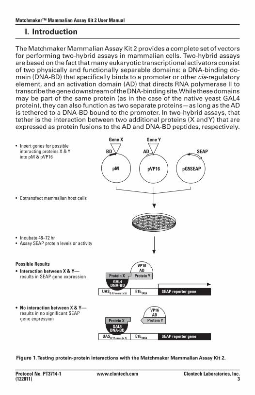
The Matchmaker Mammalian Assay Kit 2 provides a complete set of vectors for performing two-hybrid assays in mammalian cells. Two-hybrid assays are based on the fact that many eukaryotic transcriptional activators consist of two physically and functionally separable domains: a DNA-binding do-main (DNA-BD) that specifically binds to a promoter or other cis-regulatory element, and an activation domain (AD) that directs RNA polymerase II to transcribe the gene downstream of the DNA-binding site. While these domains may be part of the same protein (as in the case of the native yeast GAL4 protein), they can also function as two separate proteins—as long as the AD is tethered to a DNA-BD bound to the promoter. In two-hybrid assays, that tether is the interaction between two additional proteins (X and Y) that are expressed as protein fusions to the AD and DNA-BD peptides, respectively.
Figure 1. Testing protein-protein interactions with the Matchmaker Mammalian Assay Kit 2.
pVP16 pG5SEAP
AD
Gene Y• Insert genes for possible interacting proteins X & Y into pM & pVP16
pM
BD
Gene X
Protein X Protein YGAL4
DNA-BD
Protein XGAL4
DNA-BD
VP16AD
Protein Y
VP16AD
E1bTATA SEAP reporter geneUASG 17-mers (x 5)
E1bTATA SEAP reporter gene
SEAP
UASG 17-mers (x 5)
Possible Results
• Cotransfect mammalian host cells
• Incubate 48–72 hr• Assay SEAP protein levels or activity
• No interaction between X & Y— results in no significant SEAP gene expression
• Interaction between X & Y— results in SEAP gene expression
Matchmaker™ Mammalian Assay Kit 2 User Manual
Clontech Laboratories, Inc. www.clontech.com Protocol No. PT3714-1 4 (122811)
I. Introduction continued
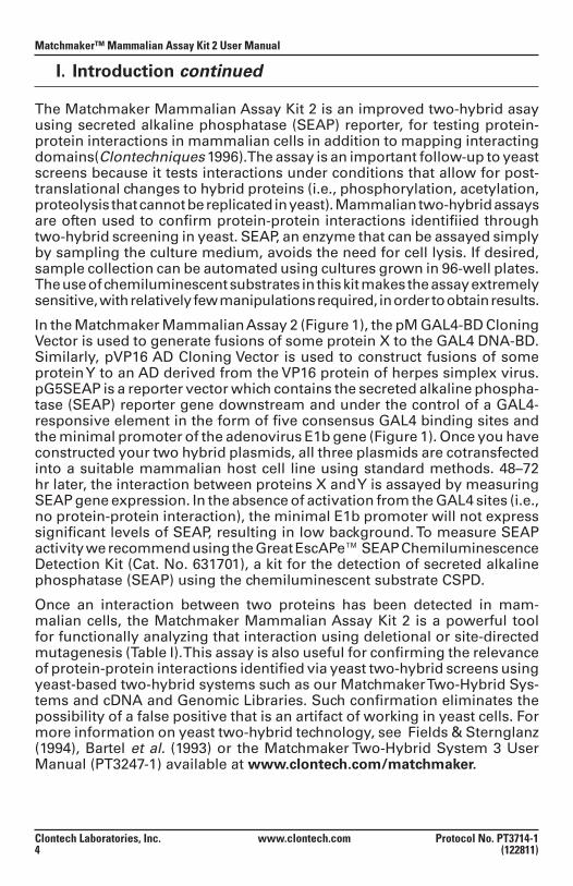
The Matchmaker Mammalian Assay Kit 2 is an improved two-hybrid asay using secreted alkaline phosphatase (SEAP) reporter, for testing protein-protein interactions in mammalian cells in addition to mapping interacting domains(Clontechniques 1996). The assay is an important follow-up to yeast screens because it tests interactions under conditions that allow for post-translational changes to hybrid proteins (i.e., phosphorylation, acetylation, proteolysis that cannot be replicated in yeast). Mammalian two-hybrid assays are often used to confirm protein-protein interactions identifiied through two-hybrid screening in yeast. SEAP, an enzyme that can be assayed simply by sampling the culture medium, avoids the need for cell lysis. If desired, sample collection can be automated using cultures grown in 96-well plates. The use of chemiluminescent substrates in this kit makes the assay extremely sensitive, with relatively few manipulations required, in order to obtain results.
In the Matchmaker Mammalian Assay 2 (Figure 1), the pM GAL4-BD Cloning Vector is used to generate fusions of some protein X to the GAL4 DNA-BD. Similarly, pVP16 AD Cloning Vector is used to construct fusions of some protein Y to an AD derived from the VP16 protein of herpes simplex virus. pG5SEAP is a reporter vector which contains the secreted alkaline phospha-tase (SEAP) reporter gene downstream and under the control of a GAL4-responsive element in the form of five consensus GAL4 binding sites and the minimal promoter of the adenovirus E1b gene (Figure 1). Once you have constructed your two hybrid plasmids, all three plasmids are cotransfected into a suitable mammalian host cell line using standard methods. 48–72 hr later, the interaction between proteins X and Y is assayed by measuring SEAP gene expression. In the absence of activation from the GAL4 sites (i.e., no protein-protein interaction), the minimal E1b promoter will not express significant levels of SEAP, resulting in low background. To measure SEAP activity we recommend using the Great EscAPe™ SEAP Chemiluminescence Detection Kit (Cat. No. 631701), a kit for the detection of secreted alkaline phosphatase (SEAP) using the chemiluminescent substrate CSPD.
Once an interaction between two proteins has been detected in mam-malian cells, the Matchmaker Mammalian Assay Kit 2 is a powerful tool for functionally analyzing that interaction using deletional or site-directed mutagenesis (Table I). This assay is also useful for confirming the relevance of protein-protein interactions identified via yeast two-hybrid screens using yeast-based two-hybrid systems such as our Matchmaker Two-Hybrid Sys-tems and cDNA and Genomic Libraries. Such confirmation eliminates the possibility of a false positive that is an artifact of working in yeast cells. For more information on yeast two-hybrid technology, see Fields & Sternglanz (1994), Bartel et al. (1993) or the Matchmaker Two-Hybrid System 3 User Manual (PT3247-1) available at www.clontech.com/matchmaker.
Protocol No. PT3714-1 www.clontech.com Clontech Laboratories, Inc. (122811) 5
Matchmaker™ Mammalian Assay Kit 2 User Manual
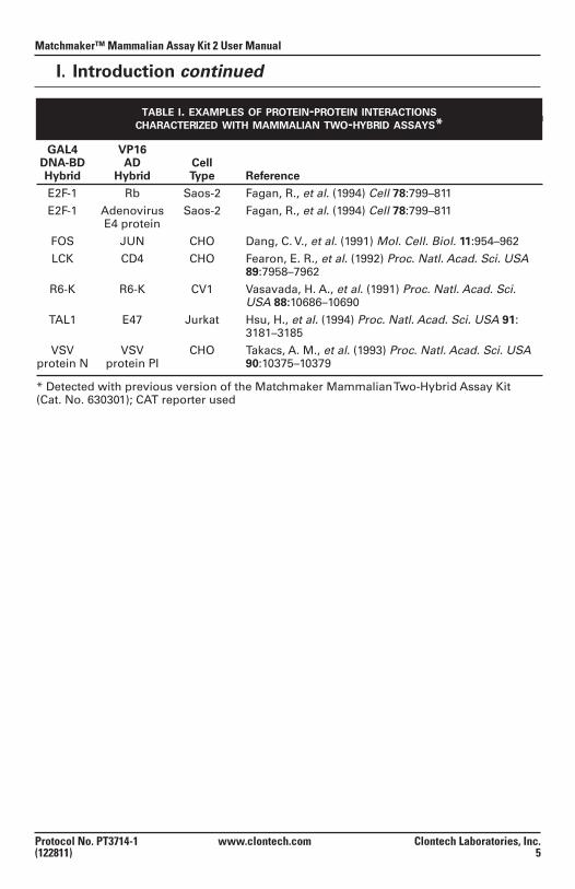
table i. examples of protein-protein interactionscharacterized with mammalian two-hybrid assays*
GAL4 VP16 DNA-BD AD Cell Hybrid Hybrid Type Reference
E2F-1 Rb Saos-2 Fagan, R., et al. (1994) Cell 78:799–811
E2F-1 Adenovirus Saos-2 Fagan, R., et al. (1994) Cell 78:799–811 E4 protein
FOS JUN CHO Dang, C. V., et al. (1991) Mol. Cell. Biol. 11:954–962
LCK CD4 CHO Fearon, E. R., et al. (1992) Proc. Natl. Acad. Sci. USA 89:7958–7962
R6-K R6-K CV1 Vasavada, H. A., et al. (1991) Proc. Natl. Acad. Sci. USA 88:10686–10690
TAL1 E47 Jurkat Hsu, H., et al. (1994) Proc. Natl. Acad. Sci. USA 91: 3181–3185
VSV VSV CHO Takacs, A. M., et al. (1993) Proc. Natl. Acad. Sci. USA protein N protein PI 90:10375–10379
* Detected with previous version of the Matchmaker Mammalian Two-Hybrid Assay Kit (Cat. No. 630301); CAT reporter used
I. Introduction continued
Matchmaker™ Mammalian Assay Kit 2 User Manual
Clontech Laboratories, Inc. www.clontech.com Protocol No. PT3714-1 6 (122811)
II. List of Components
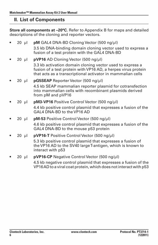
Store all components at –20°C. Refer to Appendix B for maps and detailed descriptions of the cloning and reporter vectors.
• 20 µl pM GAL4 DNA-BD Cloning Vector (500 ng/µl) 3.5 kb DNA-binding domain cloning vector used to express a
fusion of a test protein with the GAL4 DNA-BD
• 20 µl pVP16 AD Cloning Vector (500 ng/µl) 3.3 kb activation domain cloning vector used to express a
fusion of a test protein with VP16 AD, a herpes virus protein that acts as a transcriptional activator in mammalian cells
• 20 µl pG5SEAP Reporter Vector (500 ng/µl) 4.5 kb SEAP mammalian reporter plasmid for cotransfection
into mammalian cells with recombinant plasmids derived from pM and pVP16
• 20 µl pM3-VP16 Positive Control Vector (500 ng/µl) 4.4 kb positive control plasmid that expresses a fusion of the
GAL4 DNA-BD to the VP16 AD
• 20 µl pM-53 Positive Control Vector (500 ng/µl) 4.6 kb positive control plasmid that expresses a fusion of the
GAL4 DNA-BD to the mouse p53 protein
• 20 µl pVP16-T Positive Control Vector (500 ng/µl) 5.3 kb positive control plasmid that expresses a fusion of
the VP16 AD to the SV40 large T-antigen, which is known to interact with p53
• 20 µl pVP16-CP Negative Control Vector (500 ng/µl) 4.5 kb negative control plasmid that expresses a fusion of the
VP16 AD to a viral coat protein, which does not interact with p53
Protocol No. PT3714-1 www.clontech.com Clontech Laboratories, Inc. (122811) 7
Matchmaker™ Mammalian Assay Kit 2 User Manual
III. Additional Materials Required
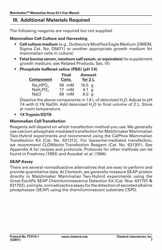
The following reagents are required but not supplied.
Mammalian Cell Culture and Harvesting • Cellculturemedium(e.g., Dulbecco’s Modified Eagle Medium [DMEM,
Sigma Cat. No. D5671] or another appropriate growth medium for mammalian cells in culture)
• Fetalbovineserum,newborncalfserum,orequivalent(to supplement growth medium; see Related Products, Sec. VI)
• Phosphate buffered saline (PBS) (pH 7.4) Final Amount
Component Conc. for 2 L Na2HPO4 58 mM 16.5 g NaH2PO4 17 mM 4.1 g NaCl 68 mM 8.0 g
Dissolve the above components in 1.8 L of deionized H2O. Adjust to pH 7.4 with 0.1N NaOH. Add deionized H2O to final volume of 2 L. Store at room temperature.
• 1XTrypsin/EDTA
Mammalian Cell TransfectionReagents will depend on which transfection method you use. We generally use calcium-phosphate-mediated transfection for Matchmaker Mammalian Two-Hybrid experiments and recommend using the CalPhos Mammalian Transfection Kit (Cat. No. 631312). For liposome-mediated transfection, we recommend CLONfectin Transfection Reagent (Cat. No. 631301). See Appendix A for recipes and protocols. Protocols for other methods can be found in Freshney (1993) and Ausubel et al. (1994).
SEAP AssayThere are several nonradioactive alternatives that are easy to perform and provide quantitative data. At Clontech, we generally measure SEAP protein directly in Matchmaker Mammalian Two-Hybrid experiments using the Great EscAPe SEAP Chemiluminescence Detection Kit (Cat. Nos. 631701 & 631702), a simple, nonradioactive assay for the detection of secreted alkaline phosphatase (SEAP) using the chemiluminescent substrate CSPD.
Matchmaker™ Mammalian Assay Kit 2 User Manual
Clontech Laboratories, Inc. www.clontech.com Protocol No. PT3714-1 8 (122811)
IV. Matchmaker Mammalian Assay Kit 2 Protocols
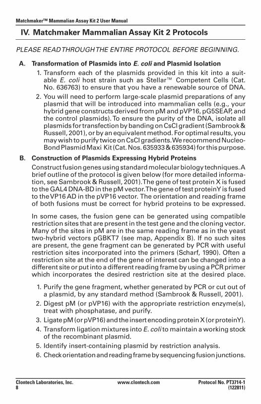
PLEASE READ THROUGH THE ENTIRE PROTOCOL BEFORE BEGINNING.
A. Transformation of Plasmids into E. coli and Plasmid Isolation 1. Transform each of the plasmids provided in this kit into a suit-
able E. coli host strain such as Stellar™ Competent Cells (Cat.No. 636763) to ensure that you have a renewable source of DNA.
2. You will need to perform large-scale plasmid preparations of any plasmid that will be introduced into mammalian cells (e.g., your hybrid gene constructs derived from pM and pVP16, pG5SEAP, and the control plasmids). To ensure the purity of the DNA, isolate all plasmids for transfection by banding on CsCl gradient (Sambrook & Russell, 2001), or by an equivalent method. For optimal results, you may wish to purify twice on CsCI gradients. We recommend Nucleo-Bond Plasmid Maxi Kit (Cat. Nos. 635933 & 635934) for this purpose.
B. Construction of Plasmids Expressing Hybrid Proteins Construct fusion genes using standard molecular biology techniques. A
brief outline of the protocol is given below (for more detailed informa-tion, see Sambrook & Russell, 2001). The gene of test protein X is fused to the GAL4 DNA-BD in the pM vector. The gene of test protein Y is fused to the VP16 AD in the pVP16 vector. The orientation and reading frame of both fusions must be correct for hybrid proteins to be expressed.
In some cases, the fusion gene can be generated using compatible restriction sites that are present in the test gene and the cloning vector. Many of the sites in pM are in the same reading frame as in the yeast two-hybrid vectors pGBKT7 (see map, Appendix B). If no such sites are present, the gene fragment can be generated by PCR with useful restriction sites incorporated into the primers (Scharf, 1990). Often a restriction site at the end of the gene of interest can be changed into a different site or put into a different reading frame by using a PCR primer which incorporates the desired restriction site at the desired place.
1. Purify the gene fragment, whether generated by PCR or cut out of a plasmid, by any standard method (Sambrook & Russell, 2001).
2. Digest pM (or pVP16) with the appropriate restriction enzyme(s), treat with phosphatase, and purify.
3. Ligate pM (or pVP16) and the insert encoding protein X (or protein Y). 4. Transform ligation mixtures into E. coli to maintain a working stock
of the recombinant plasmid. 5. Identify insert-containing plasmid by restriction analysis. 6. Check orientation and reading frame by sequencing fusion junctions.
Protocol No. PT3714-1 www.clontech.com Clontech Laboratories, Inc. (122811) 9
Matchmaker™ Mammalian Assay Kit 2 User Manual
IV. Matchmaker Mammalian Assay Kit 2 continued
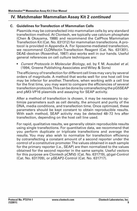
C. Guidelines for Transfection of Mammalian Cells Plasmids may be cotransfected into mammalian cells by any standard
transfection method. At Clontech, we typically use calcium phosphate (Chen & Okayama, 1988) and recommend the CalPhos Mammalian Transfection Kit (Cat. No. 631312) for mammalian transfections. A pro-tocol is provided in Appendix A. For liposome-mediated transfection, we recommend CLONfectin Transfection Reagent (Cat. No. 631301). DEAE-dextran (Rosenthal, 1987) also works well in our hands. Useful general references on cell culture techniques are:
• Current Protocols in Molecular Biology, ed. by F. M. Ausubel et al. (1994, Greene Publishing Associates and Wiley & Sons)
The efficiency of transfection for different cell lines may vary by several orders of magnitude. A method that works well for one host cell line may be inferior for another. Therefore, when working with a cell line for the first time, you may want to compare the efficiencies of several transfection protocols. This can be done by cotransfecting the pG5SEAP and pM3-VP16 plasmids and assaying for SEAP activity.
After a method of transfection is chosen, it may be necessary to op-timize parameters such as cell density, the amount and purity of the DNA, media conditions, and transfection time. Once optimized, these parameters should be kept constant to obtain reproducible results. With each method, SEAP activity may be detected 48–72 hrs after transfection, depending on the host cell line used.
For rapid, qualitative results, we generally obtain reproducible results using single transfections. For quantitative data, we recommend that you perform duplicate or triplicate transfections and average the results. You may also wish to normalize for transfection efficiency by cotransfecting a constant amount of a second reporter under the control of a constitutive promoter. The values obtained in each sample for the primary reporter (i.e., SEAP) are then normalized to the values obtained for the second reporter in the same sample. Useful vectors for this purpose are Clontech pCMVβ (Cat. No. 631719), pβgal-Control (Cat. No. 631709), or pSEAP2-Control (Cat. No. 631717).
Matchmaker™ Mammalian Assay Kit 2 User Manual
Clontech Laboratories, Inc. www.clontech.com Protocol No. PT3714-1 10 (122811)
IV. Matchmaker Mammalian Assay Kit 2 continued
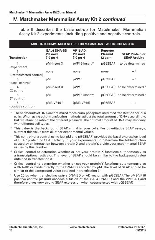
Table II describes the basic set-up for Matchmaker Mammalian Assay Kit 2 experiments, including positive and negative controls.
table ii. recommended set-up for mammalian two-hybrid assays
GAL4 DNA-BD VP16 AD Reporter Plasmid Plasmid Plasmid SEAP Protein or Transfection (10 µg a) (10 µg a) (2 µg a) SEAP Activity
1 pM-insert X pVP16-insert Y pG5SEAP to be determined (experiment) 2 none none none – b (untransfected control) 3 pM pVP16 pG5SEAP – c (basal control) 4 pM-insert X pVP16 pG5SEAP to be determined d (X control) 5 pM pVP16-insert Y pG5SEAP to be determined e
(Y control) 6 pM3-VP16 f (pM3-VP16) pG5SEAP +++ (positive control)
a These amounts of DNA are optimized for calcium-phosphate mediated transfection of HeLa cells. When using other transfection methods, adjust the total amount of DNA accordingly, but maintain the ratio of the different plasmids. The optimal amount of DNA may also vary with different cell types.
b This value is the background SEAP signal in your cells. For quantitative SEAP assays, subtract this value from all other experimental values.
c This control (or a control using just pM and pG5SEAP) provides the basal expression level of SEAP protein or SEAP activity in your experiments. To determine the fold-induction caused by an interaction between protein X and protein Y, divide your experimental SEAP values by this number.
d Critical control to determine whether or not your protein X functions autonomously as a transcriptional activator. The level of SEAP should be similar to the background value obtained in transfection 3.
e Critical control to determine whether or not your protein Y functions autonomously as a DNA-BD or binds directly to the DNA-BD encoded by pM. The level of SEAP should be similar to the background value obtained in transfection 3.
f Use 20 µg when transfecting only a DNA-BD or AD vector with pG5SEAP. The pM3-VP16 positive control plasmid encodes a fusion of the GAL4 DNA-BD and the VP16 AD and therefore gives very strong SEAP expression when cotransfected with pG5SEAP.
Protocol No. PT3714-1 www.clontech.com Clontech Laboratories, Inc. (122811) 11
Matchmaker™ Mammalian Assay Kit 2 User Manual
IV. Matchmaker Mammalian Assay Kit 2 continued
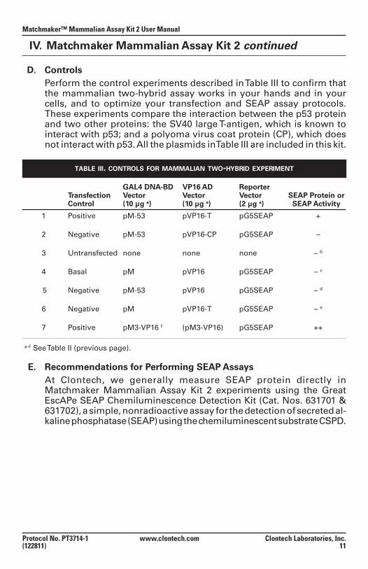
D. Controls Perform the control experiments described in Table III to confirm that
the mammalian two-hybrid assay works in your hands and in your cells, and to optimize your transfection and SEAP assay protocols. These experiments compare the interaction between the p53 protein and two other proteins: the SV40 large T-antigen, which is known to interact with p53; and a polyoma virus coat protein (CP), which does not interact with p53. All the plasmids in Table III are included in this kit.
table iii. controls for mammalian two-hybrid experiment
GAL4 DNA-BD VP16 AD Reporter Transfection Vector Vector Vector SEAP Protein or Control (10 µg a) (10 µg a) (2 µg a) SEAP Activity
1 Positive pM-53 pVP16-T pG5SEAP + 2 Negative pM-53 pVP16-CP pG5SEAP – 3 Untransfected none none none – b 4 Basal pM pVP16 pG5SEAP – c 5 Negative pM-53 pVP16 pG5SEAP – d 6 Negative pM pVP16-T pG5SEAP – e
7 Positive pM3-VP16 f (pM3-VP16) pG5SEAP ++
a–f See Table II (previous page).
E. Recommendations for Performing SEAP AssaysAt Clontech, we generally measure SEAP protein directly in Matchmaker Mammalian Assay Kit 2 experiments using the Great EscAPe SEAP Chemiluminescence Detection Kit (Cat. Nos. 631701 & 631702), a simple, nonradioactive assay for the detection of secreted al-kaline phosphatase (SEAP) using the chemiluminescent substrate CSPD.
Matchmaker™ Mammalian Assay Kit 2 User Manual
Clontech Laboratories, Inc. www.clontech.com Protocol No. PT3714-1 12 (122811)
IV. Matchmaker Mammalian Assay Kit 2 continued
F. Further Experiments 1. Verification of Hybrid Protein Expression [Optional] Expression of the GAL4 DNA-BD/Protein X fusion in mammalian
cells can be verified by preparing Western blots using soluble pro-tein extracts from the cells. Probe the blots with the GAL4 DNA-BD Monoclonal Antibody (Cat. No. 630403) using standard Western blot-ting procedures (Harlow & Lane, 1988; Sambrook & Russell, 2001).
2. Mapping Protein Structure Once an interaction between two proteins has been detected
in mammalian cells, the Matchmaker Mammalian Assay Kit 2 provides a powerful tool for functionally dissecting that interaction using deletional or site-directed mutagenesis. Transformer Site-Directed Mutagenesis Kit (Cat. No. 630702) is useful for generat-ing mutant proteins which can then be tested in the Matchmaker Mammalian Assay Kit 2.
3. Confirmation in Yeast Matchmaker Two-Hybrid System Interactions that are detected in the Matchmaker Mammalian Two-
Hybrid Assay may also be detectable in a yeast-based two-hybrid assay such as Matchmaker Two-Hybrid System 3 (Cat. No. 630303). Much less DNA is required for each two-hybrid experiment in yeast. Furthermore, yeast two-hybrid assays are much less expensive, since they do not require tissue-culture facilities and supplies for the growth and transfection of mammalian cells. For more informa-tion on yeast two-hybrid technology, see the references by Fields & Sternglanz (1994) and Bartel et al. (1993) or the references in the Matchmaker Two-Hybrid System 3 User Manual (PT3247-1) avail-able at www.clontech.com/matchmaker.
Protocol No. PT3714-1 www.clontech.com Clontech Laboratories, Inc. (122811) 13
Matchmaker™ Mammalian Assay Kit 2 User Manual
V. References
Ausubel, F. M., Brent, R., Kingdom, R. E., Moore, D. M., Seidman, J. G., Smith, J. A., & Struhl, K. (1994) Current Protocols in Molecular Biology. (Greene Publishing Associates, Inc. & John Wiley & Sons, Inc.).
Bartel, P. L, Chien, C.-T., Sternglanz, R. & Fields, S. (1993) Elimination of false positives that arise in using the two-hybrid system. BioTechniques 14:920–924.
Chen, C. & Okayama, H. (1988) Calcium phosphate-mediated gene transfer: A highly efficient transfection system for stably transforming cells with plasmid DNA. BioTechniques 6:632.
Dang, C. V., Barrett, J., Villa-Garcia, M., Resar, L. M. S., Kato, G. J. & Fearon, E. R. (1991) In-tracellular leucine zipper interactions suggest c-myc hetero-oligomerization. Mol. Cell. Biol. 11:954–962.
Fagan, R., Flint, K. J. & Jones, N (1994) Phosphorylation of E2F-1 modulates its interaction with the retinoblastoma gene product and the adenoviral E4 19-kDa protein. Cell 78:799–811.
Fearon, E. R., Finkel, T., Gillison, M. L., Kennedy, S. P., Casella, J. F., Tomaselli, G. F., Morrow, J. S. & Ding, C. V. (1992) Karyoplasmic interaction selection strategy: a general strategy to detect protein-protein interaction in mammalian cells. Proc. Natl. Acad. Sci. USA 89:7958–7962.
Fields, S. (1993) The two-hybrid system to detect protein-protein interactions. Methods: A Companion to Meth. Enzymol. 5:116–124.
Fields, S. & Sternglanz, R. (1994) The two-hybrid system: an assay for protein-protein interac-tions. Trends Genet. 10: 286–292.
Freshney, R. I. (1993) Culture of Animal Cells, Third Edition (Wiley-Liss, NY).
Harlow, E. & Lane, E. (1988) Antibodies: A Laboratory Manual (Cold Spring Harbor Laboratory, Cold Spring Harbor, NY).
Hsu, H., Wadman, I. & Baer, R. (1994) Formation of in vivo complexes between the TAL1 and E2A polypeptides of leukemic T-cells. Proc. Natl. Acad. Sci. USA 91:3181–3185.
Sadowski, I., Bell, B., Broad, P. & Hollis, M. (1992) GAL4 fusion vectors for expression in yeast or mammalian cells. Gene 118:137–141.
Sambrook, J. & Russell, D. W. (2001) Molecular Cloning: A Laboratory Manual, Third Edition (Cold Spring Harbor Laboratory Press, Cold Spring Harbor, NY).
Scharf, S. J. (1990) Cloning with PCR. In PCR Protocols: A Guide to Methods and Applications, eds. Innis, M. A., Gelfand, D. H., Sninsky, J. J. & White, T. J. (Academic Press, Inc., San Diego), pp. 84–91.
Silver, P. A., Keegan, L. P. & Ptashne, M. (1984) Amino terminus of the yeast GAL4 gene product is sufficient for nuclear localizaton. Proc. Natl. Acad. Sci. USA 81:5951–5955.
Takacs, A. M., Das, T. & Banerjee, A. K. (1993) Mapping of interacting domain between the nu-cleocapsid protein and the phosphoprotein of vesicular stomatitis virus by using a two-hybrid system. Proc. Natl. Acad. Sci.USA 90:10375–10379.
Vasavada, H. A., Ganguly, S., Germino, F. J., Wang, Z. X. & Weissman, S. M. (1991) A contingent replication assay for the detection of protein-protein interactions in animal cells. Proc. Natl. Acad.Sci. USA 88:10686–10690.
Matchmaker Mammalian Two-Hybrid Assay Kit (1996) Clontechniques XI(1):10–12.
Matchmaker™ Mammalian Assay Kit 2 User Manual
Clontech Laboratories, Inc. www.clontech.com Protocol No. PT3714-1 14 (122811)
A. Materials Required We recommend using the CalPhos Mammalian Transfection Kit
(Cat. No. 631312) for mammalian transfection. Follow the protocol in the User Manual provided.
The recipes and calcium phosphate transfection protocol below are adapted from Current Protocols in Molecular Biology, Supplement 14, Section 9.1.3.
• 2XBES-BufferedSolution(BBS,pH6.95) Final Component Conc. N, N-bis (2-hydroxyethyl)-2-aminoethane-sulfonic acid (BES; CALBIOCHEM Cat. No. 391334) 50 mM NaCl 280 mM Na2HPO4 1.5 mM
It is critical that the pH of this solution be between pH 6.95 and 6.98. We recommend that you check each new batch of 2X BES buffer against a reference stock prepared (and tested) earlier.
Filter sterilize through a 0.45 µm nitrocellulose filter (Nalgene). Store in aliquots at –20°C (can be frozen and thawed repeatedly). • 2.5 mM CaCl2 Add 183.7 g of CaCl2 dihydride (Sigma; tissue culture grade) to 500
ml of H2O. Filter sterilize through a 0.45 µm nitrocellulose filter (Nalgene). Store at –20°C in 10 ml aliquots (can be frozen and thawed repeatedly).
B. Protocol for Calcium Phosphate Transfection All plasmids should be CsCl-banded and diluted to a concentration of
1.0 µg/µl. Store the DNA solution at 4°C. For initial experiments, we recommend that each transfection be performed with a total of 22 µg of DNA (as shown in Tables II and III). However, the optimal concentra-tion of DNA may vary with different cell types.
Day 1 1. For each transfection, seed 5 x 10⁵ exponentially growing cells in
a 10 cm tissue culture plate in 10 ml of complete medium (with serum). There should be < 106 cells/plate (~30% confluency) just prior to transfection. This provides enough surface area on the plate for at least two more doublings.
Appendix A: SEAP Protocol
Protocol No. PT3714-1 www.clontech.com Clontech Laboratories, Inc. (122811) 15
Matchmaker™ Mammalian Assay Kit 2 User Manual
Day 2 2. Two hrs before transfection, replace the medium with fresh medium
(with serum) to stimulate cell growth. 3. For each transfection, combine the DNAs in a sterile 1.5 ml micro-
centrifuge tube. 4. Bring the total volume to 450 µl by adding TE or deionized H2O. 5. Add 50 µl of 2.5 M CaCl2 and vortex thoroughly. (The final concen-
tration of CaCl2 is 0.25 M.) 6. Add 500 µl of 2X BBS, mix well, and incubate at room tem-
perature for 10–20 min to allow a precipitate to form. 7. Add the calcium phosphate-DNA solution dropwise to the medium
while swirling the plate. 8. Incubate overnight in a 37°C, 5% CO2 incubator. Note: The level of carbon dioxide is critical. We recommend using a Fyrite gas
analyzer to measure percent CO2 prior to incubation.
Day 3 9. Carefully wash the cells twice with 5 ml of PBS, then add 10 ml of
complete medium (with serum).
10. Incubate the cells for an additional 24–48 hrs. Troubleshooting note: The amounts of DNA in Tables II and III have been
optimized for calcium phosphate transfection of HeLa cells; however, the optimal amount of total DNA may vary with different cell types. (The ratio of input DNAs should stay the same.) To determine the optimum amount of plasmid, transfect three plates of cells with 10, 20, and 30 µg of plasmid DNA and incubate overnight. The next day examine the plates with a microscope at 100X. A coarse, clumpy precipitate will form at DNA concentrations that are too low, a fine (almost invisible) precipitate will form at concentrations that are higher than optimal, and an even, granular precipitate will form with optimal DNA concentrations.
C. SEAP AssayThere are several nonradioactive alternatives that are easy to perform and provide quantitative data. At Clontech, we generally measure SEAP protein directly in Matchmaker Mammalian Assay Kit 2 experiments using the Great EscAPe SEAP Chemiluminescence Detection Kit (Cat. Nos. 631701 & 631702).
Appendix A: SEAP Protocol continued
Matchmaker™ Mammalian Assay Kit 2 User Manual
Clontech Laboratories, Inc. www.clontech.com Protocol No. PT3714-1 16 (122811)
Appendix B: Plasmid Maps & Multiple Cloning Sites
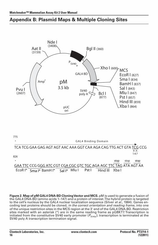
Figure 2. Map of pM GAL4 DNA-BD Cloning Vector and MCS. pM is used to generate a fusion of the GAL4 DNA-BD (amino acids 1–147) and a protein of interest. The hybrid protein is targeted to the cell’s nucleus by the GAL4 nuclear localization sequence (Silver et al., 1984). Genes en-coding test proteins should be cloned, in the correct orientation and reading frame, into one of the unique restriction sites in the MCS region at the 3' end of the GAL4 DNA-BD. Restriction sites marked with an asterisk (*) are in the same reading frame as pGBKT7. Transcription is initiated from the constitutive SV40 early promoter (PSV40e); transcription is terminated at the SV40 poly A transcription termination signal.
pM3.5 kb
pUCori
Ampr
SV40poly A
PSV40e
GAL4 BD
Aat II(3159)
Pvu I (2607)
Nde I(3408)
Bgl II (360)
Xho I (600)
Bcl I (877)
MCSEcoR I (827)Sma I (834)BamH I (837)Sal I (843)Mlu I (847)Pst I (857)Hind III (859)Xba I (864)
TCA TCG GAA GAG AGT AGT AAC AAA GGT CAA AGA CAG TTG ACT GTA TCG CCG
GAA TTC CCG GGG ATC CGT CGA CGC GTC TGC AGA AGC TTC TAG ATA AGT AA EcoR I* Sma I*
826 •
Pst ISal I*BamH I*
775 •
stop
Xba IHind IIIMlu I
a .a . 147
stopstop
GAL4 Binding Domain
Protocol No. PT3714-1 www.clontech.com Clontech Laboratories, Inc. (122811) 17
Matchmaker™ Mammalian Assay Kit 2 User Manual
Appendix B: Plasmid Maps & MCS continued
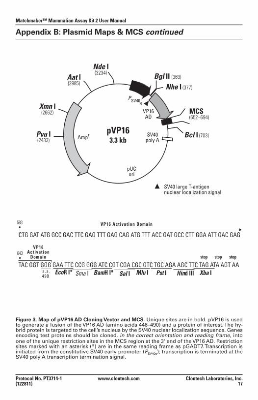
Figure 3. Map of pVP16 AD Cloning Vector and MCS. Unique sites are in bold. pVP16 is used to generate a fusion of the VP16 AD (amino acids 446–490) and a protein of interest. The hy-brid protein is targeted to the cell’s nucleus by the SV40 nuclear localization sequence. Genes encoding test proteins should be cloned, in the correct orientation and reading frame, into one of the unique restriction sites in the MCS region at the 3' end of the VP16 AD. Restriction sites marked with an asterisk (*) are in the same reading frame as pGADT7. Transcription is initiated from the constitutive SV40 early promoter (PSV40e); transcription is terminated at the SV40 poly A transcription termination signal.
pVP163.3 kb
pUCori
Ampr SV40poly A
VP16AD
MCS(652–694)
Pvu I(2433)
Nhe I (377)
Nde I(3234) Bgl II (369)
Xmn I(2662)
Aat I(2985)
Bcl I (703)
PSV40e
∆ SV40 large T-antigennuclear localization signal
∆
CTG GAT ATG GCC GAC TTC GAG TTT GAG CAG ATG TTT ACC GAT GCC CTT GGA ATT GAC GAG
TAC GGT GGG GAA TTC CCG GGG ATC CGT CGA CGC GTC TGC AGA AGC TTC TAG ATA AGT AA EcoR I* Sma I Pst ISal IBamH I*
643 •
583 •
stop
Xba IHind IIIMlu I
VP16 Act ivat ion
Domain
a .a .490
stopstop
VP16 Act ivat ion Domain
Matchmaker™ Mammalian Assay Kit 2 User Manual
Clontech Laboratories, Inc. www.clontech.com Protocol No. PT3714-1 18 (122811)
Appendix B: Plasmid Maps & MCS continued
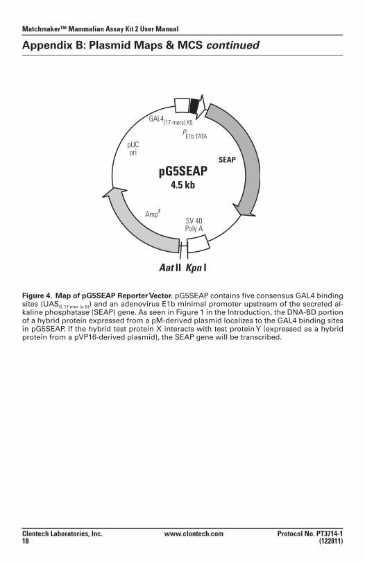
Figure 4. Map of pG5SEAP Reporter Vector. pG5SEAP contains five consensus GAL4 binding sites (UASG 17-mer (x 5)) and an adenovirus E1b minimal promoter upstream of the secreted al-kaline phosphatase (SEAP) gene. As seen in Figure 1 in the Introduction, the DNA-BD portion of a hybrid protein expressed from a pM-derived plasmid localizes to the GAL4 binding sites in pG5SEAP. If the hybrid test protein X interacts with test protein Y (expressed as a hybrid protein from a pVP16-derived plasmid), the SEAP gene will be transcribed.
pG5SEAP4.5 kb
SEAP
pUCori
AmprSV 40Poly A
PE1b TATA
GAL4(17-mers) X5
Kpn IAat II
Protocol No. PT3714-1 www.clontech.com Clontech Laboratories, Inc. (122811) 19
Matchmaker™ Mammalian Assay Kit 2 User Manual
Notes
Notice to Purchaser
Clontech products are to be used for research purposes only. They may not be used for any other purpose, including, but not limited to, use in drugs, in vitro diagnostic purposes, therapeutics, or in humans. Clontech products may not be transferred to third parties, resold, modified for resale, or used to manufacture commercial products or to provide a service to third parties without prior written approval of Clontech Laboratories, Inc.
Your use of this product is also subject to compliance with any applicable licensing requirements described on the product’s web page at http://www.clontech.com. It is your responsibility to review, understand and adhere to any restrictions imposed by such statements.
Clontech, the Clontech logo, Great EscAPe, Matchmaker, and Stellar are trademarks of Clon-tech Laboratories, Inc. All other marks are the property of their respective owners. Certain trademarks may not be registered in all jurisdictions. Clontech is a Takara Bio Company. ©2011 Clontech Laboratories, Inc.
This document has been reviewed and approved by the Clontech Quality Assurance Depart-ment.