Embed Size (px)
Citation preview
1
MASTERARBEIT / MASTER’S THESIS
Titel der Masterarbeit / Title of the Master‘s Thesis
„Quantification of synapse loss in human brains with mixed Alzheimer’s and Lewy body disease“
verfasst von / submitted by
Suzan Özugur B.Sc.
angestrebter akademischer Grad / in partial fulfilment of the requirements for the degree of
Master of Science (M.Sc.)
Wien, 2016 / Vienna 2016
Studienkennzahl lt. Studienblatt / degree programme code as it appears on the student record sheet:
A 066 863
Studienrichtung lt. Studienblatt / degree programme as it appears on the student record sheet:
Masterstudium Biologische Chemie
Betreut von / Supervisor:
Assoz. Prof. Mag. Dr. Wolfgang Wadsak, Privatdozent

! 2!
! !


! 5!
!
This!Master!Thesis!was!carried!out!from!15.!01.!2016!till!09.!09.!16!at!DZNE!in!Munich!under!leadership!from!Prof.!Dr.!Jochen!Herms,!supervised!by!Dr.!Dr.!Mario!Dorostkar.!
!
!
!
!
!
!
!
!
!
!
!
!
!
!
!
!
!
!
!
!
!
!

! 6!
Acknowledgment-!
I!want! to! thank!Dr.!Dr.!Mario!Dorostkar! for!his! supervision!of!my!master! thesis,! the!great!support! and! assistance! I! received! from! him! as! well! as! the! possibilities! and! freedom! of!research!he!was!offering.!Open!doors!and!ears!were!promoting!my!ideas!for!the!project.!
I!thank!Prof.!Dr.!Jochen!Herms!for!the!immediate!acceptance!of!my!master!thesis!project!in!form!of!an!Erasmus!internship!at!the!DZNE!in!Munich.!
Just!as!much!I!want!to!thank!all!the!members!of!the!group!of!Prof.!Dr.!Herms!for!the!help!and!support!they!gave!me,!whenever!I!asked!for!it.!
I!appreciate!the!tips!and!equipment!I!got!from!Michael!Schmidt.!
Finaly! I!might! thank!Prof.!Dr.!Wolfgang!Wadsak,!who! took! the! supervision!of!my!Erasmus!internship!from!the!University!of!Vienna.!
!
!
!
!
!
!
!
!
!
!
!
!
!
!
!

! 7!
Self0reliance-!
I! hereby! confirm!my!Master! Thesis!was!written! independently! and! just!with! the! specified!resources.!Other!works!that!were!used!are!cited!and!labeled!with!the!source.!
! !
! 8!
Abstract-!
Synapses! mediate! the! communication! between! neurons! and! their! function! alters! during!physiological! processes! such! as! learning! and! memory.! Cognitive! decline! in!neurodegenerative! disease! has! been! shown! to! result! from! loss! of! synapses.! However,!previous!studies!only!analyzed!specific!types!of!synapses!in!one!or!few!specific!brain!regions.!!The!aim!of!this!study!was!to!map!the!loss!of!excitatory!and!inhibitory!synapses!distinct!brain!regions!in!the!brains!of!patients!suffering!from!Alzheimer’s!disease!and!Lewy!body!disease.!I!analyzed!67!brains! from!donors!at! the!Newcastle!Brain!Tissue!Resource,!which!had!been!neuropathologically!classified!as!Alzheimer's!disease,!Lewy!body!disease!or!controls.!Tissue!microarrays! from!several!brain! regions!were! immunofluorescence! labelled!with!antibodies!against! DLG4! (also! known! as! PSDa95! scaffold! protein)! to! stain! excitatory! synapses! and!GABRA2!(GABAA!Receptor!subunit!α2)!to!stain!inhibitory!synapses.!Images!were!recorded!on!a! confocal!microscope! and! synapse!densities!were! quantified! automatically! using! customawritten!image!analysis!algorithms.!I! found! different! patterns! of! synapse! loss,! depending! on! the! disease.! Particularly! in!Alzheimer’s!disease!brains,! there!was!a!marked! loss!of! inhibitory!synapses! in!several!brain!regions,! including! the! entorhinal! cortex,! temporal! and! parietal! cortex,! cingulate,! visual!cortex,!putamen!and!pallidum.!This! loss! of! inhibitory! synapses! may! cause! disruption! of! longarange! neuronal! networks,!which!has!been!demonstrated!in!several!animal!studies.!!!! !
! 9!
Table-of-Contents-! !
Acknowledgment!....................................................................................................................................!6!
Selfareliance!............................................................................................................................................!7!
Abstract!...................................................................................................................................................!8!
1.! Introduction!...................................................................................................................................!11!
Anatomy!of!the!brain!........................................................................................................................!11!
2.! Neurotransmitters!.........................................................................................................................!14!
2.1.! Glutamate!–!Excitatory!neurotransmitter!..............................................................................!14!
2.2.! GABA!.......................................................................................................................................!15!
3.! Synapses!........................................................................................................................................!16!
3.1.! Excitatory!synapses!.................................................................................................................!19!
3.1.1.! Glutamatergic!synapse!....................................................................................................!19!
3.2.! Inhibitory!synapses!.................................................................................................................!21!
3.2.1.! GABAergic!synapse!..........................................................................................................!21!
3.3.! Interaction!!Inhibitory!–!Excitatory!synapses!..........................................................................!22!
4.! Disease!...........................................................................................................................................!23!
4.1.! Genetics!..................................................................................................................................!25!
4.2.! Proteins!and!indirect!factors!involved!in!Dementia!...............................................................!25!
4.2.1! Amyloid!β!..........................................................................................................................!25!
4.2.2! Tau!tangle!.........................................................................................................................!28!
4.2.3! αasynuclein!a!Lewy!bodies!................................................................................................!29!
4.2.4! Indirect!factors!.................................................................................................................!31!
4.3! Previous!findings!.....................................................................................................................!33!
4.4! Stage!of!disease!severity!.........................................................................................................!35!
CERAD!(Consortium!to!establish!a!registry!for!Alzheimer’s!Disease)!............................................!35!
5.! Objective!........................................................................................................................................!35!
6.! Material!.........................................................................................................................................!36!
6.1! Tissue!Microarrays!...................................................................................................................!37!
6.2! Chemicals!.................................................................................................................................!38!
7.! Methods!........................................................................................................................................!39!
8.! Image!acquisition!and!Analysis!......................................................................................................!42!
9.! Results!...........................................................................................................................................!43!
9.1! Statistical!Analysis!...................................................................................................................!44!
9.2! Alzheimer’s!Disease!.................................................................................................................!45!

! 10!
9.3! Lewy!Body!Disease!..................................................................................................................!56!
9.4! αaSynuclein,!AT8!and!Aβ4G8!...................................................................................................!59!
10.! Discussion!....................................................................................................................................!62!
11.! Prospects!.....................................................................................................................................!65!
12.! Abbreviations!..............................................................................................................................!66!
13.! Abstrakt!(Deutsch)!.......................................................................................................................!68!
14.! List!of!figures!...............................................................................................................................!69!
15.! List!of!tables!................................................................................................................................!70!
16.! Quotations!...................................................................................................................................!71!
!
!
! !
! 11!
1. Introduction-!
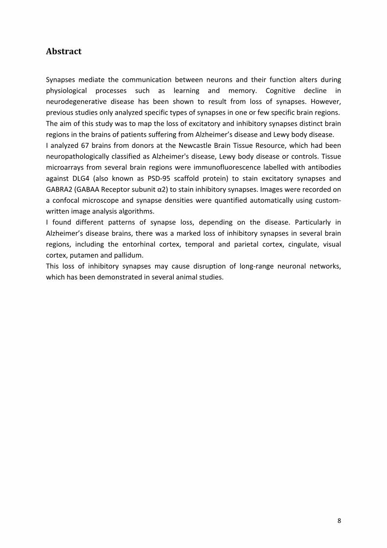
Anatomy-of-the-brain-The!human!brain!can!be!divided!anatomically!into!many!different!regions.!The!classification!into!Brodmann’s!areas!is!one!of!the!main!systems!used,!which!is!based!on!the!microscopic!appearance! of! the! different! areas.! Furthermore,! the! functions! of! the! areas! are! well!correlated! with! this! classification.! The! Brodmann’s! area! functions! can! be! summarized! in!executive!functions!(Brodmann’s!area!9,!10,!44,!45,!46),!motor!functions!(Brodmann’s!area!4,! 6,! 8),! somatosensory! functions! (Brodmann’s! area! 1,! 2,! 3,! 5,! 40),! attention! (Brodmann’s!area!7!and!39),!visual!functions!(Brodmann’s!area!17,!18,!19),!memory!(Brodmann’s!area!20,!21,! 37),! emotional! regulation! (Brodmann’s! area! 11,! 38,! 47)! and! sound! recognition! and!handling!(Brodmann’s!area!22,!41,!42).!
!
!!
!
!
!
!
!
!
!
!
!
!
!
!
Fig.%1%Brodmann’s%areas,%sagittal%picture%of%a%human%brain%
Figure%from:!Brodmann,!K.,!Beiträge!zur!histologischen!Lokalisation!der!Greosshirnrinde.!VI.!Mitteilung:!
Die!Cortexgliederung!des!Menschen.!J.!Psychol.!Neurol.!(Leipzig),!1908.!10:!p.!231a246![1]!
Schematic! picture! of! the! different! brain! regions,! differentiated! by! brodmann.! Areas! 1,! 2,! 3! are! the!primary!somatosensory!cortex,!Area!4!the!primary!motor!cortex,!Area!5!the!somatosensory!association!cortex,!Area!6:!Premotor! cortex,!Area!7:!Visuoamotor! coordination,!Area!8:! Frontal!eye! fields,!Area!9:!Dorsolateral!prefrontal!cortex,!Area!10:!Anterior!prefrontal!cortex,!Area!11:!Orbitofrontal!area,!Area!12:!Orbitofrontal!area,!Area!13,!16:!Insular!cortex,!Area!17:!Primary!visual!cortex,!Area18:!secondary!visual!cortex,!Area!19:!Associative!visual! cortex,!Area!20:! Inferior! temporal! gyrus,!Area!21:!Middle! temporal!gyrus,!Area!22:! Superior! temporal! gyrus,!Area!23:!Ventral!posterior! cingulate! cortex,!Area!24:!Ventral!anterior! cingulate! cortex,! Area! 25:! Subgenual! area,! Area! 26:! Ectosplenial! portion! of! the! retrosplenial!region! of! the! cerebral! cortex,! Area! 27! –!Piriform!cortex,! Area! 28:! Ventral! entorhinal! cortex,! Area! 29:!Retrosplenial! cingulate! cortex,! Area! 30:! Part! of! cingulate! cortex,! Area! 31:! Dorsal! Posterior! cingulate!cortex,! Area! 32:! Dorsal! anterior! cingulate! cortex,! Area! 33:! Part! of! anterior! cingulate! cortex,! Area! 34:!Dorsal!entorhinal!cortex,!Area!35:!Perirhinal!cortex,!Area!36:!Ectorhinal!area,!now!part!of!the!perirhinal!cortex,! Area! 37:! Fusiform! gyrus,! Area! 38:! Temporopolar! area,! Area! 39:! Angular! gyrus,! Area! 40:!Supramarginal!gyrus,!!Areas!41!and!42:!Auditory!cortex,!Area!43:!Primary!gustatory!cortex,!Area!44:!Pars!opercularis,!Area!45:!Pars! triangularis,!Area!46:!Dorsolateral!prefrontal! cortex,!Area!47:!Pars!orbitalis,!part! of! the! inferior! frontal! gyrus,! Area! 48:! Retrosubicular! area,! Area! 49:! Parasubicular! area! and!Brodmann’s!area!52:!Parainsular!area.!
! 12!
The!major!part!of!the!brain!is!the!cerebrum,!which!is!divided!into!two!hemispheres.!Figure!1!depicts!a!sagittal!profile!along!the!hemisphere.!The!cerebral!cortex!is!divided!into!the!frontal!lobe! (Brodmann’s! areas:! 4,! 6,! 8,! 9,! 10,! 11,! 44,! 45,! 46,! 47),! the! parietal! lobe! (Brodmann’s!areas:!1,!2,!3,!5,!7,!40),!the!temporal!lobe!(Brodmann’s!areas:!20,!21,!22,!37,!38,!39,!41,!42)!and!the!occipital!lobe!(Brodmann’s!areas:!17,!18,!19).!In!the!occipital!lobe,!visual!processing!occurs,!while! the!temporal! lobe!receives! input! from!the!ears.!Further! language,! like! fluent!speech,! understanding! and! recognition! of!words,! numbers,! body! and! faces! are! processed!there.!The!parietal! lobe! integrates!the! input!of!different!senses,! important! for!orientation.!The!frontal!lobe!is!important!in!speechalanguage!production!and!the!activation!and!control!of!body!muscles,!like!the!eyes.!
The! limbic! system!consists!of!parts!of! the! frontal,! parietal! and! temporal! lobe.! Further! the!hippocampus!and!amygdala,!which! is!part!of! the! limbic!system!(Brodmann’s!areas:!23,!24,!25,!26,!29,!30,!31,!32,!33),!as!well!as! thalamus,!basal!ganglia!and!the!cingulate!gyrus,!play!important!roles!in!memory!and!emotion.!The!brainstem!is!the!connection!of!the!spinal!cord,!consisting!of!the!midbrain,!pons,!medulla!and!oblongata.!Thalamus!and!hypothalamus!send!motor!and!sensory!signals!to!the!cortex.!The!cerebellum!coordinates!motor!and!autonomic!functions,! and! is! therefore! important! for! balance! and! coordination.! [2]! All! these! different!brain! regions! are! responsible! for! many! functions! and! disrupted! regions! therefore!responsible! for! disease.! Also! the! strong! interconnection! of! the! brain! regions! makes! it!difficult! to! distinguish! the! trigger! and! to! inhibit! the! spreading! of! disease! activators.! This!interconnection! is! due! to! neurons,! that! have! their! soma! and!most! of! its! dendrites! in! one!region,! but! their! axon! is! reaching! into! another! brain! region.! Kwon! and! Jang! showed,! that!70%!of!the!brain!is!connected!to!all!other!regions,!forming!information!processing!networks![3].!
There! are! roughly! 86,1! billion! neurons! in! an! adult! male! human! brain,! with! 19%! in! the!cerebral!cortex,!which!makes!up!82%!of!the!total!brain!mass![4].!Neurons!are!composed!of!the!soma,! the!nucleus,!one!axon!and!several!dendrites,!which!can! form!synapses! to!other!neurons!in!short!distances!4mm![5],!whereas!the!axon!can!reach!other!neurons!at!distances!of!up! to!1m![6].!One!neuron!can!be!connected!by! its!dendrites! to!100.000!other!cells! [6],!forming!a!complex!network.!Neurons!are!responsible!for!the!reception!of!stimuli,!processing!and!transfer!of!information!in!the!form!of!electrochemical!impulses.!A!neuron!consists!of!the!cell!body,!with! the!nucleus,!many!dendrites!and!also!one!axon,!which!can! form!a!synaptic!terminal.! The! axon! is! myelinated! through! oligodendrocytes,! which! forms! myelin! sheaths!around!the!axon,!to!accelerate!the!action!potential,!which! jumps!from!one!ranvier!knot!to!the!next.!This!ranvier!knot!is!the!region!between!two!myelin!sheaths!and!free!of!contact!to!oligodendrocytes.!On!the!synaptic!terminal!neurotransmitter!can!be!released!and!can!bind!on!postsynaptic!membrane!receptors,!initiating!a!new!action!potential.!
! 13!
!
!
!
!
!
!
!
!!
!
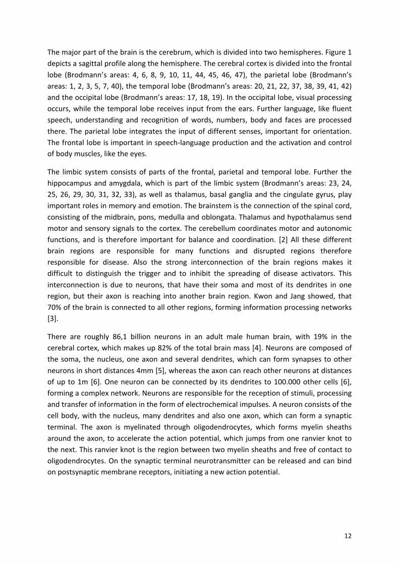
Hence! neurons! are! responsible! for! the!most! critical! and! complex! cellular! functions! in! the!brain.!”They!undergo!the!greatest!number!of!microscopic!changes!in!response!to!acute!and!chronic!cell!injury!and!are!the!principal!sites!of!damage!for!several!of!the!diseases!associated!with! the! highest! morbidity! and! mortality! in! our! society,! i.e.! cerebrovascular! and!neurodegenerative! diseases”! [8].! Neurons! require! ion! channels! and! pumps! to! hold! the!balance!between!intracellular!and!extracellular!electrolyte!concentration,!especially!for!toxic!calcium! ions,! but! also! for! sodium! and! potassium.! This! balance! is! disturbed! by! microaenvironmental! damage! to! the!membrane! and! therefore! to! the! pumps,! leading! to! energy!deprivation.! Necrosis! is! the! consequence! of! that! irreparable! cell! damage! which! may! be!caused!by!oxygen!deficit,!which!affects! cell! energy! requirements!and!membrane! integrity.!“When! neurons! undergo! cell! death! and! necrosis,! no! effective! neuronal! mitosis! or!replenishment! of! neurons! from! stem! cells! is! present! within! the! adult! human! brain”! [8].!Microglia! cells! and! macrophages! remove! these! damaged! neurons! by! phagocytosis.!Furthermore,! astrocytes! proliferate! in! response! to! the! injury.! They! seemed! to! have!important!neuroprotective! functions.! [8]!Astrocytes!outnumber!neurons! in! the!brain.!They!are!thought!to!be!responsible!for!glutamatea,! iona!and!water!homeostasis,!defense!against!oxidative!stress,!energy!storage,!scar!formation,!tissue!repair!and!synapse!formation![9].!
!
!
!
!
!
!
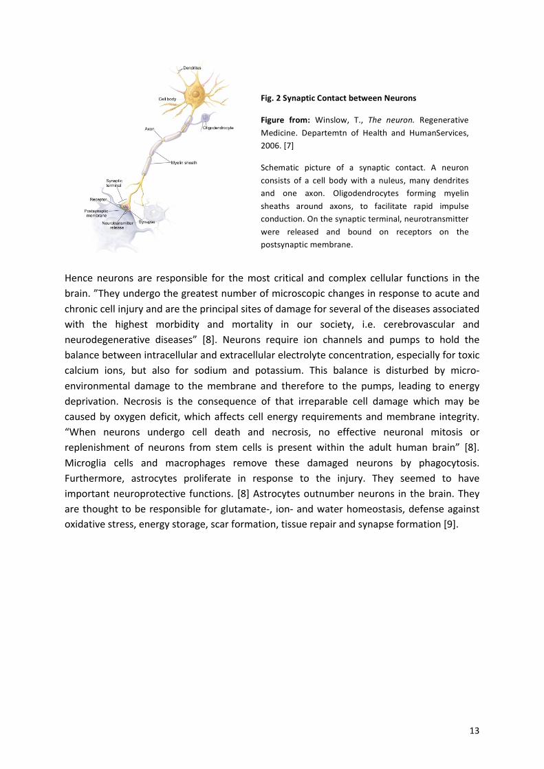
Fig.%2%Synaptic%Contact%between%Neurons%
Figure% from:% Winslow,! T.,! The$ neuron.! Regenerative!Medicine.! Departemtn! of! Health! and! HumanServices,!2006.![7]!
Schematic! picture! of! a! synaptic! contact.! A! neuron!consists! of! a! cell! body! with! a! nuleus,! many! dendrites!and! one! axon.! Oligodendrocytes! forming! myelin!sheaths! around! axons,! to! facilitate! rapid! impulse!conduction.!On!the!synaptic!terminal,!neurotransmitter!were! released! and! bound! on! receptors! on! the!postsynaptic!membrane.!
! 14!
2. Neurotransmitters-!
When! an! action! potential! reaches! a! neuron,! neurotransmitter,! packed! in! vesicles,! are!transported!to!the!membrane!and!released!into!the!synaptic!cleft.!The!synaptic!cleft!consists!of! a! prea! and! a! post! synapse! with! receptors! on! both! terminals.! The! released!neurotransmitter!can!bind!on!the!receptors!and!initiate!another!action!potential!or!inhibit!it.!Receptors!on!the!pre!synapse!can!bind!neurotransmitter!as!a!saturation!signal!to!check!the!quantity! of! neurotransmitters! in! the! cleft.! If!more! neurotransmitters! bind! on! pre! synapse!receptors! the! release! of! neurotransmitter! may! be! decreased.! There! are! many! different!neurotransmitters!which! can! be! summarized! into! different! functional! or! chemical! groups.!There!are!small!molecule!neurotransmitters!and!peptide!neurotransmitters.!The!first!can!be!divided! further! into! biogenic! amines! (catecholamine! like! dopamine!or! epinephrine,! indole!amine! like! serotonin!and! imidazole!amine! like!histamine),! amino!acids! (glutamate,!GABA),!purines!(ATP)!and!others!like!acetylcholine.!The!second!group,!peptide!neurotransmitter!can!be!between!3!–!30!amino!acids!long,!e.g.!methionine!encephalin.![10]!!
The! two! main! types! of! neurotransmitters! for! excitatory! and! inhibitory! synapses! are!glutamate!and!GABA,!which!were!also!analyzed!for!this!project.!
!
2.1. -Glutamate-–-Excitatory-neurotransmitter-Glutamate! is! the! main! excitatory! neurotransmitter! in! the! brain! and! the! most! abundant!amino!acid!in!the!human!body.!!
Signals!can!be!excitatory!or! inhibitory.!Excitatory!postsynaptic!potentials!(EPSP)!are!caused!of!an!influx!of!positive!charged!ions!(Na+)!and!results!in!a!depolarization!of!the!membrane,!that!lead!to!an!action!potential!firing!of!the!neuron,!when!the!depolarization!is!high!enough!to!overcome!the!gate!threshold.!
!
!
!
!
! ! ! ! ! ! ! !!!!!!
!
!
! 15!
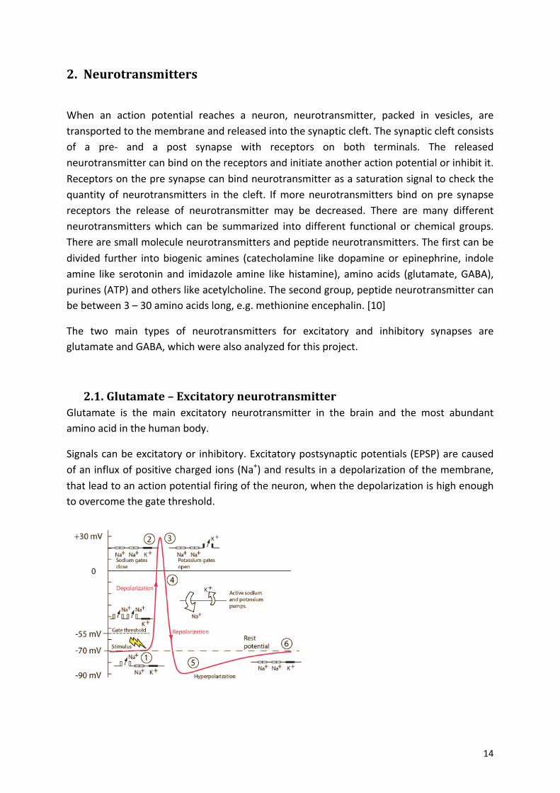
Fig.%3%%Action%potential!
Figure%from:!Charand,!K.X.,!Action$Potentials.![11]!
Demonstration!of!an!action!potential.!①"The!stimulus!opens!sodium!channels!and!drive!the!potential!from!a70mV!up!to!a55mV,!the!gate!threshold,!where!the!stimulus!creates!an!action!potential.!Under!this!threshold!no!action!potential!is!initiated.!More!sodium!channels!open!and!Na+!influx!into!the!cell!②."The!membrane!gets! depolarized! to! 30mV! and! the! sodium! gates! were! closed,!when! potassium! channels! open,! result! an!efflux! of! K+"③."④" The!membrane! repolarize! to! the! rest! potential! and! undershoot! it! to! a90mV!⑤," called"“Hyperpolarization”." This" prevents" the" neuron" from" getting" another" stimulus" in" this" time" period" to"ensure" that" the" signal" is" proceeding" in" one" direction" at" the" time."⑥" Last" the" sodium" and" potassium"pumps"drive"the"potential"back"to"E70"mV.!
!
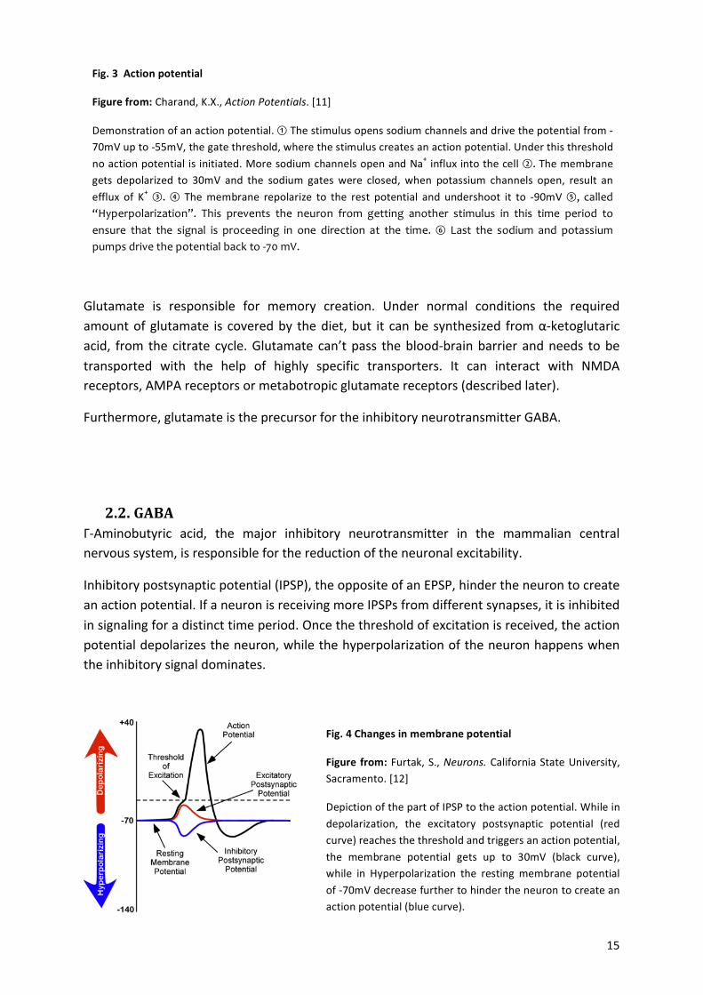
Fig.%4%Changes%in%membrane%potential%
Figure%from:!Furtak,!S.,!Neurons.!California!State!University,!Sacramento.![12]%
Depiction!of!the!part!of!IPSP!to!the!action!potential.!While!in!depolarization,! the! excitatory! postsynaptic! potential! (red!curve)!reaches!the!threshold!and!triggers!an!action!potential,!the! membrane! potential! gets! up! to! 30mV! (black! curve),!while! in! Hyperpolarization! the! resting!membrane! potential!of!a70mV!decrease!further!to!hinder!the!neuron!to!create!an!action!potential!(blue!curve).!
!
!
!
!
!
!
!
!
Glutamate! is! responsible! for! memory! creation.! Under! normal! conditions! the! required!amount!of!glutamate! is! covered!by! the!diet,!but! it! can!be!synthesized! from!αaketoglutaric!acid,! from! the!citrate! cycle.!Glutamate! can’t!pass! the!bloodabrain!barrier!and!needs! to!be!transported! with! the! help! of! highly! specific! transporters.! It! can! interact! with! NMDA!receptors,!AMPA!receptors!or!metabotropic!glutamate!receptors!(described!later).!!
Furthermore,!glutamate!is!the!precursor!for!the!inhibitory!neurotransmitter!GABA.!
!
!
2.2. -GABA-ΓaAminobutyric! acid,! the! major! inhibitory! neurotransmitter! in! the! mammalian! central!nervous!system,!is!responsible!for!the!reduction!of!the!neuronal!excitability.!
Inhibitory!postsynaptic!potential!(IPSP),!the!opposite!of!an!EPSP,!hinder!the!neuron!to!create!an!action!potential.!If!a!neuron!is!receiving!more!IPSPs!from!different!synapses,!it!is!inhibited!in!signaling!for!a!distinct!time!period.!Once!the!threshold!of!excitation!is!received,!the!action!potential!depolarizes!the!neuron,!while!the!hyperpolarization!of!the!neuron!happens!when!the!inhibitory!signal!dominates.!
!
!!
! 16!
GABA! is! synthesized! within! the! presynaptic! terminal.! Glutamic! acid! decarboxylase! (GAD)!produces!GABA!from!glutamate!with!the!cofactor!pyridoxal!phosphate.!So!only!the!absence!or! presence! of! the! enzyme! glutamate! decarboxylase! specifies! the! sort! of! synapse,! either!excitatory!or! inhibitory.!Like!glutamate,!GABA!can’t!pass!the!blood!brain!barrier!and!needs!the!presence!of!specific!receptors!and!transporters.!
!
3. Synapses--!
Neuronal!activity!runs!through!synapses.!Two!forms!of!synapse!exist:!chemical!and!electrical!(Gap!junctions).!Electrical!synapses!communicate!with! ion!flux,!whereas!chemical!synapses!convert! electrical! impulses! of! an! action! potential! into! chemical! signals! on! the! presynaptic!membrane! of! neurons.! This! triggers! an! opening! of! calcium! channels! and! the! increase! of!calcium! leads!to!secretion!of!neurotransmitter! from!the!synaptic!vesicles! into!the!synaptic!cleft.! These! neurotransmitters! bind! on! receptors! on! the! postsynaptic! membrane! of! the!synapse!from!the!second!neuron!and!can!start!a!new!action!potential.!
Signals!in!form!of!neurotransmitter,!like!adrenalin,!noradrenalin!or!dopamine,!are!released!from!the!presynapse!into!the!synaptic!cleft!and!received!by!receptors!at!the!postsynapse.!The!received!signals!on!the!surface!of!the!postsynapse!are!tansformed!in!electrical!and!biochemical!changes!in!the!postsynapse.!
!
!
!
!
!
!
!
!
!
!
!
!
! 17!
!
!
!
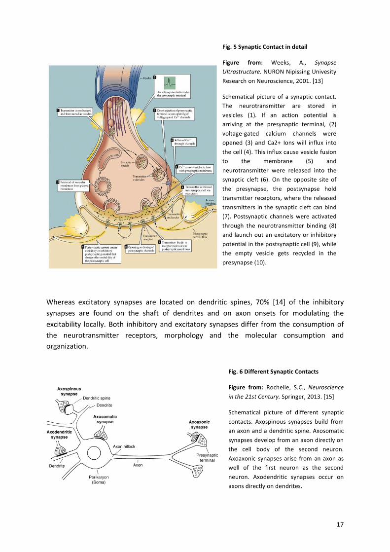
Whereas! excitatory! synapses! are! located! on! dendritic! spines,! 70%! [14]! of! the! inhibitory!synapses! are! found! on! the! shaft! of! dendrites! and! on! axon! onsets! for! modulating! the!excitability! locally.!Both! inhibitory!and!excitatory! synapses!differ! from! the!consumption!of!the! neurotransmitter! receptors,! morphology! and! the! molecular! consumption! and!organization.!
!
!
!!!
Fig.%5%Synaptic%Contact%in%detail%
Figure% from:% Weeks,! A.,! Synapse$Ultrastructure.!NURON!Nipissing!Univesity!Research!on!Neuroscience,!2001.![13]%
Schematical!picture!of!a! synaptic! contact.!The! neurotransmitter! are! stored! in!vesicles! (1).! If! an! action! potential! is!arriving! at! the! presynaptic! terminal,! (2)!voltageagated! calcium! channels! were!opened! (3)! and! Ca2+! Ions! will! influx! into!the!cell!(4).!This!influx!cause!vesicle!fusion!to! the! membrane! (5)! and!neurotransmitter! were! released! into! the!synaptic! cleft! (6).! On! the! opposite! site! of!the! presynapse,! the! postsynapse! hold!transmitter!receptors,!where!the!released!transmitters! in!the!synaptic!cleft!can!bind!(7).! Postsynaptic! channels! were! activated!through! the! neurotransmitter! binding! (8)!and! launch!out!an!excitatory!or! inhibitory!potential!in!the!postsynaptic!cell!(9),!while!the! empty! vesicle! gets! recycled! in! the!presynapse!(10).!
!
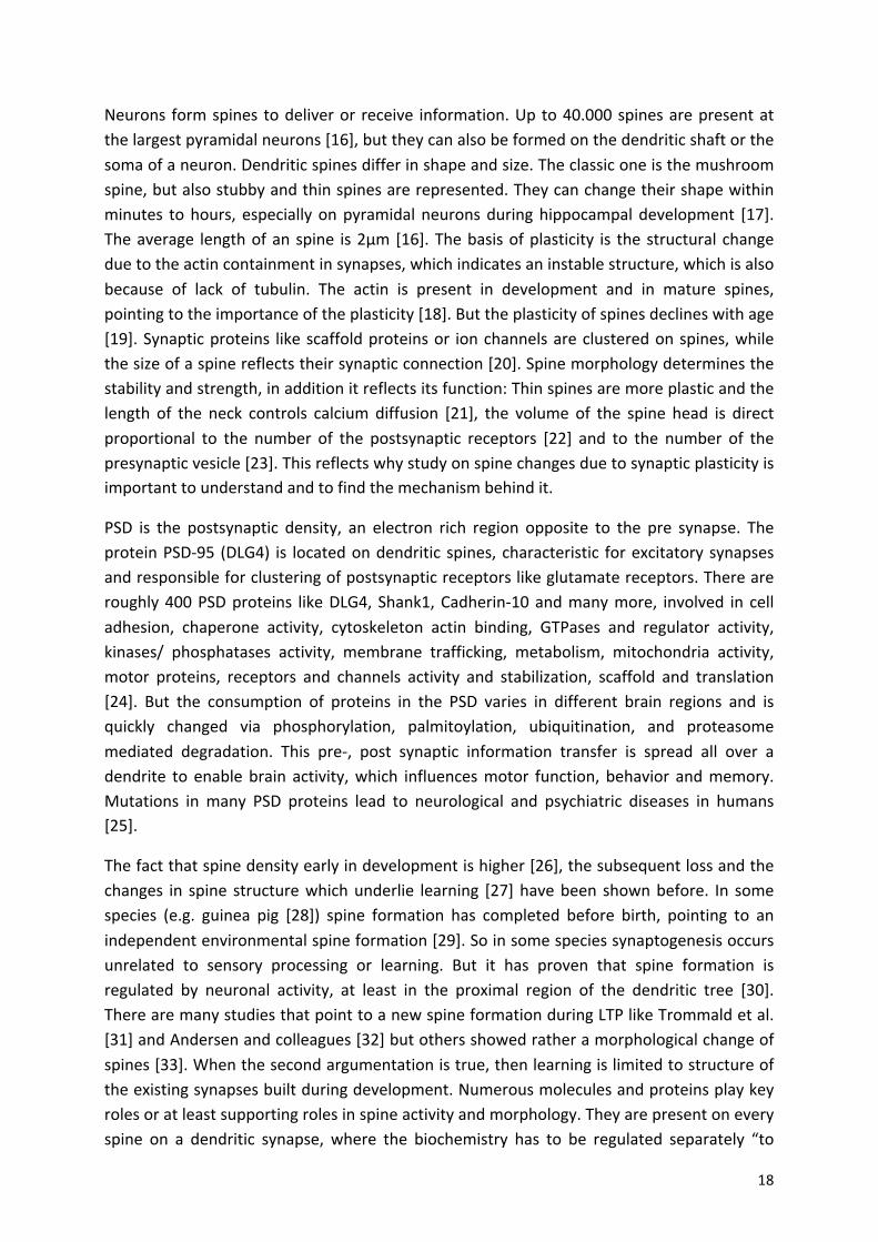
Fig.%6%Different%Synaptic%Contacts%
Figure% from:% Rochelle,! S.C.,! Neuroscience$in$the$21st$Century.!Springer,!2013.![15]%
Schematical! picture! of! different! synaptic!contacts.! Axospinous! synapses! build! from!an!axon!and!a!dendritic!spine.!Axosomatic!synapses!develop!from!an!axon!directly!on!the! cell! body! of! the! second! neuron.!Axoaxonic! synapses!arise! from!an!axon!as!well! of! the! first! neuron! as! the! second!neuron.! Axodendritic! synapses! occur! on!axons!directly!on!dendrites.!
! 18!
Neurons! form!spines! to!deliver!or! receive! information.!Up!to!40.000!spines!are!present!at!the!largest!pyramidal!neurons![16],!but!they!can!also!be!formed!on!the!dendritic!shaft!or!the!soma!of!a!neuron.!Dendritic!spines!differ!in!shape!and!size.!The!classic!one!is!the!mushroom!spine,!but!also!stubby!and!thin!spines!are!represented.!They!can!change!their!shape!within!minutes! to!hours,! especially!on!pyramidal!neurons!during!hippocampal!development! [17].!The!average! length!of!an!spine! is!2µm![16].!The!basis!of!plasticity! is! the!structural!change!due!to!the!actin!containment!in!synapses,!which!indicates!an!instable!structure,!which!is!also!because! of! lack! of! tubulin.! The! actin! is! present! in! development! and! in! mature! spines,!pointing!to!the!importance!of!the!plasticity![18].!But!the!plasticity!of!spines!declines!with!age![19].!Synaptic!proteins! like!scaffold!proteins!or! ion!channels!are!clustered!on!spines,!while!the!size!of!a!spine!reflects!their!synaptic!connection![20].!Spine!morphology!determines!the!stability!and!strength,!in!addition!it!reflects!its!function:!Thin!spines!are!more!plastic!and!the!length! of! the! neck! controls! calcium! diffusion! [21],! the! volume! of! the! spine! head! is! direct!proportional! to! the! number! of! the! postsynaptic! receptors! [22]! and! to! the! number! of! the!presynaptic!vesicle![23].!This!reflects!why!study!on!spine!changes!due!to!synaptic!plasticity!is!important!to!understand!and!to!find!the!mechanism!behind!it.!
PSD! is! the! postsynaptic! density,! an! electron! rich! region! opposite! to! the! pre! synapse.! The!protein!PSDa95! (DLG4)! is! located!on!dendritic! spines,!characteristic! for!excitatory!synapses!and!responsible!for!clustering!of!postsynaptic!receptors!like!glutamate!receptors.!There!are!roughly!400!PSD!proteins! like!DLG4,! Shank1,!Cadherina10!and!many!more,! involved! in! cell!adhesion,! chaperone! activity,! cytoskeleton! actin! binding,! GTPases! and! regulator! activity,!kinases/! phosphatases! activity,! membrane! trafficking,! metabolism,! mitochondria! activity,!motor! proteins,! receptors! and! channels! activity! and! stabilization,! scaffold! and! translation![24].! But! the! consumption! of! proteins! in! the! PSD! varies! in! different! brain! regions! and! is!quickly! changed! via! phosphorylation,! palmitoylation,! ubiquitination,! and! proteasome!mediated! degradation.! This! prea,! post! synaptic! information! transfer! is! spread! all! over! a!dendrite! to! enable!brain! activity,!which! influences!motor! function,! behavior! and!memory.!Mutations! in!many! PSD! proteins! lead! to! neurological! and! psychiatric! diseases! in! humans![25].!
The!fact!that!spine!density!early!in!development!is!higher![26],!the!subsequent!loss!and!the!changes! in! spine! structure!which!underlie! learning! [27]!have!been! shown!before.! In! some!species! (e.g.! guinea! pig! [28])! spine! formation! has! completed! before! birth,! pointing! to! an!independent!environmental!spine!formation![29].!So!in!some!species!synaptogenesis!occurs!unrelated! to! sensory! processing! or! learning.! But! it! has! proven! that! spine! formation! is!regulated! by! neuronal! activity,! at! least! in! the! proximal! region! of! the! dendritic! tree! [30].!There!are!many!studies!that!point!to!a!new!spine!formation!during!LTP!like!Trommald!et!al.![31]!and!Andersen!and!colleagues![32]!but!others!showed!rather!a!morphological!change!of!spines![33].!When!the!second!argumentation!is!true,!then!learning!is!limited!to!structure!of!the!existing!synapses!built!during!development.!Numerous!molecules!and!proteins!play!key!roles!or!at!least!supporting!roles!in!spine!activity!and!morphology.!They!are!present!on!every!spine! on! a! dendritic! synapse,! where! the! biochemistry! has! to! be! regulated! separately! “to!
! 19!
provide!a!subcellular!organization!or!isolation!of!the!biochemical!machinery!of!the!synapse”![18].!In!addition!to!spine!loss!due!to!disease,!there!are!compensation!mechanisms!that!help!to!maintain! the! synaptic! contact! [34],!but!also! some!of! the! synapses!are!more!vulnerable!than!others!in!disease.!Like!in!the!Aβ!pathology,!where!the!order!of!synapse!types!seemed!to!be!firstly!the!cholinergic,!secondly!the!glutamatergic!and!finally!the!GABAergic!terminals![35].!
!
There! are! excitatory! and! inhibitory! synapses,! which! differs! in! the! neurotransmitter!molecules! and! therefor! the! receptors,! scaffold! proteins! and! other! proteins! initiating! the!response!signal!in!the!postsynaptic!cell.!
!
3.1. -Excitatory-synapses-Excitatory!synapses,!such!as!glutamatergic!and!cholinergic,!depolarize!the!postsynaptic!cell!when! the! neurotransmitters! bind! to! the! transmembrane! receptors! in! the! synaptic! cleft.!Further! sodium! influxes! depolarize! the! neuron! from! its! resting! membrane! potential! and!increase!the!proportion!of!an!action!potential.!
For! the! communication! between!neurons,! neurotransmitter! receptors! play! a! fundamental!role.!They!can!be!localized!on!the!postsynaptic!membrane!at!the!synaptic!cleft!to!induce!an!action! potential! in! the! post! synapse,! on! the! dendritic! shaft! or! even! on! the! presynaptic!terminal!to!receive!some!feedback!information.!
!
3.1.1. Glutamatergic-synapse-The!glutamate!release!from!vesicles!is!the!major!mechanism!of!excitatory!neurotransmission!in!the!mammalian!brain!and!therefore!important!for!neuronal!activity.!80a90%!of!all!neurons!in! the! brain! are! glutamatergic! [36].! “20! glutamate! receptors! have! been! identified! in! the!mammalian! central! nervous! system”! [37].! Whereas! glutamate! can’t! pass! the! bloodabrain!barrier,! it! is! only! derived! by! local! synthesis! from! glucose.! Three! ionotropic! receptors! are!voltage! sensitive!and! located!on! the!glutamatergic! synapses:! Ionotropic! receptors,! like:!NamethylaDaaspartate! (NMDAR),! αaaminoa3ahydroxya5amethyla4aisoxazolepriopinoinic! acid!(AMPA)! and! kainate,! as! well! as! metabotropic/! Gaproteinacoupled! glutamate! receptors:!mGluR,!to!start!the!second!messenger!cascade!like!cAMP.!
The!ionotropic!receptors!include!ion!channels,!but!differ!in!the!permeability!of!the!ions,!like!Na+,!K+!and!Ca!2+![38].!Once!an!ionotropic!receptor!is!activated!it!undergoes!conformational!change!and!allows!the!influx!of!extracellular!ions.!This!process!is!fast!and!allow!neurons!to!act!fast!to!signal!transmission.!NMDAR!is!highly!expressed!on!neurons!as!well!on!astrocytes,!is!coupled!on!calcium!channels!and!plays!a!role!in!the!compensation!mechanism!of!the!body!during!injury![39]!and!is!important!for!learning!and!memory.!
! 20!
The! glutamate! binding! on! a!metabotropic! receptor! activates! the! GaProtein,! bound! to! the!cytoplasmic! tail! of! the! receptor,! which! activates! a! second! messenger! cascade! for! signal!transmission.!Metabotropic! receptors!are!slower! in! response!and!are! responsible! for!gene!expression!and!protein! synthesis,!which!enhance! the!excitability!of! glutamatergic!neurons![37],!but!also!for!direct!modulation!of!neuronal!excitability.!!
!
!
Three!transporters!are!expressed!on!glutamatergic!neurons!in!mammals:!VGlut1,!VGlut2!or!VGlut3,!which!mediate!the!glutamate!uptake!into!the!synaptic!vesicles.!The!different!types!of!transporters!are!present!in!different!regions!of!the!brain,!as!VGlut1!predominates!in!the!cortex,!cerebellum!and!hippocampus,!whereas!VGlut2!expression!is!mainly!in!the!brainstem!and! the! thalamus.! Both! isoforms! are! complementary! in! these! regions,! but! other! areas!express! both!of! them! [41].! The! third! isoform!VGlut3! is! homologous! to! the!other! two!and!expressed! at! a! very! low! level! striatum,! hippocampus,! cerebral! cortex! and! raphe! nuclei.!VGlut3!is!also!expressed!outside!the!nervous!system,!like!the!liver!and!eyes,!but!only!a!few!neurons!in!the!brain!express!VGlut3.!It!is!present!at!cholinergic!and!serotonergic!neurons!in!contrast!to!VGlut1!and!VGlut2![42].!Why!some!regions!prefer!the!expression!of!one!isoform!is!not!clear!yet.!Some!suggest!that!it!is!a!developmental!issue.!!
!
!
!
!
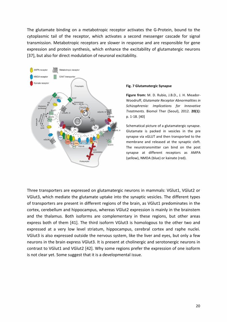
Fig.%7%Glutamatergic%Synapse%
Figure% from:%M.!D.!Rubio,! J.B.D.,! J.!H.!MeadoraWoodruff,!Glutamate$Receptor$Abnormalities$in$Schizophrenia:$ Implications$ for$ Innovative$Treatments.! Biomol! Ther! (Seoul),! 2012.! 20(1):!p.!1a18.![40]%
Schematical!picture!of!a!glutamatergic!synapse.!Glutamate! is! packed! in! vesicles! in! the! pre!synapse!via!vGLUT!and!then!transported!to!the!membrane! and! released! at! the! synaptic! cleft.!The! neurotransmitter! can! bind! on! the! post!synapse! at! different! receptors! as! AMPA!(yellow),!NMDA!(blue)!or!kainate!(red).!
!
!
! 21!
3.2. -Inhibitory-synapses-Besides! excitatory! synapses! there! are! inhibitory! synapses,! which! prevent! the! cell! from!generating!an!action!potential.!This! is!due! to!a!hyperpolarization! through!Cla! influx.! In! this!short!time!period!there!is!no!excitation!of!the!cell!possible.!
!
3.2.1. GABAergic-synapse-γaAminobutyric! acid! (GABA)! is! the!principal! inhibitory!neurotransmitter! in! the!brain!and! is!produced!via!the!precursor!glutamine!via!glutaminase!and!GAD.!Similar!to!glutamate,!both!ionotropic!and!metabotropic!receptors!exist.!GABA!can!lead!to!either!the!influx!of!negative!charged!chloride!ions!via!ionotropic!receptors!or!to!the!flow!of!positively!charged!potassium!ions! out! of! the! cell! via! metabotropic! receptors.! The! major! GABA! receptor! on! the! post!synapse! is! the!GABAaA! receptor,!which! forms!a! chloride! ion! channel.! Ionotropic! receptors!are!heteropentamers!with!the!α2!subunit!being!widely!expressed!in!the!mammalian!brain.!But!there!are!reports!showing!a!presence!of!both!receptor!subtypes,!GABAaA!and!GABAaB,!on! nonaGABAergic! neurons! and! also! GABA! transporter! (GAT)! on! the! membrane! of! nonaGABAergic!neurons!were!found,!maybe!playing!a!role! in!coatransportation!of!other! ions!or!GABA!release![16].!
!
!
!
!
!
!
!
!
!
!
!
!!
There! are! different! compositions! of! the! subtypes! in! one! GABA! receptor! cluster! and! 16!subunits! have! been! identified! yet.! This! expression! of! the! different! subunits! are! highly!variable! among! individuals! and!dependent! of! environmental! and! genetic! factors! [44].! The!most!dominant!one! is! the!α1β2γ2,! localized! in!almost!all! brain! regions.! The!α2! subtype! is!
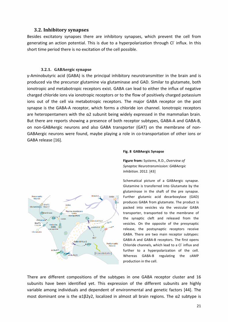
Fig.%8%%GABAergic%Synapse%
Figure%from:!Systems,!R.D.,!Overview$of$Synaptoc$Neurotransmission:$GABAergic$Inhibition.!2012.![43]!
Schematical! picture! of! a! GABAergic! synapse.!Glutamine! is!transferred! into!Glutamate!by!the!glutaminase! in! the! shaft! of! the! pre! synapse.!Further! glutamic! acid! decarboxylase! (GAD)!produces!GABA! from!glutamate.!The!product! is!packed! into! vesicles! via! the! vesicular! GABA!transporter,! transported! to! the! membrane! of!the! synaptic! cleft! and! released! from! the!vesicles.! On! the! opposite! of! the! presynaptic!release,! the! postsynaptic! receptors! receive!GABA.! There! are! two! main! receptor! subtypes:!GABAaA! and!GABAaB! receptors.! The! first!opens!Chloride!channels,!which!lead!to!a!Cla!influx!and!further! to! a! hyperpolarization! of! the! cell.!Whereas! GABAaB! regulating! the! cAMP!production!in!the!cell.!
! 22!
most! prominent! in! the! cluster! of! α2β3γ2! [45].! The! regulation! of! the! transcription! in! the!different!brain! regions!must!be!a! complex! system!with!a! tight! controlling!part,!where! the!change! in! the! expression!might! reflect! a! change! in! neuronal! activity.! Also! other! receptor!types! (also! excitatory! ones,! like! NMDAR)! can! influence! the! GABA! receptors! and! its!expression.![46]!
A!few!functional!partners!for!GABRA2!in!the!human!brain!are!ubiquilin!1,!gephyrin,!GABAA!receptor!α4/!β1/!β2/!β3/!γ2/!γ3,!GABAA!receptor!associated!protein!and!dynamin!1.!
!
!
3.3. -Interaction- -Inhibitory-–-Excitatory-synapses-Glutamate!is!a!precursor!protein!in!the!GABA!production!in!inhibitory!synapses,!but!itself!is!a!neurotransmitter! in! excitatory! synapses.! To! regulate! the! production! of! only! one!neurotransmitter!in!a!synapse,!there!is!no!GAD!present!in!excitatory!synapses,!to!allow!only!glutamate! as! neurotransmitter! and! no! further! transformation! into! GABA.! Whereas!glutamate! depolarizes! neurons,!GABA!hyperpolarizes! them,! so! in! general! those! two!pools!are! not! coalocalized.! But! in! distinct! isolated! neurons! in! some! brain! regions,! both!neurotransmitters! were! reported.! However! the! functional! significance! of! such! an!arrangement!is!not!clear![16].!
!
!
!
!
!
!
!
!!
!
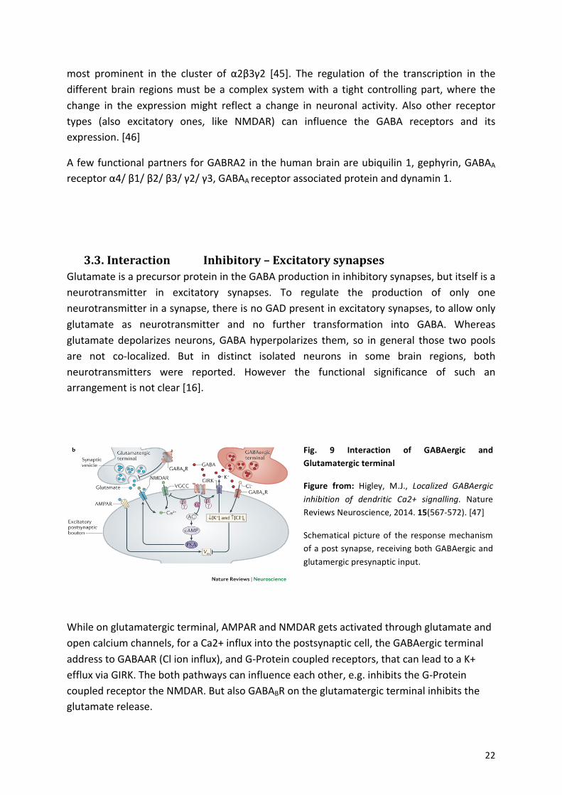
While!on!glutamatergic!terminal,!AMPAR!and!NMDAR!gets!activated!through!glutamate!and!open!calcium!channels,!for!a!Ca2+!influx!into!the!postsynaptic!cell,!the!GABAergic!terminal!address!to!GABAAR!(Cl!ion!influx),!and!GaProtein!coupled!receptors,!that!can!lead!to!a!K+!efflux!via!GIRK.!The!both!pathways!can!influence!each!other,!e.g.!inhibits!the!GaProtein!coupled!receptor!the!NMDAR.!But!also!GABABR!on!the!glutamatergic!terminal!inhibits!the!glutamate!release.!
!
Fig.% 9% Interaction% of% GABAergic% and%Glutamatergic%terminal%
Figure% from:% Higley,! M.J.,! Localized$ GABAergic$inhibition$ of$ dendritic$ Ca2+$ signalling.! Nature!Reviews!Neuroscience,!2014.!15(567a572).![47]!
Schematical! picture!of! the! response!mechanism!of!a!post!synapse,!receiving!both!GABAergic!and!glutamergic!presynaptic!input.!!
! 23!
4. Disease-!
All!neurodegenerative!disorders!have!one!thing! in!common:!pathological!accumulations!of!different!proteins!and!peptides,!specific! for!the!different!disease!types.!The!most!frequent!are! neurofibrilliary! tangles! and! βaAmyloid! plaques,! which! are! hallmarks! of! Alzheimer’s!disease.!
The!molecular! mechanism! behind!many! neurodegenerative! disorders! is! largely! unknown.!The! main! reason! is! certainly! the! complex! interaction! of! disease! supporting! factors.!Neurodegeneration!is!defined!as!neuron!loss!and!astrogliosis,!as!well!as!synapse!loss,!neuro!inflammation!and!misfolding!of!specific!proteins!that!cause!toxicity.!Each!of!these!alterations!has!a!complex!mechanism!and!many!pathways!for!activation.!For!example!the!secretion!of!interleukin! 1β! causes! Inflammation,!which! blocks! neurotrophic! factors,! that! are! necessary!for!neuron!stability,!maintenance!and!growth,!leading!to!spine!loss![48].!But!also!the!direct!alteration!of!ion!channels!by!inflammatory!cells,!like!the!phosphorylation!of!AMPA!receptors!may!cause!spine!loss![49].!
As!Herms!and!Dorostkar!depict!nicely,!there!are!three!different!structural!spine!alterations:!The!change!of! the!number! in!a!specific!brain!region!and!reduced!spine!density!because!of!reduced!dendrite!length!or!nerve!cell!loss.!Spine!shape!modification!(stubby,!mushroom!and!thin)! as! well! as! spine! head! enlargement! and! third:! Pathological! changes! like! altered!endoplasmic! reticulum! or! enlarged! PSD! [50].! All! these! abnormalities! appear! in!neurodegenerative!diseases.!!
Dementia!is!common!the!elderly!population!and!may!be!caused!by!many!different!diseases!like!Alzheimer’s!disease!(AD),!dementia!with!Lewy!bodies!(DLB),!Parkinson!disease!dementia,!frontotemporal!lobar!degeneration,!Huntington’s!disease!and!cerebrovascular!disease.!!
Alzheimer’s!disease! is!one!of! these!neurodegenerative!disorders.! It! is! a! slowly!progressive!loss!of!neuronal!functions,! including!impairments!in!processing!of! information!in!the!brain,!synapse! reduction! and! loss! of! neurons! in! certain! brain! areas.! The! hippocampus! and!entorhinal!cortex!are!affected!very!early! in!the!disease,! followed!by!the! limbic!system!and!the!neocortex,!and!finally!the!brainstem!and!the!cerebellum.!This!loss!of!important!neuronal!processes! and! cell! bodies! in! the! brain! results! in!memory! loss,! problems! in! fluent! speech,!changed!cognition!and!behavior.!!
Early! onset,! familial! forms! of! AD! and! sporadic! forms! can! be! distinguished.! While! gene!mutations! in! the! amyloid! precursor! protein! or! presenilin! 1! or! 2! genes! cause! early! onset,!familial! AD,! mutations! and! variants! in! a! large! number! of! different! genes,! such! as!apolipoprotein!E,!can!increase!the!risk!for!sporadic!AD.!The!main!risk!factor!for!sporadic!AD,!however,!is!age.!
Minimal!cognitive!impairment!(MCI)!is!a!prodromal!stage,!reflecting!the!earliest!clinical!signs!of! Alzheimer’s! disease.! Affected! individuals! show! cognitive! deficits,! atypical! for! normal!
! 24!
aging,!but!are!not!clinically!dement.!In!MCI,!there!is!typically!loss!of!presynaptic!markers!like!Synapsina1,!a!phosphoprotein!located!in!the!cytoplasm!of!small!synaptic!vesicle,!which!plays!an!important!role!in!the!neurotransmitter!release.!Other!presynaptic!markers,!which!may!be!lost!are!synaptophysin,!a!membrane!protein!of! synaptic!vesicle,!VAMP2!and!SNAP25.!Also!postsynaptic!markers!such!as!DLG4,!which!is!responsible!for!the!scaffolding!in!postsynaptic!elements,!Shank1!and!SAPa87,!may!be!lost![51,!52].!!
Further!Parkinson’s!disease!(PD)!is!the!most!common!movement!disorder!and!after!AD!the!most! common!neurodegenerative!disease.! The!hallmark!of!PD! is! the! loss!of!dopaminergic!neurons,!first!in!the!substantia!nigra!and!the!striatum.!Not!only!motor!dysfunctions!appear,!but!also!autonomic!dysfunctions,!sleep!disturbances,!depression!and!cognitive!impairment.!The!lesions!of!the!brain!are!characterized!by!the!appearance!of!Lewy!bodies!(described!later)%[53].%
Synapse!loss!is!not!specific!to!predict!AD,!because!it!is!present!in!many!dementia!diseases,!as!well! as! common! in! normal! aging.! But! it! is! the!main! predictor! for! cognitive! impairment! in!those!diseases.!According!to!World!Alzheimer!Report!2015,!a!published!statistic!shows!more!than! 46,8! million! cases! of! dementia! worldwide.! The! most! frequent! form! is! Alzheimer’s!disease!with!65%![54],!but!also!dementia!with!Lewy!bodies!accounts!for!20%!of!all!dementia!cases![55].!Most!importantly,!aging!of!the!general!population!caused!an!increasing!incidence!of! AD! that! continues! to! escalate! over! the! next! several! decades.! The! trend! predicts! a!significant! increase! to!131,5!million! (68%)!people!worldwide! in!2050,!who!will!be!affected!with!AD.!This!means!almost!a!doubling!every!20!years.! [56]!There!are!regional!differences!with! Germany,! China,! USA,! India! and! Japan! being! most! strongly! affected.! There! are! also!gender!differences:!Over!65!years,!every!second!woman!and!every!third!man!is!affected!with!dementia.!This!is!due!to!the!fact,!that!woman!become!older!and!so!the!amount!of!affected!people!is!greater![57].!The!worldwide!estimated!cost!of!dementia!in!2015!is!818!US$"[54].!
This! numbers! indicate! how! urgent! the! studies! in! neuroscience! are.! The! reduction! of!synapses! might! be! the! main! trigger! in! changed! behavior! and! recognition.! The! aim! of!neuroscience! is! to!understand!how! long! term! stability!of! synaptic! contact! can!be!ensured!and!to!develop!therapies!to!work!against!the!loss!of!synapses!in!disease!and!in!age.!
Many!factors!might!be!responsible!for!spine!loss,!like!changed!presynaptic!input!or!external!factors.!And!it!is!not!clear!if!the!presynaptic!dysfunction!or!the!postsynaptic!change!appear!first! [50].! Till! today! many! different! mechanisms! have! been! suggested! to! declare! the!alteration!in!spine!shape!and!functions!leading!to!dysfunction!and!lost!in!AD.!Therefor!many!factors!have!to!be!considered.!!
!
!
!
! 25!
4.1. -Genetics-Many! genes! are! involved! in! the! pathogenesis! of! AD.! The! main! genetic! risk! factor! for!developing!sporadic!AD!is!homozygosity!for!the!ε4!allele!of!ApolipoproteinaE![58,!59],!which!is!correlated!with!an!increase!of!amyloid!plaque!deposition!in!the!brain![60],!by!decreasing!the!clearance!of!amyloid!β!from!the!brain![61].!Two!genes!involved!in!familial!AD,!presenilin!1!(PS1)!on!chromosome!14![62]!and!presenilin!2!(PS2)!on!chromosome!1![63],!increase!the!levels!of!neurotoxic!forms!of!amyloid!β!!(Aβ42)!by!processing!the!amyloid!precursor!protein!to!amyloid!β![64,!65]!as!a!part!of!the!enzymatic!complex.!While!some!authors!argue!that!the!genetic!contribution!to!develop!AD!is!small!in!total!AD!cases![66],!others!argue!that!the!Aβ!aggregation!is!the!causative!element!in!developing!AD![50].!But!not!only!Alzheimer’s!disease!is! related!with!accumulation!of!Aβ42! in! the!brain,!also!Down’s!syndrome! Individuals! show!these!depositions![67].!
In! sporadic! and! familial! Parkinson’s! disease,! mutations! in! genes! regulating! mitochondrial!functions!(described!later)![53]!are!known.!Also!mutations!on!the!synuclein!gene!may!cause!dementia!in!the!form!of!Lewy!body!disease.!
!-
4.2. -Proteins-and-indirect-factors-involved-in-Dementia-The! hallmarks! of! AD,! which! have! been! discovered! by! Alois! Alzheimer! in! 1906,! are!intraneuronal!neurofibrially!tangles!(NFT),!consisting!of!hyperphosphorylated!and!misfolded!tau,! as! well! as! extracellular! plaques! caused! by! accumulation! of! Amyloidaβ% [52].! Amyloid!plaques! can! be! diffuse! and! are! usually! not! associated! with! synaptic! loss,! but! common!founded!in!brains!of!cognitively!intact!elderly!people.!Plaques!can!also!contain!a!dense!core,!and! these! forms! typically! occur! with! cognitive! impairment! [68].! Further,! αasynuclein,! can!accumulate!into!Lewy!bodies.!This!protein!can!bind!ubiquitin!and!initiate!cell!damage.!
These!neuropathological!changes!are!used!to!determine!the!stage!of!the!disease.!But!for!a!clear!AD!diagnosis,!a!postmortem!histological!analysis!of!the!brain!is!required!to!document!the!presence!of!characteristic!plaques!and!tangles!in!specific!brain!regions.!
!
!
!
!
!
!
4.2.1 Amyloid-β-The! “amyloid! hypothesis”! explains! the!
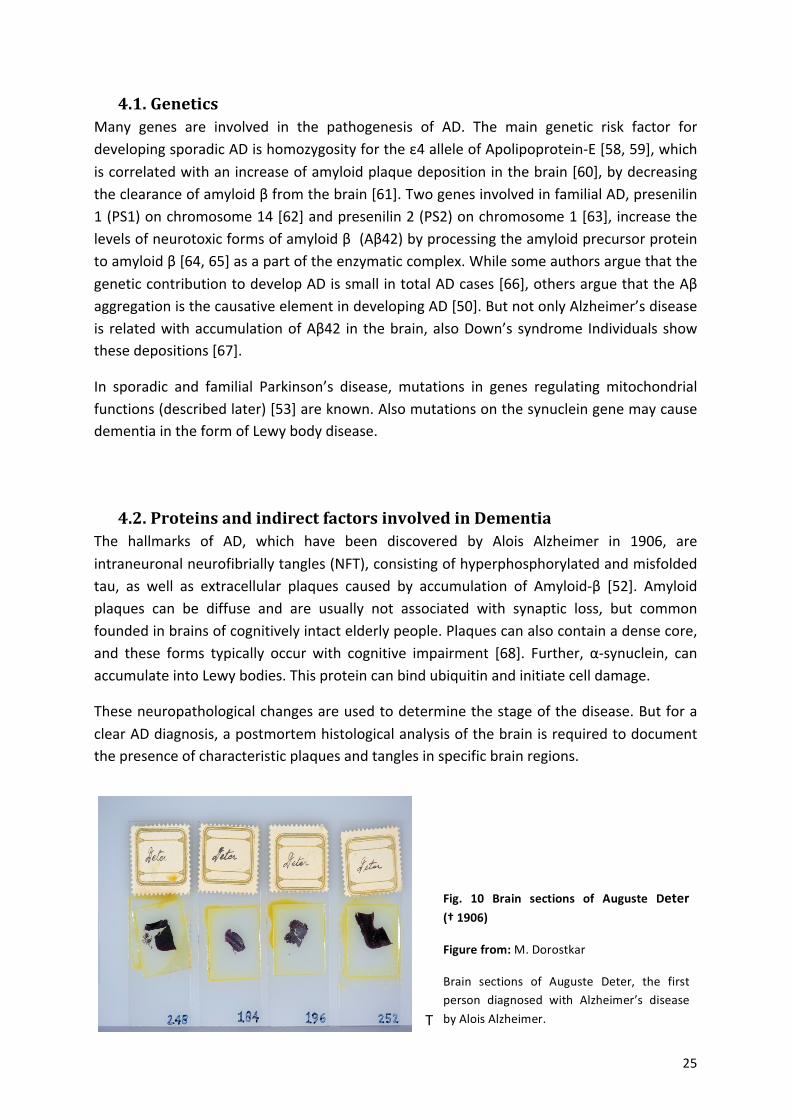
Fig.% 10% Brain% sections% of% Auguste% Deter%%%%%%%(†%1906)%
Figure%from:%M.!Dorostkar%
Brain! sections! of! Auguste! Deter,! the! first!person! diagnosed! with! Alzheimer’s! disease!by!Alois!Alzheimer.!
! 26!
pathogenesis!for!AD,!where!the!accumulation!of!neurotoxic!Aβ!in!the!cerebral!cortex!occurs!initially! [69].! The! amyloid! precursor! protein! (APP)! is! produced! by! a! single! copy! gene! on!chromosome! 21! and! cut! by! βa! and! γasecretase,! resulting! in! variable! length! Aβ! cleavage!products,! with! the! major! products! being! Aβ40! and! Aβ42.! In! healthy! Individuals! the!proportion!of!Aβ40!is!about!90!%![70].!But!in!AD!cases!the!ratio!of!Aβ40!and!Aβ42!shows!a!trend!to!increase!Aβ42!by!a!factor!of!1,5!a!1,9![69].!βaAPP!is!cut!first!by!the!βasecretase!to!a!~12!kDa!Caterminal! fragment!that! is! further!cleaved!on!the!carboxyl!side!by!γasecretase!to!release!Aβ.!An! isomer!of! ~9! kDa! is! formed!by!αasecretase,! that! can!be! cut! by! γasecretase!afterwards!to!build!p3,!a!small!Caterminal!fragment!of!Aβ![70].!!!
!!
!
!
!
!
!
!
Aβ!monomers!are!soluble!with!a!short!βasheet!region!and!a!large!αasheet!portion.!When!the!concentration!rises,!Aβ!undergoes!a!conformational!change!with!at! the!end!a!βasheet! rich!part,!which!aggregates!into!fibrils.!These!fibrils!diffuse!into!the!extracellular!space!and!form!AP.!There!can!be!diffuse!plaques!without!fibrils,!cored!plaques!with!fibrillary!cores!or!coreaonly!plaques!with!intracellular!compartments,!like!cell!bodies,!axons!and!dendrites![50].!!
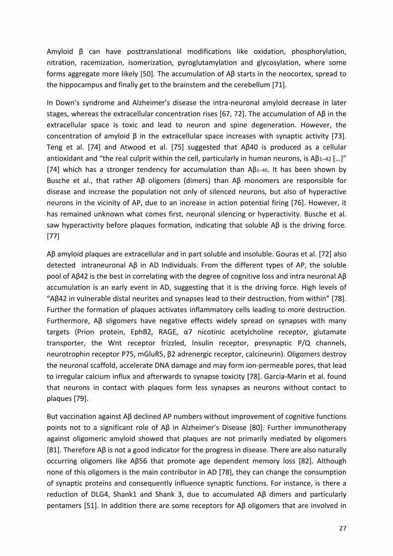
The!distribution!progress!of!βaAmyloid! is!one!of! the!diagnostic!criteria! for! the!diagnosis!of!AD!and!calling!“Thal!phasing!scale”.!Thal!described!a!stereotypic!pattern!of!five!phases,!with!no/minimal!pathology!“0”!beginning!in!the!neocortex,!up!to!high!pathology!“5”!ending!in!the!cerebellum![71].!!
!
!
!!
!
!
!
! ! ! ! ! !
!
!
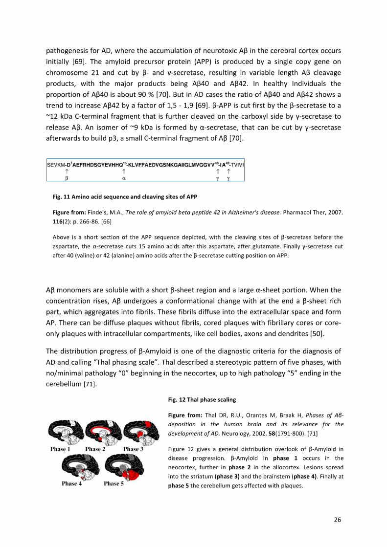
Fig.%11%Amino%acid%sequence%and%cleaving%sites%of%APP%%%%
Figure%from:%Findeis,!M.A.,!The$role$of$amyloid$beta$peptide$42$in$Alzheimer's$disease.!Pharmacol!Ther,!2007.!116(2):!p.!266a86.![66]%
Above! is! a! short! section! of! the! APP! sequence! depicted,! with! the! cleaving! sites! of! βasecretase! before! the!aspartate,! the!αasecretase! cuts!15!amino!acids!after!this! aspartate,! after!glutamate.! Finally! γasecretase!cut!after!40!(valine)!or!42!(alanine)!amino!acids!after!the!βasecretase!cutting!position!on!APP.!
Fig.%12%Thal%phase%scaling%
Figure% from:% Thal! DR,! R.U.,! Orantes! M,! Braak! H,! Phases$ of$ AβMdeposition$ in$ the$ human$ brain$ and$ its$ relevance$ for$ the$development$of$AD.!Neurology,!2002.!58(1791a800).![71]!%
Figure! 12! gives! a! general! distribution! overlook! of! βaAmyloid! in!disease! progression.! βaAmyloid! in% phase% 1! occurs! in! the!neocortex,! further! in! phase% 2% in! the! allocortex.! Lesions! spread!into!the!striatum!(phase%3)%and!the!brainstem!(phase%4).!Finally!at!phase%5!the!cerebellum!gets!affected!with!plaques.!
! 27!
Amyloid! β! can! have! posttranslational! modifications! like! oxidation,! phosphorylation,!nitration,! racemization,! isomerization,! pyroglutamylation! and! glycosylation,! where! some!forms!aggregate!more!likely![50].!The!accumulation!of!Aβ!starts!in!the!neocortex,!spread!to!the!hippocampus!and!finally!get!to!the!brainstem!and!the!cerebellum![71].!
In!Down’s! syndrome!and!Alzheimer’s! disease! the! intraaneuronal! amyloid!decrease! in! later!stages,!whereas!the!extracellular!concentration!rises![67,!72].!The!accumulation!of!Aβ!in!the!extracellular! space! is! toxic! and! lead! to! neuron! and! spine! degeneration.! However,! the!concentration!of! amyloid!β! in! the! extracellular! space! increases!with! synaptic! activity! [73].!Teng! et! al.! [74]! and! Atwood! et! al.! [75]! suggested! that! Aβ40! is! produced! as! a! cellular!antioxidant!and!“the!real!culprit!within!the!cell,!particularly!in!human!neurons,!is!Aβ1–42![…]”![74]! which! has! a! stronger! tendency! for! accumulation! than! Aβ1–40.! It! has! been! shown! by!Busche! et! al.,! that! rather! Aβ! oligomers! (dimers)! than! Aβ! monomers! are! responsible! for!disease! and! increase! the!population!not! only! of! silenced!neurons,! but! also!of! hyperactive!neurons! in!the!vicinity!of!AP,!due!to!an! increase! in!action!potential! firing![76].!However,! it!has!remained!unknown!what!comes!first,!neuronal!silencing!or!hyperactivity.!Busche!et!al.!saw!hyperactivity!before!plaques! formation,! indicating! that!soluble!Aβ! is! the!driving! force.![77]!!
Aβ!amyloid!plaques!are!extracellular!and!in!part!soluble!and!insoluble.!Gouras!et!al.![72]!also!detected! ! intraneuronal! Aβ! in! AD! Individuals.! From! the! different! types! of! AP,! the! soluble!pool!of!Aβ42!is!the!best!in!correlating!with!the!degree!of!cognitive!loss!and!intra!neuronal!Aβ!accumulation! is! an!early!event! in!AD,! suggesting! that! it! is! the!driving! force.!High! levels!of!“Aβ42!in!vulnerable!distal!neurites!and!synapses!lead!to!their!destruction,!from!within”![78].!Further!the!formation!of!plaques!activates! inflammatory!cells! leading!to!more!destruction.!Furthermore,! Aβ! sligomers! have! negative! effects! widely! spread! on! synapses! with! many!targets! (Prion! protein,! EphB2,! RAGE,! α7! nicotinic! acetylcholine! receptor,! glutamate!transporter,! the! Wnt! receptor! frizzled,! Insulin! receptor,! presynaptic! P/Q! channels,!neurotrophin!receptor!P75,!mGluR5,!β2!adrenergic!receptor,!calcineurin).!Oligomers!destroy!the!neuronal!scaffold,!accelerate!DNA!damage!and!may!form!ionapermeable!pores,!that!lead!to!irregular!calcium!influx!and!afterwards!to!synapse!toxicity![78].!GarciaaMarin!et!al.!found!that! neurons! in! contact! with! plaques! form! less! synapses! as! neurons! without! contact! to!plaques![79].!
But!vaccination!against!Aβ!declined!AP!numbers!without!improvement!of!cognitive!functions!points! not! to! a! significant! role! of! Aβ! in! Alzheimer’s! Disease! [80].! Further! immunotherapy!against! oligomeric! amyloid! showed! that! plaques! are! not! primarily!mediated! by! oligomers![81].!Therefore!Aβ!is!not!a!good!indicator!for!the!progress!in!disease.!There!are!also!naturally!occurring! oligomers! like! Aβ56! that! promote! age! dependent! memory! loss! [82].! Although!none!of!this!oligomers!is!the!main!contributor!in!AD![78],!they!can!change!the!consumption!of!synaptic!proteins!and!consequently! influence!synaptic!functions.!For! instance,! is!there!a!reduction! of! DLG4,! Shank1! and! Shank! 3,! due! to! accumulated! Aβ! dimers! and! particularly!pentamers![51].!In!addition!there!are!some!receptors!for!Aβ!oligomers!that!are!involved!in!
! 28!
signaling!cascades,!as!mGluR5![83]!and!ephrin![84].!Aβ!oligomers!bind!on!NMDA!receptors!and!are!responsible!for!abnormal!calcium!influx!that!induces!oxidative!stress![85].!White!et!al.!showed!that!cells!without!APP!were!resistant!to!the!neurotoxic!effects!of!Aβ![86].!
Importantly,!it!has!been!shown!before,!that!the!activity!of!Aβ!oligomers!is!directly!linked!to!tau!hyper!phosphorylation![87],!which!is!the!second!major!hallmark!of!Alzheimer’s!disease.!
!
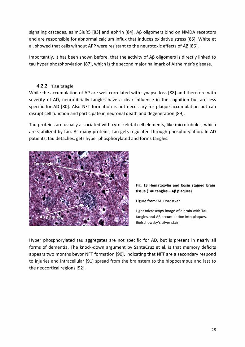
4.2.2 Tau-tangle-While!the!accumulation!of!AP!are!well!correlated!with!synapse!loss![88]!and!therefore!with!severity! of! AD,! neurofibrially! tangles! have! a! clear! influence! in! the! cognition! but! are! less!specific! for!AD! [80].!Also!NFT! formation! is!not!necessary! for!plaque!accumulation!but! can!disrupt!cell!function!and!participate!in!neuronal!death!and!degeneration![89].!!
Tau!proteins!are!usually!associated!with!cytoskeletal!cell!elements,!like!microtubules,!which!are!stabilized!by!tau.!As!many!proteins,!tau!gets!regulated!through!phosphorylation.! In!AD!patients,!tau!detaches,!gets!hyper!phosphorylated!and!forms!tangles.!!
!
-Hyper! phosphorylated! tau! aggregates! are! not! specific! for! AD,! but! is! present! in! nearly! all!forms!of!dementia.! The!knockadown!argument!by!SantaCruz!et! al.! is! that!memory!deficits!appears!two!months!bevor!NFT!formation![90],!indicating!that!NFT!are!a!secondary!respond!to!injuries!and!intracellular![91]!spread!from!the!brainstem!to!the!hippocampus!and!last!to!the!neocortical!regions![92].!
!
!
!
!
Fig.% 13% Hematoxylin% and% Eosin% stained% brain%tissue%(Tau%tangles%–%Aβ%plaques)%
Figure%from:%M.!Dorostkar%
Light!microscopy!image!of!a!brain!with!Tau!tangles!and!Aβ!accumulation!into!plaques.!Bielschowsky’s!silver!stain.!
Aβ%plaques%
Tau%tangles%
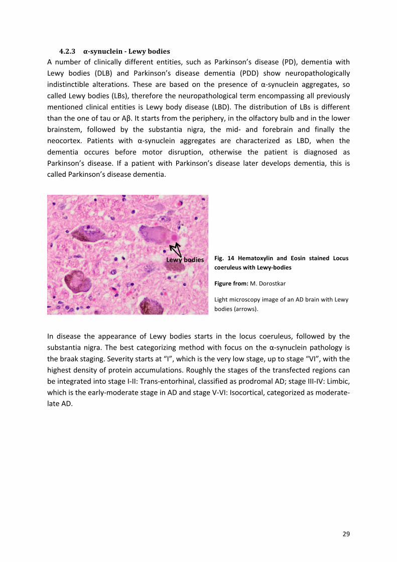
! 29!
4.2.3 α0synuclein-0-Lewy-bodies-A! number! of! clinically! different! entities,! such! as! Parkinson’s! disease! (PD),! dementia! with!Lewy! bodies! (DLB)! and! Parkinson’s! disease! dementia! (PDD)! show! neuropathologically!indistinctible! alterations.! These! are! based! on! the! presence! of! αasynuclein! aggregates,! so!called!Lewy!bodies!(LBs),!therefore!the!neuropathological!term!encompassing!all!previously!mentioned! clinical! entities! is! Lewy! body! disease! (LBD).! The! distribution! of! LBs! is! different!than!the!one!of!tau!or!Aβ.!It!starts!from!the!periphery,!in!the!olfactory!bulb!and!in!the!lower!brainstem,! followed! by! the! substantia! nigra,! the! mida! and! forebrain! and! finally! the!neocortex.! Patients! with! αasynuclein! aggregates! are! characterized! as! LBD,! when! the!dementia! occures! before! motor! disruption,! otherwise! the! patient! is! diagnosed! as!Parkinson’s! disease.! If! a! patient! with! Parkinson’s! disease! later! develops! dementia,! this! is!called!Parkinson’s!disease!dementia.!!
!!
In! disease! the! appearance! of! Lewy! bodies! starts! in! the! locus! coeruleus,! followed! by! the!substantia!nigra.! The!best! categorizing!method!with! focus!on! the!αasynuclein!pathology! is!the!braak!staging.!Severity!starts!at!“I”,!which!is!the!very!low!stage,!up!to!stage!“VI”,!with!the!highest!density!of!protein!accumulations.!Roughly!the!stages!of!the!transfected!regions!can!be!integrated!into!stage!IaII:!Transaentorhinal,!classified!as!prodromal!AD;!stage!IIIaIV:!Limbic,!which!is!the!earlyamoderate!stage!in!AD!and!stage!VaVI:!Isocortical,!categorized!as!moderatealate!AD.!
!
!
!
!
!
!
Fig.% 14% Hematoxylin% and% Eosin% stained% Locus%coeruleus%with%LewyZbodies%
Figure%from:%M.!Dorostkar!
Light!microscopy!image!of!an!AD!brain!with!Lewy!bodies!(arrows).!
Lewy%bodies%
! 30!
!
!
!
!!
!
!
!
!
!
!
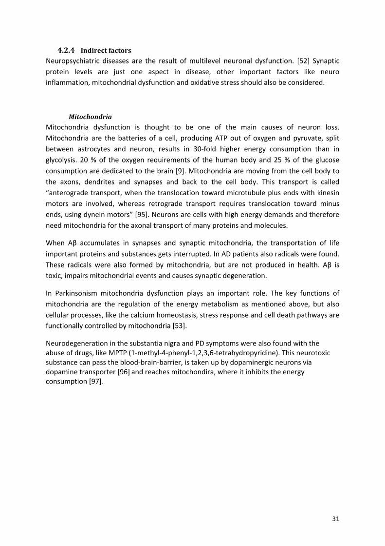
Down!below!is!an!overview!of!the!different!distributions!pathways!of!neuritic!plaques,!neurofibrillary!tangles!and!Lewy!bodies.!
!
!
!! !! !
!
!
! !
Fig.%16%Distribution%of%deposits%in%different%neurodegenerative%diseases%over%time%
Figure% from:% M.! Jucker,! L.C.W.,! SelfMpropagation$ of$ pathogenic$ protein$ aggregates$ in$ neurodegenerative$diseases.!Nature,!2013.!501:!p.!45a51.![94]!!On!the!left!different!protein!accumulations!were!depicted,!while!on!the!right!different!stages!of!diseases!were!shown.! In!a)! senile!plaques! (Amyloidaβ)!were! imaged.!Further!shows!e)! the!distribution!of!Aβ!over!the!time,!starting!neocortical,!spreading!over!the!Allo!cortex!and!ending!up! in!the!subcortical!regions.!b)!Depicts!a!tau!inclusion! as! a! neurofibrillary! tangle.! f)! Represents! the! accumulation! of! NFT! from! the! locus! coeruleus! and!transenthorhinal!area,!distribute!further!to!the!amygdala!and!interconnected!neocortical!brain!regions.!c)!Is!an!example!of!an!αasynuclein!inclusion!(Lewy!body)!which!is!typical!for!Parkinson!disease/!Lewy!body!dementia.!g)!The!accumulation!of!αasynuclein!starts!in!the!peripheral!and!autonomic!nervous!system,!followed!by!olfactory!bulb! and! lower! brainstem.! LB! spread! through! the! substantia! nigra,! over! areas! of! the! midbrain! and! basal!forebrain,!and!finally!to!the!neocortex.!
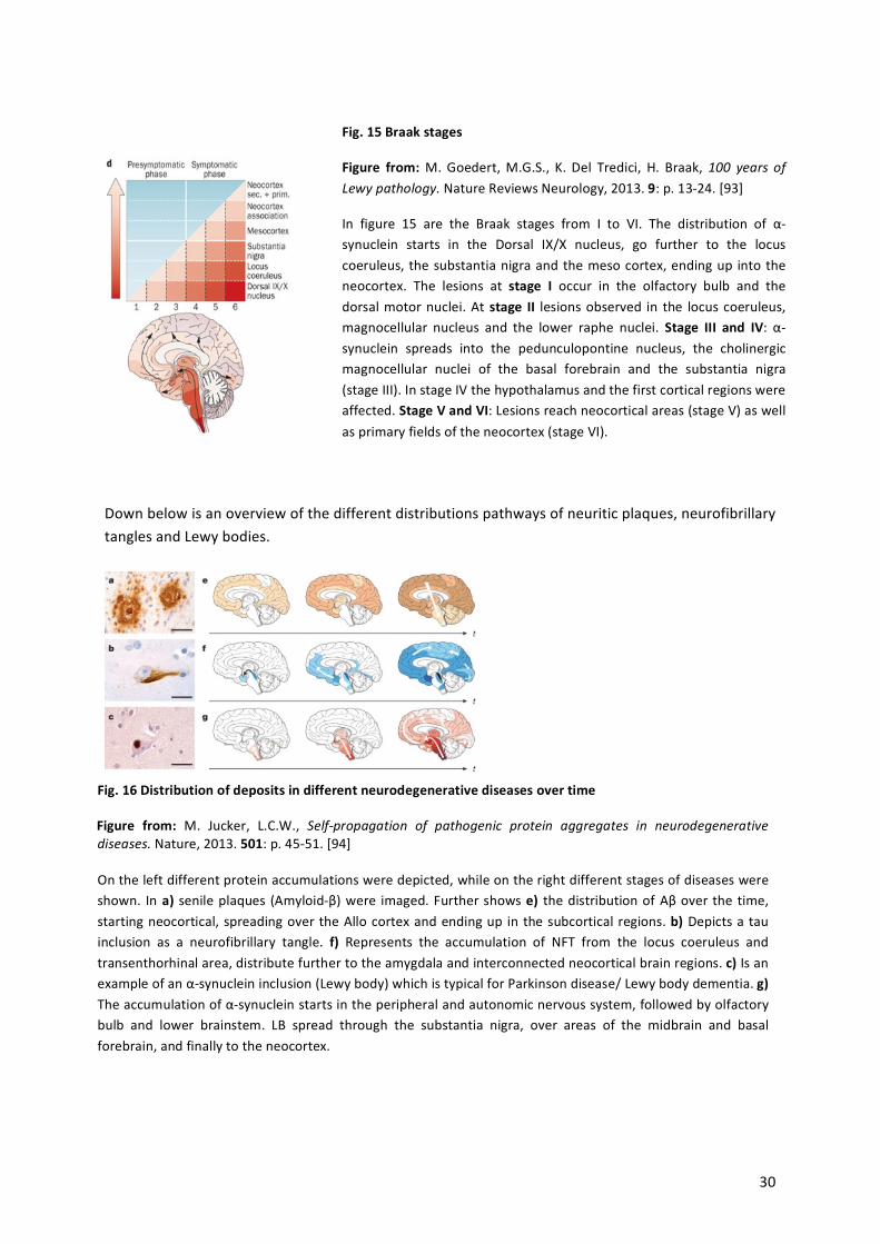
Fig.%15%Braak%stages%
Figure% from:%M.!Goedert,!M.G.S.,! K.! Del! Tredici,! H.! Braak,!100$ years$ of$Lewy$pathology.!Nature!Reviews!Neurology,!2013.!9:!p.!13a24.![93]%
In! figure! 15! are! the! Braak! stages! from! I! to! VI.! The! distribution! of! αasynuclein! starts! in! the! Dorsal! IX/X! nucleus,! go! further! to! the! locus!coeruleus,!the!substantia!nigra!and!the!meso!cortex,!ending!up! into!the!neocortex.! The! lesions! at! stage% I! occur! in! the! olfactory! bulb! and! the!dorsal!motor!nuclei.!At!stage% II! lesions!observed! in! the! locus! coeruleus,!magnocellular! nucleus! and! the! lower! raphe! nuclei.! Stage% III% and% IV:! αasynuclein! spreads! into! the! pedunculopontine! nucleus,! the! cholinergic!magnocellular! nuclei! of! the! basal! forebrain! and! the! substantia! nigra!(stage!III).!In!stage!IV!the!hypothalamus!and!the!first!cortical!regions!were!affected.!Stage%V%and%VI:!Lesions!reach!neocortical!areas!(stage!V)!as!well!as!primary!fields!of!the!neocortex!(stage!VI).!
! 31!
4.2.4 Indirect-factors-Neuropsychiatric! diseases! are! the! result! of!multilevel! neuronal! dysfunction.! [52]! Synaptic!protein! levels! are! just! one! aspect! in! disease,! other! important! factors! like! neuro!inflammation,!mitochondrial!dysfunction!and!oxidative!stress!should!also!be!considered.!
!
Mitochondria+Mitochondria! dysfunction! is! thought! to! be! one! of! the! main! causes! of! neuron! loss.!Mitochondria! are! the! batteries! of! a! cell,! producing!ATP! out! of! oxygen! and! pyruvate,! split!between! astrocytes! and! neuron,! results! in! 30afold! higher! energy! consumption! than! in!glycolysis.! 20!%! of! the! oxygen! requirements! of! the! human! body! and! 25!%! of! the! glucose!consumption!are!dedicated!to!the!brain![9].!Mitochondria!are!moving!from!the!cell!body!to!the! axons,! dendrites! and! synapses! and! back! to! the! cell! body.! This! transport! is! called!“anterograde!transport,!when!the!translocation!toward!microtubule!plus!ends!with!kinesin!motors! are! involved,! whereas! retrograde! transport! requires! translocation! toward! minus!ends,!using!dynein!motors”![95].!Neurons!are!cells!with!high!energy!demands!and!therefore!need!mitochondria!for!the!axonal!transport!of!many!proteins!and!molecules.!
When! Aβ! accumulates! in! synapses! and! synaptic! mitochondria,! the! transportation! of! life!important!proteins!and!substances!gets!interrupted.!In!AD!patients!also!radicals!were!found.!These! radicals! were! also! formed! by! mitochondria,! but! are! not! produced! in! health.! Aβ! is!toxic,!impairs!mitochondrial!events!and!causes!synaptic!degeneration.!
In! Parkinsonism! mitochondria! dysfunction! plays! an! important! role.! The! key! functions! of!mitochondria! are! the! regulation! of! the! energy!metabolism! as!mentioned! above,! but! also!cellular!processes,!like!the!calcium!homeostasis,!stress!response!and!cell!death!pathways!are!functionally!controlled!by!mitochondria![53].%
Neurodegeneration!in!the!substantia!nigra!and!PD!symptoms!were!also!found!with!the!abuse!of!drugs,!like!MPTP!(1amethyla4aphenyla1,2,3,6atetrahydropyridine).!This!neurotoxic!substance!can!pass!the!bloodabrainabarrier,!is!taken!up!by!dopaminergic!neurons!via!dopamine!transporter![96]!and!reaches!mitochondira,!where!it!inhibits!the!energy!consumption![97].!!!
!
!
!
!
! 32!
Environment+Further! factors! are! environment! influences! like! head! injuries! or! malnutrition,! leading! to!increased! oligomerization! of! intraaneuronal! Aβ42! in! distal! neurites! and! synapses,!contributing!to!their!destruction.!
There!are!findings!of!NFT!and!plaques!in!Individuals!without!dementia,!with!similar!densities!to! cases!with! dementia! [98].! As!well! as! frontal! cortex! cell! loss! not! only! in! AD! but! also! in!normal! aging! indicates! that! a! certain! level! of! neuropathology! is! a! characteristic! of! normal!aging.! However! aging! is! one! of! the! most! important! risk! factors! in! AD.! [99]! Scheff! et! al.!showed!that! the! loss!of! synapses! is!not!evenly!distributed! in!all!brain!areas,!meaning! that!age! dependent! changes! appear! not! overall,! but! in! distinct! areas,! like! the! cortex! and! the!hippocampus.! He! also! pointed! that! the! loss! of! synapses! in! Alzheimer’s! disease! is! a!consequence!of!the!disease!and!not!of!normal!aging.![100]!And!also!differences!from!human!to!human!hinder!comparisons!and!development!of!therapies!to!prevent!AD.!
Further!may!be!possible!that!education!plays!a!role!in!neurodegenerative!disease![89].!
!
!
!
All!this!phenomena!mentioned!above!run!in!parallel!and!underlie!a!yet!not!known!primary!source.!It!is!a!complex!interaction!of!proteins!in!Alzheimer’s!disease,!which!make!it!difficult!to!differ!between!disease!specific!and!nonaspecific.!A!number!of!possible!mechanisms!could!be! responsible! for! synapse!dysfunction.!Many!molecules,! like! hormones! [101],! cadherines![102],! ephrins! [103],! neurotrophins! [104],! actinarelated! [105],! PSD! [106],! cocaines! and!amphetamines![107],!are!involved!in!spine!dynamics.!The!combination!of!them!makes!it!very!hard!to!identify!one!unique!pathway!to!explain!all!problems.!
AD!has!become!a!serious!public!health!care!problem!that!will!become!increasing!concern!in!the! future.! It! is! important! to! understand! molecular! and! cellular! mechanism! of! synaptic!degeneration!to!develop!new!biomarker!and!therapy.!
!
!
!
!
!
!
! 33!
4.3 -Previous-findings-!
Scheff!and!collegues!measured!prea!(synapsisna1!and!synaptophysin)!and!postsynaptic!(PSDa95/DLG4,!drebrin!and!SAPa97)!protein!levels!and!showed!a!decline!in!synaptic!numbers!with!AD!progression.![51,!52]!But!in!another!study!in!2001,!they!claimed!that!the!synapse!loss!is!not! distributed! in! all! brain! areas! and! might! be! due! to! the! disease! process! and! not! a!consequence!of!normal!aging![51,!52].!
The!mRNA!level!of!excitatory!and!inhibitory!markers!of!hippocampal!neurons!were!analyzed!by! Loreth! et! al.! and! indicated! an! unchanged! level! of! GABRA1! in! TauPS2APP! mice,! but! a!significantly! reduced! level! of! Gad67,! which! is! glutamate! decarboxylase! and! necessary! for!GABA! production,! and! Y1R,! which! is! a! GaProtein! coupled! neuropeptide! receptor.! For! the!excitatory!synapses,!NMDAR1,!mGluR1!and!Ryr3!were!measured,!but!showed!no!alterations![108].!
In! Alzheimer’s! disease! GABAergic! neurons! are! very! well! preserved! in! comparison! to! the!other! neurotransmitter! systems! [109].! But! in! other! publications! the! focus! is! on! the!GABAergic! neurons,! because! the! inhibitory! neurons! are! supposed! to! be! the! targets! of!neurodegeneration![108].!
In!latest!papers!the!focus!of!synapse!loss!lie!on!the!inhibitory!synapses.!
Garcia! Marin! wrote! in! 2009:! “Membrane! surfaces! of! neurons! in! contact! with! AP! lack!GABAergic!perisomatic!synapses”.!The!author!suggests!that!the!GABAergic!loss!is!due!to!the!specific!selectivity!of!AP.!Further!they!described!that!“plaques! in!the!neuropil!will!produce!different! alterations! to! synaptic! circuits! than! those! associated! with! neuronal! somata“,!because! of! the! fact! that! pyramidal! cells! with! synapses! in! the! perisomatic! are! in! general!GABAergic,!whereas! other! dendrites! in! the! neuropil! build! synapses!with! all! kind! of! axons!(glutamatergic,!GABAergic,!noradrenergic,!dopaminergic,!serotonergic!and!cholinergic).!They!also!claimed,!that!not!the!cell!body,!but!dendrites!are!more!susceptible!to!the!toxic!effect!of!Aβ.![79]!
Busche! et! al.! used! two! photon! Ca2+! imaging! in! the! mouse! model! APP23! x! PS45,!overexpressing!Aβ!precursor!protein!and!presenilin1,! and! found! that! the!neuronal! activity!decreased,!as!well!as! synaptic! inhibition,!whereas! the!spontaneous!Ca2+! transients,!due! to!action! potential! firing! in! neurons,! increase! in! the! proximity! of! AP.! The! question! whether!there! is! an! increased! excitability! of! these! hyperactive! neurons! near! to! plaques,! was!answered! through! a! glutamate! receptor! inhibitor! and! the! outcome! was! no! increase! in!excitability!of!neurons,!but!maybe!an!impairment!of!inhibition.!This!thesis!was!supported!by!diazepam!treatment,!a!GABAAR!channel!activator,!which!reduced!the!activity!of!hyperactive!neurons! and! a! GABAAR! antagonist,! increasing! the! Ca2+! transients,! indicating! an! impaired!synaptic! inhibition.! The! study! showed! a! decrease! in! neuronal! activity! in! 29!%,!what! they!named! “silent”! neurons! and! an! increase! of! 20!%! in! “hyperactive”! neurons,!mostly! in! the!vicinity!of!AP.![65]!
! 34!
In! a! study! of! mouse! models! of! AD! amyloidosis,! Busche! and! colleagues! found! that! the!“impairment!can!be!rescued!by!enhancing!GABAAergic!inhibition”.!Further!GABAAR!inhibition!is! important! for! slow! Waves! development,! necessary! to! support! synaptic! plasticity!underlying!cortex!and!hippocampus!communication!and!memory!consolidation!during!sleep.!Busche!et!al.!wrote!in!2015,!that!Aβ!applied!into!the!cortex,!results!in!an!impaired!inhibition!of! GABAAR.! The! balance! of! excitatory! and! inhibitory! can! be! rescued! through! GABAAR!function!repair![77],!means!that!GABAAR!is!the!main!mediator!in!AD.!!!!!!!!!!!!!!!!!!!!
In!another!study,!Busche!and!colleagues!claimed!that!there!is!probably!a!change!in!function,!not!in!excitation!of!neurons,!in!the!GABAAR!in!Alzheimer’s!disease.!So!the!question!is!if!there!is! a! change! in! the! subunits! of! the! GABAergic! receptors! in! disease,! which! support! the!impaired!inhibition!in!synapses.![77]!
GarciaaMarin! and! colleagues! claimed,! that! in! the! vicinity! of! plaques! neurons! lack! of!GABAergic!synapses!and!further!lead!to!increased!hyperactivity.!They!suggested,!that!Aβ!is!toxic!to!the!synapses!on!dendrites!and!not!on!the!cell!body!itself,!with!sprouting!effects!to!function! impairment! [79].! But! they! only! labeled! for! GAT1! and! VGAT,! which! are! both!representatives!for!the!pre!synapse.!!
!
These! results! indicate! not! a! loss! of! inhibitory! neurons,! but! a! loss! of! the! GABA!neurotransmission!itself,!which!fits!in!our!hypothesis,!that!there!is!a!change!in!the!GABAergic!postsynaptic! receptors,!due! to! the!changed!GABA! level!and!changed!synaptic! input!of! the!pre!synapse.!To!put!Busche’s!and!GarciaaMarin’s!findings!together,!it!could!be!possible,!that!in!disease!the!pre!synapse!vanishes,!maybe!through!the!presence!of!Aβ,!and!the!GABAAR!on!the!post!synapse!therefore!change!its!subunits!to!react!to!the!loss!of!synaptic!connections.!
!
!
!
!
!
!
!
!
!
!
!
! 35!
4.4 -Stage-of-disease-severity-!
!There! are! different!methods! to! differentiate! between! the! disease! severities.! However,! a!certain!diagnosis!of!the!type!of!the!disease!and!the!actual!stage!can!only!be!made!through!an! autopsy.! Yet,! a! proper! clinical! diagnosis! is! important! to! enroll! patients! in! studies.!Diagnose! methods! were! invented,! for! example! diagnoseainterviews! (CAMDEX,! SIDAM),!general! dementia! scales! (FAST,! BRAAK),! neuropsychological! instruments! (CERAD,! AKT),!psychopathological!and!behavior!scales!(BRSD,!BEHAVEaAD)!or!function!scales!(DAD,!Barthel!index).!Some!of!them!were!used!usually!together!to!get!a!better!output!of!the!condition!of!a!patient.!But!all!of!them!test!the!cognitive!performance!(verbal!and!figure!memory,!frontala!executive!function,!attention,!speech,!visuperceptive!performance!and!areal!notion).!
!
CERAD-(Consortium-to-establish-a-registry-for-Alzheimer’s-Disease)-This! test! aims! to! differentiate! between! the! neuropsychological! features! of! Alzheimer’s!disease.!It!is!based!on!the!complete!clinical!evaluation,!the!neuropsychological!evaluation!of!the!manifestation! and! the! standardization! of! the! neuropathological! evaluation! of! AD! and!related!diseases! [110,!111].!The!composition!of! the!CERAD! test! is! verbal! fluency! (Isaacs!&!Kennie!1973),!modificated!Boston!naming!test!(Kaplan,!Goodglass!&!Weintraum,!1983),!mini!mental! state! examination! (Folstein,! Folstein,! McHugh,! 1975),! word! memory! (Atkinson! &!Shiffrin,! 1971;! Rosen,! Mohs! &! Davis! 1984),! constructive! praxis! (Rosen! et! al.,! 1984),!recognition! of!words! (Mohs! et! al.,! 1986),! constructive! praxis! recall,! trail!marking! test! and!phonetic! fluency.! The! test! results!were! evaluated! agea,! educationa! and! genderadependent!and!are!an!important!instrument!for!early!diagnostics.!The!lowest!stage!of!AD!is!marked!with!“A”,!further!severity!with!“B”!and!high!AD!with!“C”.!
!
!
5. Objective-!
Since! loss!of!cognitive! function!has!been!shown!to!result! from! loss!of! functional!synapses,!we!quantified!markers!for!excitatory!and!inhibitory!synapses.!We!aimed!to!determine!how!synapse! loss! in! specific!brain! regions! correlates!with!neuropathological!disease! stages!and!clinical!presentation.!
!
!
! 36!
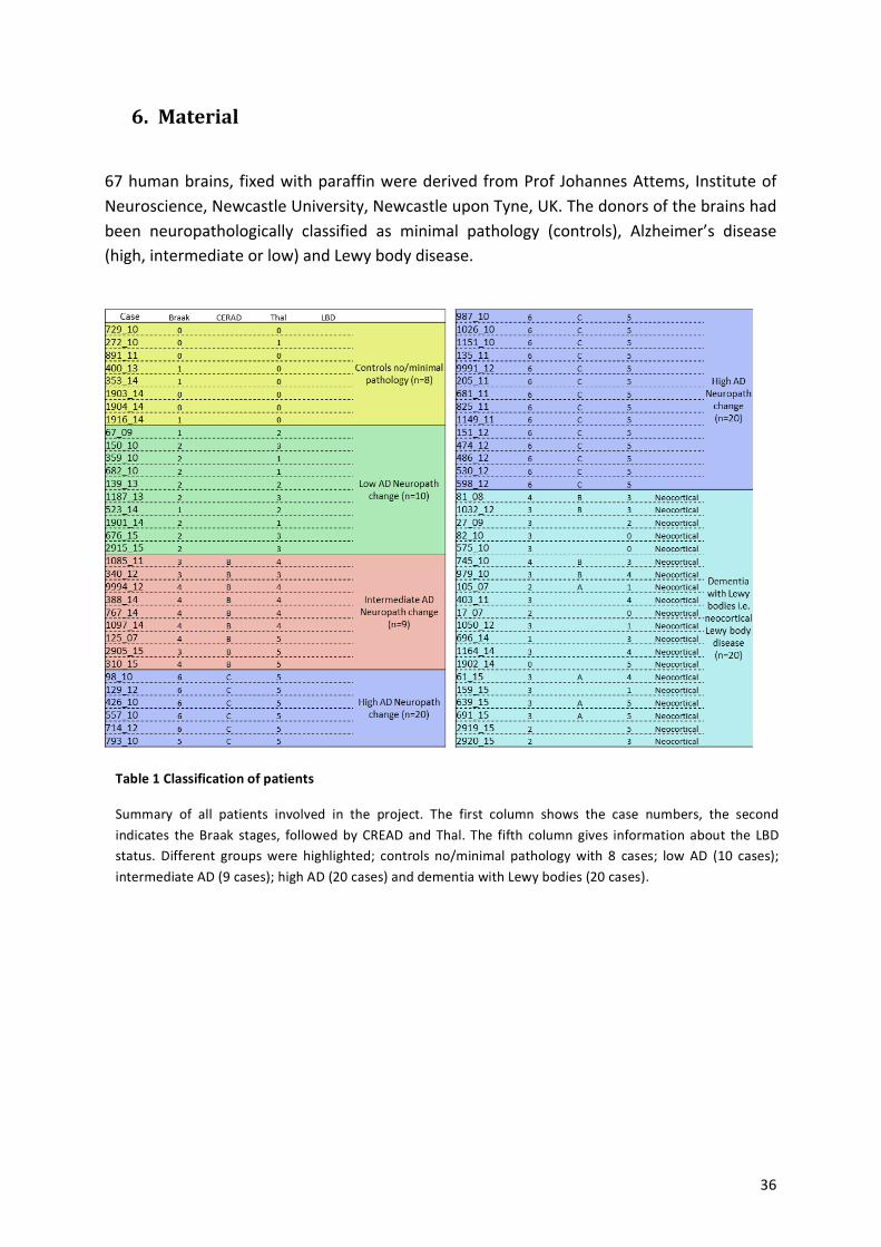
6. Material-!
67!human!brains,! fixed!with!paraffin!were!derived!from!Prof!Johannes!Attems,! Institute!of!Neuroscience,!Newcastle!University,!Newcastle!upon!Tyne,!UK.!The!donors!of!the!brains!had!been! neuropathologically! classified! as! minimal! pathology! (controls),! Alzheimer’s! disease!(high,!intermediate!or!low)!and!Lewy!body!disease.!!
!
!
!
!
!
!
!
!
!
!
!
!
Table%1%Classification%of%patients%
Summary! of! all! patients! involved! in! the! project.! The! first! column! shows! the! case! numbers,! the! second!indicates! the!Braak! stages,! followed!by!CREAD!and!Thal.! The! fifth! column!gives! information! about! the! LBD!status.! Different! groups!were! highlighted;! controls! no/minimal! pathology!with! 8! cases;! low!AD! (10! cases);!intermediate!AD!(9!cases);!high!AD!(20!cases)!and!dementia!with!Lewy!bodies!(20!cases).!
!
! 37!
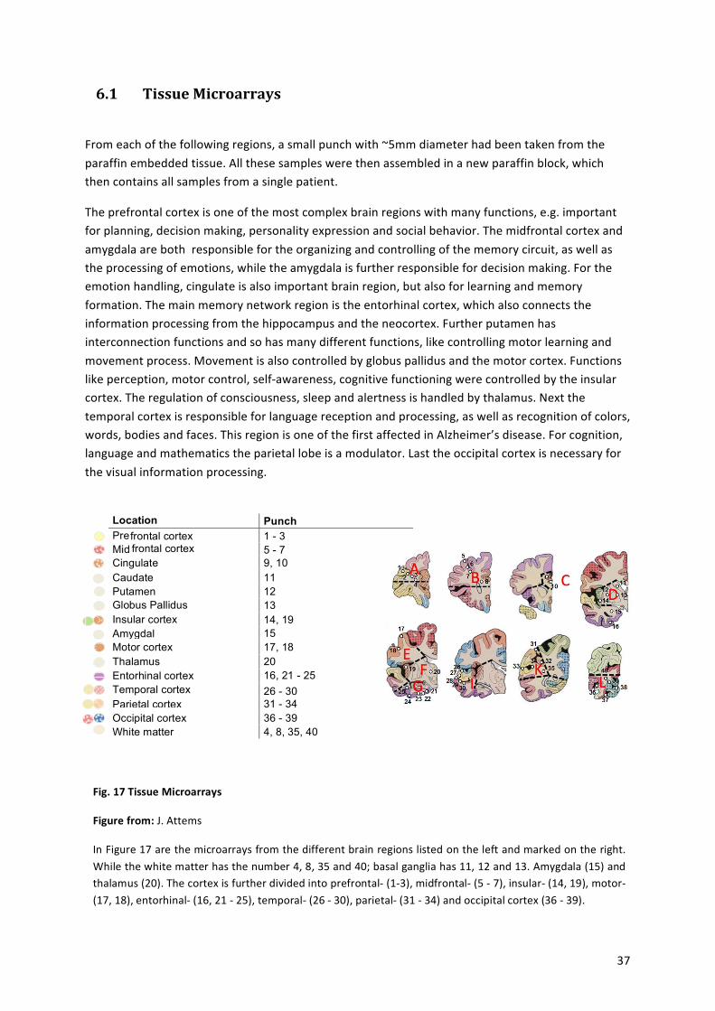
! ! ! ! ! ! Location Punch
numbers ! ! Pre frontal cortex 1 - 3 ! ! Mid frontal cortex 5 - 7 ! ! Cingulate 9, 10 ! ! Caudate 11 ! ! Putamen 12 ! ! Globus Pallidus 13 ! ! Insular cortex 14, 19 ! ! Amygdala 15 ! ! Motor cortex 17, 18 ! ! Thalamus 20 ! ! Entorhinal cortex 16, 21 - 25 ! ! Temporal cortex 26 - 30 ! ! Parietal cortex 31 - 34 ! ! Occipital cortex 36 - 39 ! ! White matter 4, 8, 35, 40 ! ! ! ! ! ! ! ! ! ! ! ! ! ! !
6.1 -Tissue-Microarrays--!
From!each!of!the!following!regions,!a!small!punch!with!~5mm!diameter!had!been!taken!from!the!paraffin!embedded!tissue.!All!these!samples!were!then!assembled!in!a!new!paraffin!block,!which!then!contains!all!samples!from!a!single!patient.!
The!prefrontal!cortex!is!one!of!the!most!complex!brain!regions!with!many!functions,!e.g.!important!for!planning,!decision!making,!personality!expression!and!social!behavior.!The!midfrontal!cortex!and!amygdala!are!both!!responsible!for!the!organizing!and!controlling!of!the!memory!circuit,!as!well!as!the!processing!of!emotions,!while!the!amygdala!is!further!responsible!for!decision!making.!For!the!emotion!handling,!cingulate!is!also!important!brain!region,!but!also!for!learning!and!memory!formation.!The!main!memory!network!region!is!the!entorhinal!cortex,!which!also!connects!the!information!processing!from!the!hippocampus!and!the!neocortex.!Further!putamen!has!interconnection!functions!and!so!has!many!different!functions,!like!controlling!motor!learning!and!movement!process.!Movement!is!also!controlled!by!globus!pallidus!and!the!motor!cortex.!Functions!like!perception,!motor!control,!selfaawareness,!cognitive!functioning!were!controlled!by!the!insular!cortex.!The!regulation!of!consciousness,!sleep!and!alertness!is!handled!by!thalamus.!Next!the!temporal!cortex!is!responsible!for!language!reception!and!processing,!as!well!as!recognition!of!colors,!words,!bodies!and!faces.!This!region!is!one!of!the!first!affected!in!Alzheimer’s!disease.!For!cognition,!language!and!mathematics!the!parietal!lobe!is!a!modulator.!Last!the!occipital!cortex!is!necessary!for!the!visual!information!processing.!
!
!
!
!
!
!
!
!
!
!
!
!
!
!
Fig.%17%Tissue%Microarrays%
Figure%from:%J.!Attems%
In!Figure!17!are!the!microarrays!from!the!different!brain!regions!listed!on!the!left!and!marked!on!the!right.!While!the!white!matter!has!the!number!4,!8,!35!and!40;!basal!ganglia!has!11,!12!and!13.!Amygdala!(15)!and!thalamus!(20).!The!cortex!is!further!divided!into!prefrontala!(1a3),!midfrontala!(5!a!7),!insulara!(14,!19),!motora!(17,!18),!entorhinala!(16,!21!a!25),!temporala!(26!a!30),!parietala!(31!a!34)!and!occipital!cortex!(36!a!39).!
!
! 38!
I!further!divided!the!optical!cortex!into!the!primary!visual!field!Brodmann’s!area!17!(Punch!38),!and!the!secondary!visual!field!Brodmann’s!area!18!(Punch!37,!39).!
!
Tissue! microarrays! from! all! patients! were! immunofluorescence! labelled! with! antibodies!against! DLG4! (also! known! as! PSDa95! scaffold! protein,! which! “is! a! hallmark! of! a! mature!synapse”! [18])! to! stain!excitatory!synapses!and!γaAminobutyric!acid! receptor!A!subunit!α2!(GABRA2)!to!stain!inhibitory!synapses.!
!
6.2 -Chemicals-�� Xylol!100!%!!�� EtOH!100!%,!96!%,!70!%!�� Citrate!Puffer!PH!6!�� dPBS!�� Normal!Goat!Serum!(NGS)!�� Triton!xa100!�� Sudan!Black!�� Sodium!Azide!�� Dako!(Fluorescence!mounting!medium)!
!
ATB-
Primary!�� Anti!PSDa95!(Mouse!monoclonal!ATB,!SYSY!124011)!�� Anti!GABAAaR!α2!(Guinea!Pig!polyclonal!ATB,!SYSY!224104)!
Secondary!�� AntiaMouse!A488!(Goat,!Molecular!probes;!A11029)!�� AntiaGuinea!Pig!A488!(Goat,!Molecular!probes;!A!11073)!
Nuclei!staining!�� DAPI!(Roth,!6335.2)!
!
Puffer-and-solutions-
�� Blocking!Buffer!(for!Blocking!and!primary!ATB!Solution)!
DPBS!+!10!%!NGS!+!1!%!Triton!xa100!(Stock!with!dPBS!and!Triton!xa100!with!0,05!%!Sodium!Azide)!
! 39!
�� Secondary!ATB!Solution!
DPBS!+!3!%!NGS!+!1!%!Triton!xa100!(Stock!with!dPBS!and!Triton!xa100!with!0,05!%!Sodium!Azide)!
�� Sudan!Black!Solution!
0,3!%!Sudan!Black!in!70!%!EtOH,!filtered!(prepared!freshly!every!week,!stored!in!fridge!at!4°C)!
!
Equipment!
Confocal!microscope! ! Images!were!taken!with!Zeiss!LSM!780!
Software! Processed! in! IgorPro7,! detecting! single! puncta,! based! on!published!algorithms![81,!112].!
!
!
7. Methods-!
Given!below!is!a!schematic!process!of!the!staining.!The!procedure!is!constructed!on!deparaffinization!steps,!antigen!retrieval,!blocking!and!antibody!and!Sudan!Black!incubations.!
!
! Day!1!
o Deparaffinization!o Antigen!retrieval!(5x!4!min)!o Blocking!(1!h)!o Primary!ATB!incubation!4°C,!Over!Night!(ON)!
Day!2! !
o Washing!with!dPBS!(3x!5!min)!o Secondary!ATB!incubation!(2!h)!o Washing!with!dPBS!(3x!5!min)!o Sudan!Black!incubation!(1,5!min)!o Washing!with!ddH2O!(3x!5!min)!o Drying!o Mounting!(ON)!
! 40!
Deparaffinization%
!First!paraffin!slices!were!brought!to!Xylol!and!EtOH!to!hydrophilize!the!tissue!and!get!rid!of!the!paraffin.!
�� 2!x!15!min!Xylol!100!%!�� 2!x!5!min!EtOH!100!%!�� 1!x!5!min!EtOH!96!%!�� 1!x!5!min!EtOH!70!%!
! For!removing!the!alcohol,!the!slides!were!washed!3!times!with!H2O.!
% %
% Antigen%retrieval%
Because!of!tissue!paraffinization!for!fixation,!crosslinking!appear!and!modify!the!tissue.!But!the!antibodies!are!not!specific!for!this!crosslinking,!so!it!has!to!be!removed.!Therefor!the!slides!were!cooked!5!x!4!min!in!the!microwave!oven!(700!Watt)!in!citrate!puffer!pH!6.! The! liquid! level! was! holding! constant! with! warm! dH2O! to! prevent! the! slices! from!drying!out.!After!that!the!sections!were!cooled!to!RT!for!20!min.!
!
% Blocking%
Next!the!slides!were!masked!to!avoid!unspecific!binding.!10!%!NGS! in!dPBS!with!10!%!Triton! xa100! were! used! as! blocking! agent! and! the! slices! were! kept! in! a! humidity!chamber!with!the!blocking!solution!for!1!h.!
!
% Primary%ATB%incubation%
Further! the! primary! ATB! were! diluted! in! blocking! solution! and! kept! in! the! humidity!chamber!at!4°C!ON.!
! Dilutions!
ATG! Host! Dilution!Anti!PSD!95! Mouse! 1!:!1000!Anti!GABAAaR!α2! Guinea!Pig! 1!:!200!
%
% Washing%
% The!slides!were!washed!with!dPBS!for!3!times,!each!5!min.!
!
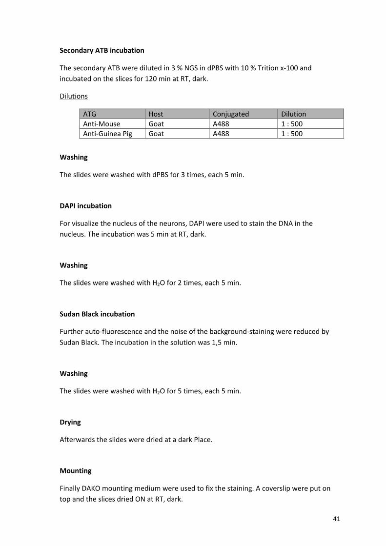
! 41!
% Secondary%ATB%incubation%
The!secondary!ATB!were!diluted!in!3!%!NGS!in!dPBS!with!10!%!Trition!xa100!and!incubated!on!the!slices!for!120!min!at!RT,!dark.!
Dilutions!
ATG! Host! Conjugated! Dilution!AntiaMouse! Goat! A488! 1!:!500!AntiaGuinea!Pig! Goat! A488! 1!:!500!
%
% Washing%
% The!slides!were!washed!with!dPBS!for!3!times,!each!5!min.!
%
% DAPI%incubation%
For!visualize!the!nucleus!of!the!neurons,!DAPI!were!used!to!stain!the!DNA!in!the!nucleus.!The!incubation!was!5!min!at!RT,!dark.!
!
% Washing%
% The!slides!were!washed!with!H2O!for!2!times,!each!5!min.!
%
% Sudan%Black%incubation%
Further!autoafluorescence!and!the!noise!of!the!backgroundastaining!were!reduced!by!Sudan!Black.!The!incubation!in!the!solution!was!1,5!min.!
%
% Washing%
% The!slides!were!washed!with!H2O!for!5!times,!each!5!min.!
%
% Drying%
% Afterwards!the!slides!were!dried!at!a!dark!Place.!
%
% Mounting%
Finally!DAKO!mounting!medium!were!used!to!fix!the!staining.!A!coverslip!were!put!on!top!and!the!slices!dried!ON!at!RT,!dark.!
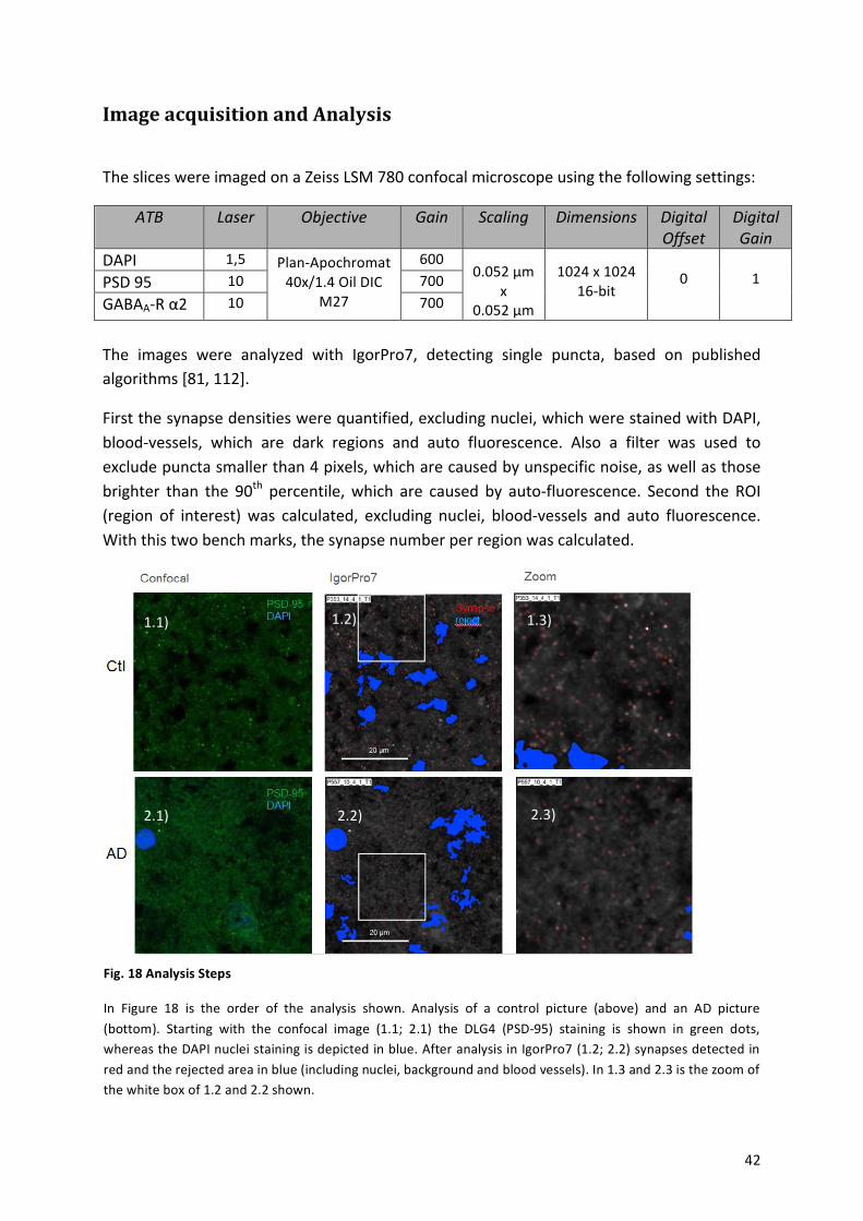
! 42!
Image-acquisition-and-Analysis-!
The!slices!were!imaged!on!a!Zeiss!LSM!780!confocal!microscope!using!the!following!settings:!
ATB$ Laser$ Objective$ Gain$ Scaling$ Dimensions$ Digital$Offset$
Digital$Gain$
DAPI! 1,5!!
PlanaApochromat!40x/1.4!Oil!DIC!
M27!
600! !
0.052!µm!x!
0.052!µm!
!
1024!x!1024!16abit!
!0!
!1!PSD!95! 10! 700!
GABAAaR!α2! 10! 700!!
The! images! were! analyzed! with! IgorPro7,! detecting! single! puncta,! based! on! published!algorithms![81,!112].!
First!the!synapse!densities!were!quantified,!excluding!nuclei,!which!were!stained!with!DAPI,!bloodavessels,! which! are! dark! regions! and! auto! fluorescence.! Also! a! filter! was! used! to!exclude!puncta!smaller!than!4!pixels,!which!are!caused!by!unspecific!noise,!as!well!as!those!brighter! than! the! 90th! percentile,!which! are! caused! by! autoafluorescence.! Second! the! ROI!(region! of! interest)!was! calculated,! excluding! nuclei,! bloodavessels! and! auto! fluorescence.!With!this!two!bench!marks,!the!synapse!number!per!region!was!calculated.!
!
!
! !
Fig.%18%Analysis%Steps%
In! Figure! 18! is! the! order! of! the! analysis! shown.! Analysis! of! a! control! picture! (above)! and! an! AD! picture!(bottom).! Starting! with! the! confocal! image! (1.1;! 2.1)! the! DLG4! (PSDa95)! staining! is! shown! in! green! dots,!whereas!the!DAPI!nuclei!staining!is!depicted! in!blue.!After!analysis!in!IgorPro7!(1.2;!2.2)!synapses!detected! in!red!and!the!rejected!area!in!blue!(including!nuclei,!background!and!blood!vessels).!In!1.3!and!2.3!is!the!zoom!of!the!white!box!of!1.2!and!2.2!shown.!
1.1)!
2.1)!
1.2)! 1.3)!
2.2)! 2.3)!
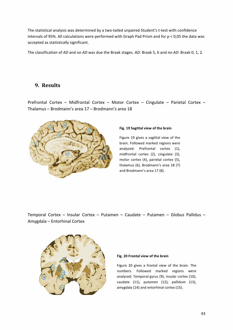
! 43!
The!statistical!analysis!was!determined!by!a!twoatailed!unpaired!Student’s!tatest!with!confidence!intervals!of!95%.!All!calculations!were!performed!with!Graph!Pad!Prism!and!for!p!<!0,05!the!data!was!accepted!as!statistically!significant.!
The!classification!of!AD$and!no$AD!was!due!the!Braak!stages.$AD:!Braak!5,!6!and!no$AD:!Braak!0,!1,!2.!
-
9. Results-!
Prefrontal! Cortex! –! Midfrontal! Cortex! –! Motor! Cortex! –! Cingulate! –! Parietal! Cortex! –!Thalamus!–!Brodmann’s!area!17!–!Brodmann’s!area!18!
!
!
!
7 !8 !
!
!
-
-!
Temporal! Cortex! –! Insular! Cortex! –! Putamen! –! Caudate! –! Putamen! –! Globus! Pallidus! –!Amygdala!–!Entorhinal!Cortex!
-
-!
!
!
!
!
!
Fig.%19%Sagittal%view%of%the%brain%
Figure! 19!gives!a! sagittal! view! of! the!brain.!Followed!marked!regions!were!analyzed:! Prefrontal! cortex! (1),!midfrontal! cortex! (2),! cingulate! (3),!motor! cortex! (4),! parietal! cortex! (5),!thalamus!(6),!Brodmann’s!area!18!(7)!and!Brodmann’s!area!17!(8).!
Fig.%20%Frontal%view%of%the%brain%
Figure! 20! gives! a! frontal! view! of! the! brain.! The!numbers.! Followed! marked! regions! were!analyzed:!Temporal!gyrus!(9),!insular!cortex!(10),!caudate! (11),! putamen! (12),! pallidum! (13),!amygdala!(14)!and!entorhinal!cortex!(15).!
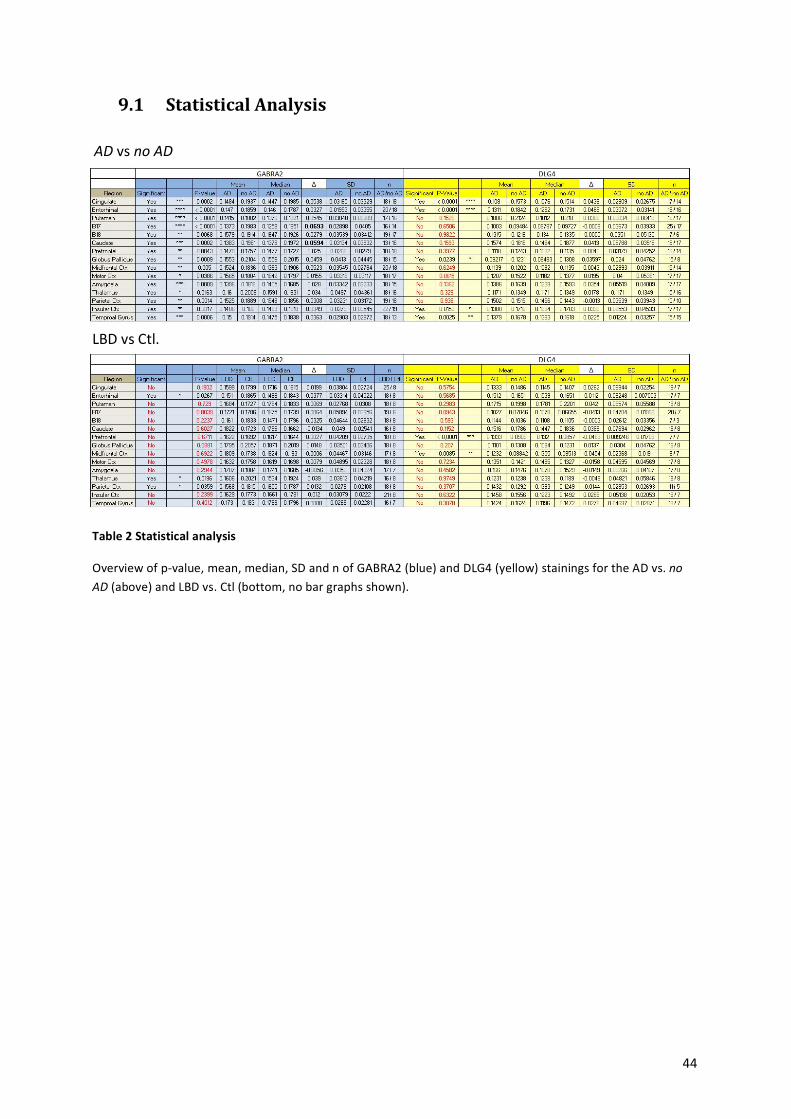
! 44!
9.1 Statistical-Analysis-!
AD!vs!no$AD!
! !Table%2%Statistical%analysis%
Overview!of!pavalue,!mean,!median,!SD!and!n!of!GABRA2!(blue)!and!DLG4!(yellow)!stainings!for!the!AD!vs.!no$AD!(above)!and!LBD!vs.!Ctl!(bottom,!no!bar!graphs!shown).!
%
LBD!vs!Ctl.!
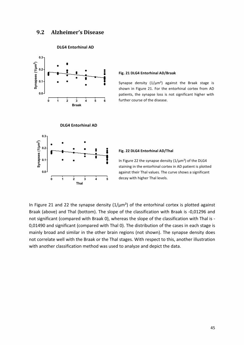
! 45!
9.2 Alzheimer’s-Disease-!
!
!
!
!
!
!
!
!
!
!
!
!
!
!
In!Figure!21!and!22!the!synapse!density!(1/µm²)!of!the!entorhinal!cortex! is!plotted!against!Braak!(above)!and!Thal!(bottom).!The!slope!of!the!classification!with!Braak!is!a0,01296!and!not!significant!(compared!with!Braak!0),!whereas!the!slope!of!the!classification!with!Thal!is!a0,01490!and!significant!(compared!with!Thal!0).!The!distribution!of!the!cases!in!each!stage!is!mainly!broad!and!similar! in!the!other!brain!regions!(not!shown).!The!synapse!density!does!not!correlate!well!with!the!Braak!or!the!Thal!stages.!With!respect!to!this,!another!illustration!with!another!classification!method!was!used!to!analyze!and!depict!the!data.!
!
!
!
!
!
!
Fig.%21%DLG4%Entorhinal%AD/Braak%
Synapse! density! (1/µm²)! against! the! Braak! stage! is!shown! in! Figure! 21.! For! the! entorhinal! cortex! from! AD!patients,! the! synapse! loss! is! not! significant! higher! with!further!course!of!the!disease.!
Fig.%22%DLG4%Entorhinal%AD/Thal%
In!Figure!22!the!synapse!density!(1/µm²)!of!the!DLG4!staining!in!the!entorhinal!cortex!in!AD!patient!is!plotted!against!their!Thal!values.!The!curve!shows!a!significant!decay!with!higher!Thal!levels.!
PSD-95 Entorhinal AD
Braak
Syn
apse
s (1
/µm
2 )
0 1 2 3 4 5 6
0.0
0.1
0.2
0.3
PSD-95 Entorhinal AD
Thal
Syn
apse
s (1
/µm
2 )
0 1 2 3 4 5
0.0
0.1
0.2
0.3
DLG4%Entorhinal%AD%
DLG4%Entorhinal%AD%
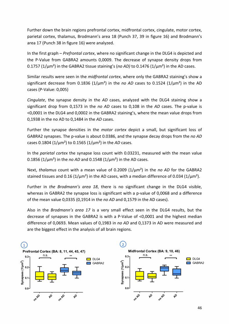
! 46!
Further!down!the!brain!regions!prefrontal!cortex,!midfrontal!cortex,!cingulate,!motor!cortex,!parietal!cortex,!thalamus,!Brodmann’s!area!18!(Punch!37,!39! in!figure!16)!and!Brodmann’s!area!17!(Punch!38!in!figure!16)!were!analyzed.!
In!the!first!graph!–!Prefrontal$cortex,!where!no!significant!change!in!the!DLG4!is!depicted!and!the! PaValue! from! GABRA2! amounts! 0,0009.! The! decrease! of! synapse! density! drops! from!0.1757!(1/µm²)!in!the!GABRA2!tissue!staining’s!(no$AD)!to!0.1476!(1/µm²)!in!the!AD!cases.!
Similar!results!were!seen!in!the!midfrontal$cortex,!where!only!the!GABRA2!staining’s!show!a!significant! decrease! from!0.1836! (1/µm²)! in! the!no$AD! cases! to! 0.1524! (1/µm²)! in! the!AD!cases!(PaValue:!0,005)!
Cingulate,! the! synapse! density! in! the! AD! cases,! analyzed! with! the! DLG4! staining! show! a!significant! drop! from! 0,1573! in! the!no$ AD! cases! to! 0,108! in! the!AD! cases.! The! pavalue! is!<0,0001!in!the!DLG4!and!0,0002!in!the!GABRA2!staining’s,!where!the!mean!value!drops!from!0,1938!in!the!no!AD!to!0,1484!in!the$AD!cases.!
Further! the! synapse! densities! in! the$ motor$ cortex$ depict! a! small,! but! significant! loss! of!GABRA2!synapses.!The!pavalue!is!about!0.0386,!and!the!synapse!decay!drops!from!the!no$AD!cases!0.1804!(1/µm²)!to!0.1565!(1/µm²)!in!the!AD!cases.!
In!the!parietal$cortex$the!synapse! loss!count!with!0.03231,!measured!with!the!mean!value!0.1856!(1/µm²)!in!the!no$AD!and!0.1548!(1/µm²)!in!the!AD!cases.!
Next,! thalamus$ count!with! a!mean! value!of! 0.2009! (1/µm²)! in! the!no$AD! for! the!GABRA2!stained!tissues!and!0.16!(1/µm²)!in!the!AD!cases,!with!a!median!difference!of!0.034!(1/µm²).!
Further! in! the$ Brodmann’s$ area$ 18,! there! is! no! significant! change! in! the! DLG4! visible,!whereas!in!GABRA2!the!synapse!loss!is!significant!with!a!pavalue!of!0,0068!and!a!difference!of!the!mean!value!0,0335!(0,1914!in!the!no$AD!and!0,1579!in!the!AD!cases).!
Also! in! the! Brodmann’s$ area$ 17! is! a! very! small! effect! seen! in! the! DLG4! results,! but! the!decrease!of! synapses! in! the!GABRA2! is!with!a!PaValue!of!<0,0001!and! the!highest!median!difference!of!0,0693.!Mean!values!of!0,1983!in!no$AD!and!0,1373!in!AD!were!measured!and!are!the!biggest!effect!in!the!analysis!of!all!brain!regions.!
!
! !1% 2%
Midfrontal Cortex (BA: 9, 10, 46)
Syn
apse
s (1
/µm
2 )
no AD AD
no AD AD
0.0
0.1
0.2
0.3DLG4GABRA2
n.s. **Prefrontal Cortex (BA: 8, 11, 44, 45, 47)
Syn
apse
s (1
/µm
2 )
no AD AD
no AD AD
0.0
0.1
0.2
0.3 n.s. **DLG4GABRA2
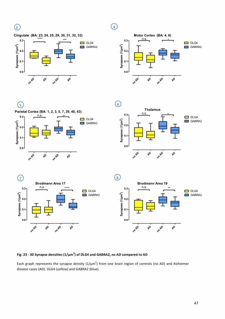
! 47!
!
!
!
!
!
!
!
!
!
!
!
!
!
!
!
!
!
!
!
!
!
!
!
!
!
!
!
Fig.%23%Z%30%Synapse%densities%(1/µm2)%of%DLG4%and%GABRA2,%no#AD%compared%to%AD%
Each!graph! represents!the!synapse!density! (1/µm2)!from!one!brain!region!of!controls! (no!AD)!and!Alzheimer!disease!cases!(AD).!DLG4!(yellow)!and!GABRA2!(blue).!
!
3% 4%
5% 6%
7% 8%Brodmann Area 18
Syn
apse
s (1
/µm
2 )
no AD AD
no AD AD
0.0
0.1
0.2
0.3 n.s. **DLG4GABRA2
Brodmann Area 17
Syn
apse
s (1
/µm
2 )
no AD AD
no AD AD
0.0
0.1
0.2
0.3 n.s. ****DLG4GABRA2
Parietal Cortex (BA: 1, 2, 3, 5, 7, 39, 40, 43)
Syn
apse
s (1
/µm
2 )
no AD AD
no AD
AD
0.0
0.1
0.2
0.3 n.s. **DLG4GABRA2
Thalamus
Syn
apse
s (1
/µm
2 )
no AD AD
no AD AD
0.0
0.1
0.2
0.3 n.s. *DLG4GABRA2
Motor Cortex (BA: 4, 6)
Syn
apse
s (1
/µm
2 )
no AD AD
no AD AD
0.0
0.1
0.2
0.3 n.s. *DLG4GABRA2
Cingulate (BA: 23, 24, 25, 29, 30, 31, 32, 33)
Syn
apse
s (1
/µm
2 )
no AD AD
no AD AD
0.0
0.1
0.2
0.3 **** ***DLG4GABRA2
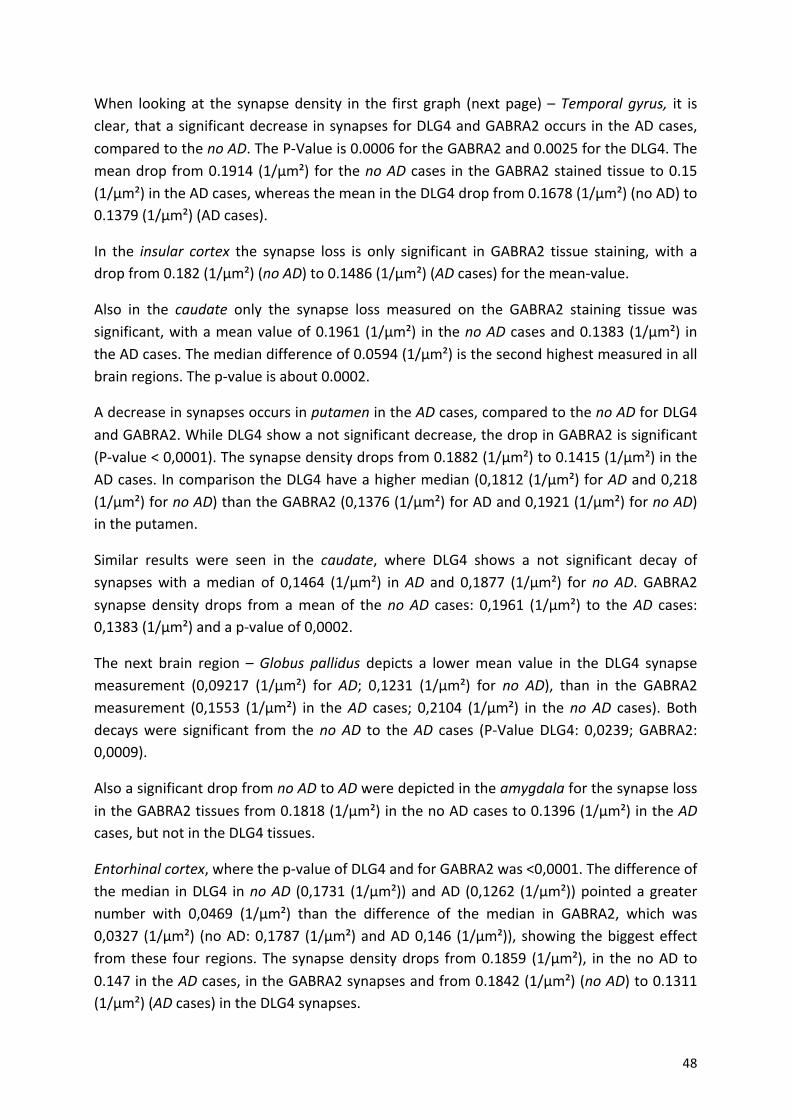
! 48!
When! looking!at! the! synapse!density! in! the! first! graph! (next!page)! –!Temporal$ gyrus,$ it! is!clear,!that!a!significant!decrease!in!synapses!for!DLG4!and!GABRA2!occurs!in!the!AD!cases,!compared!to!the!no$AD.!The!PaValue!is!0.0006!for!the!GABRA2!and!0.0025!for!the!DLG4.!The!mean!drop!from!0.1914!(1/µm²)! for! the!no$AD! cases! in! the!GABRA2!stained!tissue!to!0.15!(1/µm²)!in!the!AD!cases,!whereas!the!mean!in!the!DLG4!drop!from!0.1678!(1/µm²)!(no!AD)!to!0.1379!(1/µm²)!(AD!cases).!
In! the! insular$ cortex$ the! synapse! loss! is! only! significant! in! GABRA2! tissue! staining,!with! a!drop!from!0.182!(1/µm²)!(no$AD)!to!0.1486!(1/µm²)!(AD!cases)!for!the!meanavalue.!
Also! in! the! caudate$ only! the! synapse! loss! measured! on! the! GABRA2! staining! tissue! was!significant,!with!a!mean!value!of!0.1961!(1/µm²)! in!the!no$AD!cases!and!0.1383!(1/µm²)! in!the!AD!cases.!The!median!difference!of!0.0594!(1/µm²)!is!the!second!highest!measured!in!all!brain!regions.!The!pavalue!is!about!0.0002.!
A!decrease!in!synapses!occurs!in!putamen!in!the!AD!cases,!compared!to!the!no$AD!for!DLG4!and!GABRA2.!While!DLG4!show!a!not!significant!decrease,!the!drop!in!GABRA2!is!significant!(Pavalue!<!0,0001).!The!synapse!density!drops!from!0.1882!(1/µm²)!to!0.1415!(1/µm²)!in!the!AD!cases.!In!comparison!the!DLG4!have!a!higher!median!(0,1812!(1/µm²)!for!AD!and!0,218!(1/µm²)!for!no$AD)!than!the!GABRA2!(0,1376!(1/µm²)!for!AD!and!0,1921!(1/µm²)!for!no$AD)!in!the!putamen.!
Similar! results! were! seen! in! the! caudate,! where! DLG4! shows! a! not! significant! decay! of!synapses!with! a!median! of! 0,1464! (1/µm²)! in!AD! and! 0,1877! (1/µm²)! for!no!AD.! GABRA2!synapse! density! drops! from! a!mean! of! the!no$AD! cases:! 0,1961! (1/µm²)! to! the!AD! cases:!0,1383!(1/µm²)!and!a!pavalue!of!0,0002.!
The! next! brain! region! –!Globus$ pallidus! depicts! a! lower!mean! value! in! the! DLG4! synapse!measurement! (0,09217! (1/µm²)! for! AD;! 0,1231! (1/µm²)! for! no$ AD),! than! in! the! GABRA2!measurement! (0,1553! (1/µm²)! in! the!AD! cases;! 0,2104! (1/µm²)! in! the!no$AD! cases).! Both!decays!were! significant! from! the!no$AD! to! the!AD! cases! (PaValue!DLG4:! 0,0239;!GABRA2:!0,0009).!
Also!a!significant!drop!from!no$AD!to!AD!were!depicted!in!the!amygdala$for!the!synapse!loss!in!the!GABRA2!tissues!from!0.1818!(1/µm²)!in!the!no!AD!cases!to!0.1396!(1/µm²)!in!the!AD!cases,!but!not!in!the!DLG4!tissues.!
Entorhinal$cortex,!where!the!pavalue!of!DLG4!and!for!GABRA2!was!<0,0001.!The!difference!of!the!median! in!DLG4! in!no$AD! (0,1731! (1/µm²))!and!AD! (0,1262! (1/µm²))!pointed!a!greater!number! with! 0,0469! (1/µm²)! than! the! difference! of! the! median! in! GABRA2,! which! was!0,0327! (1/µm²)! (no!AD:!0,1787! (1/µm²)!and!AD!0,146! (1/µm²)),! showing! the!biggest!effect!from! these! four! regions.! The! synapse!density!drops! from!0.1859! (1/µm²),! in! the!no!AD! to!0.147!in!the!AD!cases,!in!the!GABRA2!synapses!and!from!0.1842!(1/µm²)!(no$AD)!to!0.1311!(1/µm²)!(AD!cases)!in!the!DLG4!synapses.!
!
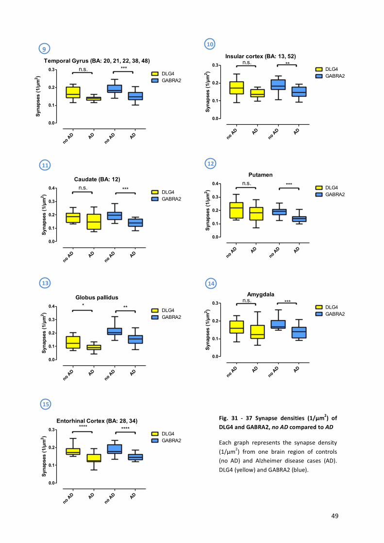
! 49!
!
!
!
!
!
!
!
! !
9%10%
12%11%
13% 14%
15%
Fig.% 31% Z% 37% Synapse% densities% (1/µm2)% of%DLG4%and%GABRA2,%no#AD%compared%to%AD%
Each! graph! represents! the! synapse! density!(1/µm2)! from! one! brain! region! of! controls!(no! AD)! and! Alzheimer! disease! cases! (AD).!DLG4!(yellow)!and!GABRA2!(blue).!
!
Insular cortex (BA: 13, 52)
Syn
apse
s (1
/µm
2 )
no AD AD
no AD AD
0.0
0.1
0.2
0.3 n.s. **DLG4GABRA2
Caudate (BA: 12)
Syn
apse
s (1
/µm
2 )
no AD AD
no AD AD
0.0
0.1
0.2
0.3
0.4DLG4
n.s. ***GABRA2
Putamen
Syn
apse
s (1
/µm
2 )
no AD AD
no AD AD
0.0
0.1
0.2
0.3
0.4
GABRA2DLG4
n.s. ***
Globus pallidus
Syn
apse
s (1
/µm
2 )
no AD AD
no AD AD
0.0
0.1
0.2
0.3
0.4 * ** DLG4GABRA2
Amygdala
Syn
apse
s (1
/µm
2 )
no AD AD
no AD AD
0.0
0.1
0.2
0.3 n.s. ***DLG4GABRA2
Entorhinal Cortex (BA: 28, 34)
Syn
apse
s (1
/µm
2 )
no AD AD
no AD AD
0.0
0.1
0.2
0.3 **** ****DLG4GABRA2
Temporal Gyrus (BA: 20, 21, 22, 38, 48)
Syn
apse
s (1
/µm
2 )
no AD AD
no AD AD
0.0
0.1
0.2
0.3DLG4
n.s. ***
GABRA2
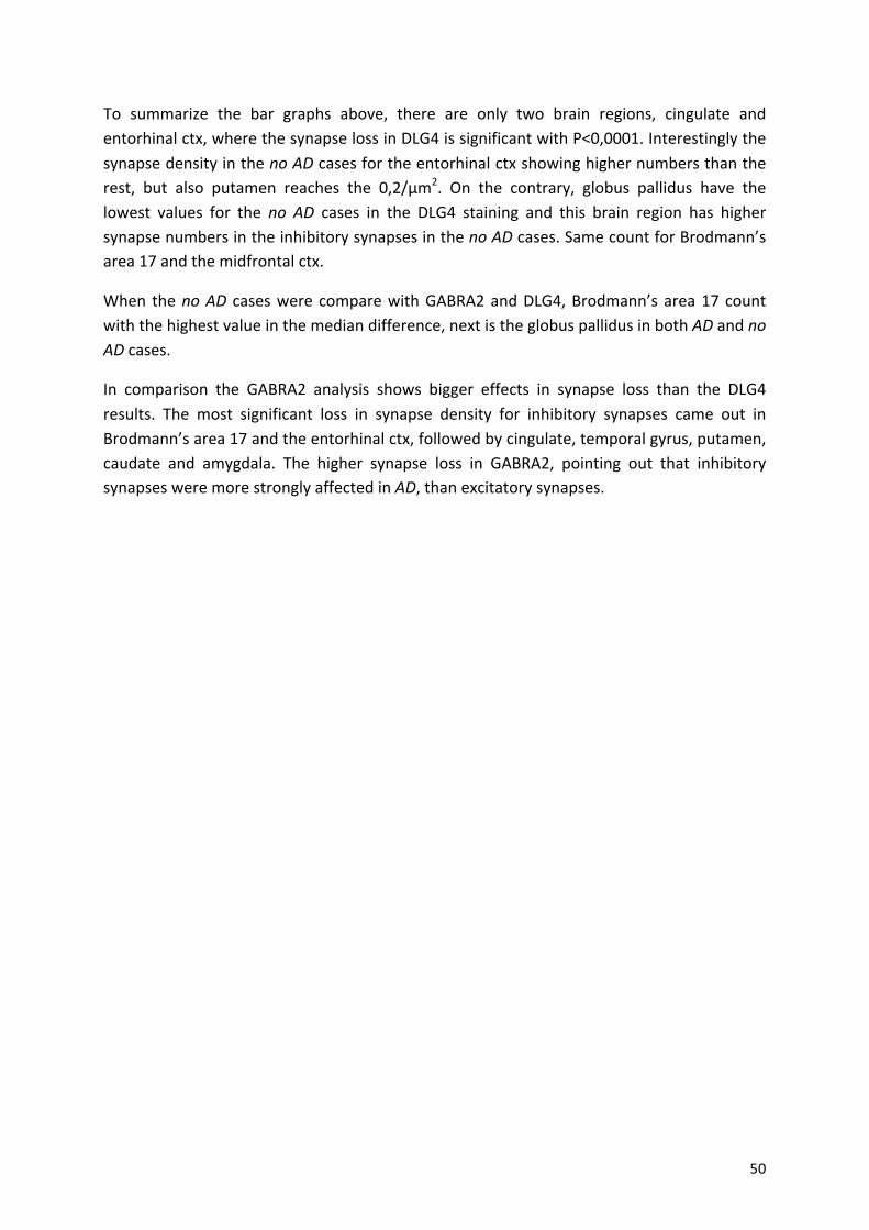
! 50!
To! summarize! the! bar! graphs! above,! there! are! only! two! brain! regions,! cingulate! and!entorhinal!ctx,!where!the!synapse!loss!in!DLG4!is!significant!with!P<0,0001.!Interestingly!the!synapse!density!in!the!no$AD!cases!for!the!entorhinal!ctx!showing!higher!numbers!than!the!rest,! but! also! putamen! reaches! the! 0,2/µm2.! On! the! contrary,! globus! pallidus! have! the!lowest! values! for! the! no$ AD! cases! in! the! DLG4! staining! and! this! brain! region! has! higher!synapse!numbers!in!the!inhibitory!synapses!in!the!no$AD!cases.!Same!count!for!Brodmann’s!area!17!and!the!midfrontal!ctx.!
When! the!no$AD! cases!were!compare!with!GABRA2!and!DLG4,!Brodmann’s!area!17!count!with!the!highest!value!in!the!median!difference,!next!is!the!globus!pallidus!in!both!AD!and!no$AD!cases.!
In! comparison! the! GABRA2! analysis! shows! bigger! effects! in! synapse! loss! than! the! DLG4!results.! The! most! significant! loss! in! synapse! density! for! inhibitory! synapses! came! out! in!Brodmann’s!area!17!and!the!entorhinal!ctx,!followed!by!cingulate,!temporal!gyrus,!putamen,!caudate! and! amygdala.! The! higher! synapse! loss! in! GABRA2,! pointing! out! that! inhibitory!synapses!were!more!strongly!affected!in!AD,!than!excitatory!synapses.!!
!
!
!
!
!
!
!
!
!
!
!
!
!
!
!
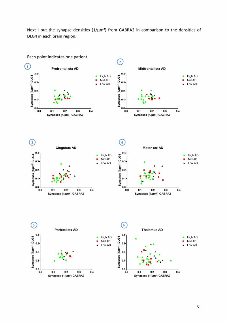
! 51!
Next! I! put! the! synapse! densities! (1/µm²)! from!GABRA2! in! comparison! to! the! densities! of!DLG4!in!each!brain!region.!!
!
Each!point!indicates!one!patient.!
Prefrontal ctx AD
Synapses (1/µm²) GABRA2
Syn
apse
s (1
/µm
2 ) D
LG4
0.0 0.1 0.2 0.3 0.40.0
0.1
0.2
0.3
0.4High ADMid ADLow AD
!
Midfrontal ctx AD
Synapses (1/µm²) GABRA2S
ynap
ses
(1/µ
m2 )
DLG
4
0.0 0.1 0.2 0.3 0.40.0
0.1
0.2
0.3
0.4High ADMid ADLow AD
!
!
!
!
Cingulate AD
Synapses (1/µm²) GABRA2
Syna
pses
(1/µ
m2 ) D
LG4
0.0 0.1 0.2 0.3 0.40.0
0.1
0.2
0.3
0.4High ADMid ADLow AD
!
Motor ctx AD
Synapses (1/µm²) GABRA2
Syn
apse
s (1
/µm
2 ) D
LG4
0.0 0.1 0.2 0.3 0.40.0
0.1
0.2
0.3
0.4High ADMid ADLow AD
!
!
!
!
Parietal ctx AD
Synapses (1/µm²) GABRA2
Syn
apse
s (1
/µm
2 ) D
LG4
0.0 0.1 0.2 0.3 0.40.0
0.1
0.2
0.3
0.4High ADMid ADLow AD
Thalamus AD
Synapses (1/µm²) GABRA2
Syna
pses
(1/µ
m2 ) D
LG4
0.0 0.1 0.2 0.3 0.40.0
0.1
0.2
0.3
0.4High ADMid ADLow AD
!
!
!!
1%
6%5%
4%3%
2%
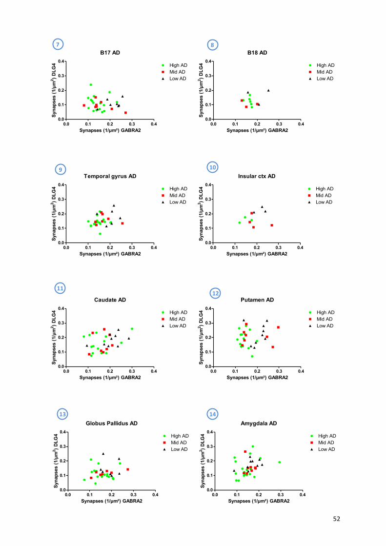
! 52!
B17 AD
Synapses (1/µm²) GABRA2
Syna
pses
(1/µ
m2 ) D
LG4
0.0 0.1 0.2 0.3 0.40.0
0.1
0.2
0.3
0.4High ADMid ADLow AD
!
B18 AD
Synapses (1/µm²) GABRA2
Syna
pses
(1/µ
m2 ) D
LG4
0.0 0.1 0.2 0.3 0.40.0
0.1
0.2
0.3
0.4High ADMid ADLow AD
!!
!!
Temporal gyrus AD
Synapses (1/µm²) GABRA2
Syn
apse
s (1
/µm
2 ) D
LG4
0.0 0.1 0.2 0.3 0.40.0
0.1
0.2
0.3
0.4High ADMid ADLow AD
!
Insular ctx AD
Synapses (1/µm²) GABRA2
Syn
apse
s (1
/µm
2 ) D
LG4
0.0 0.1 0.2 0.3 0.40.0
0.1
0.2
0.3
0.4High ADMid ADLow AD
!!
!!!
Caudate AD
Synapses (1/µm²) GABRA2
Syna
pses
(1/µ
m2 ) D
LG4
0.0 0.1 0.2 0.3 0.40.0
0.1
0.2
0.3
0.4High ADMid ADLow AD
!
Putamen AD
Synapses (1/µm²) GABRA2
Syna
pses
(1/µ
m2 ) D
LG4
0.0 0.1 0.2 0.3 0.40.0
0.1
0.2
0.3
0.4High ADMid ADLow AD
!!
!
-
Globus Pallidus AD
Synapses (1/µm²) GABRA2
Syn
apse
s (1
/µm
2 ) D
LG4
0.0 0.1 0.2 0.3 0.40.0
0.1
0.2
0.3
0.4High ADMid ADLow AD
!
Amygdala AD
Synapses (1/µm²) GABRA2
Syna
pses
(1/µ
m2 ) D
LG4
0.0 0.1 0.2 0.3 0.40.0
0.1
0.2
0.3
0.4High ADMid ADLow AD
-
9%
8%7%
13%
12%11%
10%
14%
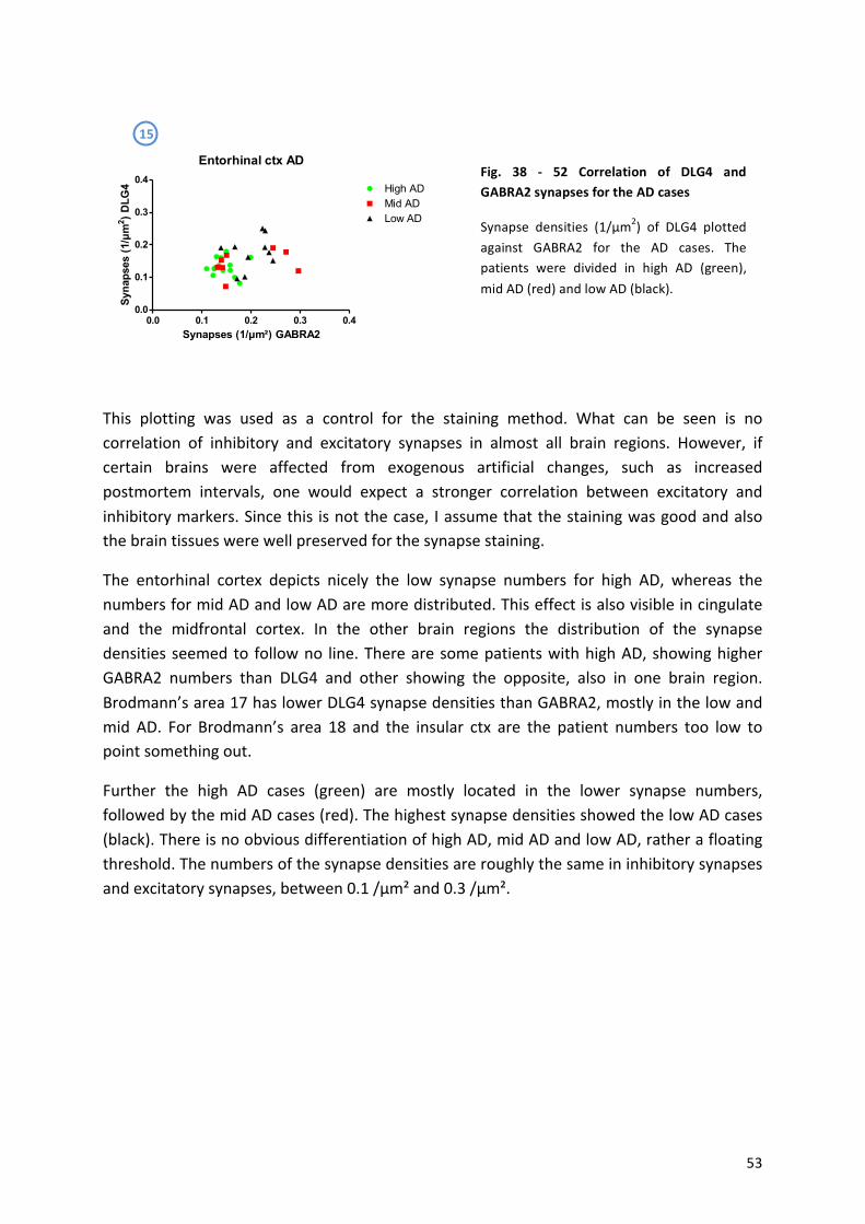
! 53!
-
-
Entorhinal ctx AD
Synapses (1/µm²) GABRA2
Syn
apse
s (1
/µm
2 ) D
LG4
0.0 0.1 0.2 0.3 0.40.0
0.1
0.2
0.3
0.4High ADMid ADLow AD
-
!
This! plotting! was! used! as! a! control! for! the! staining! method.! What! can! be! seen! is! no!correlation! of! inhibitory! and! excitatory! synapses! in! almost! all! brain! regions.! However,! if!certain! brains! were! affected! from! exogenous! artificial! changes,! such! as! increased!postmortem! intervals,! one! would! expect! a! stronger! correlation! between! excitatory! and!inhibitory!markers.!Since!this! is!not!the!case,! I!assume!that!the!staining!was!good!and!also!the!brain!tissues!were!well!preserved!for!the!synapse!staining.!
The! entorhinal! cortex! depicts! nicely! the! low! synapse! numbers! for! high! AD,! whereas! the!numbers!for!mid!AD!and!low!AD!are!more!distributed.!This!effect!is!also!visible!in!cingulate!and! the! midfrontal! cortex.! In! the! other! brain! regions! the! distribution! of! the! synapse!densities!seemed!to! follow!no! line.!There!are!some!patients!with!high!AD,!showing!higher!GABRA2! numbers! than! DLG4! and! other! showing! the! opposite,! also! in! one! brain! region.!Brodmann’s!area!17!has!lower!DLG4!synapse!densities!than!GABRA2,!mostly!in!the!low!and!mid! AD.! For! Brodmann’s! area! 18! and! the! insular! ctx! are! the! patient! numbers! too! low! to!point!something!out.!
Further! the! high! AD! cases! (green)! are! mostly! located! in! the! lower! synapse! numbers,!followed!by!the!mid!AD!cases!(red).!The!highest!synapse!densities!showed!the!low!AD!cases!(black).!There!is!no!obvious!differentiation!of!high!AD,!mid!AD!and!low!AD,!rather!a!floating!threshold.!The!numbers!of!the!synapse!densities!are!roughly!the!same!in!inhibitory!synapses!and!excitatory!synapses,!between!0.1!/µm²!and!0.3!/µm².!!
Fig.% 38% Z% 52% Correlation% of% DLG4% and%GABRA2%synapses%for%the%AD%cases%
Synapse! densities! (1/µm2)! of! DLG4! plotted!against! GABRA2! for! the! AD! cases.! The!patients! were! divided! in! high! AD! (green),!mid!AD!(red)!and!low!AD!(black).!
15%
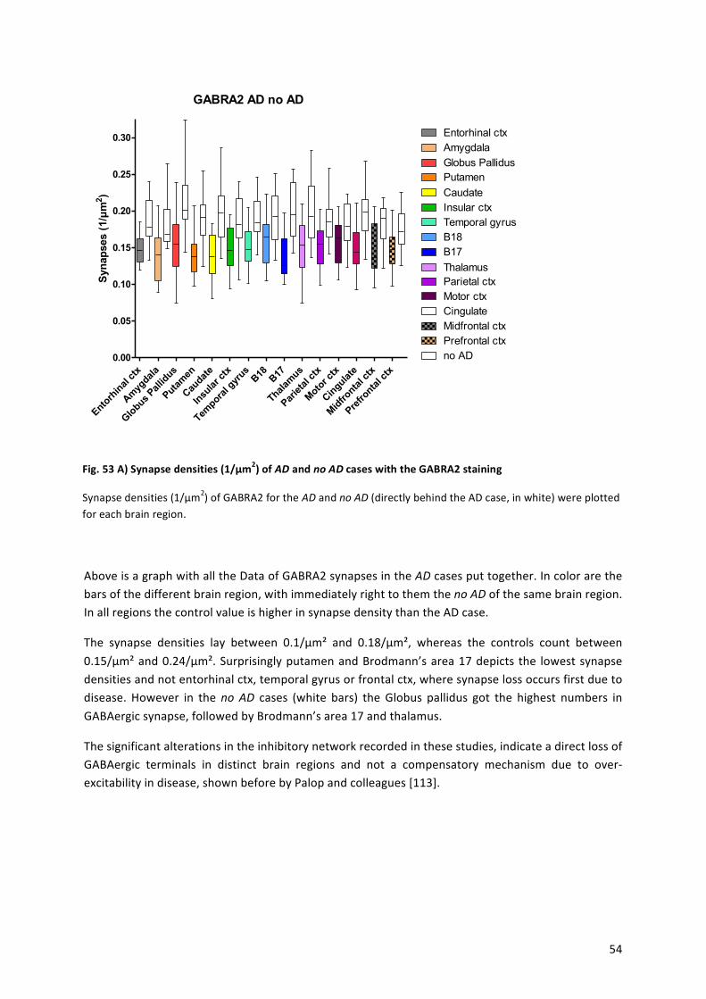
! 54!
GABRA2 AD no ADS
ynap
ses
(1/µ
m2 )
Entorh
inal
ctx
Amyg
dala
Globus P
allid
us
Putam
en
Caudat
e
Insu
lar ct
x
Tempora
l gyr
usB18 B17
Thalam
us
Pariet
al ct
x
Motor c
tx
Cingulat
e
Midfro
ntal c
tx
Prefro
ntal c
tx0.00
0.05
0.10
0.15
0.20
0.25
0.30
Prefrontal ctxMidfrontal ctx
Motor ctxParietal ctxThalamusB17B18Temporal gyrusInsular ctxCaudatePutamenGlobus PallidusAmygdalaEntorhinal ctx
Cingulate
no AD
!
!
!
!
!
Above!is!a!graph!with!all!the!Data!of!GABRA2!synapses!in!the!AD!cases!put!together.!In!color!are!the!bars!of!the!different!brain!region,!with!immediately!right!to!them!the!no$AD!of!the!same!brain!region.!In!all!regions!the!control!value!is!higher!in!synapse!density!than!the!AD!case.!
The! synapse! densities! lay! between! 0.1/µm²! and! 0.18/µm²,! whereas! the! controls! count! between!0.15/µm²!and!0.24/µm².!Surprisingly!putamen!and!Brodmann’s!area!17!depicts! the! lowest!synapse!densities!and!not!entorhinal!ctx,!temporal!gyrus!or!frontal!ctx,!where!synapse!loss!occurs!first!due!to!disease.! However! in! the!no$ AD! cases! (white! bars)! the!Globus! pallidus! got! the! highest! numbers! in!GABAergic!synapse,!followed!by!Brodmann’s!area!17!and!thalamus.!
The!significant!alterations!in!the!inhibitory!network!recorded!in!these!studies,!indicate!a!direct!loss!of!GABAergic! terminals! in! distinct! brain! regions! and! not! a! compensatory! mechanism! due! to! overaexcitability!in!disease,!shown!before!by!Palop!and!colleagues![113].!!
!
!
!
!
Fig.%53%A)%Synapse%densities%(1/µm2)%of%AD%and%no#AD%cases%with%the%GABRA2%staining%
Synapse!densities!(1/µm2)!of!GABRA2!for!the!AD!and!no$AD!(directly!behind!the!AD!case,!in!white)!were!plotted!for!each!brain!region.!!
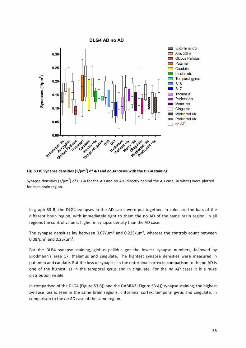
! 55!
!
!
!
!
!
!
!
!
!
!
!
!
!
!
!
!
In! graph!53!B)! the!DLG4! synapses! in! the!AD! cases!were!put! together.! In! color! are! the!bars!of! the!different! brain! region,!with! immediately! right! to! them! the! no! AD! of! the! same! brain! region.! In! all!regions!the!control!value!is!higher!in!synapse!density!than!the!AD!case.!
The! synapse!densities! lay!between!0.07/µm²!and!0.225/µm²,!whereas! the! controls! count!between!0.08/µm²!and!0.25/µm².!
For! the! DLB4! synapse! staining,! globus! pallidus! got! the! lowest! synapse! numbers,! followed! by!Brodmann’s! area! 17,! thalamus! and! cingulate.! The! hightest! synapse! densities! were! measured! in!putamen!and!caudate.!But!the!loss!of!synapses!in!the!entorhinal!cortex!in!comparison!to!the!no!AD!is!one! of! the! highest,! as! in! the! temporal! gyrus! and! in! cingulate.! For! the! no$ AD! cases! it! is! a! huge!distribution!visible.!
In!comparison!of!the!DLG4!(Figure!53!B))!and!the!GABRA2!(Figure!53!A))!synapse!staining,!the!highest!synapse! loss! is! seen! in! the! same!brain! regions:! Entorhinal! cortex,! temporal! gyrus!and! cingulate,! in!comparison!to!the!no$AD!case!of!the!same!region.!
!
Fig.%53%B)%Synapse%densities%(1/µm2)%of%AD%and%no#AD%cases%with%the%DLG4%staining%
Synapse!densities!(1/µm2)!of!DLG4!for!the!AD!and!no!AD!(directly!behind!the!AD!case,!in!white)!were!plotted!for!each!brain!region.!!
DLG4 AD no ADS
ynap
ses
(1/µ
m2 )
0.00
0.05
0.10
0.15
0.20
0.25
0.30
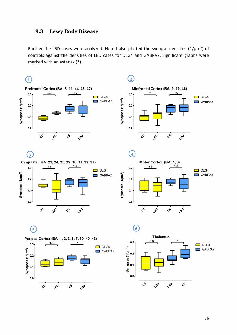
! 56!
9.3 Lewy-Body-Disease-!
Further! the!LBD!cases!were!analyzed.!Here! I!also!plotted! the!synapse!densities! (1/µm²)!of!controls! against! the!densities!of! LBD!cases! for!DLG4!and!GABRA2.! Significant! graphs!were!marked!with!an!asterisk!(*).!
!
!
!
!
!
!
!!
!
!
!
!
!
!
!
!
!
!
!
!
!
!
!
Thalamus
Syna
pses
(1/µ
m2 )
CtlLBD
LBD Ctl
0.0
0.1
0.2
0.3DLG4GABRA2
n.s. *
1% 2%
3% 4%
5% 6%
Prefrontal Cortex (BA: 8, 11, 44, 45, 47)
Syn
apse
s (1
/µm
2 )
CtlLBD Ctl
LBD
0.0
0.1
0.2
0.3
GABRA2DLG4
*** n.s.Midfrontal Cortex (BA: 9, 10, 46)
Syn
apse
s (1
/µm
2 )
CtlLBD Ctl
LBD
0.0
0.1
0.2
0.3DLG4GABRA2
** n.s.
Cingulate (BA: 23, 24, 25, 29, 30, 31, 32, 33)
Syna
pses
(1/µ
m2 )
CtlLBD Ctr
LBD
0.0
0.1
0.2
0.3DLG4GABRA2
n.s. n.s.Motor Cortex (BA: 4, 6)
Syn
apse
s (1
/µm
2 )
CtlLBD Ctl
LBD
0.0
0.1
0.2
0.3DLG4GABRA2
n.s. n.s.
Parietal Cortex (BA: 1, 2, 3, 5, 7, 39, 40, 43)
Syn
apse
s (1
/µm
2 )
CtlLBD Ctl
LBD
0.0
0.1
0.2
0.3DLG4GABRA2
n.s. *
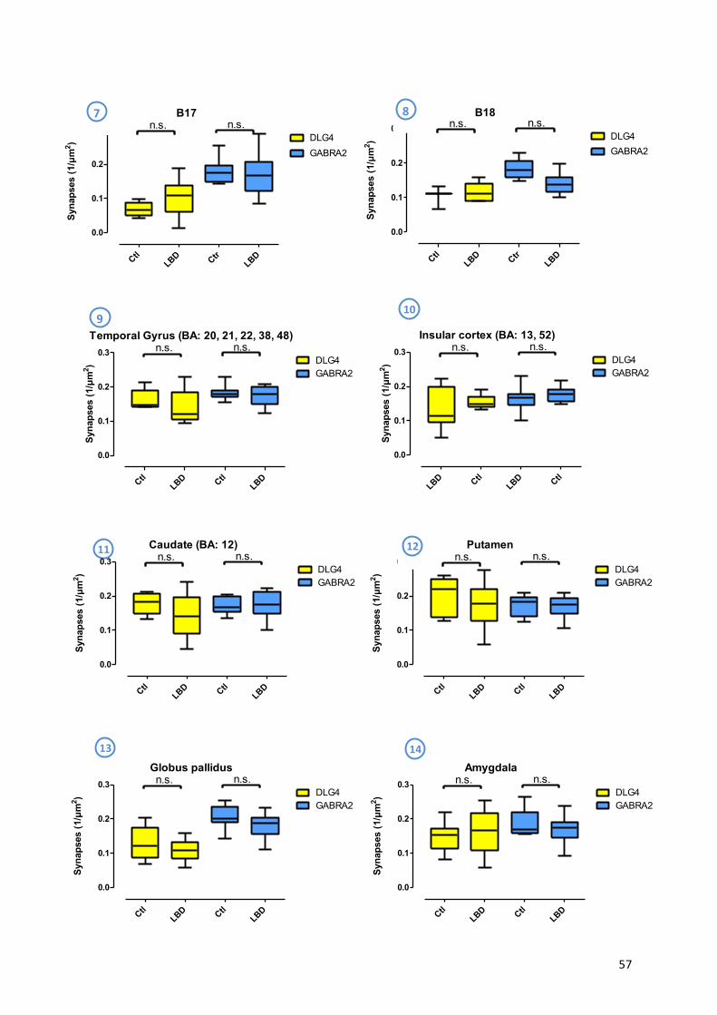
! 57!
!
!
!
!
!
!
!
!
!
!
!
!
!
!
!
!
!
!
!
!
!
!
!
B17
Syna
pses
(1/µ
m2 )
CtlLBD Ctr
LBD
0.0
0.1
0.2
0.3DLG4GABRA2
n.s. n.s.B18
Syna
pses
(1/µ
m2 )
CtlLBD Ctr
LBD
0.0
0.1
0.2
0.3DLG4GABRA2
n.s. n.s.
Putamen
Syna
pses
(1/µ
m2 )
CtlLBD Ctl
LBD
0.0
0.1
0.2
0.3
GABRA2DLG4
n.s. n.s.
Globus pallidus
Syn
apse
s (1
/µm
2 )
CtlLBD Ctl
LBD
0.0
0.1
0.2
0.3
GABRA2DLG4
n.s. n.s.Amygdala
Syna
pses
(1/µ
m2 )
CtlLBD Ctl
LBD
0.0
0.1
0.2
0.3DLG4GABRA2
n.s. n.s.
7% 8%
9%10%
12%11%
13% 14%
Temporal Gyrus (BA: 20, 21, 22, 38, 48)
Syn
apse
s (1
/µm
2 )
CtlLBD Ctl
LBD
0.0
0.1
0.2
0.3
GABRA2DLG4
n.s. n.s.Insular cortex (BA: 13, 52)
Syn
apse
s (1
/µm
2 )
LBD CtlLBD Ctl
0.0
0.1
0.2
0.3
GABRA2DLG4
n.s. n.s.
Caudate (BA: 12)
Syna
pses
(1/µ
m2 )
CtlLBD Ctl
LBD
0.0
0.1
0.2
0.3
GABRA2DLG4
n.s. n.s.
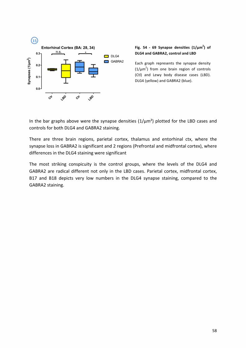
! 58!
!
!
!
!
!
!
!
!
In!the!bar!graphs!above!were!the!synapse!densities! (1/µm²)!plotted!for!the!LBD!cases!and!controls!for!both!DLG4!and!GABRA2!staining.!!
There! are! three! brain! regions,! parietal! cortex,! thalamus! and! entorhinal! ctx,! where! the!synapse!loss!in!GABRA2!is!significant!and!2!regions!(Prefrontal!and!midfrontal!cortex),!where!differences!in!the!DLG4!staining!were!significant!
The! most! striking! conspicuity! is! the! control! groups,! where! the! levels! of! the! DLG4! and!GABRA2!are! radical! different!not!only! in! the! LBD! cases.! Parietal! cortex,!midfrontal! cortex,!B17! and! B18! depicts! very! low! numbers! in! the! DLG4! synapse! staining,! compared! to! the!GABRA2!staining.!
!
!
!
!
!
!
!
!
!
!
!
15%
Fig.% 54% Z% 69% Synapse% densities% (1/µm2)% of%DLG4%and%GABRA2,%control%and%LBD%
Each! graph! represents! the! synapse! density!(1/µm2)! from! one! brain! region! of! controls!(Ctl)! and! Lewy! body! disease! cases! (LBD).!DLG4!(yellow)!and!GABRA2!(blue).!
!
Entorhinal Cortex (BA: 28, 34)
Syn
apse
s (1
/µm
2 )
CtrLBD Ctr
LBD
0.0
0.1
0.2
0.3DLG4
n.s. *
GABRA2
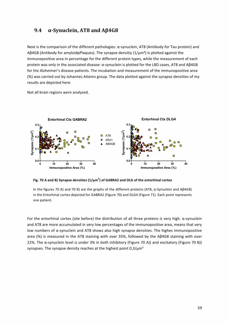
! 59!
9.4 α0Synuclein,-AT8-and-Aβ4G8-!
Next!is!the!comparison!of!the!different!pathologies:!αasynuclein,!AT8!(Antibody!for!Tau!protein)!and!Aβ4G8!(Antibody!for!amyloidpPlaques).!The!synapse!densitiy!(1/µm²)!is!plotted!against!the!Immunepositive!area!in!percentage!for!the!different!protein!types,!while!the!measurement!of!each!protein!was!only!in!the!associated!disease:!αasynuclein!is!plotted!for!the!LBD!cases,!AT8!and!Aβ4G8!for!the!Alzheimer’s!disease!patients.!The!incubation!and!measurement!of!the!immunopositive!area!(%)!was!carried!out!by!Johannes!Attems!group.!The!data!plotted!against!the!synapse!densities!of!my!results!are!depicted!here.!
Not!all!brain!regions!were!analysed.!
!!
Entorhinal Ctx GABRA2
Immunopositive Area (%)
Syn
apse
s (1
/µm
2 )
0 10 20 30 400.0
0.1
0.2
0.3
AT8
AB4G8aSyn
!
Entorhinal Ctx DLG4
Immunopositive Area (%)
Syn
apse
s (1
/µm
2 )
0 10 20 30 400.0
0.1
0.2
0.3
!
!
!
!
!
For!the!entorhinal!cortex!(site!before)!the!distribution!of!all!three!proteins!is!very!high.!αasynuclein!and!AT8!are!more!accumulated!in!very!low!percentages!of!the!immunopositive!area,!means!that!very!low!numbers!of!αasynuclein!and!AT8!shows!also!high!synapse!densities.!The!highes!immunopositive!area! (%)! is!measured! in! the!AT8!staining!with!over!35%,! followed!by! the!Aβ4G8!staining!with!over!22%.!The!αasynuclein!level!is!under!3%!in!both!inhibitory!(Figure!70!A))!and!excitatory!(Figure!70!B))!synapses.!The!synapse!density!reaches!at!the!highest!point!0,3/µm²!
!
!
!
!
Fig.%70%A%and%B)%Synapse%densities%(1/µm2)%of%GABRA2%and%DLG%of%the%entorhinal%cortex%
In!the!figures!70!A)!and!70!B)!are!the!graphs!of!the!different!proteins!(AT8,!αaSynuclein!and!Aβ4G8)!in!the!Entorhinal!cortex!depicted!for!GABRA2!(Figure!70)!and!DLG4!(Figure!71).!Each!point!represents!one!patient.!
!
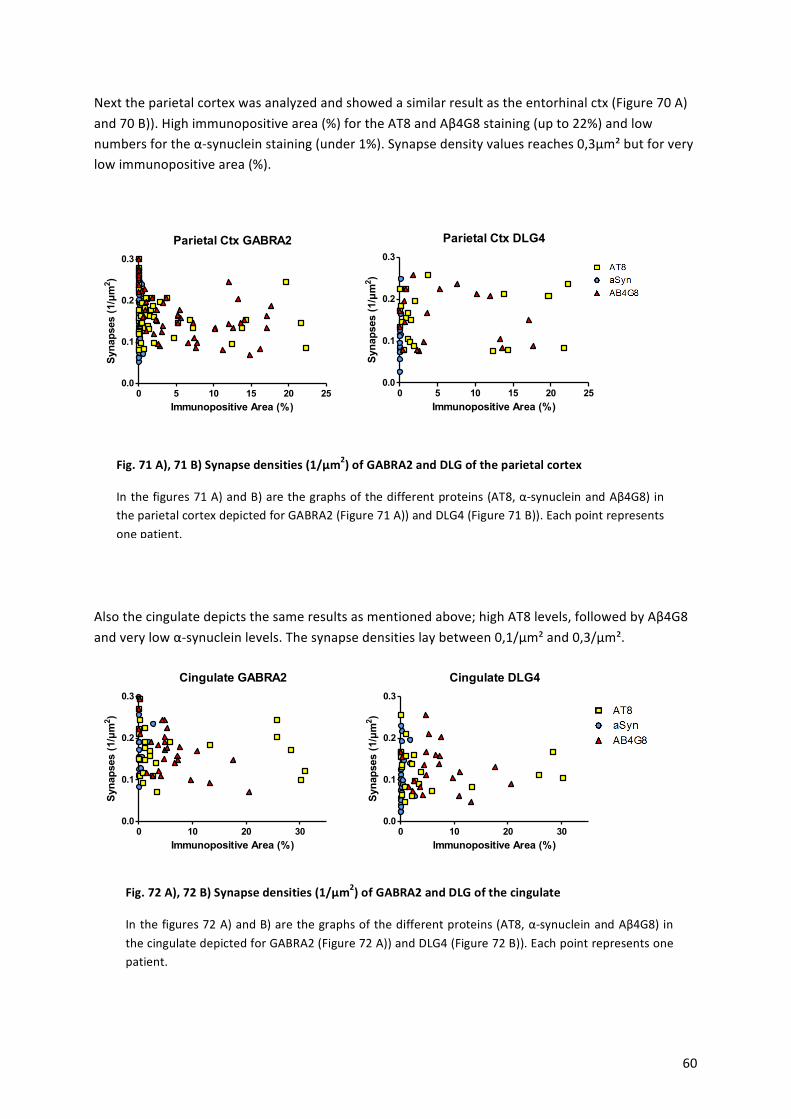
! 60!
Next!the!parietal!cortex!was!analyzed!and!showed!a!similar!result!as!the!entorhinal!ctx!(Figure!70!A)!and!70!B)).!High!immunopositive!area!(%)!for!the!AT8!and!Aβ4G8!staining!(up!to!22%)!and!low!numbers!for!the!αasynuclein!staining!(under!1%).!Synapse!density!values!reaches!0,3µm²!but!for!very!low!immunopositive!area!(%).!
!
Parietal Ctx GABRA2
Immunopositive Area (%)
Syn
apse
s (1
/µm
2 )
0 5 10 15 20 250.0
0.1
0.2
0.3
Parietal Ctx DLG4
Immunopositive Area (%)
Syn
apse
s (1
/µm
2 )
0 5 10 15 20 250.0
0.1
0.2
0.3
!
!
!
!
!
!
Also!the!cingulate!depicts!the!same!results!as!mentioned!above;!high!AT8!levels,!followed!by!Aβ4G8!and!very!low!αasynuclein!levels.!The!synapse!densities!lay!between!0,1/µm²!and!0,3/µm².!
Cingulate GABRA2
Immunopositive Area (%)
Syn
apse
s (1
/µm
2 )
0 10 20 300.0
0.1
0.2
0.3
Cingulate DLG4
Immunopositive Area (%)
Syn
apse
s (1
/µm
2 )
0 10 20 300.0
0.1
0.2
0.3
!
!
!
!
!
Fig.%72%A),%72%B)%Synapse%densities%(1/µm2)%of%GABRA2%and%DLG%of%the%cingulate%%
In!the!figures!72!A)!and!B)!are!the!graphs!of!the!different!proteins!(AT8,!αasynuclein!and!Aβ4G8)!in!the!cingulate!depicted!for!GABRA2!(Figure!72!A))!and!DLG4!(Figure!72!B)).!Each!point!represents!one!patient.!
Fig.%71%A),%71%B)%Synapse%densities%(1/µm2)%of%GABRA2%and%DLG%of%the%parietal%cortex%
In!the!figures!71!A)!and!B)!are!the!graphs!of!the!different!proteins!(AT8,!αasynuclein!and!Aβ4G8)!in!the!parietal!cortex!depicted!for!GABRA2!(Figure!71!A))!and!DLG4!(Figure!71!B)).!Each!point!represents!one!patient.!
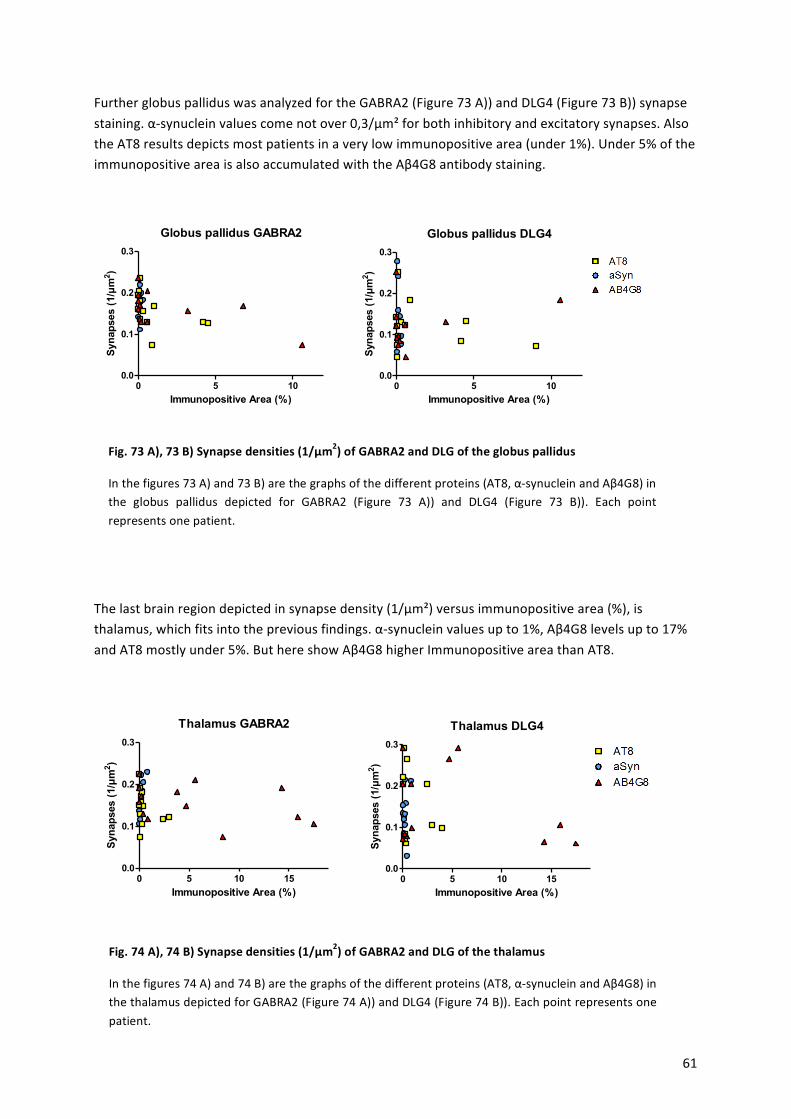
! 61!
Further!globus!pallidus!was!analyzed!for!the!GABRA2!(Figure!73!A))!and!DLG4!(Figure!73!B))!synapse!staining.!αasynuclein!values!come!not!over!0,3/µm²!for!both!inhibitory!and!excitatory!synapses.!Also!the!AT8!results!depicts!most!patients!in!a!very!low!immunopositive!area!(under!1%).!Under!5%!of!the!immunopositive!area!is!also!accumulated!with!the!Aβ4G8!antibody!staining.!
!
Globus pallidus GABRA2
Immunopositive Area (%)
Syn
apse
s (1
/µm
2 )
0 5 100.0
0.1
0.2
0.3
Globus pallidus DLG4
Immunopositive Area (%)
Syn
apse
s (1
/µm
2 )
0 5 100.0
0.1
0.2
0.3
!
!
!
!
!
!
The!last!brain!region!depicted!in!synapse!density!(1/µm²)!versus!immunopositive!area!(%),!is!thalamus,!which!fits!into!the!previous!findings.!αasynuclein!values!up!to!1%,!Aβ4G8!levels!up!to!17%!and!AT8!mostly!under!5%.!But!here!show!Aβ4G8!higher!Immunopositive!area!than!AT8.!
!
Thalamus GABRA2
Immunopositive Area (%)
Syn
apse
s (1
/µm
2 )
0 5 10 150.0
0.1
0.2
0.3
Thalamus DLG4
Immunopositive Area (%)
Syn
apse
s (1
/µm
2 )
0 5 10 150.0
0.1
0.2
0.3
!
!
!
!
!
Fig.%73%A),%73%B)%Synapse%densities%(1/µm2)%of%GABRA2%and%DLG%of%the%globus%pallidus%
In!the!figures!73!A)!and!73!B)!are!the!graphs!of!the!different!proteins!(AT8,!αasynuclein!and!Aβ4G8)!in!the! globus! pallidus! depicted! for! GABRA2! (Figure! 73! A))! and! DLG4! (Figure! 73! B)).! Each! point!represents!one!patient.!
Fig.%74%A),%74%B)%Synapse%densities%(1/µm2)%of%GABRA2%and%DLG%of%the%thalamus%
In!the!figures!74!A)!and!74!B)!are!the!graphs!of!the!different!proteins!(AT8,!αasynuclein!and!Aβ4G8)!in!the!thalamus!depicted!for!GABRA2!(Figure!74!A))!and!DLG4!(Figure!74!B)).!Each!point!represents!one!patient.!
! 62!
10. Discussion-!
In! this! study,! I! have! used! confocal! imaging! and! the! autoaanalysis! software! IgorPro7! to!provide! a! quantitative! characterization! of! inhibitory! and! excitatory! synapses! in! 15! brain!regions,!whereby!the!major! finding!was!a!significant! loss!of!GABAergic!synapse! in!all!brain!regions!for!the!AD!cases!(Braak!5,!6),!compared!to!the!no$AD!cases!(Braak!0,!1,!2).!
My! results! demonstrate! that! the! entorhinal! cortex,!which! is! usually! affected! very! early! in!disease! (Braak! 2a3),! show! the! highest! synapse! loss! in! AD! in! both! excitatory! (DLG4)! and!inhibitory!(GABRA2)!synapses!compared!to!no$AD.!
Synapses! in! the! temporal! cortex! (affected! in! Braak! stage! 4)! are! in! both! inhibitory! and!excitatory!synapses!significantly!lost.!GABRA!PaValue:!0,0006!and!DLG4!PaValue:!0,0025.!The!Median! difference! is! only! the! half! (0,0363/µm2)! of! the! highest! median! difference! in!Brodmann’s!area!17.!
While! the! frontal! and!occipital! cortex! (Braak!5)! are! affected! very! late! in!disease,! the!data!show!very!low!PaValues!and!the!highest!median!difference!measured!(Brodmann’s!area!17,!PaValue:!<0,0001,!median!difference:!0,0693)!In!comparison!of!Brodmann’s!area!17!to!18,!it!is!interesting,!that!the!values!are!not!so!similar!as!estimated.!In!GABRA2!stained!tissue,!the!synapse! densities! of! the! no$ AD! cases! is! almost! the! same! with! 0,1983/µm2! in! B17! and!0,1914/µm2! in!B18,! but! the! loss! in! the!AD! cases! is! in! the!B17!higher.!With! respect! to! the!DLG4!stained!tissue,!where!the!numbers!differ!gross!and!the!median!difference!is!almost!0!in!both!B17!and!B18,!the!excitatory!synapse!loss!is!not!seen!here.!
!
I!can!draw!two!main!conclusions!from!the!data!generated!in!this!project.!Firstly,!the!synapse!loss! for! the! excitatory! synapses! fits!well!with! the! Braak! stages! and! the! distribution! of! Aβ!(Figure! 16),! with! the! entorhinal! cortex! as! first! affected! region,! next! cingulate! and! the!temporal!gyrus.!
Secondly,!the!inhibitory!synapses!fit!better!with!the!distribution!of!tau!(Figure!16).!With!the!entorhinal! ctx! as! major! affected! region,! followed! by! putamen,! B17,! amygdala! and! the!temporal!gyrus.!
The! fact!of! the!very!high! synapse! loss! in! the!Brodmann’s!area!17! in! inhibitory! synapses! is!new!and!has!not!been!described!before.!
!
!
!
! 63!
Interestingly! there! is! a! significant! increase! in! excitatory! synapses! in! the! LBD! cases! in! the!prefrontala! and! midfrontal! cortex! and! also! in! Brodmann’s! area! 17,! whereas! the! synapse!number! decrease! in! the! putamen,! caudate! and! insular! cortex.! The! increase! in! excitatory!synapses!in!the!prefrontal!region!is!not!surprisingly!in!LBD,!where!the!action!potential!firing!in! the!motor! neurons! is! less! inhibited! than! in! healthy! patients.! The! decrease! of! DLG4! for!excitatory!synapses! in!LBD!was!also!documented!before![114]!and!fits!with!my!data! in!the!prefrontal!and!the!midfrontal!ctx,!but!not!in!the!motor!cxt.!
The!highest!synapse!loss!for!the!GABRA2!stained!tissues!was!found!in!thalamus,!entorhinal!ctx!and!the!parietal!ctx!!(Figure!12).!
Only!prefrontal!and!midfrontal!cortex!show!an!excitatory!synapse!loss!in!the!LBD!cases!and!parietal! ctx,! tahalmus! and! entrohinal! ctx! show! significant! loss! for! the! inhibitory! synapses.!The!Figures!71!–!74!show!very! low!αasynuclein! levels!but!synapse!densities!up! to!0,3/µm²!(Entorhinal! ctx,! figure! 71! A)! and! B)).! But! higher! immunopositive! area! values! in! AT8! and!Aβ4G8!staining!come!with!no!lower!synapse!density,!pointing!that!there!is!no!correlation!of!synapse!density!(1/µm²)!and!immunopositive!area!(%).!
!
For!the!Alzheimer’s!disease!data,!the!GABRA2!analysis!shows!bigger!effects!in!synapse!loss!than!the!DLG4!staining.!The!high!synapse!loss!in!GABRA2!stained!tissue!in!AD,!indicate!that!inhibitory! synapses! were! more! affected! in! AD,! than! excitatory! synapses.! Maybe! the!inhibitory! synapses! are! affected! earlier! during! the! disease! progress,! while! excitatory!synapses!are!affected!later.!But!the!numbers!of!the!synapse!densities!are!roughly!the!same!in!inhibitory!synapses!and!excitatory!synapses,!between!0.1!/µm²!and!0.3!/µm².!!
Another!reason!for!the!marked!loss!of!GABRA2!staining!may!be!an!exchange!of!the!receptor!subtype!GABAergic!neurons!in!LBD!are!functionally!altered,!as!well!as!a!loss!in!postsynaptic!GABA! receptor! markers,! like! gephyrin,! GABARAP! and! Kif5A,! which! leads! to! decreased!GABAergic! synaptic! activity.! There! is! not! a! change! in! synapse! density! in! the! LBD! in! the!Brodmann’s! area!17! [114],! as! shown! in!our!data.!GABRA2! receptors!have!been! related! to!anxiety,!depression!and!other!behavior!disorders,! like!drug!dependence!and!schizophrenia.!GABAergic!signaling!plays!an!important!role!in!neural!stem!cell!maintenance!and!renewal.!So!a! change! in! the! receptor! subtypes! can! change! the! role! and! the! targets! of! the! following!cascade.!
Finally,! in! a! study! of! Schizophrenia! and!Huntington’s! disease!GABA! subunits!were! altered!and!the!mRNA!levels!of!distinct!subunits!were!changed!in!disease![115,!116].!Mizukami!and!colleagues!reported!a!decrease!of!β3!mRNA!levels!in!human!brain!with!Alzheimer’s!disease![117].!Further!other!receptor!subtypes,!like!AMPAR!are!affected!in!Aβ!application!and!shown!a!reduced!attendance![118].!It!is!possible,!that!distinct!GABAA!receptor!subtypes!increase!in!the! concentration,! while! the! distribution! of! GABRA2! and! others! decreases,! so! that! an!exchange!of!existing!α2!subunit!occurs.!The!described!compensatory!mechanism!and!with!
! 64!
that! come! along! decreased!GABRA2! levels! is! clearly! seen! in!my!data.! So! I! suppose! that! a!changed!level!of!GABRA2!is!also!due!to!disease!characteristic!and!in!this!case!in!AD!patients,!it! is! possible! that! the! α2! subunit! is! down! regulated,! disturbed! or! exchanged! by! another!subunit.!In!my!results,!this!effect!is!seen!in!all!brain!regions,!which!promotes!this!hypothesis.!
Maybe! it! is! not! the! best! way! to! stain! and! count! for! synapses,! when! the! marker! can! be!altered!during!disease!and! is!not! reliable! to!predicate! the!number!of!GABAergic! synapses.!With!regard!to!the!changed!expression!in!disease,!such!marker!is!not!good!selected,!when!in!health!and!disease!the!consumption!of!receptors!is!changed,!the!comparison!of!the!control!and!AD!patients!is!not!possible.!
!
!
!
!
!
!
!
! -
! 65!
11. Prospects-!
Further! investigations! for! the! excitatory! synapses! as!well! as! the! LBD! cases! are! needed! to!provide! clarity! about! the! influence! of! excitatory! and! inhibitory! synapses! in! Alzheimer’s!disease!and!the!unexpected!increase!of!DGL4.!
Other!synapse!marker,!like!marker!for!cholingergic!synapses,!should!be!used!to!identify!the!network!of!synapses!and!find!correlations!between!them.!No!easily,! in!disease!substituted!protein!should!be!the!target!of!the!ATB!for!quantification!of!synapses.!DLG4!is!on!one!hand!a!better! target! than!GABRA2,!because!of! its! unaltered! consumption.!But!on! the!other!hand!DLG4!is!not!specific!for!one!synapse!type!as!GABRA2,!which!is!representative!for!GABAergic!synapses.! New! marker! should! be! found! to! identify! each! synapse! type! (GABAergic,!glutamatergic!and!cholinergic)!to!find!the!most!affected!ones!in!disease!and!also!to!identify!compensatory!mechanisms!and!the!interaction!of!the!different!synapse!types.!
!
! !
! 66!
12. Abbreviations-!
ACh! ! Acetylcholine!
AChR! ! Acetylcholine!receptor!
AD! ! Alzheimer’s!disease!
AMPAR! αaaminoa3ahydroxya5amethyla4aisoxazolepriopinoinic!acid!receptor!
AP! ! Amyloid!Plaques!
APP! ! Amyloid!precursor!protein!
BA! ! Brodmann’s!area!
βZAPP! ! Beta!Amyloid!precursor!protein!
DAPI! ! 4�,6aDiamidina2aphenylindol!
DLB! ! Dementia!with!Lewy!bodies!
eAD! ! earlyaonset!Alzheimer's!disease!
EPSP! ! Excitatory!postsynaptic!potential!
GABA! ! γaAminobutyric!acid!
GABAAZR! γaAminobutyric!acid!receptor!A!
GAD! ! Glutamic!acid!decarboxylase!
GAT! ! GABA!transporter!
IPSP! ! Inhibitory!postsynaptic!potential!
K! ! Thousand!
LBD! ! Lewy!Body!Dementia!
LTP! ! Long!term!potention!
mAChR! Muscarinic!Acetylcholine!receptor!
MCI! ! Mild!cognitive!impairment!
MM! ! Million!
nAChR!! Nicotinic!Acetylcholine!receptor!
NCI! ! No!cognitive!impairment!
! 67!
NFT! ! Neurofibrially!Tangles!
NGS! ! Normal!Goat!Serum!
NMDAR! NamethylaDaaspartate!receptor!
ON! ! Over!Night!
PD! ! Parkinsons’s!disease!
PS1! ! Presenilin!1!gene!
PS2! ! Presenilin!2!gene!
PSD! ! Postsynaptic!density!
RT! ! Room!temperature!
VGlut1!! Vesicular!Glutamate!Transporter1!
VGlut2!! Vesicular!Glutamate!Transporter!2!
5ZHT! ! Serotonin!
5ZHT3! ! Serotonin!receptor!3!
!
!
!
!
!
!
!
!
!
!
!!
!
!
! 68!
13. -Abstrakt-(Deutsch)-!
Synaptische!Plastizität!und!hohe!Synapsen!Dichte!werden!gefördert!und!aufgebaut!während!des! Lernens,! während! Spinea! und! Verbindungsverlust! die! Kennzeichen! für!Neurodegenerative!Erkrankungen!wie!die!Alzheimer!Krankheit!sind.!Viele!Faktoren!könnten!verantwortlich!sein! für!den!Spine!Verlust,!wie!ein!veränderter!pre!synaptischer! Input!oder!externe! Faktoren! und! es! ist! nicht! klar! ob! die! pre! synaptische! Fehlfunktion! oder! die!Postsynaptischen!Veränderungen!zuerst!auftreten.!Dennoch! ist!der!Synapsen!Verlust!nicht!spezifisch!um!Alzheimer!vorherzusagen.!Viele!neurodegenerative!Erkrankungen!zeigen!diese!Verluste,!ebenso!wie!es!eine!Erscheinung!des!normalen!Alterns!ist.!Jedoch!ist!es!der!Haupt!Risikofaktor!für!Demenz.!
Synapsen!Verlust!ist!nicht!gleich!verteilt!in!allen!Gehirnregionen,!was!bedeutet,!dass!altersaabhängige! Veränderungen! nicht! überall! auftreten,! aber! in! bestimmten! Regionen,! wie! im!Kortex! und! im! Hippocampus.! Körpereigene! Kompensationsmechanismen,! die! den! Spine!Verlust!entgegenwirken!und!synaptische!Kontakte!erhalten,!können!nur!bedingt!gegen!die!Neurodegeneration!halten.!
Die!Glutamat!Freisetzung!aus!Vesikeln!ist!der!Haupt!Signalweg!von!anregenden!Synapsen!im!Säuger!und!somit!wichtig!für!die!neuronale!Aktivität.!GABA!ist!der!bedeutendste!hemmende!Neurotransmitter,!bindet!auf!Rezeptoren!der!Postsynapse!und!leitet!hemmendes!Signal!ein.!!
Doch! gibt! es! Unterschiede! in! den! verschiedenen! SynapsenaTypen?! Und! welche! sind! in!Krankheit! eher! betroffen?! Um! diese! Fragen! zu! beantworten,! erregende! und! hemmende!Synapsen!wurden!evaluiert!um!mittels!typischen!synaptischen!Markern!spezifisch!markiert:!GABARa! α2! für! die! hemmenden! Synapsen! und! PSDa95! (DLG4)! für! die! Postsynapse! der!erregenden! Synapsen.! Antikörper! auf! menschlichem! Paraffin! fixierten! Schnitten! von! 15!verschiedenen! Hirnregionen,! in! Alzheimer,! Kontrolle! und! LewyaKörperaDemenz,! wurden!mittels!Konfokal!Mikroskop! (Zeiss!LSM!780)!detektiert!und!mit!Hilfe!von! IgorPro7!einzelne!Synapsen!ausgewertet.!
Der!stärkste!Synapsen!Verlust!war!in!den!hemmenden!Synapsen!des!Entorhinal!Kortex!von!Alzheimer! Patienten! zu! verzeichnen.! Für! die! hemmenden! Synapsen! waren! alle! Synapsen!Rückgänge!signifikant,!hingegen!die!Zahlen!bei!den!erregenden!Synapsen!wenig!signifikant!waren! und! teilweise! sogar! zunahmen.! Für! die! LewyaKörperaDemenz! konnte! auch! keine!signifikante!Korrelation!gefunden!werden.!
-!
!
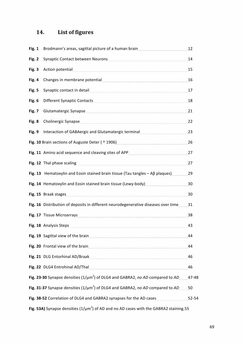
! 69!
14. List-of-figures-!
Fig.%1!! Brodmann’s!areas,!sagittal!picture!of!a!human!brain! ! ! ! 12!
Fig.%2!! Synaptic!Contact!between!Neurons! ! ! ! ! ! 14!
Fig.%3!! Action!potential! ! ! ! ! ! ! ! 15!
Fig.%4!! Changes!in!membrane!potential! ! ! ! ! ! 16!
Fig.%5!! Synaptic!contact!in!detail! ! ! ! ! ! ! 17!
Fig.%6!! Different!Synaptic!Contacts! ! ! ! ! ! ! 18!
Fig.%7!! Glutamatergic!Synapse! ! ! ! ! ! ! ! 21!
Fig.%8!! Cholinergic!Synapse! ! ! ! ! ! ! ! 22!
Fig.%9!! Interaction!of!GABAergic!and!Glutamatergic!terminal! ! ! ! 23!
Fig.%10!Brain!sections!of!Auguste!Deter!(!†!1906)! ! ! ! ! 26!
Fig.%11!!Amino!acid!sequence!and!cleaving!sites!of!APP! ! ! ! ! 27!
Fig.%12!!Thal!phase!scaling! ! ! ! ! ! ! ! 27!
Fig.%13% !Hematoxylin!and!Eosin!stained!brain!tissue!(Tau!tangles!–!Aβ!plaques)! ! 29!
Fig.%14% Hematoxylin!and!Eosin!stained!brain!tissue!(Lewyabody)! ! ! 30%
Fig.%15% Braak!stages! ! ! ! ! ! ! ! ! 30%
Fig.%16!!Distribution!of!deposits!in!different!neurodegenerative!diseases!over!time! 31!
Fig.%17!!Tissue!Microarrays! ! ! ! ! ! ! ! 38!
Fig.%18!!Analysis!Steps! ! ! ! ! ! ! ! ! 43!
Fig.%19!!Sagittal!view!of!the!brain! ! ! ! ! ! ! 44!
Fig.%20!!Frontal!view!of!the!brain! ! ! ! ! ! ! 44!
Fig.%21!!DLG!Entorhinal!AD/Braak! ! ! ! ! ! ! 46!
Fig.%22% DLG4!Entrohinal!AD/Thal! ! ! ! ! ! ! 46%
Fig.%23Z30!Synapse!densities!(1/µm2)!of!DLG4!and!GABRA2,!no$AD!compared!to!AD! 47a48!
Fig.%31Z37!Synapse!densities!(1/µm2)!of!DLG4!and!GABRA2,!no$AD!compared!to!AD! 50!
Fig.%38Z52!Correlation!of!DLG4!and!GABRA2!synapses!for!the!AD!cases! ! ! 52a54!
Fig.%53A)%Synapse!densities!(1/µm2)!of!AD!and!no!AD!cases!with!the!GABRA2!staining.55!
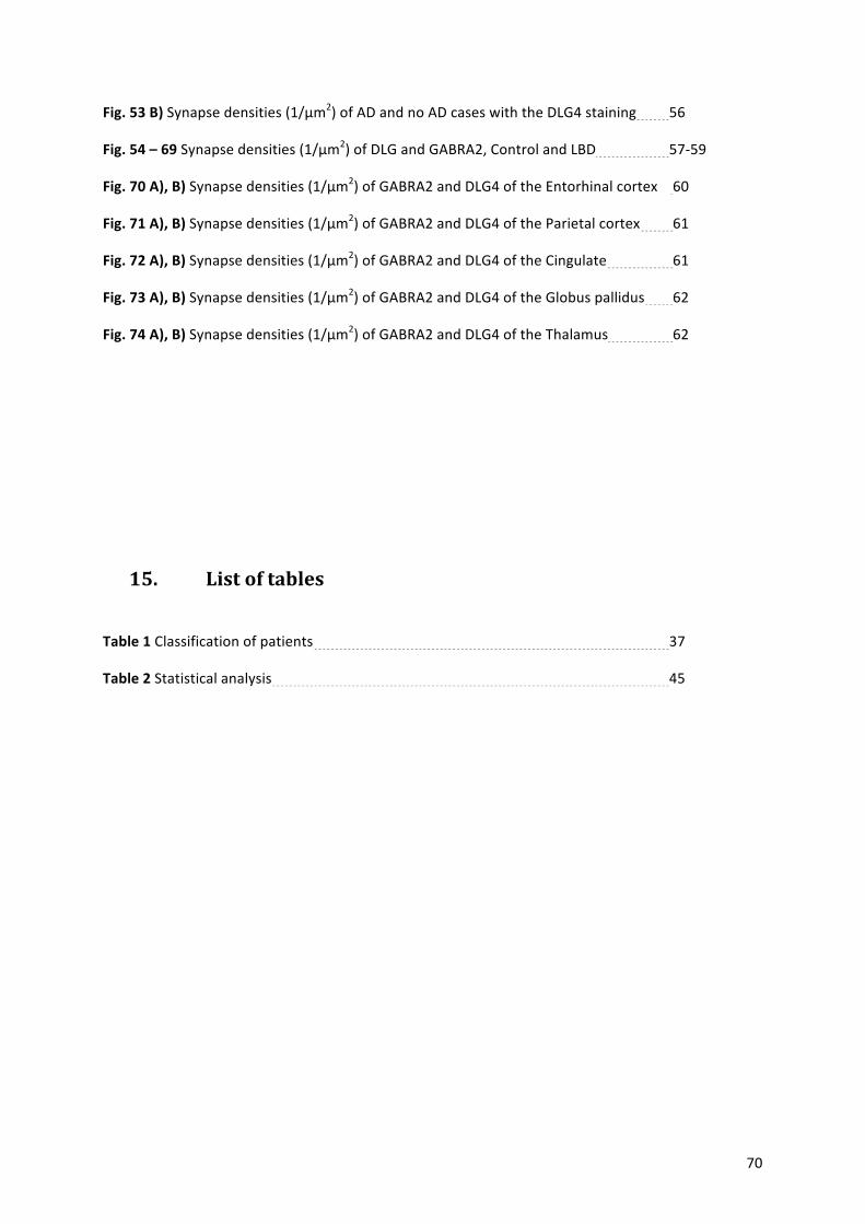
! 70!
Fig.%53%B)!Synapse!densities!(1/µm2)!of!AD!and!no!AD!cases!with!the!DLG4!staining!! 56!
Fig.%54%–%69!Synapse!densities!(1/µm2)!of!DLG!and!GABRA2,!Control!and!LBD! ! 57a59!
Fig.%70%A),%B)!Synapse!densities!(1/µm2)!of!GABRA2!and!DLG4!of!the!Entorhinal!cortex! !60!
Fig.%71%A),%B)!Synapse!densities!(1/µm2)!of!GABRA2!and!DLG4!of!the!Parietal!cortex! !61!
Fig.%72%A),%B)!Synapse!densities!(1/µm2)!of!GABRA2!and!DLG4!of!the!Cingulate! ! !61!
Fig.%73%A),%B)!Synapse!densities!(1/µm2)!of!GABRA2!and!DLG4!of!the!Globus!pallidus! !62!
Fig.%74%A),%B)!Synapse!densities!(1/µm2)!of!GABRA2!and!DLG4!of!the!Thalamus! ! !62!
!
!
!
!
!
15. List-of-tables-!
Table%1!Classification!of!patients! ! ! ! ! ! ! 37!
Table%2!Statistical!analysis! ! ! ! ! ! ! ! 45!
! !
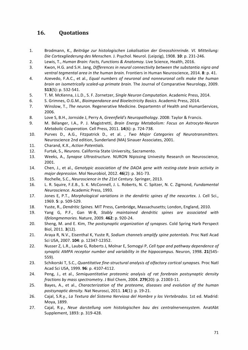
! 71!
16. Quotations-!
1.! Brodmann,! K.,$ Beiträge$ zur$ histologischen$ Lokalisation$ der$ Greosshirnrinde.$ VI.$ Mitteilung:$Die$Cortexgliederung$des$Menschen.!J.!Psychol.!Neurol.!(Leipzig),!1908.!10:!p.!231a246.!
2.! Lewis,!T.,!Human$Brain:$Facts,$Functions$&$Anatomoy.!Live!Science,!Health,!2016.!3.! Kwon,!H.G.!and!S.H.!Jang,!Differences$in$neural$connectivity$between$the$substantia$nigra$and$
ventral$tegmental$area$in$the$human$brain.!Frontiers!in!Human!Neuroscience,!2014.!8:!p.!41.!4.! Azevedo,! F.A.C.,! et! al.,!Equal$ numbers$ of$ neuronal$ and$nonneuronal$ cells$make$ the$ human$
brain$an$isometrically$scaledMup$primate$brain.!The!Journal!of!Comparative!Neurology,!2009.!513(5):!p.!532a541.!
5.! T.!M.!McKenna,!J.L.D.,!S.!F.!Zornetzer,!Single$Neuron$Computation.!Academic!Press,!2014.!6.! S.!Grimnes,!O.G.M.,!Bioimpendance$and$Bioelectricity$Basics.!Academic!Press,!2014.!7.! Winslow,!T.,!The$neuron.!Regenerative!Medicine.!Departemtn!of!Health!and!HumanServices,!
2006.!8.! Love!S,!B.H.,!Jornside!J,!Perry!A,!Greenfield's$Neuropathology.!2008:!Taylor!&!Francis.!9.! M.! Bélanger,! I.A.,! P.! J.! Magistretti,! Brain$ Energy$ Metabolism:$ Focus$ on$ AstrocyteMNeuron$
Metabolic$Cooperation.!Cell!Press,!2011.!14(6):!p.!724a738.!10.! Purves! D.,! A.G.,! Fitzpatrick! D.,! et! al.! ,! Two$ Major$ Categories$ of$ Neurotransmitters.!
Neuroscience!2nd!edition,!Sunderland!(MA)!Sinauer!Associates,!2001.!11.! Charand,!K.X.,!Action$Potentials.$12.! Furtak,!S.,!Neurons.!California!State!University,!Sacramento.!13.! Weeks,! A.,! Synapse$ Ultrastructure.! NURON! Nipissing! Univesity! Research! on! Neuroscience,!
2001.!14.! Chen,! J.,! et! al.,!Genotypic$ association$of$ the$DAOA$gene$with$ restingMstate$brain$activity$ in$
major$depression.!Mol!Neurobiol,!2012.!46(2):!p.!361a73.!15.! Rochelle,!S.C.,!Neuroscience$in$the$21st$Century.!Springer,!2013.!16.! L.!R.!Squire,!F.E.B.,!S.!K.!McConnell,!J.!L.!Roberts,!N.!C.!Spitzer,!N.!C.!Zigmond,!Fundamental$
Neuroscience.!Academic!Press,!1993.!17.! Jones! E,! P.T.,!Morphological$ variations$ in$ the$ dendirtic$ spines$ of$ the$ neocortex.! J.! Cell! Sci.,!
1969.!5:!p.!509a529.!18.! Yuste,!R.,!Dendritic$Spines.!MIT!Press,!Cambridge,!Massachusetts;!London,!England,!2010.!19.! Yang! G,! P.F.,! Gan! WaB,! Stably$ maintained$ dendritic$ spines$ are$ associated$ with$
lifelongmemories.!Nature,!2009.!462:!p.!920a24.!20.! Sheng,!M.!and!E.!Kim,!The$postsynaptic$organization$of$synapses.!Cold!Spring!Harb!Perspect!
Biol,!2011.!3(12).!21.! Araya!R,!N.V.,!Eisenthal!K,!Yuste!R,!Sodium$channels$amplify$spine$potentials.!Proc!Natl!Acad!
Sci!USA,!2007.!104:!p.!12347a12352.!22.! Nusser!Z,!L.R.,!Laube!G,!Roberts!J,!Molnar!E,!Somogyi!P,!Cell$type$and$pathway$dependence$of$
synaptic$AMPA$receptor$number$and$variability$ in$the$hippocampus.!Neuron,!1998.!21(545a559).!
23.! Schikorski!T,!S.C.,!Quantitative$fineMstructural$analysis$of$olfactory$cortical$synapses.!Proc!Natl!Acad!Sci!USA,!1999.!96:!p.!4107a4112.!
24.! Peng,! J.,! et! al.,! Semiquantitative$ proteomic$ analysis$ of$ rat$ forebrain$ postsynaptic$ density$fractions$by$mass$spectrometry.!J!Biol!Chem,!2004.!279(20):!p.!21003a11.!
25.! Bayes,! A.,! et! al.,! Characterization$ of$ the$ proteome,$ diseases$ and$ evolution$ of$ the$ human$postsynaptic$density.!Nat!Neurosci,!2011.!14(1):!p.!19a21.!
26.! Cajal,!S.R.y.,!La$Textura$del$Sistema$Nerviosa$del$Hombre$y$ los$Vertebrados.!1st!ed.!Madrid:!Moya,!1899.!
27.! Cajal,! R.y.,! Neue$ darstellung$ vom$ histologischen$ bau$ des$ centralnervensystem.! AnatAbt!Supplement,!1893:!p.!319a428.!
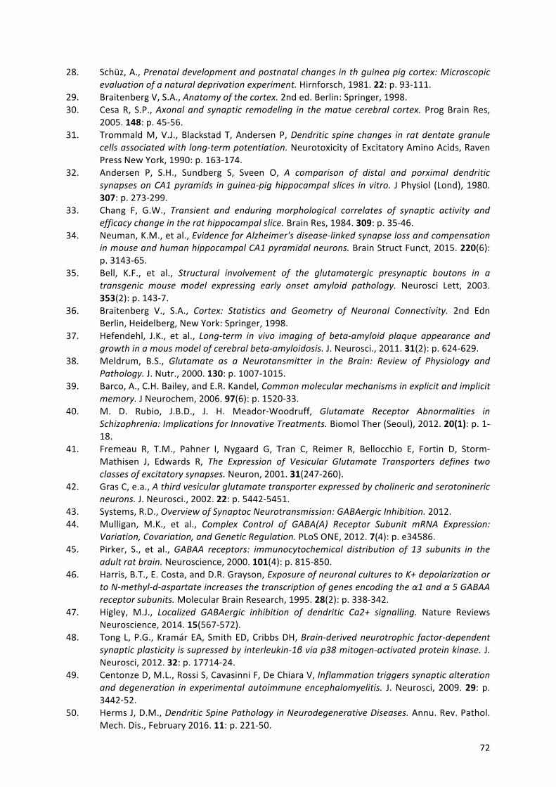
! 72!
28.! Schüz,!A.,!Prenatal$development$and$postnatal$changes$in$th$guinea$pig$cortex:$Microscopic$evaluation$of$a$natural$deprivation$experiment.!Hirnforsch,!1981.!22:!p.!93a111.!
29.! Braitenberg!V,!S.A.,!Anatomy$of$the$cortex.!2nd!ed.!Berlin:!Springer,!1998.!30.! Cesa!R,! S.P.,!Axonal$ and$ synaptic$ remodeling$ in$ the$matue$ cerebral$ cortex.! Prog!Brain!Res,!
2005.!148:!p.!45a56.!31.! Trommald!M,!V.J.,! Blackstad!T,!Andersen!P,!Dendritic$ spine$ changes$ in$ rat$dentate$granule$
cells$associated$with$longMterm$potentiation.!Neurotoxicity!of!Excitatory!Amino!Acids,!Raven!Press!New!York,!1990:!p.!163a174.!
32.! Andersen! P,! S.H.,! Sundberg! S,! Sveen! O,! A$ comparison$ of$ distal$ and$ porximal$ dendritic$synapses$on$CA1$pyramids$ in$guineaMpig$hippocampal$ slices$ in$vitro.! J!Physiol! (Lond),!1980.!307:!p.!273a299.!
33.! Chang! F,! G.W.,! Transient$ and$ enduring$ morphological$ correlates$ of$ synaptic$ activity$ and$efficacy$change$in$the$rat$hippocampal$slice.!Brain!Res,!1984.!309:!p.!35a46.!
34.! Neuman,!K.M.,!et!al.,!Evidence$for$Alzheimer's$diseaseMlinked$synapse$loss$and$compensation$in$mouse$and$human$hippocampal$CA1$pyramidal$neurons.!Brain!Struct!Funct,!2015.!220(6):!p.!3143a65.!
35.! Bell,! K.F.,! et! al.,! Structural$ involvement$ of$ the$ glutamatergic$ presynaptic$ boutons$ in$ a$transgenic$ mouse$ model$ expressing$ early$ onset$ amyloid$ pathology.! Neurosci! Lett,! 2003.!353(2):!p.!143a7.!
36.! Braitenberg! V.,! S.A.,! Cortex:$ Statistics$ and$ Geometry$ of$ Neuronal$ Connectivity.! 2nd! Edn!Berlin,!Heidelberg,!New!York:!Springer,!1998.!
37.! Hefendehl,! J.K.,! et! al.,! LongMterm$ in$ vivo$ imaging$ of$ betaMamyloid$ plaque$ appearance$ and$growth$in$a$mous$model$of$cerebral$betaMamyloidosis.!J.!Neurosci.,!2011.!31(2):!p.!624a629.!
38.! Meldrum,! B.S.,! Glutamate$ as$ a$ Neurotansmitter$ in$ the$ Brain:$ Review$ of$ Physiology$ and$Pathology.!J.!Nutr.,!2000.!130:!p.!1007a1015.!
39.! Barco,!A.,!C.H.!Bailey,!and!E.R.!Kandel,!Common$molecular$mechanisms$in$explicit$and$implicit$memory.!J!Neurochem,!2006.!97(6):!p.!1520a33.!
40.! M.! D.! Rubio,! J.B.D.,! J.! H.! MeadoraWoodruff,! Glutamate$ Receptor$ Abnormalities$ in$Schizophrenia:$Implications$for$Innovative$Treatments.!Biomol!Ther!(Seoul),!2012.!20(1):!p.!1a18.!
41.! Fremeau! R,! T.M.,! Pahner! I,! Nygaard! G,! Tran! C,! Reimer! R,! Bellocchio! E,! Fortin! D,! StormaMathisen! J,! Edwards! R,! The$ Expression$ of$ Vesicular$ Glutamate$ Transporters$ defines$ two$classes$of$excitatory$synapses.!Neuron,!2001.!31(247a260).!
42.! Gras!C,!e.a.,!A$third$vesicular$glutamate$transporter$expressed$by$cholineric$and$serotonineric$neurons.!J.!Neurosci.,!2002.!22:!p.!5442a5451.!
43.! Systems,!R.D.,!Overview$of$Synaptoc$Neurotransmission:$GABAergic$Inhibition.!2012.!44.! Mulligan,! M.K.,! et! al.,! Complex$ Control$ of$ GABA(A)$ Receptor$ Subunit$ mRNA$ Expression:$
Variation,$Covariation,$and$Genetic$Regulation.!PLoS!ONE,!2012.!7(4):!p.!e34586.!45.! Pirker,! S.,! et! al.,!GABAA$ receptors:$ immunocytochemical$ distribution$ of$ 13$ subunits$ in$ the$
adult$rat$brain.!Neuroscience,!2000.!101(4):!p.!815a850.!46.! Harris,!B.T.,!E.!Costa,!and!D.R.!Grayson,!Exposure$of$neuronal$cultures$to$K+$depolarization$or$
to$NMmethylMdMaspartate$increases$the$transcription$of$genes$encoding$the$α1$and$α$5$GABAA$receptor$subunits.!Molecular!Brain!Research,!1995.!28(2):!p.!338a342.!
47.! Higley,! M.J.,! Localized$ GABAergic$ inhibition$ of$ dendritic$ Ca2+$ signalling.! Nature! Reviews!Neuroscience,!2014.!15(567a572).!
48.! Tong!L,!P.G.,!Kramár!EA,!Smith!ED,!Cribbs!DH,!BrainMderived$neurotrophic$ factorMdependent$synaptic$plasticity$is$supressed$by$interleukinM1β$via$p38$mitogenMactivated$protein$kinase.!J.!Neurosci,!2012.!32:!p.!17714a24.!
49.! Centonze!D,!M.L.,!Rossi!S,!Cavasinni!F,!De!Chiara!V,!Inflammation$triggers$synaptic$alteration$and$degeneration$ in$ experimental$ autoimmune$encephalomyelitis.! J.!Neurosci,! 2009.!29:! p.!3442a52.!
50.! Herms!J,!D.M.,!Dendritic$Spine$Pathology$in$Neurodegenerative$Diseases.!Annu.!Rev.!Pathol.!Mech.!Dis.,!February!2016.!11:!p.!221a50.!
! 73!
51.! Pham,!E.,!et!al.,!Progressive$accumulation$of$amyloidMbeta$oligomers$ in$Alzheimer's$disease$and$ in$amyloid$precursor$protein$ transgenic$mice$ is$accompanied$by$selective$alterations$ in$synaptic$scaffold$proteins.!FEBS!J,!2010.!277(14):!p.!3051a67.!
52.! Scheff,! S.W.,! et! al.,! Synaptic$ change$ in$ the$ posterior$ cingulate$ gyrus$ in$ the$ progression$ of$Alzheimer's$disease.!Journal!of!Alzheimer's!Disease,!2015.!43(3):!p.!1073a1090.!
53.! Winklhofer,! K.F.! and! C.! Haass,!Mitochondrial$ dysfunction$ in$ Parkinson's$ disease.! Biochim!Biophys!Acta,!2010.!1802(1):!p.!29a44.!
54.! Prof!Martin!Prince,!P.A.W.,!Dr.!Maëlenn!Guerchet,!Miss!GemmaaClaire!All,!Dr.!YuaTzu!Wu,!Dr.!Matthew!Rina,!World$Alzheimer$Report$2015$M$The$Global$Impact$of$Dementia.!August!2015.!Alzheimer's%Disease%International%(ADI)%London.!
55.! McKeith,! I.G.,! Clinical$ Lewy$ Body$ Syndroms.! Annals! of! the! New! York! Ycademy! of! Science,!2000.!920(The!Molecular!Basis!of!Dementia):!p.!1a8.!
56.! Christie,! R.H.,! et! al.,!Growth$ Arrest$ of$ Individual$ Senile$ Plaques$ in$ a$Model$ of$ Alzheimer's$Disease$Observed$by$In$Vivo$Multiphoton$Microscopy.!J.!Neurosci.,!2001.!21(3):!p.!858a864.!
57.! Bickel,! H.,!Das$Wichtigste$ M$ Die$ Häufigkeit$ von$ Demenzerkrankungen.! Deutsche! Alzheimer!Gesellschaft,!2015.!
58.! SerranoaPozo,!A.,!et!al.,!APOEMε2$is$associated$with$milder$clinical$and$pathological$Alzheimer$disease.!Ann!Neurol,!2015.!77(6):!p.!917a29.!
59.! Jan!Näslund,!V.H.,!Richard!Mohs,!Kenneth!L.!Davis,!Peter!Davies,!Paul!Greengard,!Joseph!D.!Buxbaum,!Correlatin$between$elevated$levels$of$amyloid$βMpeptide$in$the$brain$and$cognitive$decline.!JAMA,!2000.!283:!p.!1571a!1577.!
60.! G.!William!Rebeck,!J.S.R.,!Dudley!K.!Strickland,!Bradley!T.!Hyman,!Apolioprotein$E$in$Sporadic$Alzheimer's$Disease:$Allelic$Variation$and$Receptor$Interactions.!Neuron,!October!1993.!11:!p.!575a580.!
61.! Castellano! JM,! K.J.,! Stewart! FR,! Jiang! H,! DeMattos! RB,!Human$ apoE$ isoforms$ differentially$regulate$brain$amyloidMβ$peptide$clearance.!Sci.!Transl.!Med.,!2011.!3:!p.!.!
62.! Sherringtion,! Cloning$ of$ a$ gene$ bearing$ missense$ mutations$ in$ earlyMonset$ familial$Alzheimer's$disease.!NATURE,!1995.!375:!p.!754a760.!
63.! LevyaLahad,! Candidate$ Gene$ for$ the$ Chromosome$ 1$ familial$ Alzheimer's$ disease$ locus.!SCIENCE,!1995.!269:!p.!973a977.!
64.! Tomita,! M.,! Saido,! Kume,! Shinozaki,! Tokuhiro,! Capell,! Walter,! Grünberg,! Haass,! Iwatsubo,!Obata,!The$presenilin$2$mutation$(N141I)$linked$to$familial$Alzheimer$disease$(Volga$German$families)$ increases$ the$ secretion$of$amyloid$β$protein$ending$at$ the$42nd$ (or$43rd)$ residue.!Proc.!Natl.!Acad.!Sci.!USA,!1997.!94:!p.!2025a2030.!
65.! Busche,!M.A.,!et!al.,!Clusters$of$hyperactive$neurons$near$amyloid$plaques$in$a$mouse$model$of$Alzheimer's$disease.!Science,!2008.!321(5896):!p.!1686a9.!
66.! Findeis,!M.A.,!The$ role$ of$ amyloid$ beta$ peptide$ 42$ in$Alzheimer's$ disease.! Pharmacol! Ther,!2007.!116(2):!p.!266a86.!
67.! Mori! C,! S.E.,! Wisniewsk! KE,! Wisniewski! TM,! Yamaguch! H,! Saido! TC,! Tolan! DR,! Selkoe! DJ,!Lemere!CA,!Intraneuronal$Abeta42$accumulation$in$Down$syndrome$brain.!Amyloid,!2002.!9:!p.!88a102.!
68.! SerranoaPozo,!A.,!et!al.,!Neuropathological$alterations$in$Alzheimer$disease.!Cold!Spring!Harb!Perspect!Med,!2011.!1(1):!p.!a006189.!
69.! Suzuki! N,! C.T.,! Cai! X,! Odaka! A,! Otvos! L,! Eckman! C,! Golde! T,! Younkin! S,! An$ Increased$percentage$ of$ long$ Amyloid$ beta$ protein$ secreted$ by$ familial$ amyloid$ β$ protein$ precursor$(βAPP717)$Mutants.!SCIENCE,!1994.!264:!p.!1336a1340.!
70.! Younkin,!S.G.,!The$role$of$Aβ42$in$Alzheimer's$disease.!Elsevier,!Paris,!1998.!92:!p.!289a292.!71.! Thal! DR,! R.U.,! Orantes! M,! Braak! H,! Phases$ of$ AβMdeposition$ in$ the$ human$ brain$ and$ its$
relevance$for$the$development$of$AD.!Neurology,!2002.!58(1791a800).!72.! Gouras,!G.K.,!et!al.,!Intraneuronal$Aβ42$Accumulation$in$Human$Brain.!The!American!Journal!
of!Pathology,!2000.!156(1):!p.!15a20.!73.! Kamenetz! F,! T.T.,! Hsieh! H,! Seabrook! G,! Borchelt! D,!APP$ Processing$ and$ synaptic$ function.!
Neuron!2003.!37(925a37).!
! 74!
74.! Teng,!F.Y.!and!B.L.!Tang,!Widespread$gammaMsecretase$activity$in$the$cell,$but$do$we$need$it$at$the$mitochondria?!Biochem!Biophys!Res!Commun,!2005.!328(1):!p.!1a5.!
75.! Atwood,!C.S.,!et!al.,!AmyloidMβ:$a$chameleon$walking$ in$two$worlds:$a$review$of$the$trophic$and$toxic$properties$of$amyloidMβ.!Brain!Research!Reviews,!2003.!43(1):!p.!1a16.!
76.! Yoshiyama,! Y.,! et! al.,! Synapse$ Loss$ and$ Microglial$ Activation$ Precede$ Tangles$ in$ a$ P301S$Tauopathy$Mouse$Model.!Neuron,!2007.!53(3):!p.!337a351.!
77.! Busche,!M.A.,!et!al.,!Rescue$of$ longMrange$circuit$dysfunction$ in$Alzheimer's$disease$models.!Nat!Neurosci,!2015.!18(11):!p.!1623a30.!
78.! Dorostkar,!M.M.,!et!al.,!Analyzing$dendritic$spine$pathology$in$Alzheimer's$disease:$problems$and$opportunities.!Acta!Neuropathol,!2015.!130(1):!p.!1a19.!
79.! GarciaaMarin,! V.,! et! al.,! Diminished$ perisomatic$ GABAergic$ terminals$ on$ cortical$ neurons$adjacent$to$amyloid$plaques.!Front!Neuroanat,!2009.!3:!p.!28.!
80.! Gouras,! G.K.,! et! al.,! Intraneuronal$ betaMamyloid$ accumulation$ and$ synapse$ pathology$ in$Alzheimer's$disease.!Acta!Neuropathol,!2010.!119(5):!p.!523a41.!
81.! Dorostkar,!M.M.,!et!al.,! Immunotherapy$alleviates$amyloidMassociated$synaptic$pathology$ in$an$Alzheimer's$disease$mouse$model.!Brain,!2014.!137(Pt!12):!p.!3319a26.!
82.! Overk,!C.R.!and!E.!Masliah,!Pathogenesis$of$synaptic$degeneration$in$Alzheimer's$disease$and$Lewy$body$disease.!Biochem!Pharmacol,!2014.!88(4):!p.!508a16.!
83.! Renner,! M.,! et! al.,! Deleterious$ Effects$ of$ Amyloid$ β$ Oligomers$ Acting$ as$ an$ Extracellular$Scaffold$for$mGluR5.!Neuron,!2010.!66(5):!p.!739a754.!
84.! Cisse,!M.,!et!al.,!Reversing$EphB2$depletion$ rescues$ cognitive$ functions$ in$Alzheimer$model.!Nature,!2011.!469(7328):!p.!47a52.!
85.! Shankar,! G.M.,! et! al.,! Natural$ oligomers$ of$ the$ Alzheimer$ amyloidMbeta$ protein$ induce$reversible$ synapse$ loss$ by$ modulating$ an$ NMDAMtype$ glutamate$ receptorMdependent$signaling$pathway.!J!Neurosci,!2007.!27(11):!p.!2866a75.!
86.! White!A,!Z.H.,!Galatis!D,!Maher!F,!Hesse!L,!Multhaup!G,!Beyreuther!K,!Masters!C,!Cappai!R,!Survival$ of$ Cultured$ Neurons$ from$ Amyloid$ Precursor$ Protein$ KnockMOut$ mice$ against$Alzheimer's$AmyloidMβ$toxicity$and$oxidative$stress.!The!Journal!of!Neuroscience,!1998.!18:!p.!6207a6217.!
87.! De!Felice,!F.G.,!et!al.,!Alzheimer's$diseaseMtype$neuronal$tau$hyperphosphorylation$induced$by$A$beta$oligomers.!Neurobiol!Aging,!2008.!29(9):!p.!1334a47.!
88.! Koffie! RM,! H.T.,! Tai! HaC,! Kay! KR,! SerranoaPozo! A,$ Apolipoprotein$ E4$ effects$ in$ Alzheimer’s$disease$are$mediated$by$synaptotoxic$oligomeric$amyloidMβ.!Brain,!2012.!135:!p.!2155–68.!
89.! Robert! D.! Terry,! E.M.,! David! P.! Salmon,! Nelson! Butters,! Richard! DeTeresa,! Robert! Hill,!Lawrence!A.!Hansen,!Robert!Katzman,!Physical$Basis$of$Cognitive$Alterations$ in$Alzheimer's$Disease:$Synpse$Loss$Is$the$Major$Correlate$of$Cognitive$Impairment.!Ann!Neurol,!1991.!30:!p.!572a580.!
90.! SantaCruz! K,! L.J.,! Spires! T,! Paulson! J,! Kotilinek! L,! Ingelsson! M,! Guimaraes! A,! DeTure! M,!Ramsden!M,!McGowan!E,!Forster!C,!Yue!M,!Orne!J,!Janus!C,!Mariash!A,!Kuskowski!M,!Hyman!B,! Hutton! M,! Ashe! K,! Tau$ suppression$ in$ a$ neurodegenerative$ mouse$ model$ improves$memory$function.!Science,!2005.!309:!p.!476.!
91.! Scheff,!S.W.,! J.H.!Neltner,!and!P.T.!Nelson,! Is$synaptic$ loss$a$unique$hallmark$of$Alzheimer's$disease?!Biochem!Pharmacol,!2014.!88(4):!p.!517a28.!
92.! Braak!H,!T.D.,!Ghebremedhin!E,!Del!Tredici!K,!Stages$of$the$pathologic$process$ in$Alzheimer$disease:$age$categories$from$1$to$100$years.!N.!Neuropathol.!Exp.!Neurol.,!2011.!70(960a69).!
93.! M.!Goedert,!M.G.S.,!K.!Del!Tredici,!H.!Braak,!100$years$of$ Lewy$pathology.!Nature!Reviews!Neurology,!2013.!9:!p.!13a24.!
94.! M.! Jucker,! L.C.W.,!SelfMpropagation$of$ pathogenic$ protein$aggregates$ in$ neurodegenerative$diseases.!Nature,!2013.!501:!p.!45a51.!
95.! W.!M.! Saxton,! P.J.H.,!The$ axonal$ transport$ of$mitochondria.! J! Cell! Sci,! 2012.!125:! p.! 2095a2104.!
96.! J.!A.! Javitch,!R.J.D.A.,! S.!M.!Strittmatter,!S.!H.!Snyder,!ParkinsonismMinducing$neurotoxin,$NMmethylM4MphenylM1,2,3,6Mtetrahydropyridine:$ uptake$ of$ the$ metabolite$ NMmethylM4M
! 75!
phenylpyridine$by$dopamine$neurons$explains$selective$toxicity.!Proc!Natl!Acad!Sci!USA,!1985.!82:!p.!2173a2177.!
97.! Nicklas,! W.J.,! I.! Vyas,! and! R.E.! Heikkila,! Inhibition$ of$ NADHMlinked$ oxidation$ in$ brain$mitochondria$ by$ 1MmethylM4MphenylMpyridine,$ a$ metabolite$ of$ the$ neurotoxin,$ 1MmethylM4MphenylM1,2,5,6Mtetrahydropyridine.!Life!Sci,!1985.!36(26):!p.!2503a8.!
98.! Morris,! J.C.,! et! al.,! Cerebral$ amyloid$ deposition$ and$ diffuse$ plaques$ in$ "normal''$ aging:$Evidence$ for$ presymptomatic$ and$ very$mild$Alzheimer's$ disease.!Neurology,! 1996.!46(3):! p.!707a719.!
99.! Shankar,! G.M.! and! D.M.!Walsh,! Alzheimer's$ disease:$ synaptic$ dysfunction$ and$ Abeta.! Mol!Neurodegener,!2009.!4:!p.!48.!
100.! Stephen!W.!Scheff,!D.A.P.,!D.!Larry!Sparks,!Quantitative$assemssment$of$possible$ageMrelated$change$ in$ synaptic$ numbers$ in$ the$ human$ frontal$ cortex.! ELSEVIER,! February! 2001.!Neurobiology%of%Aging%22:!p.!355a365.!
101.! Woolley!C,!G.E.,!Frankfurt!M,!McEwen!B,!Naturally$occurring$ fluctauation$ in$dendritic$spine$density$onadult$hippocampla$pyramidal$neurons.!J!Neurosci!1990.!10:!p.!4035a4039.!
102.! Takeichi,!M.,!Cadherins:$A$molecular$family$important$in$selective$cellMcell$adhesion.!Annu!Rev!Biochem,!1990.!59(237a252).!
103.! Ethell! I,! I.F.,! Kalo!M,! Couchman! J,! Pasquale! E,! Yamaguchi! Y,! EphB/syndecanM2$ siganling$ in$dendritic$spine$morphogenesis.!Neuon,!2001.!31:!p.!1001a1013.!
104.! Horch!H,! K.A.,! Portbury! S,! Katz! L,!Destabilization$ of$ cortical$ dendrites$ and$ spines$ by$ BDNF.!Neuron,!1999.!23(353a364).!
105.! Craig!A,!B.H.,!Molecular$heterogenety$of$central$synapses:$Afferent$and$target$regulation.!Nat!Neurosci,!2001.!4(569a578).!
106.! Hering!H,!S.M.,!Dendritic$spines:$Structure,$dynamics$and$regulation.!Nat!Rev!Neurosci,!2001.!2(880a888).!
107.! Robinson! T,! K.B.,! Alterations$ in$ the$ morphology$ of$ dendrites$ and$ dendritic$ spines$ in$ the$nucleus$accumbens$and$prefrontal$crotex$following$repeated$treatment$with$amphetamine$or$cocaine.!Eur!J!Neurosci,!1999.!11:!p.!1598a1604.!
108.! Loreth,!D.,!et!al.,!Selective$degeneration$of$septal$and$hippocampal$GABAergic$neurons$ in$a$mouse$model$of$amyloidosis$and$tauopathy.!Neurobiol!Dis,!2012.!47(1):!p.!1a12.!
109.! Young,!A.,!Cortical$amino$acidergic$pathways$in$Alzheimer's$disease.!J!Neural!Transm!Suppl.,!1987.!24:!p.!147a52.!
110.! W.!Satzger,!H.H.,!F.!Padberg,!K.!Bürger,!Th.!Nolde,!G.! Ingrassia,!R.!R.!Engel,!Zur$praktischen$Anwendung$ der$ CERADM$ Testbattereie$ als$ neuropsychologisches$ Demenzscreening.!Nervenarzt,!2001.!72:!p.!196a203.!
111.! M.! Pflüger,! C.A.,! A.! U.! Monsch,! Klinische$ Untersuchungsverfahren.! Zeitschrift! für! Klinische!Psychologie!und!Psychotherapie,!2003.!32(1):!p.!64a67.!
112.! Dorostkar,!M.M.,!et!al.,!Computational$processing$of$optical$measurements$of$neuronal$and$synaptic$activity$in$networks.!J!Neurosci!Methods,!2010.!188(1):!p.!141a50.!
113.! Palop,! J.J.,! et! al.,! Aberrant$ excitatory$ neuronal$ activity$ and$ compensatory$ remodeling$ of$inhibitory$hippocampal$circuits$in$mouse$models$of$Alzheimer's$disease.!Neuron,!2007.!55(5):!p.!697a711.!
114.! Khundakar,! A.A.,! et! al.,! Analysis$ of$ primary$ visual$ cortex$ in$ dementia$ with$ Lewy$ bodies$indicates$ GABAergic$ involvement$ associated$ with$ recurrent$ complex$ visual$ hallucinations.!Acta!Neuropathologica!Communications,!2016.!4(1).!
115.! Fatemi,! S.H.,! et! al.,!Expression$ of$GABAA$alpha2M,$ beta1M$ and$ epsilonMreceptors$ are$ altered$significantly$ in$ the$ lateral$ cerebellum$of$ subjects$with$ schizophrenia,$major$ depression$ and$bipolar$disorder.!Transl!Psychiatry,!2013.!3:!p.!e303.!
116.! ThompsonaVest,!N.M.,!et!al.,!GABA(A)$receptor$subunit$and$gephyrin$protein$changes$differ$in$the$globus$pallidus$in$Huntington's$diseased$brain.!Brain!Res,!2003.!994(2):!p.!265a70.!
117.! Mizukami,!K.,!et!al.,!GABAA$receptor$beta$2$and$beta$3$subunits$mRNA$ in$ the$hippocampal$formation$of$aged$human$brain$with$AlzheimerMrelated$neuropathology.!Brain!Res!Mol!Brain!Res,!1998.!56(1a2):!p.!268a72.!
! 76!
118.! Williams,! C.,! et! al.,!Transcriptome$Analysis$ of$ Synaptoneurosomes$ Identifies$Neuroplasticity$Genes$Overexpressed$in$Incipient$Alzheimer's$Disease.!PLoS!ONE,!2009.!4(3):!p.!e4936.!
!
!
!
!
!
!
!
!
!
!
!
!
!
!
!
!
!
!
!
!
!
!
!
!
!
!
!

! 77!
!
!