Embed Size (px)
Citation preview
[1]
MASTER OF MEDICINE IN THE SPECIALTY OF
NEUROLOGICAL SURGERY
(MMC016)
RESEARCH DISSERTATION
(For M07.2 Part II NEUS7008 – Research Report)
Title:
Analysis of the histological types of intracranial tumours at the
Chris Hani Baragwanath Hospital: 2004 – 2009
Researcher:
Dr. Shamil M. Gowan
Supervisor:
Dr. Vinod Goolab
Division: Neurosurgery
Department: Neurosciences
School of Clinical Medicine
Faculty of Health Sciences
University of the Witwatersrand
[2]
DECLARATION
I, Shamil Manilal Gowan, declare that this research report is my own work. It is being submitted for the degree of Master of Medicine (Neurosurgery) of the University of the Witwatersrand, Johannesburg. It has not been submitted before for any degree or examination at this or any other University. ________________________________ (Signature) _________ day of ________________ 2013.
[3]
ABSTRACT
Object: There is currently insufficient epidemiological data regarding the incidence of intracranial tumours in South Africa and in particular regionally, within Gauteng province. In this study, we aim to describe epidemiological data pertaining to histologically confirmed intracranial tumours at a large neurosurgery centre in Johannesburg, South Africa and to compare this data to similar international series. In addition, we will establish the frequency of different histological types of tumours and identify differences in prevalence of these tumours when the sample group has been stratified by age , race and gender. Methods: Data collection was via a retrospective analysis of patient records, operating theatre registries and histopathological reports for patients diagnosed and treated at the Chris Hani Baragwanath Hospital between 2004 and 2009. Descriptive statistical analysis was performed using variables: age, gender, race and histological types of tumours Conclusion: At our facility, the most common intracranial tumours noted were meningiomas, pituitary adenomas, glioblastomas, medulloblastomas and craniopharyngiomas. Black patients showed higher frequencies of
[4]
meningeal tumours and sellar region tumours. Tumours originating from the cranial nerves and meninges were commoner in females. Glioblastomas and craniopharyngiomas displayed a male predominance. Intracranial tumours in general occurred in younger patients at our facility than noted in the current literature. This trend was especially evident in glioblastomas and meningiomas. The benefits of both national and regional intracranial tumour registries in South Africa, would include more accurate estimates of the tumour burden and better resource allocation.
[5]
ACKNOWLEDGEMENTS
Special thanks to Dr. V. Goolab and the Department of Neurosurgery, Chris Hani Baragwanath Hospital for their assistance in general and for access to records used in this study. Without the invaluable assistance in statistical analysis provided by Nevil Cursedji, this study would not have been possible.

[6]
TABLE OF CONTENTS
Title page 1 Declaration 2 Abstract 3 Acknowledgements 5 Table of Contents 6 List of Figures 9 List of Tables 10 List of Abbreviations 10 Definition of Terms 10
Chapter 1 1.0 Introduction 12
1.1 Background 12
1.2 Problem Statement 15 1.3 Research Questions and Objectives
1.3.1 Research Question 15
1.3.2 Specific Objectives 16 1.4 Significance 16
Chapter 2 2.0 Literature Review 18
2.1 Histological types of Intracranial Tumours 18
2.2 Age Distribution of Intracranial Tumours 24 2.3 Gender Distribution of Intracranial Tumours 27 2.4 Racial Distribution of Intracranial Tumours 30

[7]
Chapter 3 3.0 Methodology 32
3.1 Study Setting 32
3.2 Study Design 33 3.3 Study Population 33
3.3.1 Inclusion Criteria 33
3.3.2 Exclusion Criteria 34 3.4 Instruments 34
3.5 Data Analysis 34 3.6 Ethical Considerations 36
Chapter 4 4.0 Results 37
4.1 Introduction 37 4.2 Description of Study Participants 37
4.3 Histological Types of Tumours 38 4.4 Age Distribution of Intracranial Tumours 41 4.5 Gender Distribution of Intracranial Tumours 50 4.6 Racial Distribution of Intracranial Tumours 52
[8]
Chapter 5 5.0 Discussion 58
5.1 Histological Types of Tumours 58
5.2 Age Distribution of Intracranial Tumours 65 5.3 Gender Distribution of Intracranial Tumours 70 5.4 Racial Distribution of Intracranial Tumours 75 5.5 Limitations of the Study 80
Chapter 6 6.0 Conclusion and Recommendations 82
6.1 Introduction 82
6.2 Conclusion 83 6.3 Recommendations 87
References 88
[9]
LIST OF FIGURES
Figure 1. Distribution of intracranial tumours by histological type Figure 2. Histological subtypes of intracranial meningiomas Figure 3. Distribution of intracranial tumours by age showing
differences between male and female patients Figure 4. Mean age of patients diagnosed with different
histological types of intracranial tumours Figure 5. Intracranial tumour distribution by age showing differences in occurrences within race groups Figure 6. Age distribution of craniopharyngiomas diagnosed at Chris Hani Baragwanath Hospital Figure 7. Absolute ratios of intracranial tumours in males and females Figure 8. Differences in relative risks for selected histological types of
intracranial tumours occurring in males and females. Figure 9. Distribution of intracranial tumours stratified by race Figure 10. Comparison of absolute numbers of selected histological
types of intracranial tumours occurring in black and white patients
Figure 11. Relative risks for selected histological types of intracranial
tumours occurring in different race groups.

[10]
Figure 12. Distribution of histologically confirmed intracranial tumours in black patients Figure 13. Distribution of histologically confirmed intracranial tumours
in white patients
LIST OF TABLES
Table1. Absolute numbers of diagnosed cases of
histologically confirmed intracranial tumours and the mean age of the patient at the time of surgery
ANNEXURES
Annexure 1 : Data Collection Sheets Annexure 2 : Ethics Committee Clearance Certificate Annexure 3 : Letter from Chris Hani Baragwanath Hospital authorizing Research Project
[11]
LIST OF ABBREVIATIONS
ALL : Acute Lymphoblastic Leukemia CBTRUS : Central Brain Tumour Registry of the United States CNS : Central Nervous System CT : Computer Tomography GBM : Glioblastoma Multiforme HLA : Human Leukocyte Antigen PNET: Primitive Neuro-ectodermal Tumour RR : Relative Risk SEER : Surveillance, Epidemiology and End Results Program
(The National Cancer Institute) UK : United Kingdom USA : United States of America
DEFINITION OF TERMS
Bimodal : having two maximal levels. Glioma : refers only to astrocytic tumours. Histology : department dealing with a tissue’s minute structure and
composition Tumourigenesis : production of tumour.
[12]
CHAPTER 1
1.0 INTRODUCTION
1.1 BACKGROUND
Intracranial tumours are a heterogeneous group of neoplasms that vary widely in terms of site of origin, pathological or morphological grouping, location, presenting features, growth potential and tendency for recurrence. These intracranial tumours, although comprising a relative small percentage of malignancies globally, have particular significance due to their associated severe morbidity and mortality . The following are examples of the types of tumours found intracranially:
1. Astrocytoma 2. Meningioma 3. Pituitary Adenoma 4. Craniopharyngioma 5. Haemangioblastoma 6. Medulloblastoma
Although epidemiological data and analysis regarding intracranial tumours in developed countries is relatively abundant, very little work of this nature is available from African Countries and in particular, from South Africa. This project aims to address this data problem by collating
[13]
demographic information regarding all of the intracranial tumours noted in patients being treated at the largest tertiary institute in Johannesburg, viz. the Chris Hani Baragwanath Hospital. Most global cancer registries and studies have been limited to tumours with malignant characteristics. However, due to their anatomical locations – i.e. intracranial - these tumours are associated with significant morbidity and poor prognosis, irrespective of whether they are benign or malignant [1,2]. This being so: the distinction of whether the tumour is benign or malignant has little clinical value; and thus the project to study intracranial tumours at Chris Hani Baragwanath Hospital will include all of the histologically confirmed types of tumours. Epidemiological data provided by centralized tumour registries such as: 1. The Central Brain Tumour Registry of the United States
(CBTRUS ) and 2. The National Cancer Institute’s: Surveillance, Epidemiology
and End Results Program (SEER ), are collected with two guiding principles: Firstly, to comprehensively describe the full spectrum of these
[14]
neoplasms, and secondly, to provide clinically relevant data analysis to neuroscientists; allowing them to predict future estimates of intracranial tumours in specifically targeted or selected population groups . Although the full demographic spectrum of brain tumours has been described in a number of publications, most of this work has been limited to the Western world. In contrast to the population of South Africa, these Western countries tend to have populations comprising mainly of Caucasians. International studies have shown a significant variance in the incidence of tumours of the central nervous system between different races [1,3,4]. When studying and comparing the incidence of intracranial tumours in Asia with global trends , it was noted that certain histological types of neoplasms, in particular meningiomas, occurred far more frequently than elsewhere in the world [5]. The authors have surmised that this was possibly due to a genetic predisposition amongst these majority Oriental ethnic groups.
[15]
Similarly, it could be hypothesized that with the differing population demographic of South Africa, the spectrum of intracranial neoplastic disease may also differ significantly. Furthermore, most of the epidemiological work has been done in countries with an increasing aging population - which may explain the high prevalence of intracranial tumours in those countries - which are known to occur in the older patient groups [6]. It should also be noted that the incidence of intracranial tumours may differ widely between different areas/regions within a country, as was noted when comparing the occurrence of brain tumours in Zaragoza and Navarre in Spain - with that of the country as a whole [7]. These differences are thought to be due to environmental exposure to carcinogens and/ or radiation in the different areas [7,8].
1.2 PROBLEM STATEMENT
There is currently insufficient epidemiological data regarding the incidence of intracranial tumours in South Africa and in particular regionally , within Gauteng province.
1.3 RESEARCH QUESTIONS AND OBJECTIVES
1.3.1 Research Question
Which are the prevalent intracranial tumours diagnosed in patients being treated at the Chris Hani Baragwanath Hospital’s department of
[16]
Neurosurgery and what are the notable patterns when this data is stratified by age , gender and race.
1.3.2 Specific Objectives
To describe epidemiological data pertaining to histologically confirmed intracranial tumours at a large neurosurgery centre in Johannesburg, South Africa and to compare this data to similar international series. In particular , the study aims to : 1. Establish frequency of different histological types of tumours 2. Identify differences in prevalence of these tumours when the sample group has been stratified by age , race and gender.
1.4 SIGNIFICANCE
An epidemiologic study of the incidence of intracranial tumours performed at the Department of Neurosurgery, Chris Hani Baragwanath Hospital, would quantify the impact of these neoplasms on the population served by that health care facility. In addition, this project aims to identify particular demographic groups most at risk from these debilitating diseases. Information of this nature could be used to formulate management strategies on a broader scale, for instance, screening programs for those individuals deemed to have a significant risk of developing an intracranial neoplasm . The identification of high as well as low

[17]
risk groups could be used to justify utilization of the available, but limited resources. The nature of this study allows it to be used as a foundation for further research, especially as a gauge of the success of future treatment or preventative strategies.
[18]
CHAPTER 2
2.0 LITERATURE REVIEW
2.1 HISTOLOGICAL TYPES OF INTRACRANIAL TUMOURS
Numerous studies have been performed describing the epidemiology of intracranial tumours. Some, like the CTBRUS and SEER studies, examine these parameters on a national scale by collating data originating from the local registries of participating counties within the USA [2,4]. Other epidemiological studies have focused on particular provinces, for example a single prefecture of Japan, Kumamoto [9], or comparisons with local regions [7]. A detailed examination of these different types of epidemiological studies allows one to grasp the overall trends in the predominant histological types of intracranial tumours occurring both on the national and local scale. Furthermore, this allows for the identification of “tumour hotspots” for particular types of tumours globally or within a particular region [10]. Most of the current research of this nature has emanated from western countries, in particular the USA, with a few reports available from Asia. By stark contrast, very little information of this nature is forthcoming from South Africa or the African continent as a whole. Most American and European studies have noted the most common intracranial group as being those tumours arising from neuro-epithelial
[19]
tissue [6,11,12,13,14,15,16,17,18,19]. Caution should be exercised when comparing these studies , as the term ‘glioma’ is often used. This term is occasionally used to refer to all tumours of glial lineage, however , in the modern context ,the term refers to astrocytic tumours [20]. Careful inspection of the tumour classifications used, distinguishes between the two groups and allows accurate comparisons to be made. Some studies from the 1990’s suggest that these tumours may account for as much as 87% of all intracranial tumours recorded, with the majority of these being malignant [13].McKinney suggested that tumours of neuro-epithelial origin, including astrocytomas, glioblastomas, oligodendroglioma and unspecified gliomas , accounted for 86% of supratentorial intracranial tumours recorded in the UK [15]. A review of intracranial tumours occurring in the Labin region of Croatia over a twenty eight year period , noted that the most frequently occurring primary intracranial tumour were those of neuroepithelial origin , accounting for 58,3% of all diagnosed CNS neoplasms [17]. In their five year review of intracranial tumours recorded in the Central Brain Tumour Registry of the United States (CBTRUS) during the early 1990’s , Surawicz , et al , found that neuroepithelial tumours accounted for over one half of all primary brain and CNS tumours in the USA [2]. Of the 221 tumours diagnosed in a French study, 47,5% were of neuro- epithelial origin [18]. In the Million Women Study performed in the UK, of the 1563 patients diagnosed with primary CNS tumours , 646 were
[20]
classified as gliomas , of which 98% were considered malignant. [19] . The incidence rate of gliomas in Finland , 4,6/100000 , was considered higher than that observed in other countries. One study suggested that 25% of all gliomas were oligodendrogiomas [21]. Within this group of tumours , many reports suggest that the most common specific histological type of tumour is the glioblastoma multiforme, sometimes referred simply as glioblastoma [4,11,22,23] . This particular type of tumour is well known to be the most aggressive and have the poorest prognosis of intracranial tumours [4,11,15]. 47% of 331 diagnosed gliomas (tumours of neuro-epithelial origin) in a Finnish study were classified as glioblastoma [ 22]. A review of six tumour registries in the USA between 1985 and 1999 , found that glioblastoma multiforme comprised 24,4% of CNS tumours, making it the most frequently occurring specific histological tumour type in that region [24]. In addition, glioblastoma was the most frequent malignant intracranial tumour in California with an incidence rate of 2,6/100000 [23]. In contrast to these regional studies, the national review of CBTRUS , found glioblastomas to be the second most common specific histological type of primary brain tumour , accounting for 22,6% of these tumours [2]. By contrast, studies from Korea and Japan recorded meningiomas as the most common specific histological type [5,11,25].Meningiomas, both
[21]
benign and malignant , tend to have a more favorable prognosis than glioblastomas [15]. In their review of cases of CNS tumours recorded in the Korean nationwide tumour registry during 2005, Lee , et al, found that tumours originating from the meninges accounted for 31,2% of all CNS tumours and in particular , meningiomas accounted for 29,6% of all CNS tumours [11]. In their comparisons between intracranial tumour incidences in Washington and West African countries, Fan, et al, noticed that higher incidences of meningiomas occurred in the African regions. [12]. A Japanese study of tumours in the elderly (defined in this study as those patients older than 75 years old) , also showed that the predominant tumour type was a meningioma. This conclusion was even more pronounced in patients under the age of 70 years old [9]. A regional study in the Kumamoto prefecture of Japan investigated 2129 patients with primary intracranial tumours and also confirmed the predominance of meningiomas , as noted in Japanese nationwide research projects [5]. European studies have showed differing results. Meningiomas accounted for only 17,67% of intracranial tumours in an English study [14]. In France, 30,3% of intracranial tumours were meningiomas, making this tumour type the second most frequent intracranial tumour [18]. Among studies performed in the USA, some regional differences were noticed. A review of intracranial tumours occurring in the registries of six regions (Connecticut , Delaware, Idaho, Massachusetts, Montana and Utah) found that meningiomas
[22]
were the second most frequent tumour , accounting for 20,9% of tumours [24]. Incidence rates of meningiomas in California were noted as 4,5/100000 , accounting for 53,5% of all benign intracranial neoplasms [23]. On a national scale , 33,8% of primary intracranial tumours recorded at the CBTRUS registry were meningiomas, making this the predominant specific histological type of tumour diagnosed [2,26]. Furthermore , meningiomas comprised 26% of all diagnosed CNS tumours in the USA [8]. Wiemals , et al found the prevalence of meningiomas to be 97,5/100000 , with over 170000 individual diagnosed with this tumour [27]. Globally , meningiomas and gliomas represent the two most common types of intracranial tumours [2,9,11,12,24]. However , western countries show a relative predominance of gliomas. In England the ratio of incidence of glioma to meningioma was recorded as 3,94 : 1,23 [14]. Gliomas outnumbered meningiomas by a ratio of 2:1 in a Washington report [16]. A similar ratio of 1,66 : 1 (glioma : meningioma) was reported in the Million Women study in the UK [19]. Pituitary adenomas were the third most common intracranial tumour type in studies performed in both Washington and Japan [9,12]. This neoplasm was found to account for 5,7% of intracranial tumours recorded in six registries in the USA [24]. Similarly, another American
[23]
study found pituitary adenomas accounted for 6% of all CNS tumours [8]. The CBTRUS national registry , noted a higher proportion of 8% of intracranial tumours[2]. The incidence rate for pituitary adenomas in California was recorded as 2,0/100000 , which is higher than that recorded in the CBTRUS registry (1,37/100000) [23]. Materljan et al noticed that these neoplasms contributed to a higher proportion of CNS tumours (12%) [17]. Another regional study showed an even greater percentage of tumours accounted for by pituitary adenomas. Of the 2129 patients with intracranial tumours residing in the Kumamato prefecture in Japan , 18,3% had a pituitary adenoma[5]. Fan and Pezeshkpour also suggested that higher incidences of pituitary adenomas have been reported in West African countries [12]. Barker et al found that out of a total of 894 patients with primary intracranial tumours , 61 arose from the cranial nerves and of these , 44 were vestibular schwannomas (sometimes referred to as acoustic neuromas) [14]. A review of vestibular schwannomas occurring in the USA, showed an overall incidence of primary nerve sheath tumours to be 1,1/100000. Furthermore, in Los Angeles county, 90% of these tumours arose from the eighth cranial nerve [28]. Pilocytic astrocytomas and embryonal tumours were the predominant malignant intracranial tumours noted in an English study [29]. In German children under the age of 15 years , the commonest tumours were
[24]
astrocytomas (41,7%), medulloblastomas (18,1%) , ependymomas (10,4%) , supratentorial PNET’s (6,7%) and craniopharyngiomas (4,4%). Germ cell tumours were diagnosed in 4,25% (n=139) patients in this study [3]. Germ cell tumours account for 3% of intracranial tumours in Japan , compared to western countries which range from 0,4 to 1% [30]. 2.2 AGE DISTRIBUTION OF INTRACRANIAL TUMOURS
Most studies suggest an increase in the incidence of intracranial neoplasms with increasing age [2,11,12,14,15,23,31]. Although this postulate is generally agreed on, some variations on this theme have been reported. The epidemiological survey conducted in southern England noted an increase in age-specific incidences of gliomas up to the age of 69 years, after which the incidence declines [14]. These findings are mirrored in national reports from Korea and Japan [9,11]. A later peak in tumour incidence was noted in American, English and Japanese (regional) studies, where the peak incidence of all CNS neoplasms occurred between 5 and 10 years later [2,29]. Both a regional investigation of CNS tumours in Washington and the SEER national program in the USA, suggested a bimodal age distribution with a small peak in childhood and a substantial peak amongst older patients [4,16]. Studies from Asia have shown the mean age of patients with tumours of neuroepithelial origin and glioblastomas to be 43,5 years and 55,5 years
[25]
respectively , with the highest incidence of malignant gliomas seen in the 60-69 year old age group [5,11]. This is consistent with a review of the CBTRUS registry in the US [2]. In the UK, peak incidence of malignant gliomas occurred at an age 10 years younger than in the Asian and American studies [14]. Meningiomas seldom occurred in childhood and their incidence increases in the third decade and peaked in the seventh decade [11]. Similarly, 96% of meningiomas in Norway occurred after the age of 30 years [17]. In a survey of CNS tumours prevalent in the Labin region, tumours of the meninges were present in almost every age group , however , the vast majority was limited to the age group 40-69 [5]. The mean age of the patients with meningiomas has been reported as 57,0- 58,1 years [11,18]. The peak incidence of pituitary adenomas in the Central Brain Tumour Registry of the United States was in the age group 65 to 85 years old [2]. In England , this parameter was measured as being between 65 and 69 years old [29]. The mean age at diagnosis of pituitary adenoma in Korea were 46,3 years. Brain tumours are the second most common cancer in children , comprising 15-25% of all paediatric malignancies [9,10,32]. Ramandeep quoted the frequency of CNS tumours in children aged less than 14
[26]
years to be 24,5% [29]. The most commonly occurring tumours found in German children under the age of 15 years , were astrocytomas (occurring in 41,7% of the sample population) , medulloblastomas (18,1%), ependymomas (10,4%), supratentorial PNET ( 6,7%) and craniopharyngiomas (4,4%) [3]. McKinney recorded similar results in a review of intracranial tumours, i.e. gliomas accounting for 40% of tumours, medulloblastomas (25%) followed by germ cell tumours and craniopharyngiomas [15]. A Korean study, in contrast, found that medulloblastomas were the most common paediatric tumour (defined in that study as tumours occurring in patients less than 14 years old) [11]. This was also apparent in cases recorded in Swedish children [33]. A survey of intracranial neoplasms registered on the CTBRUS national registry also found medulloblastomas to be the commonest childhood tumour, followed by pilocytic astrocytomas, astrocytoma NOS and malignant gliomas [2]. On the other hand, craniopharyngiomas were found to be the most frequently occurring intracranial tumour in English children [29]. Like the German study mentioned above, astrocytomas were also the commonest childhood tumour in a region of Japan. However, germ cell tumours occurred more frequently (16,5%) , with craniopharyngiomas accounting for 11,9% of recorded childhood tumours [5]. Age incidence was found to be bimodal as illustrated graphically in the CBTRUS results. The first age peak occurred between ages of 0-20 years (highest in 0-5 year group), followed by a second
[27]
peak at 45-85 years (highest in 75-85 year group) [2].At the Institute of Neuropathology in Mùnster, Germany, high grade tumours occurred more frequently in children aged less than 6 years while low grade tumours were predominantly found in older children [3]. These results are echoed in the finding from a US study, i.e. medulloblastomas were highest in 0-4 year age groups and pilocytic astrocytoma incidences peaked in 5-9 year age group [2].
2.3 GENDER DISTRIBUTION OF INTRACRANIAL TUMOURS
In general , intracranial tumours are believed to occur more frequently in men [2,3,4,12,13,15,16,22,29,33].However, ethnic variations may occur, as Asian studies report a diametrically opposed opinion [5,11]. The survey of the Central Brain Tumour Registry of the United States (CTBRUS) found a statistically significant greater incidence of intracranial neoplasms in males compared to females (12,1/100000 person years and 11,0/100000 person years respectively) [2]. This was further established in the review of cases in another large national tumour registry , which quoted the relative risk for brain tumours as 1,48 in men compared to women [4]. When reviewing findings from Africa (Nairobi, Kenya and Ibadan, Nigeria), Heshmat et al , noticed that male patients with intracranial tumours accounted for twice as many patients as females [16]. Findings from a study conducted by the University of Manchester, examining the incidence patterns of primary CNS tumours, also found a male predominance, albeit a lesser predominance. Male
[28]
and female incidence rates for intracranial neoplasms were 9,96/100000 and 8,52/100000 respectively with male-to-female ratio of 1,17 : 1 [29]. However, when comparing this data to that of smaller localized studies, some surprising results are noted. When comparing intracranial incidences in different regions within a country , the relative risk of patients may differ by as much as 1,15/100000 in favour of men in one region to1,22/100000 in favour of women (or 0.82/100000 for men) in another region [10]. A stark contradiction was noted by Provost et al , where , of the 221 patients with brain tumours studied, 57% were female and 43% were male [18]. The male predominance in CNS tumours was also apparent in the paediatric age group [3,34]. Statistically significant differences, with higher rates of male patients, were found in the following histological tumour types : glioblastoma, anaplastic astrocytoma and mixed gliomas [2]. In fact, McLendon et al found the frequency of glioblastomas to be as much as twice as high for males compared to females [35] .Even among different ethnic groups , glioblastomas are more frequently encountered in male patients [1]. Comparisons of patients across three continents confirmed the male predominance in gliomas [16]. Meningiomas were the only specific tumour with a female predominance
[29]
according to the CBTRUS review , with a relative risk of 0,5 (male-to- female) [2]. Furthermore , when comparing these results to those reported in Los Angeles, the incidence rates of female patients with meningiomas were lower than the national figure, but still reflected a higher rate in females compared to males [2]. The three-fold higher incidence of meningiomas in females noted in the Finnish Cancer Registry is higher than those reported from other Nordic countries or American and British registries [2,14,29,36]. A review by Hoffman et al , found that 72% of patients with meningiomas were female [24]. Contradictory findings were apparent among patients in the Labin region of Croatia and in West Africa, where a male predominance was noted [16,17]. US studies have found no gender preference for pituitary tumours [2,24]. Regional data from Washington found a male predominance of these tumours in both Caucasian and Black patients [16]. Medulloblastomas were twice as common in male patients [2]. In contrast, these tumours were more commonly noted among female patients in the SEER program [4]. Vestibular schwannomas had similar incidences of 0,6/100000 and 0,7/100000 for males and females respectively [28]. In contrast, a study from the British isles, found similar incidence among women (0,6/100000), but much lower rates in men (0,31/100000) [14] .
[30]
Metastatic intracranial lesions were more common in men [17].
2.4 RACIAL DISTRIBUTION OF INTRACRANIAL TUMOURS
Numerous studies performed in the USA have shown that a greater proportion of intracranial tumours occur in the Caucasian population with an annual incidence of 16,5/100000 [10,33,37]. In their review of data originating from the SEER program in USA, Deorah et al , calculated a relative risk of 1,86 (white/black) for malignant brain tumours. They compared this figure to other studies which suggested a relative risk for these tumours of 2,3 to 2,5 (white/black), but in stark contrast, one study reported a Black predominant relative risk of 6,2 (black/white) [4]. Gliomas comprised 53,67% of primary CNS tumours in whites and 40,14% in blacks.[12].A review of glioblastoma multiforme tumours occurring in Georgia, USA, found a white-to-black ratio of 6,17 to 1 (absolute ratio), which when compared to the white-to-black population ratio of 2,7 to 1, produced a racial frequency for glioblastomas of 2,3 to 1. This is further emphasized by the relative racial incidences of GBM’s in their study period (1,15/100000 in whites and 0,5/100000 in blacks) [35]. Further evidence of ethnic variation in GBM frequencies was provided in a study from the University of California, comparing the incidence of GBM in white patients with those of Middle Eastern origin, living in the same region [1]. Furthermore, the incidence of GBM’s
[31]
in Asian populations were almost half that of recorded in some American studies [5,11]. Reported rates of meningiomas are higher in blacks than whites [12,23,26]. Data from the CTBRUS registry provides a different view, suggesting no observable racial variation in this tumour’s occurrence [2]. Pituitary tumours have been shown to occur more frequently in black patients in some studies [12,23] while no racial variations in the occurrence of pituitary adenomas were noted in one large study [2]. Results obtained from the Central Brain Tumour Registry of the United States nationally, and the regional Los Angeles county registries, have shown the vast majority of cases diagnosed with acoustic neuromas occurring in the Caucasian population. During the period 1985-1999, the CBTRUS review showed that 93,1% of all acoustic neuromas occurred in white patients, while in Los Angeles, during the period 1975-1998, 82,6% of these tumours were diagnosed in white patients [28]. The racial difference in occurrence of these tumours is probably most notable in the wide discrepancy between white and black patients in South Africa (white predominance of tumour). [30]. An unusually high percentage of Japanese children were diagnosed with germ cell tumours (4,5-15%), compared to that noted in children from western countries (0,3-3,4%) [5].
[32]
CHAPTER 3
3.0 METHODOLOGY
3.1 STUDY SETTING
The study was conducted at the Chris Hani Baragwanath Hospital which is located in the southern districts of Johannesburg, within the province of Gauteng. Neurosurgical services are provided at this hospital’s 68 bed unit which operates at an estimated ninety percent bed occupancy year round . This unit primarily services the south-western parts of Johannesburg. The Department of Neurosurgery at the Chris Hani Baragwanath Hospital receives patient referrals from 13 of the state’s level 2 regional hospitals as well as from a number of privately-funded facilities. In total, the patient referral base of the neurosurgical facility includes the population of an estimated geographical area of 3400 square kilometers. Due to this large geographical referral base and consequent large numbers of patients, a study of histologically confirmed intracranial tumours at the Chris Hani Baragwanath Hospital has yielded significant data.
[33]
3.2 STUDY DESIGN
The data collection was via a retrospective analysis of patient records, operating theatre registries, and histo-pathological reports for patients diagnosed and treated with intracranial tumours who were admitted to the Chris Hani Baragwanath Hospital during the 6 year period from January 2004 to December 2009. 3.3 STUDY POPULATION
The study included patients with histologically confirmed intracranial tumours who were admitted to and surgically treated at the Department of Neurosurgery at the Chris Hani Baragwanath Hospital Patient records were sourced from the Chris Hani Baragwanath Hospital, and specifically from the operating theatre registries at the J.D. Allen Operating Theatre Complex. Pathological reports were accessed from the National Health Laboratory Services, which are based at Chris Hani Baragwanath Hospital; and from patient hospital files which are archived in the Department of Neurosurgery .
3.3.1 Inclusion Criteria
Patients were included in the study if the following criteria were met: 1. The patient was admitted to and treated at the Chris Hani
Baragwanath Hospital between 01 January 2004 and 31
[34]
December 2009.
2. The diagnosis of an intracranial tumour could be definitively confirmed
by histopathological examination.
Both male and female patients were included in the study. Patients of all races were included. All age groups were included in the study. Patients with both primary and secondary intracranial tumours were included in the study.
3.3.2 Exclusion Criteria
Patients with incomplete records were excluded from the study. 3.4 INSTRUMENTS
A data capture sheet was created to collect relevant epidemiologic data (i.e. age , gender , race group) as well as histological types of tumours (annexure 1) . Furthermore, by using a reference number on the data collection sheet which could be matched to the patient’s hospital number on a separate sheet (Annexure 1) , data could be accurate tracked while ensuring patient confidentiality.
3.5 DATA ANALYSIS
Data available included: patient age at diagnosis,
gender ,
[35]
race and histological type of tumour , for which several
determinants were calculated. NB:
The relative occurrence of the different tumour types was
expressed as a percentage.
The gender frequency was calculated on the basis of
a simple male-to-female ratio ( M:F).
To compensate for the bias created by the black race
group comprising a larger proportion of the sample group,
the racial frequency of specific tumours was assessed
as a relative ratio (e.g. black : white). This was
achieved by dividing the number of cases of a specified tumour occurring in black patients by the total number of black patients and then comparing this number to a similarly calculated value for the same tumour occurring in white patients.
Age distribution for intracranial tumours was calculated
by dividing the sample population into age groups of five- year intervals. For the purpose of this study, the patient’s age was noted as their age at the time of histological diagnosis.
In the case of commonly occurring tumours, this group
[36]
could be further divided by histological subgroups ( e.g. meningiomas could be divided by subtypes: meningothelial, psammomatous , fibrous , etc.)
3.6 ETHICAL CONSIDERATIONS
Anonymity was ensured during data collection with the utilization of two separate data collection sheets (Annexure 1). The use of unrelated reference numbers on these sheets further ensures the patient’s privacy. As this study employed a retrospective data analysis of patient records, no direct contact with patients was required. Ethical clearance was obtained from the University of the Witwatersrand’s Committee for Research on Human Subjects (Annexure 2) Permission to perform this study and access relevant records was granted by authorities at the Chris Hani Baragwanath Hospital [i.e. CEO’s Office] (Annexure 3).
[37]
CHAPTER 4
4.0 RESULTS
4.1 INTRODUCTION
This chapter is divided into the following sections:
Description of study sample group
The different histological types of intracranial tumours observed in this study population
The age distribution of this sample group
Differences in gender prevalence of intracranial tumours
The distribution of tumours noted in different race groups
4.2 DESCRIPTION OF STUDY PARTICIPANTS
For the period between 01 January 2004 and 31 December 2009, a total of 6733 patients were admitted to the Department of Neurosurgery at the Chris Hani Baragwanath Hospital. These included patients of all age groups (including pediatric patients) with a variety of neurosurgical diseases. Both cranial and spinal diseases are treated at this facility. During this period, a total of 399 patients were diagnosed with an intracranial tumour and treated surgically. Patients with intracranial tumours thus accounted for 5.93 % of all admissions. A total of 38 patients within this group were excluded from the study due to incomplete notes where a definitive histo-pathological diagnosis could
[38]
not be confirmed. This group of excluded patients accounts for 9.5 % of the previously described group of patients with intracranial tumours. The sample group for this study, thus , was made up of 361 participants (n=361).
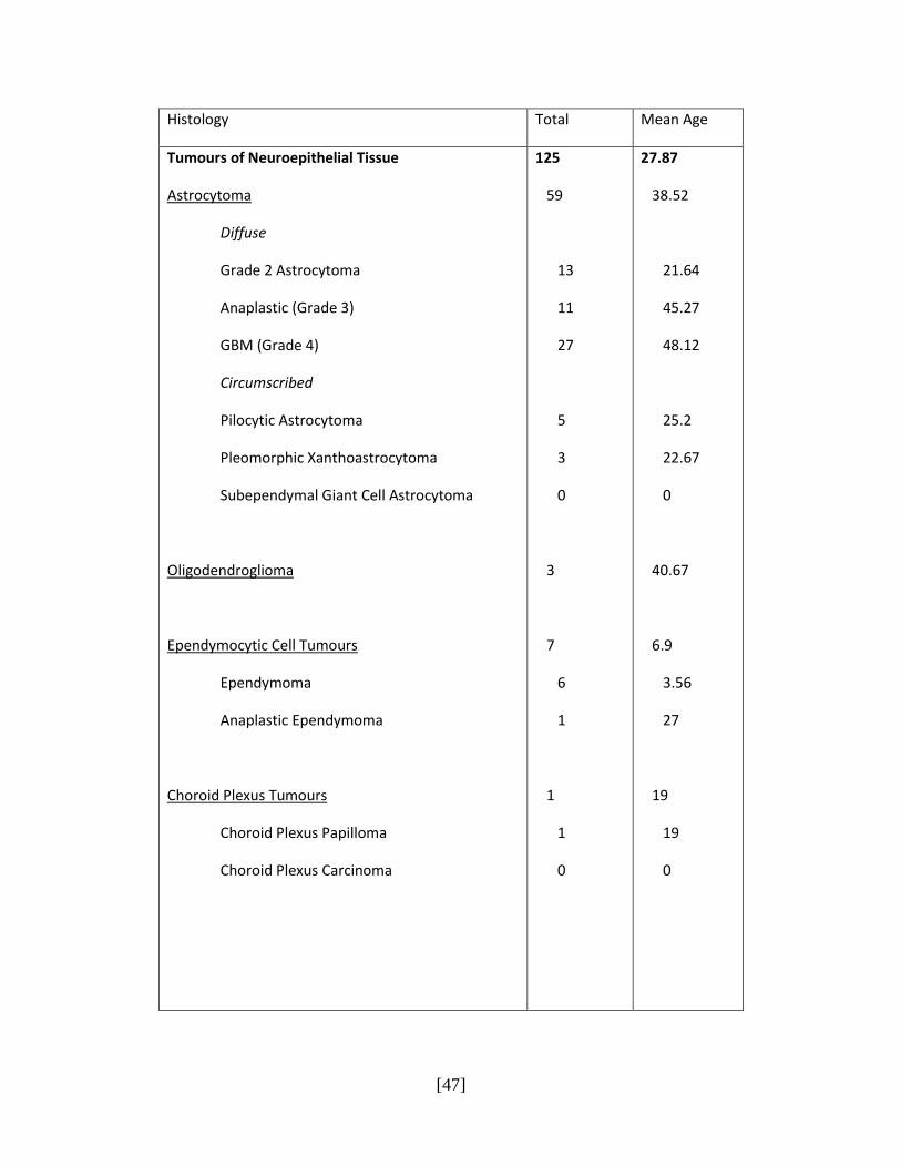
4.3 HISTOLOGICAL TYPES OF TUMOURS
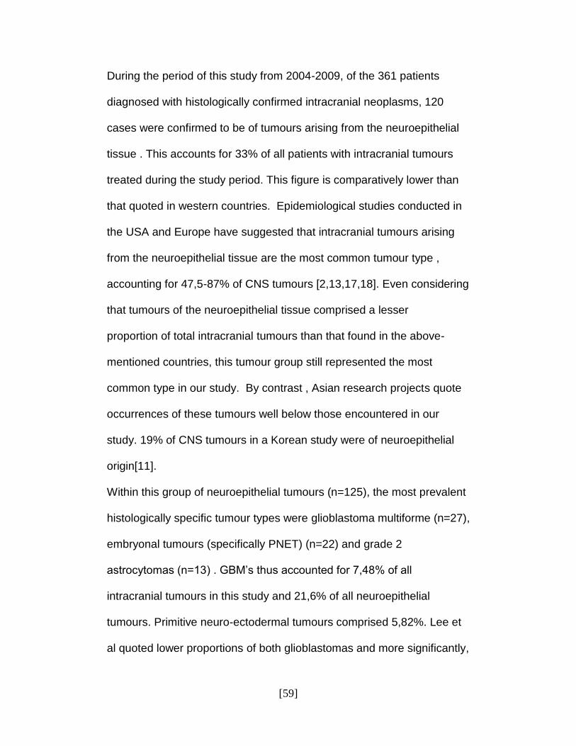
For the purposes of this study, tumours were divided into histological groups according to the WHO classification of intracranial tumours. Those tumours occurring in significant numbers were further sub-divided by their histological subgroups. In addition to primary intracranial tumours, metastatic tumours were also included. During the period between January 2004 and December 2009, intracranial tumours diagnosed at the Chris Hani Baragwanath Hospital were comprised primarily of the following subtypes : tumours of neuroepithelial tissue, tumours arising from the meninges and tumours in the sellar region. Collectively , these three subgroups accounted for 91 % of all intracranial tumours (tumours of neuroepithelial tissue = 33%, tumours of meninges = 32% and sellar tumours = 26%) as shown in figure 1. A total of 125 patients were diagnosed with tumours of the neuroepithelial tissue during the study period (Table 1). Of these cases, the majority were accounted for by the subgroup: astrocytomas (total astrocytomas = 59). The most common tumour subtype within this group were the grade 4 astrocytomas or glioblastoma multiforme (GBM)
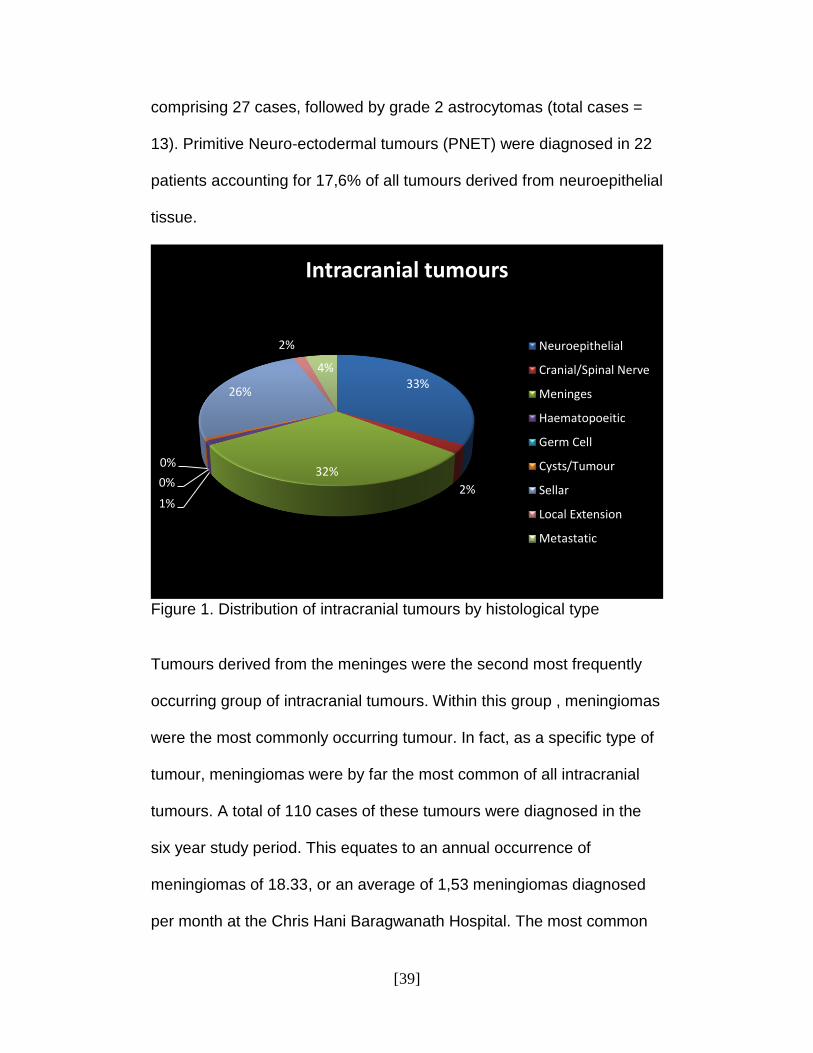
[39]
comprising 27 cases, followed by grade 2 astrocytomas (total cases = 13). Primitive Neuro-ectodermal tumours (PNET) were diagnosed in 22 patients accounting for 17,6% of all tumours derived from neuroepithelial tissue.
Figure 1. Distribution of intracranial tumours by histological type Tumours derived from the meninges were the second most frequently occurring group of intracranial tumours. Within this group , meningiomas were the most commonly occurring tumour. In fact, as a specific type of tumour, meningiomas were by far the most common of all intracranial tumours. A total of 110 cases of these tumours were diagnosed in the six year study period. This equates to an annual occurrence of meningiomas of 18.33, or an average of 1,53 meningiomas diagnosed per month at the Chris Hani Baragwanath Hospital. The most common
33%
2%
32%
1%
0%
0%
26%
2%
4%
Intracranial tumours
Neuroepithelial
Cranial/Spinal Nerve
Meninges
Haematopoeitic
Germ Cell
Cysts/Tumour
Sellar
Local Extension
Metastatic
[40]
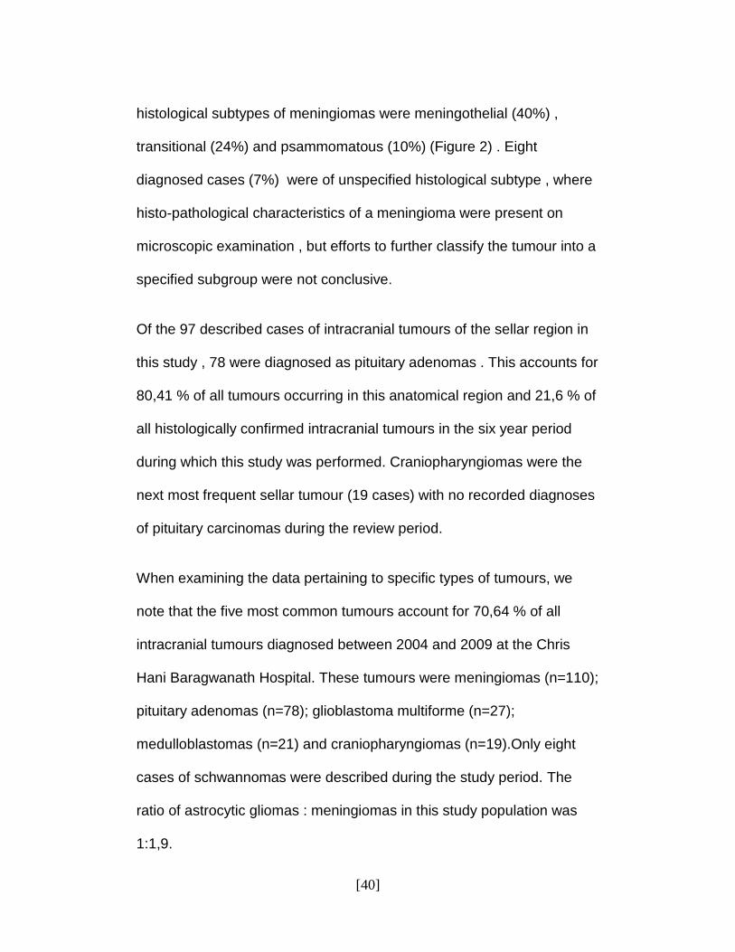
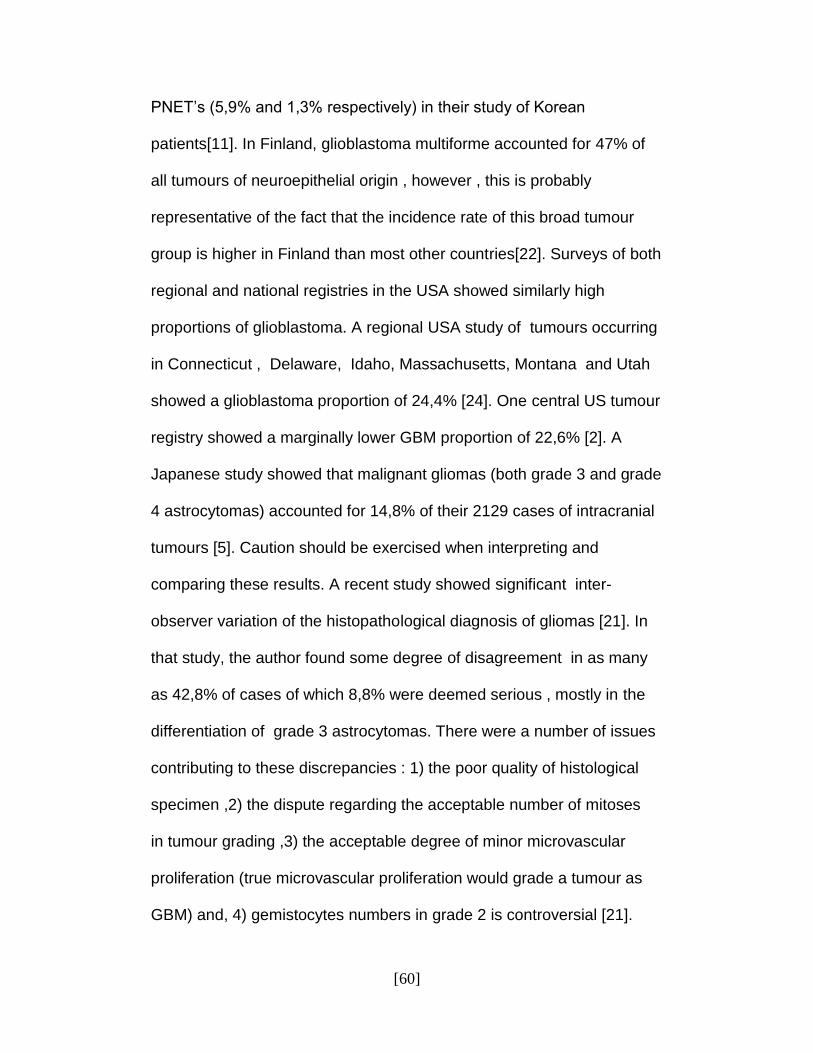
histological subtypes of meningiomas were meningothelial (40%) , transitional (24%) and psammomatous (10%) (Figure 2) . Eight diagnosed cases (7%) were of unspecified histological subtype , where histo-pathological characteristics of a meningioma were present on microscopic examination , but efforts to further classify the tumour into a specified subgroup were not conclusive. Of the 97 described cases of intracranial tumours of the sellar region in this study , 78 were diagnosed as pituitary adenomas . This accounts for 80,41 % of all tumours occurring in this anatomical region and 21,6 % of all histologically confirmed intracranial tumours in the six year period during which this study was performed. Craniopharyngiomas were the next most frequent sellar tumour (19 cases) with no recorded diagnoses of pituitary carcinomas during the review period. When examining the data pertaining to specific types of tumours, we note that the five most common tumours account for 70,64 % of all intracranial tumours diagnosed between 2004 and 2009 at the Chris Hani Baragwanath Hospital. These tumours were meningiomas (n=110); pituitary adenomas (n=78); glioblastoma multiforme (n=27); medulloblastomas (n=21) and craniopharyngiomas (n=19).Only eight cases of schwannomas were described during the study period. The ratio of astrocytic gliomas : meningiomas in this study population was 1:1,9.
[41]
Figure 2. Histological subtypes of intracranial meningioma. Meningothel = meningothelial , Malig = malignant, Anapl = anaplastic 4.4 AGE DISTRIBUTION OF INTRACRANIAL TUMOURS
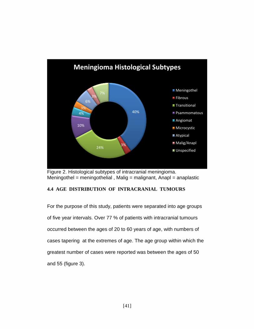
For the purpose of this study, patients were separated into age groups of five year intervals. Over 77 % of patients with intracranial tumours occurred between the ages of 20 to 60 years of age, with numbers of cases tapering at the extremes of age. The age group within which the greatest number of cases were reported was between the ages of 50 and 55 (figure 3).
40%
3% 24%
10%
4%
3%
6% 3%
7%
Meningioma Histological Subtypes
Meningothel
Fibrous
Transitional
Psammomatous
Angiomat
Microcystic
Atypical
Malig/Anapl
Unspecified
[42]
Figure 3. Distribution of intracranial tumours by age showing differences between male and female patients Paediatric cases, defined as patients aged 14 and below accounted for 11,63 % (n=42) of all reported intracranial tumours during the study period. Eighty nine intracranial tumours were found in young adults and adolescents ( aged between 15 and 35 years old) , accounting for 24,64% of all intracranial tumours. More than half of all the intracranial tumours were found in the middle aged group (51,25%). This age group, defined as older than 35 years but younger than 60 years, comprised 185 cases of histologically confirmed tumours. Only 12,47 % of cases (n=45) were in the elderly age group (aged greater than 60 years old). When examining the mean ages of patients with different tumours types
0
10
20
30
40
50
60
0-4
5-9
10
-14
15
-19
20
-24
25
-29
30
-34
35
-39
40
-44
45
-49
50
-54
55
-59
60
-64
65
-69
70
-74
>7
5
Tota
l Nu
mb
er
Age
Age Distribution - Intracranial Tumours
Total
Male
Female
[43]
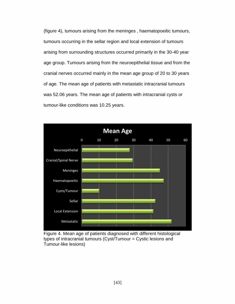
(figure 4), tumours arising from the meninges , haematopoeitic tumours, tumours occurring in the sellar region and local extension of tumours arising from surrounding structures occurred primarily in the 30-40 year age group. Tumours arising from the neuroepithelial tissue and from the cranial nerves occurred mainly in the mean age group of 20 to 30 years of age. The mean age of patients with metastatic intracranial tumours was 52.06 years. The mean age of patients with intracranial cysts or tumour-like conditions was 10.25 years.
Figure 4. Mean age of patients diagnosed with different histological types of intracranial tumours (Cyst/Tumour = Cystic lesions and Tumour-like lesions)
0 10 20 30 40 50 60
Neuroepithelial
Cranial/Spinal Nerve
Meninges
Haematopoeitic
Cysts/Tumour
Sellar
Local Extension
Metastatic
Mean Age
[44]
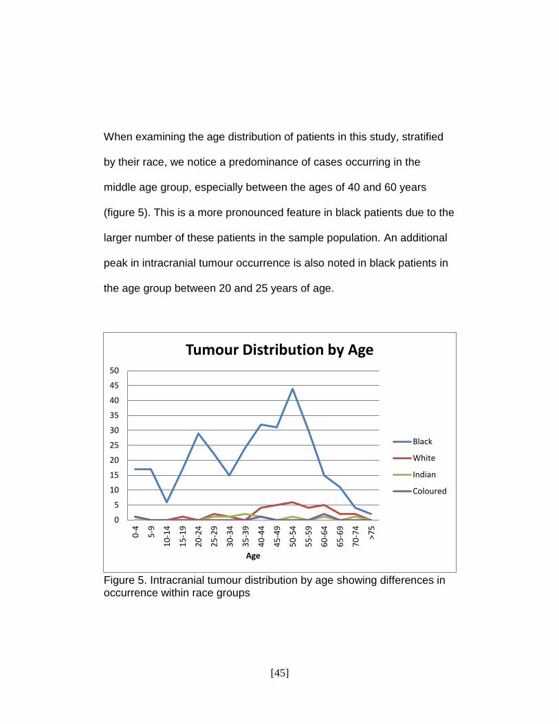
Even though the mean age of patients with tumours arising from the neuroepithelial tissue was reported as 27.87 years of age , a closer inspection of the data reveals that specific tumours within this group have considerably different age characteristics. The mean age of patients with grade 4 astrocytomas (the most commonly occurring tumour of neuroepithelial origin in this study) was 48.12 years. Cases of grade 2 astrocytomas had a mean age of 21.64 years and that of pilocytic astrocytomas was 25,2 years. The mean age of patients with primitive neuro-ectodermal tumours (another type of tumour arising from neuroepithelial tissue), by stark contrast, was 9,65 years of age (Table 1). The mean age of patients with meningiomas, the commonest tumour found in this study, was 45,65 years of age (Table1). Patients diagnosed with histologically confirmed pituitary adenomas had a mean age of 47,37 (Table1). Furthermore, almost 60 % of all reported cases of pituitary adenomas in this study occurred in the age group between 40 and 60 years of age (n=46). The mean age of the 19 patients with craniopharyngiomas was 23,58 years (Table 1). However, a more detailed analysis of the data showed a distinctive pattern characterized by two peaks in tumour occurrence (figure 6). The first peak occurs between the ages of 5 and 25 years of age and the second peak is between 45 and 60 years of age.
[45]
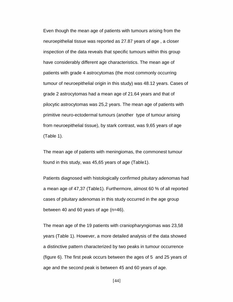
When examining the age distribution of patients in this study, stratified by their race, we notice a predominance of cases occurring in the middle age group, especially between the ages of 40 and 60 years (figure 5). This is a more pronounced feature in black patients due to the larger number of these patients in the sample population. An additional peak in intracranial tumour occurrence is also noted in black patients in the age group between 20 and 25 years of age.
Figure 5. Intracranial tumour distribution by age showing differences in occurrence within race groups
0
5
10
15
20
25
30
35
40
45
50
0-4
5-9
10
-14
15
-19
20
-24
25
-29
30
-34
35
-39
40
-44
45
-49
50
-54
55
-59
60
-64
65
-69
70
-74
>7
5
Age
Tumour Distribution by Age
Black
White
Indian
Coloured
[46]
Figure 6. Age distribution of craniopharyngiomas diagnosed at Chris Hani Baragwanath Hospital In the paediatric age group (0-15 years old), the most commonly occurring tumours were primitive neuro-ectodermal tumours (PNET) (n=16) ; craniopharyngiomas (n=7) ; ependymomas (n=6) and grade 2 astrocytomas (n=5). No pilocytic astrocytomas were noted in this age group during the six year period under review.
0
0.5
1
1.5
2
2.5
3
3.5
4
4.5
Craniopharyngioma - Age Distribution
[47]
Histology Total Mean Age
Tumours of Neuroepithelial Tissue
Astrocytoma
Diffuse
Grade 2 Astrocytoma
Anaplastic (Grade 3)
GBM (Grade 4)
Circumscribed
Pilocytic Astrocytoma
Pleomorphic Xanthoastrocytoma
Subependymal Giant Cell Astrocytoma
Oligodendroglioma
Ependymocytic Cell Tumours
Ependymoma
Anaplastic Ependymoma
Choroid Plexus Tumours
Choroid Plexus Papilloma
Choroid Plexus Carcinoma
125
59
13
11
27
5
3
0
3
7
6
1
1
1
0
27.87
38.52
21.64
45.27
48.12
25.2
22.67
0
40.67
6.9
3.56
27
19
19
0
[48]
Origin of Neurons
Desmoplastic Infantile Ganglioglioma
Ganglioglioma
Central Neurocytoma
Olfactory Neuroblastoma
Pinealocytes
Pineocytoma
Pineoblastoma
Mixed
Embryonal Tumours
Neuroblastoma
Primitive Neuro-ectodermal Tumours
Tumours of Cranial/Spinal Nerve
Schwannoma
Tumours of Meninges
Meningioma
Non Meningothelial
Haemangioblastoma
7
1
3
1
2
9
3
5
1
23
1
22
8
8
119
110
2
7
25.74
0.17
21
25
46
19.67
26.33
13.2
32
9.53
7
9.65
29.63
29.63
45.29
45.65
49
38.57
[49]
Haematopoietic Neoplasms
Germ Cell Tumour
Cysts/Tumour-like Lesions
Rathke's Cleft
Epidermoid Cyst
Dermoid Cyst
Tumours of the Sellar Region
Pit Adenoma
Pit Carcinoma
Craniopharyngioma
Local Extension from Regional Tumours Paraganglioma
Chordoma
Other
Metastatic
4
0
2
0
1
1
97
78
0
19
6
1
2
3
16
47.5
0
10.25
0
20
0.5
42.71
47.37
0
23.58
41.5
22
39
49.67
52.06
TOTAL 361 31.06
Table 1. Absolute numbers of diagnosed cases of histologically confirmed intracranial tumours and the mean age of patients at time of surgery.
[50]
4.5 GENDER DISTRIBUTION OF INTRACRANIAL TUMOURS
Of the 361 patients enrolled in this study , a total of 202 were males and 159 were females. This equates to a male:female ratio for all intracranial tumours of 1.27 : 1 . Male : Female ratios in this study showed a male predominance across most age groups with the exception of the 10-14 years, 35-39 years, 45-49 years, 55-59 years and 65-69 years age groups. The only age group with an equal number of male and female patients was the 40-45 year age group with both males and females numbering 19 patients. The male predominance was most evident at the extremes of age , notably in patients over the age of 75 and amongst children and adolescents. In particular , the age group of 15 to 19 year old patients showed five male patients diagnosed with intracranial tumours for every one female patient. When examining the ratio of absolute numbers of male and female patients stratified by the different tumour types , male patients were shown to account for more cases of tumours arising from the neuroepithelial tissue , sellar region , local extension from surrounding areas and metastatic intracranial lesions (figure 7). Tumours representing local extension into the cranial cavity from surrounding structures, in particular showed a very strong male predominance.
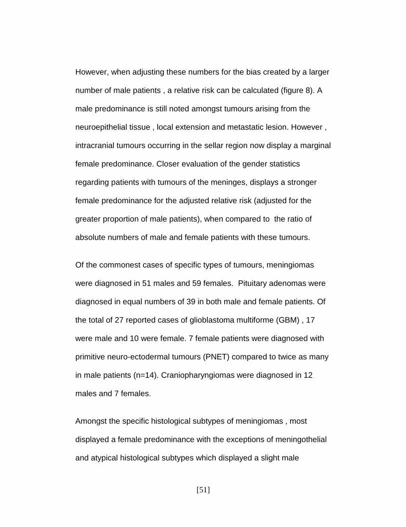
[51]
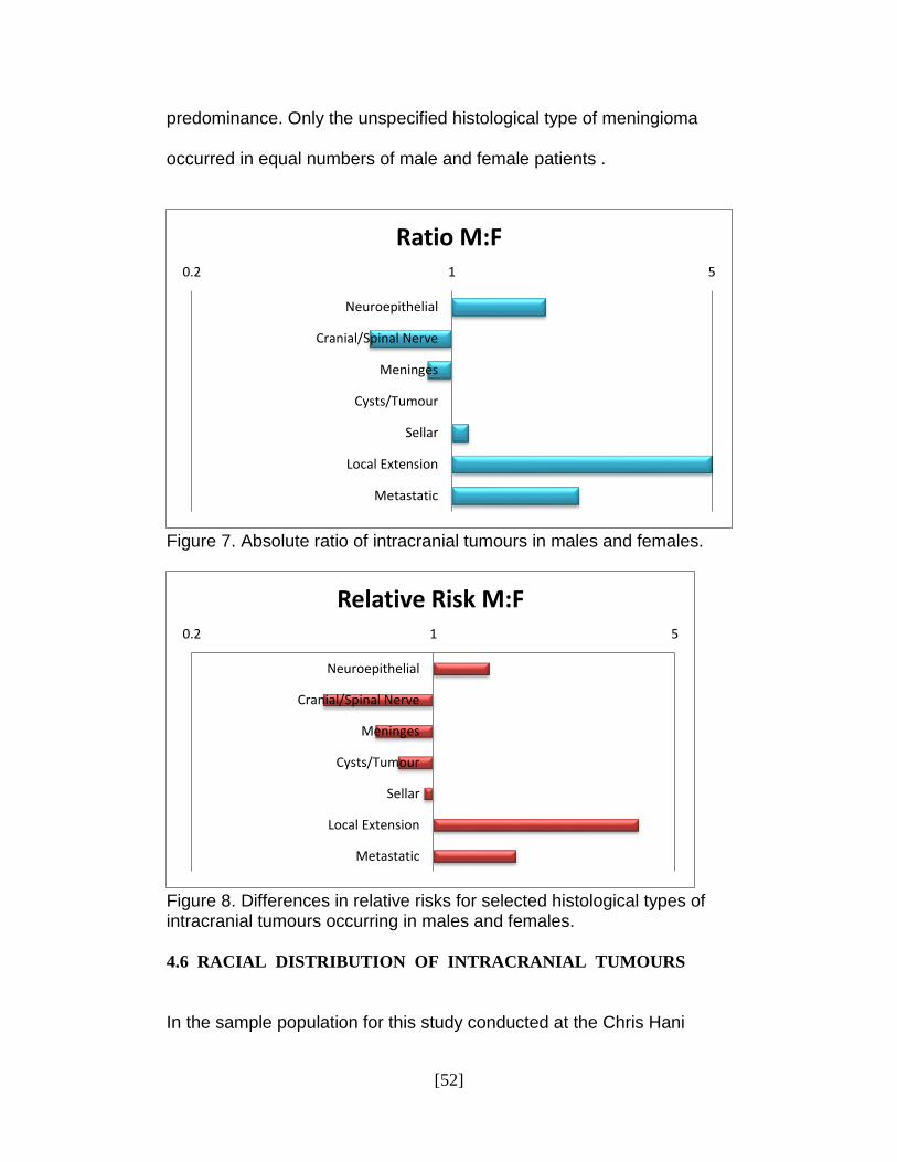
However, when adjusting these numbers for the bias created by a larger number of male patients , a relative risk can be calculated (figure 8). A male predominance is still noted amongst tumours arising from the neuroepithelial tissue , local extension and metastatic lesion. However , intracranial tumours occurring in the sellar region now display a marginal female predominance. Closer evaluation of the gender statistics regarding patients with tumours of the meninges, displays a stronger female predominance for the adjusted relative risk (adjusted for the greater proportion of male patients), when compared to the ratio of absolute numbers of male and female patients with these tumours. Of the commonest cases of specific types of tumours, meningiomas were diagnosed in 51 males and 59 females. Pituitary adenomas were diagnosed in equal numbers of 39 in both male and female patients. Of the total of 27 reported cases of glioblastoma multiforme (GBM) , 17 were male and 10 were female. 7 female patients were diagnosed with primitive neuro-ectodermal tumours (PNET) compared to twice as many in male patients (n=14). Craniopharyngiomas were diagnosed in 12 males and 7 females. Amongst the specific histological subtypes of meningiomas , most displayed a female predominance with the exceptions of meningothelial and atypical histological subtypes which displayed a slight male
[52]
predominance. Only the unspecified histological type of meningioma occurred in equal numbers of male and female patients .
Figure 7. Absolute ratio of intracranial tumours in males and females.
Figure 8. Differences in relative risks for selected histological types of intracranial tumours occurring in males and females.
4.6 RACIAL DISTRIBUTION OF INTRACRANIAL TUMOURS
In the sample population for this study conducted at the Chris Hani
0.2 1 5
Neuroepithelial
Cranial/Spinal Nerve
Meninges
Cysts/Tumour
Sellar
Local Extension
Metastatic
Ratio M:F
Neuroepithelial
Cranial/Spinal Nerve
Meninges
Cysts/Tumour
Sellar
Local Extension
Metastatic
0.2 1 5
Relative Risk M:F
[53]
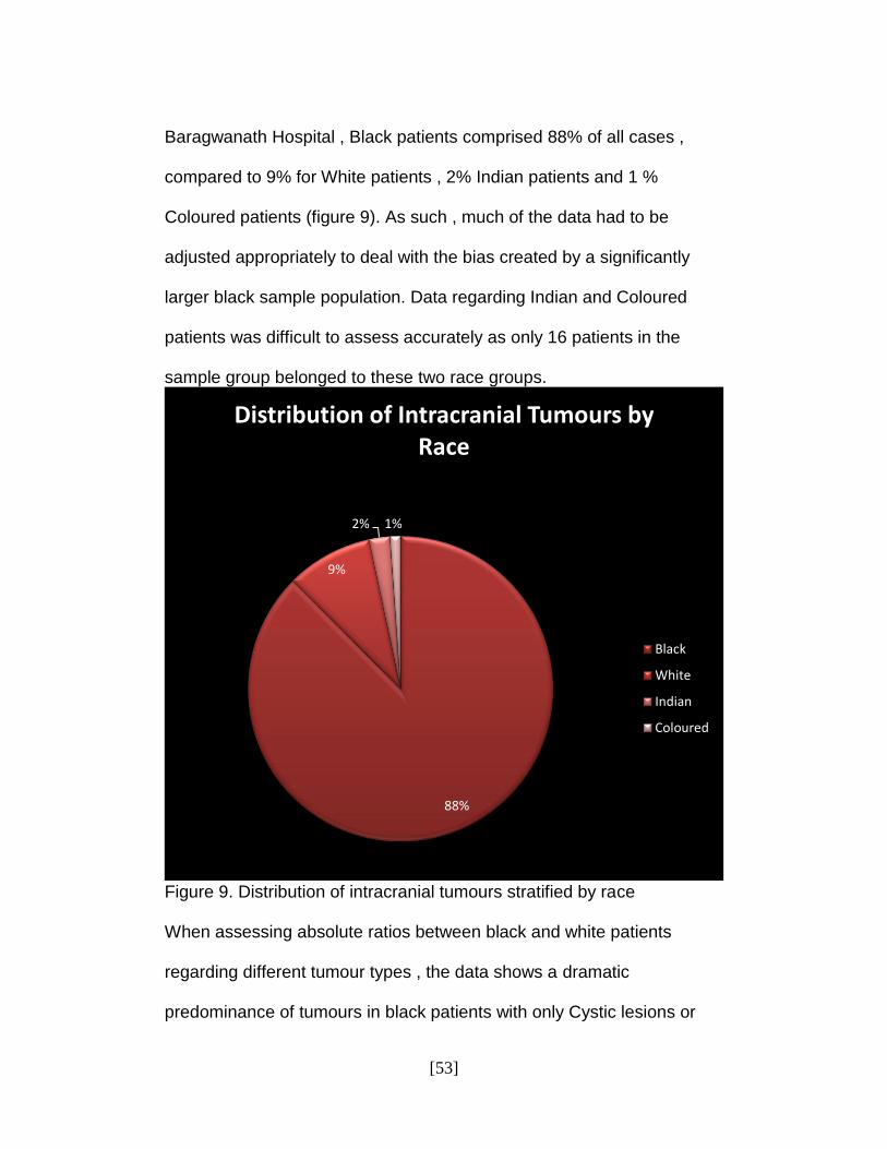
Baragwanath Hospital , Black patients comprised 88% of all cases , compared to 9% for White patients , 2% Indian patients and 1 % Coloured patients (figure 9). As such , much of the data had to be adjusted appropriately to deal with the bias created by a significantly larger black sample population. Data regarding Indian and Coloured patients was difficult to assess accurately as only 16 patients in the sample group belonged to these two race groups.
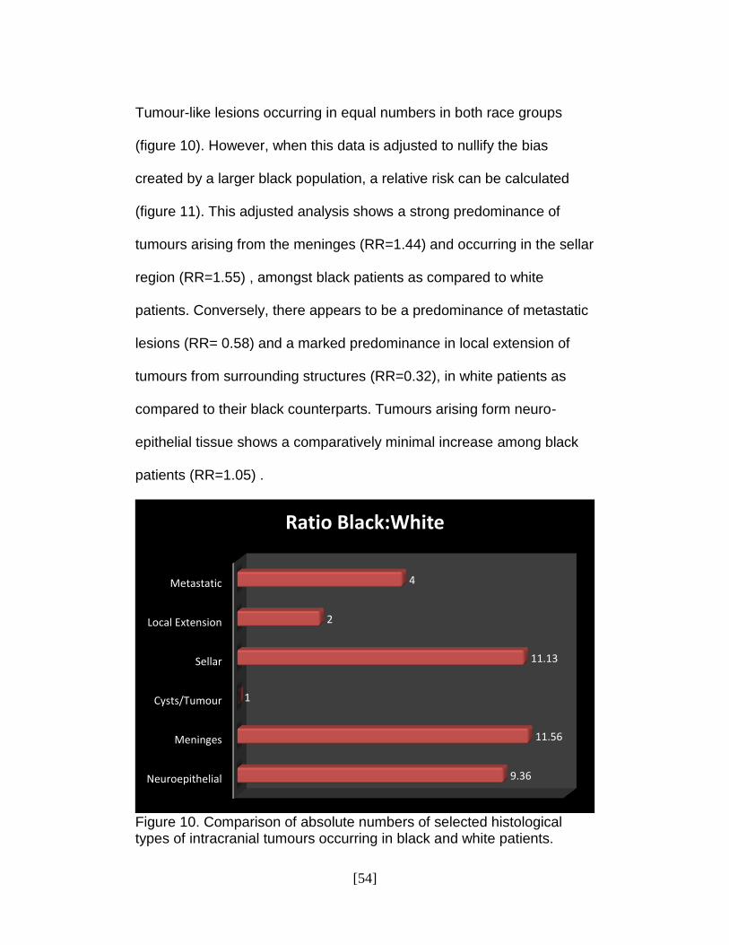
Figure 9. Distribution of intracranial tumours stratified by race When assessing absolute ratios between black and white patients regarding different tumour types , the data shows a dramatic predominance of tumours in black patients with only Cystic lesions or
88%
9%
2% 1%
Distribution of Intracranial Tumours by Race
Black
White
Indian
Coloured
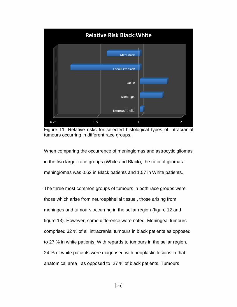
[54]
Tumour-like lesions occurring in equal numbers in both race groups (figure 10). However, when this data is adjusted to nullify the bias created by a larger black population, a relative risk can be calculated (figure 11). This adjusted analysis shows a strong predominance of tumours arising from the meninges (RR=1.44) and occurring in the sellar region (RR=1.55) , amongst black patients as compared to white patients. Conversely, there appears to be a predominance of metastatic lesions (RR= 0.58) and a marked predominance in local extension of tumours from surrounding structures (RR=0.32), in white patients as compared to their black counterparts. Tumours arising form neuro- epithelial tissue shows a comparatively minimal increase among black patients (RR=1.05) .
Figure 10. Comparison of absolute numbers of selected histological types of intracranial tumours occurring in black and white patients.
Neuroepithelial
Meninges
Cysts/Tumour
Sellar
Local Extension
Metastatic
9.36
11.56
1
11.13
2
4
Ratio Black:White
[55]
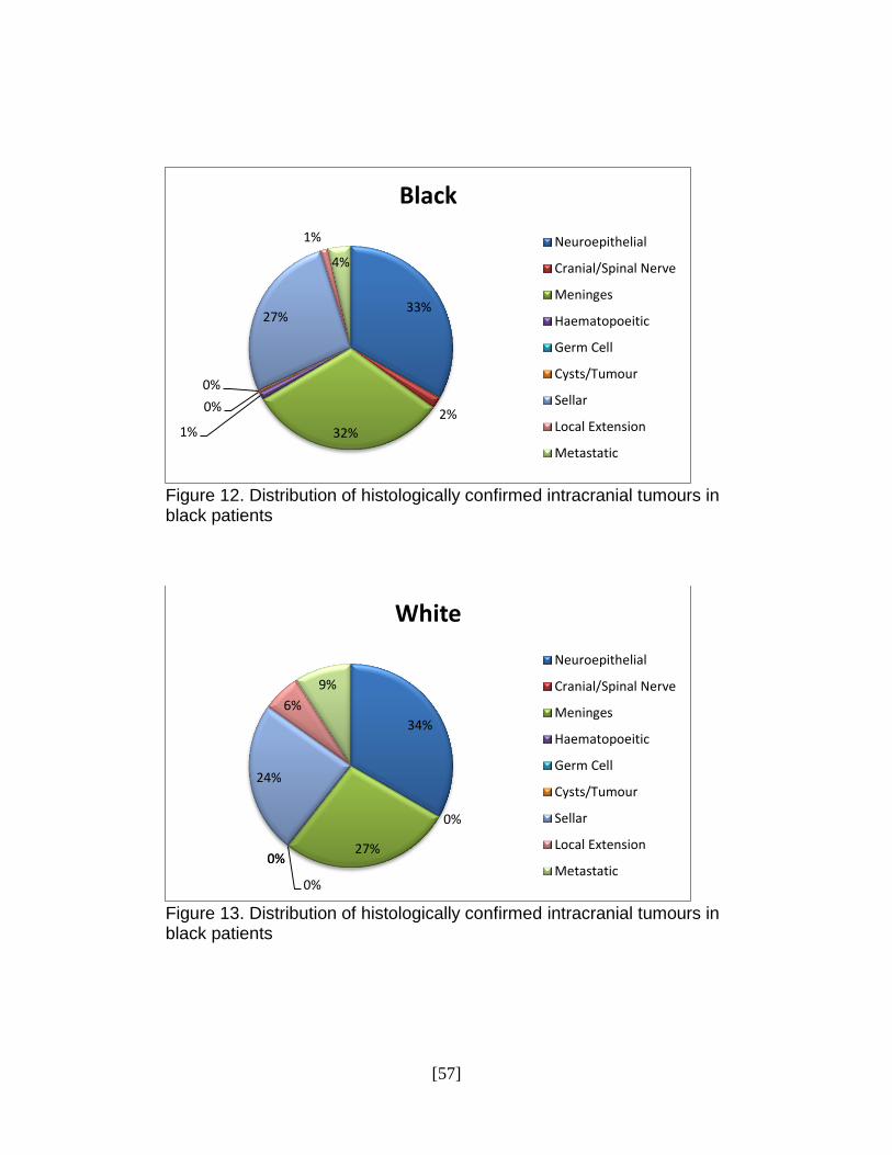
Figure 11. Relative risks for selected histological types of intracranial tumours occurring in different race groups. When comparing the occurrence of meningiomas and astrocytic gliomas in the two larger race groups (White and Black), the ratio of gliomas : meningiomas was 0.62 in Black patients and 1.57 in White patients. The three most common groups of tumours in both race groups were those which arise from neuroepithelial tissue , those arising from meninges and tumours occurring in the sellar region (figure 12 and figure 13). However, some difference were noted. Meningeal tumours comprised 32 % of all intracranial tumours in black patients as opposed to 27 % in white patients. With regards to tumours in the sellar region, 24 % of white patients were diagnosed with neoplastic lesions in that anatomical area , as opposed to 27 % of black patients. Tumours
[56]
arising from neuroepithelial tissue occurred in comparable percentages (black = 33%, white= 34 %). The diagnoses included among Indian patients with intracranial tumours were 3 meningiomas, 2 schwannomas, 1 glioblastoma multiforme (GBM), 1 haematopoeitic lesion and 1 metastatic tumour. The tumours occurring in Coloured patients at Chris Hani Baragwanath Hospital during the six year study period included 3 meningiomas and 1 dermoid cyst.
[57]
Figure 12. Distribution of histologically confirmed intracranial tumours in black patients
Figure 13. Distribution of histologically confirmed intracranial tumours in black patients
33%
2%
32% 1%
0%
0%
27%
1%
4%
Black
Neuroepithelial
Cranial/Spinal Nerve
Meninges
Haematopoeitic
Germ Cell
Cysts/Tumour
Sellar
Local Extension
Metastatic
34%
0%
27%
0%
0% 0%
24%
6%
9%
White
Neuroepithelial
Cranial/Spinal Nerve
Meninges
Haematopoeitic
Germ Cell
Cysts/Tumour
Sellar
Local Extension
Metastatic
[58]
CHAPTER 5
5.0 DISCUSSION
5.1 HISTOLOGICAL TYPES OF TUMOURS
It has been noted that substantial differences exist between countries , geographic areas and ethnic groups for incidence of malignancies of the nervous system [7]. Moreover, even within a particular country , specific regions may have differing incidences of histological tumours [2,4,12,16,17,23,24,26,27,28]. This study performed at the Chris Hani Baragwanath Hospital’s Department of Neurosurgery, aims to identify amongst others , the most prevalent intracranial tumours being treated at this facility. Patients of all ages with intracranial tumours which have undergone histo-pathological examination following surgery were included in this study. This data was then categorized according to the WHO classification for brain tumours [2,5,6] and evaluated for most commonly occurring histological types of neoplasms and the relative ratios between these tumours. In this study , the author included both primary as well as metastatic intracranial lesions as it was felt that the morbidity arising from both of these types of neoplasms were significant. The evaluation of histology-specific trends in intracranial cancer cases has important implications, both from clinical and public health standpoints, because it may lead to the improvement of diagnostic methods or the identification of potential aetiological risk factors [22].
[59]
During the period of this study from 2004-2009, of the 361 patients diagnosed with histologically confirmed intracranial neoplasms, 120 cases were confirmed to be of tumours arising from the neuroepithelial tissue . This accounts for 33% of all patients with intracranial tumours treated during the study period. This figure is comparatively lower than that quoted in western countries. Epidemiological studies conducted in the USA and Europe have suggested that intracranial tumours arising from the neuroepithelial tissue are the most common tumour type , accounting for 47,5-87% of CNS tumours [2,13,17,18]. Even considering that tumours of the neuroepithelial tissue comprised a lesser proportion of total intracranial tumours than that found in the above- mentioned countries, this tumour group still represented the most common type in our study. By contrast , Asian research projects quote occurrences of these tumours well below those encountered in our study. 19% of CNS tumours in a Korean study were of neuroepithelial origin[11]. Within this group of neuroepithelial tumours (n=125), the most prevalent histologically specific tumour types were glioblastoma multiforme (n=27), embryonal tumours (specifically PNET) (n=22) and grade 2 astrocytomas (n=13) . GBM’s thus accounted for 7,48% of all intracranial tumours in this study and 21,6% of all neuroepithelial tumours. Primitive neuro-ectodermal tumours comprised 5,82%. Lee et al quoted lower proportions of both glioblastomas and more significantly,
[60]
PNET’s (5,9% and 1,3% respectively) in their study of Korean patients[11]. In Finland, glioblastoma multiforme accounted for 47% of all tumours of neuroepithelial origin , however , this is probably representative of the fact that the incidence rate of this broad tumour group is higher in Finland than most other countries[22]. Surveys of both regional and national registries in the USA showed similarly high proportions of glioblastoma. A regional USA study of tumours occurring in Connecticut , Delaware, Idaho, Massachusetts, Montana and Utah showed a glioblastoma proportion of 24,4% [24]. One central US tumour registry showed a marginally lower GBM proportion of 22,6% [2]. A Japanese study showed that malignant gliomas (both grade 3 and grade 4 astrocytomas) accounted for 14,8% of their 2129 cases of intracranial tumours [5]. Caution should be exercised when interpreting and comparing these results. A recent study showed significant inter- observer variation of the histopathological diagnosis of gliomas [21]. In that study, the author found some degree of disagreement in as many as 42,8% of cases of which 8,8% were deemed serious , mostly in the differentiation of grade 3 astrocytomas. There were a number of issues contributing to these discrepancies : 1) the poor quality of histological specimen ,2) the dispute regarding the acceptable number of mitoses in tumour grading ,3) the acceptable degree of minor microvascular proliferation (true microvascular proliferation would grade a tumour as GBM) and, 4) gemistocytes numbers in grade 2 is controversial [21].
[61]
As such, there exists a possibility for a grade 3 astrocytoma to be incorrectly diagnosed histologically as the more benign grade 2 or a malignant grade 4 in certain cases. This may adversely skew results at some centres or in certain registries, favouring either more benign or malignant pathologies. Even so , by combining the grade 3 and grade 4 astrocytomas reported at our facility into the group “malignant astrocytic gliomas” , these would still only account for 9,14% of all intracranial tumours and 26,4% of neuroepithelial tumours. The multifactorial nature of tumourigenesis, including the effects of diet, lifestyle , viral agents , genetics , environmental or occupational exposure or indeed any combination of these factors, may attribute to the differences in the specific histological prevalences [38]. Furthermore, in their comparison of American White , African American and West African black patients , Fan et al , noticed a predominance of glial tumours in whites [12]. Since the majority of our sample population was Black (88%), this may explain the lower occurrence of these tumours in our study. This will be explored in greater detail in section 4.6 . Meningiomas were by far the most commonly occurring histologically specific intracranial tumour in this study. These tumours accounted for 30,47% of all intracranial tumours diagnosed and histo-pathologically confirmed at our facility (n=110). This equated to more than one case per month over a six year period. When comparing these results to those from studies conducted in USA, we notice a significant
[62]
prominence of meningiomas in our sample population. The incidence of these tumours in California was reported to be 4,5/100000 and they represented the most common benign intracranial tumour studied[23]. The proportion of these tumours as a percentage of all intracranial tumours in the USA was reportedly 22,6%-33,8% [2,8,26,27]. A more regional study in this country showed an even lower proportion of 20,9% prior to 2000 [24]. Studies performed in Asia show a predilection for meningiomas [5,9,11] while those from Europe tend to follow trends found in USA [14,17,18,19]. Ethnic variation in meningioma prevalence has been described globally [5,9,11,12,16]. It is the author’s opinion that these variations in meningioma occurrence may exist due to the ethnic constituency of our sample population. In addition, many meningiomas are diagnosed incidentally as a result of CT scans performed for head injuries , and higher number of these tumours may reflect a detection bias in this sense [26]. However, Wiemels et al suggest that incidentally discovered meningiomas are usually treated conservatively and do not warrant surgical excision [27]. As our study only includes histologically confirmed tumours following surgery, the possibility exists that the prevalence of meningiomas in the region served by the Chris Hani Baragwanath Hospital may be even higher than reported in this study. Pituitary adenomas comprised 21,6% of all intracranial tumours in our study population and 80,41% of all tumours of the sellar region.
[63]
American studies showed relative occurrences of pituitary tumours to be between 6-8% of all intracranial tumours [2,8]. The incidence rate in California was 2,0 per 100000 with this tumour representing 24,9% of benign tumours in their series. Materljan et al suggested frequencies of this tumour may be between 8-18% [17]. Among regional Japanese and national Korean surveys , these tumours accounted for 18,3% and 13,8% of CNS tumours respectively [5,11]. Furthermore, in separate studies conducted in Washington , Fan et al and Hesmat et al, found higher proportions of pituitary tumours among black patients in USA and Africa, compared to White Americans [12,16]. These described ethnic variations in tumour occurrence are probably fundamental to explaining the comparatively large percentage of sellar lesions noted in our sample population. In addition , Ali et al postulated a detection bias could exist in developing countries as a result of long travelling distances, limited transport facilities and economic burdens from hospitalization [39]. One could possibly infer that as a result of the obstacles encountered by a poorer individual seeking medical assistance, pathologies with more dramatic or debilitating symptomatology (e.g. loss of visual acuity or visual field deficits) would take precedence over less debilitating conditions, thus creating the detection bias. This may, in part, explain the larger percentage of pituitary tumours diagnosed at the Chris Hani Baragwanath Hospital. A relative low occurrence of vestibular schwannomas was noted in this
[64]
study. A total of eight patients were diagnosed with this intracranial tumour, out of a sample population of 361 patients. This equates to 2,21% of all intracranial tumours. By contrast , most series from around the world produce greater proportions of vestibular schwannomas in their studies. Kuratsu and Ushio named acoustic neuromas as the fourth most commonly occurring intracranial neoplasm in Japan [9]. A regional study from the same country suggested that 9,8% of all intracranial tumours were vestibular schwannomas [5]. A survey of tumours occurring in southern England noted that of the 894 patients enrolled in the study, 61 were diagnosed with tumours of the cranial nerves, and of these tumours , 44 were schwannomas [14]. Overall incidence of nerve sheath tumours in USA was 1,1/100000 , with nerve sheath tumours being 6,5% of all registered intracranial tumours [2,28]. In California , nerve sheath tumours accounted for 18,7% of benign tumours [23]. Reasons for this disparity between results shown in our study and those observed in wealthier western nations include the fact that acoustic neuromas are frequently incidental findings on imaging studies performed for other reasons [30]. The epidemiology of vestibular schwannomas reflects the distribution of medical imaging devices, as noted in Denmark where the incidence of acoustic neuromas increased about 2.5-fold between 1983 and 2001.This was attributed mainly to the increased availability MRI imaging during this period. Furthermore, the average size of the lesions in this study decreased from 35 mm to 10
[65]
mm during the study period [30], displaying a detection bias. Similarly , residents of Beverly Hills , a wealthy area, displayed a threefold higher incidence of acoustic neuromas than the national average and half of these patients had normal audiograms at the time of diagnosis [30].
5.2 AGE DISTRIBUTION OF INTRACRANIAL TUMOURS
The age distribution of all intracranial tumours showed a gradual increase in tumour cases with increasing age, peaking in the age group 50-54 years and tapering off thereafter (figure 3). Fan et al , also reported proportionally low frequencies of CNS tumours at both ends of the age spectrum (below 10 years old and greater than 70 years).The highest frequency was noted in the 50-59 year age group [12]. The rise in incidence of brain tumours is consistent with virtually all other adult tumours [15]. Cancers in late adulthood are often associated with environmental, occupational or lifestyle risk factors and can be ascribed to the cumulative effect of exposures over a prolonged period of time [25]. Conversely, this decrease in incidence in the oldest reported patients may, in part, be artefactual as symptoms arising from the presence of intracranial tumours may be mistaken for cerebrovascular disease or it may simply be that elderly patients are less likely to be investigated owing to other co-morbid conditions [15]. Furthermore, the peak incidence of tumours at our facility, occurs at a younger age when compared to studies from developed nations [2,9,11,14,29]. In addition,
[66]
the mean age of French patients with CNS tumours was 57,0 years , patients in Georgia ,USA was 58,6 years while that parameter observed in our study was 31,06 years [18]. A possible explanation for this discrepancy may lie in the aging populations in developed countries, for example , the mean population of Japanese people over the age of 70 years is 216000 [9]. A closer examination of our data reveals consistencies with the findings of Deorah et al and Heshmat et al. These suggest a bimodal trend in age distribution of intracranial neoplasms with a peak in early childhood followed by a more substantial peak in late adulthood [4,16]. Barker et al quoted peak incidence of malignant gliomas of 7,53/100000 in the age group 50-59 years [14]. Reports from Eastern Europe and Asia suggest a peak incidence in the sixth decade [5,11,17]. McLendon et al noticed that GBM occurrence was predominant in the fifth and sixth decades of life [35]. By stark contrast, those patients with glioblastoma multiforme treated surgically at the Chris Hani Baragwanath Hospital , had a mean age of 48,12 years , significantly lower than those reported in international series. The mean age of patients with neuroepithelial tumours in our study was 27,87 years , compared to that measured in Korea , 43,5 years. This may be explained by the larger proportion of medulloblastomas in our sample population (mean age 9,65 years)
[67]
compared to that of the Korean study. When comparing the incidence of intracranial tumours in the population of California, Nasseri et al noticed that the patients of Middle Eastern descent presented with tumours between 5-7 years younger than their Caucasian counterparts [1]. Furthermore, Curry and Barker noted that in addition to having lower incidences of gliomas, black American patients also presented at a younger age [30]. These described ethnic variations may provide the basis for glioblastomas and neuroepithelial tumours in general, occurring in younger patients at the Chris Hani Baragwanath Hospital. In addition, a genetic aberration in our ethnic majority may accelerate the process of tumour development. A selection bias may also exist. Due to limited resources and the dire prognosis associated with GBM, many elderly patients , especially those with comorbid illnesses, may not undergo surgical treatment and were thus selected out of this study. The mean age of patients diagnosed with meningiomas in one European report was 57,6 years, in an American report : 59 years and in an Asian report : 58,1 years [6,11,18]. By comparison, the mean age of patients with meningiomas at the Chris Hani Baragwanath Hospital was 45,65 years. Barker et al found a peak incidence of meningiomas in the age group 60-69 years [14]. The peak age group for this tumour type in our sample population was considerably younger, 50-55 years. The youngest patient with a meningioma in our sample population belonged to the age category 10-14 years, with cases of these tumours increasing
[68]
in the third decade and with the vast majority occurring between the ages of 35-59 years. Although the trend regarding increasing numbers of these tumours being diagnosed after the age of 30 years is consistent with international reports, these studies suggest peak incidence of these tumours in significantly older age groups [2,11,17,29]. No obvious reason is forthcoming for the discrepancy in age distribution of meningiomas. Several factors have been suggested as risk factors for developing meningiomas. The most common of these being exposure to ionizing radiation [27]. Tenuous links have been made between meningiomas and radiofrequency exposure (cellular telephones) , obesity and the use of long term contraceptives [15,24,27]. Head injuries have also been implicated as a risk factor , however this has been refuted as a recall bias, i.e. patients diagnosed with a meningioma may recall some form of head injury , whether significant or not [15,26]. Increasing efforts have been made in determining genetic risk factors of developing meningiomas. A predominant feature in sporadic meningiomas is a deletion and inactivation of NF2 on chromosome 22 [27]. A 1,7 fold increase in meningioma risk was noted with a single nucleotide polymorphism in the Ki-ras and ERCC2 genes, and an association was recently discovered between meningiomas and a polymorphism in the gene encoding for breast cancer susceptibility gene 1-interacting protein (BRIP-1) [26,33]. None of these factors by themselves is enough to explain the earlier occurrence of meningiomas
[69]
in our patients, but as tumourigenesis is believed to be multi-factorial, a genetic predisposition in our sample populations’ ethnic majority combined with environmental factors may predispose these patients to develop meningeal tumours at a younger age [38]. The mean age of patients diagnosed with pituitary adenomas at the Chris Hani Baragwanath Hospital was 47,37 years. This compares well with the mean age of Korean patients with this disease :46,3 years. The peak occurrence of patients with this tumour type in our sample group was in the age group 50-54 years. This is significantly lower than that quoted in western countries [2,29]. The most commonly occurring paediatric intracranial tumours in our sample population were medulloblastomas, craniopharyngiomas, ependymomas and grade 2 astrocytomas. No pilocytic astrocytomas were diagnosed during the study period. International literature reports the most common tumours as being pilocytic astrocytomas , medulloblastomas, ependymomas , craniopharyngiomas and germ cell tumours ,in varying numbers in different regions [2,3,11,15]. The absence of pilocytic astrocytomas in this study population is certainly notable , however , as the total number of paediatric patients in the sample group was relatively small, no firm conclusions can be drawn in this matter. Craniopharyngiomas show a childhood preponderance in incidence
[70]
between the ages of 5-15 years , but this accounts for only half of all cases [40]. Craniopharyngioma rates in Japan declined after a peak at 0-9 years after which the rate remained flat with some fluctuations [5]. Although craniopharyngiomas diagnosed at the Chris Hani Baragwanath Hospital also occurred most commonly in children , peaking in age groups 5-9 years and again in 15-19 years, the distribution is clearly bimodal. Following the childhood peak , the number of cases drops off rapidly and is then followed by a second adult peak (between 45-59 years). Even though the number of cases of recorded craniopharyngiomas in our sample population is small, this trend is strongly evident.
5.3 GENDER DISTRIBUTION OF INTRACRANIAL TUMOURS
Rachet et al proposed that brain tumours are 20-50% more common in men in western nations [13]. The life-time risk of being diagnosed with a CNS malignancy is estimated to be 0,67% for men and 0,52% for women [8]. In separate studies performed on two continents, McKinney et al , and Fan et al found comparable results suggesting a male-to- female ratio of 1,5 : 1 [12,15]. Regional studies in the USA , by contrast, showed only a marginal male predominance in CNS tumour occurrence or even female predominance [10]. Ethnic differences are evident as shown through studies performed at the Seoul National University College of Medicine and Kagoshima University, showing greater
[71]
proportions of female patients with CNS tumours compared to their male counterparts in Asian countries [5,11]. Differences in the age-adjusted, sex specific incidence of tumours occurring in the Zaragoza and Navarre regions of Spain were present not only geographically but also temporally. During the period, 1973-1977, the ratio of intracranial tumour incidence of males : females was 0.93 in Zaragoza (showing a female predominance) and 1,92 in Navarre (strong male predominance). However, between 1988-1990, the male : female tumour incidence ratio in Navarre and Zaragoza were 1,5 and 2,18 respectively [7,41]. During the time interval, the M:F ratio had reversed in Navarre (with a later male predominance) but had increased in Zaragoza. This lends credence to the concept of studying relative ratios both nationally and regionally as such discrepancies now become more identifiable. The male-to-female ratio in our study population was 1,27 : 1. The male predominance was evident at all ages and in particular at the extremes of age. The only exception was the age group 40-45 years , where numbers of male patients equaled females. These findings are consistent with those of western countries but are in opposition to those from Asia. Amongst German children, incidence rates of intracranial tumours were 2,8 and 2,4 for boys and girls respectively , producing a ratio (M : F) of 1,2 : 1 [3]. In our study, paediatric cases (defined for our purposes as
[72]
age less than 15 years) , comprised total of 29 male patients and 13 females , producing an absolute ratio of 2,23 : 1. The greater occurrence of tumours in male patients in our study is more pronounced , but further study with a larger paediatric population sample may reveal more information. Tumours of neuroepithelial origin were more frequent in male patients [2,14,17,37]. Our data supports this finding as a relative risk of 1,45 (male/female) was found in patients with tumours of neuroepithelial origin. Barker et al found the incidence of malignant gliomas to be especially higher among male patients [14]. Provost et al, suggested the possible explanation for a higher frequency of gliomas in males may be due to specific occupational exposures [18]. Possible reasons for the observed gender variations may arise as a result of possible variabilties in the susceptibility of X and Y chromosomes to tumourigenic stimuli, while others postulate a protective effect of female sex hormones against brain tumours [4]. Considering the increasing frequencies of these tumours with increasing age and higher proportion of male patients, one could postulate that a genetic predisposition amongst males coupled with a cumulative occupational exposure, may account for the results found [38]. Lopez-Abente et al suggested pesticides , especially containing N-nitroso compounds, may be responsible for the data in their studies [41].
[73]
Tumours of meningeal origin (comprised primarily of meningiomas) were mostly found in female patients in our study ,with a recorded relative risk (male/female) of 0,68. A review of data obtained from the SEER program suggested a higher incidence of meningiomas in females across all ages [4] , while Wiemels et al found that the previously described two-fold predominance of female meningioma cases may be inverted in the rare cases of prepubertal meningiomas [26,27]. Only 1 patient in our study ,below the age of 15 years was diagnosed with a meningioma. This patient was male , but as the number of patients recorded in this specific group is too small, no firm conclusion may be drawn. However, when comparing patients less than 30 years old, with meningiomas in our study population, 11 were male and 4 were female. When considering only the patients older than 30 years old (which comprise the vast majority of meningioma cases) , a higher female occurrence is confirmed. The association between hormones and meningiomas has been suggested , noting increased incidences among post-pubertal women especially during the peak reproductive years [27]. Furthermore, the presence of oestrogen, progesterone and androgen receptors on some meningiomas and indications of changing size of these lesions during pregnancy or with the of cessation of oestrogen agonist therapy, may explain, in part, the higher rates of meningiomas found in female patients [27]. Oddly, data from the Rijeka University School of Medicine found a greater proportion of male patients with
[74]
meningiomas [17]. A similar finding was noted when examining patients in West Africa [16]. This finding is out of keeping with world literature and may represent the effects of specific genetic and environmental factors affecting those regions. Asian reports concur with the findings in most western countries [9,11]. Surawics et al found no gender differences in the occurrence of tumours of the sellar region, however international reports suggest a higher proportion of female patients [2]. Data compiled by the University of Illinois, found equal proportions of male and female patients with pituitary adenomas [24]. Another study from the USA noted a male/female ratio of the tumours to be 0,75 [4], a finding validated in a Croatian study [17]. Regional American data noted a higher incidence of pituitary tumours amongst men , and particularly amongst black men [16]. Analysis of our data , revealed no differences in the frequency of these tumour types amongst males and females. Tumours of the cranial and spinal nerves occurred in equal frequency in a large national survey (US) [2]. In contrast, Barker et al described the incidence of vestibular schwannomas to be double in females compared to males [14]. Medulloblastomas had almost a two-fold excess in male patients [2]. Our data mirrored this finding as 14 males and 7 females were
[75]
diagnosed with these PNET’s in our sample population. No difference in sex specific incidence of tumours of local extension was noted in the CBTRUS study [2]. By contrast, this tumour group was primarily male dominated in our study. However, as the total number of tumours classified into this group is small, no firm conclusions may be made.
5.4 RACIAL DISTRIBUTION OF INTRACRANIAL TUMOURS
The comparable pattern of CNS tumours in American and African Black patients, relative to Caucasians, suggests that in the development of these neoplasms, racial factors may be important [16]. Similarly, with other types of neoplasm in the body, South African studies of childhood tumours have shown that White South African children had similar incidences of ALL to those of White American children, but South African Black children had 50% fewer cases of this neoplastic condition [42]. It is postulated that race or ethnicity may be a surrogate measure of underlying genetic factors. An epidemiological study from California was able to categorize patients into racial groups through surname, maiden name, inferences based on ethnicity of the parents or self-identification [23]. Similarly, Nasseri and Mills used patient surnames to identify patients of Middle Eastern origin living in California [1]. In our study, surnames and maiden names,
[76]
where applicable, were also used to classify patients according to their ethnicity. A statistically significantly higher incidence of brain tumours in whites (11,6/100000) was apparent when compared to blacks (7,8/100000) [2]. Black patients accounted for 88% of all recorded cases of intracranial tumours in our study, however, this is most likely a selection bias. Higher frequencies of glioblastomas have been recorded in white patients compared to black patients [23]. A study conducted in Georgia, USA, noted that 2,3 glioblastomas were found in whites for every one of these tumours in blacks [2,35]. When comparing the incidence of astrocytic gliomas in Northern European Caucasians and African Blacks, a white : black ratio of 16 : 1 was noted [35]. Nasseri et al found significantly higher proportions of glioblastomas in American whites when compared to Americans with Middle Eastern heritage [1]. In addition, when comparing Japanese and American studies , recorded incidences of gliomas were 2,1/100000 and 5,4/100000 respectively [5]. In addition to having a comparatively lower incidence of gliomas, black patients also tended to present at a younger age [30]. The relative risk of developing a neuroepithelial tumour between black and white patients at our facility, was 1,05 , which indicated an almost equal frequency of these tumours types. However, when examining, the ratio
[77]
of astrocytic glioma : meningioma (0,62 and 1,57 for Black and White patients respectively) , racial differences become more apparent . Furthermore, glioblastomas were diagnosed in 22 of 316 black patients in our study (7%), while of the 33 white patients, 4 had GBM (12%).This finding is consistent with the current literature. Investigations conducted in San Francisco, into associations between glioblastoma and HLA variants, found haplotypes B*13 and B*07-Cw07 associated with GBM. As American white patients are believed to more commonly possess haplotype B*07-Cw07 than other racial groups, this may contribute to the observed higher incidences of this tumour in white patients [30]. Another genetic aberration noted was that those patients with GBM exhibiting abnormal TP53 protein accumulation on immunohistochemistry belonged to racial groups other than whites. Caucasians commonly had tumours without either p53 mutations or TP53 protein accumulation [30]. These genetic trends may explain the differing occurrences and behaviour (i.e. age of presentation) observed in different ethnic groups. In addition, the molecular differences may become important in the development of targeted agents in the treatment of GBM [30]. Meningiomas occurred equally in black and white patients registered in a national American registry [2]. Regional data from the US, however, reported higher rates of meningiomas in black patients [2,12,23,30]. In a
[78]
comparison of meningiomas occurring in America and Africa, higher proportions of this tumour were noted in blacks compared with whites, and no significant differences were observed between the incidences of Black American and Black African patients [16]. Tumours arising from the meninges (mostly comprised of meningiomas in our study), accounted for 32% of tumours in black patients and 27% in white patients. The relative risk for this tumour was calculated to be 1,44 with a black predominance. CTBRUS data showed no racial differences in the frequency of pituitary adenomas [2]. Brown et al, however showed higher rates of pituitary adenomas in black patients [23], while other studies have shown only a modest black predominance [30]. A Washington study showed frequencies of pituitary tumours in black and white patients to be 14,44% and 6,64% respectively. At the Chris Hani Baragwanath Hospital, sellar region tumours accounted for 27% of all intracranial tumours in blacks and 24% in whites. The absolute ratio of black patients with tumours of the sellar region white patients with this tumour is 11,13 to 1, but if the figures are adjusted for the larger proportion of black patients in our study population, a relative risk of 1,55 (black/white) is noted. A predominance of these tumours in black patients is forthcoming in our study, albeit not as dramatic as reported in some studies.

[79]
Both national registries and local studies from the USA have shown an overwhelming majority of vestibular schwannomas and nerve sheath tumours in general, occurring in the White population [2,23,28,30]. The racial discrepancy in occurrence of this tumour is probably the most startling of all tumour types. In a comparison between South African patients and patients from USA and Denmark, it was found that white South Africans had incidences of this tumour, comparable with those of the patients from the western countries. Black South Africans, however, showed an incidence of acoustic neuromas which was 100-fold lower than white patients from South Africa, Denmark or USA [30]. This explains the comparatively low numbers (n=8) of nerve sheath tumours included in our study population (88% black). Germ cell tumours accounted for as much as 15% of childhood tumours in Japan. This was significantly more than was described in most western countries [5]. In addition, germ cell tumours were found in very few black patients [2] , possibly explaining the complete absence of these tumours in our study population, which was comprised primarily of black patients. Tumours representing local extension from surrounding structures showed a dramatic predominance in white patients of 42 : 1 (white : black) [12]. The calculated relative risk of these tumours (black/white) is 0,32 , displaying a marked predominance in white patients, consistent
[80]
with the current literature.
5.5 LIMITATIONS OF STUDY
As the study was limited to a specific neurosurgical centre, namely the Chris Hani Baragwanath Hospital, the data collected was limited to the socio-economic demographic serviced by that centre. In particular, a significant racial bias was noted with a larger portion of the study participants being represented by black individuals . Indian and Coloured patients, by contrast, comprised a much smaller percentage of the study population. This had to be considered in the data analysis. In addition, patients treated at the Chris Hani Baragwanath Hospital are generally of lesser affluent social standing with limited access to health care. As such, tumours with a tendency to produce more debilitating symptoms , for example : visual loss , may represent a greater proportion of tumours, as patients are more likely to seek medical assistance for these symptoms. Even though, the size of the study population included in this research project was larger enough to produce significant conclusions, a larger sample size would have revealed greater information. The study relied on a review of patient records with less than 10%

[81]
of the original sample group being excluded due to incomplete data records. Since surnames were used to classify patients into different racial groups, the impact of cross-ethnic marriages needs to be controlled. However, due to the relatively small numbers of cross- ethnic marriages in our study population, it was likely that this effect was not statistically significant.
[82]
CHAPTER 6
6.0 CONCLUSIONS AND RECOMMENDATIONS
6.1 INTRODUCTION
Developing countries contain over 75% of the world’s population, but possess only 5% of the world’s economic resources. Population-based tumour registries in these countries are few and far between, for a number of reasons, including the lack of sustained funding and infrastructure and the absence of their recognition as a national health care priority [39]. Targeting epidemiological case-control and cohort studies to specific geographical areas with high incidence rates raises the potential for identifying aetiological risk factors not commonly present in the population at large [10]. Furthermore, with the described racial and ethnic variations in specific histological types of tumours and the age-specific incidences, coupled with the recognized differences in molecular characteristics in these cases, it remains plausible that ethnicity may serve as a surrogate measure of underlying genetic factors [42]. Epidemiological studies linking individual patient information, such as demographics, occupation, social class and treatment data are vital in assessing environmental risk factors and the effects of therapeutic advances on patient survival [4].
[83]
6.2 CONCLUSION
By virtue of their anatomical location, intracranial tumours, even though rare when compared to tumours of other organ systems in the body, have severely debilitating symptomatology and significant mortality. As such, identifying specific groups at risk, by their gender, race or age, is of great value in proposing preventative or treatment strategies. Numerous studies from western nations suggests that tumours of neuroepithelial origin are the most common tumour group, quoting frequencies of between 48% to 87% [2,13,17,18]. Even though, this tumour group was also the predominant type noted during our study period, the frequency was considerably lower. However, when comparing our results to those of Asian countries, the recorded proportions of this tumour was higher at our facility [11]. The commonest specific histological type of tumour within this group, the glioblastoma multiforme, is also the most aggressive in its behaviour. GBM’s comprised a significantly lower proportion of neuroepithelial tumours and indeed of all intracranial tumours at our facility, when compared to global literature. In fact, our recorded frequencies, reflected that this tumour was half as common in comparison to American and European reports, but marginally more frequent that that reported from Asia [2,11,22,24]. Ethnic or genetic variance has been postulated as a possible explanation for these discrepancies. Another possibility for these differences may be under-reporting of the GBM, arising from
[84]
disagreements in the criteria for classification of these tumours [21]. Male predominance noted in our data analysis was consistent with the current literature, possibly as a result of variances in susceptibility of the X and Y chromosomes or occupational exposures. Meningiomas were by far the most common specific histological tumour type noted in our study accounting for nearly one third of all intracranial tumours. Even though this trend was consistent with some international studies, the frequency of this tumour in our study, was consistently higher than those reports [5,9,11]. The strong predominance of this tumour’s occurrence in female patients in our research (as well as global literature), coupled with the occurrence of specific receptors on tumour cells, suggest an association between meningiomas and female sex hormones [27]. When compared to global literature, the relative occurrence of pituitary adenomas in our study population was significantly higher [2,5,8,11,17]. In fact, these tumours were the second commonest intracranial neoplasm in our study, outnumbering even glioblastomas. This may represent the effects of genetic or environmental factors or simply be a selection bias, because in the developing world, access to health care, and in particular tertiary care, is limited [39]. Thus, patients suffering from diseases with more debilitating symptomatology (e.g. visual loss) may seek assistance more often or sooner. This high
[85]
frequency of pituitary adenomas in our study suggests that our centre may represent a tumour ‘hot spot’ (albeit in the absence of conclusive national epidemiological data). In addition to the implications for resource allocation, this also presents an opportunity for further study into this tumour, especially in terms of aetiology or genetic susceptibility. Vestibular schwannomas were extremely rare in patients treated at the Chris Hani Baragwanath Hospital, with only eight patients being diagnosed with this tumour in the six year study period. In stark contrast, Japanese studies report this neoplasm as the fourth most commonly occurring intracranial tumour in their study populations [9]. A proposed reason for these disparities were the greater access to imaging devices in developed countries, creating a detection bias in this tumour which is frequently asymptomatic [30]. However, black South African patients displayed a 100-fold lesser incidence of acoustic neuromas when compared to Caucasians from the same country and from abroad, suggesting a strong racial variance [30]. The implication of this finding not only has significance in terms of resource distribution, but also suggests that the exposure to surgical skills required in accessing the location of these tumours (cerebello-pontine angle), may be deficient in geographical regions with such low occurrences . Germ cell tumours were absent in our study population, in contrast to their common occurrence in Asian patients [5]. This suggests a strong

[86]
ethnic variance, with genetic factors or environmental (even in utero), exposures as possible causes. A startling finding in this research project was the age discrepancies between our sample population and international studies. The mean age of patients with meningiomas in our study was 8 years younger that of patients in the developed world [6,11,18]. Patients with glioblastoma multiforme in our study had a mean age which was as much as 25 years younger than their American and European counterparts. Glioblastomas were more frequent in white patients compared to black patients, while the inverse was true for meningiomas. This was consistent with international findings. Several genetic factors have been implicated in this regard, and this field represents an exciting avenue for further research [30]. The relative risk for pituitary tumours stratified by race (black/white) was1,55 in our study. There is a general lack of consensus in this regard internationally, meaning that no firm conclusions may be drawn. This study has not only shown the similarities and differences in tumour occurrence in our population compared to global trends , but has also displayed numerous opportunities for further research in this field. The overwhelming conclusion however, is the need for the development of national and regional intracranial tumour registries in South Africa.

[87]
6.3 RECOMMENDATION
1. As discussed above, the importance of epidemiologic data cannot
be over-emphasized in the identification of environmental risk factors and facilitating the planning of broader treatment strategies. As such, national and regional intracranial tumour registries would be pivotal in the management of these debilitating diseases in South Africa.
2. Further studies of this nature would have to be performed in the future to evaluate the temporal trends in the incidence of intracranial neoplasms as well as changes in gender- , racial- and age-specific frequencies of these tumours.
3. In addition, the data compiled in our study can be compared to similar investigations in other centres in South Africa to identify
specific tumour ‘hot spots’.
4. Autopsy results should also be included in future epidemiological studies. Some studies reviewed in our report, included autopsy results in their epidemiologic data. This may provide a more accurate estimate of the intracranial tumour burden encountered in that country or region as many patients with aggressive brain malignancies may succumb to their illness prior to surgical intervention being available. However, as tumours like meningiomas are very often asymptomatic, the use of autopsy findings may erroneously include incidental tumours.
[88]
REFERENCES
1. Nasseri K. & Mills J. (2009). Epidemiology of primary brain
tumors in the Middle Eastern population in California, USA. 2001–2005. Cancer Detection and Prevention 32 : 363–371
2. Surawicz T.S. , McCarthy B.J. , Kupelian V. , Jukich P.J. ,
Bruner J.M. & Davis F.G. (1999) Descriptive epidemiology of primary brain and CNS tumors: Results from the Central Brain Tumor Registry of the United States, 1990–1994 Neuro-Oncology 1 : 14–25
3. Kaatsch P, Rickert C.H. , Ku¨hl J., Schu¨z J & Michaelis J. (2001)
Population-Based Epidemiologic Data on Brain Tumors in German Children Cancer 92 (12) : 3155-3164
4. Deorah S. , Lynch C.F. , Sibenaller Z.A. & Ryken T.C. (2006)
Trends in brain cancer incidence and survival in the United States: Surveillance, Epidemiology, and End Results Program, 1973 to 2001 Neurosurg Focus 20 (4): 1-7
5. Kuratsu J. , Takeshima H. & Ushio Y. (2001)
Trends in the incidence of primary intracranial tumors in Kumamoto, Japan Int J Clin Oncol 6 : 183–191
[89]
6. Flowers A. (2000) Brain Tumors in the Older Person.
Cancer Control 7 ( 6 ) : 523-538 7. Ruiz-Tovara M. , Lo´pez-Abentea G. , Polla´na M. , Aragone´sa N. ,
Ardanazb E. , Moreo P. , Moreno C. & Vergara A. (1999) Brain cancer incidence in the provinces of Zaragoza and Navarre (Spain): effect of age, period and birth cohort Journal of the Neurological Sciences 164 : 93–99
8. Gurney J. & Kadan-Lottick N. (2001)
Brain and other central nervous system tumors: rates, trends, and epidemiology Current Opinion in Oncology 13 : 160–166
9. Kuratsu J. , Ushio Y. (1997)
Epidemiological study of primary intracranial tumours in
elderly people Journal of Neurology, Neurosurgery, and Psychiatry 63 : 116–118
10. Fang Z. , Kulldorff M. & Gregorio D.I. (2004)
Brain cancer mortality in the United States,1986 to 1995: A geographic analysis Neuro-Oncology 6 : 179–187
[90]
11. Lee C, Jung K., Yoo H. , Park S. , Lee S.H, (2010) Epidemiology of Primary Brain and Central Nervous System Tumors in Korea J Korean Neurosurg Soc 48 : 145-152
12. Fan K. & Pezeshkpour G.H. (1992)
Ethnic Distribution of Primary Central Nervous System Tumours in Washington , DC, 1971 TO 1985 J NatI Med Assoc. 84 (10) :
858-863
13. Rachet B., Mitry E. , Quinn M.J. , Cooper N & Coleman N.P. (2008)
Survival from brain tumours in England and Wales up to 2001 British Journal of Cancer 99 : S98 – S101
14. Barker D.J.P. , Weller R.O. & Garfield J.S. (1976)
Epidemiology of primary tumours of the brain and spinal cord: a regional survey in southern England J Neurol Neurosurg Psychiatry , 39 : 290-296
15. McKinney P.A. (2004)
Brain Tumours : Incidence , Survival and Aetiology
J Neurol Neurosurg Psychiatry 75 (Suppl II) :ii12–ii17.
[91]
16. Heshmamnt M.,Kovim J.,Simpson C., Kennedy J. & Fan K.J.(1976)
Neoplasms of the Central Nervous System : Incidence and Population Selectivity in the Washington DC, Metropolitan Area Cancer 38 : 2135-2142.
17. Materljan E. , Materljan B. , Sepèiæ J., Tuškan-Mohar L. ,
Zamolo G. & Erman-Baldini I. (2004) Epidemiology of Central Nervous System Tumors in Labin Area, Croatia, 1974-2001 Croat Med J 45 : 206-212
18. Provost D. , Cantagrel A. , Lebailly P. , Jaffre´ A. , Loyant V. ,
Loiseau H. , Vital A. , Brochard P. & Baldi I. (2007) Brain tumours and exposure to pesticides: a case–control study in southwestern France Occup Environ Med 64 : 509–514.
19. Benson V.S. , Pirie K. , Green J. , Casabonne D. & Beral V. (2008)
Lifestyle factors and primary glioma and meningioma tumours in the Million Women Study cohort British Journal of Cancer 99 : 185 – 190
20. Greenberg S., Handbook of Neurosurgery, Thieme New York ,
6th edition , 2001
[92]
21. Martin J. van den Bent (2010)
Interobserver variation of the histopathological diagnosis in clinical trials on glioma: a clinician’s perspective Acta Neuropathol 120 : 297–304
22. Larjavaara S.L. , Mäntylä R. , Salminen T. , Haapasalo H. , ani Raitanen J. ,Jääskeläinen J. & Auvinen A. (2007)
Incidence of gliomas by anatomic location
Neuro-Oncology 9, 319–325 23. Brown M. , Schrot R. , Bauer K. & LeTendre D. (2009)
Incidence of first primary central nervous system tumors in California, 2001–2005 J Neurooncol 94 : 249–261
24. Hoffman S., Propp J.M. & McCarthy B.J. (2006)
Temporal trends in incidence of primary brain tumors in the United States, 1985–1999 Neuro-Oncology 8 : 27–37
25. Alston R.D., Rowan S. , Eden T.O.B. ,Moran A. & Birch J.M. (2007)
Cancer incidence patterns by region and socioeconomic deprivation in teenagers and young adults in England British Journal of Cancer 96: 1760 – 1766
26. Wiemels J. , Wrensch M. , Claus E.B. (2010)
Epidemiology and etiology of meningioma J Neurooncol 99 : 307–314
[93]
27. Wiemels J. , Wrensch M. & Claus E.B. (2010)
Epidemiology and etiology of meningioma J Neurooncol 99 : 307–314
28. Propp J.M. ,McCarthy B.J. , Davis F.G. & Preston-Martin S. (2006)
Descriptive epidemiology of vestibular schwannomas Neuro-Oncology 8 : 1–11
29. Arora R.S ,Alston R.D. ,Eden T.O.B., Estlin E.J, Moran A & Birch J.
Age–incidence patterns of primary CNS tumors in children, adolescents, and adults in England Neuro-Oncology 11 : 403–413
30. Curry Jr. W.T. ,Barker II F.G. (2009)
Racial, ethnic and socioeconomic disparities in the treatment of brain tumors J Neurooncol 93 : 25–39
31. Sherwood P.R. , Stommel M. , Murman D.L. , Given C.W. &
Given B.A. (2004) Primary malignant brain tumor incidence and Medicaid enrollment Neurology 62 : 1788–1793
[94]
32. Idowu O.E. & Idowu M.A. (2008)
Environmental causes of childhood brain tumours
African Health Sciences 8 (1) : 1-4
33. Bondy M. L. ; Scheurer M. E. ; Malmer B ; Barnholtz-Sloan J ;
Davis F & Ilýasova D. ( 2008 ) Brain Tumor Epidemiology: Consensus From the Brain Tumor Epidemiology Consortium Cancer 113 (7) : 1953-1968
34. Cantwell M.M. , Forman M.R. , Middleton R.J. & Murray L.J. (2008)
Association of early life factors and brain tumour risk in a cohort study British Journal of Cancer 99 : 796 – 799
35. McLendon R.E. , Robinson Jr J.S., Chambers D.B. , Grufferman S.
& Burger P.C. The Glioblastoma Multiforme in Georgia, 1977- 1981 Cancer 56 : 894-897
36. Larjavaara S. ,Haapasalo H. , Sankila R. ,Hele´n P. & Auvinen A.
(2008) Is the incidence of meningiomas underestimated? A regional survey British Journal of Cancer 99, 182 – 184
[95]
37. Efird J.T. (2010) Season of Birth and Risk for Adult Onset Glioma
Int. J. Environ. Res. Public Health 7 : 1913-1936 38. Clapp R.W., Jacobs M.M. & Loechler E.L. (2008)
Environmental and Occupational Causes of Cancer New Evidence, 2005–2007 Rev Environ Health. 23 (1) : 1–37.
39. Ali K. , Sutaryo, Purwanto I. , Mulatsih S. , Supriyadi E. ,
Widjajanto P.H. , Sumadiono , Nurse J. (2010) Yogyakarta Pediatric Cancer Registry: An International
Collaborative Project of University Gadjah Mada, University of Saskatchewan, and the Saskatchewan Cancer Agency Asian Pacific J Cancer Prev, 10 : 131-136
40. Youmans J.R. , Neurological Surgery , Saunders, Philadelphia,
4th edition, 2004
[96]
41. López-Abente G. , Pollán M. , Ardanaz E. & Errezola M. (2003)
Geographical pattern of brain cancer incidence in the Navarre and Basque Country regions of Spain Occup Environ Med 60 : 504–508
42. Chow E.J., Puumala S.E.,Mueller B.A. , Carozza S.E., et. al. (2010)
Childhood Cancer in Relation to Parental Race and Ethnicity Cancer 116 : 3045–53

[97]
ANNEXURE 1
DATA COLLECTION SHEET
Reference
No.
Age Gender Race Histological Type
Reference No.
Hospital No.
[98]
ANNEXURE 2
ETHICS COMMITTEE CLEARANCE CERTIFICATE
[99]
ANNEXURE 3
LETTER FROM CHRIS HANI BARAGWANATH HOSPITAL
AUTHOURISING RESEARCH PROJECT

[100]