Embed Size (px)
Citation preview
Every patientdeserves theGOLD STANDARD ...
Master
Chemistry and ToxicologyChecklist
CAP Accreditation Program
College of American Pathologists325 Waukegan RoadNorthfield, IL 60093-2750www.cap.org 07.28.2015
2 of 75
Chemistry and Toxicology Checklist 07.28.2015
Disclaimer and Copyright NoticeOn-site inspections are performed with the edition of the Checklists mailed to a facility at the completionof the application or reapplication process, not necessarily those currently posted on the website. Thechecklists undergo regular revision and a new edition may be published after the inspection materialsare sent.
For questions about the use of the Checklists or Checklist interpretation, email [email protected] or call800-323-4040 or 847-832-7000 (international customers, use country code 001).
The Checklists used for inspection by the College of American Pathologists' Accreditation Programshave been created by the CAP and are copyrighted works of the CAP. The CAP has authorized copyingand use of the checklists by CAP inspectors in conducting laboratory inspections for the Commissionon Laboratory Accreditation and by laboratories that are preparing for such inspections. Except aspermitted by section 107 of the Copyright Act, 17 U.S.C. sec. 107, any other use of the Checklistsconstitutes infringement of the CAP's copyrights in the Checklists. The CAP will take appropriate legalaction to protect these copyrights.
All Checklists are ©2015. College of American Pathologists. All rights reserved.
3 of 75
Chemistry and Toxicology Checklist 07.28.2015
Chemistry and ToxicologyChecklist
TABLE OF CONTENTS
SUMMARY OF CHANGES....................................................................................................................4INTRODUCTION.................................................................................................................................... 6CHEMISTRY & TOXICOLOGY GENERAL ISSUES............................................................................ 6
QUALITY MANAGEMENT AND QUALITY CONTROL.......................................................................................................6SPECIMEN COLLECTION AND HANDLING............................................................................................................... 6CALIBRATION AND STANDARDS...............................................................................................................................7CONTROLS................................................................................................................................................................. 15
Controls – Waived Tests.......................................................................................................................................15Controls – Nonwaived Tests.................................................................................................................................16
RESULTS REPORTING..............................................................................................................................................20METHODS, INSTRUMENT SYSTEMS, AND EQUIPMENT.......................................................................................21
Radioimmunoassays............................................................................................................................................. 21Chromatography and Mass Spectrometry............................................................................................................ 22
Thin Layer Chromatography (TLC).................................................................................................................22Gas Chromatography (GC) and High Performance Liquid Chromatography (HPLC).................................... 23Mass Spectrometry (MS)................................................................................................................................ 26Inductively Coupled Plasma – Mass Spectrometry (ICP/MS)........................................................................ 28
Atomic Absorption Spectrophotometers................................................................................................................31Colorimeters, Spectrophotometers, and Fluorimeters.......................................................................................... 32Flame Photometers............................................................................................................................................... 33Glassware..............................................................................................................................................................34Pipettes - Fixed Volume, Adjustable and/or Micropipettes................................................................................... 36Analytical Balances............................................................................................................................................... 38
PERSONNEL..................................................................................................................................................................... 39LABORATORY SAFETY....................................................................................................................................................40
RADIATION SAFETY.................................................................................................................................................. 40
GENERAL CHEMISTRY......................................................................................................................42CHEMISTRY...................................................................................................................................................................... 42
THERAPEUTIC DRUG MONITORING....................................................................................................................... 42SWEAT TESTING FOR CYSTIC FIBROSIS.............................................................................................................. 44
Specimen Collection and Handling.......................................................................................................................44Analytic Methods for Sweat Testing..................................................................................................................... 49Reporting of Results..............................................................................................................................................51
PRENATAL SCREENING............................................................................................................................................52Test Panels........................................................................................................................................................... 52
Requisitions/Calculations/Reports...................................................................................................................52Interpretive Reporting for Maternal Screening............................................................................................... 58
Amniotic Fluid Alpha-fetoprotein (AFAFP)............................................................................................................ 60ELECTROPHORESIS..................................................................................................................................................62
Hemoglobin Separation.........................................................................................................................................63BLOOD GAS ANALYSIS................................................................................................................................................... 65
SPECIMEN COLLECTION AND HANDLING............................................................................................................. 66BLOOD GAS INSTRUMENTS.................................................................................................................................... 67
LEGAL TESTING.................................................................................................................................69
4 of 75
Chemistry and Toxicology Checklist 07.28.2015
ON-LINE CHECKLIST AVAILABILITY
Participants of the CAP accreditation programs may download the checklists from the CAP website(www.cap.org) by logging into e-LAB Solutions. They are available in different checklist types and formattingoptions, including:
● Master — contains ALL of the requirements and instructions available in PDF, Word/XML or Excelformats
● Custom — customized based on the laboratory's activity (test) menu; available in PDF, Word/XML orExcel formats
● Changes Only — contains only those requirements with significant changes since the previous checklistedition in a track changes format to show the differences; in PDF version only. Requirements that havebeen moved or merged appear in a table at the end of the file.
SUMMARY OF CHECKLIST EDITION CHANGESChemistry and Toxicology Checklist
07/28/2015 Edition
The information below includes a listing of checklist requirements with significant changes in the current editionand previous edition of this checklist. The list is separated into three categories:
1. New2. Revised:
● Modifications that may require a change in policy, procedure, or process for continuedcompliance; or
● A change to the Phase3. Deleted/Moved/Merged:
● Deleted● Moved — Relocation of a requirement into a different checklist (requirements that have been
resequenced within the same checklist are not listed)● Merged — The combining of similar requirements
NOTE: The listing of requirements below is from the Master version of the checklist. The customized checklistversion created for on-site inspections and self-evaluations may not list all of these requirements.
NEW Checklist Requirements
Requirement Effective DateCHM.29025 07/28/2015
REVISED Checklist Requirements
Requirement Effective DateCHM.13000 07/28/2015CHM.13600 04/21/2014CHM.13810 07/28/2015CHM.13900 07/28/2015CHM.14600 07/28/2015CHM.14916 07/28/2015CHM.18825 04/21/2014CHM.18850 04/21/2014CHM.22600 07/28/2015CHM.22700 07/28/2015CHM.24000 07/28/2015
5 of 75
Chemistry and Toxicology Checklist 07.28.2015
CHM.24300 07/28/2015CHM.25500 07/28/2015CHM.29100 07/28/2015CHM.29150 07/28/2015CHM.29600 07/28/2015CHM.32700 07/28/2015CHM.33732 07/28/2015CHM.34400 07/28/2015CHM.34600 07/28/2015
DELETED/MOVED/MERGED Checklist Requirements
Requirement Effective DateCHM.11800 07/27/2015CHM.11900 07/27/2015CHM.13800 04/20/2014CHM.13820 04/20/2014CHM.13880 04/20/2014CHM.15600 04/20/2014CHM.15700 04/20/2014CHM.17000 04/20/2014CHM.18500 04/20/2014CHM.18900 04/20/2014CHM.20300 04/20/2014CHM.21500 04/20/2014CHM.22800 04/20/2014CHM.23000 04/20/2014CHM.23100 04/20/2014CHM.23300 04/20/2014CHM.23400 04/20/2014CHM.23450 04/20/2014CHM.23500 04/20/2014CHM.23600 04/20/2014CHM.24500 04/20/2014CHM.24600 04/20/2014CHM.24700 04/20/2014CHM.24800 04/20/2014CHM.24900 04/20/2014CHM.25600 07/27/2015CHM.30200 07/27/2015CHM.33000 07/27/2015
6 of 75
Chemistry and Toxicology Checklist 07.28.2015
INTRODUCTION
This checklist is used in conjunction with the All Common and Laboratory General Checklists to inspect achemistry laboratory section or department.
Certain requirements are different for waived versus nonwaived tests. Refer to the checklist headings andexplanatory text to determine applicability based on test complexity. The current list of tests waived under CLIAmay be found at http://www.accessdata.fda.gov/scripts/cdrh/cfdocs/cfClia/analyteswaived.cfm.
Note for non-US laboratories: Checklist requirements apply to all laboratories unless a specific disclaimer ofexclusion is stated in the checklist.
CHEMISTRY & TOXICOLOGY GENERAL ISSUES
QUALITY MANAGEMENT AND QUALITY CONTROL
SPECIMEN COLLECTION AND HANDLING
Inspector Instructions:
● Aliquoting process. Determine if the process and procedure are adequate to preventcross-contamination and specimen mix-ups.
● What procedure does your laboratory follow when aliquots are made from the primaryspecimen?
CHM.12133 Aliquoting Phase II
There is a written aliquoting procedure to ensure prevention of cross contamination ofspecimens and aliquots.
NOTE: Certain limited volume specimens may warrant the use of previously aliquottedspecimens. In such cases, the laboratory must have a clearly defined, documented policyspecifying such circumstances and a procedure describing how it is performed.
7 of 75
Chemistry and Toxicology Checklist 07.28.2015
CALIBRATION AND STANDARDS
Inspector Instructions:
● Sampling of calibration and AMR policies and procedures● Sampling of calibration/calibration verification records● Sampling of AMR verification records● Sampling of patient reports/worksheets for verification of results outside of AMR● Current DEA license (for US laboratories that handle pure controlled substance(s))
● Sampling of calibration materials (quality)
● What is your course of action if calibration is unacceptable?● When was the last time you performed a calibration procedure and how did you verify
the calibration?● What is your course of action when results fall outside the AMR?● What is your course of action when you receive calibration materials for non-FDA
cleared/approved assays?● How does your laboratory verify concentration techniques?
● Further evaluate the responses, corrective actions, and resolutions for unacceptablecalibration, and unacceptable calibration verification
CHM.12950 Calibration, Calibration/Verification - Waived Tests Phase II
For waived tests, testing personnel follow manufacturer's instructions for calibration,calibration verification, and related functions.
Evidence of Compliance:✓ Written procedure consistent with the manufacturer's instructions for each waived test AND✓ Records for calibration/calibration verification/related functions as required by the
manufacturer AND✓ Records of recalibration or other appropriate corrective action when calibration verification is
unacceptable
The remaining requirements in this checklist on CALIBRATION, CALIBRATION VERIFICATION, andANALYTIC MEASUREMENT RANGE (AMR) do not apply to waived tests.
This introduction discusses the processes of calibration, calibration verification, and analytical measurementrange verification (AMR).
DEFINITIONS:
CALIBRATION is the set of operations that establish, under specified conditions, the relationship betweenreagent system/instrument response and the corresponding concentration/activity values of an analyte.
8 of 75
Chemistry and Toxicology Checklist 07.28.2015
Calibration procedures are typically specified in the manufacturer's instructions, but may also be established bythe laboratory.
CALIBRATION VERIFICATION denotes the process of confirming that the current calibration settings for eachanalyte remain valid for a test system. If calibration verification confirms that the current calibration settingsfor each analyte are valid, it is not necessary to perform a complete calibration or recalibration of the testsystem. Each laboratory must define limits for accepting or rejecting tests of calibration verification. Calibrationverification can be accomplished in several ways. If the manufacturer provides a calibration validation orverification process, it should be followed. Other techniques include (1) assay of the current method calibrationmaterials as unknown specimens, and determination that the correct target values are recovered, and (2) assayof matrix-appropriate materials with target values that are specific for the method.
REQUIRED FREQUENCY OF CALIBRATION VERIFICATIONLaboratories must calibrate a test system when it is first placed in service and perform calibration verificationat least every six months thereafter. However, a laboratory may opt to recalibrate a test system (rather thanperform calibration verification) at least every six months. If a test system has been recalibrated then it is NOTnecessary to also perform calibration verification sooner than six months following recalibration. In addition tothis six-month schedule, calibration verification or recalibration is required (regardless of the length of time sincelast performed) immediately if any of the following occurs:
1. A change of reagent lots for chemically or physically active or critical components, unless thelaboratory can demonstrate that the use of different lots does not affect the accuracy of patient/client test results, and the range used to report patient/client test data
2. If QC materials reflect an unusual trend or shift or are outside of the laboratory's acceptable limits,and other means of assessing and correcting unacceptable control values fail to identify and correctthe problem
3. After major maintenance or service. The Laboratory Director must determine what constitutes majormaintenance or service.
4. When recommended by the manufacturer
MATERIALS SUITABLE FOR CALIBRATION VERIFICATIONMaterials for calibration verification must have a matrix appropriate for the clinical specimens assayed by thatmethod and target values appropriate for the measurement system. Suitable materials may include, but are notlimited to:
1. Calibrators used to calibrate the analytical system2. Materials provided by the analytical measurement system vendor for the purpose of calibration
verification3. Previously tested unaltered patient/client specimens4. Primary or secondary standards or reference materials with matrix characteristics and target values
appropriate for the method,5. Third party general purpose reference materials that are suitable for verification of calibration
following reagent lot changes if the material is listed in the package insert or claimed by the methodmanufacturer to be commutable with patient specimens for the method. A commutable referencematerial is one that gives the same numeric result as would a patient specimen containing thesame quantity of analyte in the analytic method under discussion; i.e. matrix effects are absent.Commutability between a reference material and patient specimens can be demonstrated using theprotocol in CLSI EP14-A3,
6. Proficiency testing material or proficiency testing validated material with matrix characteristics andtarget values appropriate for the method
In general, routine control materials are not suitable for calibration verification, except in situations where thematerial is specifically designated by the method manufacturer as suitable for verification of the method'scalibration process.
ANALYTICAL MEASUREMENT RANGE
DEFINITIONS:
9 of 75
Chemistry and Toxicology Checklist 07.28.2015
The ANALYTICAL MEASUREMENT RANGE (AMR) is the range of analyte values that a method can directlymeasure on the specimen without any dilution, concentration, or other pretreatment not part of the usual assayprocess.
LINEARITY AND THE AMRAn important concept in verifying the AMR is that a plot of measured values from test samples vs. their actual(or expected) concentration or relative concentrations must be linear within defined acceptance criteria overthe AMR. Verifying linearity using such a plot verifies the AMR. Beyond the limits of the AMR, there may notbe a linear relationship between measured and actual analyte concentrations, and test results may thereforebe unreliable. For patient samples, only measured values that fall within the AMR (or can be brought into theAMR by sample dilution or concentration) should be reported. Values that fall outside the AMR may be reportedas "less than" or "greater than" the limits of the AMR (see the note below, Patent Samples with Unusually HighConcentrations of Analyte).
AMR VERIFICATION
Minimum requirements can be met by using matrix appropriate materials, which include the low, mid and highconcentration or activity range of the AMR and recovering appropriate target values, within defined acceptancecriteria. Records of AMR verification must be available.
The best practice for AMR verification is to demonstrate a linear relationship, within defined acceptance criteria,between measured concentrations of analytes and expected values for a set of four or more matrix-appropriatesamples that cover the AMR.
AMR verification may be accomplished through the calibration procedure under certain circumstances. It is notnecessary to perform a separate AMR verification if the calibration of an assay includes calibrators that span thefull range of the AMR, with low, midpoint and high values (i.e. three points) included. A one-point or two-pointcalibration does not include all of the necessary points to validate the AMR.
REQUIRED FREQUENCY OF AMR VERIFICATIONWhen initially introducing a new method, it is necessary to verify the AMR independently from the calibrationprocess. In this situation, suitable materials for the AMR verification include those listed below (see OTHERMATERIALS SUITABLE FOR AMR VERIFICATION). Additionally, when multipoint calibration that spans theAMR is utilized, a set of calibrators from a different lot number than that used to calibrate the system may besuitable for independent AMR verification.
The AMR must be verified at least every six months after a method is initially placed in service and following thecriteria defined in the checklist. If multipoint calibrators that span the AMR are used for calibration/calibrationverification, it is not necessary to independently verify the AMR, as long as the system is calibrated at leastevery 6 months.
OTHER MATERIALS SUITABLE FOR AMR VERIFICATIONThe materials used for AMR verification must be known to have matrix characteristics appropriate for themethod. The matrix of the sample (i.e. the environment in which the sample is suspended or dissolved) mayinfluence the measurement of the analyte. In many cases, the method manufacturer will recommend suitablematerials. The verification must include specimens, which at a minimum, are near the low, midpoint, and highvalues of the AMR. Suitable materials for AMR verification include the following:
1. Linearity material of appropriate matrix, e.g. CAP CVL Survey-based or other suitable linearityverification material
2. Previously tested patient/client specimens, that may be altered by admixture with other specimens,dilution, spiking in known amounts of an analyte, or other technique
3. Primary or secondary standards or reference materials with matrix characteristics and target valuesappropriate for the method
4. Patient samples that have reference method assigned target values5. Control materials, if they adequately span the AMR and have method specific target values
10 of 75
Chemistry and Toxicology Checklist 07.28.2015
CLOSENESS OF SAMPLE CONCENTRATIONS OR ACTIVITIES TO THE UPPER AND LOWER LIMITS OFTHE AMRWhen verifying the AMR, it is required that materials used are near the upper and lower limits of the AMR.Factors to consider in verifying the AMR are the expected analytic imprecision near the limits, the clinicalimpact of errors near the limits, and the availability of test specimens near the limits. It may be difficult to obtainspecimens with values near the limits for some analytes. In such cases, reasonable procedures should beadopted based on available specimen materials. The method manufacturer's instructions for verifying theAMR should be followed, when available. The Laboratory Director must define limits for accepting or rejectingverification tests of the AMR.
PATIENT SAMPLES WITH UNUSUALLY HIGH CONCENTRATIONS OF ANALYTEIn the case of samples with very high concentrations or activities of an analyte, very large dilutions may berequired to bring the concentration or activity into the AMR. Making large dilutions of patient samples canintroduce error, and the Laboratory Director should establish appropriate volumes of sample and diluent to beused to minimize dilution errors. For example, pipetting 1 µL of a sample is difficult to do accurately and largersample and diluent volumes should be specified. Note that for some analytes, an acceptable dilution protocolmay not exist because dilution would alter the analyte or the matrix causing erroneous results, e.g. free drugsor free hormones. Also note that for some analytes, there may be no clinical relevance to reporting a numericresult greater than a stated value. If it is not possible to achieve a measured value that is within the AMR byusing allowable dilutions, or there is no clinical value to reporting a higher value, then the result may be reportedas "greater than" the value of the highest allowable dilution.
**REVISED** 07/28/2015CHM.13000 Calibration Procedure Phase II
Calibration procedures for each test system are appropriate, and the calibration recordsare reviewed for acceptability.
NOTE: Calibration must be performed following manufacturer's instructions, at minimum,including the number, type, and concentration of calibration materials and criteria for acceptableperformance.
REFERENCES1) Department of Health and Human Services, Centers for Medicare & Medicaid Services. Clinical laboratory improvement amendments
of 1988; final rule. Fed Register. 1992(Feb 28):7165 [42CFR493.1217]2) Department of Health and Human Services, Centers for Medicare & Medicaid Services. Medicare, Medicaid and CLIA Programs;
Laboratory Requirements Relating to Quality Systems and Certain Personnel Qualifications; final rule. Fed Register. 2003(Jan24):3707 [42CFR493.1255]
3) Kroll MH, Emancipator K. A theoretical evaluation of linearity. Clin Chem. 1993;39:405-413
4) Clinical and Laboratory Standards Institute. Evaluation of Matrix Effects; Approved Guideline. 3rd ed. CLSI Document EP14-A3.Clinical and Laboratory Standards Institute, Wayne, PA; 2014
5) Miller WG. Quality control. In: Henry's Clinical Diagnostic and Management by Laboratory Methods, 21st Edition, , ed McPhersonRA, Pincus MR. Saunders Elsevier , 2007,p 99-111
6) Kroll MH, et al. Evaluation of the extent of nonlinearity in reportable range studies. Arch Pathol Lab Med. 2000;124:1331-1338
CHM.13100 Calibration Materials Phase II
High quality materials with test system and matrix-appropriate target values are used forcalibration and calibration verification whenever possible.
NOTE: Calibration materials establish the relationship between test system response and thecorresponding concentration/activities of an analyte. They have defined analyte target values andappropriate matrix characteristics for the clinical specimens and specific assay method. Manyinstrument systems require calibration materials with system-specific target values to produceaccurate results for clinical specimens.
Evidence of Compliance:✓ Written policy defining appropriate calibration/calibration verification materials
REFERENCES
11 of 75
Chemistry and Toxicology Checklist 07.28.2015
1) ISO 17511:2003 In vitro diagnostic medical devices--Measurement of quantities in biological samples--Metrological traceability ofvalues assigned to calibrators and control materials.
CHM.13125 Calibration Materials - Non-FDA Cleared/Approved Assays Phase II
There is a record of the quality of all calibration materials used for in vitro diagnosticdevices.
NOTE: Standards used to prepare calibrators for in vitro diagnostic devices require certificatesof purity from the vendor, or a check on purity as part of the initial assay validation process. Thelaboratory should maintain records that verify the accuracy of a new lot of calibrators by checkingthe new lot against the current lot.
For laboratories subject to US regulations, this applies to non-FDA-cleared/approved assays.
Evidence of Compliance:✓ Records of accuracy checks with each new lot
CHM.13175 Pure Controlled Substances Phase II
If the laboratory procedures require the use of chemicals (for standards, controls, etc.)covered by the Controlled Substances ACT, the laboratory maintains appropriate licenses.
NOTE: The intent is to be compliant with national and state laws.
For US laboratories, a DEA license, and in some states, a State license is required for controlledsubstances. A DEA license is not required for certain commercial solutions of controlledsubstances.
CHM.13400 Calibration/Calibration Verification Criteria Phase II
Criteria are established for frequency of recalibration or calibration verification, and theacceptability of results.
NOTE: Criteria typically include:
1. At changes of reagent lots for chemically or physically active or critical components,unless the laboratory can demonstrate that the use of different lots does not affectthe accuracy of patient/client test results and the range used to report patient/clienttest data
2. If QC materials reflect an unusual trend or shift or are outside of the laboratory'sacceptable limits, and other means of assessing and correcting unacceptable controlvalues fail to identify and correct the problem
3. After major preventive maintenance or change of a critical instrument component4. When recommended by the manufacturer5. At least every six months
Evidence of Compliance:✓ Written policy defining the method, frequency and limits of acceptability of calibration
verification for each instrument/test system AND✓ Records of calibration verification at defined frequency
REFERENCES1) Department of Health and Human Services, Centers for Medicare and Medicaid Services. Clinical laboratory improvement
amendments of 1988; final rule. Fed Register. 2003(Jan 24):3707[42CFR493.1255(b)(3)]2) Miller WG. “Quality control.” Professional Practice in Clinical Chemistry: A Companion Text, ed DR Dufour. Washington, DC: AACC
Press, 1999:12-1 to 12-22
CHM.13500 Recalibration Phase II
12 of 75
Chemistry and Toxicology Checklist 07.28.2015
The test system is recalibrated when calibration verification fails to meet the establishedcriteria of the laboratory.
Evidence of Compliance:✓ Written policy defining criteria for recalibration AND✓ Records of recalibration, if calibration or calibration verification has failed
REFERENCES1) Department of Health and Human Services, Centers for Medicare and Medicaid Services. Clinical laboratory improvement
amendments of 1988; final rule. Fed Register. 2003(Jan 24): [42CFR493.1255(a)(3)]
**REVISED** 04/21/2014CHM.13600 AMR Verification Phase II
Verification of the analytical measurement range (AMR) is performed with matrix-appropriate materials which, at a minimum, include the low, mid and high range of theAMR, appropriate acceptance criteria are defined, and the process is performed at leastevery six months and following defined criteria. Records are maintained.
NOTE: The AMR must be verified at least every six months after a method is initially placed inservice and if any of the following occur:
1. A change of reagent lots for chemically or physically active or critical components,unless the laboratory can demonstrate that the use of different lots does not affectthe accuracy of patient/client test results, and the range used to report patient/clienttest data
2. If QC materials reflect an unusual trend or shift or are outside of the laboratory'sacceptable limits and other means of assessing and correcting unacceptable controlvalues fail to identify and correct the problem
3. After major preventive maintenance or change of a critical instrument component4. When recommended by the manufacturer
AMR verification is not required for methods that measure an analyte quantitatively or semi-quantitatively, and report a qualitative value based on concentration threshold. For suchmethods, e.g. drugs of abuse, refer to checklist requirement CHM.13750.
Evidence of Compliance:✓ Written policy for AMR verification defining the types of materials used, frequency and
acceptability criteria
REFERENCES1) Department of Health and Human Services, Centers for Medicare and Medicaid Services. Clinical laboratory improvement
amendments of 1988; final rule. Fed Register. 2003(Jan 24): [42CFR493.1255]
CHM.13710 Diluted or Concentrated Samples Phase II
If a result is greater than or less than the AMR, a numeric result is not reported unless thesample is processed by dilution, a mixing procedure or concentration so that the resultfalls within the AMR.
NOTE:1. A measured value that is outside the AMR may be unreliable and should not be
reported in routine practice. Dilution, a mixing procedure* or concentration of asample may be required to achieve a measured analyte activity or concentration thatfalls within the AMR. The result must be within the AMR before it is mathematicallycorrected by the concentration or dilution factor to obtain a reportable numeric result.
2. For each analyte, the composition of the diluent solution and the appropriatevolumes of sample and diluent must be specified in the procedure manual.Specifying acceptable volumes is intended to ensure that the volumes pipetted arelarge enough to be accurate without introducing errors in the dilution ratio.
13 of 75
Chemistry and Toxicology Checklist 07.28.2015
3. All dilutions, whether automatic or manual, should be performed in a way thatensures that the diluted specimen reacts similarly to the original specimen in theassay system. For some analytes, demonstrating that more than one dilution ratiosimilarly recovers the elevated concentration may be helpful.
4. This checklist requirement does not apply if the concentration or activity of theanalyte that is outside the AMR is reported as "greater than" or " less than" the limitsof the AMR.
*This procedure is termed the "method of standard additions." In this procedure, a knownquantity (such as a control) is mixed with the unknown, and the concentration of the mixture ismeasured. If equal volumes of the two samples are used, then the result is multiplied by two, theconcentration of the known subtracted, and the concentration of the unknown is the difference.
Evidence of Compliance:✓ Patient reports or worksheets
REFERENCES1) Department of Health and Human Services, Centers for Medicare and Medicaid Services. Clinical laboratory improvement
amendments of 1988; final rule. Fed Register. 2003(Oct 1):[42CFR493.1282(b)(1)(ii)]
CHM.13720 Maximum Dilution Phase II
For analytes that may have results falling outside the limits of the AMR, the laboratoryprocedure specifies the maximum dilution that may be performed to obtain a reportablenumeric result.
NOTE:1. For each analyte, the laboratory procedure defines the maximum dilution that falls
within the AMR and that can be subsequently corrected by the dilution factor toobtain a reportable numeric result. Note that for some analytes, an acceptabledilution procedure may not exist because dilution would alter the analyte or thematrix causing erroneous results, e.g. free drugs or free hormones. Also note that,for some analytes, there may be no clinical relevance to reporting a numeric resultgreater than a stated value.
2. Analytes for which a dilution procedure is unable to bring the activity or concentrationinto the AMR should be reported as "greater than" the highest estimated values.
3. Establishment of allowable dilutions is performed when a method is first placed intoservice and is reviewed biennially thereafter as part of the procedure manual reviewby the Laboratory Director or designee. The laboratory director is responsible forestablishing the maximum allowable dilution of samples that will yield a crediblelaboratory result for clinical use.
Evidence of Compliance:✓ Patient reports or worksheets
CHM.13730 Concentration Techniques Phase I
Concentration techniques for quantitative tests are verified.
NOTE: Techniques used to concentrate specimens for analysis must be verified at specified,periodic intervals (not to exceed one year or manufacturer's recommendations).
Evidence of Compliance:✓ Written procedure for verifying the accuracy of concentration techniques AND✓ Records of concentration technique verification at defined frequency
CHM.13750 Qualitative Cut-Off Phase II
14 of 75
Chemistry and Toxicology Checklist 07.28.2015
For qualitative tests that use a cut-off value to distinguish positive from negative, the cut-off value is established initially, and verified every six months thereafter.
NOTE: This checklist requirement applies only to certain tests that report qualitative resultsbased on a quantitative measurement using a threshold (cut-off value) to discriminate between apositive and negative clinical interpretation. The cut-off value that distinguishes a positive from anegative result must be established when the test is initially placed in service, and verified everysix months thereafter. If the value of a calibrator or calibration verification material is near thatof the cut-off, then the calibration or calibration verification satisfies this checklist requirement. Ifthe laboratory is not able to access the actual numerical value from the instrument, this checklistrequirement does not apply.
Verification of the cut-off should also be performed at changes of lots of analytically criticalreagents (unless the laboratory director has determined that such changes do not affect the cut-off); after replacement of major instrument components; after major service to the instrument;and when QC materials reflect an unusual trend or shift or are outside of the laboratory'sacceptable limits, and other means of assessing and correcting unacceptable control values failto identify and correct the problem.
Appropriate materials for establishment and verification of the cut-off are identical to thoserecommended for calibration verification (listed in the introduction to the Calibration andStandards section of the Chemistry and Toxicology checklist). Note that QC materials areacceptable if the material is specifically claimed by the method manufacturer as suitable forverification of the method's calibration process.
Evidence of Compliance:✓ Written procedure for initial establishment and verification of the cut-off value AND✓ Records of initial establishment and verification of cut-off value at defined frequency
**REVISED** 07/28/2015CHM.13810 Neonatal Bilirubin Testing Phase II
Neonatal bilirubin results in the range of 5 to 25 mg/dL are accurate and suitable for usewith standardized clinical practice interpretive guidelines, with accuracy verified at leastannually.
NOTE: Each laboratory must assess the accuracy of its instrument/test system over the range ofbilirubin values appropriate for the clinical guidelines (5-25 mg/dL). In many cases, acceptableperformance can be verified using proficiency testing materials with assigned reference values.In other cases, the laboratory can meet the objective by using patient samples to performcorrelation studies against 1) a reference method; OR 2) an alternate method that consistentlydemonstrates good performance in a proficiency testing program (based on the method meanvalue as compared to the reference value). In all cases, such comparisons should include atleast one or two samples annually in the target clinical range of 5-25 mg/dL.
The reference method for total bilirubin is described in Doumas et al, Candidate referencemethod for determination of total bilirubin in serum: development and validation. Clin Chem,1985.
Evidence of Compliance:✓ Written assessment of adequacy for the agreement with target values in the range of the
clinical guidelines for clinical purposes, at least annually, by the laboratory director ordesignee
REFERENCES1) Lo SF, Doumas BT, Ashwood ER. Bilirubin proficiency testing using specimens containing unconjugated bilirubin and human serum:
results of a College of American Pathologists study. Arch Pathol Lab Med 2004;128:1219-12232) American Academy of Pediatrics Subcommittee on Hyperbilirubinemia. Management of hyperbilirubinemia in the newborn infant 35
or more weeks of gestation. Pediatrics 2004;114:297-3163) Doumas BT, Kwok-Cheung PP, Perry BW, et al. Candidate reference method for determination of total bilirubin in serum:
development and validation. Clin Chem 1985; 31:1779-1789.
15 of 75
Chemistry and Toxicology Checklist 07.28.2015
4) Lo SF, Doumas BT. The status of bilirubin measurements in U.S. Laboratories: Why is accuracy elusive? Semin Perinatol 2011;35:141-147.
5) Barrington KJ, Sankaran K. Canadian Paediatric Society, Fetus and Newborn Committee. Guidelines for detection, management,and prevention of hyperbilirubinemia in term and late preterm newborn infants. http://www.cps.ca/documents/position/hyperbilirubinemia-newborn. Accessed August 18, 2014.
6) National Collaborating Centre for Women's and Children's Health. Neonatal jaundice. London (UK): National Institute for Health andClinical Excellence (NICE); 2010. http://www.guideline.gov/content.aspx?id=23806. Accessed August 18, 2014.
CONTROLS
Controls are used to ensure that a test system is performing correctly. Traditionally, controls are samples thatact as surrogates for patient/client specimens, periodically processed like a patient/client sample to monitorthe ongoing performance of the entire analytic process. Under certain circumstances, other types of controls(electronic, procedural, built-in) may be used. (Details are in the checklist requirements in this section, below.)
CONTROLS – WAIVED TESTS
Inspector Instructions:
● Sampling of quality control policies and procedures● Sampling of QC records
● How do you determine when QC is unacceptable and when corrective actions areneeded?
● Select several occurrences in which QC is out of range and follow records todetermine if the steps taken follow the laboratory procedure for corrective action
CHM.13840 QC Results - Waived Tests Phase II
The laboratory follows manufacturer instructions for quality control, reviews results, andrecords acceptability prior to reporting patient results.
NOTE: Quality control must be performed according to manufacturer instructions. To detectproblems and evaluate trends, testing personnel or supervisory staff must review quality controldata on days when controls are run prior to the reporting of results. The laboratory director ordesignee must review QC data at least monthly or more frequently if specified in the laboratoryQC policy.
With respect to internal controls, acceptable control results must be recorded, at a minimum,once per day of patient testing for each device.*
*Acceptable internal control results need not be recorded, if (and only if) an unacceptableinstrument control automatically locks the instrument and prevents release of patient results.
Evidence of Compliance:✓ Written procedure consistent with manufacturer instructions for each waived test AND
16 of 75
Chemistry and Toxicology Checklist 07.28.2015
✓ Records showing confirmation of acceptable QC results
CHM.13860 QC Corrective Action - Waived Tests Phase II
There is a record of corrective action when control results exceed defined acceptancelimits.
CONTROLS – NONWAIVED TESTS
Inspector Instructions:
● Sampling of quality control policies and procedures● Sampling of QC records, including external and internal quality control processes
● How do you determine when quality control is unacceptable and when correctiveactions are needed?
● How does your laboratory verify or establish acceptable quality control ranges?● What is your course of action when monthly precision data changes significantly from
the previous month’s data?● What is your course of action when you perform test procedures that do not have
commercially available calibration or control materials?
● Select several occurrences in which QC is out of range and follow records todetermine if the steps taken follow the laboratory procedure for corrective action
● Use QC data to identify tests that utilize internal quality control processes to confirmthat any individualized quality control plan (IQCP) is used as approved by thelaboratory director
**REVISED** 07/28/2015CHM.13900 Daily QC - Nonwaived Tests Phase II
Controls are run at least daily, or more frequently if specified in manufacturer'sinstructions, laboratory procedure, or the CAP Checklist, for quantitative and qualitativetests.
NOTE: The laboratory must define the number and type of quality control used and the frequencyof testing in its quality control procedures. Control testing is not required on days when patienttesting is not performed.
Controls must be run prior to reporting patient results, after a change of analytically criticalreagents, major preventive maintenance, or change of a critical instrument component. Dailyquality control must be run as follows:
1. Quantitative tests - two controls at different concentrations at least daily2. Qualitative tests - a negative control and a positive control (when applicable) at least
daily
Controls should verify assay performance at relevant decision points. The selection of thesepoints may be based on clinical or analytical criteria.
17 of 75
Chemistry and Toxicology Checklist 07.28.2015
If an internal quality control process (e.g. electronic/procedural/built-in) is used instead of anexternal control material to meet daily quality control requirements, the laboratory must have anindividualized quality control plan (IQCP) approved by the laboratory director to address the useof the alternative control system. Please refer to the Individualized Quality Control Plan section ofthe All Common Checklist for the eligibility of tests for IQCP and requirements for implementationand ongoing monitoring of an IQCP.
Evidence of Compliance:✓ Records of QC results including external and internal control processes AND✓ Written quality control procedures AND✓ Manufacturer product insert or manual
REFERENCES1) Department of Health and Human Services, Centers for Medicare and Medicaid Services. Medicare, Medicaid and CLIA programs;
CLIA fee collection; correction and final rule. Fed Register. 2003(Jan 24):5232 [42CFR493.1256(d)(3) (i, ii)]2) Steindel SJ, Tetrault G. Quality control practices for calcium, cholesterol, digoxin, and hemoglobin. A College of American
Pathologists Q-Probes study in 505 hospital laboratories. Arch Pathol Lab Med. 1998;122:401-4083) Voss EM, et al. Determining acceptability of blood glucose meters. Statistical methods for determining error. Lab Med.
1996;27:601-6064) Clinical and Laboratory Standards Institute (CLSI). Statistical Quality Control for Quantitative Measurement Procedures: Principles
and Definitions; Approved Guideline—Third Edition. CLSI document C24-A3 (ISBN 1-56238-613-1). Clinical and LaboratoryStandards Institute, 940 West Valley Road, Suite 1400, Wayne, Pennsylvania 19087-1898 USA, 2006
5) Ye JJ, et al. Performance evaluation and planning for patient/client-based quality control procedures. Am J Clin Pathol.2000;113:240-248
6) Clinical and Laboratory Standards Institute (CLSI). User Protocol for Evaluation of Qualitative Test Performance; Approved Guideline—Second Edition. CLSI document EP12-A2 (ISBN 1-56238-654-9). Clinical and Laboratory Standards Institute, 940 West ValleyRoad, Suite 1400, Wayne, Pennsylvania 19087-1898 USA, 2008.
7) Clinical and Laboratory Standards Institute. Laboratory Quality Control Based on Risk Management; Approved Guideline. CLSIdocument EP23-A. Clinical and Laboratory Standards Institute, Wayne, PA, 2011.
8) Department of Health and Human Services, Centers for Medicare and Medicaid Services, Brochure #11. CLIA IndividualizedQuality Control Plan Introduction. July 2013. http://www.cms.gov/Regulations-and-Guidance/Legislation/CLIA/Downloads/CLIAbrochure11.pdf
9) Centers for Medicare and Medicaid Services (CMS), Individual Quality Control Plan (IQCP) for Clinical Laboratory ImprovementAmendments (CLIA) laboratory nonwaived testing. http://www.cms.gov/Regulations-and-Guidance/Legislation/CLIA/Downloads/IQCP-announcement-letter-for-CLIA-CoC-and-PPM-labs.pdf (Accessed June 2014).
CHM.14000 QC Acceptable Range Verification Phase II
For quantitative tests, a valid acceptable range has been established or verified for eachlot of control material.
NOTE: For unassayed controls, the laboratory must establish a valid acceptable range byrepetitive analysis in runs that include previously tested control material. For assayed controls,the laboratory must verify the acceptability ranges supplied by the manufacturer.
Evidence of Compliance:✓ Written procedure to establish or verify control ranges AND✓ Records for control range verification of each lot
REFERENCES1) Clinical and Laboratory Standards Institute. Evaluation of Precision Performance of Quantitative Measurement Methods; Approved
Guideline. 3rd ed. CLSI Document EP05-A3. Clinical and Laboratory Standards Institute, Wayne, PA; 2014.2) Clinical and Laboratory Standards Institute. User Verification of Performance to Precision and Trueness; Approved Guideline. 3rd ed.
CLSI Document EP15-A3. Clinical and Laboratory Standards Institute, Wayne, PA; 2014.3) Ross JW, Lawson NS. Analytic goals, concentration relationships, and the state of the art of clinical laboratory precision. Arch Pathol
Lab Med 1995;119:495-5134) Steindel SJ, Tetrault G. Quality control practices for calcium, cholesterol, digoxin, and hemoglobin. A College of American
Pathologists Q-Probes study in 505 hospital laboratories. Arch Pathol Lab Med 1998;122:401-4085) Clinical and Laboratory Standards Institute (CLSI). Risk Management Techniques to Identify and Control Error Sources - Approved
Guideline - Second Edition. CLSI Document EP18-A2 (ISBN 1-56238-712-X). Clinical and Laboratory Standards Institute, 940 WestValley Road, Suite 1400, Wayne, PA , 19087-1898, USA, 2009.
CHM.14125 Calibrator Preparation Phase II
If the laboratory prepares calibrators and controls in-house, these materials are preparedseparately.
18 of 75
Chemistry and Toxicology Checklist 07.28.2015
NOTE: In general, calibrators should not be used as QC materials. If calibrators are used ascontrols, then different preparations should be used for these two functions.
Evidence of Compliance:✓ Written policy for in-house preparation of calibrators and controls
REFERENCES1) Department of Health and Human Services, Centers for Medicare and Medicaid Services. Clinical laboratory improvement
amendments of 1988; final rule. Fed Register. 2003(Jan 24):3708 [42CFR493.1256(d)(9)]
CHM.14150 Calibrators as Controls Phase I
If a calibrator obtained from an outside supplier is used as a control, it is a different lotnumber from that used to calibrate the method.
NOTE: In general, calibrators should not be used as QC materials. However, this practice maybe necessary for some methods when a separate control product is not available. In such cases,the calibrator used as a control must be from a different lot number than that used to calibrate themethod.
Evidence of Compliance:✓ Written policy for the use of calibrators as controls AND✓ QC/calibrator records
REFERENCES1) Department of Health and Human Services, Centers for Medicare and Medicaid Services. Clinical laboratory improvement
amendments of 1988; final rule. Fed Register. 2003(Jan 24):3708 [42CFR493.1256(d)(9)]
CHM.14200 Validation of Accuracy Phase II
If the laboratory performs test procedures for which calibration and control materials arenot commercially available, guidelines have been established to validate the accuracy ofpatient/client test results.
CHM.14300 QC Data Phase II
Quality control data are organized and presented so they can be evaluated daily by thetechnical staff to detect problems, trends, etc.
NOTE: Results of controls must be recorded or plotted to readily detect a malfunction in theinstrument or in the analytic system. These control records must be readily available to theperson performing the test.
REFERENCES1) Clinical and Laboratory Standards Institute (CLSI). Statistical Quality Control for Quantitative Measurement Procedures: Principles
and Definitions; Approved Guideline—Third Edition. CLSI document C24-A3 (ISBN 1-56238-613-1). Clinical and LaboratoryStandards Institute, 940 West Valley Road, Suite 1400, Wayne, Pennsylvania 19087-1898 USA, 2006
CHM.14500 Numeric QC Data Phase I
For numeric QC data, quality control statistics (e.g. SD and CV) are calculated monthly todefine and monitor analytic imprecision.
NOTE: The laboratory must evaluate the imprecision statistics (e.g. SD and CV, or otherappropriate statistics) monthly to confirm that the test system is performing within acceptablelimits. For whole blood methods, where stabilized whole blood or other suitable material is notavailable for QC, such statistics may be generated from previous patient/client samples using theSD of duplicate pairs or other patient data based statistical procedures.
This checklist requirement does not apply to external controls run only to verify new lots/shipments of test materials. However the laboratory should have defined acceptable limits forsuch controls (either from the manufacturer, or developed by the laboratory).
19 of 75
Chemistry and Toxicology Checklist 07.28.2015
Evidence of Compliance:✓ Written procedure for monitoring analytic imprecision including statistical analysis of data
AND✓ QC records showing monthly monitoring for imprecision
REFERENCES1) Mukherjee KL. Introductory mathematics for the clinical laboratory. Chicago, IL: American Society of Clinical Pathology, 1979:81-94
2) Barnett RN. Clinical laboratory statistics, 2nd ed. Boston, MA: Little, Brown, 1979
3) Weisbrodt IM. Statistics for the clinical laboratory. Philadelphia. PA: JB Lippincott, 1985
4) Matthews DF, Farewell VT. Understanding and using medical statistics. New York, NY: Karger, 1988
5) Department of Health and Human Services, Centers for Medicare and Medicaid Services. Clinical laboratory improvementamendments of 1988; final rule. Fed Register. 2003(Jan 24):7146 [42CFR493.1256(d)(10)(i)]
6) Ross JW, Lawson NS. Analytic goals, concentrations relationships, and the state of the art for clinical laboratory precision. ArchPathol Lab Med. 1995;119:495-513
7) Clinical and Laboratory Standards Institute (CLSI). Statistical Quality Control for Quantitative Measurement Procedures: Principlesand Definitions; Approved Guideline—Third Edition. CLSI document C24-A3 (ISBN 1-56238-613-1). Clinical and LaboratoryStandards Institute, 940 West Valley Road, Suite 1400, Wayne, Pennsylvania 19087-1898 USA, 2006
8) Brooks ZC, et al. Critical systematic error supports used of varied QC rules in routine chemistry. Clin Chem. 2000;46:A70
**REVISED** 07/28/2015CHM.14600 QC Corrective Action Phase II
There are records of corrective action when control results exceed defined acceptabilitylimits.
NOTE: Patient/client test results obtained in an analytically unacceptable test run or since the lastacceptable test run must be re-evaluated to determine if there is a significant clinical differencein patient/client results. Re-evaluation may or may not include re-testing patient samples,depending on the circumstances.
Even if patient samples are no longer available, test results can be re-evaluated to search forevidence of an out-of-control condition that might have affected patient results. For example,evaluation could include comparison of patient means for the run in question to historical patientmeans, and/or review of selected patient results against previous results to see if there areconsistent biases (all results higher or lower currently than previously) for the test(s) in question).
The corrective action for tests that have an IQCP approved by the laboratory director mustinclude an assessment of whether further evaluation of the risk assessment and quality controlplan is needed based on the problems identified (e.g. trending for repeat failures, etc.).
REFERENCES1) Department of Health and Human Services, Centers for Medicare and Medicaid Services. Clinical laboratory improvement
amendments of 1988; final rule. Fed Register. 2003(Oct 1):1046[42CFR493.1282(b)(2)]2) Department of Health and Human Services, Centers for Medicare and Medicaid Services. Clinical laboratory improvement
amendments of 1988; final rule. Fed Register. 2003(Oct 1):[42CFR493.1282(b)(1)(i)]
CHM.14800 QC Handling Phase II
Control specimens are tested in the same manner and by the same personnel as patient/client samples.
NOTE: QC specimens must be analyzed by personnel who routinely perform patient/client testing- this does not imply that each operator must perform QC daily, so long as each instrument and/or test system has QC performed at required frequencies, and all analysts participate in QCon a regular basis. To the extent possible, all steps of the testing process must be controlled,recognizing that pre-analytic and post-analytic variables may differ from those encountered withpatient/clients.
Evidence of Compliance:✓ Records reflecting that QC is run by the same personnel performing patient testing
REFERENCES1) Department of Health and Human Services, Centers for Medicare and Medicaid Services. Clinical laboratory improvement
amendments of 1988; final rule. Fed Register. 2003(Jan 24):7166 [42CFR493.1256(d)(8)]2) ibid, 2003(Jan 24):3708[42CFR493.1256(d)(7-8)
20 of 75
Chemistry and Toxicology Checklist 07.28.2015
CHM.14900 QC Confirmation of Acceptability Phase II
The results of controls are reviewed for acceptability before reporting results.
NOTE: Control results must be reviewed before reporting patient/client results. It is implicitin quality control that patient/client test results will not be reported when controls do not yieldacceptable results. Controls must be run prior to reporting patient results after a change ofanalytically critical reagents, major preventive maintenance, or change of a critical instrumentcomponent.
Evidence of Compliance:✓ Written policy stating that controls are reviewed and acceptable prior to reporting patient
results AND✓ Evidence of corrective action taken when QC results are not acceptable
REFERENCES1) Department of Health and Human Services, Centers for Medicare and Medicaid Services. Clinical laboratory improvement
amendments of 1988; final rule. Fed Register. 2003(Jan 24):7166 [42CFR493.1256(f)]2) Department of Health and Human Services, Centers for Medicare and Medicaid Services. Clinical laboratory improvement
amendments of 1988; final rule. Fed Register. 2003(Jan 24):3708 [42CFR493.1256(d)(6)]
**REVISED** 07/28/2015CHM.14916 Monthly QC Review Phase II
Quality control data are reviewed and assessed at least monthly by the laboratory directoror designee.
NOTE: The review of quality control data must be recorded and include follow-up for outliers,trends, or omissions that were not previously addressed.
The QC data for tests performed less frequently than once per month should be reviewed whenthe tests are performed.
The review of quality control data for tests that have an IQCP approved by the laboratory directormust include an assessment of whether further evaluation of the risk assessment and qualitycontrol plan is needed based on problems identified (e.g. trending for repeat failures, etc.).
Evidence of Compliance:✓ Records of QC review including follow-up for outliers, trends or omissions
RESULTS REPORTING
Inspector Instructions:
● Sampling of reporting policies and procedures● Sampling of patient reports (reference range included)● Sampling of patient toxicology reports
CHM.15250 Toxicology Results Phase II
There are written procedures for the reporting of toxicology results.
NOTE: In addition to the requirements found in the Laboratory General Checklist, the followinginformation must be included in toxicology reports:
21 of 75
Chemistry and Toxicology Checklist 07.28.2015
1. If appropriate, substances or classes of substances analyzed as part of thetoxicology test
2. Specimen type3. Report status for positive results (i.e., unconfirmed, confirmed or pending
confirmation)4. For immunoassays, the assay cut-off concentration for each drug or drug class*5. If the report includes unconfirmed screening results, a statement that such results
are to be used only for medical (i.e., treatment) purposes. Unconfirmed screeningresults must not be used for non-medical purposes (e.g., employment testing, legaltesting)
*The cut-off concentrations may either be included in the report or in a separate chart/memorandum available to clinicians.
REFERENCES1) Clinical and Laboratory Standards Institute (CLSI). Toxicology and Drug Testing in the Clinical Laboratory; Approved Guideline—
Second Edition. CLSI document C52-A2 (ISBN 1-56238-639-5). Clinical and Laboratory Standards Institute, 940 West Valley Road,Suite 1400, Wayne, Pennsylvania 19087-1898 USA, 2007.
METHODS, INSTRUMENT SYSTEMS, AND EQUIPMENT
The checklist requirements in this section should be used in conjunction with the requirements in the AllCommon Checklist relating to instruments and equipment.
Inspector Instructions:
● If problems are identified during the review of the methods, instrument systems, andequipment or when asking questions, further evaluate the laboratory’s responses,corrective actions and resolutions
● Select a representative assay and follow the entire process from specimen receipt tofinal result reporting
RADIOIMMUNOASSAYS
Inspector Instructions:
● Sampling of radioimmunoassay policies and procedures● Sampling of calibration records● Sampling of background radioactivity records
CHM.15900 Gamma Counter Calibration Phase II
Gamma counters and/or scintillation counters are calibrated, results recorded andcompared to previous values each day of use.
Evidence of Compliance:✓ Written procedure for calibration
CHM.16000 Background Radioactivity Phase II
The background radioactivity is determined each day of use, including the background ineach well of a multi-well counter, with defined upper limits of acceptability.
22 of 75
Chemistry and Toxicology Checklist 07.28.2015
Evidence of Compliance:✓ Records of background radioactivity determinations at defined frequency
CHM.16200 Counting Times Phase II
Counting times for quantitative procedures are sufficiently long for statistical accuracyand precision.
Evidence of Compliance:✓ Written procedure defining counting times for each quantitative assay
REFERENCES1) Klee G, Post G. Effect of counting errors on immunoassay precision. Clin Chem. 1989;35:1362-1366
CHROMATOGRAPHY AND MASS SPECTROMETRY
THIN LAYER CHROMATOGRAPHY (TLC)
Inspector Instructions:
● Sampling of TLC policies and procedures● Sampling of control, standards/calibrator records
CHM.16300 Standard/Calibration Materials Phase II
Appropriate standards, calibrators, or controls (as applicable) are included with each TLCplate.
NOTE: Appropriate standards must include compounds that test the chromatographic range ofthe TLC plate, and that test all phases of the staining/development system. This may consist of astandard, previously tested positive patient sample, or dot that contains appropriate compounds.
Evidence of Compliance:✓ Written policy defining appropriate use of standards/calibrators for TLC AND✓ Records showing use of appropriate standards/calibrators with each plate
REFERENCES1) Clinical and Laboratory Standards Institute. Toxicology and Drug Testing in the Clinical Laboratory; Approved Guideline. 2nd ed.
CLSI Document C52-A2. Clinical and Laboratory Standards Institute, Wayne, PA; 2007.
CHM.16400 Daily QC - TLC Phase II
Negative and appropriate positive controls are extracted and run through the entireprocedure.
NOTE: Positive and negative controls must be extracted and carried through the entire procedurewith each plate or card.
Appropriate positive controls must include drugs/compounds that test the extraction,chromatographic range of the TLC plate, and the staining/development system.
Evidence of Compliance:✓ Written QC procedure defining QC requirements appropriate to the complexity of the test
system AND
23 of 75
Chemistry and Toxicology Checklist 07.28.2015
✓ QC records at defined frequency
REFERENCES1) Department of Health and Human Services, Centers for Medicare and Medicaid Services. Medicare, Medicaid and CLIA programs;
CLIA fee collection; correction and final rule. Fed Register. 2003(Jan 24): [42CFR493.1256(d)(4)]2) Clinical and Laboratory Standards Institute. Toxicology and Drug Testing in the Clinical Laboratory; Approved Guideline. 2nd ed.
CLSI Document C52-A2. Clinical and Laboratory Standards Institute, Wayne, PA; 2007.
CHM.16500 Solvent Mixtures Phase II
Solvent mixtures are prepared fresh as needed.
NOTE: If a mixture of solvents is used, certain components will evaporate with time faster thanothers. This leads to poor extraction or reproducibility of migration rates. If a commercial kit isused, the manufacturer's instructions should be followed.
Evidence of Compliance:✓ Written procedure for preparation of solvent mixture
GAS CHROMATOGRAPHY (GC) AND HIGHPERFORMANCE LIQUID CHROMATOGRAPHY (HPLC)
Inspector Instructions:
● Sampling of GC/HPLC policies and procedures● Sampling of control, calibration/standards records● Sampling of column verification records● Sampling of records of sample order● Records of signal intensity monitoring
● How does your laboratory evaluate the effectiveness of hydrolysis?● How does your laboratory evaluate potential carryover?● How have you determined the limit of detection and the AMR?
CHM.16550 Calibration and Calibration Verification Phase II
Appropriate calibration or calibration verification is performed on each day of patienttesting or following the manufacturer's instructions.
NOTE: For qualitative assays, an appropriate calibrator should be run at normal and abnormallevels. For quantitative assays, a multipoint calibration may be required if the measurement hasa non-linear response. For some assays, a level near the assay's limit of detection (LOD) orat critical decision point(s) is needed. For measurement systems that have a linear responseverified by periodic multipoint calibration verification and AMR verification protocols, a calibrationprocedure that uses a single calibrator at an appropriate concentration is acceptable. Analysesbased on a single point calibration must be controlled by appropriate quality control samples. Inaddition, inclusion of a negative control (reagent blank) is good laboratory practice.
Evidence of Compliance:✓ Written procedure for calibration/calibration verification AND✓ Records of calibration/calibration verification
REFERENCES1) Department of Health and Human Services, Centers for Medicare and Medicaid Services. Clinical laboratory improvement
amendments of 1988; final rule. Fed Register. 2003(Jan 24): [42CFR493.1255]
24 of 75
Chemistry and Toxicology Checklist 07.28.2015
2) Clinical and Laboratory Standards Institute. Gas Chromatography/Mass Spectrometry Confirmation of Drugs; Approved Guideline.
2nd ed. CLSI Document C43-A2. Clinical and Laboratory Standards Institute, Wayne, PA; 2010.
CHM.16650 Quality Control Phase II
Appropriate controls are extracted and run through the entire procedure on each day ofpatient testing.
NOTE: Controls used in chromatographic procedures must evaluate as much of the completetesting process as is technically feasible. The control process includes any pre-treatment,pre-purification or extraction steps, unless non-pretreated control material is inappropriate.For qualitative assays, the negative and positive controls should be at concentrations thatmeaningfully confirm performance below and above the decision threshold for the analyte. Forquantitative assays, appropriate controls must include at least one normal sample, and at leastone sample reflecting a disease range. For some assays, an additional control concentration maybe useful to confirm performance near the assay's LOD*, LOQ** or cut-off, if appropriate, or at aconcentration consistent with highly abnormal levels that test the AMR.
*LOD - limit of detection
**LOQ - limit of quantitation
If a hydrolysis step is required in the assay, the laboratory includes a control (when available)with each batch to evaluate the effectiveness of hydrolysis.
Evidence of Compliance:✓ Written procedure defining QC requirements for each test system AND✓ QC records at defined frequency
REFERENCES1) Department of Health and Human Services, Centers for Medicare and Medicaid Services. Medicare, Medicaid and CLIA programs;
CLIA fee collection; correction and final rule. Fed Register. 2003(Jan 24):5232 [42CFR493.1256(d)(3)(ii)]2) Clinical and Laboratory Standards Institute. Gas Chromatography/Mass Spectrometry Confirmation of Drugs; Approved Guideline.
2nd ed. CLSI Document C43-A2. Clinical and Laboratory Standards Institute, Wayne, PA; 2010.
CHM.16750 Sample Run Order Phase II
A record of sample run order is maintained for review.
NOTE: The run list must include blanks, standards, controls, and patients included in each runand be stored with the results of each batch run.
CHM.16770 Chromatographic Characteristics/Column Review Phase II
Chromatographic characteristics and column performance are reviewed and approved foreach run before results are released.
NOTE: Checks should record testing variables such as flow rate of carrier gas and amount ofsample injected and indications of error, including split peaks, doublets, and tailing.
CHM.16800 Carryover Detection Phase II
There is a procedure for detection and evaluation of potential carryover.
NOTE: No matter what type of injection is used, the procedure must address criteria for theevaluation of potential carryover from a preceding elevated (high concentration) sample to thefollowing sample in each analytical batch analysis.
Evidence of Compliance:✓ Records of reassessment of samples with potential carryover
25 of 75
Chemistry and Toxicology Checklist 07.28.2015
REFERENCES1) Clinical and Laboratory Standards Institute. Preliminary Evaluation of Quantitative Clinical Laboratory Methods; Approved Guideline.
3rd ed. CLSI Document EP10-A3-AMD. Clinical and Laboratory Standards Institute, Wayne, PA; 2014.2) Society of Forensic Toxicologists/American Academy of Forensic Sciences. Forensic Toxicology Laboratory Guidelines. 2002;
8.2.8:133) Clinical and Laboratory Standards Institute. Gas Chromatography/Mass Spectrometry Confirmation of Drugs; Approved Guideline.
2nd ed. CLSI Document C43-A2. Clinical and Laboratory Standards Institute, Wayne, PA; 2010.
CHM.16850 Column Verification Phase II
New columns are verified for performance before use.
Evidence of Compliance:✓ Written procedure for column verification AND✓ Records of column verification
CHM.16950 Column/Detector Monitoring Phase II
The written procedure requires monitoring the performance of the column and detector oneach day of use.
NOTE: Unextracted standards, extracted calibrators or controls, typically containing the targetcompound(s), may be analyzed each day to monitor critical aspects of GC performance.Appropriate criteria for evaluating such parameters as retention time, relative retention time,separation of closely eluting compounds of interest, plates, chromatography quality, and detectorresponse should be established and monitored.
Evidence of Compliance:✓ Records for column and detector monitoring at defined frequency
CHM.17050 Gas Leakage - GC Phase I
A written procedure specifies the checking of gas lines and connections for leaks everytime tubing or a connection has been manipulated.
Evidence of Compliance:✓ Records of gas line checks
CHM.17100 Reagent Grade Phase II
Reagents, solvents and gases are of appropriate grade.
Evidence of Compliance:✓ Written procedure detailing appropriate grade for materials used.
CHM.17150 Limit of Detection/AMR Phase II
There are records that the limit of detection (sensitivity) and the AMR for quantitativemethods have been determined for each procedure.
REFERENCES1) Clinical and Laboratory Standards Institute. Gas Chromatography/Mass Spectrometry Confirmation of Drugs; Approved Guideline.
2nd ed. CLSI Document C43-A2. Clinical and Laboratory Standards Institute, Wayne, PA; 2010.
26 of 75
Chemistry and Toxicology Checklist 07.28.2015
MASS SPECTROMETRY (MS)
Inspector Instructions:
● Sampling of MS policies and procedures● Identification criteria compliance
● How does your laboratory identify possible ion-suppression?
CHM.18400 Instrument Operation Phase II
There are written procedures for operation and calibration of the mass spectrometer.
REFERENCES1) Clinical and Laboratory Standards Institute (CLSI). Gas Chromatography/Mass Spectrometry Confirmation of Drugs; Approved
Guideline - Second Edition. CLSI Document C43-A2. (ISBN 1-56238-720-0). Clinical and Laboratory Standards Institute, 940 WestValley Road, Wayne, PA 19087-1898, USA, 2010.
CHM.18600 Mass Spectrometer Tuning Phase II
The mass spectrometers are tuned each day of patient/client testing, or according tomanufacturer's recommendations and tune records are maintained.
NOTE: Acceptable tolerance limits for tune parameters must be defined, and tune recordsmaintained.
CHM.18700 Identification Criteria Phase II
The identification criteria for single stage mass spectrometry (i.e. GC/MS, LC/MS) are incompliance with recommendations.
NOTE: One acceptable criterion for compound identification by GC/MS using ion ratios is that theunknown result must have ion ratios within a predefined or tolerance limit. This limit should besupported by either literature references or through experimental means. Such ion ratio tolerancelimits may differ based on the technique applied (e.g. GC/MS versus LC/MS) as well as theanalyte(s) being determined (e.g. compounds with mainly ions of low abundance); thus, a definedlimit to cover all methods and analytes cannot be given.
Identification using ion ratios typically requires the use of at least two ion ratios. However, oneion ratio of two characteristic ions may be acceptable if there are only a few characteristic ratiosAND if there are other identifying characteristics, e.g. retention time. The internal standard'sidentification should be monitored with at least one ion ratio. An acceptable criterion forcompound identification using total spectra is that the unknown result must have a "spectralmatch" quality or fit that is within the defined limits that the laboratory has set and validated. Ionratios determined from total spectra analysis are an acceptable identification method, and shouldfulfill the same criteria as given above for ion ratio identification.
Laboratories using mass spectrometric methods for quantitative purposes based on totalion current measurements without ion ratios should have ancillary information and assay
27 of 75
Chemistry and Toxicology Checklist 07.28.2015
characteristics that validate this process, e.g. known compound of interest, retention times,potential interferences by endogenous compounds or other drugs/metabolites, etc.
Evidence of Compliance:✓ QC and test records
REFERENCES1) Clinical and Laboratory Standards Institute (CLSI). Gas Chromatography/Mass Spectrometry Confirmation of Drugs; Approved
Guideline - Second Edition. CLSI Document C43-A2. (ISBN 1-56238-720-0). Clinical and Laboratory Standards Institute, 940 WestValley Road, Wayne, PA 19087-1898, USA, 2010.
2) Official Journal of the European Communities. Commission Decision implementing Council Directive 96/23/EC concerning theperformance of analytical methods and the interpretation of results (17.8.2002)
CHM.18800 Identification Criteria Phase II
The identification criteria for tandem mass spectrometry (MS/MS) are validated andrecorded.
NOTE: In tandem mass spectrometry using multiple reaction monitoring (MRM) there is at leastone transition monitored for the internal standard and another for the analyte.
Evidence of Compliance:✓ QC and test records
REFERENCES1) Clinical and Laboratory Standards Institute (CLSI). Gas Chromatography/Mass Spectrometry Confirmation of Drugs; Approved
Guideline - Second Edition. CLSI Document C43-A2. (ISBN 1-56238-720-0). Clinical and Laboratory Standards Institute, 940 WestValley Road, Wayne, PA 19087-1898, USA, 2010.
2) Official Journal of the European Communities. Commission Decision implementing Council Directive 96/23/EC concerning theperformance of analytical methods and the interpretation of results (17.8.2002)
**REVISED** 04/21/2014CHM.18825 Matrix Effect Assessment Phase II
There is a record of assessment of matrix effects in LC-MS test development.
NOTE: Matrix effect on analyte ionization can be in both directions: suppression or, lessfrequently seen, enhancement of ionization. Evaluation of matrix effect on ionization must beperformed during assay development and validation.
Examples of evaluation protocols may include:1. Post Column Infusion - Constant infusion of analyte followed by injection of blank
matrix specimen extracts to measure ionization response2. Mobile Phase/Post Extractions Spiking - Compare response of analyte spiked into
mobile phase to that of analyte spiked into blank matrix specimen extracts
A minimum of 10 different sources of the matrix of interest should be used during ionsuppression/enhancement evaluation. If the average suppression/enhancement exceeds ±25%or the %CV of the ion suppression/enhancement exceeds ±15%, the validation studies mustinclude data to demonstrate that matrix effects do not affect assay accuracy. One approachto validating accuracy is to assess calibration curve (slope) variation with calibrators in mobilephase and different matrices.
While the above represents recognized approaches to ion suppression/enhancement, otherreferenced approaches may also suffice.
REFERENCES1) Annesley, TM. Ion Suppression in Mass Spectrometry. Clin. Chem. 49, pp. 1041-1044 (2003)
2) Krull, I, and Swartz, M. Quantitation in Method Validation. LC-GC, 16, pp. 1084-1090 (1998)
3) Scientific Wording Group for Forensic Toxicology (SWGTOX). Standard Practices for Method Validation in Forensic Toxicology. JAnal Tox 2013;37:452-474
**REVISED** 04/21/2014CHM.18850 Matrix Effect Evaluation - Patient Samples Phase II
28 of 75
Chemistry and Toxicology Checklist 07.28.2015
The laboratory LC-MS assay procedure includes an evaluation for possible ion-suppression or enhancement in patient samples during routine testing.
NOTE: Ion suppression (or less frequently, ion enhancement) is a recognized analytical anomalyin LC-MS assays. Such suppression can lead to false negative results or poor quantitativeanalyses (especially near assay limit of quantitation). While difficult to predict and observefrom specimen to specimen, certain precautions should be used to try to recognize when ionsuppression or enhancement occurs.
Routine monitoring of the signal intensity of internal standard(s) is an effective way to recognizesignal suppression in a single patient sample, due to unexpected interfering components ofthe matrix. Internal standards to be used are those that cover the areas of the elution profilewhere matrix effects are most pronounced, and that the suitability of these internal standardshas been determined (i.e. with acceptance limits) during assay development and validation.Internal standard abundance acceptance criteria may be based on signal to noise ratio or maybe compared to internal standard abundance in QC samples. As an example, for isotopically-labeled internal standards, if there is poor recovery of the internal standard, a signal to noise ratiogreater than 3:1 should still suffice for acceptance of the specimen in question. If recovery of theisotopically-labeled internal standard is considered poor, then an alternate analysis should beconsidered, e.g. the method of standard addition. For analogue-type internal standards, internalstandard recovery may be used as a guide for identification of ion suppression/enhancement,although another option, such as the method of standard addition, would be a reasonablealternative. It should be noted that even isotopically-labeled internal standards do not alwaysreadily identify ion suppression or enhancement.
Evidence of Compliance:✓ Written procedure requiring monitoring of internal standards OR records of alternative
methods used
INDUCTIVELY COUPLED PLASMA – MASS SPECTROMETRY (ICP/MS)
Inspector Instructions:
● Sampling of ICP/MS policies and procedures● Sampling of calibration records
● How does your laboratory verify assay performance each day of use?
CHM.20400 Tuning Solution Phase II
An appropriate tuning solution or autotune is used to verify assay performance each dayof use.
NOTE: Tuning solutions may contain a single element or multiple elements. Use of suchsolutions and/or autotuning verifies general system performance, control for potentialinterferences and mass resolution, optimization of ion lens voltages and check signal stability.Failure to use a tuning solution or autotune may affect ICP/MS sensitivity and selectivity.
29 of 75
Chemistry and Toxicology Checklist 07.28.2015
Evidence of Compliance:✓ Records of instrument tuning at defined frequency
CHM.20500 Peak Width Phase I
The peak width is optimized.
NOTE: ICP/MS peak width must be optimized. In quadrupole ICP/MS experiments, there isgenerally mass unit resolution. If a mass spectral peak is too broad, a false positive finding mayoccur, since it may overlap with another analyte. If a mass spectral peak is too narrow, sensitivityis sacrificed. Most manufacturers of ICP/MS instrumentation designate an acceptable peak widthrange. The peak width range is generally determined using a tuning solution. Some softwarepackages automatically check and alter peak width range. Peak width optimization is generallyverified daily. There may be times when it may be desirable to go outside the manufacturersspecified peak width range. For example, brass is an alloy of copper and zinc. In ICP/MS, copperpeaks surround that of zinc. Therefore, the copper peaks may interfere with the ability to detectzinc. Hence, by narrowing the zinc peak width, the possible interference due to copper may bemitigated or eliminated. With high resolution ICP/MS, it may be acceptable to have designatedacceptable peak width range levels for different analytes.
Evidence of Compliance:✓ Records of verification of peak width optimization
CHM.20600 Common Interferences Minimized Phase II
When appropriate, oxides and doubly-charged species are minimized.
NOTE: Oxides and doubly-charged species are common interferences in ICP/MS. Oxides ofvarious elements may have overlapping signals with elements of the same mass, thus leading tofalse-positive findings. Special techniques such as high resolution ICP/MS, dynamic-reaction celland collision-reaction cell processes may eliminate the concern for oxide interference. Elementswith a second ionization potential greater than or equal to 15.8 eV (the ionization potential ofargon) may be doubly-charged. Such doubly-charged species may suggest the presence ofan element that is not truly present. For example, gadolinium has an isotope at m/e154. It hasa doubly-charged species at m/e 77, which is also the same mass as an isotope of selenium,
as well as a mass used as a correction factor for arsenic interference by ArCl37. Despite thepotential for a doubly-charged species, if the analyte of interest cannot be interfered with byknown doubly-charged species, then such concern is unwarranted.
Evidence of Compliance:✓ Written procedure defining technique to minimize common interference
CHM.20700 Dual Detector Mode Phase II
If the dual detector mode is applied, the calibration is verified.
NOTE: In ICP/MS, calibration can be performed in two modes – pulse counting for lowerconcentrations and analog for higher concentrations. If a range is necessary that overlapswith both modes, then the laboratory should employ a cross-calibration. This is generallyaccomplished by use of a tuning solution whereby a full calibration is performed in both modesfollowed by software adjustment for a smooth transition. If a concentration range is needed thatonly encompasses one mode or the other, then a cross-calibration is unnecessary as long as theappropriate mode is employed.
Evidence of Compliance:✓ Records of calibration verification and cross-calibration, if needed
30 of 75
Chemistry and Toxicology Checklist 07.28.2015
CHM.20800 Reaction/Collision Cell Phase I
If a reaction/collision cell is utilized, the reaction/collision gases are optimized.
NOTE: Optimization of reaction/collision gases will allow for maximization of sensitivity andminimization of background counts. Such optimization is generally accomplished through use ofa separate tuning solution and is controlled by a separate part of most software packages thanthat used for autotuning.
Evidence of Compliance:✓ Records of optimization of reaction/collision gases
CHM.20900 Calibration Curve Phase II
An adequate and appropriate calibration curve is established for quantitative testing.
Evidence of Compliance:✓ Written procedure defining criteria for establishing the calibration curve for quantitative
testing
CHM.21000 Instrument Operation Phase II
There are written procedures for operation, calibration and detection of drift inperformance for ICP/MS equipment.
NOTE: Procedures for ICP/MS equipment must include criteria for performance and proceduresto detect drift, which can occur rapidly. One way in which instrument drift can be detected is byevaluating control materials at defined intervals during a run.
CHM.21100 Isotope/Standard Criteria Phase II
Appropriate criteria are defined for selection of both the isotope(s) and the associatedinternal standard(s) related to each quantified element.
NOTE: When isotopes and internal standards are measured by ICP/MS, interferences (isobaricand polyatomic species) and relative abundances must be considered and described in writtenprocedures and/or assay validation materials.
CHM.21200 Contamination Phase I
There are written procedures to minimize and detect contamination of results obtained byICP/MS.
NOTE: Potential sources of contamination that should be evaluated and managed in an ICP/MS laboratory include specimen collection, reagent handling, carryover between samples, andengineering controls within the analytical environment.
CHM.21300 Gas/Reagent Purity Phase I
The purity of each gas and reagent used with ICP/MS is defined in a written procedure andappropriate for the intended use.
NOTE: Purity of gasses and reagents (including water) used with ICP/MS should be defined andvalidated to identify and minimize interferences and sources of contamination.
CHM.21400 Controls/Calibrators/Blanks Phase I
31 of 75
Chemistry and Toxicology Checklist 07.28.2015
Controls, calibrators and blanks are matrix-matched to the sample type.
NOTE: The matrices of controls, calibrators and blanks may affect the ions generated and shouldbe considered in the design and validation of each ICP/MS assay. If matrices are not an issue,the laboratory should have a record that matrix-matching is not necessary.
Evidence of Compliance:✓ Written procedure defining the use of matrix-matched controls/calibrators/blanks OR a record
that matrix-matching is unnecessary
ATOMIC ABSORPTION SPECTROPHOTOMETERS
Inspector Instructions:
● Sampling of AA Spectrophotometer policies and procedures● Calibration records
● How does your laboratory ensure optimal lamp performance?
CHM.21600 Burner Head/Aspirator Phase II
The burner head and aspirator are flushed thoroughly with water each day of use.
Evidence of Compliance:✓ Record of burner head and aspirator maintenance
CHM.21700 Optical Beam Alignment Phase II
The optical beam alignment is checked at defined frequencies, and results are recorded.
NOTE: This should be done at least weekly, although daily checking is preferred.
CHM.21800 Atomizer Phase II
The atomizer is cleaned and flow rate optimized at regular, specified intervals, and theresults recorded.
CHM.21900 Sampler System Phase II
Automatic sampler systems (e.g. on graphite furnace) are checked for precision atspecified periodic intervals.
Evidence of Compliance:✓ Records of sampler system checks at defined frequency
CHM.22000 Graphite Furnace Phase II
32 of 75
Chemistry and Toxicology Checklist 07.28.2015
If a graphite furnace is used, the blank value of a graphite tube is verified for each elementtested.
NOTE: Residue from assayed samples may accumulate on the graphite tube, thus potentiallyresulting in false positive findings should the residue contain the element of interest. In addition,checking for the response of a blank may also serve as one of the indicators that the graphitetube may need replacement.
Evidence of Compliance:✓ Record of graphite tube checks at defined frequency
CHM.22100 Calibration Curve Phase II
An adequate and appropriate calibration curve is established for quantitative testing.
Evidence of Compliance:✓ Written procedure for establishing the calibration curve for quantitative testing
CHM.22200 Lamp Energy Phase II
The lamp's energy is verified and recorded for each run.
NOTE: Atomic absorption spectrophotometric lamp energy must be verified and recorded foreach run. Lamps lose performance characteristics over time. Decrements in lamp performancemay be observed by a loss of sensitivity. Poor lamp performance may also serve as an indicatorof another system failure, e.g. loose connections.
COLORIMETERS, SPECTROPHOTOMETERS, AND FLUORIMETERS
The following requirements apply to stand-alone instruments; they are not applicable to instruments embeddedin automated equipment for which the manufacturer's instructions must be followed.
Inspector Instructions:
● Sampling of colorimeter/spectrophotometer policies and procedures● Sampling of manufacturer required system checks
● How does your laboratory verify calibration curves?
CHM.22300 Absorbance/Linearity Phase II
Absorbance and/or fluorescence linearity is checked and recorded at least annually or asoften as specified by the manufacturer, with filters or standard solutions.
Evidence of Compliance:✓ Records of absorbance and linearity checks at required frequency
CHM.22400 Wavelength Calibration Phase II
33 of 75
Chemistry and Toxicology Checklist 07.28.2015
Spectrophotometer (including ELISA plate readers) wavelength calibration, absorbanceand linearity are checked at least annually (or as often as specified by the manufacturer),with appropriate solutions, filters or emission line source lamps, and the results recorded.
NOTE: Some spectrophotometer designs, e.g. diode array, have no moving parts that can alterwavelength accuracy and do not require routine verification. The manufacturer's instructionsshould be followed.
Evidence of Compliance:✓ Records of wavelength calibration checks at required frequency
CHM.22500 Stray Light Phase II
Stray light is checked at least annually with extinction filters or appropriate solutions, ifrequired by the instrument manufacturer.
Evidence of Compliance:✓ Records of stray light checks at required frequency
**REVISED** 07/28/2015CHM.22600 Calibration Curves Phase II
For procedures using calibration curves, all the curves are rerun at defined intervals and/or verified after servicing or recalibration of instruments.
NOTE: Calibration curves must be run following manufacturer's instructions, at minimum, and asdefined in laboratory procedure.
Evidence of Compliance:✓ Records of calibration curve rerun and/or verification at defined frequency
FLAME PHOTOMETERS
Inspector Instructions:
● Sampling of flame photometer policies and procedures● Sampling of system cleaning
● Filters (clean, not scratched, not deteriorated)
**REVISED** 07/28/2015CHM.22700 Filter Photometers Phase II
Filters (filter photometers) are checked at least annually to ensure they are in goodcondition (e.g. clean, free of scratches).
Evidence of Compliance:✓ Records of filter checks at defined frequency
34 of 75
Chemistry and Toxicology Checklist 07.28.2015
CHM.22900 Burner/Chimney Phase II
The burner, chimney and appropriate optical surfaces are checked for dirt and film andcleaned at defined intervals.
Evidence of Compliance:✓ Record of maintenance at defined frequency
GLASSWARE
Inspector Instructions:
● Pipette verification procedure● Sampling of pipette/dilutor checks
● What is your laboratory’s course of action prior to using non-certified volumetricglassware?
CHM.23800 Glassware Accuracy Phase II
Glass volumetric flasks are of certified accuracy (Class A, National Institute of Standardsand Technology (NIST) Standard or equivalent) or if non-certified volumetric glassware isused, all items are checked for accuracy of calibration before initial use.
Evidence of Compliance:✓ Pipettes marked Class A OR NIST certificate OR validation study of accuracy for non-
certified glassware
CHM.23900 Pipette Accuracy Phase II
Glass volumetric pipettes are of certified accuracy (Class A); or they are checked bygravimetric, colorimetric, or some other validation procedure before initial use.
NOTE: The following Table shows the American Society for Testing and Materials' calibration(accuracy) specifications for Class A volumetric pipettes:
Reconstitution of lyophilized calibrators, controls, or proficiency testing materials, or any othertasks requiring accurate volumetric measurement, must be performed only with measuringdevices of Class A accuracy, or those for which accuracy has been defined and deemedacceptable for the intended use.
If initial calibration is performed by the manufacturer or other outside facility, sufficient informationmust be provided to justify acceptance of the pipette's calibration based on the laboratory'swritten specifications of acceptable bias and imprecision. The outside facility must also provide arecord of the technique used to check calibration and ship the pipette in a manner that protects itfrom damage in transit.
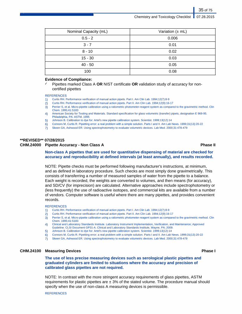
35 of 75
Chemistry and Toxicology Checklist 07.28.2015
Nominal Capacity (mL) Variation (± mL)
0.5 - 2 0.006
3 - 7 0.01
8 - 10 0.02
15 - 30 0.03
40 - 50 0.05
100 0.08
Evidence of Compliance:✓ Pipettes marked Class A OR NIST certificate OR validation study of accuracy for non-
certified pipettes
REFERENCES1) Curtis RH. Performance verification of manual action pipets. Part I. Am Clin Lab. 1994;12(7):8-9
2) Curtis RH. Performance verification of manual action pipets. Part II. Am Clin Lab. 1994;12(9):16-17
3) Perrier S, et al. Micro-pipette calibration using a ratiometric photometer-reagent system as compared to the gravimetric method. ClinChem. 1995;41:S183
4) American Society for Testing and Materials. Standard specification for glass volumetric (transfer) pipets, designation E 969-95.Philadelphia, PA: ASTM, 1995
5) Johnson B. Calibration to dye for: Artel's new pipette calibration system. Scientist. 1999;13(12):14
6) Connors M, Curtis R. Pipetting error: a real problem with a simple solution. Parts I and II. Am Lab News. 1999;31(13):20-22
7) Skeen GA, Ashwood ER. Using spectrophotometry to evaluate volumetric devices. Lab Med. 2000;31:478-479
**REVISED** 07/28/2015CHM.24000 Pipette Accuracy - Non Class A Phase II
Non-class A pipettes that are used for quantitative dispensing of material are checked foraccuracy and reproducibility at defined intervals (at least annually), and results recorded.
NOTE: Pipette checks must be performed following manufacturer's instructions, at minimum,and as defined in laboratory procedure. Such checks are most simply done gravimetrically. Thisconsists of transferring a number of measured samples of water from the pipette to a balance.Each weight is recorded, the weights are converted to volumes, and then means (for accuracy),and SD/CV (for imprecision) are calculated. Alternative approaches include spectrophotometry or(less frequently) the use of radioactive isotopes, and commercial kits are available from a numberof vendors. Computer software is useful where there are many pipettes, and provides convenientrecords.
REFERENCES1) Curtis RH. Performance verification of manual action pipets. Part I. Am Clin Lab. 1994;12(7):8-9
2) Curtis RH. Performance verification of manual action pipets. Part II. Am Clin Lab. 1994;12(9):16-17
3) Perrier S, et al. Micro-pipette calibration using a ratiometric photometer-reagent system as compared to the gravimetric method. ClinChem. 1995;41:S183
4) Clinical and Laboratory Standards Institute. Laboratory Instrument Implementation, Verification, and Maintenance; ApprovedGuideline. CLSI Document GP31-A. Clinical and Laboratory Standards Institute, Wayne, PA; 2009.
5) Johnson B. Calibration to dye for: Artel's new pipette calibration system. Scientist. 1999;13(12):14
6) Connors M, Curtis R. Pipetting error: a real problem with a simple solution. Parts I and II. Am Lab News. 1999;31(13):20-22
7) Skeen GA, Ashwood ER. Using spectrophotometry to evaluate volumetric devices. Lab Med. 2000;31:478-479
CHM.24100 Measuring Devices Phase I
The use of less precise measuring devices such as serological plastic pipettes andgraduated cylinders are limited to situations where the accuracy and precision ofcalibrated glass pipettes are not required.
NOTE: In contrast with the more stringent accuracy requirements of glass pipettes, ASTMrequirements for plastic pipettes are ± 3% of the stated volume. The procedure manual shouldspecify when the use of non-class A measuring devices is permissible.
REFERENCES
36 of 75
Chemistry and Toxicology Checklist 07.28.2015
1) American Society for Testing and Materials. Standard specification for serological pipets, disposable plastic, designation E934-88, In 1993 Annual Book of ASTM Standards, section 14 (general methods and instrumentation). Philadelphia, PA: ASTM,1993:14.02:485-486
PIPETTES - FIXED VOLUME, ADJUSTABLE AND/OR MICROPIPETTES
Pipettes and diluting devices of all types must be checked for accuracy and reproducibility before being placedin service and at least annually thereafter.
Inspector Instructions:
● Pipette calibration procedure● Sampling of pipette/dilutor checks
● How are you assured your automatic pipetting systems exhibit no carryover effects?
CHM.24200 Pipette Accuracy Phase II
There is a written procedure defining how pipettes used for quantitative dispensing(fixed volume, adjustable volume, micropipettes, and analytic instruments with integralautomatic pipettors) are checked for accuracy of calibration and reproducibility(gravimetric, colorimetric, volumetric or other verification procedure) before being placedin service initially, and the result recorded.
NOTE: The initial calibration may be performed by the manufacturer or other outside facility,but in such cases the laboratory must have a record from the manufacturer or other facility thatincludes the technique used to check calibration, the method of shipment to prevent damage intransit, and the bias and imprecision of the pipette(s). The bias and imprecision must meet thespecifications established by the laboratory.
REFERENCES1) Curtis RH. Performance verification of manual action pipets. Part I. Am Clin Lab. 1994;12(7):8-9
2) Curtis RH. Performance verification of manual action pipets. Part II. Am Clin Lab. 1994;12(9):16-17
3) Perrier S, et al. Micro-pipette calibration using a ratiometric photometer-reagent system as compared to the gravimetric method. ClinChem. 1995;41:S183
4) Clinical and Laboratory Standards Institute. Laboratory Instrument Implementation, Verification, and Maintenance; ApprovedGuideline. CLSI Document GP31-A. Clinical and Laboratory Standards Institute, Wayne, PA; 2009.
5) Johnson B. Calibration to dye for: Artel's new pipette calibration system. Scientist. 1999;13(12):14
6) Connors M, Curtis R. Pipetting error: a real problem with a simple solution. Parts I and II. Am Lab News. 1999;31(13):20-22
7) Skeen GA, Ashwood ER. Using spectrophotometry to evaluate volumetric devices. Lab Med. 2000;31:478-479
**REVISED** 07/28/2015CHM.24300 Pipette Accuracy Phase II
Pipettes used for quantitative dispensing are checked for accuracy and reproducibility(gravimetric, colorimetric, volumetric or other verification procedure) at defined intervals(at least annually), and results recorded.
NOTE: Pipette checks must be performed following manufacturer's instructions, at minimum, andas defined in laboratory procedure.
37 of 75
Chemistry and Toxicology Checklist 07.28.2015
For analytic instruments with integral automatic pipettors, the accuracy and precision of thepipetting system should be checked at least annually, unless that is not practical for the end-userlaboratory. Manufacturers' recommendations should be followed.
REFERENCES1) Curtis RH. Performance verification of manual action pipets. Part I. Am Clin Lab. 1994;12(7):8-9
2) Curtis RH. Performance verification of manual action pipets. Part II. Am Clin Lab. 1994;12(9):16-17
3) Perrier S, et al. Micro-pipette calibration using a ratiometric photometer-reagent system as compared to the gravimetric method. ClinChem. 1995;41:S183
4) Clinical and Laboratory Standards Institute. Laboratory Instrument Implementation, Verification, and Maintenance; ApprovedGuideline. CLSI Document GP31-A. Clinical and Laboratory Standards Institute, Wayne, PA; 2009.
5) Johnson B. Calibration to dye for: Artel's new pipette calibration system. Scientist. 1999;13(12):14
6) Connors M, Curtis R. Pipetting error: a real problem with a simple solution. Parts I and II. Am Lab News. 1999;31(13):20-22
7) Skeen GA, Ashwood ER. Using spectrophotometry to evaluate volumetric devices. Lab Med. 2000;31:478-479
CHM.24400 Pipette Carryover Phase II
The laboratory evaluates its automatic pipetting systems for carryover.
NOTE: The laboratory must have written procedures for evaluating whether carryover effects arepresent. This requirement applies to both stand-alone pipette systems and to sample pipettesintegrated with analytic instruments.
In practice, carryover is a problem only for analytes with a wide clinical range of analyteconcentration, such that a minute degree of carry-over could have significant clinical implications.Examples include immunoassays such as β-HCG, certain enzymes (e.g. CK), and certaindrugs of abuse (e.g. benzoylecgonine [cocaine metabolite], which may be present in highconcentrations). The laboratory should select representative examples of such analytes forcarryover studies.
Evaluation for carryover is not required for automatic pipettes that use disposable tips.
One suggested method to study carryover is to run known high patient samples, followed byknown low samples to see if the results of the low-level material are affected. If carryover isdetected, the laboratory must determine the analyte concentration above which subsequentsamples may be affected, and define this value in the procedure. Results of each analytical runmust be reviewed to ensure that no results exceed this level. If results that exceed the definedlevel are detected, then the appropriate course of action must be defined (repeat analysis ofsubsequent samples, for example).
Carryover studies must be performed, as applicable, as part of the initial evaluation of aninstrument. (The laboratory may use the data from carryover studies performed by instrumentmanufacturers, as appropriate.) Carryover studies should be repeated after major maintenanceor repair of the pipetting assembly of the instrument.
Evidence of Compliance:✓ Record of carryover studies at defined frequency
REFERENCES1) Clinical and Laboratory Standards Institute. Laboratory Instrument Implementation, Verification, and Maintenance; Approved
Guideline. CLSI Document GP31-A. Clinical and Laboratory Standards Institute, Wayne, PA; 2009.
38 of 75
Chemistry and Toxicology Checklist 07.28.2015
ANALYTICAL BALANCES
Inspector Instructions:
● Sampling of balance service records● Sampling of balance accuracy check records
● Analytical balances (mounting)● Standard weights (maintained, appropriate class)
CHM.25300 Balance Maintenance Phase I
Balances are cleaned, serviced and checked at least annually only by qualified servicepersonnel (i.e. service contract or as needed).
Evidence of Compliance:✓ Records of balance maintenance
CHM.25400 Balance Mounting Phase I
Analytical balances are mounted such that vibrations do not interfere with readings.
**REVISED** 07/28/2015CHM.25500 Analytical Balance Accuracy Phase II
Standard weights of the appropriate ANSI/ASTM Class are available and used for verifyingaccuracy of analytical balances at defined intervals, with records maintained.
NOTE: The verification of accuracy of the analytical balance must be performed on a regularschedule to ensure accurate creation of analytical calibrators and/or weighed-in controls fromstandard materials, as well as when gravimetrically checking the accuracy of pipettes.
Accuracy must be verified at least every six months if used for weighing materials to makestandard solutions for method calibration. Accuracy must be verified at the time of installation andwhenever a balance is moved. Acceptable ranges must be defined.
External verification of accuracy requires the appropriate class of ASTM specification weights.ASTM Class 1 weights are appropriate for calibrating high precision analytical balances (0.01to 0.1 mg limit of precision). ASTM Class 2 weights are appropriate for calibrating precisiontop-loading balances (0.001 to 0.01 g precision).w ASTM Class 3 weights are appropriate forcalibrating moderate precision balances, (0.01 to 0.1 g precision).
Evidence of Compliance:✓ Written procedure defining criteria for the use of standard weights for accuracy checks of
analytical balances
REFERENCES1) ASTM E617-13. Standard Specification for Laboratory Weights and Precision Mass Standards, ASTM International, West
Conshohocken, PA, 2013. http://www.astm.org2) ASTM E898-88 (2013). Standard Test Method of Testing Top-Loading, Direct-Reading Laboratory Scales and Balances. ASTM
International, West Conshohocken, PA, 2013. http://www.astm.org
39 of 75
Chemistry and Toxicology Checklist 07.28.2015
CHM.25700 Weight Maintenance Phase II
Weights are well-maintained (clean, in a covered container, not corroded) and appropriatelifting or handling devices are available.
NOTE: Weights must be well-maintained (covered when not in use, not corroded) and only behandled by devices that will not allow residual contaminants to remain on the masses. Certifiedmasses will only meet their specifications if maintained in pristine condition.
PERSONNEL
Inspector Instructions:
● Records of education and experience
CHM.25800 Bench Testing Supervision Phase II
The person in charge of bench testing/section supervisor in chemistry has educationequivalent to an associate's degree (or beyond) in chemical, physical or biological scienceor medical technology and at least four years experience (one of which must be in clinicalchemistry) under a qualified section director.
Evidence of Compliance:✓ Records of qualifications including degree or transcript, current license (if required) and work
history in related field
CHM.25900 Bench Testing Supervision Phase II
The person in charge of bench testing/section supervisor in toxicology has educationequivalent to an associate's degree (or beyond) in chemical, physical or biological scienceor medical technology, and at least four years experience (one of which must be intoxicology) under a qualified section director.
Evidence of Compliance:✓ Records of qualifications including degree or transcript, current license (if required) and work
history in related field
CHM.25950 Bench Testing Supervision Phase II
The person in charge of bench testing/section supervisor in the blood gas section haseducation and experience equivalent to an associate's degree (or beyond) in a chemical,physical or biological science or medical technology (or certified or registered respiratorytherapist) and at least four years experience (one of which must be in blood gas testing)under a qualified section director.
Evidence of Compliance:✓ Records of qualifications including degree or transcript, current license (if required) and work
history in related field
40 of 75
Chemistry and Toxicology Checklist 07.28.2015
LABORATORY SAFETY
The inspector should review relevant requirements from the Safety section of the Laboratory General checklist,to assure that the chemistry laboratory is in compliance. Please elaborate upon the location and the details ofeach deficiency in the Inspector's Summation Report.
RADIATION SAFETY
Inspector Instructions:
● Sampling of radiation safety policies and procedures● Sampling of radiation area surveys/wipe tests records● Sampling of radioactive waste disposal records● Sampling of personnel records of radionuclide training
● Radionuclide storage areas (properly shielded)● Appropriate signage where radioactive materials are used/stored
● Does your laboratory have representation at radiation safety committee meetings?● How does your laboratory check the effectiveness of workbench decontamination?
CHM.27900 Radiation Safety Manual Phase II
There is an up-to-date radiation safety manual that includes sections on decontaminationand radioactive waste.
NOTE: For US laboratories, this is required by the Nuclear Regulatory Commission (NRC).
REFERENCES1) U.S. Nuclear Regulatory Commission. Guide for the preparation of applications for medical use programs. Regulatory guide 10.8:
Appendix H model procedure for area surveys. Washington, DC: USNRC, 19872) Clinical and Laboratory Standards Institute (CLSI). Clinical Laboratory Waste Management; Approved Guideline—Third Edition. CLSI
document GP05-A3 (ISBN 1-56238-744-8). Clinical and Laboratory Standards Institute, 940 West Valley Road, Suite 1400, Wayne,Pennsylvania 19087-1898, USA 2011.
CHM.28000 Workspace Decontamination Phase II
Workbenches and sinks are decontaminated each day of use, and the effectiveness testedat least monthly.
NOTE: If the laboratory uses only Iodine-125 either a wipe test or a portable scintillation probecan be used.
Evidence of Compliance:✓ Records of daily workbench/sink decontamination AND✓ Records of monthly effectiveness tests
41 of 75
Chemistry and Toxicology Checklist 07.28.2015
REFERENCES1) U.S. Nuclear Regulatory Commission. Guide for the preparation of applications for medical use programs. Regulatory guide 10.8:
Appendix H - model procedure for area surveys. Washington, DC: USNRC, 1987.
CHM.28100 Radionuclides Handling Phase I
There are written policies regarding authorization or restriction of personnel handlingradionuclides.
NOTE: These policies should be incorporated into the department's radiation safety manual.
CHM.28200 Radionuclide Leak Phase II
There are written procedures for notification if a damaged or leaking radionuclideshipment is received.
NOTE: Procedures must include inspection, monitoring of shipments, and instructions fornotification, if leakage or damage is noted in a radionuclide shipment.
Evidence of Compliance:✓ Records of inspections and notifications
REFERENCES1) Clinical and Laboratory Standards Institute (CLSI). Clinical Laboratory Safety; Approved Guideline, Third Edition. CLSI document
GP17-A3 [ISBN 1-56238-797-9 (Print); ISBN 1-56238-798-7 (Electronic)]. Clinical and Laboratory Standards Institute, 940 WestValley Road, Suite 1400, Wayne, Pennsylvania 19087-1898 USA, 2012.
2) Department of Transportation, Research and Special Programs Administration. Hazardous materials table, special provisions,hazardous materials communications, emergency response information and training requirements. Fed Register. 2003 (Oct 1)[49CFR172.403, 600, 602, 604]
CHM.28300 Radionuclide Storage Phase II
Radionuclide storage and decay areas are properly shielded, if required for specificisotopic materials.
NOTE: Radionuclide storage and decay areas must be properly shielded, if required for specificisotopic materials, to avoid excessive exposure to personnel and interference with countingprocedures.
Evidence of Compliance:✓ Written procedure defining shielding requirements for radionuclide storage and decay areas
CHM.28400 Radiation Surveys Phase II
There are regular radiation area surveys and wipe tests, with records maintained.
NOTE: Routine radiation surveys and wipe tests to determine exposure rates and detectcontamination must be performed and recorded at defined frequency.
Evidence of Compliance:✓ Written procedure defining frequency of radiation survey and wipe tests to determine
exposure rates and detect contamination
CHM.28500 Radioactive Material Sign Phase I
All areas or rooms where radioactive materials are being used or stored are posted toindicate the presence of radioactive materials.
42 of 75
Chemistry and Toxicology Checklist 07.28.2015
NOTE: For US laboratories, all areas or rooms where radioactive materials are being usedor stored must be posted to indicate the presence of radioactive materials, consistent with10CFR20, Appendix C.
REFERENCES1) Nuclear Regulatory Commission; Standards for Protection Against Radiation. Fed Register 2004(Jan1):354-421[10CFR20.2402]
CHM.28600 Radionuclide Training Phase II
Personnel receive training in decontamination routines and in the safe handling andproper disposal of radionuclides (wastes, syringes, needles, and sponges) with recordsmaintained.
CHM.28700 Radioactive Waste Phase II
Radioactive waste is stored separately, under required conditions, and appropriatelydiscarded, with records maintained.
NOTE: Records of the radioactive trash disposal must be maintained. For US laboratories,NRC regulations specify that separate areas be established for the receipt of radioactive wasteand that these areas be properly shielded to reduce radiation levels below those maximumpermissible limits specified in 10CFR20.
Evidence of Compliance:✓ Written procedure defining criteria for proper storage and disposal of radioactive waste
REFERENCES1) Clinical and Laboratory Standards Institute (CLSI). Clinical Laboratory Waste Management; Approved Guideline—Third Edition. CLSI
document GP05-A3 (ISBN 1-56238-744-8). Clinical and Laboratory Standards Institute, 940 West Valley Road, Suite 1400, Wayne,Pennsylvania 19087-1898, USA 2011.
CHM.28800 Safety Committee Representation Phase II
There are records indicating that a laboratory representative is a member of and/orattends institutional radiation safety committee meetings regularly.
NOTE: Independent laboratories must have a radiation safety officer who fulfills the functions ofan institutional radiation safety committee.
Evidence of Compliance:✓ Records of laboratory participation in institutional Safety Committee meeting OR participation
in other appropriate group responsible for radiation safety
GENERAL CHEMISTRY
CHEMISTRY
THERAPEUTIC DRUG MONITORING
Inspector Instructions:
● Sampling of TDM policies and procedures● Sampling of TDM patient reports (dosage, time of drug administration)
43 of 75
Chemistry and Toxicology Checklist 07.28.2015
● How is the clinician able to link TDM laboratory results to the dosage and time thepatient received the drug?
CHM.28900 Specimen Collection/Drug Dosing Phase I
As applicable, the laboratory has provided information to clinical personnel of the optimalspecimen collection time in relation to drug dosing.
Evidence of Compliance:✓ Written procedure defining criteria for specimen collection for TDM
REFERENCES1) Nicholson PW, et al. Ideal sampling time for drug assays. Br J Clin Pharm. 1980;9:467470
2) Howanitz PJ, Steindel SJ. Digoxin therapeutic drug monitoring practices. A College of American Pathologists Q-Probes study of 666institutions and 18679 toxic levels. Arch Pathol Lab Med. 1993;117:684-690
3) Schoenenberge RA, et al. Appropriateness of antiepileptic drug level monitoring. JAMA. 1995;274:1622-1626
4) Williamson KM, et al. Digoxin toxicity: an evaluation in current clinical practice. Arch Intern Med. 1998;158:2444-2499
CHM.29000 TDM Results Phase II
Where applicable, TDM results are reported in relation to patient dosing and/or timinginformation.
NOTE: The intent is to have a mechanism whereby the clinician can easily and accurately linkTDM results from the laboratory to the dosage and time of drug administration. Ideally, the testresult, dose and administration time would be reported in juxtaposition on the patient chart.This may be the responsibility of the laboratory, or an integrating function of reported laboratoryanalytic data with clinical information from other sources.
Evidence of Compliance:✓ Written procedure defining criteria for reporting TDM results
REFERENCES1) Elin RJ. Computer-assisted therapeutic drug monitoring. Clin Lab Med. 1987;7:485-492
2) Howanitz PJ, Steindel SJ. Digoxin therapeutic drug monitoring practices. A College of American Pathologists Q-Probes study of 666institutions and 18679 toxic levels. Arch Pathol Lab Med. 1993;117:684-690
3) Schoenenberge RA, et al. Appropriateness of antiepileptic drug level monitoring. JAMA. 1995;274:16221626
4) Williamson KM, et al. Digoxin toxicity: an evaluation in current clinical practice. Arch Intern Med. 1998;158:2444-2499
5) Steele BW, et al. An evaluation of analytic goals for assays of drugs. A College of American Pathologists therapeutic drug monitoringsurvey study. Arch Pathol Lab Med. 2001;125:729-735
**NEW** 07/28/2015CHM.29025 Immunosuppressive Drug Result Reporting Phase II
For the reporting of immunosuppressive drug results, the patient report contains all of thefollowing:
1. Appropriate therapeutic ranges based on the test method used2. Analytical method (all tests) and method platform (immunoassays only)3. Elements required in GEN.41096
NOTE: For immunosuppressive drugs (e.g. cyclosporine, sirolimus, tacrolimus, mycophenolicacid, everolimus), the therapeutic range may depend upon the test method, type of transplant,and length of time since the transplant procedure. Results from different types of samples anddifferent methods are not interchangeable.
Evidence of Compliance:✓ Written procedure for reporting immunosuppressive drug results AND✓ Patient results showing required report elements
44 of 75
Chemistry and Toxicology Checklist 07.28.2015
SWEAT TESTING FOR CYSTIC FIBROSIS
The laboratory diagnosis of cystic fibrosis includes SCREENING and CONFIRMATORY sweat testing.Screening tests include: sweat conductivity, cystic fibrosis indicator patch system, Orion skin measuringelectrode, and sweat osmolality. Confirmatory tests include quantitative analysis of sweat chloride.
SPECIMEN COLLECTION AND HANDLING
Inspector Instructions:
● Sampling of sweat testing policies and procedures● Sampling of records/log of insufficient sweat samples● Sampling of iontophoresis unit maintenance records
● An employee performing the sweat collection procedure, if possible
● How do you ensure effective disinfection of the sweat collection equipment?● What is your course of action if the patient is receiving oxygen from an open delivery
system?
**REVISED** 07/28/2015CHM.29100 Sweat Test/Appropriate Age Phase I
The sweat test is offered only to patients at an appropriate age.
NOTE: To increase the likelihood of collecting an adequate sweat specimen, sweat chloridetesting can be evaluated in asymptomatic newborns with a positive newborn screen result whenthe infant is at least two weeks of age and weighs >2kg.
In symptomatic newborns (e.g. those with meconiuim ileus), sweat chloride can be evaluatedas early as 48 hours after birth if an adequate sweat volume can be collected; although, thelikelihood of an inconclusive result may be greater at this age.
Evidence of Compliance:✓ Written policy defining criteria for sweat testing OR collection manual that includes
parameters for patient age
REFERENCES1) Farrell PM, et al. Guidelines for diagnosis of cystic fibrosis in newborns through older adults: Cystic Fibrosis Foundation consensus
report. J. Pediatr, 2008: 153(2):S4-S14.2) Clinical and Laboratory Standards Institute (CLSI). Sweat Testing: Sample Collection and Quantitative Analysis; Approved Guideline-
Third Edition. CLSI document C34-A3 (ISBN 1-56238-713-8). Clinical and Laboratory Standards Institute, 940 West Valley Road,Wayne, PA, 19087-1898, USA, 2009.
**REVISED** 07/28/2015CHM.29150 Biennial Review Equipment Disinfection Phase I
45 of 75
Chemistry and Toxicology Checklist 07.28.2015
The laboratory reviews the procedures employed for disinfection of equipment andfacilities used for sweat collection at least biennially.
NOTE: The purpose of this review is to assure continued effectiveness. One suggested approachis biennial evaluation by the infection control department of the institution.
Evidence of Compliance:✓ Written procedure for disinfection of equipment and facility
CHM.29200 Sweat Collection and Analysis Procedure Phase II
The laboratory follows generally accepted procedures for sweat collection and analysis,including steps to minimize sample evaporation or contamination.
NOTE: Because sample evaporation and contamination can have significant impact on thevalidity of test results, laboratories must incorporate the following steps into their procedure and/or follow manufacturer's recommendations:
A. When using gauze or filter paper collection pads:
1. Use gauze and/or filter paper that is low in electrolyte content2. Wash the patient's skin thoroughly with distilled or deionized water, then dry before
stimulation. Repeat after stimulation and before collection3. Do not touch the weighing vial, wax film, collection site, or collection pad. Always use
forceps or powder-free gloves4. Use two pieces of waterproof adhesive tape on all sides of the paraffin wax film or
wrap with a disposable stretch bandage to produce an airtight seal5. Blot back into the collection pad any condensate that may have formed on the wax
film during collection. Failure to collect the condensate can result in false positive testresults
6. After collection, quickly transfer the specimen pad to the weighing vial and reweighpromptly
B. When collecting sweat into Macroduct coils:
1. Wash the patient's skin thoroughly with distilled, deionized water. Leave the skinslightly wet after washing the area where the electrode will be attached or add adrop of distilled or deionized water to either the skin or the Pilogel surface (afterinstallation in the electrode). Repeat after stimulation and before collection
2. Avoid touching the collecting surface of the coil3. Fasten the collector to the extremity with firm strap pressure. Test for proper
attachment after sweat appears in the coils4. Do not attempt to remove the entire collector assembly from the patient's extremity
before separating the coil from the main body. Loss of specimen may occur5. Do not contaminate the nippers or sweat dispensing needle with sweat sample
C. When collecting and analyzing sweat using the Nanoduct system:
1. Wash the patient's skin thoroughly with distilled, deionized water. Leave the skinslightly wet after washing the area where the electrode will be attached or add adrop of distilled or deionized water to either the skin or the pilogel surface (afterinstallation in the electrode). Repeat after stimulation and before collection
2. Avoid touching the collecting surface of the device
Evidence of Compliance:✓ Written procedure defining criteria for sweat collection and analysis including required
elements OR other procedure following manufacturer's recommendations
46 of 75
Chemistry and Toxicology Checklist 07.28.2015
REFERENCES1) Scott MG, et al. Electrolytes and blood gases, In Burtis C and Ashwood E (eds), Tietz textbook of clinical chemistry. Philadelphia: WB
Saunders, 4th edition, 20062) Clinical and Laboratory Standards Institute (CLSI). Sweat Testing: Sample Collection and Quantitative Analysis; Approved Guideline-
Third Edition. CLSI document C34-A3 (ISBN 1-56238-713-8). Clinical and Laboratory Standards Institute, 940 West Valley Road,Wayne, PA, 19087-1898, USA, 2009.
CHM.29300 Sweat Stimulation Phase II
The protocol requires sweat stimulation and collection from the patient's lower arm orupper leg, using a site that is free from diffuse inflammation or rash.
NOTE: The protocol should require that sweat is stimulated and collected from the patient'slower arm or upper leg. Sweat must not be stimulated or collected from the head or trunk. Sweatmust not be stimulated or collected from an area of diffuse inflammation, such as a rash oreczematous lesion, because of the likelihood of contamination by serous fluid.
REFERENCES1) Liebke C, et al. Sweat electrolyte concentrations in children with atopic dermatitis. Lancet. 1997;350:1678
2) Clinical and Laboratory Standards Institute (CLSI). Sweat Testing: Sample Collection and Quantitative Analysis; Approved Guideline-Third Edition. CLSI document C34-A3 (ISBN 1-56238-713-8). Clinical and Laboratory Standards Institute, 940 West Valley Road,Wayne, PA, 19087-1898, USA, 2009.
CHM.29400 Pilocarpine Grade Phase II
If the laboratory prepares the pilocarpine solution for iontophoresis, the source of thepilocarpine is USP grade or equivalent.
CHM.29500 Electrode Placement Phase II
The protocol requires that the electrodes used for stimulation are placed such thationtophoretic current never crosses the patient's trunk.
NOTE: The protocol must specify that electrodes used for stimulation be placed so that currentdoes not cross the trunk, to avoid the possibility of current crossing the heart, resulting in cardiacdepolarization.
**REVISED** 07/28/2015CHM.29600 Iontophoresis Conditions and Equipment Maintenance Phase II
The written procedure specifies the conditions of iontophoresis and maintenancerequirements.
NOTE: For safety reasons, the iontophoretic current source must be battery-powered, to avoidthe possibility of patient exposure to line voltage. For manually controlled devices, iontophoresismust be performed for no more than five minutes at a current less than 4 mA, to prevent burns.The iontophoresis unit must be tested by qualified personnel (such as engineering personnel) forcurrent leakage and current control at defined frequencies and records maintained.
CHM.29700 Iontophoresis Oxygen Phase II
The protocol specifies that iontophoresis is withheld from patients receiving oxygen by anopen delivery system.
NOTE: While the possibility of an explosion due to the generation of an electrical spark is remote,it cannot be ignored. Often, these patients can temporarily receive oxygen via a facemask ornasal cannula, in which case sweat testing can be done.
CHM.29800 Iontophoretic Stimulation Phase II
47 of 75
Chemistry and Toxicology Checklist 07.28.2015
The procedure specifies that the area of iontophoretic stimulation is equivalent to the areaof sweat collection.
NOTE: The procedure must specify that the area of iontophoretic stimulation is equivalent to thearea of sweat collection. Sweat electrolyte concentration is related to sweat rate. At low sweatrates, sweat electrolyte concentration decreases. The average sweat rate should exceed 1 g/m2/min.
To ensure adequate sweat stimulation and accurately reflect sweat electrolyte concentration, aminimum acceptable sweat volume or weight is required. This requirement is based on the sizeof the electrode and stimulation area, the type and size of collecting media, and the durationof sweat collection. To standardize the process, the stimulation and collection area shouldbe equivalent, and the time of collection consistent. For example, for the positive electrode,use a 1 ½ inch x 1 ½ inch electrode over a 2 x 2 inch gauze pad saturated with pilocarpine forstimulation, then collect sweat onto a 2 x 2 inch pre-weighed gauze pad.
REFERENCES1) Clinical and Laboratory Standards Institute (CLSI). Sweat Testing: Sample Collection and Quantitative Analysis; Approved Guideline-
Third Edition. CLSI document C34-A3 (ISBN 1-56238-713-8). Clinical and Laboratory Standards Institute, 940 West Valley Road,Wayne, PA, 19087-1898, USA, 2009.
CHM.29850 Appropriate Sweat Collection Device Phase II
The sweat collection device is appropriate for the iontophoresis system.
NOTE: The sweat collection device must be designed for use with the appropriate iontophoresissystem so that the stimulation and collection area are equivalent and the appropriate minimumacceptable sweat volume or weight can be achieved. Examples of acceptable combinationsinclude:
■ Stimulation with Wescor Pilogel iontophoresis and collection into WescorMacroduct coils
■ Stimulation with copper electrodes over gauze/filter paper pilocarpine pads andcollection into gauze/filter paper
■ Stimulation and collection into Wescor Nanoduct conductivity cell
Examples of unacceptable combinations include:
■ Stimulation with Wescor Pilogel iontophoresis and collection into gauze/filterpaper
■ Stimulation with Polychrome iontophoresis and collection into Wescor Macroductcoils, gauze or filter paper
■ Stimulation with copper electrodes over gauze/filter paper pilocarpine pads andcollection into Wescor Macroduct coils
REFERENCES1) Clinical and Laboratory Standards Institute (CLSI). Sweat Testing: Sample Collection and Quantitative Analysis; Approved Guideline-
Third Edition. CLSI document C34-A3 (ISBN 1-56238-713-8). Clinical and Laboratory Standards Institute, 940 West Valley Road,Wayne, PA, 19087-1898, USA, 2009.
2) Cystic Fibrosis Foundation Center Accreditation Guidelines. Bethesda, MD, 2004
CHM.29900 Sweat Collection Time Phase II
The procedure specifies that sweat collection time may not exceed 30 minutes.
NOTE: Extending the collection time will not significantly increase the sweat yield and may leadto sample evaporation. Samples may be taken from less than maximally stimulated glands. Thismay lead to a false-negative result. In addition, altering the collection time will affect the minimum
48 of 75
Chemistry and Toxicology Checklist 07.28.2015
acceptable sweat weight or volume, because the time parameter of the rate equation has beenchanged.
Evidence of Compliance:✓ Written procedure for sweat collection
CHM.30000 Sweat Collection Parameters Phase II
The protocol specifies the parameters of sweat collection.
NOTE: These must include an established minimum acceptable sweat volume or weight basedon the area of stimulation, area of collection and standardized time for collection. The averagesweat rate should exceed 1 g/m2/min, which in general corresponds to a minimum sampleweight of about 75 mg of sweat collected on a 2x2 inch gauze or filter paper and about 15 µL ofsweat collected in Macroduct coil in 30 min. Samples less than the required volume or weightmust not be analyzed.
REFERENCES1) Hjelm M, et al. Sweat sodium related to amount of sweat after sweat test in children with and without cystic fibrosis. Acta Paediatr
Scand. 1986;75:652-6562) Clinical and Laboratory Standards Institute (CLSI). Sweat Testing: Sample Collection and Quantitative Analysis; Approved Guideline-
Third Edition. CLSI document C34-A3 (ISBN 1-56238-713-8). Clinical and Laboratory Standards Institute, 940 West Valley Road,Wayne, PA, 19087-1898, USA, 2009.
CHM.30100 Sweat Sample Rejection Phase II
The procedure specifies that multiple insufficient sweat samples are rejected and notpooled for analysis.
NOTE: The laboratory must reject individual samples that do not meet minimum sample sizerequirements. The average sweat rate of 1 g/m2/min is determined independently for eachsite. The requirement is a physiologic one, not an analytic one. Samples less than the requiredvolume or weight must not be pooled to achieve the weight/volume requirement. Measurementon samples from less than maximally stimulated sweat glands may lead to false-negative results.
Evidence of Compliance:✓ Records of specimen rejection
CHM.30150 Sweat Rejection Incidence Rate Phase I
The incidence of insufficient sweat samples is routinely monitored.
NOTE: Laboratories should collect data on the number of patients from whom a sufficientsweat sample has not been obtained. For patients older than three months of age, the annualinsufficient rate should not exceed 5%. For patients three months of age or younger, the rateof insufficient samples should not exceed 10%. If these rates are exceeded, the collectionprocedure should be reevaluated for consistency with the CLSI document C34-A3. The mostcommon cause of insufficient samples is the use of inappropriate collection devices (seeCHM.29850).
Evidence of Compliance:✓ Records of insufficient collection AND✓ Records of corrective action if rate exceeds the norm
REFERENCES1) Cystic Fibrosis Foundation Center Accreditation Guidelines. Bethesda, MD, 2009
CHM.30250 Sweat Sample Storage Phase I
The procedure describes appropriate storage conditions for collected sweat samples.
49 of 75
Chemistry and Toxicology Checklist 07.28.2015
NOTE: If there is a significant delay between collection and analysis, appropriate storageconditions must be followed: a) Sweat collected on gauze is stable at refrigerator temperaturesfor up to 72 hours once reweighed and secured in a vial with a tightly fitting cap; and b) Sweatcollected in Macroduct is stable at refrigeration or room temperature for up to 72 hours in a0.2 mL microcentrifuge tube with a tight fitting cap. Storage of sweat in microbore tubing is notrecommended.
REFERENCES1) Cystic Fibrosis Foundation Center Accreditation Guidelines. Bethesda, MD, 2009
CHM.30300 Sweat Collection Skin Reaction Phase II
The procedure describes the recognition of, and appropriate treatment for, patient skinreactions (allergic or burns) to pilocarpine and/or other reagents used in iontophoresis.
NOTE: Rarely, some patients may develop an area of, urticaria (hives) or small localized burns.In such cases, the procedure must be discontinued immediately and appropriate medicalattention obtained. Sweat must not be collected over areas of urticaria or burns.
Evidence of Compliance:✓ Records of follow-up treatment
ANALYTIC METHODS FOR SWEAT TESTING
Inspector Instructions:
● Sweat testing method validation records● Sampling of QC logs
CHM.30400 Sweat Test Validation Phase II
For sweat testing, the analytical method is validated by the laboratory prior to patienttesting using specimens equivalent to the volume and concentration of patient sweatsamples.
NOTE: Validation procedures must include studies of accuracy, precision, and upper/lower limitsof the analytic measurement range. The laboratory should be aware that some instrumentsdesigned for serum or urine electrolyte determination may lack the sensitivity required for sweattesting.
Evidence of Compliance:✓ Written procedure defining criteria for validation of the analytical method AND✓ Records of method validation study(ies)
REFERENCES1) Pillion DJ, Meczan E. Chloride measurement by microelectrode in cystic fibrosis and normal sweat. Miner Electrolyte Metab.
1987;13:196-2002) Barnes GL, et al. Sweat testing by capillary collection and osmometry: suitability of the Wescor macroduct system for screening
suspected cystic fibrosis patients. Aust Paediatr J. 1988;24:191-1933) Department of Health and Human Services, Centers for Medicare and Medicaid Services. Clinical laboratory improvement
amendments of 1988; final rule. Fed Register. 2003(Jan 24):3707 [42CFR493.1255]4) Clinical and Laboratory Standards Institute (CLSI). Sweat Testing: Sample Collection and Quantitative Analysis; Approved Guideline-
Third Edition. CLSI document C34-A3 (ISBN 1-56238-713-8). Clinical and Laboratory Standards Institute, 940 West Valley Road,Wayne, PA, 19087-1898, USA, 2009.
5) Department of Health and Human Services, Centers for Medicare and Medicaid Services Center for Disease Control and Prevention.Medicare, Medicaid and CLIA Programs; Laboratory Requirements Relating to Quality Systems and Certain Personnel Qualifications;final rule. Fed Register. 2003 (Jan 24): Vol. 68, No. 16 [42CFR493]
50 of 75
Chemistry and Toxicology Checklist 07.28.2015
CHM.30550 Sweat Chloride AMR Phase II
The lower limit of the sweat chloride analytical measurement range is less than or equal to10 mmol/L.
NOTE: The lower limit of the sweat chloride analytical measurement range must be less than orequal to 10 mmol/L without any dilution, concentration or other pretreatment that is not part of theusual assay procedure.
REFERENCES1) Cystic Fibrosis Foundation Center Accreditation Guidelines. Bethesda, MD, 2009
2) Farrell PM, et al. Guidelines for diagnosis of cystic fibrosis in newborns through older adults: Cystic Fibrosis Foundation consensusreport. J Pediatr 2008;153:S4-S14
CHM.30575 Confirmatory Sweat Test Report Phase II
If the test performed is a confirmatory test (i.e. quantitative analysis of sweat chloride), theupper limit of AMR for sweat chloride results is less than or equal to 160 mmol/L.
NOTE: Even though the analytical instrument may have a higher upper limit of its AMR, sweatchloride concentrations > 160 mmol/L are not physiologically possible. Results of sweat chloridetesting greater than 160 mmol/L must not be reported, and the patient must be retested.
Evidence of Compliance:✓ Patient reports or worksheets
REFERENCES1) Schultz IJ. Micropuncture studies of the sweat formation in cystic fibrosis patients. J Clin Invest. 1969;48:1470-1477
CHM.30600 Daily QC - Sweat Testing Phase II
The laboratory analyzes two levels of controls (one in the negative range and one in thepositive range) at least once each day patient specimens are assayed.
NOTE: If sweat is collected from patients on gauze or filter paper, controls should be placeddirectly onto the same collection material, eluted, and treated in the same manner as a patientspecimen.
For test systems with internal controls, the laboratory may limit daily quality control to the internalcontrols ONLY if all CAP requirements for internal controls are met, as listed in CHM.13900.
Evidence of Compliance:✓ Written QC procedure AND✓ Records of QC results at defined frequency
REFERENCES1) Department of Health and Human Services, Centers for Medicare and Medicaid Services. Clinical laboratory improvement
amendments of 1988; final rule. Fed Register. 2003(Jan 24):7165 [42CFR493.1255]2) Clinical and Laboratory Standards Institute (CLSI). Sweat Testing: Sample Collection and Quantitative Analysis; Approved Guideline-
Third Edition. CLSI document C34-A3 (ISBN 1-56238-713-8). Clinical and Laboratory Standards Institute, 940 West Valley Road,Wayne, PA, 19087-1898, USA, 2009.
3) Department of Health and Human Services, Centers for Medicare and Medicaid Services Center for Disease Control and Prevention.Medicare, Medicaid and CLIA Programs; Laboratory Requirements Relating to Quality Systems and Certain Personnel Qualifications;final rule. Fed Register. 2003 (Jan 24): Vol. 68, No. 16 [42CFR493]
51 of 75
Chemistry and Toxicology Checklist 07.28.2015
REPORTING OF RESULTS
Inspector Instructions:
● Sampling of sweat analysis reports (appropriate reference ranges and disclaimer ifapplicable)
CHM.30700 Sweat Test Reporting Phase II
The laboratory report indicates the specific analytes measured in the sweat analysis, andapplies the appropriate reference ranges and/or decision levels to patient results.
NOTE: The laboratory report must clearly indicate the analytes measured in the sweat test, andapply the appropriate reference intervals and/or decision levels to patient results. Osmolalityand conductivity are nonselective methods for sweat analysis; they are not equivalent to sweatchloride concentrations and therefore have their own unique set of reference ranges. Whensweat conductivity is expressed as units of aqueous sodium chloride solution, the values areapproximately 15 mmol/L higher than when chloride is measured directly.
The Cystic Fibrosis Foundation reference intervals for chloride and conductivity are:
SWEAT CHLORIDE: For infants up to and including six months of age: less than or equal to 29mmol/L, CF unlikely; 30-59 mmol/L, intermediate; greater than or equal to 60 mmol/L, indicativeof CF. For individuals older than six months of age: < 40 mmol/L, CF unlikely; 40-59 mmol/L,intermediate; > 60 mmol/L, indicative of CF. The result must be interpreted with regard to thepatient's age and clinical presentation.
SWEAT CONDUCTIVITY: A patient having a sweat conductivity greater than or equal to 50mmol/L should be referred to a specialized cystic fibrosis care center for a quantitative analysis ofsweat chloride with or without sweat sodium.
SWEAT OSMOLALITY: 50-150 mmol/kg, negative; 151-200 mmol/kg, equivocal; > 200 mmol/kg,positive for cystic fibrosis.
REFERENCES1) Farrell PM, et al. Guidelines for diagnosis of cystic fibrosis in newborns through older adults: Cystic Fibrosis Foundation consensus
report. J Pediatr 2008;153:S4-S142) Clinical and Laboratory Standards Institute (CLSI). Sweat Testing: Sample Collection and Quantitative Analysis; Approved Guideline-
Third Edition. CLSI document C34-A3 (ISBN 1-56238-713-8). Clinical and Laboratory Standards Institute, 940 West Valley Road,Wayne, PA, 19087-1898, USA, 2009.
CHM.30800 Sweat Test Report Disclaimer Phase II
If the test performed is a screening test (e.g. sweat conductivity, CF Indicator patchsystem, Orion skin measuring electrode, etc.), the report includes a statement regardingthe limits of clinical interpretation.
NOTE: Suggested wording for such a disclaimer might be: "This result represents a screeningtest for cystic fibrosis. Patients having borderline or positive results should be referred for aquantitative sweat chloride concentration."
REFERENCES1) Rosenstein BJ, Langbaum TS. Misdiagnosis of cystic fibrosis. Need for continuing follow-up and reevaluation. Clin Pediatr.
1987;26:78-822) Clinical and Laboratory Standards Institute (CLSI). Sweat Testing: Sample Collection and Quantitative Analysis; Approved Guideline-
Third Edition. CLSI document C34-A3 (ISBN 1-56238-713-8). Clinical and Laboratory Standards Institute, 940 West Valley Road,Wayne, PA, 19087-1898, USA, 2009.
52 of 75
Chemistry and Toxicology Checklist 07.28.2015
PRENATAL SCREENING
TEST PANELS
Test panels include: 1) Second trimester quadruple panel: maternal serum alpha-fetoprotein (MSAFP),unconjugated estriol (uE3), and beta-human chorionic gonadotropin (beta-hCG) and dimeric inhibin-A (DIA); 2)First trimester panel: total or free beta-hCG, pregnancy associated placental protein A (PAPP-A), and nuchaltranslucency (NT); 3) Sequential and integrated panels: various combinations of first and second trimester tests
REQUISITIONS/CALCULATIONS/REPORTS
Requests for prenatal screening (neural tube defects, Down syndrome, etc.) must include specific informationfor meaningful interpretation of laboratory tests. For clinical screening purposes, analyte concentrations mustbe converted to multiple of the median (MoM) values, using gestational-age specific medians. The MoMvalue is used directly as the interpretative result for neural tube defect screening and for calculating risk forfetal trisomies. Gestational age-specific MoM values need to be adjusted for each patient, based on severalvariables. The laboratory must work cooperatively with the clinician to ensure that all necessary information isobtained.
Inspector Instructions:
● Sampling of prenatal screen policies and procedures● Sampling of prenatal screening requisitions for required elements● Sampling of median value records● Sampling of records verifying calculated gestational age, maternal age and patient-
specific risks
● How does your laboratory verify or establish acceptable median values?● How does your laboratory monitor assay quality, appropriateness of medians and
accuracy of gestational dating?● What is your course of action if the requisition does not include all necessary
information?
CHM.31100 Prenatal Screen Requisitions Phase II
Prenatal screening requisitions solicit the first day of the last menstrual period (LMP), theestimated date of confinement (EDC), or estimated gestational age by ultrasound dating.
NOTE: Accurate screening requires that the laboratory know the clinician's best estimate of thegestational age at the time of the specimen collection. The gestational age allows determinationof the best median values for a given screen. The optimum time for neural tube defect detectionusing MSAFP measurements is between 16-18 weeks gestation.
REFERENCES1) Wald NJ, et al. Maternal serum-alphafetoprotein measurement in antenatal screening for anencephaly and spina bifida in early
pregnancy. Report of U.K. collaborative study on alpha-fetoprotein in relation to neural-tube defects. Lancet. 1977;i:1323-13322) Wald NJ, et al. Maternal serum screening for Down's syndrome in early pregnancy. Brit Med J. 1988;297:883-887
3) Bock JL. Current issues in maternal serum alphafetoprotein screening. Am J Clin Pathol. 1992;97:541-554
CHM.31200 Prenatal Screen Requisitions Phase II
53 of 75
Chemistry and Toxicology Checklist 07.28.2015
Prenatal screening requisitions solicit maternal birth date.
NOTE: Maternal birth date must be included as part of the test requesting process. Maternal ageis not necessary when screening for neural tube defects, but is needed to calculate the patient-specific risk for Down syndrome. The patient-specific risk, not the analyte concentration, is usedas the screening variable to identify pregnancies at high-risk for Down syndrome.
REFERENCES1) Cuckle HS, et al. Estimating a woman's risk of having a pregnancy associated with Down's syndrome using her age and serum
alphafetoprotein level. Brit J Obstet Gynecol. 1987;94:387-4022) Wald NJ, et al. Maternal serum screening for Down's syndrome in early pregnancy. Brit Med J. 1988;297:883-887
CHM.31300 Prenatal Screen Requisitions Phase II
Prenatal screening requisitions solicit patient race.
NOTE: Patient race must be included as part of the test requesting process.
Both MSAFP and hCG values in black women are approximately 10% to 15% higher thanin Caucasian women. Depending upon the racial distribution of the patient population, thelaboratory should either have separate MSAFP medians for blacks if enough data is availableand Caucasians, or apply an appropriate correction factor for patients from the less commonraces. Black women also have a lower birth prevalence of neural tube defects, and somescreening programs raise the MoM cut-off to take this difference into account (after adjusting theAFP MoM value).
uE3 levels are similar in the two racial groups, uE3 medians need not be adjusted.
DIA medians in blacks are approximately 8% lower than those in Caucasians, and adjustment isrecommended.
PAPP-A values are approximately 25% higher in black women, therefore medians should beadjusted.
Current data indicate that statistically significant differences in median values exist for otherraces and ethnic groups (e.g. Hispanic, Asian, Native American). Most differences tend to besmall and do not materially affect the risk calculations for Down syndrome and neural tubedefects. However, laboratories that screen large numbers of women may consider using medianvalues specific to ethnic or racial groups if significant differences are identified in their screeningpopulation.
REFERENCES1) Crandall BF, et al. Alphafetoprotein concentrations in maternal serum: relation to race and body weight. Clin Chem. 1983;29:531-533
2) Greenberg F, et al. Estimates of birth prevalence rates of spina bifida in the United States from computer generated maps. Am JObstet Gynecol. 1983;145:570-573
3) Bock JL. Current issues in maternal serum alphafetoprotein screening. Am J Clin Pathol. 1992;97:541-554
4) Kulch P, et al. Racial differences in maternal serum human chorionic gonadotrophin and unconjugated oestriol levels. Prenat Diag.1993;13:191-195
5) O/Brien JE, Dvorin E, Drugan A, Johnson MP, Yaron TY, Evans MI. Race-ethnicity -specific variation in multiple-marker biochemicalscreening: alpha-fetoprotein, hCG, and estriol. Obstet Gynecol 1997 89(3):355-8
6) Benn PA, Clive JM, Collins R. Medians for second-trimester maternal serum alpha-fetoprotein human chorionic gonadotrophin, andunconjugated estriol; differences between races or ethnic groups. Clin Chem 1997 43(2):333-7
7) Spencer K, et al. The influence of ethnic origin on first trimester biochemical markers of chromosomal abnormalities. Prenat Diagn2000 June;20(6):491-4
CHM.31400 Prenatal Screen Requisitions Phase II
Prenatal screening requisitions solicit maternal weight.
NOTE: Maternal weight must be included as part of the test requesting process. The averageconcentration of all of the analytes used for second trimester screening decreases withincreasing maternal weight. Heavier women have lower analyte values, and lighter womenhave higher levels. This is presumably due to a greater maternal blood volume in the formergroup. The influence of weight adjustment on neural tube defects screening is small, but
54 of 75
Chemistry and Toxicology Checklist 07.28.2015
adjustment aids in equalizing false positive and false negative rates in all weight categories.The concentration of first trimester markers also decrease with increasing maternal weight; therelationship between PAPP-A and maternal weight is greater than for any other marker. All firsttrimester markers should be adjusted for maternal weight. Laboratories can utilize publishedweight correction equations until sufficient data are collected to establish correction equationsusing in-house patient data. The linear-reciprocal equation model allows the correction equationto apply to a broader range of maternal weights than the log-linear model and only requiresapproximately 1000 data points for calculation.
REFERENCES1) Johnson AM, et al. The effect of adjusting maternal serum alphafetoprotein levels for maternal weight in pregnancies with open spina
bifida: a United States collaborative study. Am J Obstet Gynecol. 1990;163:9112) Bock JL. Current issues in maternal serum alphafetoprotein screening. Am J Clin Pathol. 1992;97:541-554
3) Neveux LM, et al. Refinements in managing maternal weight adjustment for interpreting prenatal screening results. Prenat Diag.1996;16:1115-1119
CHM.31500 Prenatal Screen Requisitions Phase II
Prenatal screening requisitions solicit a history of the patient receiving medication (e.g.insulin) to control diabetes at the time of conception.
NOTE: A history of insulin-dependent diabetes mellitus (IDDM) at the time of conception mustbe included as part of the test requesting process. Pregnant women who have diabetes priorto conception have 12% lower MSAFP levels than nondiabetics of the same gestational age,but have a higher birth prevalence of neural tube defects. If medians derived from nondiabeticwomen are used to screen women with IDDM, the percentage of women with values exceedingthe laboratory's MoM screening cut-off will be inappropriately low, resulting in a lower detectionrate. Conversely, if medians from nondiabetic women are used for screening pregnancies fromwomen with IDDM for Down syndrome, the AFP MoM values calculated using non-diabeticmedians will be inappropriately low, resulting in an increased number of patients in the screenpositive group. Differences are slight for the other analytes. uE3 is 8% lower, second trimesterhCG is 5% lower, and DIA is 9% lower in pregnant women with DM. Adjustment for theseanalytes is optional. PAPP-A and first trimester hCG are not significantly different in DM, andadjustment is not recommended for these analytes. There is no consensus whether correction forgestational diabetes is warranted.
REFERENCES1) Wald NJ, et al. Maternal serum alphafetoprotein and diabetes mellitus. Br J Obstet Gynecol. 1979;86:101-105
2) Kucera J. Rate and type of congenital anomalies among offspring of diabetic women. J Reprod Med. 1979;7:61-70
3) Greene MF, et al. Maternal serum alphafetoprotein levels in diabetic pregnancies. Lancet. 1988;ii:345-346
4) Wald NJ, et al. Maternal serum unconjugated oestriol, human chorionic gonadotrophin, and alpha-fetoprotein levels in pregnancieswith insulin-dependent diabetes: implications for Down's syndrome screening. Brit J Obstet Gynaecol. 1992;99:51-53
5) Hutley W, et al. Second-trimester prenatal screening markers for Down syndrome in women with insulin-dependent diabetes mellitus.Prenat Diagn 2004;24:804-807
CHM.31600 Prenatal Screen Requisitions Phase II
Prenatal screening requisitions solicit clinical evidence of multiple gestations (twins, etc.).
NOTE: Clinical evidence of multiple gestations (twins, etc.) must be included as part of thetest requesting process. Women with ultrasonographically confirmed twin pregnancies have,on average, twice the level of MSAFP than that seen in women with singleton pregnancies atthe same gestational age. Most laboratories use a screening cutoff level for twin pregnanciesbetween 3.5 and 5.0 MoM, or divide the MoM by two and use an adjusted cutoff level as forsingleton pregnancies. However, screening for neural tube defects in twin pregnancies is lessefficient than singleton pregnancies. Approximately 50% of twin pregnancies with an openneural tube defect and 5% of unaffected twin pregnancies have a MoM of 4.5 or greater. If twinpregnancies are to be screened for Down syndrome, laboratories need to calculate risks usinga method specifically designed for this application. hCG and DIA levels are also approximatelytwice as high in twin pregnancies, while uE3 levels in twin pregnancies are approximately1.7 times as high as in singleton pregnancies. There are few data on analyte concentrations
55 of 75
Chemistry and Toxicology Checklist 07.28.2015
from twin pregnancies with one or both fetuses affected with Down’s syndrome; therefore,only “pseudo risks” can be calculated. Pseudo risks are less reliable than those calculated forsingleton pregnancy and some laboratories choose not to report the actual risk, but only reportspecimens as screen negative or screen positive. Insufficient data are available to calculateDown syndrome risk in pregnancies with three or more fetuses.
REFERENCES1) Cuckle HS, et al. Maternal serum alphafetoprotein screening for open neural tube defects in twin pregnancies. Prenat Diagn.
1990;10:71-772) Bock JL. Current issues in maternal serum alphafetoprotein screening. Am J Clin Pathol. 1992;97:541-554
3) Wald NJ, et al. Maternal serum unconjugated oestriol and human chorionic levels in twin pregnancies: implications for screening forDowns syndrome. Brit J Obstet Gynaecol. 1991;98:905-908
4) Wald NJ, Rish S. Prenatal screening for Down syndrome and neural tube defects in twin pregnancies. Prenat Diagn 2005;25:740-745
CHM.31700 Prenatal Screen Requisitions Phase I
The test requisitions ask if this is the initial screening sample or a repeat test for thispregnancy.
NOTE: The interpretation of a repeat maternal serum sample may be different from theinterpretation of an initial serum specimen. In neural tube defect screening using AFP, a repeatsample can be interpreted as if it were an initial sample if fixed MoM cut-offs are used to identifyhigh-risk women. If patient-specific risks are calculated for a repeat test result, one shouldcombine the results of both the initial and repeat sample, using published algorithms. Repeattesting is not recommended for Down syndrome screening unless a sample is drawn too early forreliable interpretation. If a test on a second sample is performed, it is essential that the revisedrisk be calculated using the results from both samples. A method has been published for thesecalculations.
REFERENCES1) Estimating an individual's risk of having a fetus with open spina bifida and the value of repeat alphafetoprotein testing. Fourth report
of the U.K. collaborative study on alphafetoprotein in relation to neural tube defects. J Epidemiol Commun Health. 1982;36:87-952) Cuckle HS, et al. Repeat maternal serum testing in multiple marker Downs syndrome screening programmes. Prenat Diagn.
1994;14:603-6073) Hackshaw AK. et al. Repeat testing in antenatal screening for Down syndrome using dimeric inhibin-A in combination with other
maternal serum markers. Prenat Diagn, 2001;21:58-61
CHM.31800 Median Values Phase II
There is a record that the laboratory has established its own median values or verified thatthe medians from another source are appropriate for the population being screened.
NOTE: Systematic biases in maternal serum assay values of up to 30% can occur when kitsfrom different manufacturers are used. In addition, between laboratory differences in equipment,reagents, and technique may introduce bias in assay results even when the same kit lot is used.These differences can be minimized by reporting results in multiples of the normal median(assuming that the medians are calculated using values measured on the population to be testedusing the kit designated for screening). Ideally, week-specific medians would be establishedby testing approximately 100 patients per week of gestation. Because analytes are stable, it ispossible to use stored frozen specimens collected over a period of years. A second approachis to perform a split specimen study with a reference laboratory and transfer the referencelaboratory's medians using the comparison regression equation from the split specimen study.However, in practice the most practical method is to measure values on 300 consecutivelycollected specimens spread over the appropriate gestational age range, and perform weightedregression analysis using published models. It is not necessary to document that all specimensare collected from unaffected pregnancies. Smoothing data by weighted regression analysisallows median values to be calculated for weeks with limited data. Package insert medians maybe outdated or inappropriate and should not be used even for a short time. Incorrect referencedata may lead to inappropriate recommendations in the laboratory report.
56 of 75
Chemistry and Toxicology Checklist 07.28.2015
Evidence of Compliance:✓ Records for median value determination OR records for verification of package inserts or
other sources
REFERENCES1) Report of a workshop sponsored by the National Institute of Child Health and Human Development (NICHD), Bethesda, Maryland.
The quality control of alphafetoprotein reagents and assay for the antenatal screening and diagnosis of open neuraltube defects. ClinChim Acta. 1980;105:9-24
2) Knight GJ, et al. Assessing reliability of AFP test kits. Contemp Obstet Gynecol. 1987 (Technology issue);30:37-52
3) Clinical and Laboratory Standards Institute (CLSI). Maternal Serum Screening; Approved Guideline—Second Edition. CLSIdocument I/LA25-A2 (ISBN 1-56238-749-9). CLSI, 940 West Valley Road, Suite 1400, Wayne, Pennsylvania 19087-1898 USA, 2011
4) Knight GJ. Quality assessment of a prenatal screening program. Early Human Development. 1996;47(Supp):S49-S53
5) Haddow et al. Screening for maternal serum for fetal Down's syndrome in the first trimester. N Eng J Med, 1998;338:955-961
CHM.31900 Median Value Reverification Phase II
Medians are reverified at specified intervals and when new reagent lots are introduced,and the medians are recalculated if necessary.
NOTE: Changing reagent lots can introduce significant bias. One method for assessing a new lotis performing a split specimen comparison study between the new and old lot.
In addition, re-evaluation of medians at specified intervals is a valuable quality controlmechanism to ensure validity of reported MoMs. Epidemiological monitoring of Down syndromescreening can be accomplished by determining the median MoMs at frequent intervals (e.g.every 500-1000 patients, weekly or monthly). If a persistent shift is noted, (less than 0.95 orgreater than 1.05), new medians should be determined. Records of the median MoMs must bemaintained.
Evidence of Compliance:✓ Written procedure defining criteria and frequency for recalculation or reverification of median
values AND✓ Records of median value recalculation or reverification data at defined frequency
REFERENCES1) Report of a workshop sponsored by the National Institute of Child Health and Human Development (NICHD), Bethesda, Maryland.
The quality control of alphafetoprotein reagents and assay for the antenatal screening and diagnosis of open neuraltube defects. ClinChim Acta. 1980;105:9-24
2) Bock JL. Current issues in maternal serum alphafetoprotein screening. Am J Clin Pathol. 1992;97:541-554
3) Ashwood ER, Knight GJ. Clinical Chemistry of Pregnancy. In: Tietz Textbook of Clinical Chemistry and Molecular Pathology, 4th ed.Burtis CA, Ashwood ER, Bruns DE, Eds. Philadelphia, Elsevier Saunders, 2006, pp. 2153-206
4) Erickson JA, Ashwood ER, Gin CA. Evaluation of a dimeric inhibitin-A assay for assessing fetal Down syndrome: establishment,comparison, and monitoring of median concentrations for normal pregnancies. Arch Pathol Lab Med, 2004 Apr;128(4):415-20
CHM.31950 Establishing Nuchal Translucency (NT) Measurements Phase I
If screening panels are offered using nuchal translucency (NT) values, the laboratoryshould have a written procedure for their establishment and use.
NOTE: Some potential content of these procedures might include:● Requirements for verifying the credentialing of each sonographer● Having sonographers provide a set number of NT/CRL measurements prior to initial
interpretations● Implementing sonographer-specific medians when the median NT MoM levels are
outside of set limits● Reviewing data for sonographers available from certifying organization (e.g. NT Quality
Review Program - NTQR (https://ntqr.org) or the Fetal Medicine Foundation - FMF(http://fetalmedicineusa.com)
● Establishing communication with sonographers to inform them of monitoring results● Establishing communications with credentialing organizations to inform them of
sonographer performance
57 of 75
Chemistry and Toxicology Checklist 07.28.2015
Evidence of Compliance:✓ Written procedure defining criteria for establishing a new sonographer AND✓ Records of sonographer establishment
REFERENCES1) Palomaki GE, Neveux LM, Donnenfeld A, Lee JE, McDowell G, Canick JA, Summers A, Lambert-Messerlian G, Kellner LH,
Zebelman A, Haddow JE. Quality assessment of routine nuchal translucency measurements: A North American laboratoryperspective. Genet Med, 2008 Feb;10(2):131-8
CHM.31960 Nuchal Translucency (NT) Measurements, Monitoring Phase I
If screening panels are offered using nuchal translucency (NT) values, the laboratoryshould routinely perform epidemiological monitoring of these measurements.
NOTE: An example of such a monitoring procedure (with action limits) is provided below. Foreach sonographer with sufficient data (typically at least 30 to 50 measurements over six months),monitor and provide limits for three quality parameters.
● Percent increase in NT measurements (in mm) by gestational age (e.g. 15% to 35%)● The NT median MoM (e.g. 0.90 to 1.10)● The distribution of NT MoMs after a logarithmic transformation (log standard deviation),
(e.g. 0.08 to 0.13)
Evidence of Compliance:✓ Records of NT median data study(ies) AND✓ Records of review at defined frequency
REFERENCES1) Palomaki GE, Neveux LM, Donnenfeld A, Lee JE, McDowell G, Canick JA, Summers A, Lambert-Messerlian G, Kellner LH,
Zebelman A, Haddow JE. Quality assessment of routine nuchal translucency measurements: A North American laboratoryperspective. Genet Med, 2008 Feb;10(2):131-8
2) Palomaki GE, et al. Technical standards and guidelines: Prenatal screening for Down syndrome that includes first-trimesterbiochemistry and/or ultrasound measurements. Genet Med, 2009;11(9):669-681
CHM.32000 Screen Result Statistics Phase II
The percentages of women with screen-positive test results for neural tube defects (NTD),Down syndrome, and Trisomy 18 are calculated and reviewed at least quarterly.
NOTE: Data from large studies provide guidelines for the percentage of pregnancies that willfall above specified maternal serum AFP MoM levels (NTD screening) or with risks greater thanspecified risk cutoff levels (Down syndrome screening). Regular comparison of a laboratory'sscreen-positive rates with expected rates serves as a continuing measure of assay quality,appropriateness of medians, and accuracy of gestational dating.
Evidence of Compliance:✓ Written procedure defining criteria for comparison of screen-positive tests to expected rates
to include frequency of analysis AND✓ Records of statistical analysis and evaluation of screen-positive test results at least quarterly
REFERENCES1) Report of a workshop sponsored by the National Institute of Child Health and Human Development (NICHD), Bethesda Maryland.
The quality control of alphafetoprotein reagents and assay for the antenatal screening and diagnosis of open neuraltube defects. ClinChim Acta. 1980;105:9-24
2) Palomaki GJ, et al. Risk based screening for trisomy 18 using alphaphetoprotein, unconjugated estriol, and human chorionic. PrenatDiagn 1995;15:713-723
3) Knight GJ, et al. Epidemiologic monitoring of prenatal screening for neural tube defects and Down syndrome. Clin Lab Med2003;22:531-551
4) Wald NJ, Hackshaw AK, George LM. Assay precision of serum alpha fetoprotein in antenatal screening for neural tube defects andDown;s Syndrome. J Med Screen. 2000;7(2):74-7
5) Wald NJ, Rodeck C, Hackshaw AK, Walters J, Chitty L, Mackinson AM. First and second trimester antenatal screening for Down'ssyndrome: the results of the Serum, Urine and Ultrasound Screening Study (SURUSS). Health Technol Asses 2003;7(11)
CHM.32100 Dimeric Inhibin A Phase II
58 of 75
Chemistry and Toxicology Checklist 07.28.2015
If the laboratory adds dimeric inhibin A (DIA) (or another marker) to its screening panel, ithas followed the same requirements outlined for established markers.
NOTE: Many screening laboratories have added a fourth marker, dimeric inhibin A, to the tripletest. If so, the laboratory must demonstrate that it adheres to the checklist items outlined forestablished markers in the Prenatal Screening section. These include establishing median valuesappropriate for the population being screened, and adjusting where appropriate for variables thathave been shown to influence analyte values, such as maternal weight, maternal race, insulindependent diabetes, and twin pregnancy. Laboratories must verify that the risks calculated usingthe additional marker are valid.
Evidence of Compliance:✓ Records of validation studies for dimeric inhibin A or other markers added to the screening
panel
REFERENCES1) Haddow JE, Palomaki GE, Knight GJ, Foster DL, Neveux LM. Second trimester screening for Down's syndrome using maternal
serum dimeric inhibin A. J Med Screen 1998;5(3):115-92) Cuckle HS, Holding S, Jones R, Groome NP, Wallace EM. Combining inhibin A with existing second-trimester markers in maternal
serum screening for Down's syndrome. Prenat Diagn 1996 16(12):1095-11003) Clinical and Laboratory Standards Institute. Maternal Serum Screening; Approved Guideline. 2nd ed. CLSI Document I/LA25-A2.
Clinical and Laboratory Standards Institute, Wayne, PA; 2011.
CHM.32200 Computer Calculations Phase II
There are records that neural tube defect and Down syndrome risk calculations wereinitially verified for accuracy and reverified with any software updates or changes.
NOTE: Verification can be accomplished by interlaboratory comparisons, by comparison withresults calculated or reported by proficiency testing programs, or by use of risk tables availableon the CAP website (located in the CAP/APMG Biochemical and Molecular Genetics ResourceCommittee Genetics Topic Center section). At a minimum, the accuracy of calculated gestationalage, maternal age, and patient-specific risks must be verified.
Evidence of Compliance:✓ Written procedure for verifying accuracy of calculations initially and with changes AND✓ Records of initial and subsequent calculation checks
INTERPRETIVE REPORTING FOR MATERNAL SCREENING
Inspector Instructions:
● Sampling of maternal screen patient reports (demographics, clinical information,results reported in MoM, cut-off values)
CHM.32300 Maternal Screen Reports Phase II
All of the following demographic and clinical information is included in the report: date ofbirth; maternal weight; maternal race; first day of the last menstrual period or gestationalage as determined by ultrasound examination; specimen draw date; initial or repeatspecimen; presence of medication-dependent diabetes; family history of neural tubedefect, and presence of multiple gestation if known.
59 of 75
Chemistry and Toxicology Checklist 07.28.2015
NOTE: Reports must include data essential to the interpretation of the test results to enableboth the laboratory and physician to ensure that the interpretations on the report are based oncomplete and correct information.
In addition to the above, smoking history is recommended.
CHM.32400 Multiple of Population Median Phase II
Test results are reported as multiples of the population median (MoM).
NOTE: Reporting of results in terms of multiple of the population median (MoM) simplifiesinterpretation at various gestational ages, reduces possible systematic between-laboratory andbetween-kit bias in assay results, and facilitates comparison among laboratories. Laboratoriescan also compare their experiences with large-scale published studies more readily by usingMoM as the reportable interpretive unit. The initial MoM is calculated as the measured analytevalue divided by the median value for the appropriate gestational age. The MoM should also beadjusted for the other clinical variables known to influence the concentration of each analyte,generally by dividing by a factor specific for each variable.
Evidence of Compliance:✓ Written procedure defining criteria for reporting results as MoM
REFERENCES1) Report of a workshop sponsored by the National Institute of Child Health and Human Development (NICHD), Bethesda Maryland.
The quality control of alphafetoprotein reagents and assay for the antenatal screening and diagnosis of open neuraltube defects. ClinChim Acta. 1980;105:9-24
2) Clinical and Laboratory Standards Institute (CLSI). Maternal Serum Screening; Approved Guideline—Second Edition. CLSIdocument I/LA25-A2 (ISBN 1-56238-749-9). CLSI, 940 West Valley Road, Suite 1400, Wayne, Pennsylvania 19087-1898 USA,2011.
CHM.32500 Result Cut-off Values Phase II
The report classifies a pregnancy as screen-positive or screen-negative for open neuraltube defects, based on the MSAFP test results.
NOTE: Cut-off levels based on maternal serum AFP MoM values or risk have been establishedby large screening programs. Use of these cut-off values in the laboratory report can assist thephysician in making clinical decisions about pregnancy management.
REFERENCES1) Burton BK. Elevated maternal serum alphafetoprotein (MSAFP): interpretation and followup. Clin Obstet Gynecol. 1988;31:293-305
2) Milunsky A, et al. Predictive values, relative risks, and overall benefits of high and low maternal serum alphafetoprotein screening insingleton pregnancies: new epidemiologic data. Am J Obstet Gynecol. 1989;161:291-297
3) Crandall BF, et al. Risks associated with an elevated maternal serum alphafetoprotein level. Am J Obstet Gynecol.1991;165:581-586
4) Bock JL. Current issues in maternal serum alphafetoprotein screening. Am J Clin Pathol. 1992;97:541-554
CHM.32600 Result Cut-off Values Phase II
The report classifies a pregnancy as screen-positive or screen-negative for fetal Downsyndrome, based on the calculated risk.
NOTE: Cut-off levels based on risk for fetal Down syndrome have been established by largescreening programs. Use of the cut-off values in the laboratory report can assist the physician inmaking clinical decisions about pregnancy management.
REFERENCES1) Wald NJ, et al. Maternal serum screening for Down's syndrome in early pregnancy. Brit Med J. 1988;425:883-887
60 of 75
Chemistry and Toxicology Checklist 07.28.2015
AMNIOTIC FLUID ALPHA-FETOPROTEIN (AFAFP)
Inspector Instructions:
● Sampling of AFAFP policies and procedures● Sampling of AFAFP patient reports (results reported in MoM)● Sampling of QC logs
● How does your laboratory verify or establish acceptable median AFAFP values?● What is your laboratory’s course of action when you receive an amniotic fluid sample
that is visibly contaminated with blood?● What is your laboratory’s course of action when an amniotic fluid has an elevated
AFP?
● Select an abnormal AFAFP result and review records for the confirmatory testingperformed, including QC for AChE testing
**REVISED** 07/28/2015CHM.32700 Median AFAFP Values Phase II
There are records that the laboratory has established its own median AFAFP values orverified that medians provided from another source are appropriate for the populationbeing screened.
NOTE: Systematic biases in AFAFP assay values of up to 30% can occur when kits fromdifferent manufacturers are used. In addition, between-laboratory differences in equipment,reagents, and technique may introduce bias in assay results even when the same kit is used.These differences can be minimized by reporting results in multiples of the median (assumingthat the medians are calculated using values measured on the population to be tested usingthe kit designed for screening). Package insert medians may be outdated or inappropriate andshould only be used as a general guide as to expected medians. When computing AFAFPmedians from laboratory data, samples from pregnancies with known (or suspected) neural tubeor ventral wall defects should be removed. A reasonable practice would be to trim all values over2.5 or 3.0 MoM prior to computing medians using a log-linear model (for data between 15 and22 weeks' gestation). Medians below 15 weeks do not follow the log-linear model and alternativecurve fitting is required.
Evidence of Compliance:✓ Records for median value determination OR records for median values from package inserts
or other sources
REFERENCES1) Amniotic fluid alphafetoprotein measurement in antenatal diagnosis of anencephaly and open spina bifida in early pregnancy.
Second report of the U.K. collaborative study on alphafetoprotein in relation to neural tube defects. Lancet. 1979;ii:6512) Report of a workshop sponsored by the National Institute of Child Health and Human Development (NICHD), Bethesda Maryland:
the quality control of alphafetoprotein reagents and assay for the antenatal screening and diagnosis of open neuraltube defects. ClinChim Acta. 1980;105:9-24
3) Knight GJ. Maternal serum alphafetoprotein screening. In: techniques in diagnostic human biochemical genetics. New York, NY:WileyLiss, 1991:503
4) Clinical and Laboratory Standards Institute (CLSI). Maternal Serum Screening; Approved Guideline—Second Edition. CLSIdocument I/LA25-A2 (ISBN 1-56238-749-9). CLSI, 940 West Valley Road, Suite 1400, Wayne, Pennsylvania 19087-1898 USA, 2011
61 of 75
Chemistry and Toxicology Checklist 07.28.2015
5) Palomaki GE, Knight GJ, Haddow JE. Calculating amniotic fluid alpha-fetoprotein medial values in the first trimester. Prenat Diagn.1993;13:887-9.
CHM.32800 Median Value Reverification Phase II
AFAFP medians are recalculated or reverified at specified intervals.
Evidence of Compliance:✓ Written procedure defining criteria and frequency for recalculation or reverification of the
AFAFP median AND✓ Records of median values recalculation or reverification at defined frequency
CHM.32900 Multiple of Median Phase II
AFAFP results are reported in multiples of the median (MoM).
NOTE: Reporting of AFAFP results in terms of multiples of the median (MoM) simplifiesinterpretation at various gestational weeks, reduces the systematic between-laboratoryand between-kit bias in results, and facilitates comparison of results between laboratories.Laboratories may compare their experiences with large-scale published studies much morereadily when using MoM as the interpretive unit for AFP measurements. AFAFP concentrationsare higher in black women than in whites and may be different in other racial groups as well.Although no simple correction factors are available at present, the use of race-specific medians isworth consideration.
Evidence of Compliance:✓ Written procedure defining criteria for reporting results as MoM
REFERENCES1) Report of a workshop sponsored by the National Institute of Child Health and Human Development (NICHD), Bethesda, Maryland:
the quality control of alphafetoprotein reagents and assay for the antenatal screening and diagnosis of open neuraltube defects. ClinChim Acta. 1980;105:9-24
2) Wald NJ, et al. Amniotic fluid acetylcholinesterase measurement in the prenatal diagnosis of open neural tube defects. Prenat Diagn.1989;9:813-829
3) Clinical and Laboratory Standards Institute (CLSI). Maternal Serum Screening; Approved Guideline—Second Edition. CLSIdocument I/LA25-A2 (ISBN 1-56238-749-9). CLSI, 940 West Valley Road, Suite 1400, Wayne, Pennsylvania 19087-1898 USA, 2011
CHM.33100 Dilution Control Phase II
At least one amniotic fluid dilution control is processed with each analytic run of amnioticfluids.
Evidence of Compliance:✓ Records of dilution control with each run
CHM.33200 AChE Testing Phase II
Acetylcholinesterase (AChE) testing is performed on ALL amniotic fluids having elevatedAFAFP concentrations.
NOTE: Acetylcholinesterase (AChE) testing is an essential confirmatory test for amniotic fluidswith abnormal AFP results. The odds of having a fetus with a neural tube defect are considerablygreater if both the AFAFP is elevated and the AChE is positive. The addition of AChE for thedetection of neural tube defects will reduce the false positive rate while maintaining a highdetection rate. This procedure may be performed in-house or referred to a reference laboratory. Iffetal blood is present, acetylcholinesterase results must be interpreted with caution.
Evidence of Compliance:✓ Written policy defining criteria for AChE testing on abnormal AFAFP results AND✓ Patient reports showing AChE results, as applicable
REFERENCES
62 of 75
Chemistry and Toxicology Checklist 07.28.2015
1) Haddow JE, et al. Acetylcholinesterase and fetal malformations: modified qualitative technique for diagnosis of neural tube defects.Clin Chem. 1981;27:61-63
2) Report of the collaborative acetylcholinesterase study. Amniotic fluid acetylcholinesterase electrophoresis as a secondary test in thediagnosis of anencephaly and open spina bifida in early pregnancy. Lancet. 1981;ii:321324
3) Wald N, et al. Amniotic fluid acetylcholinesterase measurement in the prenatal diagnosis of open neural tube defects. Prenat Diagn.1989;9:813-829
4) Crandall BF et al. Risks for fetal abnormalities after very and moderately elevated AFAFPS. Prenat Diagn 1997;17:837-841.
CHM.33300 Daily QC - Acetylcholinesterase Phase II
If acetylcholinesterase is run in-house, both positive and negative controls are includedwith each analytic run.
Evidence of Compliance:✓ QC records for appropriate controls with each run
REFERENCES1) Haddow JE, et al. Acetylcholinesterase and fetal malformations: modified qualitative technique for diagnosis of neural tube defects.
Clin Chem. 1981;27:61-632) Report of the collaborative acetylcholinesterase study. Amniotic fluid acetylcholinesterase electrophoresis as a secondary test in the
diagnosis of anencephaly and open spina bifida in early pregnancy. Lancet. 1981;ii:3213243) Wald NJ, et al. Amniotic fluid acetylcholinesterase measurement in the prenatal diagnosis of open neural tube defects. Prenat Diagn.
1989;9:813-829
CHM.33400 Acetylcholinesterase Confirmation Phase II
If acetylcholinesterase is run in-house, acetylcholinesterase-positive results areconfirmed by addition of a specific inhibitor.
NOTE: Positive acetylcholinesterase results must be confirmed by the addition of a specificinhibitor of acetylcholinesterase, such as BW284C51.
Evidence of Compliance:✓ Written procedure for AChE confirmation testing AND✓ Records of inhibitor testing for positive acetylcholinesterase results prior to reporting results
REFERENCES1) Barlow RD, et al. A single method for amniotic fluid gelacetylcholinesterase determination, suitable for routine use in antenatal
diagnosis of open neuraltube defects. Clin Chim Acta. 1982;119:137-142
ELECTROPHORESIS
Inspector Instructions:
● Sampling of electrophoresis policies and procedures● Sampling of electrophoresis QC logs
● Electrophoretic patterns (appropriate separations)
CHM.33500 Daily QC - Electrophoresis Phase II
Suitable control samples are run and reviewed with each batch of patient samples for allelectrophoresis procedures for which controls are available.
63 of 75
Chemistry and Toxicology Checklist 07.28.2015
Evidence of Compliance:✓ Records of electrophoresis QC
CHM.33600 Electrophoresis Separations Phase II
Electrophoretic separations are satisfactory.
CHM.33700 Acceptable Limits - Controls Phase II
Acceptable limits are set for controls of procedures where the electrophoretic bands arequantified.
Evidence of Compliance:✓ Records of defined acceptable limits for control range of each lot
HEMOGLOBIN SEPARATION
Hemoglobin solubility testing alone is NOT sufficient for detecting or confirming the presence of sicklinghemoglobins in all situations. For purposes of diagnosing hemoglobinopathies, additional tests are required.
Inspector Instructions:
● Sampling of abnormal hemoglobin policies and procedures● Sampling of patient reports (confirmatory testing, comments)● Sampling of QC records
● Hemoglobin electrophoretic patterns (appropriate separations and controls)● Examine a sampling of medium (media) used to identify hemoglobin variants
including alkaline/acid electrophoresis, isoelectric focusing, HPLC or other method
● What is your course of action when the primary screening method appears to showHb S?
● What is your course of action when the primary Hb electrophoresis method shows Hbvariants migrating in nonA/nonS positions?
CHM.33708 Hb S Primary Screen Phase II
For samples that appear to have Hb S in the primary screening (by any method), thelaboratory either 1) performs a second procedure (solubility testing, or other acceptablemethod) to confirm the presence of Hb S, or 2) includes a comment in the patient reportrecommending that confirmatory testing be performed.
NOTE: For primary definitive diagnosis screening by electrophoresis or other separationmethods, all samples with hemoglobins migrating in the "S" positions or peak must be tested forsolubility or by other acceptable confirmatory testing for sickling hemoglobin(s). Known sicklingand non-sickling controls both must be included with each run of patient specimens tested.
Evidence of Compliance:✓ Written policy for follow-up when Hb S appears in the primary screen
64 of 75
Chemistry and Toxicology Checklist 07.28.2015
CHM.33716 Daily QC - Hgb Separation Phase II
Controls containing at least three known major hemoglobins, including both a sicklingand a nonsickling hemoglobin (e.g. A, F and S) are performed with the patient specimen(s)and separations are satisfactory.
Evidence of Compliance:✓ Written procedure defining QC requirements for hemoglobin separation AND✓ QC records reflecting the use of appropriate controls AND✓ Electrophoresis media/separation tracings demonstrating appropriate controls and separation
REFERENCES1) Fairbanks VF. Hemoglobinopathies and thalassemias. Laboratory methods and case studies. New York, NY: BC Decker, 1980
2) Beuzard Y, et al. Isoelectric focusing of human hemoglobins, In Hanash, Brewer, eds. Advances in hemoglobin analysis. New York,NY: Alan R. Liss, 1981:177-195
3) Cossu G, et al. Neonatal screening of betathalassemias by thin layer isoelectric focusing. Am J Hematol. 1982;13:149
4) Bunn HF, Forget BG. Hemoglobin: molecular, genetic and clinical aspects. Philadelphia, PA: WB Saunders, 1986
5) Honig GR, Adams JG III. Human hemoglobin genetics. Vienna, Austria: SpringerVerlag, 1986
6) Jacobs S, et al. Newborn screening for hemoglobin abnormalities. A comparison of methods. Am J Clin Pathol. 1986;85:713-715
7) Fishleder AJ, Hoffman GC. A practical approach to the detection of hemoglobinopathies: part I. The introduction and thalassemiasyndromes. Lab Med. 1987;18:368-372
8) Fishleder AJ, Hoffman GC. A practical approach to the detection of hemoglobinopathies: part II. The sickle cell disorders. Lab Med.1987;18:441-443
9) Fishleder AJ, Hoffman GC. A practical approach to the detection of hemoglobinopathies: part III. Nonsickling disorders and cordblood screening. Lab Med. 1987;18:513-518
10) Armbruster DA. Neonatal hemoglobinopathy screening. Lab Med. 1990;21:815-822
11) Adams JG III, Steinberg MH. Analysis of hemoglobins, In Hoffman R, et al, eds. Hematology: basic principles and practice. NewYork, NY: ChurchillLivingstone, 1991:1815-1827
12) Mallory PA, et al. Comparison of isoelectric focusing and cellulose acetate electrophoresis for hemoglobin separation. Clin Lab Sci.1994;7:348- 352
13) Awalt E, et al. Tandem mass spectrometry (MS) – A screening tool for hemoglobinopathies. Clin Chem. 2001;47(suppl):A165
14) Bradley CA, Kelly A. Comparison of high performance liquid chromatography with electrophoresis for measurement of hemoglobinsA, A2, S, F, and C. Clin Chem. 2001;47(suppl):A172
15) Bradley CA, Kelly A. Calibration verification of hemoglobins A, A2, S, and F with an automated chromatography system. Clin Chem.2001;47(suppl):A17315)
16) Hoyer JD, et al. Flow cytometric measurement of hemoglobin F in RBCs: diagnostic usefulness in the distinction of hereditarypersistence of fetal hemoglobin (HPFH) and hemoglobin S-HPFH from other conditions with elevated levels of hemoglobin F. Am JClin Pathol. 2002;117:857-863
**REVISED** 07/28/2015CHM.33732 Hemoglobin Variants Phase II
All samples with hemoglobin variants migrating in "nonA, nonS" positions on alkalineelectrophoresis, or other low resolution procedure are further defined with otheracceptable methods where clinically and technically appropriate.
NOTE: If all clinically significant variants are not clearly separated by the primary method,additional testing is performed to further characterize these hemoglobin variants. This mayinclude, but is not limited to, performance by a complementary, separate methodology orincreasing the duration of the assay (for HPLC) where the hemoglobins migrate/elute at differentconfigurations. Further workup of such variants, including referral to a reference laboratory, isdependent upon the patient's overall clinical situation, such as findings of erythrocytosis or ahemolytic anemia.
Evidence of Compliance:✓ Written policy defining criteria for further identification of hemoglobin variants AND✓ Patient reports and records reflecting further work-up, when appropriate
REFERENCES1) Glacteros F, et al. Cord blood screening for hemoglobin abnormalities by thin layer isoelectric focusing. Blood. 1980;56:1068
2) Beuzard Y, et al. Isoelectric focusing of human hemoglobins, In Hanash, Brewer, eds. Advances in hemoglobin analysis. New York,NY: Alan R. Liss, 1981:177-195
3) Black J. Isoelectric focusing in agarose gel for detection and identification of hemoglobin variants. Hemoglobin. 1984;8:117
4) Bunn HF, Forget BG. Hemoglobin: molecular, genetic and clinical aspects. Philadelphia, PA: WB Saunders, 1986
5) Fishleder AJ, Hoffman GC. A practical approach to the detection of hemoglobinopathies: part I. The introduction and thalassemiasyndromes. Lab Med. 1987;18:368-372
6) Fishleder AJ, Hoffman GC. A practical approach to the detection of hemoglobinopathies: part II. The sickle cell disorders. Lab Med.1987;18:441-443
65 of 75
Chemistry and Toxicology Checklist 07.28.2015
7) Fishleder AJ, Hoffman GC. A practical approach to the detection of hemoglobinopathies: part III. Nonsickling disorders and cordblood screening. Lab Med. 1987;18:513-518
8) Adams JG III, Steinberg MH. Analysis of hemoglobins, In Hoffman R, et al, eds. Hematology: basic principles and practice. NewYork, NY: ChurchillLivingstone, 1991:1815-1827
9) Mallory PA, et al. Comparison of isoelectric focusing and cellulose acetate electrophoresis for hemoglobin separation. Clin Lab Sci.1994;7:348-352
10) Ou CN, Rognerud CL. Rapid analysis of hemoglobin variants by cation exchange HPLC. Clin Chem. 1993;39:820-824
11) Awalt E, et al. Tandem mass spectrometry (MS) – A screening tool for hemoglobinopathies. Clin Chem. 2001;47(suppl):A165
12) Bradley CA, Kelly A. Comparison of high performance liquid chromatography with electrophoresis for measurement of hemoglobinsA, A2, S, F, and C. Clin Chem. 2001;47(suppl):A172
13) Bradley CA, Kelly A. Calibration verification of hemoglobins A, A2, S, and F with an automated chromatography system. Clin Chem.2001;47(suppl):A173
14) Hoyer JD, et al. Flow cytometric measurement of hemoglobin F in RBCs: diagnostic usefulness in the distinction of hereditarypersistence of fetal hemoglobin (HPFH) and hemoglobin S-HPFH from other conditions with elevated levels of hemoglobin F. Am JClin Pathol. 2002;117:857-863
CHM.33764 Hb S Predominant Band Phase II
All samples that appear to have Hb S as the predominant band by the primary screening(by whatever method) and that are confirmed as sickling by appropriate methods arefurther examined to ascertain whether the "Hb S" band or peak contains solely Hb S orboth Hb S and Hb D, Hb G or other variant hemoglobins.
NOTE: When the predominant hemoglobin component appears to be Hb S, it is necessary todetermine whether this represents homozygous Hb S or a heterozygote for Hb S and anothervariant such as Hb D, Hb G, Hb-Lepore, or other hemoglobin variant(s). Given the clinicalimplications of homozygous Hb S (or Hb S/ß zero thalassemia) it is imperative to exclude otherhemoglobin variants, however rare. Referral of these specimens to a reference laboratory forfurther workup is acceptable.
Evidence of Compliance:✓ Written policy defining criteria for determination of homozygous versus heterozygous Hb S
AND✓ Patient records or worksheets showing the exclusion of hemoglobin variants OR referral for
further work-up
REFERENCES1) Black J. Isoelectric focusing in agarose gel for detection and identification of hemoglobin variants. Hemoglobin. 1984;8:117
2) Bunn HF, Forget BG. Hemoglobin: molecular, genetic and clinical aspects. Philadelphia, PA: WB Saunders, 1986
3) Fishleder AJ, Hoffman GC. A practical approach to the detection of hemoglobinopathies: part I. The introduction and thalassemiasyndromes. Lab Med. 1987;18:368-372
4) Fishleder AJ, Hoffman GC. A practical approach to the detection of hemoglobinopathies: part II. The sickle cell disorders. Lab Med.1987;18:441-443
5) Fishleder AJ, Hoffman GC. A practical approach to the detection of hemoglobinopathies: part III. Nonsickling disorders and cordblood screening. Lab Med. 1987;18:513-518
6) Adams JG III, Steinberg MH. Analysis of hemoglobins, In Hoffman R, et al, eds. Hematology: basic principles and practice. NewYork, NY: ChurchillLivingstone, 1991:18151-827
7) Mallory PA, et al. Comparison of isoelectric focusing and cellulose acetate electrophoresis for hemoglobin separation. Clin Lab Sci.1994;7:348-352
BLOOD GAS ANALYSIS
The Chemistry and Toxicology Checklist is intended for inspection of laboratory sections performing testing ina dedicated space (e.g. main laboratory, respiratory therapy). Laboratories performing testing at or near thepatient bedside (e.g. portable instruments) must use the Point-of-Care Testing Checklist.
The number of checklists needed for test sites under the same CLIA number and CAP number is determined asfollows:
● Blood gas testing performed in more than one area under the same supervision use one Chemistryand Toxicology Checklist (e.g. main laboratory and stat lab);
66 of 75
Chemistry and Toxicology Checklist 07.28.2015
● Blood gas testing performed in more than one area under different supervision use separateChemistry and Toxicology Checklists for each separately supervised site (e.g. main laboratory andrespiratory therapy department);
Testing sites within an institution with different CLIA and CAP numbers must submit separate applications andhave separate full inspections.
SPECIMEN COLLECTION AND HANDLING
Inspector Instructions:
● Blood gas collection policy and procedure● Sampling of records for performance of collateral circulation tests
● How are personnel that perform arterial punctures made aware of possiblecomplications?
CHM.33800 Knowledgeable - Arterial Punctures Phase II
Personnel performing arterial punctures are knowledgeable about the more significantcomplications of this procedure compared with venipuncture.
Evidence of Compliance:✓ Records of training in personnel files
REFERENCES1) Clinical and Laboratory Standards Institute. Procedures for the Collection of Arterial Blood Specimens; Approved Standard. 4th ed.
CLSI Document GP43-A4. Clinical and Laboratory Standards Institute. Wayne, PA; 2004.2) Clinical and Laboratory Standards Institute (CLSI). Blood Gas and pH Analysis and Related Measurements; Approved Guideline -
Second Edition. CLSI document C46-A2 (ISBN 1-56238-694-8). Clinical and Laboratory Standards Institute, 940 West Valley Road,Suite 1400, Wayne, PA 19087-1898, USA 2009.
CHM.33900 Collateral Circulation Phase II
For radial artery sampling, a test for collateral circulation is performed and recordedbefore arterial puncture, as applicable.
NOTE: The various technologies available have been evaluated in the published literature.Consensus should be established between the laboratory and involved clinicians to identify thepatient/clients in whom such a test is medically useful in averting potential patient/client injury.The site from where the sample was obtained should be recorded.
Evidence of Compliance:✓ Written collection procedure defining situations that require testing for collateral circulation to
include preferred technique(s)✓ Records of collection site and results of applicable collateral circulation testing
REFERENCES1) Vaghadia H, et al. Evaluation of a postocclusive circulatory hyperaemia (PORCH) test for the assessment of ulnar collateral
circulation. Can J Anaesth. 1988;35:591-5982) Cheng EY, et al. Evaluation of the palmar circulation by pulse oximetry. J Clin Monit. 1989;5:1-3
3) Levinsohn DG, et al. The Allen's test: analysis of four methods. J Hand Surg. 1991;16:279-282
4) Fuhrman TM, et al. Evaluation of collateral circulation of the hand. J Clin Monit. 1992;8:28-32
5) Furhman TM, et al. Evaluation of digital blood pressure, plethysmography, and the modified Allen's test as a means of evaluating thecollateral circulation to the hand. Anaesthesia. 1992;47:959-961
67 of 75
Chemistry and Toxicology Checklist 07.28.2015
6) Fuhrman TM, McSweeney E. Noninvasive evaluation of the collateral circulation to the hand. Acad Emerg Med. 1995;2:195-199
7) O'Mara K, Sullivan B. A simple bedside test to identify ulnar collateral flow. Ann Intern Med. 1995;123:637
8) Starnes SL, et al. Noninvasive evaluation of hand circulation before radial artery harvest for coronary artery bypass grafting. J ThoracCardiovasc Surg. 1999;117:261-266
9) Cable DG, et al. The Allen test. Ann Thorac Surg. 1999;67:876-877
10) Clinical and Laboratory Standards Institute. Procedures for the Collection of Arterial Blood Specimens; Approved Standard. 4th ed.CLSI Document GP43-A4. Clinical and Laboratory Standards Institute. Wayne, PA; 2004.
CHM.34000 Ambient Air Contamination Phase II
There is a procedure to prevent ambient air contamination of blood gas samples beforeanalysis.
Evidence of Compliance:✓ Written procedure for prevention of ambient air contamination
REFERENCES1) Ishikawa S, et al. The effects of air bubbles and time delay on blood gas analysis. Ann Allergy. 1974;33:72-77
2) Mueller RG, et al. Bubbles in samples for blood gas determinations. Am J Clin Pathol. 1976;65:242-249
3) Madiedo G, et al. Air bubbles and temperature effect on blood gas analysis. J Clin Pathol. 1980;33:864-867
4) Biswas CK, et al. Blood gas analysis: effect of air bubbles in syringe and delay in estimation. Brit Med J. 1982;284:923-927
5) McKane MH, et al. Sending blood gas specimens through pressurized transport tube systems exaggerates the error in oxygentension measurements created by the presence of air bubbles. Anesth Analg. 1995;81:179-182
6) Astles JR, et al. Pneumatic transport exacerbates interference of room air contamination in blood gas samples. Arch Pathol Lab Med.1996;120:642-647
7) Clinical and Laboratory Standards Institute (CLSI). Blood Gas and pH Analysis and Related Measurements; Approved Guideline -Second Edition. CLSI document C46-A2 (ISBN 1-56238-694-8). Clinical and Laboratory Standards Institute, 940 West Valley Road,Suite 1400, Wayne, PA 19087-1898, USA 2009.
BLOOD GAS INSTRUMENTS
Inspector Instructions:
● Blood Gas analysis policy and procedure● Sampling of blood gas calibration records● Sampling of blood gas QC records
CHM.34200 Calibration Materials Phase II
The materials used for calibration of the pH, CO2, and O2 sensors are either inconformance with the instrument manufacturer's specifications or traceable to NISTStandard Reference Materials.
REFERENCES1) Clinical and Laboratory Standards Institute (CLSI). Blood Gas and pH Analysis and Related Measurements; Approved Guideline -
Second Edition. CLSI document C46-A2 (ISBN 1-56238-694-8). Clinical and Laboratory Standards Institute, 940 West Valley Road,Suite 1400, Wayne, PA 19087-1898, USA 2009.
CHM.34300 Calibration - Blood Gas Instruments Phase II
Blood gas instruments are calibrated according to manufacturer's specifications and atleast as frequently as recommended by the manufacturer.
NOTE: Instruments used infrequently must be recalibrated each time of use. Some instrumentshave built in calibration that is performed automatically by the instrument; however, there must besome defined procedure for verifying the reliability of this process. If appropriate, the calibrationmust compensate for the influence of barometric pressure.
Evidence of Compliance:✓ Written calibration procedure including defined frequency AND
68 of 75
Chemistry and Toxicology Checklist 07.28.2015
✓ Records for calibration at defined frequency
REFERENCES1) Department of Health and Human Services, Centers for Medicare and Medicaid Services. Clinical laboratory improvement
amendments of 1988; final rule. Fed Register. 2003(Jan 24):3709 [42CFR493.1267(a)]
**REVISED** 07/28/2015CHM.34400 Daily QC - Blood Gas Instruments Phase II
A minimum of one level of quality control for pH, pCO2 and pO2 is analyzed at leastevery eight hours of operation when patient specimens are tested, or more frequently ifspecified in the manufacturer's instructions or laboratory procedure.
NOTE: The laboratory must define the number and type of quality control used and the frequencyof testing in its quality control procedures. Control testing is not required on days when patienttesting is not performed. Controls must be run prior to reporting patient results after a change ofanalytically critical reagents, major preventive maintenance, or change of a critical instrumentcomponent.
If an internal quality control process (e.g. electronic/procedural/built-in) is used instead of anexternal control material to meet daily quality control requirements, the laboratory must have anindividualized quality control plan (IQCP) approved by the laboratory director to address the useof the alternative control system. Please refer to the Individualized Quality Control Plan section ofthe All Common Checklist for the eligibility of tests for IQCP and requirements for implementationand ongoing monitoring of an IQCP.
Evidence of Compliance:✓ Written quality control procedures AND✓ Records of QC results including external and internal control processes AND✓ Manufacturer product insert or manual
REFERENCES1) Department of Health and Human Services, Centers for Medicare and Medicaid Services. Medicare, Medicaid and CLIA programs;
CLIA fee collection; correction and final rule. Fed Register. 2003(Jan 24) [42CFR493.1267(b)]2) Clinical and Laboratory Standards Institute (CLSI). Statistical Quality Control for Quantitative Measurement Procedures: Principles
and Definitions; Approved Guideline—Third Edition. CLSI document C24-A3 (ISBN 1-56238-613-1). Clinical and LaboratoryStandards Institute, 940 West Valley Road, Suite 1400, Wayne, Pennsylvania 19087-1898 USA, 2006
CHM.34500 Daily QC - Blood Gas Instruments Phase II
The control materials for pH, pCO2 and pO2 represent both high and low values on eachday of patient testing.
NOTE: If using internal controls, the electronic simulators should challenge at high and lowvalues.
Evidence of Compliance:✓ Written procedure defining QC requirements AND✓ QC records reflecting the appropriate use of controls
REFERENCES1) Department of Health and Human Services, Centers for Medicare and Medicaid Services. Medicare, Medicaid and CLIA programs;
CLIA fee collection; correction and final rule. Fed Register. 2003(Jan 24) [42CFR493.1267(b)]2) Clinical and Laboratory Standards Institute (CLSI). Statistical Quality Control for Quantitative Measurement Procedures: Principles
and Definitions; Approved Guideline—Third Edition. CLSI document C24-A3 (ISBN 1-56238-613-1). Clinical and LaboratoryStandards Institute, 940 West Valley Road, Suite 1400, Wayne, Pennsylvania 19087-1898 USA, 2006
3) Ng VL, et al. The rise and fall of i-STAT point-of-care blood gas testing in an acute care hospital. Am J Clin Pathol. 2000;114:128-138
**REVISED** 07/28/2015CHM.34600 QC - Blood Gas Instruments Phase II
69 of 75
Chemistry and Toxicology Checklist 07.28.2015
At least one level of quality control material for pH, pCO2 and pO2 is included each timepatient specimens are tested, except for automated instruments that internally calibrate atleast once every 30 minutes of use.
NOTE: An internal quality control process (e.g. electronic/procedural/built-in) may be used tomeet this requirement if an individualized quality control plan (IQCP) approved by the laboratorydirector addresses the use of the alternative control system
Evidence of Compliance:✓ Written policy defining QC requirements AND✓ QC results OR record of internal calibrator
REFERENCES1) Department of Health and Human Services, Centers for Medicare and Medicaid Services. Medicare, Medicaid and CLIA programs;
CLIA fee collection; correction and final rule. Fed Register. 2003(Jan 24): 3709 [42CFR493.1267(c)]2) Clinical and Laboratory Standards Institute (CLSI). Statistical Quality Control for Quantitative Measurement Procedures: Principles
and Definitions; Approved Guideline—Third Edition. CLSI document C24-A3 (ISBN 1-56238-613-1). Clinical and LaboratoryStandards Institute, 940 West Valley Road, Suite 1400, Wayne, Pennsylvania 19087-1898 USA, 2006
LEGAL TESTING
Some laboratories may choose to perform certain tests exclusively for legal purposes (e.g. alcohol for traffic lawenforcement, criminal justice and medical examiner systems). In this case, the performance of legal testing mustmeet forensic, not clinical laboratory, standards. These forensic standards include the requirements for chain-of-custody protocols for specimens and aliquots, specimen seals, increased specimen and record security,appropriate confirmation testing, and a certifying review process.
Certain clinical tests have a higher potential for being involved in a legal proceeding, e.g. blood alcohol tests formotor vehicle accident patients and drugs of abuse tests for patients undergoing drug treatment or neonatessuspected of drug exposure, in utero. Therefore, a laboratory may choose to conduct these clinical tests usingpolicies and procedures that meet both forensic and clinical laboratory standards. It is not a requirement,however, to conduct any clinical testing using the standards of legal testing; it is an administrative decision todo so. Toxicology testing for diagnosis, treatment or other clinical purposes must meet only clinical laboratorypractice standards.
This section includes requirements that specifically relate to legal or "forensic" toxicology testing requirements. Itis not intended for accreditation of workplace drug testing, for testing related to monitoring compliance with drug-of-abuse oversight programs or for post-mortem toxicology. The CAP Forensic Drug Testing (FDT) program isappropriate for workplace testing.
70 of 75
Chemistry and Toxicology Checklist 07.28.2015
Inspector Instructions:
● Sampling of legal testing policies and procedures (includes collection, accessioning,specimen retention/storage, record retention)
● Copies of or written evaluation of applicable laws and regulations● Sampling of external and internal chain-of-custody records● Sampling of certifying review of analytical and forensic records
● Locked limited-access secured area (contains original specimens/containers)● Locked limited-access secured area (contains forensic records)
● How do you know your specimen collection containers maintain analyte stability?● How have you evaluated ethanol specificity in your method for testing legal alcohols?● Who has access to the secure area where original specimens are stored?● What is your course of action when unacceptable specimens are received?● What is your course of action after obtaining an unconfirmed positive test result?● How do you ensure the confidentiality of patient reports?
CHM.36700 Regulations/Laws Phase II
The laboratory is aware of rules and regulations that may affect any legal testingperformed by the laboratory.
Evidence of Compliance:✓ Copies of applicable regulations and laws OR records that applicable regulations and laws
were evaluated to ensure compliance
REFERENCES1) Urry FM, Wong Y-W. Current issues in alcohol testing. Lab Med. 1995;26:194-197
2) Williams RH, Erickson T. Evaluating toxic alcohol poisoning in the emergency setting. Lab Med. 1998;29:102-107
CHM.36800 Specimen Collection Manual - Legal Testing Phase I
There is a written procedure for collecting blood samples for legal testing, includingaspects of skin preparation and use of preservatives.
REFERENCES1) Dubowski KM, Essary NA. Contamination of blood specimens for alcohol analysis during collection. Abst Rev Alcohol Driving.
1983;4:3-72) Peek GJ, et al. The effects of swabbing the skin on apparent blood ethanol concentration. Alcohol & Alcoholism. 1990;25:639-640
3) Jones AW. Severe isopropanolemia without acetonemia: contamination of specimens during venipuncture? Clin Chem. 1995;41:123
4) Clinical and Laboratory Standards Institute. Blood Alcohol Testing in the Clinical Laboratory; Approved Guideline. CLSI DocumentC60-A. Clinical and Laboratory Standards Institute. Wayne, PA; 1997.
5) Sylvester PA, et al. Unacceptably high site variability in postmortem blood alcohol analysis. J Clin Pathol. 1998;51:250-252
6) Williams RH, Leikin JB. Medicolegal issues and specimen collection for ethanol testing. Lab Med. 1999;30:530-537
CHM.36900 Specimen Collection Container Effectiveness Phase I
The laboratory has evaluated the effectiveness of its specimen collection containers inmaintaining analyte stability.
NOTE: The laboratory should evaluate the effectiveness of its specimen collection containersin accurately maintaining analyte stability over time, as changes may occur that would alter thevalidity of reported measurements. For example, both metabolic consumption of alcohol andproduction of alcohol by microorganisms must be considered.
71 of 75
Chemistry and Toxicology Checklist 07.28.2015
Evidence of Compliance:✓ Written procedure defining criteria for evaluation of specimen collection containers AND✓ Records of evaluation studies
REFERENCES1) Brown GA, et al. The stability of ethanol in stored blood. Part 1. Important variables and interpretation of results. Analyt Chim Acta.
1973;66:271-2832) Kaye S. The collection and handling of the blood alcohol specimen. Am J Clin Pathol. 1980;74:743-746
3) Winek CL, Paul LJ. Effect of short-term storage conditions on alcohol concentrations in blood from living human subjects. Clin Chem.1983;29:1959-1960
4) Dubowski KM, et al. The stability of ethanol in human whole blood controls: an inter-laboratory study. J Analyt Toxicol.1997;21:486-491
CHM.37000 Ethanol Specificity Phase II
If the laboratory tests for legal alcohols, the method has been evaluated for ethanolspecificity.
NOTE: The laboratory director must ensure that the method is sufficiently specific for ethanol inthe setting in which the test is used. The alcohol dehydrogenase-mediated enzymatic assays arevariably susceptible to positive interference by high lactate dehydrogenase and/or high lactatelevels, resulting in false positive or falsely elevated ethanol levels. The use of an enzymaticassay is acceptable if the laboratory director has determined the extent of LD and lactateinterferences and that the specificity is adequate for the setting in which it is used. The laboratoryshould have a procedure to manage this specificity problem.
Evidence of Compliance:✓ Written procedure defining actions to be taken for potential interferences in testing for legal
alcohols AND✓ Records of ethanol specificity evaluation studies OR evaluation of manufacturer's record of
specificity
REFERENCES1) Dubowski KM. Alcohol determination in the clinical laboratory. Am J Clin Pathol. 1980;74:747-750
2) NCCLS. Blood Alcohol Testing in the Clinical Laboratory; Approved Guideline. NCCLS document T/DM6A. NCCLS, 940 West ValleyRoad, Suite 1400, Wayne, Pennsylvania 19087-1898 USA, 1997.
3) Osterloh JD, et al. Discrepancies in osmolal gaps and calculated alcohol concentrations. Arch Pathol Lab Med. 1996;120:637-341
4) Williams RH, Leikin JB. Assessment of ethanol intoxication and regulatory issues. Lab Med. 1999;30:587-594
5) Nine JS et al. J Anal Toxicol. 1995;19:192-196
6) Clinical and Laboratory Standards Institute. Blood Alcohol Testing in the Clinical Laboratory; Approved Guideline. CLSI DocumentC60-A. Clinical and Laboratory Standards Institute. Wayne, PA; 1997.
CHM.37100 Receiving/Accessioning Phase II
The specimen receiving/accessioning procedure requires a record of type of specimen,verification of specimen identity, completeness of external chain-of-custody records, andintegrity (tamper-evident) of the transmittal or shipping container.
Evidence of Compliance:✓ Written procedure defining criteria for receiving/accessioning of specimens for legal testing
CHM.37200 Chain-of-Custody Records Phase II
The laboratory properly completes appropriate sections of external chain-of-custodyrecords.
Evidence of Compliance:✓ Written procedure for chain-of-custody
CHM.37300 Chain-of-Custody Records Phase II
There is a requirement for preparation of internal chain-of-custody records for specimensreceived.
72 of 75
Chemistry and Toxicology Checklist 07.28.2015
Evidence of Compliance:✓ Written procedure for chain-of-custody
CHM.37400 Chain-of Custody Documents Phase II
The laboratory generates and properly completes internal chain-of-custody records toaccount for the specimens and aliquots.
NOTE: The chain-of-custody procedure must account for all authorized individuals who handlethe specimens/aliquots, or the storage location when not in the possession of an authorizedindividual. The reason for the transfer of custody should be recorded along with the date of thetransfer.
Evidence of Compliance:✓ Written procedure for chain-of-custody
CHM.37500 Secured Specimen Storage Phase II
The original specimens are always maintained in the original containers, and in a limited-access secured area, when not in the possession of an authorized individual.
NOTE: The original specimens must always be maintained either in the direct custody of anauthorized individual, or be in a locked secured area accessible only to authorized individuals.This locked and limited-access area may be a refrigerator, freezer or storage room within thelaboratory.
Evidence of Compliance:✓ Written policy defining criteria for storage of and access to specimens for legal testing AND✓ Records for internal chain-of-custody reflecting limited-access storage OR record of direct
custody of the specimen by an authorized person at all times
CHM.37600 Limited-Access Area Phase II
Access to the limited-access area is restricted to authorized laboratory personnel.
CHM.37700 Accessioning Procedure Phase II
The accessioning procedure for specimens defines criteria for determining the forensicacceptability of specimens for analysis, and there is a procedure for the course of action(i.e. reporting back to the client) that must be followed when unacceptable specimens areidentified.
NOTE: Clients and laboratories may have different rules for evaluating a specimen for its forensicacceptability for analysis (chain-of-custody failures, missing information, specimen leakage,inadequate volume, wrong type of specimen submitted, etc.). These evaluation criteria mustbe recorded in the accessioning procedure along with the actions that laboratory personnel arerequired to take in reporting these problems to the client. Unacceptable specimens must bemonitored by the laboratory as part of its quality management program.
Evidence of Compliance:✓ Specimen rejection records
CHM.37800 Specimen Retention and Storage Phase II
Appropriate specimen retention and storage conditions are defined for positive andnegative specimens.
73 of 75
Chemistry and Toxicology Checklist 07.28.2015
NOTE: The policies and procedures for specimen security, storage, location, retention, and finaldisposition must be appropriate for each type of specimen tested by the laboratory. The minimumspecimen retention time and storage condition must comply with applicable laws and regulations.
CHM.37900 Positive Result Confirmation Phase II
The laboratory requires confirmation of all positive results before release.
NOTE: Some clients may request that unconfirmed positive results be reported, with thelaboratory storing the specimen for potential confirmation at a later date. If it is locally acceptableto report unconfirmed positive results, a disclaimer must accompany those results to indicate thatunconfirmed positive results may NOT meet forensic requirements.
Evidence of Compliance:✓ Written procedure requiring confirmation of all positive results or the use of a disclaimer on
report if unconfirmed results are reported, when applicable AND✓ Records reflecting the confirmatory testing performed or patient reports including the use of
the disclaimer for unconfirmed results
CHM.38000 Second Method Confirmation Phase II
The laboratory confirms all positive results, using a second method that is scientificallyvalid and legally defensible, and which is analytically different from the initial testingmethod.
NOTE: An exception is that for legal alcohol confirmation testing, the standard of practice doesnot necessarily require the use of a confirmation method that is analytically different from thatof the initial test. However, legal alcohol testing must use a methodology with high specificityfor ethanol. The alcohol dehydrogenase-mediated enzymatic assays are variably susceptibleto positive interference by high lactate dehydrogenase and/or high lactate levels resulting infalse positive or falsely elevated ethanol level. The use of an enzymatic assay is acceptable ifthe laboratory director has determined the extent of LD and lactate interferences and that thespecificity is adequate for the setting in which it is used. The laboratory should have a procedureto manage this specificity problem.
Evidence of Compliance:✓ Written procedure defining the method for confirmatory testing of all positive results for tests
other than legal alcohol AND✓ Records reflecting the confirmatory testing performed on positive results
REFERENCES1) Nine JS et al. J Anal Toxicol. 1995;19:192-196
CHM.38100 Review of Analytic Procedure Phase II
There is a written procedure that requires that each step of the analysis be reviewed andrecorded, and the review of the following information for both screening and confirmatorytesting, at a minimum.
1. Results of standards or calibrators2. Results of quality controls3. Laboratory identification of samples tested in each batch and the testing
sequence of calibrators, controls, and unknowns4. Identity of analyst(s) performing and reviewing the test results
CHM.38200 Certifying Review Phase II
74 of 75
Chemistry and Toxicology Checklist 07.28.2015
There is a written procedure for the certifying review of the analytical and forensic recordsfor all specimens (negative and positive) before results are released, and directs thecertifying review to include the following.
1. External chain-of-custody records2. Internal chain-of-custody records3. Review and comparison of both screening and confirmation results4. Acceptability of quality control results5. Review of critical analytical data for the identification, quantitation (if required)
of each drug in confirmation analyses for calibrators/standards, controls, andunknown specimens
6. Final report for completeness, accuracy, and agreement with the analyticaldata
CHM.38300 Certifying Review Phase II
The certifying review procedure requires a record of the identity of the reviewer and thedate review was completed.
CHM.38400 Report Confidentiality Phase II
Documented procedures for reporting results emphasize and ensure maintenance ofconfidentiality of reports.
NOTE: The reporting of legal testing results should be done in a confidential manner to ensurethat only authorized client representatives or authorized laboratory personnel can receive,review, or print these results regardless of the methods used for reporting (e.g. telephone, FAX,remote printer, computer terminal).
CHM.38500 Record Retention Phase II
There is a written policy that defines the records that must be retained to meet client,legal, regulatory, and accreditation requirements, and the length of retention time.
NOTE: The CAP Laboratory Accreditation Program requires the following forensic records beretained for at least two years. However, the laboratory must be able to store forensic records aslong as any legal action is pending.
■ Laboratory specimen security logs■ Laboratory accessioning logs■ Chain-of-custody records and requisitions■ Analytical data from screening and confirmation analyses■ Specimen reports■ Quality control program records■ Instrument maintenance/service records■ Instrument calibration records■ Reagent/standard/calibrator/control preparation and verification records■ Method performance validation records■ Personnel files on all laboratory personnel who are involved with the forensic
testing performed by the laboratory■ Proficiency testing survey results, reports, and corrective actions■ CAP accreditation reports and corrective actions
CHM.38600 Secured Forensic Records Phase II

75 of 75
Chemistry and Toxicology Checklist 07.28.2015
The forensic records are maintained in a limited-access secured (locked) area that is onlyaccessible to authorized laboratory personnel.
Evidence of Compliance:✓ Written policy addressing restricted access to forensic records