Embed Size (px)
Citation preview
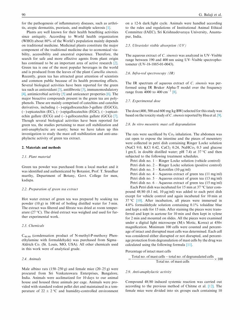
International Journal of Veterinary Science and Medicine (2014) 2, 89–94
Cairo University
International Journal of Veterinary Science and Medicine
www.vet.cu.edu.egwww.sciencedirect.com
Short Communication
Mast cell stabilizing and anti-anaphylactic
activity of aqueous extract of green tea(Camellia sinensis)
* Corresponding author. Tel.: +91 9490135291.E-mail address: [email protected] (G. Balaji).
Peer review under responsibility of Faculty of Veterinary Medicine,
Cairo University.
Production and hosting by Elsevier
2314-4599 ª 2014 Production and hosting by Elsevier B.V. on behalf of Faculty of Veterinary Medicine, Cairo University.
http://dx.doi.org/10.1016/j.ijvsm.2014.03.001
G. Balajia,*, M. Chalamaiah
b, P. Hanumanna
c, B. Vamsikrishna
d,
D. Jagadeesh Kumar e, V. Venu babu b
a Department of Pharmacology, Sri Krishnadevaraya University, Anantapur 515 003, A.P., Indiab Food and Drug Toxicology Research Centre, National Institute of Nutrition, Hyderabad 500 604, A.P., Indiac Department of Pharmacology, Nirmala College of Pharmacy, Kadapa, A.P., Indiad Department of Research and Development, Hetero Labs Limited, Unit-III, Jeedimetla, Hyderabad, A.P., Indiae Department of Pharmaceutics, Sri Krishnadevaraya University, Anantapur 515 003, A.P., India
Received 9 January 2014; revised 23 March 2014; accepted 23 March 2014
Available online 2 June 2014
KEYWORDS
Green tea extract;
Mast cell stabilization;
Anti-anaphylactic activity;
Compound 48/80;
IR and UV–Visible
spectroscopy
Abstract Green tea (Camellia sinensis) is one of the most popular and widely consumed beverages
in the world. In the current study, aqueous extract of green tea (C. sinensis) was evaluated for mast
cell stabilizing and anti-anaphylactic activities. Green tea extract (11, 13, 15 mg/ml) significantly
(P < 0.05) inhibited compound 48/80-induced rat mesentric mast cell degranulation in a dose
dependent manner. Anti-anaphylactic activity of green tea extract was performed in female mice.
At a dose of 400, 500, 600 mg/kg BW, green tea extract showed significant reduction in the mortal-
ity of mice subjected to anaphylactic shock by compound C48/80. Ketotifen was used for compari-
son. In addition, IR and UV–Visible spectroscopy analysis of green tea extract revealed the presence
of functional groups of bioactive compounds. These results suggest that green tea could be useful in
the treatment of asthma and allergic rhinitis.ª 2014 Production and hosting by Elsevier B.V. on behalf of Faculty of Veterinary Medicine, Cairo
University.
1. Introduction
Mast cells derive from the bone marrow and mature under theinfluence of local tissue microenvironmental conditions [1].Mast cells are important mediators of inflammatory responses,
such as allergy and anaphylaxis. The activated mast cellssecrete numerous vasoactive and proinflammatory mediators.These include pre-formed molecules such as histamine, seroto-
nin, TNF, kinins and proteases stored in secretory granules.The scientific evidence indicates that mast cells are critical
90 G. Balaji et al.
for the pathogenesis of inflammatory diseases, such as arthri-tis, atopic dermatitis, psoriasis, and multiple sclerosis [1].
Plants are well known for their health benefiting activities
since antiquity. According to World health organization(WHO) about 80% of the World’s population mainly dependson traditional medicine. Medicinal plants constitute the major
component of the traditional medicine due to economical via-bility, accessibility and ancestral experience. Therefore, thesearch for safe and more effective agents from plant origin
has continued to be an important area of active research [2].Green tea is one of the most popular beverages in the worldand is produced from the leaves of the plant Camellia sinensis.Recently, green tea has attracted great attention of scientists
and common public because of its health promoting effects.Several biological activities have been reported for the greentea such as antioxidant [3], antifibrotic [3], immunomodulatory
[4], antimicrobial activity [5] and anticancer properties [6]. Themajor bioactive compounds present in the green tea are poly-phenols. These are mainly comprised of catechins and catechin
derivatives, including (�)-epigallocatechin-3-gallate (EGCG),(�)-epicatechin (EC), (�)-epigallocatechin (EGC), (�)-epicat-echin gallate (ECG) and (�)-gallocatechin gallate (GCG) [7].
Though several biological activities have been reported forgreen tea, the studies pertaining to mast cell stabilization andanti-anaphylactic are scanty; hence we have taken up thisinvestigation to study the mast cell stabilization and anti-ana-
phylactic activity of green tea extract.
2. Materials and methods
2.1. Plant material
Green tea powder was purchased from a local market and itwas identified and authenticated by Botanist, Prof. T. Sreedharmurthy, Department of Botany, Govt. College for men,
kadapa.
2.2. Preparation of green tea extract
Hot water extract of green tea was prepared by soaking teapowder (10 g) in 100 ml of boiling distilled water for 5 min.The extract was filtered and filtrate was dried at room temper-ature (27 �C). The dried extract was weighed and used for fur-
ther experimental work.
2.3. Chemicals
C48/80 (condensation product of N-methyl-P-methoxy Phen-ethylamine with formaldehyde) was purchased from Sigma–Aldrich Co. (St. Louis, MO, USA). All other chemicals used
in this work were of analytical grade.
2.4. Animals
Male albino rats (150–250 g) and female mice (20–25 g) wereprocured from Sri Venkateswara Enterprises, Bangalore,India. Animals were acclimatized for 10 days to our animalhouse and housed three animals per cage. Animals were pro-
vided with standard rodent pellet diet and maintained in a tem-perature of 22 ± 2 �C and humidity-controlled environment
on a 12-h dark/light cycle. Animals were handled accordingto the rules and regulations of Institutional Animal EthicalCommittee (IAEC), Sri Krishnadevaraya University, Ananta-
pur, India.
2.5. Ultraviolet visible absorption (UV)
The aqueous extract of C. sinensis was analyzed in UV–Visiblerange between 190 and 400 nm using UV–Visible spectropho-tometer (UV-18-1885-01-0043).
2.6. Infra-red spectroscopy (IR)
The IR spectrum of aqueous extract of C. sinensis was per-
formed using IR Bruker Alpha-T model over the frequencyrange from 4000 to 400 cm�1 [8].
2.7. Experimental dose
The dose (400, 500 and 600 mg/kgBW) selected for this studywasbasedon the toxicity study ofC. sinensis reported byHsu et al. [9].
2.8. In vitro mesentric mast cell degranulation
The rats were sacrificed by Co2 inhalation. The abdomen wascut open to expose the intestine and the pieces of mesentery
were collected in petri dish containing Ringer Locke solution(NaCl 9.0, KCl 0.42, CaCl2 0.24, NaHCo3 0.5 and glucose1 gm/L in double distilled water; pH 7.4) at 37 �C and then
subjected to the following treatment schedules.Petri dish no. 1 – Ringer Locke solution (vehicle control)Petri dish no. 2 – Ringer Locke solution (positive control)Petri dish no. 3 – Ketotifen (10 lg/ml)
Petri dish no. 4 – Aqueous extract of green tea (11 mg/ml)Petri dish no. 5 – Aqueous extract of green tea (13 mg/ml)Petri dish no. 6 – Aqueous extract of green tea (15 mg/ml)
Each Petri dish was incubated for 15 min at 37 �C later com-pound 48/80 (0.1 ml, 10 lg/ml) was added to each petri dishexcept for vehicle control and again incubated for 10 min at
37 �C [10]. After incubation, all pieces were immersed in4.0% formaldehyde solution containing 0.1% toluidine blueand kept a side for 15 min. After staining the pieces were trans-ferred and kept in acetone for 10 min and then kept in xylene
for 2 min and mounted on slides. All the pieces were examinedunder a digital light microscope (M/s Motic, Korea) at 450·magnification. Minimum 100 cells were counted and percent-
age of intact and disrupted mast cells was determined. Each cellwas considered either disrupted or not disrupted, and percent-age protection from degranulation of mast cells by the drug was
calculated using the following formula [11].
Percentage of intact mast cells
¼Total no: of mast cells� total no: of degranulated cells
Total no: of mast cells�100
2.9. Anti-anaphylactic activity
Compound 48/80 induced systemic reaction was carried outaccording to the previous method of Chitme et al. [12]. The
female mice were divided into six groups each containing 10
Mast cell stabilizing and anti-anaphylactic activity of aqueous extract of green tea (Camellia sinensis) 91
animals. Green tea extract was administered daily for 7 daysby oral gavage using three different doses (400, 500 and600 mg/kg). The standard drug ketotifen (50 mg/kg) was
administered intraperitoneally. The following schedule oftreatment was administered.
Group I: distilled water (control)
Group II: C48/80 (sensitized)Group III: standard drug (ketotifen)Group IV: green tea extract (400 mg/kg)
Group V: green tea extract (500 mg/kg)Group VI: green tea extract (600 mg/kg)One hour after the last dose of drug administration (on 7th
day), all the animals except group I were given an injection of
8 mg/kg i.p. of C48/80 (toxicant). Mortality was monitored for1 h after induction of anaphylactic reaction. Mortality was cal-culated using the following formula.
Mortality ð%Þ ¼ No: of dead mice
Total no: of experimental mice� 100
3. Statistical analysis
All data were reported as Mean ± SD. The statistical analysis
was done using one-way analysis of variance (ANOVA) fol-lowed by Duncan’s Multiple Range (DMR) test. The level ofsignificance was evaluated at P < 0.05.
4. Results
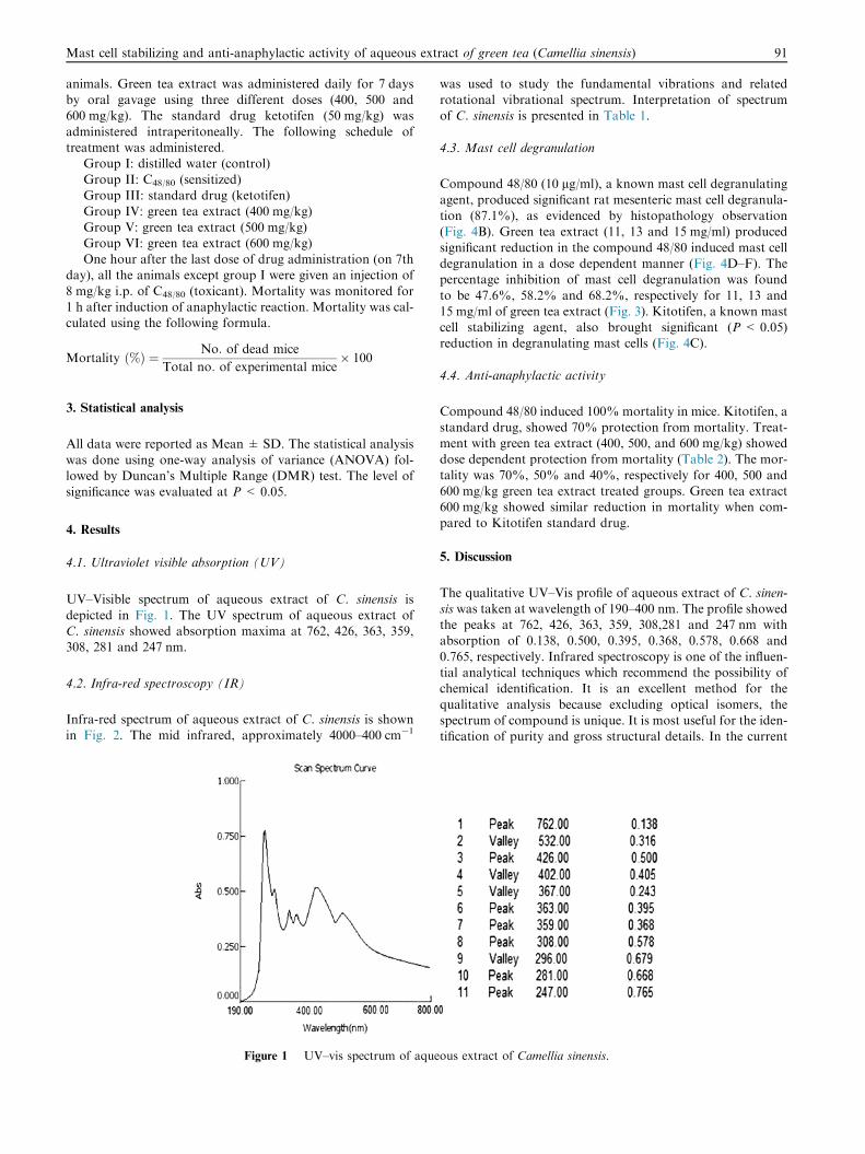
4.1. Ultraviolet visible absorption (UV)
UV–Visible spectrum of aqueous extract of C. sinensis is
depicted in Fig. 1. The UV spectrum of aqueous extract ofC. sinensis showed absorption maxima at 762, 426, 363, 359,308, 281 and 247 nm.
4.2. Infra-red spectroscopy (IR)
Infra-red spectrum of aqueous extract of C. sinensis is shownin Fig. 2. The mid infrared, approximately 4000–400 cm�1
Figure 1 UV–vis spectrum of aque
was used to study the fundamental vibrations and relatedrotational vibrational spectrum. Interpretation of spectrumof C. sinensis is presented in Table 1.
4.3. Mast cell degranulation
Compound 48/80 (10 lg/ml), a known mast cell degranulating
agent, produced significant rat mesenteric mast cell degranula-tion (87.1%), as evidenced by histopathology observation(Fig. 4B). Green tea extract (11, 13 and 15 mg/ml) produced
significant reduction in the compound 48/80 induced mast celldegranulation in a dose dependent manner (Fig. 4D–F). Thepercentage inhibition of mast cell degranulation was found
to be 47.6%, 58.2% and 68.2%, respectively for 11, 13 and15 mg/ml of green tea extract (Fig. 3). Kitotifen, a known mastcell stabilizing agent, also brought significant (P < 0.05)reduction in degranulating mast cells (Fig. 4C).
4.4. Anti-anaphylactic activity
Compound 48/80 induced 100% mortality in mice. Kitotifen, a
standard drug, showed 70% protection from mortality. Treat-ment with green tea extract (400, 500, and 600 mg/kg) showeddose dependent protection from mortality (Table 2). The mor-
tality was 70%, 50% and 40%, respectively for 400, 500 and600 mg/kg green tea extract treated groups. Green tea extract600 mg/kg showed similar reduction in mortality when com-pared to Kitotifen standard drug.
5. Discussion
The qualitative UV–Vis profile of aqueous extract of C. sinen-sis was taken at wavelength of 190–400 nm. The profile showedthe peaks at 762, 426, 363, 359, 308,281 and 247 nm withabsorption of 0.138, 0.500, 0.395, 0.368, 0.578, 0.668 and
0.765, respectively. Infrared spectroscopy is one of the influen-tial analytical techniques which recommend the possibility ofchemical identification. It is an excellent method for the
qualitative analysis because excluding optical isomers, thespectrum of compound is unique. It is most useful for the iden-tification of purity and gross structural details. In the current
ous extract of Camellia sinensis.
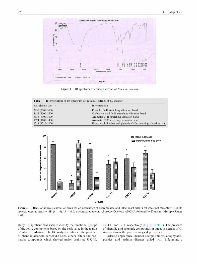
Figure 2 IR spectrum of aqueous extract of Camellia sinensis.
Table 1 Interpretation of IR spectrum of aqueous extract of C. sinensis.
Wavelength (cm�1) Interpretation
3135 (3500–3100) Phenolic O–H stretching vibration band
3135 (3300–2500) Carboxylic acid O–H stretching vibration band
3135 (3100–3000) Aromatic C–H stretching vibration band
1594 (1600–1400) Aromatic C–C stretching vibration band
1214 (1320–1000) Ester, alcohol, ether and phenolic C–O stretching vibration band
Figure 3 Effects of aqueous extract of green tea on percentage of degranulated and intact mast cells in rat intestinal mesentery. Results
are expressed as mean ± SD (n= 6). *P < 0.05 as compared to control group (One way ANOVA followed by Duncan’s Multiple Range
test).
92 G. Balaji et al.
study, IR spectrum was used to identify the functional groups
of the active components based on the peak value in the regionof infrared radiation. The IR analysis confirmed the presenceof phenolic alcohols, carboxylic acids, ethers, esters and aro-
matics compounds which showed major peaks at 3135.86,
1594.41 and 1214, respectively (Fig. 2; Table 1). The presence
of phenolic and aromatic compounds in aqueous extract of C.sinensis shows the pharmacological properties.
Allergic appearance includes allergic rhinitis, anaphylaxis,
purities and asthma diseases allied with inflammatory
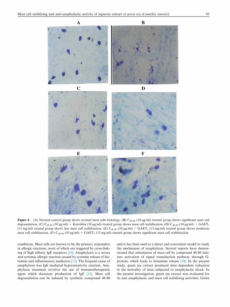
Figure 4 (A) Normal control group shows normal mast cells histology, (B) C48/80 (10 lg/ml) treated group shows significant mast cell
degranulation, (C) C48/80 (10 lg/ml) + Ketotifen (10 lg/ml) treated group shows mast cell stabilization, (D) C48/80 (10 lg/ml) + GAET1
(11 mg/ml) treated group shows less mast cell stabilization, (E) C48/80 (10 lg/ml) + GAET2 (13 mg/ml) treated group shows moderate
mast cell stabilization, (F) C48/80 (10 lg/ml) + GAET3 (15 mg/ml) treated group shows significant mast cell stabilization.
Mast cell stabilizing and anti-anaphylactic activity of aqueous extract of green tea (Camellia sinensis) 93
conditions. Mast cells are known to be the primary respondersin allergic reactions, most of which are triggered by cross-link-
ing of high affinity IgE receptors [10]. Anaphylaxis is a severeand systemic allergic reaction caused by systemic release of his-tamine and inflammatory mediators [13]. The frequent cause of
anaphylaxis was IgE mediated hypersensitivity reaction. Ana-phylaxis treatment involves the use of immunotherapeuticagent which decreases production of IgE [12]. Mast celldegranulation can be induced by synthetic compound 48/80
and it has been used as a direct and convenient model to studythe mechanism of anaphylaxis. Several reports have demon-
strated that stimulation of mast cell by compound 48/80 initi-ates activation of signal transduction pathway through G-protein, which leads to histamine release [10]. In the present
study, green tea extract produced dose dependent reductionin the mortality of mice subjected to anaphylactic shock. Inthe present investigation, green tea extract was evaluated forits anti anaphylactic and mast cell stabilizing activities. Green
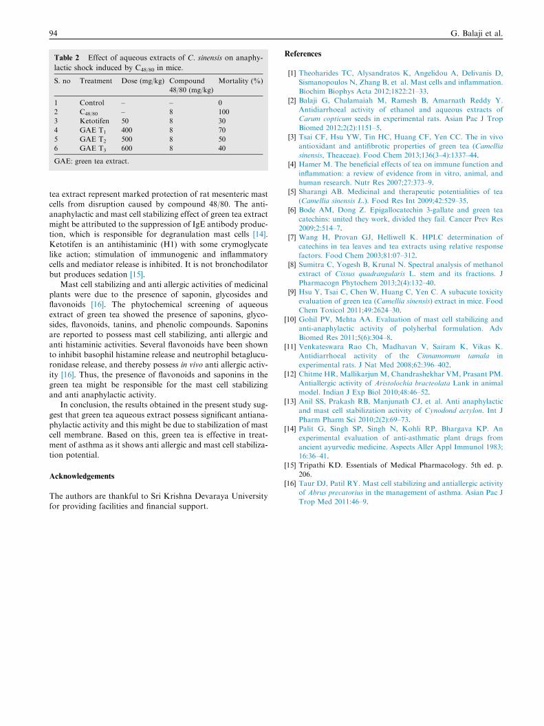
Table 2 Effect of aqueous extracts of C. sinensis on anaphy-
lactic shock induced by C48/80 in mice.
S. no Treatment Dose (mg/kg) Compound
48/80 (mg/kg)
Mortality (%)
1 Control – – 0
2 C48/80 – 8 100
3 Ketotifen 50 8 30
4 GAE T1 400 8 70
5 GAE T2 500 8 50
6 GAE T3 600 8 40
GAE: green tea extract.
94 G. Balaji et al.
tea extract represent marked protection of rat mesenteric mastcells from disruption caused by compound 48/80. The anti-
anaphylactic and mast cell stabilizing effect of green tea extractmight be attributed to the suppression of IgE antibody produc-tion, which is responsible for degranulation mast cells [14].
Ketotifen is an antihistaminic (H1) with some crymoglycatelike action; stimulation of immunogenic and inflammatorycells and mediator release is inhibited. It is not bronchodilator
but produces sedation [15].Mast cell stabilizing and anti allergic activities of medicinal
plants were due to the presence of saponin, glycosides andflavonoids [16]. The phytochemical screening of aqueous
extract of green tea showed the presence of saponins, glyco-sides, flavonoids, tanins, and phenolic compounds. Saponinsare reported to possess mast cell stabilizing, anti allergic and
anti histaminic activities. Several flavonoids have been shownto inhibit basophil histamine release and neutrophil betaglucu-ronidase release, and thereby possess in vivo anti allergic activ-
ity [16]. Thus, the presence of flavonoids and saponins in thegreen tea might be responsible for the mast cell stabilizingand anti anaphylactic activity.
In conclusion, the results obtained in the present study sug-
gest that green tea aqueous extract possess significant antiana-phylactic activity and this might be due to stabilization of mastcell membrane. Based on this, green tea is effective in treat-
ment of asthma as it shows anti allergic and mast cell stabiliza-tion potential.
Acknowledgements
The authors are thankful to Sri Krishna Devaraya University
for providing facilities and financial support.
References
[1] Theoharides TC, Alysandratos K, Angelidou A, Delivanis D,
Sismanopoulos N, Zhang B, et al. Mast cells and inflammation.
Biochim Biophys Acta 2012;1822:21–33.
[2] Balaji G, Chalamaiah M, Ramesh B, Amarnath Reddy Y.
Antidiarrhoeal activity of ethanol and aqueous extracts of
Carum copticum seeds in experimental rats. Asian Pac J Trop
Biomed 2012;2(2):1151–5.
[3] Tsai CF, Hsu YW, Tin HC, Huang CF, Yen CC. The in vivo
antioxidant and antifibrotic properties of green tea (Camellia
sinensis, Theaceae). Food Chem 2013;136(3–4):1337–44.
[4] Hamer M. The beneficial effects of tea on immune function and
inflammation: a review of evidence from in vitro, animal, and
human research. Nutr Res 2007;27:373–9.
[5] Sharangi AB. Medicinal and therapeutic potentialities of tea
(Camellia sinensis L.). Food Res Int 2009;42:529–35.
[6] Bode AM, Dong Z. Epigallocatechin 3-gallate and green tea
catechins: united they work, divided they fail. Cancer Prev Res
2009;2:514–7.
[7] Wang H, Provan GJ, Helliwell K. HPLC determination of
catechins in tea leaves and tea extracts using relative response
factors. Food Chem 2003;81:07–312.
[8] Sumitra C, Yogesh B, Krunal N. Spectral analysis of methanol
extract of Cissus quadrangularis L. stem and its fractions. J
Pharmacogn Phytochem 2013;2(4):132–40.
[9] Hsu Y, Tsai C, Chen W, Huang C, Yen C. A subacute toxicity
evaluation of green tea (Camellia sinensis) extract in mice. Food
Chem Toxicol 2011;49:2624–30.
[10] Gohil PV, Mehta AA. Evaluation of mast cell stabilizing and
anti-anaphylactic activity of polyherbal formulation. Adv
Biomed Res 2011;5(6):304–8.
[11] Venkateswara Rao Ch, Madhavan V, Sairam K, Vikas K.
Antidiarrhoeal activity of the Cinnamomum tamala in
experimental rats. J Nat Med 2008;62:396–402.
[12] Chitme HR, Mallikarjun M, Chandrashekhar VM, Prasant PM.
Antiallergic activity of Aristolochia bracteolata Lank in animal
model. Indian J Exp Biol 2010;48:46–52.
[13] Anil SS, Prakash RB, Manjunath CJ, et al. Anti anaphylactic
and mast cell stabilization activity of Cynodond actylon. Int J
Pharm Pharm Sci 2010;2(2):69–73.
[14] Palit G, Singh SP, Singh N, Kohli RP, Bhargava KP. An
experimental evaluation of anti-asthmatic plant drugs from
ancient ayurvedic medicine. Aspects Aller Appl Immunol 1983;
16:36–41.
[15] Tripathi KD. Essentials of Medical Pharmacology. 5th ed. p.
206.
[16] Taur DJ, Patil RY. Mast cell stabilizing and antiallergic activity
of Abrus precatorius in the management of asthma. Asian Pac J
Trop Med 2011:46–9.