Embed Size (px)
Citation preview
www.kardiologiapolska.pl
Kardiologia Polska 2014; 72, 1: 67–69; DOI: 10.5603/KP.2014.0006 ISSN 0022–9032
ELEKTROKARDIOGRAFIA / ECG
Masquerading bundle branch blockZamaskowany blok odnogi pęczka Hisa
Piotr Kukla1, Adrian Baranchuk2, Marek Jastrzębski3, Leszek Bryniarski3
1Department of Internal Disease and Cardiology, Specialistic Hospital, Gorlice, Poland2Division of Cardiology, Kingston General Hospital, Queen’s University, Kingston, Canada31st Department of Cardiology, Interventional Electrocardiology and Hypertension, University Hospital, Krakow, Poland
A b s t r a c t
We here describe a surface 12-lead electrocardiogram (ECG) of a 72-year-old female with a prior history of breast cancer and chemotherapy-induced cardiomyopathy. An echocardiogram revealed left ventricular dysfunction, ejection fraction of 23%, with mild enlarged left ventricle. The 12-lead ECG showed atrial fibrillation with a mean heart rate of about 100 bpm, QRS duration 160 ms, QT interval 400 ms, right bundle branch block (RBBB) and left anterior fascicular block (LAFB). The combination of RBBB features in the precordial leads and LAFB features in the limb leads is known as ‘‘masquerading bundle branch block’’. In most cases of RBBB and LAFB, the QRS axis deviation is located between – 80 to –120 degrees. Rarely, when predominant left ventricular forces are present, the QRS axis deviation is near about – 90 degrees, turning the pattern into an atypical form. In a situation of RBBB associated with LAFB, the S wave can be absent or very small in lead I. Such a situ-ation is the result of not only purely LAFB but also with left ventricular hypertrophy and/or focal block due to scar (extensive anterior myocardial infarction) or fibrosis (cardiomyopathy). Sometimes, this specific ECG pattern is mistaken for LBBB. RBBB with LAFB may imitate LBBB either in the limb leads (known as ‘standard masquerading’ — absence of S wave in lead I), or in the precordial leads (called ‘precordial masquerading’ — absence of S wave in leads V5 and V6).
Our ECG showed both these types of masquerading bundle branch block — absence of S wave in lead I and in leads V5 and V6.
Key words: right bundle branch block, left anterior fascicular block, masquerading branch block
Kardiol Pol 2014; 72, 1: 67–69
Address for correspondence: Piotr Kukla, MD, PhD, Department of Internal Disease and Cardiology, Specialistic Hospital, ul. Węgierska 21, 38–300 Gorlice, Poland, tel: +48 18 355 34 15, e-mail: [email protected]
Copyright © Polskie Towarzystwo Kardiologiczne
CASE REPORTWe present the case of a surface 12-lead electrocardiogram (ECG) of a 72-year-old female with a prior history of breast cancer and chemotherapy-induced cardiomyopathy. An echocardiogram revealed left ventricular dysfunction, ejection fraction of 23%, with mild enlarged left ventricle.
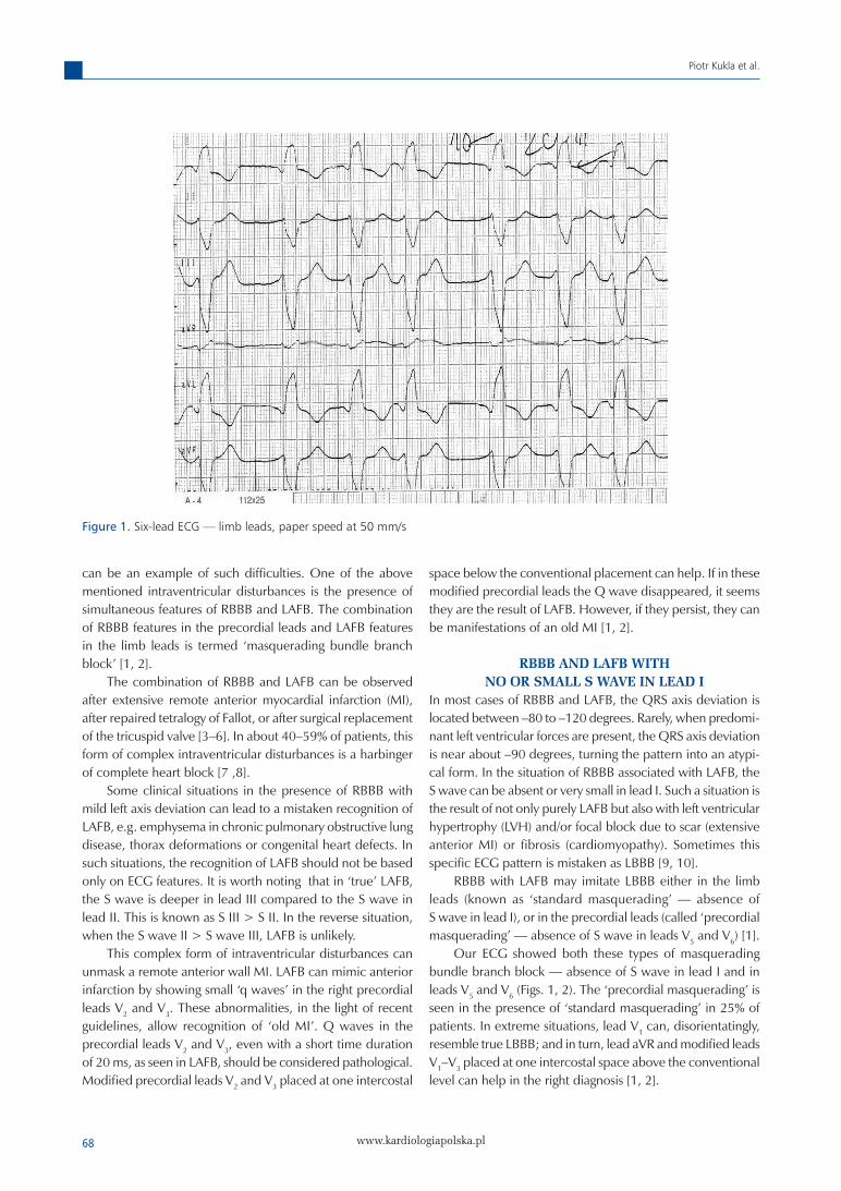
The 12-lead ECG showed atrial fibrillation with a mean heart rate of about 100 bpm, QRS duration 160 ms, QT in-terval 400 ms, right bundle-branch block (RBBB) based on the following criteria: (1) duration of QRS ≥ 120 ms; (2) R wave peak time in lead V1 > 50 ms; (3) QRS morphology in lead V1, V2: rsR’; and (4) secondary ST-T segment changes, downsloping ST segment depression with negative T wave in leads V1–V3.
There was one criterion of RBBB, according to the actual definition, that was not met: the presence of wide S wave in leads I or V6. The duration of the S wave should be more than
40 ms or wider than the R wave in leads I or V6. This criterion cannot always be seen when a left anterior fascicular block (LAFB) is present. The current ECG showed atypical QRS morphology in leads I, aVL and leads V5, V6 to be considered a typical RBBB. The QRS morphology in these leads resembles left bundle-branch block (LBBB). Additionally, this ECG met the criteria for LAFB: (1) QRS axis between –45 degrees and –90 degrees; (2) presence of qR morphology in lead aVL; (3) R wave peak time in lead aVL ≥ 45 ms; and (4) QRS duration of below 120 ms, but in the presence of RBBB this criterion does not need to be fulfilled. How can we explain a morphology that resembles LBBB in the presence of RBBB?
DISCUSSIONSome ECG patterns can be difficult to interpret, leading to difficult clinical decision making. Intraventricular disturbances
www.kardiologiapolska.pl
Piotr Kukla et al.
68
can be an example of such difficulties. One of the above mentioned intraventricular disturbances is the presence of simultaneous features of RBBB and LAFB. The combination of RBBB features in the precordial leads and LAFB features in the limb leads is termed ‘masquerading bundle branch block’ [1, 2].
The combination of RBBB and LAFB can be observed after extensive remote anterior myocardial infarction (MI), after repaired tetralogy of Fallot, or after surgical replacement of the tricuspid valve [3–6]. In about 40–59% of patients, this form of complex intraventricular disturbances is a harbinger of complete heart block [7 ,8].
Some clinical situations in the presence of RBBB with mild left axis deviation can lead to a mistaken recognition of LAFB, e.g. emphysema in chronic pulmonary obstructive lung disease, thorax deformations or congenital heart defects. In such situations, the recognition of LAFB should not be based only on ECG features. It is worth noting that in ‘true’ LAFB, the S wave is deeper in lead III compared to the S wave in lead II. This is known as S III > S II. In the reverse situation, when the S wave II > S wave III, LAFB is unlikely.
This complex form of intraventricular disturbances can unmask a remote anterior wall MI. LAFB can mimic anterior infarction by showing small ‘q waves’ in the right precordial leads V2 and V3. These abnormalities, in the light of recent guidelines, allow recognition of ‘old MI’. Q waves in the precordial leads V2 and V3, even with a short time duration of 20 ms, as seen in LAFB, should be considered pathological. Modified precordial leads V2 and V3 placed at one intercostal
space below the conventional placement can help. If in these modified precordial leads the Q wave disappeared, it seems they are the result of LAFB. However, if they persist, they can be manifestations of an old MI [1, 2].
RBBB AND LAFB WITH NO OR SMALL S WAVE IN LEAD I
In most cases of RBBB and LAFB, the QRS axis deviation is located between –80 to –120 degrees. Rarely, when predomi-nant left ventricular forces are present, the QRS axis deviation is near about –90 degrees, turning the pattern into an atypi-cal form. In the situation of RBBB associated with LAFB, the S wave can be absent or very small in lead I. Such a situation is the result of not only purely LAFB but also with left ventricular hypertrophy (LVH) and/or focal block due to scar (extensive anterior MI) or fibrosis (cardiomyopathy). Sometimes this specific ECG pattern is mistaken as LBBB [9, 10].
RBBB with LAFB may imitate LBBB either in the limb leads (known as ‘standard masquerading’ — absence of S wave in lead I), or in the precordial leads (called ‘precordial masquerading’ — absence of S wave in leads V5 and V6) [1].
Our ECG showed both these types of masquerading bundle branch block — absence of S wave in lead I and in leads V5 and V6 (Figs. 1, 2). The ‘precordial masquerading’ is seen in the presence of ‘standard masquerading’ in 25% of patients. In extreme situations, lead V1 can, disorientatingly, resemble true LBBB; and in turn, lead aVR and modified leads V1–V3 placed at one intercostal space above the conventional level can help in the right diagnosis [1, 2].
Figure 1. Six-lead ECG — limb leads, paper speed at 50 mm/s

www.kardiologiapolska.pl
Masquerading bundle branch block
69
The proper recognition of ‘masquerading bundle branch block’ may allow in making the proper clinical decision.
Firstly, it gives us information about the poor prognosis because of coexisting extensive heart damage and conduction disturbances associated with LVH, scar and/or fibrosis [1].
Secondly, it increases the risk of complete heart block [7, 8]. Thirdly, in the era of resynchronisation therapy (CRT),
mistaken LBBB could lead to improper CRT implantation [11].
Conflict of interest: none declared
References1. Elizari MV, Baranchuk A, Chiale PA. Masquerading bundle
branch block: a variety of right bundle branch block with left ante-rior fascicular block. Expert Rev Cardiovasc Ther, 2013; 11: 69–75.
2. Acunzo RS, Konopka IV, Sanchéz RA et al. Right bundle branch block and middle septal fiber block with or without left anterior fascicular block manifested as aberrant conduction in apparent healthy individuals. Electro-vectorcardiographic characteriza-tion. J Electrocardiol, 2013 (In press).
3. Rosenbaum MB, Corrado G, Oliveri R et al. Right bundle branch block with left anterior hemiblock surgically induced in tetralogy of Fallot. Relation to the mechanism of electrocardiographic changes in endocardial cushion defects. Am J Cardiol, 1970; 26: 12–19.
4. Downing JW, Kaplan S, Bove KE. Post surgical left anterior hemi-block and right bundle branch block. Br Heart J, 1972; 34: 263–270.
5. Steeg CN, Krongrad E, Davachi F et al. Postoperative left anterior and right bundle branch block following repair of tetralogy of Fallot. Clinical and etiologi consideration. Circulation, 1975; 51: 1026–1029.
6. Aravindakshan V, Elizari MV, Rosenbaum MB. Right bundle branch block and left anterior fascicular block following tricuspid valve replacement. Circulation, 1970; 42: 895–902.
7. Lasser RP, Haft JI, Fredberg CK. Relationship of right bundle branch block and marked left axis deviation (with left parietal or peri-infarction block) to complete heart block and syncope. Circulation, 1968; 37: 429–437.
8. Rothfeld EL, Zucker IR, Tiu R. Parsonnet V. The electrocardio-graphic syndrome of superior axis and right bundle branch block. Dis Chest, 1969; 55: 306–309.
9. Unger PN, Lesser ME, Kugel VH, Lev M. The concept of masquer-ading bundle branch block ; an electrocardiographic-pathologic correlation. Circulation, 1958; 17: 397–409.
10. Ortega-Carnicer J, Malillos M, Munoz L, Rodruguez-Garcia J. Left anterior hemiblock masking the diagnosis or right bundle branch block. J Electrocardiol, 1986; 19: 97–98.
11. Sipahi I, Chou JC, Hyden M et al. Effect of QRS morphology on clinical event reduction with cardiac resynchronization therapy: meta-analysis of randomized controlled trials. Am Heart J, 2012; 163: 260–267.
Figure 2. Six-lead ECG — precordial leads, paper speed at 50 mm/s