Embed Size (px)
Citation preview
Marker-less Reconstruction of Dense 4-D SurfaceMotion Fields using Active Laser Triangulation
for Respiratory Motion Management
Sebastian Bauer1, Benjamin Berkels3, Svenja Ettl2, Oliver Arold2, JoachimHornegger1, Martin Rumpf4
1 Pattern Recognition Lab, Dept. of Computer Science2 Institute of Optics, Information and Photonics
Friedrich-Alexander-Universitat Erlangen-Nurnberg, Erlangen, [email protected]
3 Interdisciplinary Mathematics Institute,University of South Carolina, Columbia, SC, USA
4 Institute for Numerical Simulation,Rheinische Friedrich-Wilhelms-Universitat Bonn, Bonn, Germany
Abstract. To manage respiratory motion in image-guided interventionsa novel sparse-to-dense registration approach is presented. We applyan emerging laser-based active triangulation (AT) sensor that deliverssparse but highly accurate 3-D measurements in real-time. These sparseposition measurements are registered with a dense reference surface ex-tracted from planning data. Thereby a dense displacement field is recon-structed which describes the 4-D deformation of the complete patientbody surface and recovers a multi-dimensional respiratory signal for ap-plication in respiratory motion management. The method is validatedon real data from an AT prototype and synthetic data sampled fromdense surface scans acquired with a structured light scanner. In a studyon 16 subjects, the proposed algorithm achieved a mean reconstructionaccuracy of ±0.22 mm w.r.t. ground truth data.
1 Introduction
Respiration-synchronized image-guided radiation therapy (IGRT) techniques aimat tracking the tumor location and reposition the beam dynamically. To reduceadditional radiation exposure, recent hybrid tumor-tracking techniques combineepisodic radiographic imaging with continuous monitoring of external breathingsurrogates based on the premise that the internal tumor position can be accu-rately predicted from external motion. The underlying correlation model can beestablished from a series of simultaneously acquired external-internal positionmeasurements [1] or 4-D CT planning data [2]. Clinically available solutions forhybrid tumor-tracking [1, 3] measure external motion using a single or a fewpassive markers on the patient’s chest as a low-dimensional surrogate. Thus,these techniques are incapable of depicting the full complexity of respiratorymotion, they involve extensive patient preparation, and require reproducible
marker placement with a substantial impact on model accuracy. Modern IGRTsolutions that allow to monitor the motion of the complete external patient sur-face help to reduce correlation model uncertainties. In particular, range imaging(RI) technologies can acquire a dense 3-D surface model of the patient [4–6].Based on the estimation of a dense displacement field representing the deforma-tion of the instantaneous torso surface w.r.t. a reference surface (either from RIor planning CT data), a highly accurate correlation model can be established [7,8]. The deformation estimation from dense surface scans for application in RThas been investigated recently [8, 9]. Available RI-based IGRT solutions are ca-pable of delivering dense surface information in a marker-less manner but focuson patient positioning, do not support dense sampling in real-time [4, 5] or atthe cost of a limited field of view [6], often imply high costs in terms of hardwareand are subject to measurement uncertainties due to the sampling principlese.g. active stereo [6] or swept lasers [4, 5]. The temporal resolution of these so-lutions may be insufficient to characterize respiratory motion. In this paper, wepropose a marker-less system based on a non-moving active laser triangulation(AT) sensor that delivers sparse but accurate measurements in real-time (30 Hz).Using prior patient shape knowledge from planning data, a variational model isproposed to recover a dense and accurate displacement field and to reconstructa complete and reliable patient surface model at the instantaneous respirationphase. Estimating the dense deformation is combined with recovering a sparsedisplacement field from AT measurements to planning data, thus the approachis closely related to the field of inverse-consistent registration [10, 11]. The varia-tional model is discretized using Finite Elements, the optimization is guided bya step-size controlled gradient flow to guarantee fast and smooth relaxation.
2 Method
In this section, we derive the variational model for the reconstruction of a densedisplacement field from sparse measurements. Given is a reference shape G ⊂ R3
extracted from planning data and the instantaneous body surfaceM representedby a sparse sampling Y . For instance, let us assume that the AT sensor acquiresa set of n measurements Y = y1, . . . , yn, yi ∈ R3, arranged in a grid-like struc-ture (Fig. 1). We assume that G is given as a graph, i. e. there is a domain Ω ⊂ R2
usually associated with the plane of the patient table and a function g : Ω → Rsuch that G =
(ζ, g(ζ)) ∈ R3 : ζ ∈ Ω
. Due to respiration, the intra-fractional
sampling Y is not aligned with G. Now, the goal is to estimate the unknown,non-rigid, dense deformation φ of G with Y ⊂ φ(G). For this purpose, in a jointmanner, we estimate φ together with an inverse deformation ψ matching Y andG in the sense that ψ(Y ) ⊂ G. When registering Y onto G we solely deal with asparse displacement field (ψ(yi))i=1,...,n on the n positions measured by the ATsensor. A geometric sketch of the registration configuration is depicted in Fig. 1.Estimating ψ allows us to establish a correspondence between the AT measure-ments and the reference patient surface, whereas the dense deformation φ enablesthe reconstruction of the complete instantaneous patient surface. We represent
Fig. 1. Geometric configuration for reconstructing the dense deformation φ withφ(ζ, g(ζ)) = (ζ, g(ζ)) + u(ζ) from sparse sampling data Y = y1, . . . , yn and the ap-proximate sparse inverse ψ with ψ(yi) = yi + wi (for a better visibility G and Y havebeen pulled apart). Furthermore, the projection P onto G and the orthogonal projectionQ from the graph G onto the parameter domain Ω are sketched.
ψ by a vector of displacements W = w1, . . . , wn with ψ(yi) = yi+wi . Further-more, the deformation φ is represented by a displacement u : Ω → R3 definedon the parameter domain Ω of the graph G with φ(ζ, g(ζ)) = (ζ, g(ζ)) + u(ζ) .To quantify the matching of ψ(Y ) onto G let us assume that the signed distancefunction d with respect to G is precomputed in a sufficiently large neighborhoodin R3. We set d(x) := ±dist(x,G), where the sign is positive outside the body,i. e. above the graph, and negative inside. Then ∇d(x) is the outward pointingnormal on G and |∇d(x)| = 1. Based on this signed distance map d we can definethe projection P (x) := x− d(x)∇d(x) of a point x ∈ R3 in a neighborhood of Gonto the closest point on G and compute the mismatch of ψ(Y ) and G pointwisevia |P (ψ(yi)) − ψ(yi)| = |d(yi + wi)|. Let us emphasize that we do not expectψ to be equal to the projection P . Indeed, the computational results discussedbelow underline that it is the prior in the deformation φ which leads to generalmatching correspondences for a minimizer of our variational approach.
2.1 Definition of the Registration Energy
Now, we define a functional E on dense displacement fields u and sparse vectorsof displacements W such that a minimizer represents a suitable matching of theplanning data and AT measurements:
E [u,W ] := Ematch[W ] + κ Econ[u,W ] + λ Ereg[u] (1)
where κ and λ are nonnegative constants controlling the contributions of theindividual terms. Ematch denotes a term measuring closeness of ψ(Y ) to G. Theconsistency functional Econ is responsible for establishing the relation betweenboth displacement fields. Finally, Ereg ensures a regularization of the dense dis-placement u. The detailed definitions of these functionals are as follows.
Matching Energy. In order to measure closeness of ψ(Y ) to G, we use thepointwise mismatch measure discussed above and define
Ematch[W ] :=1
2n
n∑i=1
|d(yi + wi)|2 . (2)
Consistency Energy. For a known instantaneous deformation φ of the patientsurface G and an exact deformation correspondence ψ(Y ) ⊂ G of the AT mea-surement Y the identity φ(ψ(Y )) = Y holds. But for an arbitrary deformationψ described by some vector of displacements W in general ψ(Y ) 6⊂ G. To relateφ and ψ in this case we have to incorporate the projection P because φ is onlydefined on G. In fact, to ensure that (φ P ψ)(W ) ≈W for a minimizer of thetotal energy we introduce the consistency energy
Econ[u,W ] :=1
2n
n∑i=1
|P (yi + wi) + u(QP (yi + wi))− yi|2 , (3)
where Q ∈ R2×3 denotes the orthographic projection matrix with Q(ζ, g(ζ)) = ζ.Here, we have used that φ(P (ψ(yi))) = P (yi + wi) + u(QP (yi + wi)) . Indeed,this definition of the consistency energy allows us to compute a dense smoothdisplacement of the patient planning surface even though only a sparse set ofmeasurements is available.
Prior for the Displacement. To ensure smoothness of the deformation φ onG we incorporate a thin plate spline type regularization of the correspondingdisplacement u [12] and define
Ereg[u] :=1
2
∫Ω
|4u|2 dx , (4)
where 4u = (4u1,4u2,4u3) and thus |4u|2 =∑3k=1(4uk)2. Indeed, since
our input data Y only implicitly provide information for φ on a sparse set, afirst order regularizer is inadequate to ensure sufficient regularity for the defor-mation. Let us emphasize that (discrete) smoothness of the approximate inversedeformation ψ is implicitly controlled by the regularization of φ.
2.2 Numerical Optimization
To minimize the functional E (Eq. 1), we apply a Finite Element approximationand optimize the functional using a gradient descent scheme. In particular, afteran appropriate scaling of G we choose Ω = [0, 1]2 and consider a piecewise bi-linear, continuous Finite Element approximation on a uniform rectangular meshcovering Ω. In the experiments we used a 129×129 grid. Furthermore, the signeddistance function d is precomputed using a fast marching method on a uniformrectangular 3-D grid covering the unit cube [0, 1]3 and stored on the nodes ofthis grid. In the algorithm d and ∇d are evaluated using trilinear interpolationof nodal values. For the gradient descent, derivatives of the energy have to becomputed numerically. The derivatives of Ematch and Econ w.r.t. wj are given as:
∂wjEmatch[W ] =
1
nd(yj + wj)∇d(yj + wj)
∂wjEcon[u,W ] =
1
n(P (yj + wj) + u(QP (yj + wj))− yj)T
(DP (yj + wj) +∇u(QP (yj + wj))QDP (yj + wj))

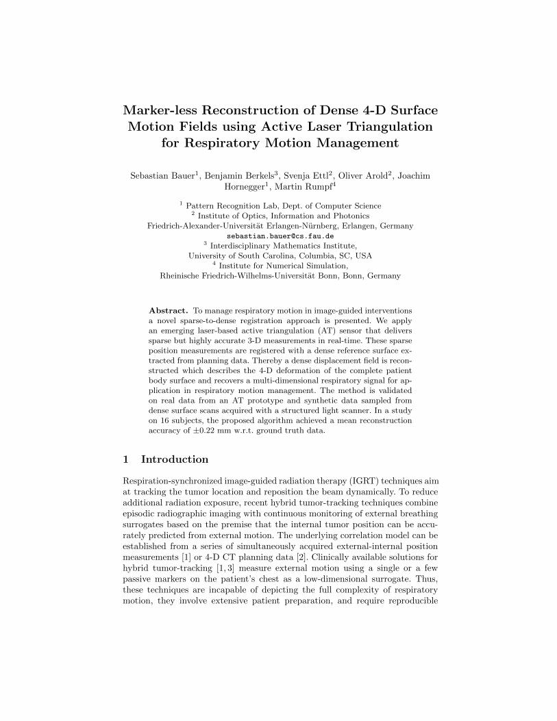
G and Y2 to Y4 φ2 on G φ3 on G φ4 on G
Fig. 2. Validation on real AT data. Estimation of φp transforming G intoMp, from ATsampling data Yp. For the glyph visualization of φp on G, |u(ζ)| is color coded [mm].
where DP denotes the Jacobian of the projection P . The variations of Econ andEreg w.r.t. u in a direction ϑ : Ω → R3 are given by:
〈∂uEcon[u,W ], ϑ〉 =1
n
n∑i=1
(P (yi + wi) + u(QP (yi + wi))− yi)ϑ(QP (yi + wi))
〈∂uEreg[u], ϑ〉 =
3∑k=1
∫Ω
4uk4ϑk dx
The evaluation of DP involves the Hessian D2d(x) of the distance function.One can either compute D2d(x) based on second order finite differences or - asactually implemented here - replace the projection direction in P by the alreadycomputed direction from the last update. Furthermore, the Laplacian of a FiniteElement function is evaluated by the discrete Finite Element Laplacian. In thegradient descent scheme we stop iterating as soon as the energy decay is smallerthan a threshold value ε, ε = 10−4 proved to be sufficient to achieve the accuracyreported below. For the first frame of the respiratory motion we initialize u = 0and wj = P (yj) − yj leading to approx. 60 gradient descent steps on average.For all subsequent frames we take u from the previous step and wj = P (yj)− yjas initial data resulting in approx. 45 descent steps on average.
3 Experiments and Results
Experimental Setup. For validation of the method, we have used an eye-safeAT prototype that acquires a sparse grid of 11×10 accurate 3-D sampling lines inreal-time (30 Hz), using two perpendicular laser line pattern projection systemsand a 1024×768 px resolution CCD chip [13]. Within the measurement volume,the mean AT measurement uncertainty is σ = 0.39 mm. The evaluation datasetis composed of 32 datasets from 16 subjects, each performing abdominal andthoracic breathing, respectively. Per subject, we synchronously acquired bothreal AT data and surface data using a moderately accurate but rather densestructured light (SL) system with a resolution of 320×240 px. Both sensors weremounted at a height of 1.2 m above the patient table, at a viewing angle of 30.AT and SL data were aligned using calibration. From each dataset, we extractedsparse AT measurements Yp and dense SL meshes Mp for 8 phases within one
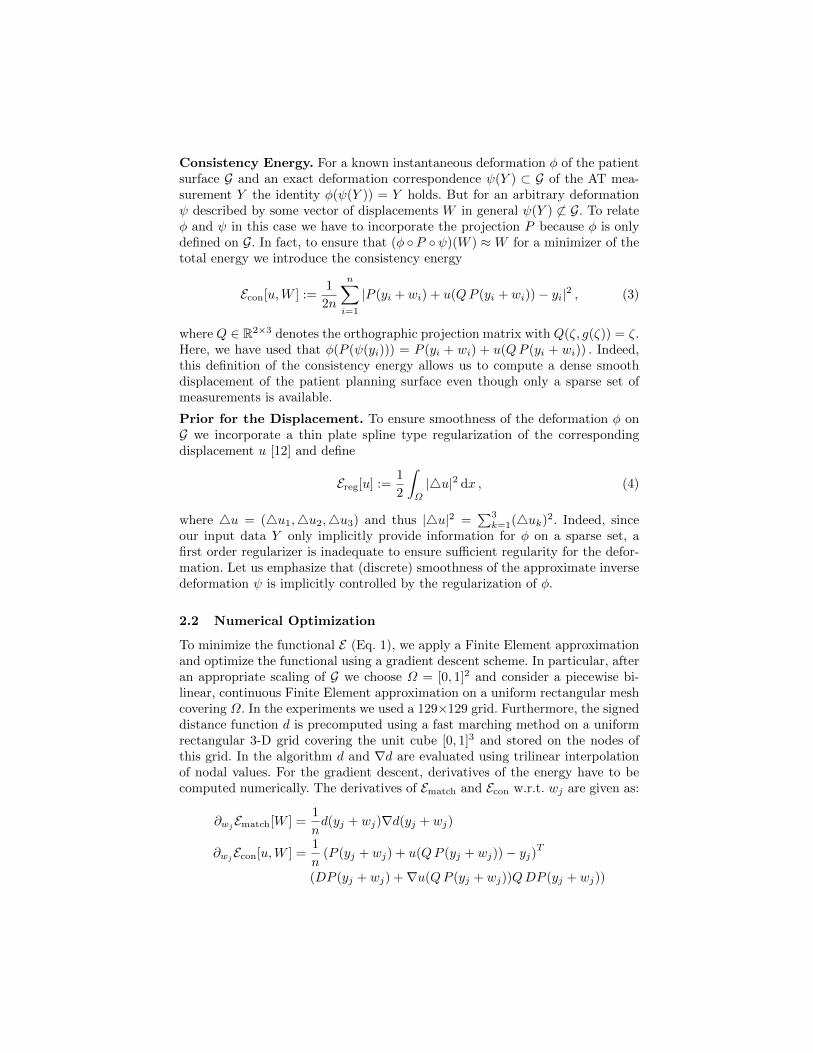
G and Y4 dist(G,M4) dist(φ4(G),M4) φ4 on G
Fig. 3. Estimation of φp transforming G into Mp from realistic AT sampling data Yp,for thoracic (top row) and abdominal respiration (bottom row). p = 4 represents therespiration state of fully inhale, roughly. For the glyph visualization of φ on G, |u(ζ)|is color coded [mm]. Please note that the color coding differs by a factor of 10.
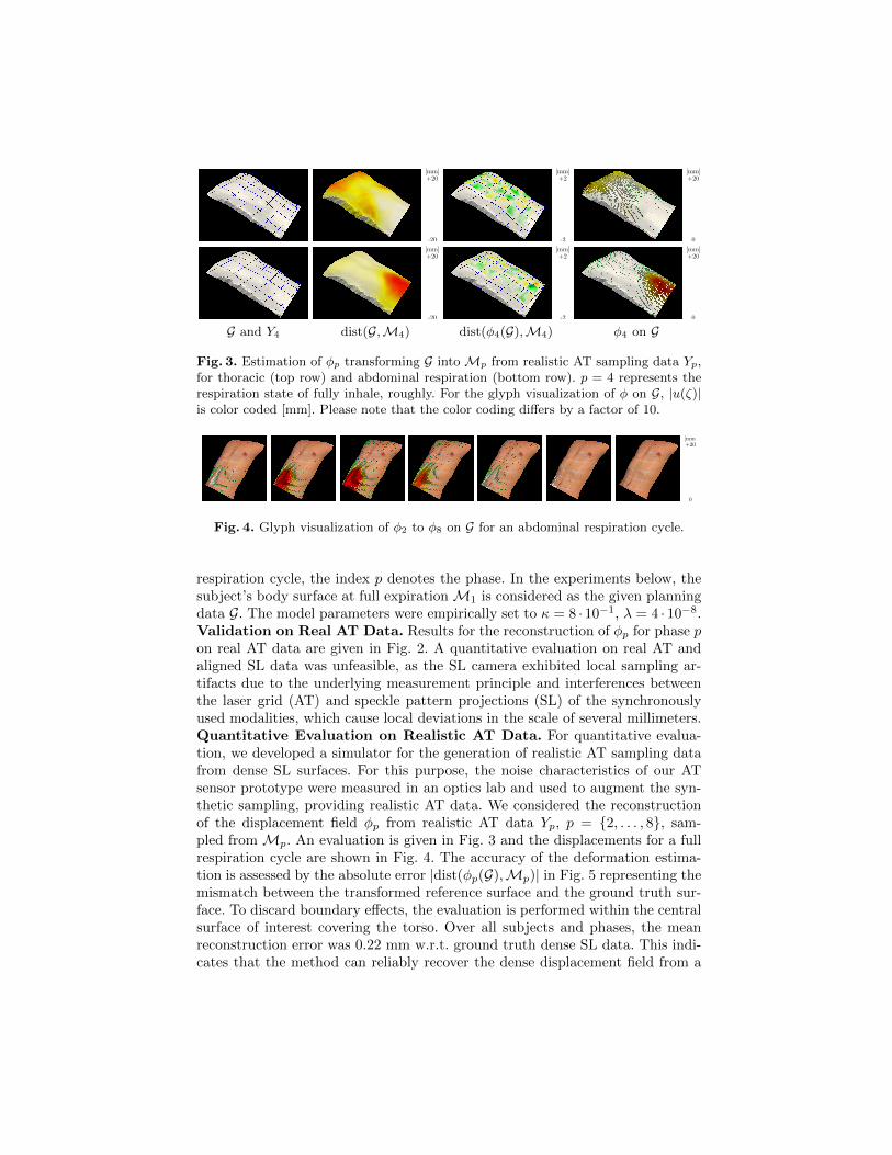
Fig. 4. Glyph visualization of φ2 to φ8 on G for an abdominal respiration cycle.
respiration cycle, the index p denotes the phase. In the experiments below, thesubject’s body surface at full expirationM1 is considered as the given planningdata G. The model parameters were empirically set to κ = 8 · 10−1, λ = 4 · 10−8.Validation on Real AT Data. Results for the reconstruction of φp for phase pon real AT data are given in Fig. 2. A quantitative evaluation on real AT andaligned SL data was unfeasible, as the SL camera exhibited local sampling ar-tifacts due to the underlying measurement principle and interferences betweenthe laser grid (AT) and speckle pattern projections (SL) of the synchronouslyused modalities, which cause local deviations in the scale of several millimeters.Quantitative Evaluation on Realistic AT Data. For quantitative evalua-tion, we developed a simulator for the generation of realistic AT sampling datafrom dense SL surfaces. For this purpose, the noise characteristics of our ATsensor prototype were measured in an optics lab and used to augment the syn-thetic sampling, providing realistic AT data. We considered the reconstructionof the displacement field φp from realistic AT data Yp, p = 2, . . . , 8, sam-pled from Mp. An evaluation is given in Fig. 3 and the displacements for a fullrespiration cycle are shown in Fig. 4. The accuracy of the deformation estima-tion is assessed by the absolute error |dist(φp(G),Mp)| in Fig. 5 representing themismatch between the transformed reference surface and the ground truth sur-face. To discard boundary effects, the evaluation is performed within the centralsurface of interest covering the torso. Over all subjects and phases, the meanreconstruction error was 0.22 mm w.r.t. ground truth dense SL data. This indi-cates that the method can reliably recover the dense displacement field from a

Fig. 5. Box plots of |dist(φp(G),Mp)| for realistic AT sampling data from 16 subjects,for abdominal (top row) and thoracic (bottom row) respiration. Given are plots fordifferent phases of the respiration cycle (left), w.r.t. the respiration amplitude (center),and for the individual subjects (right). The reconstruction error scales approximatelylinearly with the respiration amplitude observing a peak at the respiration state of fullyinhale (phase 4/5). The whiskers indicate that >99% of the residual error is <1 mm.
sparse sampling of the instantaneous patient state using prior shape knowledge.Performance. With our proof of concept implementation, a single gradient de-scent step on a single core of a Xeon X5550 2.67GHz CPU takes ≈ 60 ms. Overall subjects, we achieved total runtimes of 2.6±0.7 s, thus significantly outper-forming related work on dense-to-dense surface registration [8] with runtimes inthe scale of minutes (25 iterations, 11.9 s per iteration on comparable CPU andfor a surface mesh with a comparable number of vertices).
4 Conclusions and Outlook
In this paper, a variational approach to marker-less reconstruction of dense non-rigid 4-D surface motion fields from sparse but accurate AT sampling data hasbeen introduced. The algorithm can precisely reconstruct the dense respiratorydisplacement field using prior shape knowledge from planning data. The implica-tions for RT motion management are manifold. The motion fields can be used asmulti-dimensional respiration surrogates, as input for accurate external-internalmotion correlation models, and to reconstruct the body shape for patient posi-tioning. Beyond its application in RT, the approach holds potential for motion-compensated tomographic reconstruction and image-guided interventions.
Acknowledgments. S. Bauer acknowledges support by the European RegionalDevelopment Fund & the Bayerisches Staatsministerium fur Wirtschaft, Infras-

truktur, Verkehr und Technologie under Grant IUK338/001, and by the Gradu-ate School of Information Science in Health (GSISH) & TUM Graduate School.
References
1. Hoogeman, M., Prvost, J.B., Nuyttens, J., Pll, J., Levendag, P., Heijmen, B.: Clini-cal accuracy of the respiratory tumor tracking system of the Cyberknife: assessmentby analysis of log files. Int J Radiat Oncol Biol Phys 74(1) (2009) 297–303
2. Verellen, D., Depuydt, T., Gevaert, T., Linthout, N., Tournel, K., Duchateau, M.,Reynders, T., Storme, G., Ridder, M.D.: Gating and tracking, 4D in thoracictumours. Cancer Radiother 14(67) (2010) 446–454
3. Willoughby, T.R., Forbes, A.R., Buchholz, D., Langen, K.M., Wagner, T.H., Zei-dan, O.A., Kupelian, P.A., Meeks, S.L.: Evaluation of an infrared camera andX-ray system using implanted fiducials in patients with lung tumors for gatedradiation therapy. Int J Radiat Oncol Biol Phys 66(2) (2006) 568–575
4. Brahme, A., Nyman, P., Skatt, B.: 4D laser camera for accurate patient positioning,collision avoidance, image fusion and adaptive approaches during diagnostic andtherapeutic procedures. Med Phys 35(5) (2008) 1670–1681
5. Moser, T., Fleischhacker, S., Schubert, K., Sroka-Perez, G., Karger, C.P.: Tech-nical performance of a commercial laser surface scanning system for patient setupcorrection in radiotherapy. Phys Med 27(4) (2011) 224–232
6. Peng, J.L., Kahler, D., Li, J.G., Samant, S., Yan, G., Amdur, R., Liu, C.: Charac-terization of a real-time surface image-guided stereotactic positioning system. MedPhys 37(10) (2010) 5421–5433
7. Fayad, H., Pan, T., Clement, J.F., Visvikis, D.: Correlation of respiratory motionbetween external patient surface and internal anatomical landmarks. Med Phys38(6) (2011) 3157–3164
8. Schaerer, J., Fassi, A., Riboldi, M., Cerveri, P., Baroni, G., Sarrut, D.: Multi-dimensional respiratory motion tracking from markerless optical surface imagingbased on deformable mesh registration. Phys Med Biol 57(2) (2012) 357–373
9. Bauer, S., Berkels, B., Hornegger, J., Rumpf, M.: Joint ToF image denoisingand registration with a CT surface in radiation therapy. In Bruckstein, A., terHaar Romeny, B., Bronstein, A., Bronstein, M., eds.: Scale Space and VariationalMethods in Computer Vision. Volume 6667 of LNCS. Springer Berlin/Heidelberg(2012) 98–109
10. Cachier, P., Rey, D.: Symmetrization of the non-rigid registration problem us-ing inversion-invariant energies: Application to multiple sclerosis. In Delp, S.,DiGoia, A., Jaramaz, B., eds.: MICCAI 2000. Volume 1935 of LNCS. SpringerBerlin/Heidelberg (2000) 697–708
11. Christensen, G., Johnson, H.: Consistent image registration. IEEE Trans MedImaging 20(7) (2001) 568–582
12. Modersitzki, J., Fischer, B.: Curvature based image registration. Journal of Math-ematical Imaging and Vision 18(1) (2003) 81–85
13. Ettl, S., Arold, O., Yang, Z., Hausler, G.: Flying triangulation—an optical 3Dsensor for the motion-robust acquisition of complex objects. Appl Opt 51(2) (2012)281–289