Embed Size (px)
Citation preview

Marine Natural Product Honaucin A Attenuates Inflammation by Activating the
Nrf2-ARE Pathway
Samantha J. Mascuch†, Paul D. Boudreau
†, Tristan M. Carland
‡, N.Tessa Pierce
†,
Joshua Olson§, Mary E. Hensler
§, Hyukjae Choi⏊ Joseph Campanale
†, Amro
Hamdoun†, Victor Nizet
§,║, William H. Gerwick
†,║, Teresa Gaasterland
†,▽, and Lena
Gerwick†,°
† Center for Marine Biotechnology and Biomedicine, Scripps Institution of
Oceanography, University of California, San Diego, La Jolla, CA 92093, USA
‡ Illumina Inc., San Diego, CA 92122, USA
§ Department of Pediatrics, University of California, San Diego, School of Medicine,
La Jolla, CA 92093, USA
⏊ College of Pharmacy, Yeungnam University, Gyeongsan, 38541, Republic of
Korea
║Skaggs School of Pharmacy and Pharmaceutical Sciences, University of California,
San Diego, La Jolla, CA 92093, USA
▽Bioinformatics Contact: [email protected]
° Lead Contact: [email protected]

TABLE OF CONTENTS
S1. Reaction scheme for honaucin A and N-Acetyl-L-cysteine or reduced glutathione……….…3
S2. Structure of honaucin A with Michael Acceptor motif and electron withdrawing
group highlighted……………………………………..………………………………….…..…..4
S3. Synthetic scheme for the generation of a fluorescent ethyl 7- dimethylaminocoumarin-
4-acetate honaucin probe, Fl-OCT-honaucin A………………………………………….……..5
S4. Activity data for Fl-OCT-honaucin A and ethyl 7- dimethylaminocoumarin-4-acetate
in an assay of nitric oxide inhibition………………...…………………….………………….....6
S5. Confocal images of RAW264.7 cells incubated with Fl-OCT-honaucin A and ethyl
7- dimethylaminocoumarin-4-acetate.…………………………………………………..……...7
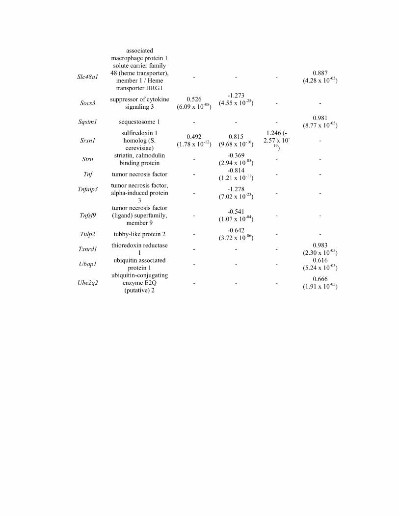
Table S1. Information for genes that were differentially expressed upon honaucin
A exposure………..…………………………………………………………………….…..8
SUPPORTING EXPERIMENTAL PROCEDURES……………………………………………...11
General experimental procedures for the generation of fluorescent ethyl 7-
dimethylaminocoumarin-4-acetate probe, assay for the detection of nitric oxide, treatment of
RAW264.7 cells with ethyl 7- dimethylaminocoumarin-4-acetate probe and confocal imaging
ACKNOWLEDGEMENT…………………………………………………………………...………13
SUPPORTING REFERENCES……………………………………………………………………..13

SUPPORTING FIGURES AND TABLES
S1. Reaction scheme for honaucin A and N-Acetyl-L-cysteine or reduced glutathione showing
predicted adducts.

S2. Structure of honaucin A with Michael Acceptor motif (red) and electron withdrawing group
(blue) highlighted.

S3. Synthetic scheme for the generation of a fluorescent ethyl 7- dimethylaminocoumarin-4-acetate
honaucin probe, Fl-OCT-honaucin A.

S4. Activity data for Fl-OCT-honaucin A and ethyl 7- dimethylaminocoumarin-4-acetate in an assay
of nitric oxide inhibition. Error bars represent the standard deviations of 3 replicates.

S5. Confocal images showing the subcellular localization of Fl-OCT-honaucin A and ethyl 7-
dimethylaminocoumarin-4-acetate in RAW264.7 cells. Cells were exposed to either honaucin probe
or coumarin for 15 minutes (A, B) or one hour (C, D) before being washed with PBS. The probe was
better retained than coumarin and showed both a punctate as well as a more diffuse cytosolic
distribution. (E) Close-up showing the distribution of the honaucin probe at one hour.
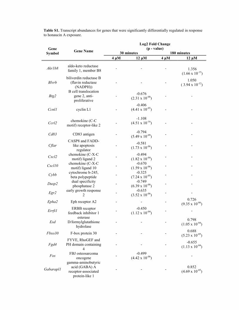
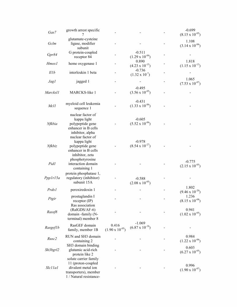
Table S1. Transcript abundances for genes that were significantly differentially regulated in response
to honaucin A exposure.
Gene
Symbol Gene Name
Log2 Fold Change
(p - value)
30 minutes 180 minutes
4 μM 12 μM 4 μM 12 μM
Akr1b8 aldo-keto reductase
family 1, member B8
- - - 1.356
(1.66 x 10⁻¹⁵
)
Blvrb
biliverdin reductase B
(flavin reductase
(NADPH))
- - - 1.050
( 3.94 x 10⁻¹²
)
Btg2
B cell translocation
gene 2, anti-
proliferative
- -0.676
(2.31 x 10-08
) - -
Ccnl1 cyclin L1 -
-0.406
(4.41 x 10-05
)
- -
Ccrl2 chemokine (C-C
motif) receptor-like 2 -
-1.108
(4.51 x 10⁻¹⁶
)
- -
Cd83 CD83 antigen - -0.794
(5.49 x 10-09
) - -
Cflar
CASP8 and FADD-
like apoptosis
regulator
- -0.581
(1.73 x 10-06
) - -
Cxcl2 chemokine (C-X-C
motif) ligand 2 -
-0.494
(1.82 x 10-04
) - -
Cxcl10 chemokine (C-X-C
motif) ligand 10 -
-0.670
(1.59 x 10-06
) - -
Cybb cytochrome b-245,
beta polypeptide -
-0.325
(7.24 x 10-05
) - -
Dusp2 dual specificity
phosphatase 2 -
-0.749
(6.39 x 10-08
) - -
Egr2 early growth response
2 -
-0.635
(3.52 x 10-06
) - -
Epha2 Eph receptor A2 - - - 0.726
(9.35 x 10-06
)
Errfi1 ERBB receptor
feedback inhibitor 1 -
-0.450
(1.12 x 10-04
) - -
Esd
esterase
D/formylglutathione
hydrolase
- - - 0.798
(1.05 x 10-04
)
Fbxo30 F-box protein 30 - - - 0.688
(5.23 x 10-05
)
Fgd4
FYVE, RhoGEF and
PH domain containing
4
- - - -0.655
(1.13 x 10-04
)
Fos FBJ osteosarcoma
oncogene -
-0.499
(4.42 x 10-06
) - -
Gabarapl1
gamma-aminobutyric
acid (GABA) A
receptor-associated
protein-like 1
- - - 0.852
(4.69 x 10-05
)
Gas7 growth arrest specific
7 - - -
-0.699
(8.15 x 10-05
)
Gclm
glutamate-cysteine
ligase, modifier
subunit
- - - 1.108
(3.14 x 10-06
)
Gpr84 G protein-coupled
receptor 84 -
-0.511
(1.29 x 10-04
) - -
Hmox1 heme oxygenase 1 - 0.890
(4.23 x 10⁻¹⁵
) -
1.818
(1.15 x 10⁻¹³
)
Il1b interleukin 1 beta - -0.736
(1.32 x 10⁻⁷
) - -
Jag1 jagged 1 - - - 1.065
(7.53 x 10-07
)
Marcksl1 MARCKS-like 1 -
-0.495
(3.56 x 10-05
)
- -
Mcl1 myeloid cell leukemia
sequence 1 -
-0.431
(1.33 x 10-06
)
- -
Nfkbia
nuclear factor of
kappa light
polypeptide gene
enhancer in B cells
inhibitor, alpha
-
-0.605
(5.52 x 10-06
)
- -
Nfkbiz
nuclear factor of
kappa light
polypeptide gene
enhancer in B cells
inhibitor, zeta
-
-0.978
(8.54 x 10⁻²⁷
)
- -
Pid1
phosphotyrosine
interaction domain
containing 1
- - - -0.775
(2.15 x 10-05
)
Ppp1r15a
protein phosphatase 1,
regulatory (inhibitor)
subunit 15A
- -0.588
(2.08 x 10-05
)
- -
Prdx1 peroxiredoxin 1 - - - 1.802
(9.46 x 10⁻²⁰
)
Ptgir prostaglandin I
receptor (IP) - - -
1.236
(8.15 x 10-08
)
Rassf8
Ras association
(RalGDS/AF-6)
domain -family (N-
terminal) member 8
- - - 0.941
(1.02 x 10-05
)
Rasgef1b RasGEF domain
family, member 1B
0.416
(1.90 x 10-05
)
-1.069
(6.87 x 10⁻¹⁸
)
- -
Rusc2 RUN and SH3 domain
containing 2 - - -
0.984
(1.22 x 10-06
)
Sh3bgrl2
SH3 domain binding
glutamic acid-rich
protein like 2
- - - 0.603
(6.27 x 10-05
)
Slc11a1
solute carrier family
11 (proton-coupled
divalent metal ion
transporters), member
1 / Natural resistance-
- - - 0.996
(1.90 x 10-07
)
associated
macrophage protein 1
Slc48a1
solute carrier family
48 (heme transporter),
member 1 / Heme
transporter HRG1
- - - 0.887
(4.28 x 10-05
)
Socs3 suppressor of cytokine
signaling 3
0.526
(6.09 x 10-08
)
-1.273
(4.55 x 10⁻²⁵
)
- -
Sqstm1 sequestosome 1 - - - 0.981
(8.77 x 10-05
)
Srxn1
sulfiredoxin 1
homolog (S.
cerevisiae)
0.492
(1.78 x 10⁻¹²
)
0.815
(9.68 x 10⁻¹⁶
)
1.246 (-
2.57 x 10-
19)
-
Strn striatin, calmodulin
binding protein -
-0.369
(2.94 x 10-05
) - -
Tnf tumor necrosis factor - -0.814
(1.21 x 10⁻¹¹
) - -
Tnfaip3
tumor necrosis factor,
alpha-induced protein
3
- -1.278
(7.02 x 10⁻²³
) - -
Tnfsf9
tumor necrosis factor
(ligand) superfamily,
member 9
- -0.541
(1.07 x 10-04
) - -
Tulp2 tubby-like protein 2 - -0.642
(3.72 x 10-06
) - -
Txnrd1 thioredoxin reductase
1 - - -
0.983
(2.30 x 10-05
)
Ubap1 ubiquitin associated
protein 1 - - -
0.616
(5.24 x 10-05
)
Ube2q2
ubiquitin-conjugating
enzyme E2Q
(putative) 2
- - - 0.666
(1.91 x 10-05
)

SUPPORTING EXPERIMENTAL PROCEDURES
Generation of fluorescent ethyl 7- dimethylaminocoumarin-4-acetate honaucin probe Briefly, 3S-Hydroxy butyrolactone (1, 1.02 g) and 3-butenoic acid (2) were coupled via Steglich
esterification in the presence of N,N'-dicyclohexylcarbodiimide (DCC) and a catalytic amount of 4-
(dimethylamino)pyridine (DMAP) to give a compound 3.1 An alcohol of hex-5-ene-1-ol (4) was
treated with trityl chloride to give tritylated compound 5. Then compounds 3 and 5 were coupled by
Grubb’s reaction with the Grubbs catalyst 2nd generation.2 The resulting compound 6 was treated
with N-chlorosuccinimide under the presence of PhSeCl for allylic chlorination to produce compound
7.3 The trityl group of compound 7 was deprotected under mild acidic condition and its primary
alcohol (8) was coupled with coumarin-4-acetic acid (9) via Steglich esterification to give fluorescent
honaucin A (10).
Specifically, 3-Butenoic acid (2, 851 mg) and 1.2 eq. of N,N'-dicyclohexylcarbodiimide (DCC, 2.4 g)
with a catalytic amount of 4-(dimethylamino)pyridine (DMAP) were dissolved in 10 mL of distilled
CH2Cl2 and the mixture was stirred for 5 min at room temperature. To the solution, 3S-hydroxy
butyrolactone (1, 1.02 g) was added and the mixture was incubated for 16 h at room temperature. The
solution was dried under N2 and the residue was subjected to silica open column chromatography with
a stepped gradient elution (hexanes and ethyl acetate). The fraction eluting with 70% hexanes in ethyl
acetate was further purified by RP-HPLC (Phenomenex Luna C18, 250 x 10 mm, 3 mL/min,
H2O:CH3CN=7:3, 210 nm) to give a purified compound 3 (885 mg, yield 52%). Hex-5-ene-1-ol (4,
500 mg) was dissolved in 3 mL of anhydrous pyridine with the addition of trityl chloride (1.394 g).
The solution was incubated with stirring for 16 h at room temperature, and the tritylation was
quenched with the addition of 1 mL of MeOH. Then the solution was evaporated under reduced
pressure and the residue was partitioned between CHCl3 (50 mL) and H2O (20 mL). The organic layer
was washed with brine (20 mL), treated with Na2SO4, and evaporated under reduced pressure. The
tritylated product (5, 1.576 g, 92%) was purified by normal phase Sep-Pak column chromatography.
Compounds 3 (425 mg) and 5 (85.6 mg) were coupled by Grubb’s reaction with Grubbs catalyst 2nd
generation (0.2 eq, 42.5 mg) under distilled CH2Cl2 for 16 h with stirring. The reaction mixture was
filtered and evaporated under reduced pressure and then subjected to RP-HPLC (Phenomenex Hydro
RP, 250 x 10 mm, 3 mL/min, A=90% H2O in CH3CN, B=100% CH3CN, 3:7 for 20 min, 3:7 to 100%
CH3CN for 20 min, 210 nm) to give a pure compound 6 (15.7 mg, yield 13%). Compound 6 (160 mg)
was added to a vial containing PhSeCl (16 mg) and anhydrous CH3CN (150 mL). To a reaction
mixture, N-chlorosuccinimide (52.3 mg) in anhydrous CH3CN was added dropwise through a gas
tight syringe and the reaction mixture was incubated for 16 h with stirring at room temperature. The
reaction mixture was concentrated under N2 and the residual material was partitioned between Et2O
and H2O. The Et2O soluble material was purified by normal phase Sep-Pak chromatography to give
pure compound 7 (128 mg, 75%). Compound 7 (51.9 mg) was dissolved in Et2O and treated with 0.1
N formic acid for 4 h to give the detritylated product, compound 8 (23.7 mg, 86%). Coumarin-4-acetic
acid (9, 4.3 mg) and 3.58 mg of DCC with a catalytic amount of DMAP were dissolved in 3 mL of
distilled CH2Cl2 and the mixture was stirred for 5 min at room temperature. To the solution,
compound 8 (4 mg) was added and the mixture was incubated for 16 h at room temperature. The
solution was dried under N2 and the residue was subjected to silica open column chromatography with
a stepped gradient elution (hexanes and ethyl acetate). The fraction eluting with 70% hexanes in ethyl
acetate was further purified by RP-HPLC (Phenomenex Luna C18, 250 x 4.6 mm, 1 mL/min,
H2O:CH3CN=7:3, 210 nm) to give a purified compound 10 (4.5 mg, yield 52%).
Compound 3: 1H NMR (500 MHz, CDCl3) δ 5.88 (ddt, J = 17.0, 10.4, 7.2 Hz, 1H), 5.45 (m, 1H), 5.21
(dd, J = 10.4, 1.1 Hz, 1H), 5.20 (dd, J = 17.0, 1.1 Hz, 1H), 4.51 (dd, J = 11.2, 4.9 Hz, 1H), 4.37 (d, J
= 11.2 Hz, 1H), 3.13 (d, J = 7.2 Hz, 2H), 2.86 (dd, J = 18.6, 6.6 Hz, 1H), 2.62 (d, J = 18.6 Hz, 1H); 13
C NMR (75 MHz, CDCl3) δ 174.6, 171.1, 129.3, 119.7, 73.2, 70.2, 39.0, 34.7; HRESITOFMS m/z
[M+H]+ 171.0657 (calcd for C8H11O4 171.0652).
Compound 5: 1H NMR (500 MHz, CDCl3) δ 7.55 (d, J = 8.1 Hz, 1H), 7.37 (t, J = 7.6 Hz, 1H), 7.30 (t,
J = 7.3 Hz, 1H), 5.87 (ddt, J = 16.9, 10.2, 6.7 Hz, 1H), 5.06 (ddd, J = 17.2, 3.5, 1.6 Hz, 1H), 5.04 –

5.00 (m, 1H), 3.15 (t, J = 6.6 Hz, 1H), 2.11 (q, J = 7.2 Hz, 1H), 1.60 – 1.53 (m, 1H); 13
C NMR (75
MHz, CDCl3) δ 144.7, 139.0, 128.9, 127.9, 127.0, 114.6, 86.5, 63.6, 33.8, 29.7, 25.8; HREIMS m/z
[M]+ 342.1981 (calcd for C28H26O 342.1978).
Compound 6: 1H NMR (500 MHz, CDCl3) δ 7.44 (d, J = 7.3 Hz, 6H), 7.30 (t, J = 7.5 Hz, 6H), 7.23 (t,
J = 7.3 Hz, 3H), 5.57 (m, 1H), 5.46 (m, 1H), 5.43 (ddd, J = 6.7, 3.2, 1.6 Hz, 1H), 4.50 (dd, J = 11.1,
4.8 Hz, 1H), 4.36 (d, J = 11.1 Hz, 1H), 3.05 (ddd, J = 6.5, 3.4, 2.7 Hz, 2H), 2.85 (dd, J = 18.5, 6.8
Hz, 1H), 2.62 (d, J = 18.4 Hz, 1H), 2.02 (q, J = 7.0 Hz, 2H), 1.63 (m, 2H), 1.47 (dq, J = 15.0, 7.6 Hz,
2H); 13
C NMR (75 MHz, CDCl3) δ 174.6, 171.7, 144.7, 135.9, 128.9, 127.9, 127.0, 120.8, 86.5, 73.2,
70.0, 63.5, 38.0, 34.7, 32.5, 29.7, 26.0; HRESITOFMS m/z [M+Na]+ 507.2143 (calcd for
C31H32O5Na 507.2142).
Compound 7: 1H NMR (500 MHz, CDCl3) δ 7.44 (d, J = 7.2 Hz, 6H), 7.30 (t, J = 7.5 Hz, 6H), 7.24 (t,
J = 7.3 Hz, 3H), 6.93 (dd, J = 15.4, 7.5 Hz, 1H), 6.01 (d, J = 15.4 Hz, 1H), 5.51 (dd, J = 6.5, 5.0 Hz,
1H), 4.54 (dd, J = 11.1, 4.8 Hz, 1H), 4.44 (m, 1H), 4.40 (d, J = 11.0 Hz, 1H), 3.08 (t, J = 6.4 Hz,
2H), 2.89 (dd, J = 18.5, 6.8 Hz, 1H), 2.66 (d, J = 18.5 Hz, 1H), 1.82 (dd, J = 14.7, 7.6 Hz, 2H), 1.65
(ddd, J = 14.1, 8.7, 4.2 Hz, 2H), 1.50 (dt, J = 20.5, 6.8 Hz, 2H); 13
C NMR (125 MHz, CDCl3) δ
174.7, 165.2, 148.5, 144.5, 128.8, 128.0, 127.1, 121.3, 86.6, 73.1, 70.3, 63.1, 59.6, 37.4, 34.7, 29.4,
23.2; HRESITOFMS m/z [M+Na]+ 541.1753 (calcd for C31H31ClO5Na 541.1752).
Compound 8: 1H NMR (500 MHz, CDCl3) δ 6.96 (dd, J = 15.4, 7.5 Hz, 1H), 6.05 (d, J = 15.4 Hz,
1H), 5.53 (dd, J = 6.5, 5.0 Hz, 1H), 4.55 (dd, J = 11.1, 4.8 Hz, 1H), 4.48 (dd, J = 14.2, 7.2 Hz, 1H),
4.43 (d, J = 11.2 Hz, 1H), 3.68 (t, J = 6.1 Hz, 2H), 2.91 (dd, J = 18.5, 6.7 Hz, 1H), 2.68 (d, J = 18.4
Hz, 1H), 1.91 (dd, J = 14.3, 7.5 Hz, 2H), 1.64-1.50 (m, 4H); 13
C NMR (125 MHz, CDCl3) δ 174.60,
165.15, 148.34, 121.39, 73.15, 70.32, 62.65, 59.62, 37.47, 34.77, 32.09, 22.79; HRESITOFMS m/z
[M+Na]+ 299.0655 (calcd for C12H17ClO5Na 299.0657).
Compound 10: 1H NMR (500 MHz, CDCl3) δ 7.41 (d, J = 8.9 Hz, 1H), 6.91 (dd, J = 15.4, 7.5 Hz,
1H), 6.64 (dd, J = 9.0, 2.5 Hz, 1H), 6.54 (d, J = 2.5 Hz, 1H), 6.05 (s, 1H), 6.02 (dd, J = 15.4, 1.1 Hz,
1H), 5.53 (dd, J = 6.3, 4.9 Hz, 1H), 4.55 (dd, J = 11.2, 4.8 Hz, 1H), 4.44 (d, J = 11.2 Hz, 1H), 4.37
(dd, J = 13.6, 6.6 Hz, 1H), 4.13 (t, J = 6.4 Hz, 2H), 3.69 (s, 2H), 3.07 (s, 6H), 2.91 (dd, J = 18.5, 6.7
Hz, 1H), 2.69 (d, J = 18.4 Hz, 1H), 1.80 (dt, J = 10.7, 4.3 Hz, 2H), 1.65 (m, 2H), 1.53 – 1.42 (m,
1H), 1.42 – 1.33 (m, 1H); 13
C NMR (125 MHz, CDCl3) δ 174.6, 169.3, 165.1, 161.9, 156.2, 153.2,
148.5, 148.0, 125.5, 121.5, 111.0, 109.3, 108.7, 98.6, 73.2, 70.4, 65.2, 59.4, 40.4, 38.6, 37.1, 34.8,
28.0, 22.8; HRESITOFMS m/z [M+Na]+ 528.1394 (calcd for C25H28ClNO8Na 528.1396).
Detection of NO Production in Murine Macrophages treated with Fl-OCT honaucin A (Villa et
al. 2010)4
A 96-well plate was seeded with RAW 264.7 cells at a density of 5 x 10⁴ cells/180 μL medium/well in
Dulbecco’s Modified Eagle Medium (Gibco, Carlsbad, CA) supplemented with 10% fetal bovine
serum and penicillin/streptomycin. The plate was incubated overnight at 37 °C with 5% CO2 in order
to achieve a confluent cellular monolayer. Fl-OCT honaucin A, consisting of honaucin A conjugated
to ethyl 7- dimethylaminocoumarin-4-acetate, as well as ethyl 7- dimethylaminocoumarin-4-acetate
itself were serially diluted in 30% EtOH:PBS and each dilution was added to the plate in triplicate
wells (10 μL/well ) to give a final concentration series of 30, 10, 3.0, 1.0, 0.3 and 0.1 μg/mL. The
plate with compounds was incubated for 1 hr at 37 °C with 5% CO2 prior to the addition of 10
μL/well bacterial endotoxin yielding a final concentration of 3.0 μg/mL. Controls included the
positive control dimethylsulfoxide (5%), an LPS-free control, and a compound-free LPS only control.
Plates were incubated overnight 1 hr at 37 °C with 5% CO2 and the amount of nitric oxide in each
well was determined the following morning by measurement of the NO breakdown product nitrite via
Griess reaction.5 Briefly, the amount of nitrite in each sample was compared to a prepared nitrite
standard curve (0-100 μM). 50 μL 1% sulfanilamide in 5% phosphoric acid was added to 50 uL of
supernatant from each well of the overnight assay plate, as well as to the prepared nitrite standard
curve, and incubated in the dark at room temperature for 10 minutes. Following this incubation, 0.1%
N-1-napthylethylenediamine dihydrochloride in water (50 μL, Ricca Chemical Company LLC,
Pocomoke City, MD) was added to the plate and the incubation was repeated. The absorbance of the
wells was measured at 570 nm and sample nitrite concentration was calculated by regression using the
nitrite standard curve. Average nitrite and standard deviation for each sample treatment was reported.
An MTT assay of cell viability ensured that cell mortality was not observed at the IC50 values of the
compounds.
Cell tracking with a fluorescent honaucin A probe
RAW 264.7 macrophages were seeded at a density of 1x10⁶ cells per milliliter in
MatTek (Ashland, MA, USA) 35 mm glass-bottom dishes and allowed to achieve confluency in a
CO2 incubator overnight. Either coumarin or the coumarin-conjugated honaucin A probe (2 μM) was
introduced to the cells. Cells were incubated for 15 minutes or 1 hour at 37 °C with 5% CO2 and then
washed 5 times with phosphate buffered saline. The nuclear stain DRAQ5 was then added and the
cells were incubated for an additional 10 minutes. A Zeiss LSM-700 laser scanning confocal
microscope (Jena, Germany) running Zen 2010 software (Zeiss, revision 5.5) was used to visualize
compound localization in the cells (40x magnification). This was done by visualizing 390 nm Z-
sections that were ultimately compiled into a maximum intensity projection using ImageJ (National
Institutes of Health, Bethesda, MD, USA). Settings were maintained between treatments so that
differences in fluorescence were comparable.
ACKNOWLEDGEMENT
Fluorescent compound (9) was gifted by J.J. La Clair in the Chemistry Department at University of
California, San Diego.
SUPPORTING REFERENCES
(1) Neises, B.; Steglich, W. Simple method for the esterification of carboxylic acids. Angew. Chem.
Int. Ed. 1978, 17, 522-524.
(2) Scholl, M.; Ding, S.; Lee, C. W.; Grubbs, R.H. Synthesis and activity of a new generation of
ruthenium-based olefin metathesis catalysts coordinated with 1,3-dimesityl-4,5-dihydroimidazol-2-
ylidene ligands. Org. Lett. 1999, 1, 953-956.
(3) Tunge, J. A.; Mellegaard, S. R. Selective selenocatalytic allylic halogenation. Org. Lett. 2004, 6,
1205-1207.
(4) Villa, F. A.; Lieske, K.; Gerwick, L. Selective MyD88-dependent pathway inhibition by the
cyanobacterial natural product malyngamide F acetate. Eur. J. Pharmacol. 2010, 629, 140-146.
(5) Green, L. C.; Wagner, D. A.; Glogowski, J.; Skipper, P. L.; Wishnok, J.S.; Tannenbaum, S.R.
Analysis of nitrate, nitrite, and N-15-labeled nitrate in biological fluids. Anal. Biochem. 1982, 126,
131-138.