Embed Size (px)
Citation preview
manuscript su5383 for review
Structure of Equilenin at 100 K: an estrone related steroid
Christopher S. Frampton* and David D. MacNicol
CONFIDENTIAL – NOT TO BE REPRODUCED, QUOTED NOR SHOWN TO OTHERS
SCIENTIFIC MANUSCRIPT
For review only.
Sunday 16 July 2017
Category: research communications
Co-editor:
Professor H. Stoeckli-Evans
Telephone: +41 32 7182400
Fax: +41 32 7182511
Email: [email protected]
Contact author:
Christopher S. Frampton
United Kingdom
Telephone: 01895 265337
Fax: ?
Email: [email protected]
checkCIF/PLATON results for paper su5383
checkCIF/PLATON results
Ellipsoid plot
checkCIF/PLATON results
No syntax errors found. CIF dictionary Interpreting this report
Datablock: I
Bond precision: C-C = 0.0020 A Wavelength=1.54184
Cell: a=7.27709(7) b=7.32686(6) c=25.5179(2)
alpha=90 beta=90 gamma=90
Temperature: 100 K
Calculated Reported
---------- --------
Volume 1360.57(2) 1360.57(2)
Space group P 21 21 21 P 21 21 21
Hall group P 2ac 2ab P 2ac 2ab
Moiety formula C18 H18 O2 C18 H18 O2
Sum formula C18 H18 O2 C18 H18 O2
Mr 266.32 266.32
Dx,g cm-3 1.300 1.300
Z 4 4
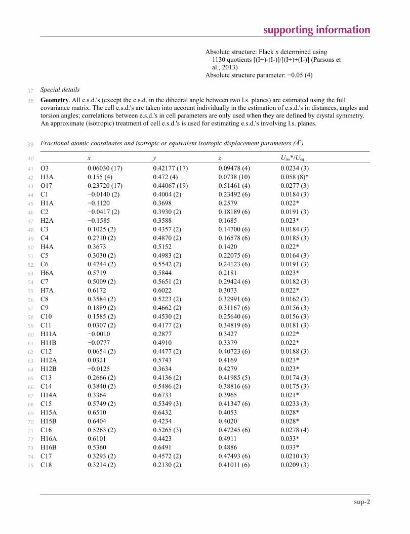
Mu (mm-1) 0.658 0.658
F000 568.0 568.0
F000’ 569.63
h,k,lmax 9,9,31 9,9,31
Nref 2767[ 1632] 2769
Tmin,Tmax 0.911,0.955 0.853,0.960
Tmin’ 0.765
Correction method= # Reported T Limits: Tmin=0.853 Tmax=0.960 AbsCorr = MULTI-SCAN
Data completeness= 1.70/1.00
Theta(max)= 74.465
R(reflections)= 0.0292( 2754) wR2(reflections)= 0.0804( 2769)
S = 1.008 Npar= 186
Alert level G
PLAT142_ALERT_4_G s.u. on b - Axis Small or Missing .............. 0.00006 Ang.
PLAT143_ALERT_4_G s.u. on c - Axis Small or Missing .............. 0.00020 Ang.
PLAT791_ALERT_4_G The Model has Chirality at C13 (Chiral SPGR) S Verify
PLAT791_ALERT_4_G The Model has Chirality at C14 (Chiral SPGR) S Verify
PLAT802_ALERT_4_G CIF Input Record(s) with more than 80 Characters 1 Info
PLAT978_ALERT_2_G Number C-C Bonds with Positive Residual Density. 19 Note
0 ALERT level A = Most likely a serious problem - resolve or explain
0 ALERT level B = A potentially serious problem, consider carefully
0 ALERT level C = Check. Ensure it is not caused by an omission or oversight
6 ALERT level G = General information/check it is not something unexpected
0 ALERT type 1 CIF construction/syntax error, inconsistent or missing data
1 ALERT type 2 Indicator that the structure model may be wrong or deficient
0 ALERT type 3 Indicator that the structure quality may be low
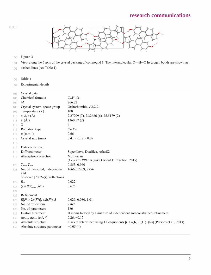
5 ALERT type 4 Improvement, methodology, query or suggestion
0 ALERT type 5 Informative message, check
database duplication summary
Datablock: ciftbxwarning
Chemical name =
R factor =
Space group =
Formula =
a= b= c=
alpha= beta= gamma=
Datablock: Inputlinelengthexceedsline_
Chemical name =
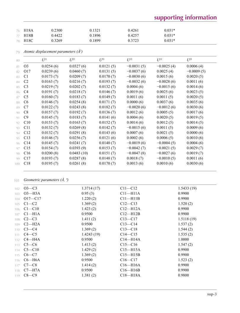
R factor =
Space group =
Formula =
a= b= c=
alpha= beta= gamma=
Datablock: I
Chemical name = Equilenin
R factor = 0.029
Space group =
Formula = C18 H18 O2
a=7.27709 b=7.32686 c=25.5179
alpha=90 beta=90 gamma=90
Ohrt, J. M., Haner, B. A. & Norton, D. A. (1967). Acta Cryst.23, 1100-1100
[Cell: 7.48,25.528,7.279(90,90,90) R= T= Room Temp.(283-303)]
Author Response:This paper was a unit cell determination only. No 3-dimensional structural
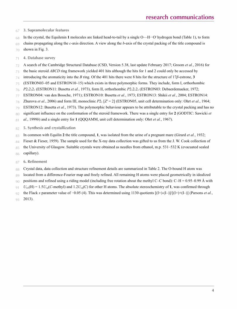
data exists for this important steroid.
reference checking results
The following references were not checked in detail as they were not recognized as journal
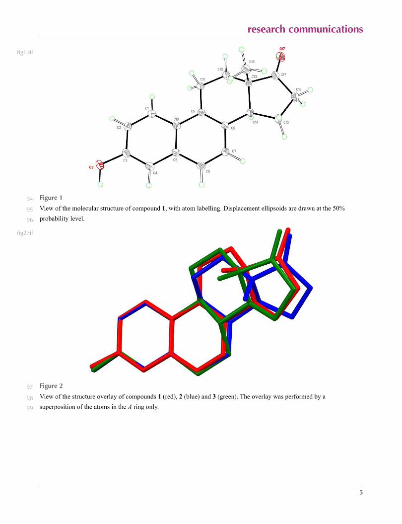
references
Fieser, L. F. & Fieser, M. (1959). Steroids. Reinhold Publishing Corporation, New York, 460--461.
Marshall, P. G. (1970). Rodd’s Chemistry of Carbon Compounds, 2nd Edition., ed. Coffey S. Vol. IID,
Elsevier, B. V., 216--222.
Rigaku Oxford Diffraction (2015). CrysAlis PRO. Rigaku Corporation, Oxford, UK.
The following references may be incorrectly formatted
Bossche, G. van den (1971). Bull. Soc. Roy. Sci. Liege, 40, 614--?.
[Unrecognized journal title.]
Cruickshank, D. W. J. & Sparks, R. A. (1960). Proc. Roy. Soc. A. 258, 270--285.
[Unrecognized journal title.]
Duax, W. L., Weeks, C. M. & Rohrer, D. C. (1976). Crystal Structures of Steroids, in Topics in
Stereochemistry, eds. Allinger, N. L., Eliel, E. L., 9, John Wiley & Sons, Inc., Hoboken, NJ, USA, pp.
271--383.
[Missing final page numbers?]
All references appear to be cited unambiguously
Citation comments
1 date found in data_I _publ_body_contents that could be part of a citation but not found in reference
list: 2017

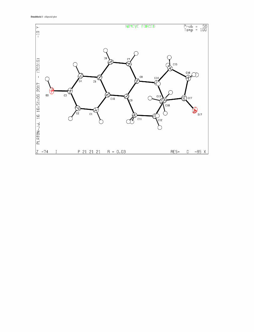
Datablock I - ellipsoid plot
research communications
1
Structure of Equilenin at 100 K: an estrone related steroid1
Christopher S. Framptona* and David D. MacNicolb2
aWolfson Centre for Materials Processing, Brunel University London, Kingston Lane, Uxbridge, UB8 3PH, U.K., and bDepartment of Chemistry, 3University of Glasgow, Glasgow, G12 8QQ, Scotland, U.K.4Correspondence email: [email protected]
Abstract 6
The structure of the estrone related steroid, Equilenin, C18H18O2 (systematic name 3-hydroxy-13-7
methyl-11,12,13,14,15,16-hexahydro-cyclopenta[a]phenanthren-17-one), has been determined at 100 K. The crystals are 8
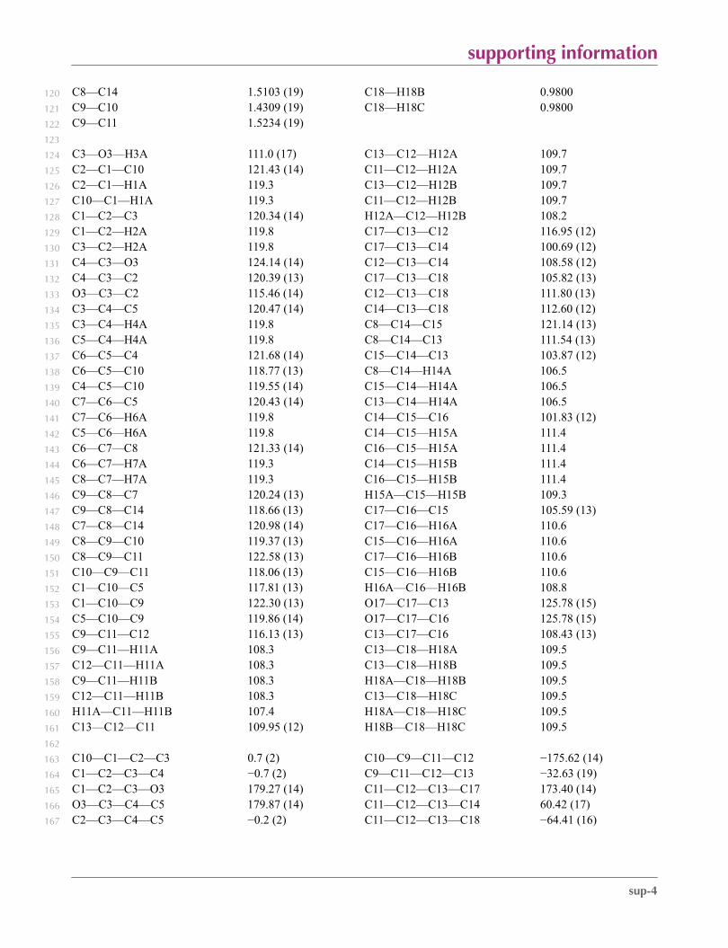
orthorhombic, P212121, and the absolute structure of the molecule in the crystal has been determined by resonant 9
scattering [Flack parameter = 0.05 (4)]. The carbon atoms of the A and B rings, are coplanar with an r.m.s. deviation from 10
planarity of 0.0104 Å. The C ring has a sofa conformation while the D ring has an envelope conformation with the 11
methine C atom as the flap. The keto oxygen and the methyl group are translated 0.78 Å and 0.79 Å, respectively, from 12
the equivalent positions on 17β-estrone 3. In the crystal, molecules are linked by O—H···O hydrogen bonds forming 13
chains parallel to the c-axis direction.14
Keywords: crystal structure; Equilenin; Equilin; estrone; steroid; conformation; hydrogen bonding. 15
research communications
2
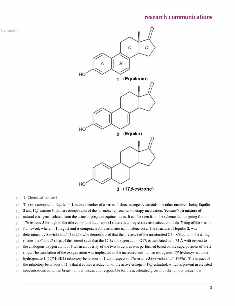
83scheme1.tif
1. Chemical context 16
The title compound, Equilenin 1, is one member of a series of three estrogenic steroids, the other members being Equilin 17
2 and 17β-estrone 3, that are components of the hormone replacement therapy medication, ′Premarin′, a mixture of 18
natural estrogens isolated from the urine of pregnant equine mares. It can be seen from the scheme that on going from 19
17β-estrone 3 through to the title compound Equilenin (1), there is a progressive aromatization of the B ring of the steroid 20
framework where in 1 rings A and B comprise a fully aromatic naphthalene core. The structure of Equilin 2, was 21
determined by Sawicki et al. (1999b), who demonstrated that the presence of the unsaturated C7—C8 bond in the B ring 22
rotates the C and D rings of the steroid such that the 17-keto oxygen atom, O17, is translated by 0.73 Å with respect to 23
the analogous oxygen atom of 3 when an overlay of the two structures was performed based on the superposition of the A 24
rings. The translation of the oxygen atom was implicated in the increased anti-human estrogenic 17β-hydroxysteroid de-25
hydrogenase 1 (17β-HSD1) inhibitory behaviour of 2 with respect to 17β-estone 3 (Sawicki et al., 1999a). The impact of 26
the inhibitory behaviour of 2 is that it causes a reduction of the active estrogen, 17β-estradiol, which is present in elevated 27
concentrations in human breast tumour tissues and responsible for the accelerated growth of the tumour tissue. It is 28
research communications
3
therefore of great interest to investigate what the structural and conformational consequences are on the C and D rings of 29
the steroid framework of 1 by having fully unsaturated A and B rings. Although the unit-cell parameters of 1 at room 30
temperature have been previously reported by Ohrt et al. (1967), no three-dimensional structure analysis of this important 31
estrone steroid has been determined. Herein, we report on the crystal structure of this final member of the estrone series 32
of steroids, Equilenin 1, at 100 K. 33
2. Structural commentary 34
The crystal structure of Equilenin 1, is orthorhombic, space group P212121 (Z ′= 1) and its molecular structure is 35
illustrated in Fig. 1. The unit cell data agree with the previously reported values (Ohrt et al., 1967) with the caveat that 36
they are slightly smaller owing to some modest isotropic contraction due to the lower temperature. The atoms C1 through 37
C10, which define the AB (napthalene) plane, are little affected by the chiral centres at C13 and C14, and are coplanar 38
with an r.m.s. deviation of the fitted atoms of 0.0104 Å and a total puckering amplitude, Q, of 0.033 (2) Å. The greatest 39
displacement from the ten atom mean plane is atom C10 at −0.019 (1) Å. The C—C bond lengths of the AB rings follow 40
the pattern in which C1–C2, C3–C4, C6–C7 and C8–C9 are significantly shorter, (mean value 1.372 Å), than the 41
remaining 7 bonds (mean value 1.421 Å) [Ahmed & Cruickshank, 1952; Cruickshank & Sparks, 1960], thus 42
demonstrating that the AB ring is a true aromatic naphthalene core. The aromatization of ring B does however, have a 43
significant effect on the conformations of both the C and D rings of 1, compared to 2 and 3. In contrast to the regular 44
chair conformation of the C rings of 2 and 3, the C ring of 1, has a highly symmetric 13β-envelope conformation 45
characterized by a ∆Cs(9) asymmetry parameter of 0.50° (Duax et al., 1976); and related pairs of torsion angles [C14–46
C8–C9–C11, C8–C9–C11–C12, −4.1 (2), 4.1 (2)°; C9–C11–C12–C13, C9–C8–C14–C13, −32.6 (2), 32.7 (2)°; C11–47
C12–C13–C14, C12–C13–C14–C8, 60.4 (2), −61.3 (1)°]. The downside impact of this conformational change in the C 48
ring of 1 is such that in place of the asymmetric twist or half-chair D ring conformation demonstrated by 2 and 3, the D 49
ring of Equilenin 1 displays a 14α-envelope conformation with a ∆Cs(14) of 4.20°; the torsion angles for 1, (with related 50
torsion angles for 2/3 are given in [/]) are C13–C14–C15–C16, C17–C13–C14–C15) −41.3 (2)[−40.2/-39.0]°, 43.3 (2)51
[44.5/42.9]°; C14–C13–C17–C16, C14–C15–C16–C17, −28.6 (2)[−31.0/-30.9]°, 22.3 (2)[19.6/19.4]°; and C15–C16–52
C17–C13, 3.6 (2)[8.1/7.5]°. Torsional angle data for 2 and 3 was extracted from structures GODTIC (Sawicki et al., 53
1999b) and ESTRON13 (Shikii et al., 2004), respectively [see Section 4, Database survey]. Compounds 1 and 2, possibly 54
owing to increased conformational constraint in the B ring, have lower oestrongenic activity than 17β-estrone itself, 55
which has the B ring as the principal point of molecular flexibility (Duax et al., 1976; Busetta et al., 1973). Interestingly 56
this reduction in activity (Marshall, 1970) does not directly relate to the crystallographically determined degree to which 57
the A and B rings of the steroid are constrained to coplanarity, since 1, possessing an essentially planar naphthalene core, 58
is about five times more estrogenic than 2 which features only approximate coplanarity of its A and B rings with an r.m.s. 59
deviation of the fitted atoms of 0.102 Å, and a total puckering amplitude of 0.270 (2) Å (Sawicki et al., 1999b). An 60
overlay of structures 1 (red), 2 (blue) and 3 (green) is shown in Fig. 2. The overlay was performed by a superposition of 61
the atoms in the A ring only. From this overlay it can be calculated that the keto oxygen atom is translated by 0.78 Å and 62
0.69 Å, respectively, for compounds 1 and 2 from its position on 3. Perhaps more significant is the degree of translation 63
of the methyl group C18 which is translated by 0.79 Å and 1.40 Å, respectively, for compounds 1 and 2 from its position 64
on 3 which may account for the increased estrogenic activity of 1 over 2. The stereochemistry assignments at C13 and 65
C14 are S, S; confirmed by resonant scattering through the Flack x parameter value of −0.05 (4). 66
research communications
4
3. Supramolecular features 67
In the crystal, the Equilenin 1 molecules are linked head-to-tail by a single O—H···Oi hydrogen bond (Table 1), to form 68
chains propagating along the c-axis direction. A view along the b-axis of the crystal packing of the title compound is 69
shown in Fig. 3. 70
4. Database survey 71
A search of the Cambridge Structural Database (CSD, Version 5.38, last update February 2017; Groom et al., 2016) for 72
the basic steroid ABCD ring framework yielded 401 hits although the hits for 1 and 2 could only be accessed by 73
introducing the aromaticity into the B ring. Of the 401 hits there were 8 hits for the structure of 17β-estrone, 3 74
(ESTRON03–05 and ESTRON10–15) which exists in three polymorphic forms. They include, form I, orthorhombic 75
P212121 (ESTRON11: Busetta et al., 1973), form II, orthorhombic P212121 (ESTRON03: Debaerdemaeker, 1972; 76
ESTRON04: van den Bossche, 1971); ESTRON10: Busetta et al., 1973; ESTRON13: Shikii et al., 2004; ESTRON14: 77
Zhurova et al., 2006) and form III, monoclinic P21 [Z′ = 2] (ESTRON05, unit cell determination only: Ohrt et al., 1964; 78
ESTRON12: Busetta et al., 1973). The polymorphic behaviour appears to be attributable to the crystal packing and has no 79
significant influence on the conformation of the steroid framework. There was a single entry for 2 (GODTIC: Sawicki et 80
al., 1999b) and a single entry for 1 (QQQAMM, unit cell determination only: Ohrt et al., 1967). 81
5. Synthesis and crystallization 82
In common with Equilin 2 the title compound, 1, was isolated from the urine of a pregnant mare (Girard et al., 1932; 83
Fieser & Fieser, 1959). The sample used for the X-ray data collection was gifted to us from the J. W. Cook collection of 84
the University of Glasgow. Suitable crystals were obtained as needles from ethanol, m.p. 531–532 K (evacuated sealed 85
capillary). 86
6. Refinement 87
Crystal data, data collection and structure refinement details are summarized in Table 2. The O-bound H atom was 88
located from a difference-Fourier map and freely refined. All remaining H atoms were placed geometrically in idealized 89
positions and refined using a riding model (including free rotation about the methyl C–C bond): C–H = 0.95–0.99 Å with 90
Uiso(H) = 1.5Ueq(C-methyl) and 1.2Ueq(C) for other H atoms. The absolute stereochemistry of 1, was confirmed through 91
the Flack x parameter value of −0.05 (4). This was determined using 1130 quotients [(I+)-(I–)]/[(I+)+(I–)] (Parsons et al., 92
2013). 93
research communications
5
fig1.tif
Figure 194
View of the molecular structure of compound 1, with atom labelling. Displacement ellipsoids are drawn at the 50% 95
probability level. 96
fig2.tif
Figure 297
View of the structure overlay of compounds 1 (red), 2 (blue) and 3 (green). The overlay was performed by a 98
superposition of the atoms in the A ring only. 99
research communications
6
fig3.tif
Figure 3100
View along the b axis of the crystal packing of compound 1. The intermolecular O—H···O hydrogen bonds are shown as 101
dashed lines (see Table 1). 102
Table 1103
Experimental details104
105 Crystal data
106 Chemical formula C18H18O2
107 Mr 266.32
108 Crystal system, space group Orthorhombic, P212121
109 Temperature (K) 100
110 a, b, c (Å) 7.27709 (7), 7.32686 (6), 25.5179 (2)
111 V (Å3) 1360.57 (2)
112 Z 4
113 Radiation type Cu Kα
114 µ (mm−1) 0.66
115 Crystal size (mm) 0.41 × 0.12 × 0.07
116
117 Data collection
118 Diffractometer SuperNova, Dualflex, AtlasS2
119 Absorption correction Multi-scan (CrysAlis PRO; Rigaku Oxford Diffraction, 2015)
120 Tmin, Tmax 0.853, 0.960
121 No. of measured, independent andobserved [I > 2σ(I)] reflections
16660, 2769, 2754
122 Rint 0.022
123 (sin θ/λ)max (Å−1) 0.625
124
125 Refinement
126 R[F2 > 2σ(F2)], wR(F2), S 0.029, 0.080, 1.01
127 No. of reflections 2769
128 No. of parameters 186
129 H-atom treatment H atoms treated by a mixture of independent and constrained refinement
130 ∆ρmax, ∆ρmin (e Å−3) 0.26, −0.17
131 Absolute structure Flack x determined using 1130 quotients [(I+)-(I-)]/[(I+)+(I-)] (Parsons et al., 2013)
132 Absolute structure parameter −0.05 (4)
research communications
7
Computer programs: CrysAlis PRO (Rigaku Oxford Diffraction, 2015), CrysAlis PRO (Rigaku Oxford Diffraction, 2015), SHELXD2014/6 (Sheldrick, 1332010), SHELXL2014/6 (Sheldrick, 2015), SHELXTL (Sheldrick, 2008) and Mercury CSD 2.0 (Macrae et al., 2008), SHELXTL (Sheldrick, 2008) and 134publCIF (Westrip, 2010).
135
Table 2136
Hydrogen-bond geometry (Å, º) 137
138 D—H···A D—H H···A D···A D—H···A
139 O3—H3A···O17i 0.95 (3) 1.82 (3) 2.7153 (17) 157 (3)
140 Symmetry code: (i) −x+1/2, −y+1, z−1/2.
Acknowledgements 141
We thank the University of Glasgow for the gift of the sample from the J. W. Cook collection.142
References 143
Ahmed, F. R. & Cruickshank, D. W. J. (1952). Acta Cryst. 5, 852–853.144
Bossche, G. van den (1971). Bull. Soc. Roy. Sci. Liege, 40, 614–?.145
Busetta, B., Courseille, C. & Hospital, M. (1973). Acta Cryst. B29, 298–313.146
Cruickshank, D. W. J. & Sparks, R. A. (1960). Proc. Roy. Soc. A. 258, 270–285.147
Debaerdemaeker, T. D. J. (1972). Cryst. Struct. Commun., 1, 39–42.148
Duax, W. L., Weeks, C. M. & Rohrer, D. C. (1976). Crystal Structures of Steroids, in Topics in Stereochemistry, eds. 149
Allinger, N. L., Eliel, E. L., 9, John Wiley & Sons, Inc., Hoboken, NJ, USA, pp. 271–383.150
Fieser, L. F. & Fieser, M. (1959). Steroids. Reinhold Publishing Corporation, New York, 460–461.151
Girard, A., Sandulesco, G., Fridenson, A., Gudefroy, C. & Rutgers, J. J. (1932). Compt. Rend. Acad. Sci. 194, 1020-1022.152
Groom, C. R., Bruno, I. J., Lightfoot, M. P. & Ward, S. C. (2016). Acta Cryst. B72, 171–179.153
Macrae, C. F., Bruno, I. J., Chisholm, J. A., Edgington, P. R., McCabe, P., Pidcock, E., Rodriguez-Monge, L., Taylor, R., 154
van de Streek, J. & Wood, P. A. (2008). J. Appl. Cryst. 41, 466–470.155
Marshall, P. G. (1970). Rodd's Chemistry of Carbon Compounds, 2nd Edition., ed. Coffey S. Vol. IID, Elsevier, B. V., 156
216–222.157
Ohrt, J. M., Haner, B. A. & Norton, D. A. (1964). Acta Cryst. 17, 1611.158
Ohrt, J. M., Haner, B. A. & Norton, D. A. (1967). Acta Cryst. 23, 1100.159
Parsons, S., Flack, H. D. & Wagner, T. (2013). Acta Cryst. B69, 249–259.160
Rigaku Oxford Diffraction (2015). CrysAlis PRO. Rigaku Corporation, Oxford, UK.161
Sawicki, M. W., Erman, M., Puranen, T., Vihko, P. & Ghosh, D. (1999a). Proc. Natl. Acad. Sci. 96, 840–845.162
Sawicki, M. W., Li, N. & Ghosh, D. (1999b). Acta Cryst. C55, 425–427.163
Sheldrick, G. M. (2008). Acta Cryst. A64, 112–122.164
Sheldrick, G. M. (2010). Acta Cryst. D66, 479–485.165
Sheldrick, G. M. (2015). Acta Cryst. C71, 3–8.166
Shikii, K., Sakamoto, S., Seki, H., Utsumi, H. & Yamaguchi, K. (2004). Tetrahedron, 60, 3487–3492.167
research communications
8
Westrip, S. P. (2010). J. Appl. Cryst. 43, 920–925.168
Zhurova, E. A., Matta, C. F., Wu, N., Zhurov, V. V. & Pinkerton, A. A. (2006). J. Am. Chem. Soc. 128, 8849–8861. 169

supporting information
sup-1
supporting information1
Structure of Equilenin at 100 K: an estrone related steroid2
Christopher S. Frampton* and David D. MacNicol3
Computing details 4
Data collection: CrysAlis PRO (Rigaku Oxford Diffraction, 2015); cell refinement: CrysAlis PRO (Rigaku Oxford 5
Diffraction, 2015); data reduction: CrysAlis PRO (Rigaku Oxford Diffraction, 2015); program(s) used to solve structure: 6
SHELXD2014/6 (Sheldrick, 2010); program(s) used to refine structure: SHELXL2014/6 (Sheldrick, 2015); molecular 7
graphics: SHELXTL (Sheldrick, 2008) and Mercury CSD 2.0 (Macrae et al., 2008); software used to prepare material for 8
publication: SHELXTL (Sheldrick, 2008) and publCIF (Westrip, 2010).9
3-Hydroxy-13-methyl-11,12,13,14,15,16-hexahydro- cyclopenta[a]phenanthren-17-one 10
Crystal data 11
C18H18O212Mr = 266.3213Orthorhombic, P21212114a = 7.27709 (7) Å15b = 7.32686 (6) Å16c = 25.5179 (2) Å17V = 1360.57 (2) Å3
18Z = 419F(000) = 56820
Dx = 1.300 Mg m−3
Melting point: 531 KCu Kα radiation, λ = 1.54184 ÅCell parameters from 13177 reflectionsθ = 3.5–76.4°µ = 0.66 mm−1
T = 100 KRod, colourless0.41 × 0.12 × 0.07 mm
Data collection 21
SuperNova, Dualflex, AtlasS2 22diffractometer
Radiation source: fine-focus sealed X-ray tube, 23Enhance (Cu) X-ray Source
Detector resolution: 5.2921 pixels mm-124
ω scans25Absorption correction: multi-scan 26
(CrysAlis PRO; Rigaku Oxford Diffraction, 2015)
Tmin = 0.853, Tmax = 0.96016660 measured reflections2769 independent reflections2754 reflections with I > 2σ(I)Rint = 0.022θmax = 74.5°, θmin = 3.5°h = −9→7k = −9→9l = −31→31
Refinement 27
Refinement on F228
Least-squares matrix: full29R[F2 > 2σ(F2)] = 0.02930wR(F2) = 0.08031S = 1.01322769 reflections33186 parameters340 restraints35Primary atom site location: structure-invariant 36
direct methods
Secondary atom site location: difference Fourier map
Hydrogen site location: mixedH atoms treated by a mixture of independent
and constrained refinementw = 1/[σ2(Fo
2) + (0.0525P)2 + 0.310P] where P = (Fo
2 + 2Fc2)/3
(∆/σ)max < 0.001∆ρmax = 0.26 e Å−3
∆ρmin = −0.17 e Å−3
supporting information
sup-2
Absolute structure: Flack x determined using 1130 quotients [(I+)-(I-)]/[(I+)+(I-)] (Parsons et al., 2013)
Absolute structure parameter: −0.05 (4)
Special details 37
38 Geometry. All e.s.d.'s (except the e.s.d. in the dihedral angle between two l.s. planes) are estimated using the full covariance matrix. The cell e.s.d.'s are taken into account individually in the estimation of e.s.d.'s in distances, angles and torsion angles; correlations between e.s.d.'s in cell parameters are only used when they are defined by crystal symmetry. An approximate (isotropic) treatment of cell e.s.d.'s is used for estimating e.s.d.'s involving l.s. planes.
Fractional atomic coordinates and isotropic or equivalent isotropic displacement parameters (Å2) 39
40 x y z Uiso*/Ueq
41 O3 0.06030 (17) 0.42177 (17) 0.09478 (4) 0.0234 (3)
42 H3A 0.155 (4) 0.472 (4) 0.0738 (10) 0.058 (8)*
43 O17 0.23720 (17) 0.44067 (19) 0.51461 (4) 0.0277 (3)
44 C1 −0.0140 (2) 0.4004 (2) 0.23492 (6) 0.0184 (3)
45 H1A −0.1120 0.3698 0.2579 0.022*
46 C2 −0.0417 (2) 0.3930 (2) 0.18189 (6) 0.0191 (3)
47 H2A −0.1585 0.3588 0.1685 0.023*
48 C3 0.1025 (2) 0.4357 (2) 0.14700 (6) 0.0184 (3)
49 C4 0.2710 (2) 0.4870 (2) 0.16578 (6) 0.0185 (3)
50 H4A 0.3673 0.5152 0.1420 0.022*
51 C5 0.3030 (2) 0.4983 (2) 0.22075 (6) 0.0164 (3)
52 C6 0.4744 (2) 0.5542 (2) 0.24123 (6) 0.0191 (3)
53 H6A 0.5719 0.5844 0.2181 0.023*
54 C7 0.5009 (2) 0.5651 (2) 0.29424 (6) 0.0182 (3)
55 H7A 0.6172 0.6022 0.3073 0.022*
56 C8 0.3584 (2) 0.5223 (2) 0.32991 (6) 0.0162 (3)
57 C9 0.1889 (2) 0.4662 (2) 0.31167 (6) 0.0156 (3)
58 C10 0.1585 (2) 0.4530 (2) 0.25640 (6) 0.0156 (3)
59 C11 0.0307 (2) 0.4177 (2) 0.34819 (6) 0.0181 (3)
60 H11A −0.0010 0.2877 0.3427 0.022*
61 H11B −0.0777 0.4910 0.3379 0.022*
62 C12 0.0654 (2) 0.4477 (2) 0.40723 (6) 0.0188 (3)
63 H12A 0.0321 0.5743 0.4169 0.023*
64 H12B −0.0125 0.3634 0.4279 0.023*
65 C13 0.2666 (2) 0.4136 (2) 0.41985 (5) 0.0174 (3)
66 C14 0.3840 (2) 0.5486 (2) 0.38816 (6) 0.0175 (3)
67 H14A 0.3364 0.6733 0.3965 0.021*
68 C15 0.5749 (2) 0.5349 (3) 0.41347 (6) 0.0233 (3)
69 H15A 0.6510 0.6432 0.4053 0.028*
70 H15B 0.6404 0.4234 0.4020 0.028*
71 C16 0.5263 (2) 0.5265 (3) 0.47245 (6) 0.0278 (4)
72 H16A 0.6101 0.4423 0.4911 0.033*
73 H16B 0.5360 0.6491 0.4886 0.033*
74 C17 0.3293 (2) 0.4572 (2) 0.47493 (6) 0.0210 (3)
75 C18 0.3214 (2) 0.2130 (2) 0.41011 (6) 0.0209 (3)
supporting information
sup-3
76 H18A 0.2300 0.1321 0.4261 0.031*
77 H18B 0.4422 0.1896 0.4257 0.031*
78 H18C 0.3269 0.1899 0.3723 0.031*
Atomic displacement parameters (Å2) 79
80 U11 U22 U33 U12 U13 U23
81 O3 0.0254 (6) 0.0327 (6) 0.0121 (5) −0.0031 (5) −0.0025 (4) 0.0006 (4)
82 O17 0.0239 (6) 0.0460 (7) 0.0131 (5) −0.0037 (6) 0.0025 (4) −0.0009 (5)
83 C1 0.0173 (7) 0.0209 (7) 0.0170 (7) −0.0030 (6) 0.0015 (6) 0.0020 (5)
84 C2 0.0163 (7) 0.0216 (7) 0.0193 (7) −0.0032 (6) −0.0028 (6) 0.0011 (6)
85 C3 0.0219 (7) 0.0202 (7) 0.0132 (7) 0.0004 (6) −0.0015 (6) 0.0014 (6)
86 C4 0.0191 (7) 0.0218 (7) 0.0146 (7) 0.0019 (6) 0.0025 (6) 0.0023 (5)
87 C5 0.0160 (7) 0.0183 (7) 0.0149 (7) 0.0011 (6) 0.0011 (5) 0.0020 (5)
88 C6 0.0146 (7) 0.0254 (8) 0.0171 (7) 0.0000 (6) 0.0037 (6) 0.0035 (6)
89 C7 0.0122 (7) 0.0243 (8) 0.0182 (7) −0.0020 (6) −0.0012 (6) 0.0030 (6)
90 C8 0.0157 (7) 0.0192 (7) 0.0136 (7) 0.0012 (6) 0.0005 (5) 0.0017 (6)
91 C9 0.0145 (7) 0.0183 (7) 0.0141 (6) 0.0004 (6) 0.0020 (5) 0.0019 (5)
92 C10 0.0153 (7) 0.0163 (7) 0.0152 (7) 0.0014 (6) 0.0012 (5) 0.0014 (5)
93 C11 0.0132 (7) 0.0269 (8) 0.0142 (7) −0.0015 (6) 0.0011 (5) 0.0009 (6)
94 C12 0.0132 (7) 0.0291 (8) 0.0143 (6) 0.0007 (6) 0.0021 (5) 0.0000 (6)
95 C13 0.0146 (7) 0.0256 (7) 0.0121 (6) 0.0002 (6) 0.0006 (5) 0.0010 (6)
96 C14 0.0145 (7) 0.0241 (7) 0.0140 (7) −0.0019 (6) −0.0004 (5) 0.0004 (6)
97 C15 0.0154 (7) 0.0393 (9) 0.0153 (7) −0.0042 (7) −0.0021 (5) 0.0029 (7)
98 C16 0.0200 (8) 0.0483 (10) 0.0151 (7) −0.0047 (8) −0.0027 (6) 0.0019 (7)
99 C17 0.0193 (7) 0.0287 (8) 0.0148 (7) 0.0018 (7) −0.0010 (5) 0.0011 (6)
100 C18 0.0195 (7) 0.0261 (8) 0.0170 (7) 0.0013 (6) 0.0010 (6) 0.0030 (6)
Geometric parameters (Å, º) 101
102 O3—C3 1.3714 (17) C11—C12 1.5433 (19)
103 O3—H3A 0.95 (3) C11—H11A 0.9900
104 O17—C17 1.220 (2) C11—H11B 0.9900
105 C1—C2 1.369 (2) C12—C13 1.520 (2)
106 C1—C10 1.423 (2) C12—H12A 0.9900
107 C1—H1A 0.9500 C12—H12B 0.9900
108 C2—C3 1.411 (2) C13—C17 1.5118 (19)
109 C2—H2A 0.9500 C13—C14 1.537 (2)
110 C3—C4 1.369 (2) C13—C18 1.544 (2)
111 C4—C5 1.4243 (19) C14—C15 1.535 (2)
112 C4—H4A 0.9500 C14—H14A 1.0000
113 C5—C6 1.413 (2) C15—C16 1.547 (2)
114 C5—C10 1.429 (2) C15—H15A 0.9900
115 C6—C7 1.369 (2) C15—H15B 0.9900
116 C6—H6A 0.9500 C16—C17 1.523 (2)
117 C7—C8 1.414 (2) C16—H16A 0.9900
118 C7—H7A 0.9500 C16—H16B 0.9900
119 C8—C9 1.381 (2) C18—H18A 0.9800
supporting information
sup-4
120 C8—C14 1.5103 (19) C18—H18B 0.9800
121 C9—C10 1.4309 (19) C18—H18C 0.9800
122 C9—C11 1.5234 (19)
123
124 C3—O3—H3A 111.0 (17) C13—C12—H12A 109.7
125 C2—C1—C10 121.43 (14) C11—C12—H12A 109.7
126 C2—C1—H1A 119.3 C13—C12—H12B 109.7
127 C10—C1—H1A 119.3 C11—C12—H12B 109.7
128 C1—C2—C3 120.34 (14) H12A—C12—H12B 108.2
129 C1—C2—H2A 119.8 C17—C13—C12 116.95 (12)
130 C3—C2—H2A 119.8 C17—C13—C14 100.69 (12)
131 C4—C3—O3 124.14 (14) C12—C13—C14 108.58 (12)
132 C4—C3—C2 120.39 (13) C17—C13—C18 105.82 (13)
133 O3—C3—C2 115.46 (14) C12—C13—C18 111.80 (13)
134 C3—C4—C5 120.47 (14) C14—C13—C18 112.60 (12)
135 C3—C4—H4A 119.8 C8—C14—C15 121.14 (13)
136 C5—C4—H4A 119.8 C8—C14—C13 111.54 (13)
137 C6—C5—C4 121.68 (14) C15—C14—C13 103.87 (12)
138 C6—C5—C10 118.77 (13) C8—C14—H14A 106.5
139 C4—C5—C10 119.55 (14) C15—C14—H14A 106.5
140 C7—C6—C5 120.43 (14) C13—C14—H14A 106.5
141 C7—C6—H6A 119.8 C14—C15—C16 101.83 (12)
142 C5—C6—H6A 119.8 C14—C15—H15A 111.4
143 C6—C7—C8 121.33 (14) C16—C15—H15A 111.4
144 C6—C7—H7A 119.3 C14—C15—H15B 111.4
145 C8—C7—H7A 119.3 C16—C15—H15B 111.4
146 C9—C8—C7 120.24 (13) H15A—C15—H15B 109.3
147 C9—C8—C14 118.66 (13) C17—C16—C15 105.59 (13)
148 C7—C8—C14 120.98 (14) C17—C16—H16A 110.6
149 C8—C9—C10 119.37 (13) C15—C16—H16A 110.6
150 C8—C9—C11 122.58 (13) C17—C16—H16B 110.6
151 C10—C9—C11 118.06 (13) C15—C16—H16B 110.6
152 C1—C10—C5 117.81 (13) H16A—C16—H16B 108.8
153 C1—C10—C9 122.30 (13) O17—C17—C13 125.78 (15)
154 C5—C10—C9 119.86 (14) O17—C17—C16 125.78 (15)
155 C9—C11—C12 116.13 (13) C13—C17—C16 108.43 (13)
156 C9—C11—H11A 108.3 C13—C18—H18A 109.5
157 C12—C11—H11A 108.3 C13—C18—H18B 109.5
158 C9—C11—H11B 108.3 H18A—C18—H18B 109.5
159 C12—C11—H11B 108.3 C13—C18—H18C 109.5
160 H11A—C11—H11B 107.4 H18A—C18—H18C 109.5
161 C13—C12—C11 109.95 (12) H18B—C18—H18C 109.5
162
163 C10—C1—C2—C3 0.7 (2) C10—C9—C11—C12 −175.62 (14)
164 C1—C2—C3—C4 −0.7 (2) C9—C11—C12—C13 −32.63 (19)
165 C1—C2—C3—O3 179.27 (14) C11—C12—C13—C17 173.40 (14)
166 O3—C3—C4—C5 179.87 (14) C11—C12—C13—C14 60.42 (17)
167 C2—C3—C4—C5 −0.2 (2) C11—C12—C13—C18 −64.41 (16)
supporting information
sup-5
168 C3—C4—C5—C6 −178.75 (15) C9—C8—C14—C15 155.31 (15)
169 C3—C4—C5—C10 1.0 (2) C7—C8—C14—C15 −28.7 (2)
170 C4—C5—C6—C7 179.52 (14) C9—C8—C14—C13 32.65 (19)
171 C10—C5—C6—C7 −0.2 (2) C7—C8—C14—C13 −151.31 (14)
172 C5—C6—C7—C8 −0.4 (2) C17—C13—C14—C8 175.33 (13)
173 C6—C7—C8—C9 0.7 (2) C12—C13—C14—C8 −61.31 (16)
174 C6—C7—C8—C14 −175.24 (15) C18—C13—C14—C8 63.05 (16)
175 C7—C8—C9—C10 −0.4 (2) C17—C13—C14—C15 43.25 (15)
176 C14—C8—C9—C10 175.63 (14) C12—C13—C14—C15 166.61 (13)
177 C7—C8—C9—C11 179.82 (14) C18—C13—C14—C15 −69.03 (16)
178 C14—C8—C9—C11 −4.1 (2) C8—C14—C15—C16 −167.57 (14)
179 C2—C1—C10—C5 0.1 (2) C13—C14—C15—C16 −41.33 (17)
180 C2—C1—C10—C9 178.36 (15) C14—C15—C16—C17 22.97 (18)
181 C6—C5—C10—C1 178.82 (14) C12—C13—C17—O17 33.7 (3)
182 C4—C5—C10—C1 −0.9 (2) C14—C13—C17—O17 151.02 (18)
183 C6—C5—C10—C9 0.5 (2) C18—C13—C17—O17 −91.6 (2)
184 C4—C5—C10—C9 −179.25 (14) C12—C13—C17—C16 −145.92 (15)
185 C8—C9—C10—C1 −178.42 (14) C14—C13—C17—C16 −28.56 (16)
186 C11—C9—C10—C1 1.3 (2) C18—C13—C17—C16 88.83 (16)
187 C8—C9—C10—C5 −0.2 (2) C15—C16—C17—O17 −175.96 (18)
188 C11—C9—C10—C5 179.60 (13) C15—C16—C17—C13 3.62 (19)
189 C8—C9—C11—C12 4.1 (2)
Hydrogen-bond geometry (Å, º) 190
191 D—H···A D—H H···A D···A D—H···A
192 O3—H3A···O17i 0.95 (3) 1.82 (3) 2.7153 (17) 157 (3)
193 Symmetry code: (i) −x+1/2, −y+1, z−1/2.

supporting information
sup-6
other supporting information194
Crystallographic Information File. su5383.cif195
Structure factors. su5383Isup2.hkl 196