Embed Size (px)
Citation preview
Lec. 7 ENDODONTICS Ass. Prof. Dr.
Anas F Mahdee Manual or Hand instrumentation techniques:
Several methods were developed for manual root canal preparation:
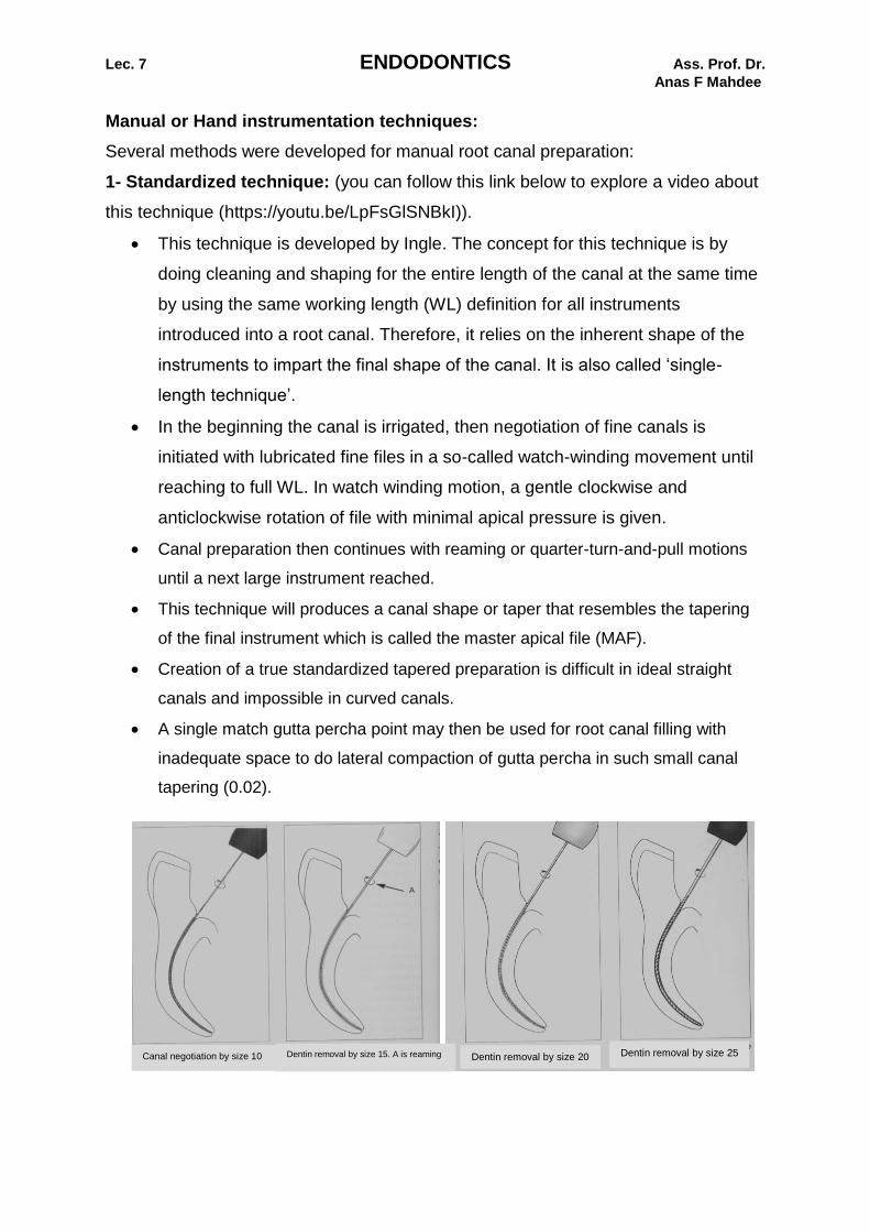
1- Standardized technique: (you can follow this link below to explore a video about
this technique (https://youtu.be/LpFsGlSNBkI)).
This technique is developed by Ingle. The concept for this technique is by
doing cleaning and shaping for the entire length of the canal at the same time
by using the same working length (WL) definition for all instruments
introduced into a root canal. Therefore, it relies on the inherent shape of the
instruments to impart the final shape of the canal. It is also called ‘single-
length technique’.
In the beginning the canal is irrigated, then negotiation of fine canals is
initiated with lubricated fine files in a so-called watch-winding movement until
reaching to full WL. In watch winding motion, a gentle clockwise and
anticlockwise rotation of file with minimal apical pressure is given.
Canal preparation then continues with reaming or quarter-turn-and-pull motions
until a next large instrument reached.
This technique will produces a canal shape or taper that resembles the tapering
of the final instrument which is called the master apical file (MAF).
Creation of a true standardized tapered preparation is difficult in ideal straight
canals and impossible in curved canals.
A single match gutta percha point may then be used for root canal filling with
inadequate space to do lateral compaction of gutta percha in such small canal
tapering (0.02).
Canal negotiation by size 10 Dentin removal by size 15. A is reaming Dentin removal by size 20 Dentin removal by size 25
2
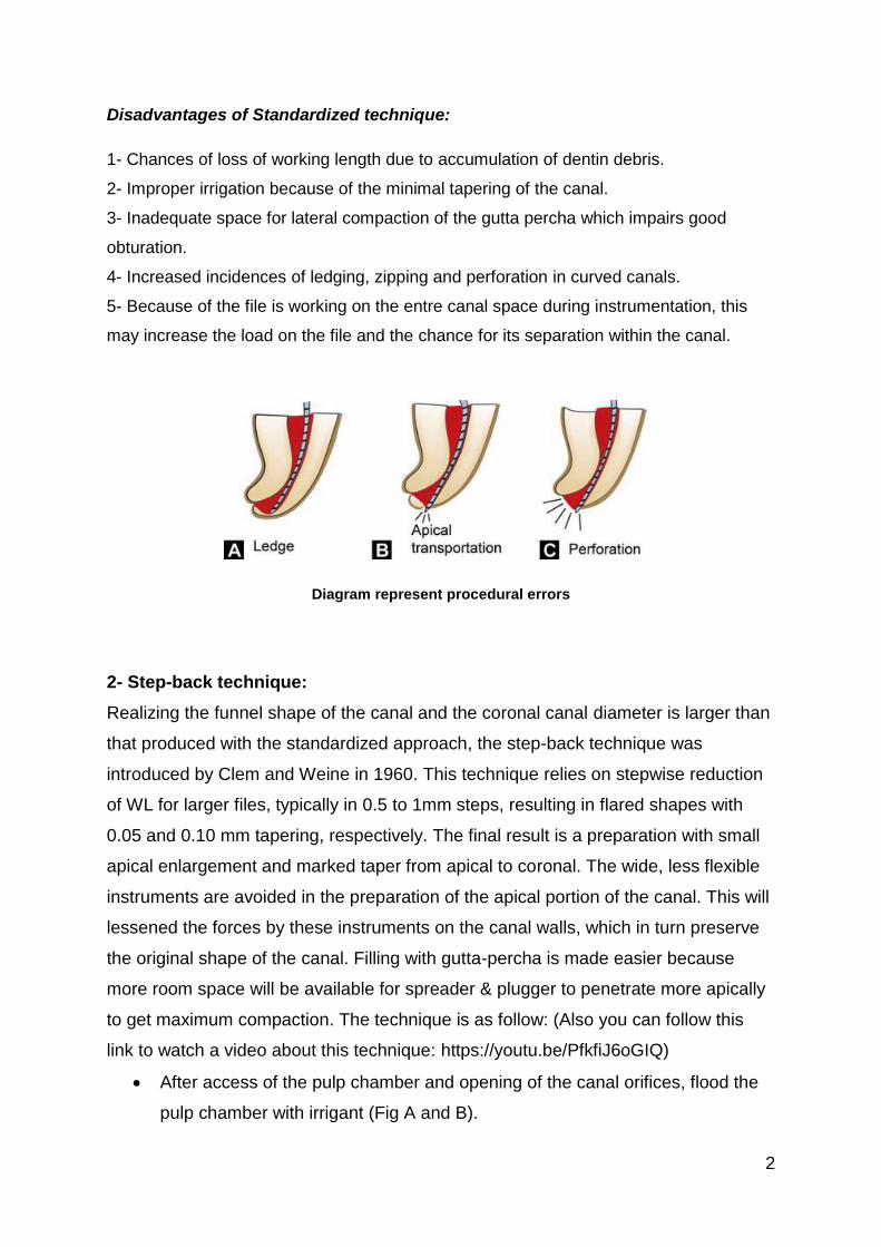
Disadvantages of Standardized technique:
1- Chances of loss of working length due to accumulation of dentin debris.
2- Improper irrigation because of the minimal tapering of the canal.
3- Inadequate space for lateral compaction of the gutta percha which impairs good
obturation.
4- Increased incidences of ledging, zipping and perforation in curved canals.
5- Because of the file is working on the entre canal space during instrumentation, this
may increase the load on the file and the chance for its separation within the canal.
Diagram represent procedural errors
2- Step-back technique:
Realizing the funnel shape of the canal and the coronal canal diameter is larger than
that produced with the standardized approach, the step-back technique was
introduced by Clem and Weine in 1960. This technique relies on stepwise reduction
of WL for larger files, typically in 0.5 to 1mm steps, resulting in flared shapes with
0.05 and 0.10 mm tapering, respectively. The final result is a preparation with small
apical enlargement and marked taper from apical to coronal. The wide, less flexible
instruments are avoided in the preparation of the apical portion of the canal. This will
lessened the forces by these instruments on the canal walls, which in turn preserve
the original shape of the canal. Filling with gutta-percha is made easier because
more room space will be available for spreader & plugger to penetrate more apically
to get maximum compaction. The technique is as follow: (Also you can follow this
link to watch a video about this technique: https://youtu.be/PfkfiJ6oGIQ)
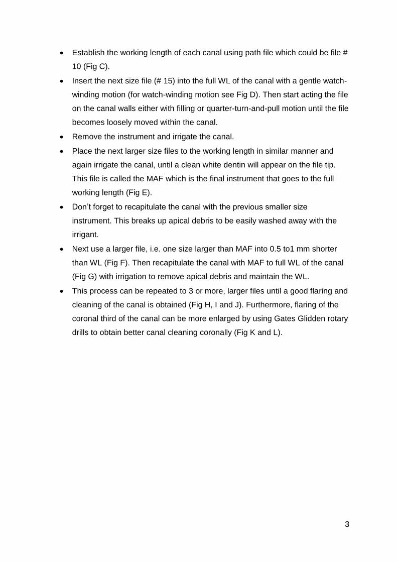
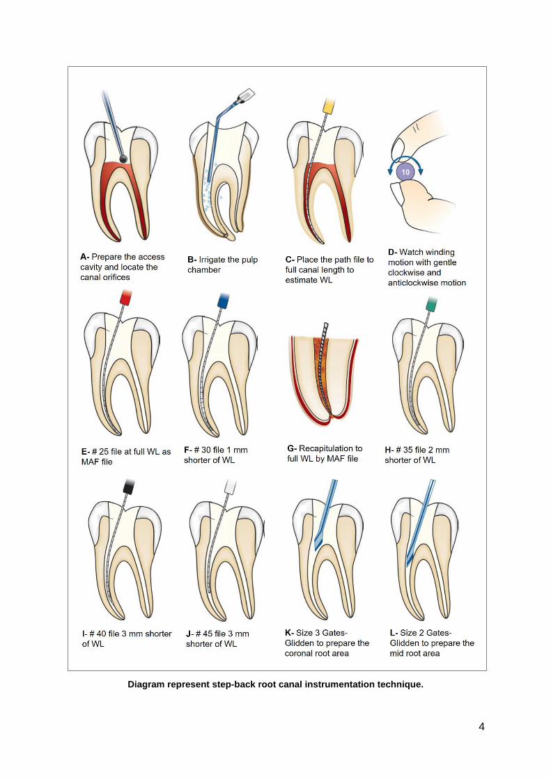
After access of the pulp chamber and opening of the canal orifices, flood the
pulp chamber with irrigant (Fig A and B).
3
Establish the working length of each canal using path file which could be file #
10 (Fig C).
Insert the next size file (# 15) into the full WL of the canal with a gentle watch-
winding motion (for watch-winding motion see Fig D). Then start acting the file
on the canal walls either with filling or quarter-turn-and-pull motion until the file
becomes loosely moved within the canal.
Remove the instrument and irrigate the canal.
Place the next larger size files to the working length in similar manner and
again irrigate the canal, until a clean white dentin will appear on the file tip.
This file is called the MAF which is the final instrument that goes to the full
working length (Fig E).
Don’t forget to recapitulate the canal with the previous smaller size
instrument. This breaks up apical debris to be easily washed away with the
irrigant.
Next use a larger file, i.e. one size larger than MAF into 0.5 to1 mm shorter
than WL (Fig F). Then recapitulate the canal with MAF to full WL of the canal
(Fig G) with irrigation to remove apical debris and maintain the WL.
This process can be repeated to 3 or more, larger files until a good flaring and
cleaning of the canal is obtained (Fig H, I and J). Furthermore, flaring of the
coronal third of the canal can be more enlarged by using Gates Glidden rotary
drills to obtain better canal cleaning coronally (Fig K and L).
4
Diagram represent step-back root canal instrumentation technique.
5
Advantages of step-back technique:
More flaring of the canal at the coronal part with proper apical stop.
Disadvantages of step-back technique:
1. Difficult to irrigate apical region.
2. Alteration of the WL after canal flaring.
3. More chances of pushing debris periapically.
4. Time consuming.
5. Increased chances of iatrogenic errors for example ledge formation in curved
canals.
6. Difficult to penetrate instruments in the canal.
7. More chances of instrument fracture.
Step-down technique:
This technique was developed to shape the coronal part (coronal pre-flaring) of the
canal before instrumentation of the apical part.
The objectives of this technique is
1- To permit straight access to the apical region of the canal by eliminating coronal
interference
2- To remove the bulk of necrotic tissue and microorganisms before apical shaping
to minimize extruded debris through the apical foramen during instrumentation.
3- To allow deeper penetration of irrigant deeply into the apical part of the canal. In
addition, it provide coronal escape way for debris extrusion from the apex.
4- The WL is less likely to change with less chance of zipping near the apical
constriction.
Procedure: (you can follow this link to watch a video about this technique:
https://youtu.be/uLAstzZeSc0)
Preparation of two coronal root canal thirds using Hedstrom files of size #15,
#20, and #25 to 16 to 18 mm or where they bind. These files are used with
circumferential filing motion on the canal walls.
Thereafter, increasing the coronal flaring of the canal by using Gates-Glidden
drills size 2, 3, and 4, in sequential order and 1mm shorter length between
each file.
6
Followed by canal WL estimation, then instrumentation of the remaining apical
part of the canal. This includes using small K-file # 15, 20 and 25 to prepare
the apical seat.
Combining the two parts, step-down and apical shape, by stepwise
decreasing of WL of incrementally larger files. Frequent recapitulation with a
#25 K-file to WL is advised to prevent blockage.
Disadvantages of step-down technique:
It is only time consuming technique.
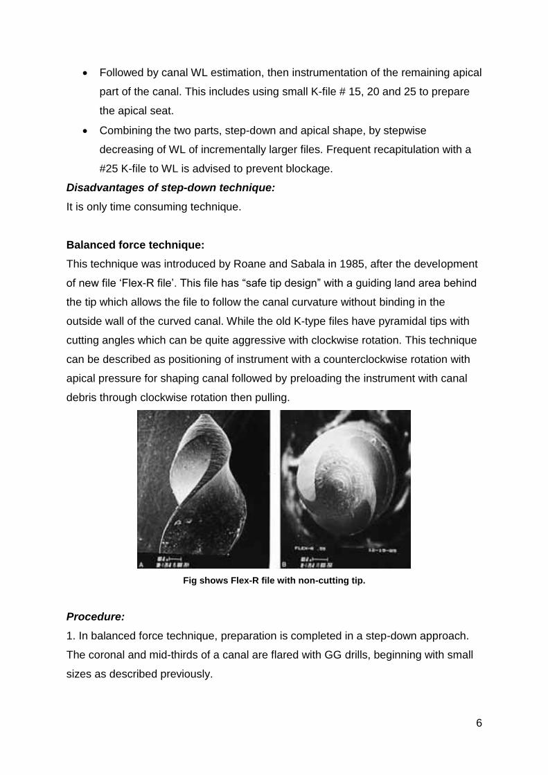
Balanced force technique:
This technique was introduced by Roane and Sabala in 1985, after the development
of new file ‘Flex-R file’. This file has “safe tip design” with a guiding land area behind
the tip which allows the file to follow the canal curvature without binding in the
outside wall of the curved canal. While the old K-type files have pyramidal tips with
cutting angles which can be quite aggressive with clockwise rotation. This technique
can be described as positioning of instrument with a counterclockwise rotation with
apical pressure for shaping canal followed by preloading the instrument with canal
debris through clockwise rotation then pulling.
Fig shows Flex-R file with non-cutting tip.
Procedure:
1. In balanced force technique, preparation is completed in a step-down approach.
The coronal and mid-thirds of a canal are flared with GG drills, beginning with small
sizes as described previously.
7
2- After that, the balanced force hand instrumentation begins in the apical
preparation by placing, cutting, and removing instrument using only rotation motion.
First file which binds short of working length is inserted into the canal and rotated
clockwise a quarter of a turn. This movement causes flutes to engage a small
amount of dentin.
3. Now file is rotated counterclockwise with apical pressure at least one third of a
revolution. It is the counterclockwise rotation with apical pressure which actually
provides the cutting action by shearing off small amount of dentin engaged during
clockwise rotation.
4. Then a final clockwise rotation is given to the instrument which loads the flutes of
file with loosened debris and the file is withdrawn.
5. Balanced Force instrumentation initiated from the belief that the apical area should
be shaped to sizes larger than were generally practiced. The original Balanced Force
concept then refers to apical control zones by, for example, first using sizes #15 and
#20 files to the periodontal ligament (i.e., through the apical foramen) and then
reducing the working depth by 0.5 mm for subsequent sizes #25, #30, and #35. The
apical shape is then completed 1 mm short using sizes #40 and #45 under
continuing irrigation with NaOCl.
Advantages of balanced force technique
Lesser chances of creating a ledge, blockage or canal transportation.
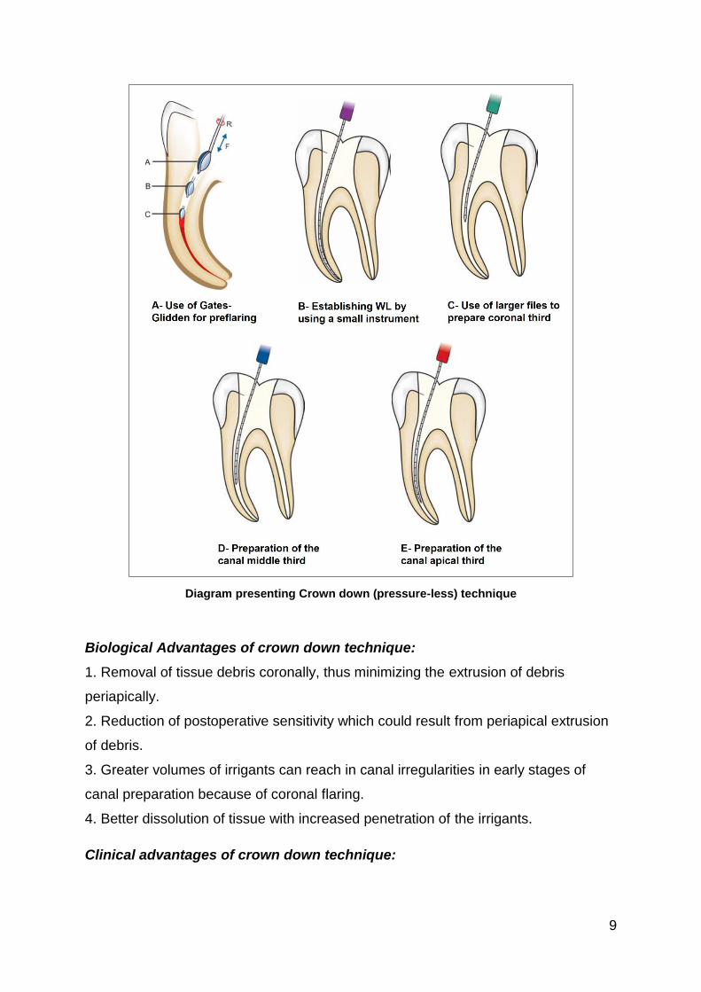
Crown-down (pressure-less) technique
The crown-down instrumentation concept based on the canal shaping technique
moving from the crown toward the apical portion of the canal. This concept was the
introductory for the most recent rotary instrumentation technology.
Procedure: (please follow this link to watch a video about the steps of this
technique: https://youtu.be/qfBYMA2_evQ)
1. After preparing the access opening and locating the canal, flood the pulp chamber
with irrigation solution and start pre-flaring of the canal orifices. This can be done by
using hand instruments, Gates-Glidden drills or the nickle-titanium rotary
instruments. After that a glide-path for each canal have to be obtained from the canal
orifice till the apical foramen by using # 10 or 15 file.
8
2. Coronal preparation of the canal can be started with Gates-Glidden drills. The
crown down approach begins with larger Gates-Glidden first (Fig A) (size 4 or 5),
followed by smaller diameter Gates-Gliddens are worked into the canal with
additional mm to complete coronal flaring. A care should be taken to avoid carrying
all the Gates-Glidden drills to same level which may lead to excessive cutting of the
dentin.
3. Frequent irrigation with sodium hypochlorite and recapitulation with a smaller file
(usually No. 10 file) to prevent canal blockage.
4. After establishing coronal and mid root enlargement, explore the canal and
establish the working length with small instruments (# 10 or 15 file) (Fig B).
5. Introduce larger files to coronal part of the canal and prepare it (Fig C and D).
Subsequently introduce progressively smaller number files deeper into the canal in
sequential order and prepare the apical part of the canal (Fig E).
6. Final apical preparation is prepared and finished along with frequent irrigation of
the canal system.
9
Diagram presenting Crown down (pressure-less) technique
Biological Advantages of crown down technique:
1. Removal of tissue debris coronally, thus minimizing the extrusion of debris
periapically.
2. Reduction of postoperative sensitivity which could result from periapical extrusion
of debris.
3. Greater volumes of irrigants can reach in canal irregularities in early stages of
canal preparation because of coronal flaring.
4. Better dissolution of tissue with increased penetration of the irrigants.
Clinical advantages of crown down technique:
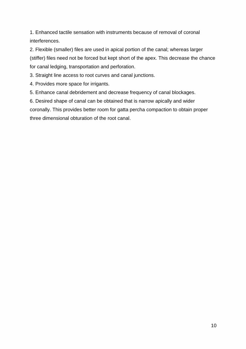
10
1. Enhanced tactile sensation with instruments because of removal of coronal
interferences.
2. Flexible (smaller) files are used in apical portion of the canal; whereas larger
(stiffer) files need not be forced but kept short of the apex. This decrease the chance
for canal ledging, transportation and perforation.
3. Straight line access to root curves and canal junctions.
4. Provides more space for irrigants.
5. Enhance canal debridement and decrease frequency of canal blockages.
6. Desired shape of canal can be obtained that is narrow apically and wider
coronally. This provides better room for gatta percha compaction to obtain proper
three dimensional obturation of the root canal.
11
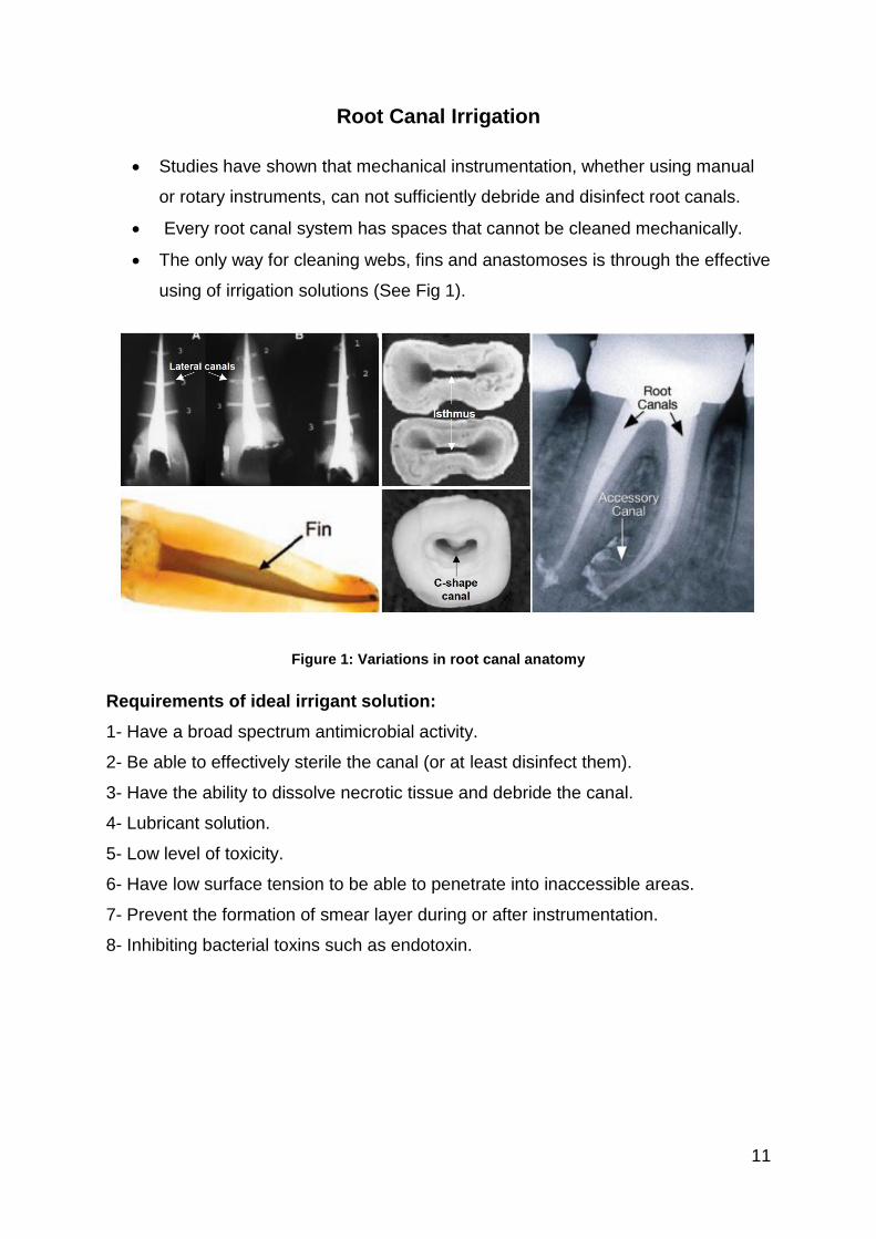
Root Canal Irrigation
Studies have shown that mechanical instrumentation, whether using manual
or rotary instruments, can not sufficiently debride and disinfect root canals.
Every root canal system has spaces that cannot be cleaned mechanically.
The only way for cleaning webs, fins and anastomoses is through the effective
using of irrigation solutions (See Fig 1).
Figure 1: Variations in root canal anatomy
Requirements of ideal irrigant solution:
1- Have a broad spectrum antimicrobial activity.
2- Be able to effectively sterile the canal (or at least disinfect them).
3- Have the ability to dissolve necrotic tissue and debride the canal.
4- Lubricant solution.
5- Low level of toxicity.
6- Have low surface tension to be able to penetrate into inaccessible areas.
7- Prevent the formation of smear layer during or after instrumentation.
8- Inhibiting bacterial toxins such as endotoxin.
12
Functions of irrigants
1- Removal of dentinal shavings by physical flushing to prevent their packing at the
apical region of the root canal.
2- Canal wetting material which effectively increase the efficacy of root canal
instruments. Instruments are less likely to break when the canal walls are lubricated
by irrigant.
3- Irrigants act as a solvent for necrotic tissue, so they loosen debris, pulp tissue and
microorganisms from irregular dentinal walls.
4- Irrigants facilitate the removal of debris from inaccessible regions of root canals.
5- Most irrigants have germicidal and antibacterial properties.
6- Irrigants also have bleaching action to lighten teeth discolored by necrotic pulp
tissue, caries or restorative material.
7- Irrigats facilitate the removal of smear layer and opening of the dentinal tubules.
Factors that modifying the activity of irrigating solution
There are several factors that can be controlled to increase the efficacy of irrigant
solutions:
1- Concentration: the dissolving capacity of some irrigation solution, such as sodium
hypochlorite, can be increased with higher concentration (5.2 rather than 2.5%).
However the cytotoxicity of higher concentrations is extremely higher.
2- Contact: the irrigant must contact the intracanal substrate (organic tissue, or
microbes) to be effective, otherwise it won’t be able to dissolve or flushout the debris.
Therefore, it is critical that the canal diameter should be mechanically enlarged to
facilitate the delivery of the irrigant solution up to the apical region of the prepared
canal.
3- Presence of organic tissue: the organic tissue must be removed mechanically or
chemomechanically to increase the efficacy of intracanal irrigation. This can be
obtained by simultaneous use of instruments and irrigating solutions.
4- Quantity and frequency of the irrigant used:
More irrigation causes better tissue debridement.
Each time a flush of fresh potent irrigant plays an action.
5- Gauge of irrigating needle: usually the 27 or 28 irrigation needle is preferable for
better penetration into the canal.
13
6- Surface tension of irrigation solution: the lower surface tension, the better
wettability and the more penetration into narrowest areas of the canals, and even
into the dentinal tubules.
7- Level of penetration of the irrigant: Maximum actions of irrigant occurs on coronal
part of root canal whereas minimal on apical end.
8- Age of irrigant: Freshly prepared solution is more effective than older one.
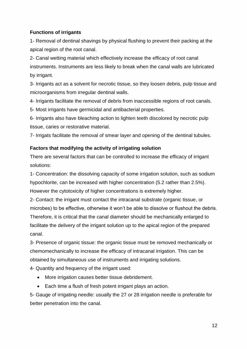
Irrigant solutions:
There are several irrigation solution that are currently used in root canal
chemomechanical debridement nowadays (See Figure 2). But none of these irrigats
fulfil all the required criteria. The main irrigants include sodium hypochlorite,
chlorhexidine and ethylene diamine tetraacetic acid. A combination of several
irrigants can be used to get maximum action.
Figure 2: A list of the currently used irrigant in root canal treatment.
Normal saline:
Normal saline as 0.9% W/V is commonly used irrigant in endodontics. It is very mild
in action and can be used in adjunct to chemical irrigant. It causes gross
debridement and lubrication of the root canal. Normal saline basically acts in flushing
action. It can also be used as a final rinse for root canals to remove the chemical
irrigant left after root canal preparation.
Advantages: it is a biocompatible solution with no adverse effect even if extruded
periapically, because its osmotic pressure is the same as that of the blood.
14
Disadvantages:
It has no dissolution, disinfectant and antimicrobial properties.
Too mild to thoroughly clean the canal.
Does not remove sear layer.
Sodium hypochlorite (NaOCl):
NaOCl encompasses many desirable properties of the main root canal irrigant and
has therefore been described as the most ideal irrigant solution. It can be used with
different concentrations (0.5 to 6%) but the recommended concentration in many
studies is 5.25%. Commercially available household bleach (Clorox) contains 6.15%
NaOCl.
NaOCl dissolve organic material such as pulp tissue, collagen, organic
material in smear layer and bacteria. With lower concentrations (0.5%) it
dissolve only necrotic tissue, however in higher concentrations dissolve both
necrotic and vital which is not always a desirable property.
NaOCl possess a broad-spectrum antimicrobial activity against endodontic
microorganisms and biofilms, including microbiota difficult to eradicate from
root canals, such as Enterococcus, Actinomyces, and Candida organisms.
This depends on its concentration and the contact time. With higher
concentration and longer contact time its antimicrobial action increase.
NaOCl minimally remove dentin debris or smear layer. Therefore, the use
dentin demineralizing agent (EDTA) is recommended post instrumentation to
eliminate smear layer and enhance cleaning of difficult-to-reach areas such
as dentinal tubules and lateral canals.
When using NaOCl over extended periods of time during treatment, it has an
undesired side effect by decreasing the flexural strength and modulus of
elasticity of dentin. Therefore it has to be flushed out by using normal saline
after the end of instrumentation visit.
NaOCl also has bleaching action by the function of the hypochlorite ions
which is important in whitening the discolouration caused by pulp necrosis or
endodontic and restorative material such as some endodontic sealers, and
amalgam restoration. However, NaOCl cause bleaching in contact with
clothes, so cautions have to be taken during its use.
15
Although NaOCl is nontoxic during intracanal use, it could cause serious
tissue damage if it injected periapically especially with higher concentration.
This is associated with severe pain, swelling and periapical bleeding.
Medication like antibiotics, analgesics, antihistamine should be prescribed
accordingly. In addition to these, reassurance to the patient is the prime
consideration. Thus irrigation with NaOCl should always be performed
passively especially in cases with larger apical diameters and needles with
very small diameter.
Advantages of NaOCl:
1- It has antibacterial and bleaching action.
2- It help in canal debridement by dissolution of the organic debris.
3- It cause lubrication of canals
4- Economical.
5- Easily available.
Disadvantages:
1- Because of high surface tension, its ability to wet dentin is less.
2- Irritant to tissues, if extruded periapically, it can result in severe cellular
damage.
3- If comes in contact, it cause inflammation of gingiva because of its caustic
nature.
4- It causes clothes bleaching in contact.
5- It has bad odor and taste
6- Vapours of NaOCl can irritate the eyes.
7- It has a corrosive effect to instruments.
References:
1- Garg N., Garg A. Textbook of Endodontics. Jaypee Brother Medical Publisher
(P) LTD. 2nd Edition, 2010.
2- Hargreaves K M., Cohen S., Berman L H., Cohen’s Pathways of the Pulp.
Mosby. 10th Edition, 2011.
3- Hülsmann M., Peters O A., Dummer P MH. Mechanical preparation of root
canals: shaping goals, techniques and means. Endodontic Topics. 2005; 10
(1): 30-76.
16
4- Ingle J I., Bakland L K., Baumgartner J C., Ingle’s ENdodontics 6. BC Decker
Inc Hamilton. 6th Edition, 2008.
5- Ruddle C J. Endodontic Access Preparation The Tool for Success. Just in
Time Online Education, Dental Products Report. 2007: 1-9.
6- Vertucci F J., Fla G., Root Canal Anatomy of the Human Permanent Teeth.
Oral surgery, oral medicine, oral pathology. 1984; 85 (5): 589-599.
7- West J., Endodontic Update 2006. Journal of Esthetic and Restorative
Dentistry. 2006; 18 (5): 280- 300.