Embed Size (px)
Citation preview

MANUAL ON THERAPEUTIC
HYPOTHERMIA FOR PERINATAL
ASPHYXIA
NATIONAL NEONATOLOGY FORUM
An NNF Publication
NATIONAL NEONATOLOGY FORUM OF INDIA803, 8th Floor, A-9, GDITL Tower, Netaji Subhash Place, Pitampura, Delhi-110034
Phone: 27353535, Mobile: 8527453535, E-mail: [email protected], Website: www.nnfi.org
NNF Office Bearers
Dr. B.D. BhatiaPresident, NNF
Dr. Ajay GambhirPast President, NNF
Dr. Alok BhandariHony. Secretary, NNF
Dr. Lalan Kr. BhartiJt. Secy.
cum Treasurer, NNF
Dr. Utpal Kant SinghMember
Dr. Mohit SahniMember
Dr. Anurag SinghMember
Dr. Himanshu KelkarMember
Dr. Vishnu BhatMember
NNF Governing Body Members
Dr. VISHNU BHAT. B Dr. ADHISIVAM. B
EDITORS:

OREWORD F
Cooling is standard of care in perinatal asphyxia for term and late
preterms in developed countries. In India therapeutic
hypothermia (TH) in perinatal asphyxia with HIE management has
started picking up and most of level 3 neonatal units either in
private or medical college set up have started practicing it with
good results. The National Neonatology Forum in its accreditation
programme has made it mandatory for level 3 B accreditation. It is
now appropriate time for NNF to bring out a manual on
“Therapeutic hypothermia for perinatal asphyxia” so that it can
help the new units to start cooling therapy and also provide
standardized care. The NNF at its annual convention every year
conducts workshops on Therapeutic hypothermia. Hence, it was
further necessary to have a manual on the same. The NNF is
thankful to Prof. B Vishnu Bhatt and Dr. Adhisivam B and all the
other contributors to have accepted the request and complete
the project in the record short time of couple of weeks. We are
®also thankful to the Miracradle team (Pluss Advanced
Technologies Pvt. Ltd.) for sponsoring the printing of this manual to
be distributed to all the delegates attending the 37th annual
convention of NNF-2017 at Gurgaon.
Dr. Alok Bhandari Secretary NNF
Dr. B. D. BhatiaPresident NNF
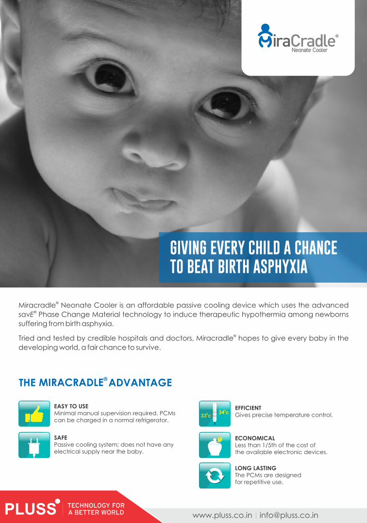
GIVING EVERY CHILD A CHANCE
TO BEAT BIRTH ASPHYXIA
www.pluss.co.in [email protected]
SAFE Passive cooling system; does not have any electrical supply near the baby.
EASY TO USE Minimal manual supervision required. PCMs can be charged in a normal refrigerator.
033 c034 c
ECONOMICALLess than 1/5th of the cost of the available electronic devices.
EFFICIENTGives precise temperature control.
LONG LASTINGThe PCMs are designed for repetitive use.
® MIRACRADLETHE ADVANTAGE
®Miracradle Neonate Cooler is an affordable passive cooling device which uses the advanced
savE Phase Change Material technology to induce therapeutic hypothermia among newborns
suffering from birth asphyxia.
®Tried and tested by credible hospitals and doctors, Miracradle hopes to give every baby in the
developing world, a fair chance to survive.
®

REFACEP
There is ample evidence for the benefit of therapeutic hypothermia
for term and late preterm neonates with perinatal asphyxia. Though
it is the standard of care practiced in most of the developed
countries, it is yet to gain momentum in India. We are happy that
National Neonatology Forum (NNF), India is bringing out a manual
on therapeutic hypothermia for perinatal asphyxia. The chapters in
this manual have been contributed by clinicians with expertise in
the field. Both theory and practical aspects of therapeutic
hypothermia with special relevance to the Indian context have
been covered. We are sure that this manual will be of great help to
Neonatology fel lows and Pediatr ic postgraduates in
understanding the principles of therapeutic hypothermia and the
nuances of practical application of this intervention.
DR. B. VISHNU BHATDean (Research)
Professor & HOD
Department of Neonatology,
JIPMER Pondicherry
Dr. B. Vishnu Bhat

CONTRIBUTING AUTHORS
S. No. AUTHORS
1 Dr. VISHNU BHAT B
Senior Professor and Head, Dept. of Neonatology, JIPMER, Pondicherry
2 Dr. ADHISIVAM B
Additional Professor, Dept. of Neonatology, JIPMER, Pondicherry
3 Dr. NIVEDITA M
Associate Professor, Dept. of Neonatology, JIPMER, Pondicherry
4 Dr. NISHAD P
Assistant Professor, Dept. of Neonatology, JIPMER, Pondicherry
5 Dr. SRIDHAR SANTHANAM
Professor and Head, Dept. of Neonatology, CMC, Vellore
6 Dr. NIRANJAN THOMAS
Professor, Dept. of Neonatology, CMC, Vellore
7 Dr. MANISH KUMAR
Associate Professor, Dept. of Neonatology, CMC, Vellore
8 Dr. SUMITHA ARUN
Assistant Professor, Dept. of Neonatology,CMC, Vellore
9 Dr. KUMUTHA J
Professor and Head, Dept. of Neonatology, Saveetha Medical College, Chennai
10 Dr. PRAKASH AMBOIRAM
Professor and Head, Dept. of Neonatology, SRMC, Chennai
11 Dr. UMAMAHESWARI B
Associate Professor, Dept. of Neonatology, SRMC, Chennai
12 Dr. MANIGANDAN CHANDRASEKARAN
Consultant Neonatologist, Cloudnine Hospital, Chennai
13 Dr. MINTOO TERGESTINA
Senior Resident, Dept. of Neonatology, CMC, Vellore
14 Dr. NITHYA J PONMUDI
Senior Resident, Dept. of Neonatology, CMC, Vellore
15 Dr. VASANTHAN T
Neonatology Fellow, JIPMER, Pondicherry

TABLE OF CONTENTS
1. Hypoxic Ischemic Encephalopathy - Pathophysiology and clinical features
2. Therapeutic hypothermia - Mechanisms of action
3. Therapeutic hypothermia for neonatal encephalopathy - Evidence
4. Asphyxia and therapeutic hypothermia in India - An overview
5. Cooling devices for therapeutic hypothermia in newborn babies
6. Procedure for whole body cooling
7. Monitoring of neonates during therapeutic hypothermia
8. Prevention and management of complications of therapeutic hypothermia
9. Adjuvants with therapeutic hypothermia
10. Amplitude integrated electroencephalography in HIE
11. Cranial ultrasonography in perinatal asphyxia
12. Magnetic Resonance Imaging in HIE
13. Follow up of cooled babies
1
11
13
22
29
37
39
43
46
53
63
69
79

HYPOXIC ISCHEMIC ENCEPHALOPATHYPATHOPHYSIOLOGY & CLINICAL FEATURES
CHAPTER
1
Every year,
around 4 million
babies die in the
neonatal period
(first 28 days of
life) globally and
asphyxia is one of
the major causes
accounting for
23% of these
neonatal deaths.
INTRODUCTION
The WHO definition of perinatal asphyxia is “failure to initiate and
sustain breathing at birth”. Perinatal asphyxia leads to multi-organ
dysfunction in the neonate and the neurological dysfunction inherent
to this clinical condition is referred as Hypoxic Ischemic
Encephalopathy (HIE). HIE is characterized by clinical and laboratory
evidence of acute or sub-acute brain injury secondary to asphyxia.
The primary causes of HIE are systemic hypoxemia and/or reduced
cerebral blood flow. Every year, around 4 million babies die in the
neonatal period (first 28 days of life) globally and asphyxia is one of the
major causes accounting for 23% of these neonatal deaths. Among
the survivors of asphyxia, cerebral palsy is a dreaded complication
associated with loss of potential productive member for the society
and direct burden lasting for the entire life on the individual and family
and social institutions. Hence every effort should be made to prevent
perinatal asphyxia and pediatricians should be well versed with the
management of this important clinical condition.
EPIDEMIOLOGY
It is a sad fact that most of the neonatal deaths due to asphyxia (99%)
occur in developing countries. HIE occurs in 1.5 per1000 full term births.
While 15 - 20% of neonates with HIE die early, 25% will survive with
disabilities. Moreover HIE is a major problem at all levels - individual,
family and society, contributing to 15-28% of children with cerebral
palsy and 25% of all children with developmental delay. Despite
significant advances in perinatal care, cerebral palsy among term
infants continues to occur. The long term neuro-developmental
outcome depends on the severity of the neonatal encephalopathy.
Dr. Adhisivam B

pg 2
According to NNPD 2003 data, Apgar score <7 at 1 minute (including moderate and severe
asphyxia) was noted in 9% of all intramural deliveries while 2.5% babies continued to have
Apgar scores <7 at 5 minutes of age. Bag and mask ventilation was required in 4.5% infants
while less than 1% infants required cardiac compressions and/ or medications as part of
resuscitation at birth. Clinical features of HIE were noted in 1.5% of all babies and it
accounted for 20% of neonatal deaths.
ETIOLOGY
It is practically impossible to ascertain the exact time of the hypoxic insult sustained by a
newborn unless some convincing evidence is available. Some of the common etiology for
HIE are listed in Table 1. The clinical presentation of neonatal encephalopathy due to varied
etiologies can overlap and hence it is difficult for the clinician to identify the etiology using
neurological examination alone. It is always prudent to exclude other causes of neonatal
encephalopathy when the perinatal risk factors are not clear.
PATHOPHYSIOLOGY
HIE does not refer to a single event but rather refers to a continuing process beginning from
the time of hypoxic insult. There are two different episodes of neuronal impairment which
are known to occur during HIE. The immediate hypoxic insult is called the primary phase
and this is followed by a short period of recovery (latent phase) lasting for approximately six
hours. Subsequently there is a longer phase during which there is a release of chemical
mediators causing secondary neuronal damage. Neurons may die during the actual
ischemic or primary phase itself. Several neurons however recover at least partially in the
'latent' phase but die hours or even days later (secondary or delayed cell death) (Fig.1).
Based on the cerebral energy state, HIE involves two phases of energy failure - primary and
secondary. The blood flow and oxygen substrates in the brain are decreased in primary
energy failure. Reduction in ATP and other phosphorylated compounds like
phosphocreatine and significant tissue acidosis are important features during this phase.
An “excito toxic - oxidative cascade” characterized by excessive stimulation of
neurotransmitter receptors and membrane depolarization causing increased intracellular
calcium is also observed. Increased levels of intracellular calcium activate nitric oxide
synthase to perpetuate the release of nitric oxide, which in turn affects mitochondrial
respiration (Fig.2). Signals from damaged mitochondria lead to apoptosis or programmed
cell death till energy stores are available. However cessation of these energy supply results
in cell necrosis. Activation of caspase enzyme system can also trigger apoptosis. Resolution
of hypoxia can reverse the decrease in ATP and intracellular pH and enhances recycling of

pg 3
neurotransmitters. In case of prolonged and severe insult, the initial cascade of events will
cause a second phase of energy failure in the mitochondria. This secondary energy failure
differs from the primary in that the decline in the levels of ATP and other phosphorylated
compounds are not associated with brain acidosis. The secondary energy failure is
characterized by continuing excite toxic-oxidation cascade, apoptosis, inflammation and
altered growth factor levels and protein synthesis (Table 2). The interval between the
primary and secondary energy failure is called the latent phase, an important therapeutic
window (approximately 6 hours). When neuronal tissue sustains a hypoxic insult, cell death
may be either delayed or remain progressive depending on the region and severity of the
injury. Gestational age has a role in the susceptibility of the brain to hypoxic damage. In term
neonates, the gray matter is primarily affected (selective neuronal necrosis) while in the
preterm it is the white matter leading to periventricular leucomalacia. The other factors
which contribute to the degree of damage include cellular susceptibility, watershed areas,
regional metabolic rates and degree of asphyxia.
OXIDATIVE STRESS AND DNA DAMAGE IN ASPHYXIA
There are three important pathways that lead to free radical production. First one is Fenton
reaction. During hypoxic ischemia, protein-bound iron is liberated from its binding proteins in
the neuronal and microglial cells. Non protein bound iron (NPBI) or free iron usually
accumulates during hypoxic ischemia. When the damaged brain is reperfused and re-
oxygenated, toxic hydroxyl free radical will be formed as NPBI react with hydrogen
peroxide. Thus NPBI is related to excessive neuronal damage immediately following the
insult. Second, the activation of neuronal and inducible nitric oxide synthase leads to the
generation of the nitric oxide radical (NO), which reacts with superoxide to form the toxic
peroxynitrite (ONOO). Peroxynitrite and reactive oxygen species cause DNA damage and
cell death. Finally, hypoxanthine, accumulated during the hypoxic-ischemic episode as a
degradation product of ATP, is metabolized to uric acid by xanthine oxidase (XO). This
reaction gives rise to further formation of superoxide radicals (Fig.3). The toxicity of these free
radicals contribute substantially to reperfusion injury of the neuronal tissue after the hypoxic
insult. Neonatal brain is more susceptible to oxidative stress because of low concentrations
of antioxidants, a high consumption of oxygen and presence of high concentrations of
unsaturated fatty acids that break down to form more oxygen free radicals.
CLINICAL FEATURES
The important CNS clinical features of HIE include altered level of sensorium, seizures and
tone abnormalities. Their appearance is variable depending on the progression of energy
failure and severity of insult and the usual time frame of these clinical features is depicted in
Fig. 4. For quantifying the severity of HIE, Sarnat and Sarnat classification is commonly used
while Levene's classification is also helpful (Tables 3 and 4). Though the CNS features are sine
qua non of HIE all other organ systems including the kidney and heart are also affected in
variable proportions depending on the disease severity and quality of care (Table 5). When
pg 4
the neonate sustains a hypoxic insult, almost all organs are at risk of cell injury and death.
However, certain organs are more vulnerable to injury than others despite inbuilt
physiological reflexes trying to protect these vital organs.
Acute kidney injury (AKI) is a common occurrence in infants with neonatal encephalopathy
(NE), with a reported incidence of 50–72 %. Most of these studies are small and employ
varying definitions for AKI based on elevation of serum creatinine, oliguria, decreased
glomerular filtration rate and presence of haematuria and proteinuria. Nevertheless, these
studies emphasize the frequency of AKI complicating NE and underline the uncertainty
surrounding the precise and early diagnosis of AKI in critically ill neonates. The majority of AKI
following perinatal hypoxia-ischemia is prerenal in origin and often non oliguric.
Cardiac dysfunction is part of the clinical spectrum of multi organ dysfunction in term infants
with HIE. In children with HIE, the reported incidence of cardiovascular dysfunction ranges
from 29 - 78%. Oxygen deprivation secondary to a hypoxic and /or ischaemic insult causes
myocardial damage. This eventually leads to decreased cardiac output, impaired
myocardial contractility, systemic hypotension and pulmonary hypertension.
DIFFERENTIAL DIAGNOSES
History of prolonged and difficult labor coupled with need for significant resuscitation, low
apgar scores, altered sensorium and early onset seizures will usually point towards HIE.
However, other differential diagnoses likes inborn errors of metabolism, neuromuscular
disorders, developmental defects of brain and sepsis should be kept in mind as their clinical
features my overlap with HIE. It is not uncommon to find meconium aspiration syndrome and
sepsis associated with HIE in term and preterm babies respectively.
APPROACH TO DIAGNOSIS
A detailed history including antenatal and delivery details reflecting events leading to
compromised blood supply and/or oxygenation of the fetus should be obtained. History of
placental abruption, cord around neck, cord prolapse, maternal hemorrhage, trauma,
cardiorespiratory arrest, uterine rupture or significant fetal decelerations if present should
be recorded. There is increased risk of neonatal encephalopathy if the mother has fever
during antepartum or intrapartum period. A careful neurologic examination needs to be
performed to diagnose encephalopathy.

pg 5
BIBLIOGRAPHY
1. Shankaran S. Hypoxic-ischemic encephalopathy and novel strategies for neuroprotection. Clin
Perinatol. 2012; 39:919-29.
2. Wachtel EV, Hendricks-Muñoz KD. Current management of the infant who presents with
neonatal encephalopathy. CurrProbl Pediatr Adolesc Health Care. 2011; 41:132-53.
3. Bhat BV, Adhisivam B. Therapeutic cooling for perinatal asphyxia-Indian experience. Indian
J Pediatr. 2014; 81:585-91
4. Bharadwaj SK, Bhat BV. Therapeutic hypothermia using gel packs for term neonates with
hypoxic ischaemic encephalopathy in resource-limited settings: A randomized controlled trial.
J Trop Pediatr. 2012; 58:382-8.
5. Perrone S, Stazzoni G, Tataranno ML, Buonocore G. New pharmacologic and therapeutic
approaches for hypoxic-ischemic encephalopathy in the newborn. J Matern Fetal Neonatal
Med. 2012; 25Suppl 1:83-8.
6. Galvao TF, Silva MT, Marques MC, de Oliveira ND, Pereira MG. Hypothermia for perinatal brain
hypoxia-ischemia in different resource settings: a systematic review. J Trop Pediatr. 2013;
59:453-9.
7. Thayyil S, Shankaran S, Wade A, Cowan FM, Ayer M, Satheesan K, et al. Whole-body cooling in
neonatal encephalopathy using phase changing material. Arch Dis Child Fetal Neonatal Ed.
2013; 98(3):F280-1
8. Joy R, Pournami F, Bethou A, Bhat VB, Bobby Z. Effect of therapeutic hypothermia on oxidative
stress and outcome interm neonates with perinatal asphyxia: a randomized controlled trial.
J Trop Pediatr 2013; 59:17–22
9. Aggarwal R, Deorari AK, Paul VK. Post-resuscitation management of asphyxiated neonates.
Indian J Pediatr. 2001; 68:1149-53
10. AIIMS protocol 2014 - Post-resuscitation management of asphyxiated neonate. Available from:
URL: http://www.newbornwhocc.org/clinical_proto.html. Accessed May6, 2014.
11. Pauliah SS, Shankaran S, Wade A, Cady EB, Thayyil S. Therapeutic Hypothermia for Neonatal
Encephalopathy in Low and Middle Income Countries: A Systematic Review and Meta-
Analysis. PLoS ONE 2013; 8: e58834.
12. Gane BD, Bhat V, Rao R, Nandhakumar S, Harichandrakumar KT, Adhisivam B. Effect of
therapeutic hypothermia on DNA damage and neuro-developmental outcome among term
neonates with perinatal asphyxia: A randomized controlled trial. J Trop Pediatr. 2014 ; 60(2):
134-40.
13. Thomas N, Chakrapani Y, Rebekah G, Kareti K, Devasahayam S. Phase changing material: An
alternative method for cooling babies with hypoxic ischaemic encephalopathy. Neonatology.
2015; 107(4):266-70.
14. Tanigasalam V, Bhat V, Adhisivam B, Sridhar MG. Does therapeutic hypothermia reduce acute
kidney injury among term neonates with perinatal asphyxia? – a randomized controlled trial.
J MaternFetal Neonatal Med. 2016; 29(15):2544-7

pg 6
Hypoxia or Anoxia: A partial (hypoxia) or complete (anoxia) lack of oxygen
in the brain or blood.
Asphyxia: The state in which placental or pulmonary gas exchange is
compromised or ceases altogether.
Ischemia: The reduction or cessation of blood flow to an organ which
compromises both oxygen and substrate delivery to the tissue.
Hypoxic-Ischemic Encephalopathy: Abnormal neurologic behavior in the
neonatal period arising as a result of a hypoxic-ischemic event.
Neonatal Encephalopathy: A clinical syndrome of “disturbed neurological
function in the earliest days of life in the term infant, manifested by difficulty
with initiating and maintaining respiration, depression of tone and reflexes,
subnormal level of consciousness, and often seizures.”
Definition of Perinatal Asphyxia (PA)
— World Health Organization: Failure to initiate and sustain breathing.
— NNPD Network:
o Moderate PA: Slow/gasping breathing or an Apgar score of 4 to 6
at 1 minute.
o Severe PA: No breathing or an Apgar score of 0-3 at 1 minute of age.
— American Academy of Pediatrics and American College of Obstetrics
and Gynecology: Presence of all of following criteria-
o Profound metabolic or mixed acidemia (pH< 7.00) in umbilical
cord blood.
o Persistence of low Apgar scores less than 3 for more than 5 minutes
o Signs of neonatal neurologic dysfunction (e.g., seizures,
encephalopathy, tone abnormalities).
o Evidence of multiple organ involvement (such as that of kidneys,
lungs, liver, heart and intestine).
BOX 1: DEFINITIONS

pg 7
Fig. 2: Pathophysiology of HIE - Key Events
Fig.1: Pathophysiology of HIE - Overview
Hypoxicdepolarization
Cell lysisExcitotoxins
Calcium entry
Primary energy failure
Latent phase(6-15 hours)
Secondaryenergy failure(6-48 hours)
Oxidativemetabolism
recoveryApoptosis
Inflammation
Reducedmitochondrial
activitySeizuresEdema
Cell death
REPERFUSION
RECOVERY BRAINDAMAGE
BRAIN DAMAGE
Chemial toxins(metals, CCI ,4
quinones, peroxides)
Impaired2+Ca transport
Perturbationof cytoskeletalorganization
Phospholipaseactivation
Glucocorticoids TCDD
Receptor activation
Protein synthesis
2+Ca influx
Impairedmitochondrial
function
Endonucleaseactivation
Proteaseactivation
2+Cytosolic Ca increase

pg 8
Fig. 3: Free radicals production in HIE
Birth -12 hours Depressed level of alertness, periodic breathing or
respiratory failure, intact pupillary and occulomotor
responses, hypotonia, seizures
12 - 24 hours Change in level of alertness, seizures, apnoeic spells,
jitteriness, weakness in proximal limbs, upper > lower
(term), lower > upper (preterm)
24 -72 hours Stupor or coma, respiratory arrest, brain stem pupillary
and occulomotor disturbances, catastrophic
deteoriation with severe intraventricular hemorrhage
and periventricular hemorrhagic infarction (premature)
> 72 hours Persistent yet diminishing stupor, disturbed sucking,
swallowing, gag and tongue movements, hypotonia /
hypertonia, limb weakness
Fig.4: HIE - Clinical Features
ANAEROBIC GLYCOLYSIS
HYPOXIA-ISCHEMIA
Decreased ATP
INCREASED GLUTAMATE
NMDA RECEPTOR ACTIVATED
INCREASED INTRACELLULAR CALCIUM
ACTIVATES LIPASES AND LIPID PEROXIDATION
FREE RADICALS
INCREASEDLACTATE
ACTIVATES NO SYNTHASE
NITRIC OXIDE
FREE RADICALS
XANTHINE
HYPOXANTHINE
ADENOSINE
FREE RADICALS

pg 9
Maternal Uteroplacental Fetal
Cardiac arrest Placental abruption Feto maternal hemorrhage
Asphyxiation Cordprolapse Twin to twin transfusion
Severe anaphylaxis Uterine rupture Severe iso immune hemolytic disease
Status epilepticus Hyper stimulation with Cardiac arrhythmia
Hypovolemic shock oxytocic agents
Table 1: Etiology of HIE
Primary energy failure
Decrease in cerebral blood flow, oxygen
substrates and ATP
Excito toxic-oxidative cascade
Loss of ionic homeostasis across
membranes
Entry of intracellular calcium
Mitochondrial disruption
Brain acidosis
Table 2: Characteristics of energy failures related to HIE
Secondary energy failure
Continuing of excite toxic-oxidative
cascade
Activation of microglia-inflammatory
response
Activation of caspase proteins
Reduction in levels of growth factors,
protein synthesis
Continuing Apoptosis and necrosis
Table 3: Sarnat and Sarnat classification of HIE
STAGE 1
Level of consciousness Neuromuscular control
Muscle tone
Posture
Stretch reflexes
Segmental myoclonus
Complex reflexes
Suck
Moro
Oculovestibular
Tonic neck
Autonomic fuction
Hyperalert
Normal
Mild distal flexion
Overactive
Present
Weak
Strong: low threshold
Normal
Slight
Generalized sympathetic
STAGE 2
Lethargic or obtunded
Mild hypotonia
Strong distal flexion
Overactive
Present
Weak or absent
Weak; incomplete; high threshold
Overactive
Strong
Generalized parasympathetic
Stuporous
Flaccid
Intermittent decerebration
Decreased or absent
Absent
Absent
Absent
Weak or absent
Absent
Both systems decreased
STAGE 3
Continued on next page...

pg 10
Table 4: Levene's classification of HIE
Feature Mild Moderate Severe
Consciousness Irritable Lethargy Comatose
Tone Hypotonia Marked hypotonia Severe hypotonia
Seizures No Yes Prolonged
Sucking/respiration Poor suck Unable to suck Unable to sustain
spontaneous respiration
CNS Hypoxic ischemic encephalopathy, intracranial hemorrhage,
seizures, long-term neurological sequelae
Cardiac Myocardial dysfunction, valvular dysfunction, rhythm
abnormalities, congestive cardiac failure
Renal Hematuria, acute tubular necrosis, renal vein thrombosis
Pulmonary Delayed adaptation, respiratory failure, meconium aspiration,
surfactant depletion, primary pulmonary hypertension
GI tract Necrotizing enterocolitis, hepatic dysfunction
Hematological Thrombocytopenia, coagulation abnormalities
Metabolic Acidosis, hypoglycemia, hypocalcemia, hyponatremia
Table 5: Organ system dysfunction in perinatal asphyxia
Autonomic fuction
Pupils
Generalized sympathetic
Mydriasis
Generalized parasympathetic
Miosis
Both systems decreased
Variable: often unequal; poor light reflex
Heart rate Tachycardia Bradycardia Variable
Bronchial and salivary secretions
Gastrointestinal motility
Seizures
Electroencephalogram findings
Spars
Normal or decreased
None
Normal (awake)
Profuse
Increased: diarrhea
Common: focal or multifocal
Early low-voltage continuous delta and theta
Later, periodic pattern (awake),Seizures: focal 1- to 1.5-Hz spike-and-wave
2-14 days
Variable
Variable
Uncommon (excluding decerebration)
Early: periodic pattern with isopotential phases.
Later: totally isopotential
Hours to weekLess than 24 hoursDuration
THERAPEUTIC HYPOTHERMIA
MECHANISMS OF ACTION
Therapeutic Hypothermia (TH) has been proven to be effective in
reducing morbidity associated with HIE and has become the
standard of care for HIE in developed countries. However in
underdeveloped and transitional countries where the problem is
more common, therapeutic cooling is still in the nascent phase. There
are several reasons for this in resource restricted settings. Similarly,
despite availability of compelling clinical evidence that TH initiated
within a few hours after hypoxic insult can improve neurological
outcome in term infants, implementing the same in resource restricted
settings of India is not that easy mainly because of the non-availability
of expensive devices used for providing TH in developed countries.
Recently, two systematic reviews on the efficacy and safety of TH in
low and middle income resource settings have been published. The
systematic review by Pauliah et al did not find any statistically
significant reduction in neonatal mortality in underdeveloped
countries although the confidence intervals were wide. Galvao et al
observed that there is ample evidence for designating hypothermia
as the standard of care for HIE but more evidence from low income
countries is required.
TH is neuroprotective by inhibiting several steps in the excito toxic
oxidative cascade which include inhibiting the increase in the
concentration of lactic acid, glutamate and nitric oxide in the brain
(Fig. 1 and Box 1). Moreover, TH inhibits protease activation,
mitochondrial failure, free radical damage, lipid peroxidation and
inflammation. TH has been shown to decrease brain energy use,
prolong the latent phase, reduce infarct size, decrease neuronal cell
loss, retain sensory motor function, and preserve hippocampal
structures. Early application of TH preferably within 6 hours i.e. before
the onset of the secondary phase of energy failure is likely to be
effective and improve neurodevelopmental outcome. Usually it is
CHAPTER
2
Therapeutic
Hypothermia (TH)
has been proven
to be effective in
reducing
morbidity
associated with
HIE and has
become the
standard of care
for HIE in
developed
countries.
Dr. Vishnu Bhat B

continued for a period of 72 hours for better neuro protection. Applying TH immediately or
within a few hours after reperfusion and continued for 72 hours has been shown to favorably
affect outcome in newborn and adult animals.
pg 12
Fig.1: Pathophysiology of HIE - Key Events
Ÿ
Ÿ Decrease in energy utilization
Ÿ Reduction of cytotoxic amino acid accumulation and nitric oxide
Ÿ Inhibition of platelet-activating factor and inflammatory cascade
Ÿ Suppression of free radical activity
Ÿ Attenuation of secondary energy failure
Ÿ Inhibition of apoptosis (cell death)
Ÿ Reduction of the extent of brain injury
Reduction of cerebral metabolism and prevention of edema
Box 1: Summary – Mechanisms of action of Therapeutic Hypothermia
HYPOXIA-ISCHEMIA
ANEROBIC GLYCOLYSIS
Decreased ATP
INCREASEDLACTATEINCREASED GLUTAMATE
NMDA RECEPTOR ACTIVATED
INCREASED INTRACELLULAR CALCIUM
ACTIVATES LIPASES AND LIPIDPERROXIDATION
FREE RADICALS
ACTIVATES NO SYNTHASE
NITRIC OXIDE
FREE RADICALS
ADENOSINE
HYPOXANTHINE
XANTHINE
FREE RADICALS
Therapeutic Hypothermia

THERAPEUTIC HYPOTHERMIA FOR NEONATAL ENCEPHALOPATHY
EVIDENCE
CHAPTER
3
Cooling appears to
reduce DNA
damage induced by
oxidative stress and
improve
neurodevelopmental
outcome.
Dr. Nishad P
SUMMARY OF EVIDENCE OF BENEFIT
There is now unequivocal and high-quality evidence from multiple
large RCTs that neonates with moderate to severe hypoxic-ischemic
encephalopathy (HIE) following a perinatal event benefit from
hypothermia under tightly controlled conditions. The trials used either 1-5head cooling devices (with mild systemic hypothermia), or whole-
6-12body cooling alone. A recent Cochrane review included 11 RCTs,
comprising a total of 1,505 infants, and concluded that TH resulted in a
r e d u c t i o n i n t h e c o m b i n e d o u t c o m e o f d e a t h o r 13neurodevelopmental disability (to 18 months of age).
Cooling resulted in an absolute risk reduction (risk difference, RD) of
15% in the composite outcome. Put another way, the number needed
to treat for one additional beneficial outcome (NNTB) was 7 (95% CI: 5
to 10). The RD for mortality with cooling was -9% (95% CI: -13% to -4%);
NNTB 11 (95% CI: 8 to 25) and for neurodevelopmental disability in
survivors was -13% (95% CI: -19% to -7%); NNTB 8 (95% CI: 5 to 14). The
most influential RCTs are listed in Table 1.
Cooling appears to reduce DNA damage induced by oxidative stress 14and improve neurodevelopmental outcome. There is also evidence
that therapeutic hypothermia limits myocardial and renal injury in 15, 16term infants with HIE.
METHOD OF COOLING
A metaanalysis of the trials that used selective head cooling did not
show a statistically significant difference in mortality (RR 0.78, 95% CI
0.59 to 1.04) or major neurodevelopmental disability (all participants,

pg 14
13RR 0.72, 95% CI 0.50 to 1.05). But it is worth noting that the trend toward benefit was strong
and consistent, the magnitude of benefit was comparable to the trials that used whole
body cooling, and the intervention in the head-cooling trials also included mild systemic
hypothermia. In addition, there was a statistically significant reduction in the hypothermia
groups when neurodevelopmental disability was assessed in survivors as opposed to all
participants (RR 0.66, 95% CI 0.47 to 0.94).
Metaanalysis of the trials that used whole body cooling showed a significant reduction in
mortality (RR 0.73, 95% CI 0.61 to 0.89) and a trend towards reduction in
neurodevelopmental disability (all participants, RR 0.79, 95% CI 0.62 to 1.01). There was also
a significant reduction in neurodevelopmental disability among survivors (RR 0.67, 95% CI 13 0.53 to 0.83). Apart from concerns about selective head cooling being less effective in
cooling deeper brain structures, this method offers no advantages in cost or convenience
over whole body hypothermia. Whole body hypothermia can also be delivered using
inexpensive devices (discussed next) in low and middle income countries, hence should be
the preferred method of cooling.
COOLING DEVICES
While most western centers currently prefer systemic hypothermia delivered using servo-
controlled mattresses, these devices are expensive. Other techniques have been used to
cool infants, ranging from passive hypothermia to fans, ice/gel packs and phase-changing 11materials. Of the large RCTs, the ICE trial notably used gel packs to cool infants. The use of
gel packs to cool infants has been studied in more resource-limited settings and appears to 17, 18reduce the risk of death or developmental delay. In practice, ice packs are associated
with wider fluctuations in temperature and are labor intensive, because of the need to
rearrange the number and position of the gel packs based on infant temperature.
Passive cooling is a very practical and inexpensive option, especially for transporting
outborn infants who may not arrive in an NICU within the 6-hour window for initiating
hypothermia. But some infants do not cool enough with passive cooling, while others may
overshoot the target temperature. In a retrospective observational study, only 39% of
passively cooled transported infants were within the target temperature range upon arrival 19at the regional unit,compared to 100% of actively cooled infants.
The use of phase changing material for cooling is increasingly popular in India because of
the availability of a relatively inexpensive device. This has been studied as an alternative to
servo-controlled cooling and appears to perform reasonably well, with one study reporting 20 that the target temperature was maintained during 96.2% of the cooling phase. As the
study authors noted, however, careful monitoring is required, especially during the

induction phase and the rewarming phase. This has also been our experience, and
overcooling appears to be a more common problem than failure to induce hypothermia.
DURATION OF COOLING
8The duration of hypothermia in most trials was 72 hours. One trial cooled infants for 48 hours 1and another for 48 to 72 hours depending on the neurological status of the infant. In the ICE
11study, infants were re-warmed by 0.5°C every 2 hours and in two other studies, infants were 4,5allowed to re-warm spontaneously at room temperature. All the other studies rewarmed
1-3, 6-8, 10, 12infants by 0.5°C per hour.
In 2014, NICHD investigators reported the initial results of a 2X2 factorial RCT which aimed to
determine if cooling infants for a longer duration (for 120 hours), or to a lower temperature
(to 32°C), or both are better than cooling at 33.5°C for 72 hours in term infants with moderate 21to severe HIE. They reported an adjusted risk ratio for NICU deaths of 1.37 (95% CI: 0.92 to
2.04) for the 120 hours group Vs. the 72 hours group. Safety outcomes were similar. The trial
was stopped early for safety and futility after enrolling 364 instead of the planned 726 infants.
Follow up data were available for 95% (n=347) of these infants at 18-22 months, and showed
that death or moderate to severe disability occurred in 56 of 176 infants (31.8%) cooled for
72 hours and in 54 of 171 infants (31.6%) cooled for 120 hours (adjusted RR 0.92, 95% CI 0.68 to 221.25). The adjusted RD was -1.0% (95% CI -10.2% to 8.1%), indicating an NNTB of around 100.
It is interesting to note that infants who were cooled for 120 hours at 32°C had the lowest
disability rates but the highest mortality rates. Although the authors noted an interaction
between longer and deeper cooling, the study was not powered to examine this
interaction. Current evidence therefore suggests that cooling for a duration longer than 72
hours is not beneficial.
DEPTH OF COOLING
In the NICHD trial discussed above, death or disability occurred in 59 of 185 infants (31.9%)
cooled to 33.5°C and in 51 of 162 infants (31.5%) cooled to 32.0°C (adjusted RR 0.92, 95%CI 220.68 to 1.26); adjusted RD was −3.1% (95%CI, −12.3% to 6.1%). Deeper cooling did not
appear to be beneficial. The authors observed a significant interaction between longer
and deeper cooling (P = .048); the primary outcome rates were 34.5% at 32.0°C for 72 hours,
29.3% at 33.5°C for 72 hours, 28.2% at 32.0°C for 120 hours, and 34.4% at 33.5°C for 120 hours.
There is currently no evidence to cool below the commonly used target temperature of
33.5°C (range of 33°C-34°C).
pg 15

REWARMING
23There is concern about decrease in systemic blood pressure during rewarming. Rebound 24, 25seizures can occur during rewarming, and in at least one case, the infant had to be
25recooled for another 24 hours to control the seizures. Rewarming has also been reported to 26affect the EEG background. However, it is not clear if another regime for rewarming will be
better than the ones used in the RCTs. With current evidence, it appears prudent to rewarm
slowly, at a rate of no more than 0.5°C per hour.
COOLING PRETERM INFANTS
As can be seen from Table 1, most RCTs have been done in term and late preterm infants,
with a gestational age of 35 weeks or higher. The vast majority of enrolled infants in these
trials were born at term. Preterm animal models of asphyxia indicate that hypothermia may 27 offer short-term neuroprotection. This would appear to support the idea of offering
therapeutic hypothermia to preterm neonates with HIE, but there are serious obstacles to
overcome.
In term infants, encephalopathy is more easily attributable to an acute peripartum event,
and hence indicates a clinically significant hypoxic-ischemic event. In preterm infants,
attributing an abnormal neurologic status to an intrapartum event is harder, because acute
pulmonary and cardiac problems are common after birth, as is sepsis. EEG or aEEG, used in
some RCTs of hypothermia, is harder to interpret in preterm infants, owing to maturational 28, 29changes.
30,31Initial reports of cooling preterm infants indicate that caution is warranted. An RCT of
whole body hypothermia in preterm infants between 33 and 35 weeks gestational age and
weighing ≥ 1500g, with neonatal encephalopathy within 6 hours of birth, is currently
recruiting infants (Clinicaltrials.gov identifier: NCT01793129). This is a multicentric trial
conducted by the NICHD Neonatal Research Network, and the primary outcome is death
or moderate or severe disability assessed at 18 to 22 months corrected age. Until more
evidence is generated by this and other trials, hypothermia should not be offered to infants
below 35 weeks of gestation.
pg 16
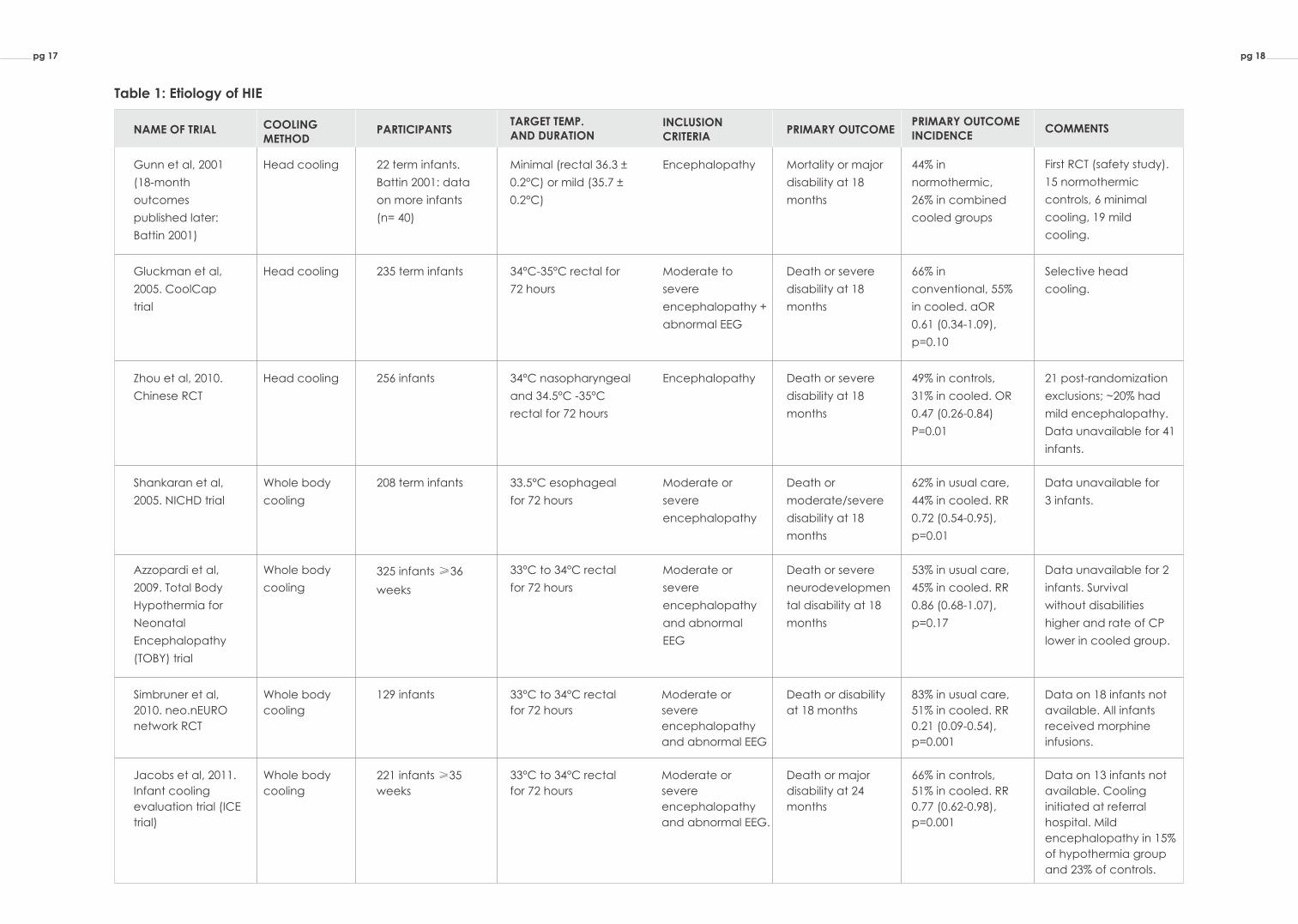
INCLUSION CRITERIA
PRIMARY OUTCOMEPRIMARY OUTCOME INCIDENCE
COMMENTS
Encephalopathy Mortality or major
disability at 18
months
44% in
normothermic,
26% in combined
cooled groups
First RCT (safety study).
15 normothermic
controls, 6 minimal
cooling, 19 mild
cooling.
Moderate to
severe
encephalopathy +
abnormal EEG
Death or severe
disability at 18
months
66% in
conventional, 55%
in cooled. aOR
0.61 (0.34-1.09),
p=0.10
Selective head
cooling.
Encephalopathy Death or severe
disability at 18
months
49% in controls,
31% in cooled. OR
0.47 (0.26-0.84)
P=0.01
21 post-randomization
exclusions; ~20% had
mild encephalopathy.
Data unavailable for 41
infants.
Moderate or
severe
encephalopathy
Death or
moderate/severe
disability at 18
months
62% in usual care,
44% in cooled. RR
0.72 (0.54-0.95),
p=0.01
Data unavailable for
3 infants.
Moderate or
severe
encephalopathy
and abnormal
EEG
Death or severe
neurodevelopmen
tal disability at 18
months
53% in usual care,
45% in cooled. RR
0.86 (0.68-1.07),
p=0.17
Data unavailable for 2
infants. Survival
without disabilities
higher and rate of CP
lower in cooled group.
Moderate or
severe
encephalopathy
and abnormal EEG
Death or disability
at 18 months
83% in usual care,
51% in cooled. RR
0.21 (0.09-0.54),
p=0.001
Data on 18 infants not
available. All infants
received morphine
infusions.
Death or major
disability at 24
months
66% in controls,
51% in cooled. RR
0.77 (0.62-0.98),
p=0.001
Data on 13 infants not
available. Cooling
initiated at referral
hospital. Mild
encephalopathy in 15%
of hypothermia group
and 23% of controls.
Moderate or
severe
encephalopathy
and abnormal EEG.
pg 18
Table 1: Etiology of HIE
NAME OF TRIAL COOLING METHOD
PARTICIPANTSTARGET TEMP. AND DURATION
Gunn et al, 2001
(18-month
outcomes
published later:
Battin 2001)
Head cooling 22 term infants.
Battin 2001: data
on more infants
(n= 40)
Minimal (rectal 36.3 ±
0.2°C) or mild (35.7 ±
0.2°C)
Gluckman et al,
2005. CoolCap
trial
Head cooling 235 term infants 34°C-35°C rectal for
72 hours
Zhou et al, 2010.
Chinese RCT
Head cooling 256 infants 34°C nasopharyngeal
and 34.5°C -35°C
rectal for 72 hours
Shankaran et al,
2005. NICHD trial
Whole body
cooling
208 term infants 33.5°C esophageal
for 72 hours
Azzopardi et al,
2009. Total Body
Hypothermia for
Neonatal
Encephalopathy
(TOBY) trial
Whole body
cooling
325 infants ≥36
weeks
33°C to 34°C rectal
for 72 hours
Simbruner et al,
2010. neo.nEURO
network RCT
Whole body
cooling
129 infants 33°C to 34°C rectal
for 72 hours
Jacobs et al, 2011.
Infant cooling
evaluation trial (ICE
trial)
Whole body
cooling
221 infants ≥35
weeks
33°C to 34°C rectal
for 72 hours
pg 17
pg 19
1. Gunn AJ, Gluckman PD, Gunn TR. Selective head cooling in newborn infants after perinatal
asphyxia: a safety study. Pediatrics. 1998 Oct;102(4 Pt 1):885-92.
2. Akisu M, Huseyinov A, Yalaz M, Cetin H, Kultursay N. Selective head cooling with hypothermia
suppresses the generation of platelet-activating factor in cerebrospinal fluid of newborn infants
with perinatal asphyxia. Prostaglandins LeukotEssent Fatty Acids. 2003 Jul;69(1):45-50.
3. Gluckman PD, Wyatt JS, Azzopardi D, Ballard R, Edwards AD, Ferriero DM, et al. Selective head
cooling with mild systemic hypothermia after neonatal encephalopathy: multicentrerandomised
trial. Lancet. 2005 Feb 19-25;365(9460):663-70.
4. Zhou WH, Cheng GQ, Shao XM, Liu XZ, Shan RB, Zhuang DY, et al. Selective head cooling with mild
systemic hypothermia after neonatal hypoxic-ischemic encephalopathy: a multicenter
randomized controlled trial in China. J Pediatr. 2010 Sep;157(3):367-72, 72 e1-3.
5. Lin ZL, Yu HM, Lin J, Chen SQ, Liang ZQ, Zhang ZY. Mild hypothermia via selective head cooling as
neuroprotective therapy in term neonates with perinatal asphyxia: an experience from a single
neonatal intensive care unit. J Perinatol. 2006 Mar;26(3):180-4.
6. Shankaran S, Laptook A, Wright LL, Ehrenkranz RA, Donovan EF, Fanaroff AA, et al. Whole-body
hypothermia for neonatal encephalopathy: animal observations as a basis for a randomized,
controlled pilot study in term infants. Pediatrics. 2002 Aug;110(2 Pt 1):377-85.
7. Shankaran S, Laptook AR, Ehrenkranz RA, Tyson JE, McDonald SA, Donovan EF, et al. Whole-body
hypothermia for neonates with hypoxic-ischemic encephalopathy. N Engl J Med. 2005 Oct
13;353(15):1574-84.
8. Eicher DJ, Wagner CL, Katikaneni LP, Hulsey TC, Bass WT, Kaufman DA, et al. Moderate
hypothermia in neonatal encephalopathy: safety outcomes. Pediatr Neurol. 2005
Jan; 32(1):18-24.
9. Eicher DJ, Wagner CL, Katikaneni LP, Hulsey TC, Bass WT, Kaufman DA, et al. Moderate
hypothermia in neonatal encephalopathy: efficacy outcomes. Pediatr Neurol.
2005 Jan; 32(1):11-7.
10. Azzopardi DV, Strohm B, Edwards AD, Dyet L, Halliday HL, Juszczak E, et al. Moderate hypothermia
to treat perinatal asphyxial encephalopathy. N Engl J Med. 2009 Oct 01;361(14):1349-58.
11. Jacobs SE, Morley CJ, Inder TE, Stewart MJ, Smith KR, McNamara PJ, et al. Whole-body
hypothermia for term and near-term newborns with hypoxic-ischemic encephalopathy: a
randomized controlled trial. Arch Pediatr Adolesc Med. 2011 Aug;165(8):692-700.
12. Simbruner G, Mittal RA, Rohlmann F, Muche R. Systemic hypothermia after neonatal
encephalopathy: outcomes of neo.nEURO.network RCT. Pediatrics. 2010 Oct;126(4):e771-8.
13. Jacobs SE, Berg M, Hunt R, Tarnow-Mordi WO, Inder TE, Davis PG. Cooling for newborns with
hypoxic ischaemic encephalopathy. Cochrane Database Syst Rev. 2013 Jan 31(1):CD003311.
14. Joy R, Pournami F, Bethou A, Bhat VB, Bobby Z. Effect of therapeutic hypothermia on oxidative
stress and outcome in term neonates with perinatal asphyxia: a randomized controlled trial. J Trop
Pediatr. 2013 Feb;59(1):17-22.
15. Rakesh K, Vishnu Bhat B, Adhisivam B, Ajith P. Effect of therapeutic hypothermia on myocardial
BIBLIOGRAPHY

pg 20
dysfunction in term neonates with perinatal asphyxia - a randomized controlled trial. J Matern
Fetal Neonatal Med. 2017 Jul 06:1-6.
16. Tanigasalam V, Bhat V, Adhisivam B, Sridhar MG. Does therapeutic hypothermia reduce acute
kidney injury among term neonates with perinatal asphyxia?--a randomized controlled trial. J
Matern Fetal Neonatal Med. 2016;29(15):2545-8.
17. Bharadwaj SK, Bhat BV. Therapeutic hypothermia using gel packs for term neonates with hypoxic
ischaemic encephalopathy in resource-limited settings: a randomized controlled trial. J Trop
Pediatr. 2012 Oct;58(5):382-8.
18. Bhat BV, Adhisivam B. Therapeutic cooling for perinatal asphyxia-Indian experience. Indian J
Pediatr. 2014 Jun;81(6):585-91.
19. Chaudhary R, Farrer K, Broster S, McRitchie L, Austin T. Active versus passive cooling during
neonatal transport. Pediatrics. 2013 Nov;132(5):841-6.
20. Thomas N, Chakrapani Y, Rebekah G, Kareti K, Devasahayam S. Phase changing material: an
alternative method for cooling babies with hypoxic ischaemic encephalopathy. Neonatology.
2015;107(4):266-70.
21. Shankaran S, Laptook AR, Pappas A, McDonald SA, Das A, Tyson JE, et al. Effect of depth and
duration of cooling on deaths in the NICU among neonates with hypoxic ischemic
encephalopathy: a randomized clinical trial. JAMA. 2014 Dec 24-31;312(24):2629-39.
22. Shankaran S, Laptook AR, Pappas A, McDonald SA, Das A, Tyson JE, et al. Effect of Depth and
Duration of Cooling on Death or Disability at Age 18 Months Among Neonates With Hypoxic-
Ischemic Encephalopathy: A Randomized Clinical Trial. JAMA. 2017 Jul 04;318(1):57-67.
23. Thoresen M, Whitelaw A. Cardiovascular changes during mild therapeutic hypothermia and
rewarming in infants with hypoxic-ischemic encephalopathy. Pediatrics. 2000 Jul;106(1 Pt 1):92-9.
24. Battin M, Bennet L, Gunn AJ. Rebound seizures during rewarming. Pediatrics. 2004
Nov;114(5):1369.
25. Kendall GS, Mathieson S, Meek J, Rennie JM. Recooling for rebound seizures after rewarming in
neonatal encephalopathy. Pediatrics. 2012 Aug;130(2):e451-5.
26. Birca A, Lortie A, Birca V, Decarie JC, Veilleux A, Gallagher A, et al. Rewarming affects EEG
background in term newborns with hypoxic-ischemic encephalopathy undergoing therapeutic
hypothermia. Clin Neurophysiol. 2016 Apr;127(4):2087-94.
27. Laptook AR. Birth Asphyxia and Hypoxic-Ischemic Brain Injury in the Preterm Infant. Clin Perinatol.
2016 Sep;43(3):529-45.
28. Sisman J, Campbell DE, Brion LP. Amplitude-integrated EEG in preterm infants: maturation of
background pattern and amplitude voltage with postmenstrual age and gestational age. J
Perinatol. 2005 Jun;25(6):391-6.
29. Sommers R, Tucker R, Harini C, Laptook AR. Neurological maturation of late preterm infants at 34
wk assessed by amplitude integrated electroencephalogram. Pediatr Res. 2013
Dec;74(6):705-11.
30. Smit E, Liu X, Jary S, Cowan F, Thoresen M. Cooling neonates who do not fulfil the standard cooling
criteria - short- and long-term outcomes. Acta Paediatr. 2015 Feb;104(2):138-45.

31. Walsh W. Report of a pilot study of Cooling four preterm infants 32-35 weeks gestation with HIE. J
Neonatal Perinatal Med. 2015 Mar 10.
32. Azzopardi D, Brocklehurst P, Edwards D, Halliday H, Levene M, Thoresen M, et al. The TOBY Study.
Whole body hypothermia for the treatment of perinatal asphyxial encephalopathy: a
randomised controlled trial. BMC Pediatr. 2008 Apr 30;8:17.
33. Rutherford M, Ramenghi LA, Edwards AD, Brocklehurst P, Halliday H, Levene M, et al. Assessment
of brain tissue injury after moderate hypothermia in neonates with hypoxic-ischaemic
encephalopathy: a nested substudy of a randomised controlled trial. Lancet Neurol. 2010
Jan;9(1):39-45.
34. Shankaran S, Barnes PD, Hintz SR, Laptook AR, Zaterka-Baxter KM, McDonald SA, et al. Brain injury
following trial of hypothermia for neonatal hypoxic-ischaemic encephalopathy. Arch Dis Child
Fetal Neonatal Ed. 2012 Nov;97(6):F398-404.
35. Cheong JL, Coleman L, Hunt RW, Lee KJ, Doyle LW, Inder TE, et al. Prognostic utility of magnetic
resonance imaging in neonatal hypoxic-ischemic encephalopathy: substudy of a randomized
trial. Arch Pediatr Adolesc Med. 2012 Jul 01;166(7):634-40.
36. Weeke LC, Boylan GB, Pressler RM, Hallberg B, Blennow M, Toet MC, et al. Role of EEG background
activity, seizure burden and MRI in predicting neurodevelopmental outcome in full-term infants
with hypoxic-ischaemic encephalopathy in the era of therapeutic hypothermia. Eur J Paediatr
Neurol. 2016 Nov;20(6):855-64.
pg 21

An estimated 4
million babies
die every year
in the neonatal
period.
THERAPEUTIC HYPOTHERMIA FOR NEONATAL ENCEPHALOPATHY
EVIDENCE ASPHYXIA AND THERAPEUTIC HYPOTHERMIA IN INDIA-AN OVERVIEW
CHAPTER
4
Dr. Niranjan Thomas, Dr. Mintoo Tergestina
According to the Lancet Global Series on Neonatal survival, an 1 estimated 4 million babies die every year in the neonatal period.
Ninety nine percentage of these deaths are in low to middle income
countries. India contributes to one-fifth of global live births and more
than a quarter of neonatal deaths. The neonatal mortality rate (NMR)
as of 2013 is 28 per 1000 live births, of which the NMR for rural and urban 2 is 31 and 15 per 100 live births respectively. Intrapartum complications
and birth asphyxia account for 23% of neonatal deaths globally and
19.2% of neonatal deaths in India, of which 97.8% of deaths due to
asphyxia occur in the first week of life and 70% within the first 24 hours.
The reported incidence of deaths due to hypoxia varies from 2 to
16.2% in community-based studies, with case fatality rates ranging 3 from 38.5 to 74%. Survivors of Hypoxic ischemia encephalopathy
carry an additional burden of long term morbidity, neurological
disability and functional impairment. As of today, the only modality
for treatment and a means to decrease long term morbidity in babies
with perinatal asphyxia is the initiation of therapeutic hypothermia
within the first 6 hours of life.
COOLING IN LOW AND MIDDLE INCOME COUNTRIES
There has been considerable debate regarding the use of cooling in
low and middle income countries. Increased rates of maternal
malnutrition, inadequate maternal pelvis size, poor antenatal

obstetric care, late referral of complicated pregnancies, lack of trained birth attendants for
and basic equipment for neonatal resuscitation, lack of referral centers that can manage
neonates that require ventilation, increased rates of HIV, malaria and puerperal sepsis are 6factors that increase the risk of perinatal asphyxia.
While the perinatal services offered in high income-low neonatal mortality countries are
either institutional deliveries or home deliveries with rapid transport to a tertiary level 3 neonatal unit, 25% of deliveries in India are home deliveries with minimal care thereafter.
Lack of access to transport, large distances and costs that are required to be borne for
transport, coupled with weak communication systems and deliveries in resource poor
settings can lead to delay in care that can be offered to a sick neonate. Even in cases of
institutional delivery, delay in transport and referral can lead to loss of the window for
initiation of therapeutic hypothermia.
An increased rate of perinatal infections has also raised concern when cooling in India.
Data from animal studies suggest that hypothermia may not be neuroprotective after
bacterial lipopolysaccharide-sensitized brain injury as compared to hypothermia without 7bacterial lipopolysaccharide. There has also been concern that hypothermia can lead to
decreased neutrophil function and chemotaxis and increased or worsening sepsis.
However, reported rates of culture proven sepsis in a trial of cooling with gel packs done in
India was 6.5%, with similar rates reported from the National Institute of Child Health and 8Human Development (NICHD) trial. In a case series of 40 babies from a tertiary level NICU, of
40 babies who received therapeutic hypothermia, only one had a positive blood culture 9result and 11 had results that were positive for C-reactive protein.
Other concerns that have been expressed are that the population has increased rates of
intrauterine growth retarded babies, and an increased incidence of meconium aspiration
syndrome. Temperature fluctuations and shivering have been pointed out as drawbacks to
cooling with low-tech methods. However, when done under close monitoring, the
fluctuations seen with manual cooling devices are no different from those seen in the NICHD
and Total Body Hypothermia for Neonatal Encephalopathy (TOBY) trials.
A constant criticism of therapeutic hypothermia by hypothermia-sceptics is that
hypothermia decreases mortality but leads to an increase in survival of neonates with
neuro-developmental morbidities. In India, there is a lack of institutionalized developmental
follow up and support for children with developmental delay and disability is limited from
governmental organizations and the society at large. This situation, however, has the
potential to change and improve. While the economics of cooling in low to middle income
countries has not yet been worked out, data from the TOBY trials show a clear benefit to
cooling.
pg 23

EQUIPMENT USED AND TRIALS FROM INDIA
Cooling equipment used in the high-income countries is expensive and unsuitable for wider
use in low and middle-income countries. Studies have looked at passive cooling (keeping
the baby without clothes, and without the use of a radiant warmer) during transport and the
induction phase of therapeutic hypothermia. This was found to lead to over cooling, with
temperatures < 32°C noted. It was also thought to be unlikely to be able to sustain 11temperatures within the target range for the 72 hour duration of cooling that is required.
Whole body cooling has been described from other countries using water bottles, gloves
filled with water, a laminar flow device and a servo controlled fan. The first trial of whole
body cooling in India was reported from the Christian Medical College, Vellore, and was a
study done to determine the feasibility and safety of whole body cooling in newborn infants
with perinatal encephalopathy in a low resource setting. This described hypothermia using
ice gel packs. This study involved neonates >35 weeks gestation who were cooled to a
rectal temperature of 33±0.5°C for 72 hours. Twenty infants were included in this study that
used reusable ice gel packs obtained from the immunization clinic at no added expense.
While this low cost alternative required frequent changes of packs, more nursing input and
constant monitoring, the target temperature was achieved and maintained with ease. The
study concluded that whole body cooling in term infants with perinatal asphyxia was
achievable, safe and inexpensive in a low-resource setting. However, the swings of
temperature, and shivering of the cooled infant lead to the beginning of a search for a 12better low cost solution.
The second published trial in India came from the Jawaharlal Institute of Postgraduate
Medical Education and Research (JIPMER), Pondicherry, a year later, which cooled babies
with cloth covered gel packs and followed them up to an age of 6 months. These infants
were assessed at 6 months using Baroda Developmental Screening Test, and the group that
received therapeutic hypothermia showed a significant reduction in the combined rate of
death or developmental delay at 6 months of age by 21% (8.1% in the TH group vs. 29% in the 13control, RR 0.28, 95% CI: 0.11-0.70; p = 0.003). The same researchers from JIPMER also
showed that therapeutic hypothermia reduced oxidative stress as measured by total 14antioxidant status and malondialdehyde, and reduced oxidative stress-induced DNA
15damage.
In 2009, Iwata et al published their report of the successful use of phase changing material
(PCM) for cooling newborn piglets that had been exposed to hypoxia. The use of the PCM
mattress was found to give more stable cooling, and a longer period within the target range 16of temperature. The first randomized controlled trial of whole body cooling using phase
changing material was conducted in Calicut in south India in 2013. Thirty three infants with
Thompsons encephalopathy score >5 were cooled. However, the study found that cooling
with PCM was effective only when the ambient temperature was <28°C, and required close 17temperature monitoring by the nursing staff.
pg 24

In 2014, a study was conducted on 28 newborns in the neonatal unit of the Madras Medical
College, Tamil Nadu, which assessed the feasibility of the use of the Tecotherm-HELIX
cooling mattress. This was a servo-controlled cooling device developed for low to middle 18income countries, with an estimated cost of US$1000. It worked on the basis of re-
circulating water mixed with alcohol through a re-usable cooling mattress to achieve a set
temperature of 33.5°C. The advantage of the Tecotherm-HELIX cooling mattress was that
refilling the machine with water every 6 – 8 hours was the only additional nursing input
required. However, the cooling mattress required replacement once in the 6 month study
period.
During the years from 2011 to 2014, a wholly indigenous passive cooling mattress that was
based on phase changing material was in development at the Christian Medical College,
Vellore. Multiple attempts were made to find the ideal PCM that was easy to use, safe,
lightweight, portable and gave precise temperature control of 33-34°C for 72 hours with
minimal manual supervision and no requirement of constant electricity. The feasibility of the
technology was reported in 2015, on a retrospective series of 41 babies with HIE who had
been cooled with PCM (OM 32™ or HS 29™) to a target rectal temperature of 33-34°C. This
was successful in maintaining the target temperature range 96.2% of the time in the cooling 19phase.
The final product was the CMC PCM Mattress- Patent-3184/DEL/2013. This used a cascaded
system of PCMs that were incorporated in a polymer matrix that provided form stability and
ensured that the PCM retained its shape and form, preventing leakage. The melting points,
thicknesses, conductivities and placement of the involved layers were such that a “quasi-
automated” cooling system was created, which functioned similar to a servo controlled
system. Pluss Advanced Technologies, who supplied the phase change material worked
closely with the research team from Christian Medical College Vellore, and later marketed it ®as the MiraCradle . Over the years, from 2011, more than 200 babies have been cooled
successfully, with minimal complications and successful maintenance of temperature
within the target ranges. More than 150 commercial PCM mattresses are currently in use in
India and data of 103 babies cooled in 10 different hospitals in India with good results has
been sent for publication. They are also used across the world, in South Africa, Turkey and in
Kenya.
SYSTEMATIC REVIEW AND META-ANALYSIS
20A systematic review by Pauliah et al, did not show any significant improvement in mortality
and concluded that cooling cannot be recommended in both low and middle income
countries until further data suggest benefit. However, the confidence intervals in this review
were wide, and it was speculated by the authors that the reasons for the lack of treatment
effect observed were poor quality of the included studies; lack of efficiency of low
technology cooling devices; lack of optimal neonatal intensive care, sedation and
ventilation support; intrauterine growth restriction; and maternal malnutrition.
pg 25

pg 26
The review concluded that therapeutic hypothermia using low technology methods was
achievable, safe and inexpensive. When combined with intensive care, this is proven to
significantly reduce the mortality and neurological morbidity in survivors at discharge. The
Meta-Analysis by Rossouw et al shows that low technology cooling devices are effective,
and make a difference in the mortality and morbidity of the babies when used in an
intensive care setting where mechanical ventilation is available, and when it is possible to
provide more intensive nursing. The initial feasibility study from India had shown that cooling
with manual application of cloth-covered gel packs was feasible with a nurse-to-infant ratio 12of 1:3.
23A meta-analysis by Galvao et al. of 16 studies and a total of 1889 neonates (of which 8 of
the studies included were conducted in lower income countries) showed that hypothermia
significantly reduced mortality (RR = 0.77; 95% CI: 0.65-0.92). Meta-regression analysis also
revealed that hypothermia efficacy does not increase as the gross domestic product per
capita of the country in which it is done rises, and they concluded that there is enough
evidence to support therapeutic hypothermia as the standard of care for hypoxic-ischemic
encephalopathy.
CONCLUSION
India, with diverse patient profiles, has centers that have facilities that equal those in high
income countries. A trial of cooling can no longer be done in a high income country due to
the loss of equipoise. To do so in our country – to offer cooling versus normothermia in babies
with perinatal asphyxia should raise ethical questions. In areas that lack basic facilities, the
focus should remain on improving antenatal and neonatal care to prevent asphyxia.
Training of personnel, following standard protocols for cooling, setting up and maintaining a
national registry to monitor neurodevelopmental outcome (similar to the TOBY registry) is
what we should achieve. While the equipment used in high income countries cannot be
sustainably used in low and middle income countries, therapeutic hypothermia using low
cost technology can provide sustainable, efficient and effective treatment with minimal
costs and maximal benefits, and should be implemented as a standard of care for infants
with moderate or severe HIE and refusal cannot be justified where facilities exist.
BIBLIOGRAPHY
1. Lawn JE, Cousens S, Zupan J, Lancet Neonatal Survival Steering Team. 4 million neonatal deaths:
when? Where? Why? Lancet Lond Engl. 2005 Mar 5;365(9462):891–900.
2. Neo Natal Mortality Rate (NMR) (per 1000 live births) | NITI Aayog, (National Institution for
Transforming India), Government of India [Internet]. [cited 2017 Oct 6]. Available from:
http://niti.gov.in/content/neo-natal-mortality-rate-nmr-1000-live-births
3. National Family Health Survey [Internet]. [cited 2017 Oct 6]. Available from:
http://rchiips.org/NFHS/factsheet_NFHS-4.shtml
4. Edwards AD. The discovery of hypothermic neural rescue therapy for perinatal hypoxic-ischemic
encephalopathy. Semin Pediatr Neurol. 2009 Dec;16(4):200–6.
5. Jacobs SE, Berg M, Hunt R, Tarnow-Mordi WO, Inder TE, Davis PG. Cooling for newborns with
hypoxic ischaemic encephalopathy. Cochrane Database Syst Rev. 2013 Jan 31;(1):CD003311.
6. Montaldo P, Pauliah SS, Lally PJ, Olson L, Thayyil S. Cooling in a low-resource environment: lost in
translation. Semin Fetal Neonatal Med. 2015 Apr;20(2):72–9.
7. Osredkar D, Thoresen M, Maes E, Flatebø T, Elstad M, Sabir H. Hypothermia is not neuroprotective
after infection-sensitized neonatal hypoxic-ischemic brain injury. Resuscitation. 2014
Apr;85(4):567–72.
8. Shankaran S, Laptook AR, Ehrenkranz RA, Tyson JE, McDonald SA, Donovan EF, et al. Whole-body
hypothermia for neonates with hypoxic-ischemic encephalopathy. N Engl J Med. 2005 Oct
13;353(15):1574–84.
9. Aker K. Birth asphyxia and whole body cooling: a descriptive study of newborns admitted with
perinatal asphyxia in a tertiary care Perinatal center in south India. Masters thesis. Trondheim,
Norway: Norweigian University of Science and Technology; 2009.
10. Azzopardi D, Brocklehurst P, Edwards D, Halliday H, Levene M, Thoresen M, et al. The TOBY Study.
Whole body hypothermia for the treatment of perinatal asphyxial encephalopathy: a
randomised controlled trial. BMC Pediatr. 2008 Apr 30;8:17.
11. Akula VP, Gould JB, Davis AS, Hackel A, Oehlert J, Van Meurs KP. Therapeutic hypothermia during
neonatal transport: data from the California Perinatal quality care collaborative (CPQCC) and
California perinatal transport system(CPeTS) for 2010. J Perinatol 2013;33(3):194e7.
12. Thomas N, George KC, Sridhar S, Kumar M, Kuruvilla KA, Jana AK. Whole body cooling in newborn
infants with perinatal asphyxial encephalopathy in a low resource setting: a feasibility trial. Indian
Pediatr. 2011 Jun;48(6):445–51.
13. Bharadwaj SK, Bhat BV. Therapeutic hypothermia using gel packs for term neonates with hypoxic
ischaemic encephalopathy in resource-limited settings: a randomized controlled trial. J Trop
Pediatr. 2012 Oct;58(5):382–8.
14. Joy R, Pournami F, Bethou A, Bhat VB, Bobby Z. Effect of therapeutic hypothermia on oxidative
stress and outcome in term neonates with perinatal asphyxia: a randomized controlled trial. J Trop
Pediatr. 2013 Feb;59(1):17–22.
pg 27

pg 28
15. Effect of Therapeutic Hypothermia on DNA Damage and Neurodevelopmental Outcome
Among Term Neonates With Perinatal Asphyxia: A Randomized Controlled Trial [Internet].
PubMed Journals. Available from: https://ncbi.nlm.nih.gov/labs/articles/24343823/
16. Iwata S, Iwata O, Olson L, Kapetanakis A, Kato T, Evans S, et al. Therapeutic hypothermia can be
induced and maintained using either commercial water bottles or a “phase changing material”
mattress in a newborn piglet model. Arch Dis Child. 2009 May;94(5):387–91.
17. Thayyil S, Shankaran S, Wade A, Cowan FM, Ayer M, Satheesan K, et al. Whole-body cooling in
neonatal encephalopathy using phase changing material. Arch Dis Child Fetal Neonatal Ed. 2013
May;98(3):F280-281.
18. Pauliah S, Narayanan E, Kumutha K, Vijayakumar M, Nair M, Shankaran S. Hypothermia for
Encephalopathy in Low and Middle-income Countries (HELIX): a feasibility study. Archs Dis Child
Fetal Neonatal Ed 2014;99. A74.
19. Thomas N, Chakrapani Y, Rebekah G, Kareti K, Devasahayam S. Phase Changing Material: An
Alternative Method for Cooling Babies with Hypoxic Ischaemic Encephalopathy. Neonatology.
2015 Feb 26;107(4):266–70.
20. Pauliah SS, Shankaran S, Wade A, Cady EB, Thayyil S. Therapeutic hypothermia for neonatal
encephalopathy in low- and middle-income countries: a systematic review and meta-analysis.
PloS One. 2013;8(3):e58834.
21. Rossouw G, Irlam J, Horn AR. Therapeutic hypothermia for hypoxic ischaemic encephalopathy
using low-technology methods: a systematic review and meta-analysis. Acta Paediatr Oslo Nor
1992. 2015 Dec;104(12):1217–28.
22. Jacobs SE, Morley CJ, Inder TE, Stewart MJ, Smith KR, McNamara PJ, et al. Whole-body
hypothermia for term and near-term newborns with hypoxic-ischemic encephalopathy: a
randomized controlled trial. Arch Pediatr Adolesc Med. 2011 Aug;165(8):692–700.
23. Galvao TF, Silva MT, Marques MC, de Oliveira ND, Pereira MG. Hypothermia for perinatal brain
hypoxia-ischemia in different resource settings: a systematic review. J Trop Pediatr. 2013
Dec;59(6):453–9.

COOLING DEVICES FOR THERAPEUTIC HYPOTHERMIA IN NEWBORN BABIES
CHAPTER
5
One of the
limiting factors
to provide
therapeutic
hypothermia in
India is
availability of
cooling device.
Dr. Sumitha Arun, Dr. Manish Kumar
Many tertiary level hospitals in India have started providing
therapeutic hypothermia as standard of care to newborn with
moderate to severe degree of hypoxic ischemic encephalopathy.
One of the limiting factors to provide therapeutic hypothermia in India
is availability of cooling device. Therapeutic hypothermia can be
provided as selective head cooling or whole body cooling. In this
article, we will discuss what would constitute an ideal cooling device,
what are the devices available and a description on how they
function.
THE CHARACTERISTICS OF AN IDEAL COOLING DEVICE
Ÿ It should induce cooling rapidly to the desired core temperature
Ÿ It should maintain the core temperature tightly within the target
range
Ÿ It should allow re-warming in a slow and controlled manner
Ÿ It should be easy to use
Ÿ It should require minimal nursing input
Ÿ It should not interfere with access to the baby
Ÿ Environmental temperature should not affect cooling efficacy
Ÿ Safety systems in case of accidental probe displacement

pg 30
Since the early 2000's, therapeutic hypothermia has been tried either as selective head
cooling or as whole body cooling. Many of these devices are cost-prohibitive. Hence
alternate low cost cooling systems have been developed for use in resource limited settings.
The various high technology devices with their cooling methods are summarized in Table 1.
Blanketrol 2 used in NICHD
2. Criticool from MTRE, Charter Kontron, Milton Keynes,
UK, is a microprocessor controlled servo device. It has a
large basal unit and disposable cure wraps through
which water is circulated. Access to baby is restricted
due to wraps covering the baby. The rewarming phase
can be set in manual or controlled mode. Controlled
rewarming is set at 0.2°C per 30 minutes. It takes 7.5 hours
to rewarm.
Cooling Equipment- High technology devices
1.BLANKETROL 2 (Cincinnatti subzero products Inc, USA) 1 was used in the NICHD trial. It is a microprocessor
controlled device based on conductive water therapy.
Target temperature is achieved by heating and cooling
water circulated through mattress and wraps. Due to
fluctuations in temperature, additional blankets were
used around the baby. The rewarming process is
manually done in the device.
BLANKETROL 3 used gradient programme technology to
minimize fluctuations in water temperature. It did not
require use of additional blankets. It involved precooling
to 5°C 15 minutes prior to starting. Oesophageal probes
were used for temperature recording. Data export
software was incorporated.

3. Tecotherm TS med 200 from Tec-com, Halle Germany:
Is a manual cooling device which was used in the TOBY 2trial.
4. Tecotherm Servo from Tec-com, Halle Germany: This
consists of a mattress that is wrapped around baby's
trunk and legs. The device is fully automated and
requires no further input after initiation. The baby's
temperature is cooled to 33.5°C within 30 minutes. This
temperature is maintained for 72 hours and a fully
automated rewarming process starts. The rewarming
occurs to 37°C at 0.2-0.5°C/hour (requiring around 7
hours for rewarming). The monitor displays rectal and
skin temperature. Data can be transported out of the
machine and analyzed. Default temperature settings
on the machine can be altered according to
individual needs.
5. Cool cap (Olympic Medical Cool Care System,
Olympic Medical, Seattle, WA, USA): This was used for
selective head cooling. Olympic cool cap system
maintains the water in the fitted cap at an operator
specified temperature. The rest of the body is exposed
to radiant warmer. It requires lot of nursing input. Rectal
temperature is used to guide the set temperature.
Target temperature is 34-35°C. Rewarming is manual in
cool cap.
There is a statistically significant reduction in the composite outcome of death or major
neurodevelopmental disability in both selective head cooling and whole body cooling as 3 per Cochrane meta-analysis. During selective head cooling there is a temperature
gradient between the surface and deeper structures of the brain. Even when we effectively
cool the surface of brain to desired temperature, the deep brain structures may not be
adequately cooled. This is a significant concern as profound hypoxic injury particularly
affects the deep brain structures. In a comparative study there were lesser lesions on MRI 4after whole body cooling in comparison to selective head cooling.
pg 31

NAME
OF DEVICEDESIGN FEATURES ISSUES
Blanketrol 2
RECURRENT
EXPENSES
Blanketrol 3
Criticool
Tecotherm
Servo
Cool cap
Mattress Wraps
Mattress Wraps
Wraps
Mattress
Cap
Coolant- water
Wraps
Coolant –water
Less fluctuations in
temperature
Less fluctuations in
temperature
Wraps
1. Coolant- water Rewarming –automated
2. Portable
3. Alcohol based coolant
1. Fully automated Rewarming –automated
2. Portable
3. Alcohol based coolant
1. Selective head cooling
1. Temperature fluctuation
2. Rewarming –manual
3. Not portable
1. Pre-cooling required.
1. Rewarming –manual/controlled
2. Not portable
Manual rewarming
More nursing input
Cooling wraps
Cooling wraps
Coolant top
ups and
mattress
Cooling cap
Cooling wraps
pg 32
Table 1:

COOLING EQUIPMENT-LOW COST DEVICES
Various low technologies methods like passive cooling, ice gel pack, cooling fan, etc.; have
been tried to achieve therapeutic hypothermia because of the high cost and unavailability
of servo controlled equipment. There have been concerns of overcooling, fluctuations in
temperature, cold injury like subcutaneous fat necrosis, increased shivering and need of
more nursing input. Increase in awareness, intensive monitoring, refinement of technology
(e.g., use of phase changing material) and appropriate intervention (e.g., use of analgesia,
frequent change in position) have helped to achieve therapeutic hypothermia without
overcooling and minimal fluctuations of temperature. We have shown the efficacy and
safety of low technologies devices like frozen gel pack and phase changing materials 5which is comparable to standard servo-controlledcooling equipment. A recent systematic
analysis has shown that low technology device in tertiary level settings has improved survival 6and neurological outcome in HIE babies with moderate to severe encephalopathy.
PASSIVE COOLING
Babies are kept naked with radiant warmer switched off. This has been mainly tried during
transportation for induction phase of cooling. It is feasible to achieve cooling passively but
there has been concern of overcooling. Several studies have reported significant 7overcooling (<32°C) in approximately one third of the babies. There is no published data
regarding use of passive cooling for maintenance and rewarming phase.
FROZEN/ICE GEL PACK
ICE trial, a multi-center randomized controlled trial, showed improved disability free survival
in babies with moderate to severe HIE after achieving therapeutic hypothermia with frozen 8 gel pack. In India, the feasibility trial from Christian Medical College, Vellore showed safety
9and effectiveness of frozen gel pack (FGP) for therapeutic hypothermia.
Method: Insert rectal probe 5 cm inside and fix to thigh. Switch off the radiant warmer. For
induction phase, distribute 4-5 packs over the head, both axillae, back and abdomen.
Once the temperature reaches 34°C, remove packs one by one, targeting the
temperature of 33.5± 0.3°C. The packs on the head and back are to be removed last.
Gel packs should be stored in deep freezer. Wrap the FGPs with cloth and avoid direct skin
contact for longer duration. This minimizes cold injury to skin. Always have rectal probes for 10 temperature monitoring. Skin probes are NOT an acceptable alternative. Leave only one
FGP at the temperature of 33.5°C. Set the alarm limit at 33.2°C and 33.8°C on monitor during
maintenance phase. At the end of 72 hours of induced hypothermia, remove cool packs
and turn on the radiant warmer to raise the body temperature by 0.3°C/hour to reach a
target temperature of 36.5°C. (This should take 12-15 hours). The temperature probe should
pg 33

Equipment description
be removed after monitoring the temperature for a further 12 hours (72 + 12 hours after
initiation of cooling).
If the infant's rectal temperature increases to 33.8°C, more cloth covered cooling-gel packs
should be placed and then subsequently removed when the rectal temperature reaches
33.5°C.
If the temperature decreases to 33°C, remove cooling gel packs/turn warmer on till
temperature reaches 33.5°C
PHASE CHANGING MATERIALS
Phase changing materials (PCM) are made of salt hydride, fatty acid and esters or paraffin,
melting at a set point. PCMs absorb and release heat at a nearly constant temperature and
can store 5–14 times more heat per unit volume compared to material like water or masonry.
When a baby is placed on a bed made of PCM, heat from the baby is transferred to the
PCM which absorbs the heat till it melts. Hence, PCM has the potential to provide a
mechanism of heat removal which will be easier and safer to use than ice packs. Keeping
the melting point of the PCM at the target temperature will ensure that the baby's
temperature will not fall below this temperature. The device designed by our unit has used
PCM with 2 different melting point: PCM 21 with melting point at 21°C for induction phase
and PCM 29 with the melting point at 29°C for maintenance phase. The theory and 11 methodology has been described in detail. PCM should not be frozen. It should be stored in
the lower compartment of refrigerator.
In our study with PCM, induction time was 45 minutes, only 3.8% of temperature readings 5were outside the target range and fluctuation of core body temperature was 0.28°C.
pg 34
1. Outer covering layer - good conductor (only lines)2. Foam layer (orange-yellow)3. Insulating layer (green)4. PCM layer (red)5. Water pack layer (blue)
Cross-section withouter covering
Top view of mattress with PCM and water packs

Table 2: Comparison of different cooling device
Time taken to reach 45 (10, 100) 90 (25, 150) 90 minutes (NICHD)
target temperature, 30 minutes (TOBY)
minutes; median (IQR)
Temperature fluctuation 0.4 0.28 0.4 (TOBY)
during cooling phase, 0.5 (NICHD)oC; mean (SD) (semi-automated)
Temperature readings 9.8 % 3.8 %
outside the target range
Subcutaneous Fat 13 % 2.2 % 2.8 %
Necrosis
Laborious nursing input More Less Least
Frozen gel pack Phase changing Servo controlled
material device
METHODS
Switch off radiant warmer. Fix the rectal probe.
Remove the PCM from refrigerator and keep it in room temperature for 30 minutes before
commencing cooling. Use PCM 21 for induction and PCM 29 for maintenance phase if
nursery temperature is 27°C or above. If nursery temperature is 26°C or less, use 29 PCM for
both the phases.
If you are using 21 PCM for induction, remove it once rectal temperature is 34°C.
If the infant's rectal temperature increases to 33.8°C, change 29 PCM if it is melted. If 29 PCM
is good, add a 21 PCM and then subsequently remove when the rectal temperature
reaches 33.5°C.
If the temperature decreases to 33.2°C, introduce a bed sheet between baby and PCM. If
temperature remains low, turn warmer on till temperature reaches 33.5°C.
PCM based devices have been successfully used in CMCH, JIPMER and various other
hospitals in India over the past few years with documented evidence of safety and efficacy.
OTHERS
Laminar flow device, cooling fans, clay pots have been used for therapeutic hypothermia in
various studies.
pg 35

Fully automated servo controlled Cooling devices are ideal for providing therapeutic
hypothermia to newborn with asphyxia. Affordability is the main issue. Low technology
cooling devices like frozen gel packs and Phase change materials are effective, safe and
an affordable alternative in intensive care settings.
1. Shankaran S, Laptook AR, Ehrenkranz RA, Tyson JE, McDonald SA, Donovan EF, et al. Whole-body
hypothermia for neonates with hypoxic–ischemic encephalopathy. N Engl J Med.
2005;353(15):1574–1584.
2. The TOBY Study Group, Azzopardi D, Brocklehurst P, Edwards D, Halliday H, Levene M, et al. The
TOBY Study. Whole body hypothermia for the treatment of perinatal asphyxial encephalopathy:
A randomised controlled trial. BMC Pediatr [Internet]. 2008 Dec [cited 2017 Oct 2];8(1). Available
from: http://bmcpediatr.biomedcentral.com/articles/10.1186/1471-2431-8-17
3. Jacobs SE, Berg M, Hunt R, Tarnow-Mordi WO, Inder TE, Davis PG. Cooling for newborns with
hypoxic is chaemic encephalopathy. Cochrane Database Syst Rev. 2013 Jan 31;(1):CD003311.
4. Sarkar S, Donn SM, Bapuraj JR, Bhagat I, Barks JD. Distribution and severity of hypoxic-ischaemic
lesions on brain MRI following therapeutic cooling: selective head versus whole body cooling.
Arch Dis Child Fetal Neonatal Ed. 2012 Sep;97(5):F335-339.
5. Shabeer MP, Abiramalatha T, Smith A, Shrestha P, Rebekah G, Meghala A, et al. Comparison of
Two Low-cost Methods of Cooling Neonates with Hypoxic Ischemic Encephalopathy. J Trop
Pediatr. 2016 Oct 6;fmw067.
6. Rossouw G, Irlam J, Horn AR. Therapeutic hypothermia for hypoxic ischaemic encephalopathy
using low-technology methods: a systematic review and meta-analysis. Acta Paediatr. 2015
Dec;104(12):1217–28.
7. Fairchild K, Sokora D, Scott J, Zanelli S. Therapeutic hypothermia on neonatal transport: 4-year
experience in a single NICU. J Perinatol Off J CalifPerinat Assoc. 2010 May;30(5):324–9.
8. Jacobs SE, Morley CJ, Inder TE, Stewart MJ, Smith KR, McNamara PJ, et al. Whole-body
hypothermia for term and near-term newborns with hypoxic-ischemic encephalopathy: a
randomized controlled trial. Arch Pediatr Adolesc Med. 2011 Aug;165(8):692–700.
9. Thomas N, George KC, Sridhar S, Kumar M, Kuruvilla KA, Jana AK. Whole body cooling in newborn
infants with perinatal asphyxial encephalopathy in a low resource setting: a feasibility trial. Indian
Pediatr. 2011 Jun;48(6):445–51.
10. Thomas N, Rebekah G, Sridhar S, Kumar M, Kuruvilla KA, Jana AK. Can skin temperature replace
rectal temperature monitoring in babies undergoing therapeutic hypothermia in low-resource
settings?: Temperature monitoring during therapeutic hypothermia. Acta Paediatr. 2012
Dec;101(12):e564–7.
11. Thomas N, Chakrapani Y, Rebekah G, Kareti K, Devasahayam S. Phase Changing Material: An
Alternative Method for Cooling Babies with Hypoxic Ischaemic Encephalopathy. Neonatology.
2015 Feb 26;107(4):266–70.
pg 36
BIBLIOGRAPHY

PROCEDURE FOR WHOLE BODY COOLING
CHAPTER
6
Neonates can be
considered for
cooling if the
following criteria
are met (NICHD
criteria)
Dr. Sumitha Arun, Dr. Manish Kumar
WHICH NEONATES TO COOL?
Neonates can be considered for cooling if the following criteria are
met (NICHD criteria):
1. Gestational age ≥ 36 weeks
2. Less than 6 hours old
3. A pH of 7.0 or less or a base deficit of 16 mmol per liter or more in a
sample of umbilical cord blood or any blood during the first hour after
birth. If the pH is between 7.01 and 7.15, a base deficit is between 10
and 15.9 mmol per liter, or a blood gas is not available, additional
criteria are required. These include an acute perinatal event (e.g.,
late or variable decelerations, cord prolapse, cord rupture, uterine
rupture, maternal trauma, hemorrhage, or cardiorespiratory arrest)
and either a 10-minute Apgar score of 5 or less or assisted ventilation
initiated at birth and continued for at least 10 minutes.
4. Moderate to severe encephalopathy on clinical examination.
TYPES OF COOLING
Two different types of hypothermia were practiced:
a) Selective head cooling
b) Whole body cooling
Though selective head cooling appears to be attractive, it does not
correlate well with core body temperature. There is also differential
cooling between the superficial and deeper parts of the brain
causing differential perfusion. In contrast, whole body cooling
correlates well with core body temperature but associated with more
adverse effects.

pg 38
PREREQUISITES TO PRACTICE THERAPEUTIC HYPOTHERMIA
The minimum prerequisites to start the practice of therapeutic hypothermia have been
depicted in Table.1:
Table 1: Minimum requirements to practice therapeutic hypothermia.
DIFFERENT PHASES OF COOLING THERAPY
The different phases of therapeutic hypothermia are depicted in the Table 2.
Table 2: Phases of therapeutic hypothermia.
Place Personnel Paraphernalia Protocols
Well
established
level-3 NICU
care.
Trained
doctors &
Staff nurse
Radiant warmer,
Cooling device, ABG
machine, Multi-
parametric
parameters, Ventilators
Evidence based
standard protocol.
Neuro-development
follow up.
Strategies
INDUCTION
Rapid cooling to 33-34°C. Duration: 45 minutes-1 hour. Monitor temperature
and vitals. Treatment of shock and seizures. Avoid shivering and
overcooling. Rectal probe inserted 5 cm into the rectum and skin probe
over the abdomen are connected to the monitor for continuous
monitoring. The radiant warmer is switched off.
The target temperature is monitored every 15 minutes for the first four hours
and later hourly for 72 hours. Vital parameters like heart rate, capillary filling
time and blood pressure are recorded hourly. Close monitoring of
temperature and vitals. Maintain temperature: 33-34°C. Total Duration: 72
hours. Monitor urine output and adjust total fluid intake. Monitor for multi-
organ damage. Adjust drug dosages. Look for bed sores and skin damage.
Slow rewarming initiated. Duration: 6-10 hours.
Temperature raise: 0.5°C/hour.
Look for seizures. Avoid fluid shifts. Careful vitals monitoring.
After rewarming feeding is started gradually.
Hypothermia
phases
MAINTENANCE
REWARMING PHASE

MONITORING OF NEONATES DURING THERAPEUTIC HYPOTHERMIA
CHAPTER
7
As cooling is
used more widely
and has been
newly introduced
in neonatal units,
continued
surveillance of its
use in clinical
practice is
mandatory.
Dr. KumuthaJ, Dr. Manigandan Chandrasekaran
INTRODUCTION
As cooling is used more widely and has been newly introduced in
neonatal units, continued surveillance of its use in clinical practice is
mandatory. The initial management of infants with HIE following
admission to the neonatal unit consists of standard neonatal intensive
care measures, continuous core temperature monitoring using a
rectal probe and initiation of therapeutic hypothermia. Monitoring of
such infants is critical to the neurological outcome. We describe here
the systematic approach to monitor these infants.
TEMPERATURE MONITORING
Both the core temperature and skin temperature are monitored
continuously. The core temperature is usually monitored by rectal or
oesophageal probe. The rectal probe should be inserted 3-6 cm and
secured to the thigh. The oesophageal probe is inserted to a length of
measured distance (Angle of mouth to tragus and up to a point 2 cm
above Xiphisternum). The temperature is recorded every 15 minutes till
a core temperature of 33.5°C is achieved and thereafter every hour till
rewarming is completed. The core temperature should be
maintained between 33.5°C to 34°C. The fluctuation of temperature
during the maintenance phase should be very minimal as it can
adversely affect the neurological outcome.
CARDIORESPIRATORY CARE
Hypothermia is consistently associated with sinus bradycardia as it
slows the atrial pacemaker and intracardiac conduction. The QT

pg 40
Ÿ
Ÿ Start mechanical ventilation if repeated desaturations associated with seizures or
incipient respiratory failure with rising oxygen requirements and increasing respiratory
acidosis are observed.
Ÿ Avoid hypocapnia. Aim for pCO2 around 45 - 50 mm during cooling
Ÿ Maintain SaO2≥92%to lessen risk of pulmonary hypertension
Ÿ Obtain arterial access to monitor blood pressure if mechanical ventilation is required
Ÿ Monitor regional perfusion using capillary refill scores and core-peripheral temperature
gap as guides to peripheral blood distribution
Ÿ Aim for mean blood pressure (BP) ≥ 40 mm Hg. A low BP requires assessment. Perform
echocardiographic assessment (if available) of cardiac contractility and stroke volume
to guide fluid/inotrope administration
Correct respiratory acidosis by manipulating ventilator support
CARDIO-RESPIRATORY MANAGEMENT
interval also can be prolonged. The heart rate can be even lower than 100. Heart rate drops
by nearly 14 beats per one degree drop in temperature. HR up to 70 can be tolerated as
long as it is normal sinus rhythm and normal SPO2 and BP are observed. Therefore, it is vital to
observe these neonates with continuous cardiac monitoring preferably with multi-channel
monitoring. Sinus bradycardia usually does not need intervention. Close monitoring of
blood pressure is very essential to identify shock since CRT will be not a reliable indicator of
hypo perfusion in babies receiving cooling therapy. All infants should be monitored closely
with invasive blood pressure monitoring (if possible) and treated with inotropes accordingly.
MECHANICAL VENTILATION
Ventilation may not be needed for all neonates during cooling, however, it is important to
maintain normal pCO , pO , pH and respiratory effort. Decreasing body temperature lowers 2 2
metabolic rate by about 5-8% per °C. Furthermore, partial pressure of blood gases and pH
are also affected because of altered gas solubility during hypothermia. With each degree
Celsius decrease in core temperature, pH increases by 0.015, pCO and pO decrease by 2 2
4% and 7% respectively. Excessively low pCO during therapeutic hypothermia may result in 2
altered cerebral blood flow auto regulation, and reduced cerebral perfusion, and may
lower the seizure threshold. Hence, blood gas values should be corrected for core body
temperature. The corrected blood gas values should be maintained within the normal
range by appropriate ventilator adjustments.

Ÿ Hypoglycemia can be a serious complicating factor in HIE. Monitor plasma glucose
closely (4 hourly) and adjust glucose intake accordingly.
Ÿ Oliguria/anuria is common following HIE; Monitor urinary output and aim for urine output≥1ml/kg/hour. Observe for bladder retention. Urinary catheterization may be useful.
Ÿ Daily weight monitoring should be carried out.
Ÿ Initial intravenous fluid requirements are approximately 40 ml/kg/day 10% Dextrose
solution. Monitor blood electrolyte levels 8 hourly for first 24–48 hours.
Ÿ Grade up fluid depending on the weight gain and presence of oedema. as these babies
tend to retain lot of fluid.
Ÿ Consider electrolyte additives or parenteral nutrition after 24 - 48 hours when electrolytes/
renal function stable.
Ÿ Give maintenance potassium supplementation if renal function is adequate (2 mmol/
kg/day). Avoid potassium supplementation during cooling, as hyperkalemia may occur
on rewarming.
FLUID BALANCE
SKIN MONITORING
Poor skin perfusion occurs during cooling. The skin has to be inspected periodically and the
back to be inspected at least 12 hourly. The position has to be changed 6 to 12 hourly from
flat to slightly tilted in the supine position. Cyanosis of the hands and feet is common and
usually transient.
FLUID BALANCE
Disturbances in electrolytes and glucose homeostasis are common in infants with HIE,
including those receiving therapeutic hypothermia. Fluid balance is essential in cooling
neonates as the metabolic rate is low, and they may not need large volume of fluids. It is
challenging to restrict fluids and supplement adequately. Fluid boluses should be avoided
as it can lead to exacerbation of oedema which is due to capillary leak. The cooled babies
may need more volume during rewarming phase due to redistribution of fluid into the tissues
and increased diuresis.
It is recommended that serum electrolytes and plasma glucose should be kept within the
normal range during hypothermia treatment. Hypocalcemia, hypomagnesemia, and
hypoglycemia are common in asphyxiated newborn infants. Hence close monitoring of
electolytes and blood gas should be performed as per needs of the baby.
pg 41

It is preferable to monitor lab parameters during cooling as mentioned below.
INVESTIGATIONS
CBC 0, 24, 48, 72 hours
TIMINGS
Electrolytes /RFT 12, 24, 48, 72 hours
Coagulation profile 0, 24, 72 hours
LFT 0, 24, 72 hours
ABG 0, 12, 24, 48, 72 hours
1. Shankaran S, Laptook AR, Ehrenkranz RA, Tyson JE, McDonald SA, Donovan EF, et al. Whole-body
hypothermia for neonates with hypoxic–ischemic encephalopathy. N Engl J Med. 2005;353(15):
1574–1584.
2. Sarkar S, Barks JD. Systemic complications and hypothermia. Semin Fetal Neonatal Med. 2010 Oct;
15(5):270-5.
3. Bhat BV, Adhisivam B. Therapeutic cooling for perinatal asphyxia-Indian experience. Indian J
Pediatr. 2014; 81:585-91
pg 42
NEUROLOGICAL MONITORING
Details are elaborated in chapters 10, 11 and 12.
Meticulous neonatal care provided by multi disciplinary team is critical for ensuring better
outcomes for infants with HIE on therapeutic hypothermia.
CONCLUSION
BIBLIOGRAPHY
HEMATOLOGICAL MONITORING
PREVENTION & MANAGEMENT OF COMPLICATIONS OF THERAPEUTIC HYPOTHERMIA
Inadvertent excessive cooling (cold-injury syndrome) may occur
during therapeutic hypothermia and may be due to inadequate
monitoring, inexperienced staff or non-servocontrolled cooling
systems. Asphyxiated newborns with HIE per se are known to have
impaired thermo-regulation. The effects of excessive cooling,
especially pulmonary and cardiac dysfunction, are expected to be
more serious in asphyxiated infants who already have multiorgan
dysfunction, or the multi-organ dysfunction initially triggered by the HIE
may be exaggerated by superimposed cold-injury syndrome.
Excessive cooling can cause cardiovascular instability and re-
emergence of seizures. Some of the problems associated with cold-
injury syndrome include increased mortality; development of
sclerema, skin erythema, and acrocyanosis; pulmonary hemorrhage;
renal failure; increased blood viscosity and disseminated
intravascular coagulation; hypoglycemia; acid base and electrolyte
disturbances; increased risk ofinfections (secondary to decreased
leukocytemobility and phagocytosis); significant cardiovascular
disturbances including sudden cardiac arrest and ventricular
tachyarrhythmia. Sinus bradycardia and thrombocytopenia have
been reported to be the only significant adverse effects of
hypothermia in all of the current meta-analysis of the cooling trials.
Advances in intensive care including continuous cardio-respiratory
monitoring, mechanical ventilation, and better understanding of
pathophysiology of hypothermia have reduced many of the adverse
effects previously reported with therapeutic hypothermia.
CHAPTER
8
Inadvertent
excessive cooling
(cold-injury
syndrome) may
occur during
therapeutic
hypothermia and
may be due to
inadequate
monitoring.
Dr. Vishnu Bhat B, Dr. Adhisivam B

pg 44
CARDIOVASCULAR
Therapeutic hypothermia is consistently associated with sinus bradycardia as it slows the a
trial pacemaker and intra-cardiac conduction. The QT interval can also be prolonged, and
hypotension can occur.
HEMATOLOGICAL
Hypothermia-induced coagulation abnormalities include platelet dysfunction, increased
fibrinolytic activity and prolongation of prothrombin time and partial thromboplastin time.
However, no increase in bleeding complications is reported in any of the three large
randomized cooling trials.
RENAL
Therapeutic hypothermia can suppress antidiuretic hormone, and in experimental animal
models cooling was associated with decrease in renal perfusion and glomerular filtration
rate. However, according to a recent meta-analysis, there was no statistically significant
difference in the rate of oliguria in cooled infants.
DRUG METABOLISM
Drugs including anti convulsants, sedatives, neuromuscular paralyzing agents, antibiotics,
and inotropic agents are commonly used in asphyxiated infants while receiving therapeutic
hypothermia. Metabolism and excretion of these drugs and their metabolites may be
modified by cooling, as well as by the frequent presence of hypoxic ischemic
hepatocellular and renal injury complicating HIE. Close monitoring of anti convulsant levels
and clinical level of sedation and paralysis while morphine and paralyzing agents are used,
is warranted during treatment with hypothermia. Typical adjustment of gentamicin dosing
for impaired renal function based on trough serum concentrations is recommended when
treating actual or suspected infection during therapeutic hypothermia.
The common complications noted with therapeutic hypothermia from JIPMER, Pondicherry
are listed in Table 1.

Ÿ Bradycardia (25%) and other cardiac arrhythmia (1%)
Ÿ Thrombocytopenia (13% - 25%)
Ÿ Hypoglycemia (10%)
Ÿ Hypocalcemia (6%)
Ÿ Shock (8%)
Ÿ Sclerema and Subcutaneous fat necrosis (6%)
Ÿ Acid-base and electrolyte disturbances
Ÿ DIC (5%)
Ÿ Pulmonary hemorrhage
Ÿ Increased blood viscosity- hemoconcentration
Table 1: Complications of Therapeutic hypothermia
Though therapeutic hypothermia is generally safe, it is important to cool neonates using
strict protocols and often at centers with considerable experience with cooling.
1. Sarkar S, Barks JD. Systemic complications and hypothermia. Semin Fetal Neonatal Med. 2010 Oct;
15(5):270-5.
pg 45
BIBLIOGRAPHY

ADJUVANT THERAPIES
FOR THERAPEUTIC
HYPOTHERMIA
CHAPTER
9
For every
infant with HIE
that benefits
from
hypothermia,
many more
do not.
Dr. Nishad P
For every infant with HIE that benefits from hypothermia, many more
do not. Hence, there is an urgent need to focus on adjuvant therapies
that may be neuro protective in this condition. Enhancing the
endogenous neuro protective responses of the body using melatonin,
endogenous endocannabinoids, or erythropoietin may also be more 2physiologic, and less likely to cause adverse effects.
XENON
Xenon is one of the leading adjuvant therapies being investigated in
neonatal HIE. Xenon is a potent NMDA receptor antagonist and can
decrease neuronal apoptosis, and also inhibits AMPA and kainate 3receptors.
The multicenter Total Body hypothermia plus Xenon trial (TOBY-Xe)
enrolled 92 infants (36–43 weeks, 46 randomly assigned to 4hypothermia alone and 46 to xenon plus hypothermia. The primary
outcomes were reduced thalamic lactate to N-acetyl aspartate
(NAA) ratios (on magnetic resonance spectroscopy) and preserved
fractional anisotropy (FA) in the posterior limb of the internal capsule
on MRI within 15 days of birth. Lactate to NAA ratios can predict
neurodevelopmental outcomes. Changes in FA also correlate well
with outcomes in infants with HIE. No significant differences on MRI
were detected between the groups. Based on these results, the
authors concluded that combined treatment with hypothermia and
30% xenon for 24 hours may not improve clinical outcomes compared
to hypothermia alone. The CoolXenon3 trial (ClinicalTrials.gov
Identifier: NCT02071394) is currently underway, and is likely to be

pg 47
completed by August 2019. Xenon is, however, very expensive. The need for special
ventilation circuits that can reuse the gas implies that even if it is shown to be effective,
xenon is unlikely to be a favored adjuvant therapy in more resource-limited countries
including India.
ERYTHROPOIETIN/DARBEPOIETIN
Although originally identified as an erythropoietic factor, Erythropoietin (Epo) plays many
roles in the human body. It has myriad effects on immunomodulation, inflammation,
angiogenesis, and neurodevelopment. Many cell types in the central nervous system
express EPO receptors during different stages of development, suggesting a role for Epo. It
has been shown to be neuroprotective in a variety of experimental settings in animal
models, using a wide range of doses (1000 to 30,000 U/kg). Recombinant human Epohas 5-7also been shown to improve neurological outcomes in preterm infant.
The use of Epo in term infants with hypoxic-ischemic encephalopathy is relatively more
recent. When compared to hypothermia, a potential advantage is that Epo may be
effective even when started more than 6 hours (but <48 hours) after birth. In a randomized
trial of Epo (without hypothermia) involving 167 infants in China, those receiving Epo had 8significantly lower Thompson scores by day 7. The composite outcome of death or
moderate-severe disability at 18 months was lower (24.6%) in the Epo group compared to
controls (43.8%). On subgroup analysis, benefits were limited to infants with moderate
hypothermia. In this study, the Epo dose used was 300 (52 infants) or 500 U/kg (31 infants) on
alternate days for 2 weeks. There was no obvious toxicity or hematopoietic effect
in the Epo group.
A small, prospective non-randomized study of Epo in infants with HIE found fewer neurologic
and developmental abnormalities at 6 months in infants administered a 5-day course of Epo 9at a dose of 2500 IU/kg, when compared to control groups. Another phase II placebo-
controlled trial of Epo used an Epo dose of 1000 U/kg (n = 24) or placebo (n = 26) at 1, 2, 3, 5,
and 7 days and found improved motor function at 1 year and less MRI brain injury in the Epo 10group. This group had earlier conducted a phase I trial of Epo with hypothermia and
shown that Epo at 1000 U/kg given intravenously resulted in plasma Epo levels similar to
animal studies of neuro protection. Some adult studies have reported an increased risk of
adverse outcomes with the use of Epo. In addition, questions about dose and duration of
therapy remain. A recent pharmacokinetic analysis of 47 infants who previously received
Epo in 2 trials suggested that in infants receiving hypothermia, a dose of 1000 U/kg every 24 11hours for the first 2 days resulted in achievement of target levels.
MELATONIN
Melatonin, like erythropoietin, is endogenously produced and serves normal functions in the
human body, but is also available in a synthetic form. The pineal gland synthesizes
melatonin from tryptophan, with secretion peaking at around 3-4 am in normal adults. In

adults, melatonin secretion follows a circadian rhythm (one cycle per 24 hours). In neonates, 12the rhythm is ultradian (many cycles per 24 hours).
Melatonin has antioxidant and anti-inflammatory properties and can reduce apoptosis. In
mouse models of excitotoxic brain injury, melatonin has a dose-dependant neuro 12protective effect. It also has no known adverse effects, making it an attractive candidate
for adjuvant therapy.
In a small study, 10 asphyxiated infants were given oral melatonin (8 doses of 10 mg each at
2-hour intervals) and were noted to have reductions in the levels of malondialdehyde and 15nitrite/nitrate at 12 and 24 hours. The study used 10 asphyxiated infants and 10 healthy
infants as controls. There were no deaths in the melatonin group, and 3 deaths among the
asphyxiated controls. A randomized controlled trial of melatonin in infants with HIE reported
that infants who received melatonin in addition to hypothermia had fewer seizures and less 16white matter abnormalities on MRI. Controls included a group of infants who received
hypothermia alone, and a group of healthy infants. Survival without neurological
abnormalities to 6 months of age was higher in the melatonin/hypothermia group.
An advantage of melatonin is that it crosses the placenta and the blood-brain barrier and
appears to be safe, and can potentially serve as a neuro protectant when administered 3antenatally.
MAGNESIUM
The role of magnesium sulfate in neuro protection in the preterm fetus at risk of imminent
delivery is now well established. Magnesium salts modulate NMDA-mediated neuronal
injury and may be neuro protective in infants being cooled for HIE. An RCT of magnesium
sulfate infusion involving 50 neonates in Bangladesh reported better neurologic status at 17discharge in the experimental group. While others have also reported better outcomes
with the use of magnesium sulfate, these studies did not include infants undergoing 18-20 hypothermia. A multicenter RCT of hypothermia plus magnesium sulfate versus
hypothermia plus placebo in 60 term and near-term infants with moderate to severe HIE
reported no differences in neonatal death, seizures, hypotension or other adverse 21outcomes but long-term outcomes are not reported.
ANTIEPILEPTIC DRUGS
Antiepileptic drugs are commonly used in neonates with HIE to control seizures. In theory,
control of seizures itself may protect neurons from the effects of repetitive firing and energy
depletion. However, some of the newer anticonvulsants have been proposed to have other
neuroprotective effects that may be beneficial in HIE.
Topiramate (TPM) is a well-known anticonvulsant that modulates AMPA and kainite 22receptors and other ion channels. In neonatal rats, topiramate helps to limit damage from
oxygen-glucose deprivation. Topiramate may reduce excitotoxic injury and limit neuronal
pg 48

cell loss in neonatal HIE, and this effect is probably independent of its anticonvulsant effect.
However, hypothermia has been shown to prolong the half-life of TPM, hence more
pharmacokinetic data may be required. The NeoNATI trial was a pilot randomized
multicentric trial designed to assess the safety and efficacy of TPM in neonates undergoing 23hypothermia for HIE. The trial involved 44 term infants, 23 in the hypothermia group and 21
in the hypothermia + TPM group. There were no significant differences in mortality, severe
neurological disability, MRI changes or safety outcomes between the groups, but the
combined treatment group had a lower prevalence of epilepsy.
Levetiracetam is increasingly used for neonatal seizure control. Besides its anticonvulsant
effect, levetiracetam regulates AMPA and NMDA receptor mediated transmission, and 24reduces reducing glutamate release. Since levetiracetam also has reasonable efficacy in
controlling neonatal seizures and a low toxicity, it is an attractive candidate for adjuvant
treatment. However, evidence from RCTs is yet to be generated.
2-IMINOBIOTIN
2-Iminobiotin (2-IB) is a biotin analogue which inhibits inducible nitric oxide synthase, and
may prevent NO-mediated mitochondrial injury. 2-IB was shown to hypoxia-mediated 25neurotoxicity in an in-vitro human neuronal cell model.
N-ACETYL CYSTEINE
N-Acetylcysteine (NAC) scavenges oxygen radicals and decreases inflammation and nitric 26oxide (NO) production in experimental stroke models. In rat models of stroke, NAC reduces
infarct volume and improves neurologic scores. In newborn piglets with hypoxic brain injury, 27 NAC helps maintain normal cerebral amino acid chemistry. A large RCT of NAC to
evaluate its effect on broncho pulmonary dysplasia (BPD) in extremely low birth infants
found no reduction in BPD, but there was less periventricular leukomalacia in the NAC 28 group. Studies of NAC in term infants with asphyxia are yet to be done.
CANNABINOIDS
Cannabinoids may be neuro protective by inducing hypothermia or through other specific
mechanisms. However, since the only reported benefit of cannabinoids is in animal models
and cannabis use during pregnancy has been linked to cognitive defects in infants, it is likely 2that more experimental data will be required before human trials can be conducted.
pg 49

pg 50
HUMAN CORD BLOOD
Stem cell therapy appears to be neuro protective in animal models of hypoxic-ischemic
injury. Since umbilical cord blood (UCB) is an easily accessible and ethically acceptable
source of stem cells, this therapy would be attractive, if proven to be effective in human
infants with HIE. In an open-label feasibility trial in 23 infants cooled for HIE, non-29cyropreserved autologous UCB cells were administered. 74% of cell recipients survived to 1
year with BSID scores >85, compared to 41% of concurrent cooled infants.
REMOTE ISCHEMIC POST CONDITIONING
In clinical studies of transient ischemic attack and angina, brief non-lethal episodes of
ischemia have been shown to be protective. This is called ischemic preconditioning. When
this technique is used after the major ischemic episode, we call it ischemic post
conditioning. In remote ischemic post conditioning (RIPC), the brief ischemic stimulus is 2applied to a non-vital organ (e.g. a limb) away from the affected organ (e.g. the brain).
While the exact mechanisms by which RIPC confers protection are yet to be described, a
neuronal pathway (involving the autonomic nervous system), a humoral pathway
(involving, as the name suggests, mediators transported through the bloodstream) and a
systemic response (including reduced neutrophil activation and decreased expression of 2genes involved in inflammation and apoptosis) are believed to be involved. Studies in
30 animal models of HIE indicate that RIPC may be beneficial. Clinical trials in neonates with
HIE are yet to be conducted.
CONCLUSION
While a number of adjunct therapies are being studied, many are currently not ready for
wide spread adoption due to reasons of limited evidence, cost, or convenience. However,
it appears likely that this situation will change very soon, given the number of trials that are in
progress.

1. Jacobs SE, Berg M, Hunt R, Tarnow-Mordi WO, Inder TE, Davis PG. Cooling for newborns with
hypoxic ischaemic encephalopathy. Cochrane Database Syst Rev. 2013 Jan 31(1):CD003311.
2. Hassell KJ, Ezzati M, Alonso-Alconada D, Hausenloy DJ, Robertson NJ. New horizons for newborn
brain protection: enhancing endogenous neuroprotection. Archives of Disease in Childhood
Fetal and Neonatal Edition. 2015;100(6):F541-F52.
3. McAdams RM, Juul SE. Neonatal Encephalopathy: Update on Therapeutic Hypothermia and
Other Novel Therapeutics. Clin Perinatol. 2016 Sep;43(3):485-500.
4. Azzopardi D, Robertson NJ, Bainbridge A, Cady E, Charles-Edwards G, Deierl A, et al. Moderate
hypothermia within 6 h of birth plus inhaled xenon versus moderate hypothermia alone after birth
asphyxia (TOBY-Xe): a proof-of-concept, open-label, randomised controlled trial. Lancet Neurol.
2016 Feb;15(2):145-53.
5. Song J, Sun H, Xu F, Kang W, Gao L, Guo J, et al. Recombinant human erythropoietin improves
neurological outcomes in very preterm infants. Ann Neurol. 2016 Jul;80(1):24-34.
6. Ohls RK, Cannon DC, Phillips J, Caprihan A, Patel S, Winter S, et al. Preschool Assessment of Preterm
Infants Treated With Darbepoetin and Erythropoietin. Pediatrics. 2016 Mar;137(3):e20153859.
7. Ohls RK, Kamath-Rayne BD, Christensen RD, Wiedmeier SE, Rosenberg A, Fuller J, et al. Cognitive
outcomes of preterm infants randomized to darbepoetin, erythropoietin, or placebo. Pediatrics.
2014 Jun;133(6):1023-30.
8. Zhu C, Kang W, Xu F, Cheng X, Zhang Z, Jia L, et al. Erythropoietin improved neurologic outcomes
in newborns with hypoxic-ischemic encephalopathy. Pediatrics. 2009 Aug;124(2):e218-26.
9. Elmahdy H, El-Mashad AR, El-Bahrawy H, El-Gohary T, El-Barbary A, Aly H. Human recombinant
erythropoietin in asphyxia neonatorum: pilot trial. Pediatrics. 2010 May;125(5):e1135-42.
10. Wu YW, Mathur AM, Chang T, McKinstry RC, Mulkey SB, Mayock DE, et al. High-Dose Erythropoietin
and Hypothermia for Hypoxic-Ischemic Encephalopathy: A Phase II Trial. Pediatrics. 2016
Jun;137(6).
11. Frymoyer A, Juul SE, Massaro AN, Bammler TK, Wu YW. High-dose erythropoietin population
pharmacokinetics in neonates with hypoxic-ischemic encephalopathy receiving hypothermia.
Pediatr Res. 2017 Jun;81(6):865-72.
12. Biran V, Phan Duy A, Decobert F, Bednarek N, Alberti C, Baud O. Is melatonin ready to be used in
preterm infants as a neuroprotectant? Dev Med Child Neurol. 2014 Aug;56(8):717-23.
13. Welin AK, Svedin P, Lapatto R, Sultan B, Hagberg H, Gressens P, et al. Melatonin reduces
inflammation and cell death in white matter in the mid-gestation fetal sheep following umbilical
cord occlusion. Pediatr Res. 2007 Feb;61(2):153-8.
14. Robertson NJ, Faulkner S, Fleiss B, Bainbridge A, Andorka C, Price D, et al. Melatonin augments
hypothermic neuroprotection in a perinatal asphyxia model. Brain. 2013 Jan;136(Pt 1):90-105.
15. Fulia F, Gitto E, Cuzzocrea S, Reiter RJ, Dugo L, Gitto P, et al. Increased levels of malondialdehyde
and nitrite/nitrate in the blood of asphyxiated newborns: reduction by melatonin. J Pineal Res.
2001 Nov;31(4):343-9.
16. Aly H, Elmahdy H, El-Dib M, Rowisha M, Awny M, El-Gohary T, et al. Melatonin use for
pg 51
BIBLIOGRAPHY

pg 52
neuroprotection in perinatal asphyxia: a randomized controlled pilot study. J Perinatol. 2015
Mar;35(3):186-91.
17. Hossain MM, Mannan MA, Yeasmin F, Shaha CK, Rahman MH, Shahidullah M. Short-term outcome
of magnesium sulfate infusion in perinatal asphyxia. Mymensingh Med J. 2013 Oct;22(4):727-35.
18. Ichiba H, Tamai H, Negishi H, Ueda T, Kim TJ, Sumida Y, et al. Randomized controlled trial of
magnesium sulfate infusion for severe birth asphyxia. Pediatr Int. 2002 Oct;44(5):505-9.
19. Ichiba H, Yokoi T, Tamai H, Ueda T, Kim TJ, Yamano T. Neurodevelopmental outcome of infants
with birth asphyxia treated with magnesium sulfate. Pediatr Int. 2006 Feb;48(1):70-5.
20. Bhat MA, Charoo BA, Bhat JI, Ahmad SM, Ali SW, Mufti MU. Magnesium sulfate in severe perinatal
asphyxia: a randomized, placebo-controlled trial. Pediatrics. 2009 May;123(5):e764-9.
21. Rahman S, Canpolat F, Oncel M, Evli A, Dilmen U, Parappil H, et al. Multicenter randomized
controlled trial of therapeutic hypothermia plus magnesium sulfate versus therapeutic
hypothermia plus placebo in the management of term and near-term infants with hypoxic
ischemic encephalopathy (The Mag Cool study): A pilot study. Journal of Clinical Neonatology.
[Original Article]. 2015 July 1, 2015;4(3):158-63.
22. Noh MR, Kim SK, Sun W, Park SK, Choi HC, Lim JH, et al. Neuroprotective effect of topiramate on
hypoxic ischemic brain injury in neonatal rats. Exp Neurol. 2006 Oct;201(2):470-8.
23. Filippi L, Fiorini P, Catarzi S, Berti E, Padrini L, Landucci E, et al. Safety and efficacy of topiramate in
neonates with hypoxic ischemic encephalopathy treated with hypothermia (NeoNATI): a
feasibility study. J Matern Fetal Neonatal Med. 2017 Mar 28:1-8.
24. Lee CY, Chen CC, Liou HH. Levetiracetam inhibits glutamate transmission through presynaptic
P/Q-type calcium channels on the granule cells of the dentate gyrus. Br J Pharmacol. 2009
Dec;158(7):1753-62.
25. Zitta K, Peeters-Scholte C, Sommer L, Parczany K, Steinfath M, Albrecht M. Insights into the
neuroprotective mechanisms of 2-iminobiotin employing an in-vitro model of hypoxic-ischemic
cell injury. Eur J Pharmacol. 2016 Dec 05;792:63-9.
26. Khan M, Sekhon B, Jatana M, Giri S, Gilg AG, Sekhon C, et al. Administration of N-acetylcysteine
after focal cerebral ischemia protects brain and reduces inflammation in a rat model of
experimental stroke. J Neurosci Res. 2004 May 15;76(4):519-27.
27. Jantzie LL, Cheung PY, Johnson ST, Bigam DL, Todd KG. Cerebral amino acid profiles after hypoxia-
reoxygenation and N-acetylcysteine treatment in the newborn piglet. Neonatology.
2010;97(3):195-203.
28. Ahola T, Lapatto R, Raivio KO, Selander B, Stigson L, Jonsson B, et al. N-acetylcysteine does not
prevent bronchopulmonary dysplasia in immature infants: a randomized controlled trial. J
Pediatr. 2003 Dec;143(6):713-9.
29. Cotten CM, Murtha AP, Goldberg RN, Grotegut CA, Smith PB, Goldstein RF, et al. Feasibility of
autologous cord blood cells for infants with hypoxic-ischemic encephalopathy. J Pediatr. 2014
May;164(5):973-9 e1.
30. Drunalini Perera PN, Hu Q, Tang J, Li L, Barnhart M, Doycheva DM, et al. Delayed remote ischemic
postconditioning improves long term sensory motor deficits in a neonatal hypoxic ischemic rat
model. PLoS One. 2014;9(2):e90258.
AMPLITUDE INTEGRATED EEG
IN ASPHYXIA AND
THERAPEUTIC HYPOTHERMIA
CHAPTER
10
In the 1960s,
Dr. Maynard and
Prior introduced
aEEG as an
adjunct to
continuous EEG
(cEEG) to monitor
cerebral function
during surgical
anaesthesia.
Dr. Sridhar Santhanam, Dr. Nithya J Ponmudi
INTRODUCTION
Amplitude integrated electro-encephalography (aEEG) is a method
of continuous tracing of EEG which evaluates long term changes and
trends in electro cortical background activity. In the 1960s,
Dr. Maynard and Prior introduced aEEG as an adjunct to continuous
EEG (cEEG) to monitor cerebral function during surgical anaesthesia.
Full array electro-encephalography uses about 16 electrodes and
requires the expertise of a neurophysicist who is trained to interpret
EEG. The advantage of aEEG over continuous EEG include limited
number of electrodes, ease of operation with shorter training of
personnel to interpret, immediate interpretation at the bedside itself
and condensed output. The equipment is more cost effective than
cEEG. With advent of therapeutic hypothermia in asphyxiated
babies, aEEG is increasingly being used as eligibility to start cooling, to
detect subclinical seizure activity and to assess as a long term
prognosticator.
HOW DOES aEEG WORK ?
All aEEG machines will give a trace of raw EEG as well as either
bilateral or cross-cerebral aEEG traces. In aEEG, the signals from
continuous EEG are amplified, filtered, rectified and compressed
using a piecewise semi-logarithmic transformation and is portrayed as
a slow trend display. It is useful for continuous monitoring of brain
function.

pg 54
Fig. 1: Electrode placement
PLACEMENT OF ELECTRODES
The recording measures differences in electrical potential and displays the changes in
electrical activity over time. Unlike cEEG, a minimum of 3 electrodes are required in aEEG
(single channel monitoring). Of these two are biparietal (P3 and P4) and the third is the
ground electrode. It can use two channel monitoring (Brainz Monitor and Olympic Brainz
Monitor) where two additional electrodes are used in the central (C3 and C4) or frontal (F3
and F4) regions which improves the sensitivity of detecting seizures and also detecting
laterality of lesions. The number of electrodes can be increased on witnessing seizures. The
logic behind parietal placement of electrodes is that this overlies the cerebral cortex at the
junction of the anterior, middle and posterior cerebral circulations and thus might be most
affected in is chaemia. The electrode placement is shown in Fig. 1.
WHAT TO LOOK FOR IN AN aEEG TRACE?
While analyzing an aEEG trace, it is necessary to comment on the following:
A. Background activity
B. Seizure activity
C. Sleep wake cycle
Fz
C4C3 T4T3 Cz
Fp1 Fp2
P4
O2O1
P3
Pz
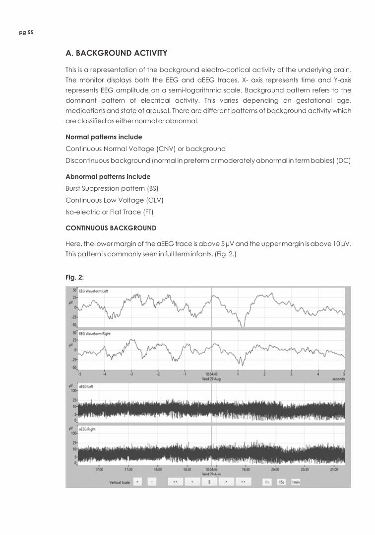
A. BACKGROUND ACTIVITY
This is a representation of the background electro-cortical activity of the underlying brain.
The monitor displays both the EEG and aEEG traces. X- axis represents time and Y-axis
represents EEG amplitude on a semi-logarithmic scale. Background pattern refers to the
dominant pattern of electrical activity. This varies depending on gestational age,
medications and state of arousal. There are different patterns of background activity which
are classified as either normal or abnormal.
Normal patterns include
Continuous Normal Voltage (CNV) or background
Discontinuous background (normal in preterm or moderately abnormal in term babies) (DC)
Abnormal patterns include
Burst Suppression pattern (BS)
Continuous Low Voltage (CLV)
Iso-electric or Flat Trace (FT)
CONTINUOUS BACKGROUND
Here, the lower margin of the aEEG trace is above 5 µV and the upper margin is above 10 µV.
This pattern is commonly seen in full term infants. (Fig. 2.)
Fig. 2:
pg 55

DISCONTINUOUS PATTERN
EEG: In discontinuous pattern, there are periods of lower amplitude interspersed with periods
of higher amplitude.
aEEG: There is a wide depressed trace with a lower voltage of <5µV and upper voltage of
>10µV. (Fig. 3.)
Fig. 3:
INTERPRETATION: Discontinuous pattern may be normally seen in prematurity. However, it is
sometimes encountered after cerebral injury in term babies and after anti-convulsant
therapy. In very preterm babies, the discontinuous pattern mimics burst suppression and is
called “tracédiscontinu”. As the premature infant approaches term, the pattern changes
to a continuous pattern.
BURST SUPPRESSION PATTERN
EEG: There are periods of abnormally high amplitude activity (bursts) interspersed with
periods of markedly suppressed amplitude (suppression).
aEEG: There are burst spikes (with amplitudes of >25µV) interspersed with iso electric
suppression where there is no or minimal electrical activity with flat lower margins having
voltage 0-1µV. (Fig.4.)
Fig. 4:
pg 56

INTERPRETATION: Prolonged burst suppression pattern is highly suggestive of severe brain
injury and is often seen in asphyxiated babies with severe encephalopathy. Since EEG
inactivity is associated with damage to non-specific but widespread regions of the brain,
the time of interburst interval (IBI) (the period of suppressed activity between bursts) may
correlate with long term outcome. It is thought that there is high probability of adverse
neurological outcome if IBI exceeds 30 seconds.
CONTINUOUS LOW VOLTAGE PATTERN
EEG: There is continuous low amplitude
aEEG: There is narrowing of EEG trace with both the upper and lower margins having and
amplitude of <5µV. (Fig. 5.)
Fig. 5:
INTERPRETATION: A prolonged continuous low voltage pattern is highly suggestive of
severe brain injury. This is often seen in asphyxiated babies with severe encephalopathy.
FLAT TRACE
EEG: In this pattern, the amplitude is very low (close to zero)
aEEG: aEEG shows low amplitude with a narrow trace , upper and lower margins are
<5µV and almost close to zero µV (Fig. 6.)
Fig. 6:
pg 57
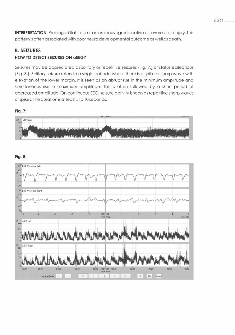
INTERPRETATION: Prolonged flat trace is an ominous sign indicative of severe brain injury. This
pattern is often associated with poor neuro developmental outcome as well as death.
B. SEIZURES
HOW TO DETECT SEIZURES ON aEEG?
Seizures may be appreciated as solitary or repetitive seizures (Fig. 7.) or status epilepticus
(Fig. 8.). Solitary seizure refers to a single episode where there is a spike or sharp wave with
elevation of the lower margin. It is seen as an abrupt rise in the minimum amplitude and
simultaneous rise in maximum amplitude. This is often followed by a short period of
decreased amplitude. On continuous EEG, seizure activity is seen as repetitive sharp waves
or spikes. The duration is at least 5 to 10 seconds.
Fig. 7:
Fig. 8:
pg 58

C. SLEEP WAKE CYCLE (SWC)
Normal sleep wake cycle is seen as smooth sinusoidal variations, mostly in the minimum
amplitude. Continuous pattern with good variability and no repetitive waveform (like spike
or sharp waves) is witnessed while the newborn is awake or in active sleep. This is seen as
narrowing of the wave on aEEG. This alternates with widening that represents discontinuous
pattern seen when a baby is in quiet sleep. A mature or developed cycle lasts about 20
minutes. Therefore, sleep wake cycles are seen as alternating widening and narrowing of
the aEEG trace.(Fig. 9.)
Fig. 9:
ASPHYXIA AND aEEG
Hypoxia or ischemia is associated with suppression of the EEG waveform. The duration and
amplitude of suppression can correlate with the severity of the hypoxic event. Thus, early
aEEG background pattern may be used as an adjunct in selecting newborn who may
benefit by therapeutic hypothermia. Various researchers have also tried to use the aEEG
trace either with or without hypothermia as a prognosticator.
aEEG AS CRITERIA TO INITIATE HYPOTHERMIA
The TOBY trial in the UK used three criteria to cool newborn:
1. Evidence of asphyxia
2. Evidence of moderate or severe encephalopathy
3. aEEG evidence of hypoxic injury. This included a 30 minute aEEG with any of the
following:
a. normal background with some seizure activity
b. moderately abnormal activity
c. suppressed activity
d. continuous seizure activity
Similarly, the Cool cap trial and the European trials on hypothermia used aEEG as a criteria
for recruitment. However, the NICHD trial and the ICE trial did not have aEEG as entry criteria
to initiate cooling. Though we have been using aEEG in asphyxiated newborn since 2009
and have practised therapeutic hypothermia since 2007, we do not use aEEG abnormality
as essential criteria for cooling newborn.
pg 59

A study by Skranes et al from Norway assessed if aEEG as an entry criteria improved selection
of infants for cooling. They compared the trace at 6 hours of life with MRI lesion load and 24
months outcome. They found that babies most babies with continuous normal background
at entry were normal at 24 months. Babies with severe EEG voltage criteria (BS,CNV or FT)
were more likely to have adverse outcome with time to normal trace in these babies being
the best predictor of outcome. Hence they suggested that recruitment for cooling should
be only in babies who had abnormal aEEG background.
aEEG AS A PROGNOSTICATOR
Researchers have looked at the prognostic value of early aEEG (at <6 hours or prior to
initiation of cooling) as well as changes in aEEG over time to assess if it would prognosticate
on long term neurological outcome. The results when comparing early aEEG to outcome
have been variable.
In a follow up analysis of the TOBY trial data, the value of early aEEG to accurately predict
death or disability at 18 months was studied. In a cohort of 315 babies, it was seen that the
positive predictive value (PPV) of a severely abnormal aEEG by voltage and pattern was
0.63 and 0.59 respectively in non-cooled infants and 0.55 and 0.51 in cooled infants (p>0.05).
The authors attributed the lower PPV in cooled babies to the neuro protective effect of
cooling.
Similarly, Marianne Thoresen et al studied the value of early as well as changes in pattern
and voltage over 72 hours in cooled and non-cooled babies. They recruited seventy-four
infants whose outcome was assessed at 18 months. The positive predictive value of an
abnormal aEEG pattern at <6 hours was 84% in non-cooled babies vs 59% for cooled infants.
The recovery time to normal background pattern was the best predictor of poor outcome in
both cooled and non-cooled babies (96.2% and 90.9% respectively). Never developing
sleep wake cycling over 72-96 hours always predicted poor outcome. Infants treated with
hypothermia had good outcome if the background became continuous by 48 hours.
Thus, early aEEG abnormalities are more likely to predict adverse outcome in babies who do
not receive hypothermia. Both these studies show that early aEEG may not accurately
predict outcome in cooled infants. Change in pattern from abnormal (BS. CNV or FT) to
continuous normal voltage by 48 hours predicted good outcome and never achieving
sleep wake cycle by 96 hours uniformly predicted poor outcome.
Chandrasekharan et al published a meta-anlysis of all studies using aEEG for
prognostication. This looked at aEEG criteria at various time periods (6, 24, 48 and 72 hours).
They considered only continuous background as normal pattern and all other patterns (DC,
BS, CLV and FT) as abnormal and used death or disability at 1 year as adverse outcome
measure. Nine studies involving 520 infants were used in the meta-analysis. The pooled
sensitivity and specificity for an abnormal trace at 6 hours of age in predicting adverse were
96% and 39%. The diagnostic odds ratio of an abnormal trace was highest (66.9%). They also
showed that a normal background at 6 hours predicted good outcome.
pg 60

Similarly, a systematic review of prognostic tests in term infants with HIE showed that aEEG
had the highest sensitivity and specificity in predicting outcome (0.93 and 0.9 respectively)
followed by EEG, visual evoked potential. Diffusion weighted MRI had higher specificity
whereas T1, T2 weighted MRI had better sensitivity.
WHAT IS THE EFFECT OF COOLING ON aEEG?
In most infants with asphyxia, the aEEG background will eventually become normal over
few days. As seen in the few studies quoted, aEEG may normalize earlier in cooled infants
because of the neuro protective role of hypothermia. A study was done to see if the
amplitude of the aEEG trace would change with cooling but no change was found.
However, it is possible that the amplitude of seizures can decrease with cooling and hence it
may be more difficult to diagnose electrical seizures in cooled infants. An EEG is always
necessary to diagnose seizures in hypothermis infants. Studies also show that the frequency
and duration of seizures is less in hypothermic infants. Hence aEEG may be a poor to
diagnose electrical seizures in cooled babies.
Therefore, aEEG may be used as an adjunct to clinical criteria to select babies for cooling.
Babies with continuous normal background at 6 hours may not benefit much with cooling.
Babies with persistently abnormal trace are most likely to have death or adverse long term
neurological outcome. Seizures may not be adequately detected by aEEG especially in
cooled babies and hence it may useful to study the raw EEG to identify seizures. Hence
aEEG may be a useful adjunct in babies who are offered therapeutic hypothermia.
TAKE HOME POINTS
Ÿ Though not essential, amplitude integrated EEG may be a useful tool to identify babies
who would most benefit by hypothermia.
Ÿ Continuous normal background at initiation of hypothermia is a good prognosticator.
Ÿ Persistence of abnormal trace at 48 or 72 hours may indicate poor prognosis.
Ÿ Never achieving sleep wake cycle by 96 hours always denotes poor long term prognosis.
Ÿ Seizures cannot be adequately diagnosed by aEEG especially in babies undergoing
therapeutic hypothermia.
pg 61

1. Hellström-Westas L, De Vries LS, Rosén I. An atlas of amplitude-integrated EEGs in the newborn.
Parthenon Publishing; 2003.
2. Shah NA, Wusthoff CJ. How to use: amplitude-integrated eeg (aeeg). Archives of Disease in
Childhood-Education and Practice. 2015 Apr 1;100(2):75-81.
3. Azzopardi D. Predictive value of the amplitude integrated EEG in infants with hypoxic ischaemic
encephalopathy: data from a randomised trial of therapeutic hypothermia. Archives of Disease
in Childhood-Fetal and Neonatal Edition. 2013 Jun 22:fetalneonatal-2013.
4. Thoresen M, Hellström-Westas L, Liu X, de Vries LS. Effect of hypothermia on amplitude-integrated
electroencephalogram in infants with asphyxia. Pediatrics. 2010 Jul 1;126(1):e131-9.
5. Chandrasekaran M, Chaban B, Montaldo P, Thayyil S. Predictive value of amplitude-integrated
EEG (aEEG) after rescue hypothermic neuro protection for hypoxic ischemic encephalopathy: a
meta-analysis. Journal of Perinatology. 2017 Jun 1;37(6):684-9.
pg 62
BIBLIOGRAPHY

CRANIAL ULTRASONOGRAPHYIN PERINATAL ASPHYXIA
Technique: Brain is usually visualized through anterior fontanelle in two
planes namely the coronal and sagittal. The angle of the transducer
may be changed to evaluate the periphery of the brain with special
attention to the subcortical white matter and the grey-white matter
differentiation. The visualization of the cerebellar hemispheres is
enhanced by obtaining images through the right and left mastoid
fontanels. This technique has also been shown to improve the
detection of posterior fossa hemorrhages. The modern cranial US
requires switching from the curved - to a linear - array transducer,
which allows high-resolution imaging of the brain with the detailed
interrogation of the subarachnoid space and superficial cortex as well
as deeper brain structures. Important factors influencing USG findings
are brain maturity, the duration and severity of insult, and timing of
imaging studies.
CHAPTER
11
Cranial
ultrasonography
(CUG) is the first
choice for initial
radiological
evaluation as it is
very convenient,
portable and
non-invasive.
Dr. Prakash Amboiram
Fig. 1: Brain structures as seen in CUG
Thalamus
Caudate
Caudothalamc groove
Acute changes in grey–scale sonography
The brain sites that are more vulnerable to hypoxic injury are
determined largely by maturity of the brain. Less intense insults cause
intraventricular hemorrhages and periventricular white matter injury in
preterm neonates and parasagittal watershed territory infarcts in term 1neonates. Severe hypoxia-ischemia in both preterm and term
Cranial ultrasonography (CUG) is the first choice for initial radiological
evaluation as it is very convenient, portable and non-invasive.

pg 64
neonates preferentially damages the deep grey matter like Basal ganglion (BG) and
thalamus. The neurons of the cerebral cortex are the most vulnerable, most often
hippocampus. Regional vascular factors play a certain role because of neuronal injury is
more marked in vascular water-shed zones during ischemia, in parasagittal cerebral 3cortex. The brainstem, cerebellum, and the deep grey matter are generally spared from
injury in mild to moderate hypoxic-ischemic insults due to auto regulatory mechanisms.
However, more prolonged insults may cause injury to the watershed zones, which are
relatively hypoperfused as a result of shunting during a pressure passive state of cerebral 7circulation.
USG changes in HIE can be classified as:
• Peripheral - mild to moderate Hypoxic ischemic insult
• Central - severe Hypoxic ischemic insult
Peripheral brain abnormalities include changes in the grey-white matter differentiation and
changes in the echogenicity of the cortex and subcortical white matter (Fig. 2&3). Early US
findings include a global increase in cerebral echogenicity and the obliteration of the CSF
spaces, suggesting diffuse cerebral oedema (Fig. 2,3,4). However, the small size of liquor
spaces alone is not specific for brain oedema and slit like ventricles can be seen in healthy 4newborns during the first 3 days of life.
An increase of parenchymal echogenicity with partial or total obliteration of normally visible
structures is correlated with severity of HIE. Although the phenomenon of increased
echogenicity (the so- called “bright brain”) is fully developed by the second–third day of
life, the moderate to severe increase of parenchymal echogenicity as early as at the age of
12 ± 2 h has a high sensitivity (88%) for death or severe disability and can be an early 4predictor of permanent brain damage in asphyxiated infants. By the second-third day of
5life, the predictive value of the “bright brain“ phenomenon can be as high as 90-100% .
Long-lasting increase in parenchymal echogenicity (5-7 days and more) is a predictor of 4,5poor outcome at 18 months of age.
Fig. 2: Asymmetrical
hyperechoic
appearance of the brain
in perinatal asphyxia

Central brain abnormalities in infants with severe global asphyxia include changes in
echogenicity of the deep grey matter (basal ganglia and thalami), brainstem and the 6 periventricular white matter (Fig. 5, 6). The presence of thalamic echogenicity (so-called
“bright thalamus”) generally suggests a more severe injury and correlates with a poorer 8,9 outcome Increased echogenicity in the basal ganglia, thalami, and brainstem is readily
5,10apparent after 7 days of severe HIE.
Fig. 3:Left: Coronal US image shows ill-defined structures including basal ganglia, and slit like ventricles at the age of 12 hours.
Right: Magnified sagittal view using linear transducer shows the accentuation of the grey-white matter differentiation with prominent sulci at the age of 12 hours.
Fig. 4: Focused view of the periphery shows sulci effacement and blurring of the gray-white matter differentiation
pg 65
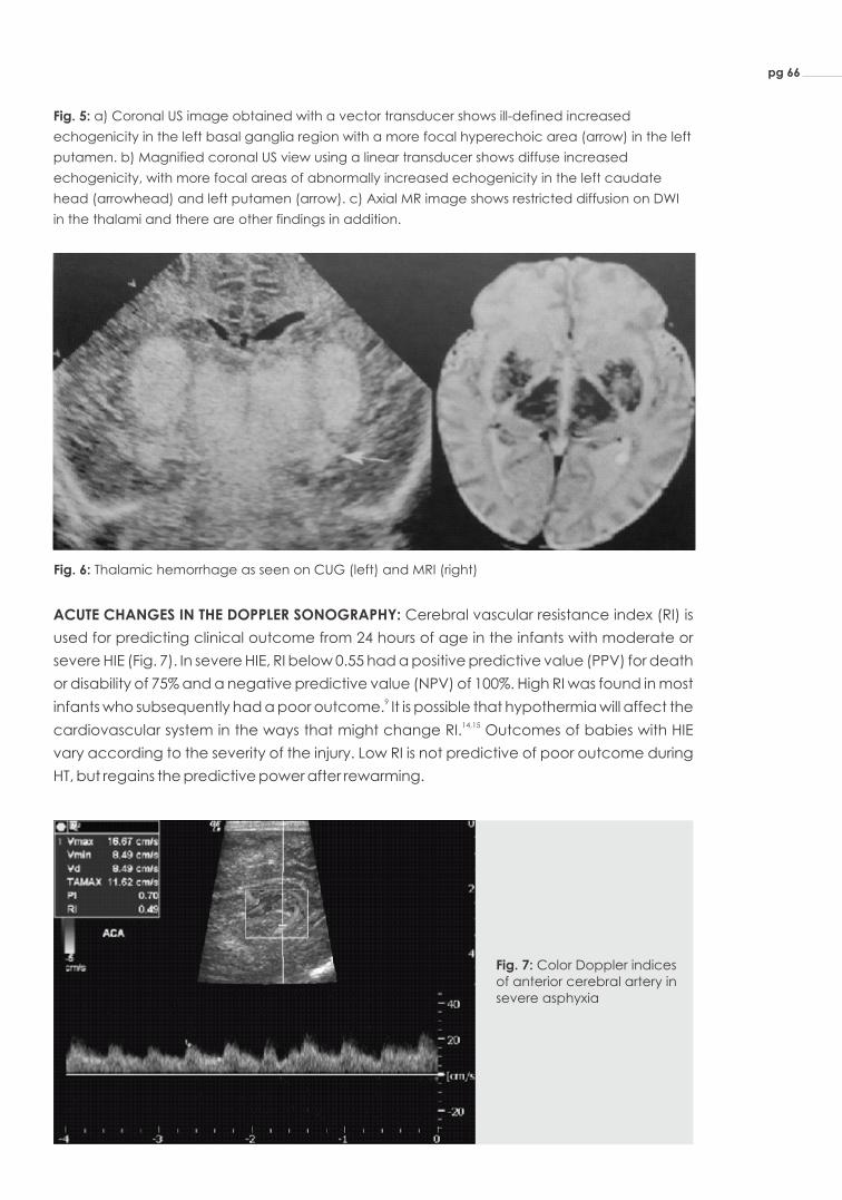
Fig. 5: a) Coronal US image obtained with a vector transducer shows ill-defined increased
echogenicity in the left basal ganglia region with a more focal hyperechoic area (arrow) in the left
putamen. b) Magnified coronal US view using a linear transducer shows diffuse increased
echogenicity, with more focal areas of abnormally increased echogenicity in the left caudate
head (arrowhead) and left putamen (arrow). c) Axial MR image shows restricted diffusion on DWI
in the thalami and there are other findings in addition.
Fig. 6: Thalamic hemorrhage as seen on CUG (left) and MRI (right)
ACUTE CHANGES IN THE DOPPLER SONOGRAPHY: Cerebral vascular resistance index (RI) is
used for predicting clinical outcome from 24 hours of age in the infants with moderate or
severe HIE (Fig. 7). In severe HIE, RI below 0.55 had a positive predictive value (PPV) for death
or disability of 75% and a negative predictive value (NPV) of 100%. High RI was found in most 9infants who subsequently had a poor outcome. It is possible that hypothermia will affect the
14,15cardiovascular system in the ways that might change RI. Outcomes of babies with HIE
vary according to the severity of the injury. Low RI is not predictive of poor outcome during
HT, but regains the predictive power after rewarming.
Fig. 7: Color Doppler indices of anterior cerebral artery in severe asphyxia
pg 66

EVOLUTION OF SONOGRAPHIC CHANGES: The best way to visualize white matter is at 7.5 MHz
or higher frequencies. Sonographic changes in the earlier phase are uncertain as cell
reaction and microvascular changes develop after 48 hours. Under normal conditions a
difference in echogenicity can be observed between relatively hypoechoic cortex and
mildly hyperechoic white matter. Gyral white matter core echogenicity can be compared
in patients in a sagittal or coronal section through the gyrus cinguli. Early indicators hinting at
the need for further imaging are 1) fuzzy, swollen brain, 2) relative hypoechoic aspect of
caudate, 3) appearance on day 2 or day 3 of four hyperechoic columns in coronal section
through deep grey matter, 4) luxury perfusion or reversed diastolic flow in large arteries, 5)
early hyperechoic haemorrhage in ischemic areas and 6) gyral core hyperechogenicity.
MRI and CUS: in the recently published data shows that modern CUS can pick up
abnormalities including parenchymal almost similar to MRI except focal lesions near the 6,17cortex.
IN SUMMARY
1. CUG is an excellent tool in screening and monitoring of asphyxiated infants with HIE
and evaluating the prognosis of asphyxiated infants.
2. CUS may miss some lesions as compared to MRI but still very useful especially in
babies who are critical.
3. In the infants with HIE treated with therapeutic hypothermia, the low resistance
index is probably not as good a predictor of a poor outcome as in normothermia
and further studies are needed in this area.
4. Repeat CUG at regular intervals may improve identification of brain abnormalities
especially in centers where MRI facility is not available.
pg 67

1. Huang, BY. & Castillo, M. Hypoxic-ischemic brain injury: imaging findings from birth to
adulthood. Radiographics2008; 28: 417–439.
2. Barkovich, AJ. MR and CT evaluation of profound neonatal and infantile asphyxia. Am J
Neuroradiol1992;13: 959–972.
3. Volpe JJ. Neurology of the newborn. WB Saunders Company. 5th ed 2008.
4. Ilves, P., Lintrop, M., Talvik, I., Muug, K., Maipuu, L. &Metsvaht, T. Low cerebral blood flow
velocity and head circumference in infants with severe hypoxic ischemic encephalopathy
and poor outcome. Acta Paediatr 2009; 98: 459–465.
5. Babcock, DS. & Ball, W. Jr. Postasphyxial encephalopathy in full-term infants: US diagnosis.
Radiology 1983; 148: 417–423.
6. Epelman, M., Daneman, A., Kellenberger, CJ., Aziz, A., Konen, O., Moineddin, R., White, H.
&Blaser, S. Neonatal encephalopathy: a prospective comparison of head US and MRI. Pediatr
Radiol2010; 40: 1640–1650.
7. Barkovich, AJ. 2005. Brain and spine injuries in infancy and childhood. In: Barkovich AJ,
ed.Pediatric neuroimaging: 190–290, 4th ed, Lippincott Williams & Wilkins, ISBN 978-1-4160-4918-
0, Philadelphia.
8. Connolly, B., Kelehan, P., O´Brien, N., Gorman, W., Murphy, JF., King, M. & Donoghue, V.The
echogenic thalamus in hypoxic ischaemic encephalopathy. Pediatr Radiol1994;24: 268–271.
9. Eken, P., Toet, MC. Groenendaal, F. & de Vries, LS. Predictive value of early neuroimaging,
pulsed Doppler and neurophysiology in full term infants with hypoxic-ischaemic
encephalopathy.“Arch Dis Child 1995; 73: F75–80.
10. Hertzberg, BS., Pasto, ME., Needleman, L., Kurtz, AB. & Rifkin, MD. Postasphyxial
encephalopathy in term infants: sonographic demonstration of increased echogenicity of the
thalamus and basal ganglia. J Ultrasound Med 1987; 6: 197–202.
11. North, K., & Lowe, L. Modern head ultrasound: normal anatomy, variants, and pitfalls that may
simulate disease. Ultrasound Clin 2009; 4: 497–512.
12. Lowe, LH.,& Bailey, Z. Sate-of-the-art cranial sonongraphy: Part I, modern techniques and
image interpretation. AJR Am J Roentgenol2011; 196: 1028–1033.
13. Bennhagen, RG., Weintraub, RG., Lundström, NR. &Svenningsen, NW. Hypoxic-ischaemic
encephalopathy is associated with regional changes in cerebral bblood flow velocity
and alterations in cardiovascular function. Biol Neonate 1998;75:275–286
14. Elstad, M., Whitelaw, A. &Thoresen, M. Cerebral resistance index is less predictive in
hypothermic encephalopathic newborns. Acta Paediatr 2011; 21.
15. Thoresen, M. & Whitelaw, A. Cardiovascular changes during mild therapeutic hypothermia and
rewarming in infants with hypoxic-ischemic encephalopathy. Pediatrics 2000;106: 92–99.
16. Archer, LN., Levene, MI. & Evans, DH. Cerebral artery Doppler ultrasonography for prediction of
outcome after perinatal asphyxia. Lancet 1986; 2: 1116–1118.
17. Wei Shen, Jia-Hua Pan, Wei-Dong Chen. Comparison of transcranial ultrasound and cranial
MRI in evaluations of brain injuries from neonatal asphyxia. Int J Clin Exp Med 2015;8(10):18319-
18326.
pg 68
BIBLIOGRAPHY

CHAPTER
12
MAGNETIC RESONANCE
IMAGING IN HYPOXIC ISCHEMIC
ENCEPHALOPATHY PROCEDURE
FOR WHOLE BODY COOLING
Hypoxic Ischemic Injury in term neonates causes global Hypoxic
Ischemic Injury (HII) in MRI. HII occurs due to interruption of placental
flow (antepartum), or events during delivery (intrapartum) or hypoxic
damage after delivery (postnatal). MRI imaging is the most sensitive
imaging technique to detect early hypoxic injury secondary to
perinatal asphyxia. MRI injury is also considerably less in infants who
have undergone TH. MRI helps in both diagnosis and prognosis of HIE.
This chapter has been written with objective of highlighting the various
hypoxic injuries occurring in term neonates.
ROLE OF NEUROIMAGING IN THERAPEUTIC HYPOTHERMIA (TH)
INCLUDE
Ÿ Diagnosis
Ÿ Assess the pattern and severity of injury
Ÿ Prediction of clinical outcome based on pathologic lesions
observed on imaging
Ÿ Information about long term prognosis
MR IMAGING- PRACTICAL TIPS
Transporting MR imaging in any sick neonate could be complex.
The following should be done:
Ÿ Proper Communication - ICU and MRI
Ÿ Replace standard length tubes with extension tubes
Ÿ Transport in incubator to maintain temperature
Ÿ Monitoring of vital signs during transport and scan
Hypoxic Ischemic
Injury in term
neonates causes
global hypoxic
ischemic injury
(HII) in MRI.
Dr. Umamaheswari B

pg 70
Ÿ Time taken to complete all sequences: 30 to 45 minutes
Ÿ Use MR compatible equipment: Pulse oximeter, ECG system, Infusion pump,
Ventilator, Resuscitation equipment. MR compatible incubator if available
Ÿ Temperature maintenance during the imaging
Ÿ Preventing Movement and Noise: Swaddling, Ear plugs/Muffs
Ÿ As far as possible avoid oral sedation, swaddling and timing after feeding if
permitted
Ÿ If HIE stage III, baby may be already drowsy especially with the use of anti-
convulsants
Ÿ If needed Oral sedation (Chloral hydrate) 25-50mg/kg could be used. Or
Intranasal sedation with Midazolam (100 microgram) could be used
PROTOCOL
Ÿ MRI-preferably 3 Tesla; if unavailable 1.5 Tesla could be used
Ÿ Sequences to be included: Axial and sagittal T1 Weighted Images (T1WI) and
T2WI, coronal T2 FLAIR(fluid attenuated inversion recovery), axial DWI(Diffusion
weighted Images) and GRE (Gradient recalled echo) images, MRA (MR
angiogram), MRV (MR venogram) and 1 or 2 MR spectroscopy sequences in
ventro-lateral thalamus and lentiform nucleus and centrum semi-ovale
(Calculate lactate/NAA ratio)
Ÿ MRI usually done between 5 and 14 days in infants who have undergone TH
PATTERN OF INJURY IN HIE IN TERM BABIES
Pattern of injury depends on the brain maturity, duration and severity of insult and type of
MRI sequencing and timing of imaging
Profound hypoxic injury
EventAcute severe event like cord prolapse, abruption, uterine rupture
Pathology
Pattern of Injury
Partial hypoxic injury
Prolonged partial hypoxia-ischemia like difficult delivery, maternal fever
Energy failure leading to cell necrosis followed by secondary energy failure and programmed cell death
Cortical necrosis; laminar necrosis and selective neuronal necrosis
Basal ganglia and thalamus (BGT) pattern and posterior limb of internal capsule (PLIC) pattern.If severe entire cerebrum affected
Cortical/subcortical pattern
HIE Stage II and III Clinical manifestation may be subtle
Sarnat Staging

Structures in MRI (Fig. 1) Corresponding anatomical structure (Fig. 2)
Term MRI normal structure
Fig. 3: T1 W image Fig. 4: T2 W image
Myelination of posterior limb of internal capsule (PLIC) appearing as hyperechoeic in T1W
and hypoechoeic in T2W images (Fig. 3 and 4.). Under normal circumstances, PLIC is
myelinated in term neonates. In HIE, myelination could be affected.
DW imaging detects the alteration in diffusion of free water. With acute injury, the
intracellular water content increases and there is restriction in the movement of water in and
out of the cell. Areas of diffusion restriction would be shown as increased signal. DW imaging
signal intensity starts appearing within1 to 2 days of injury, peak by 4 to 5 days and pseudo-
normalizes by day 7 of injury. T 1 and T2 W imaging changes starts by 3 days of injury and scan
on day 7 continue to show more obvious changes. Interpretation of MRI should take into
account the time of MRI imaging and the type of imaging.
INTERPRETATION OF NORMAL MRI
pg 71

stConventional T1 and T2WI are most useful at end of 1 week. BGT pattern indicates acute
hypoxic ischemic injury.
Fig. 6: DW changes (Diffusion weighted images in a term neonate with acute ischemic insult)
Fig. 6 is showing diffusion restriction in basal ganglia region. DW changes occur within 24 hours of stinjury, peaks at 5 days of life and pseudo-normalizes by end of 1 week.
PATTERN OF MRI INJURY
BGT-Basal Ganglia thalamus pattern: Occur with or without PLIC changes
PLIC- Posterior limb of internal capsule myelination changes
Cerebral cortex and sub cortical white matter pattern
Fig. 5: BGT pattern (With or without PLIC changes) (T1W Images)
pg 72
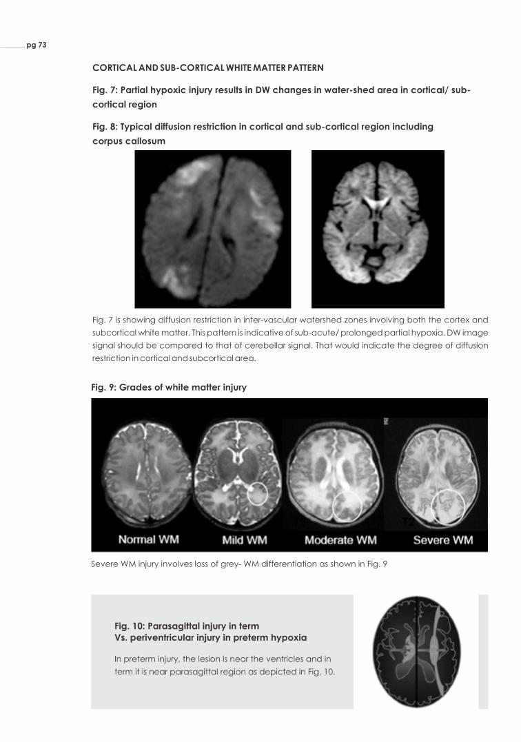
CORTICAL AND SUB-CORTICAL WHITE MATTER PATTERN
Fig. 7: Partial hypoxic injury results in DW changes in water-shed area in cortical/ sub-
cortical region
Fig. 8: Typical diffusion restriction in cortical and sub-cortical region including
corpus callosum
Fig. 7 is showing diffusion restriction in inter-vascular watershed zones involving both the cortex and
subcortical white matter. This pattern is indicative of sub-acute/ prolonged partial hypoxia. DW image
signal should be compared to that of cerebellar signal. That would indicate the degree of diffusion
restriction in cortical and subcortical area.
Fig. 9: Grades of white matter injury
Severe WM injury involves loss of grey- WM differentiation as shown in Fig. 9
Fig. 10: Parasagittal injury in term Vs. periventricular injury in preterm hypoxia
In preterm injury, the lesion is near the ventricles and in
term it is near parasagittal region as depicted in Fig. 10.
pg 73
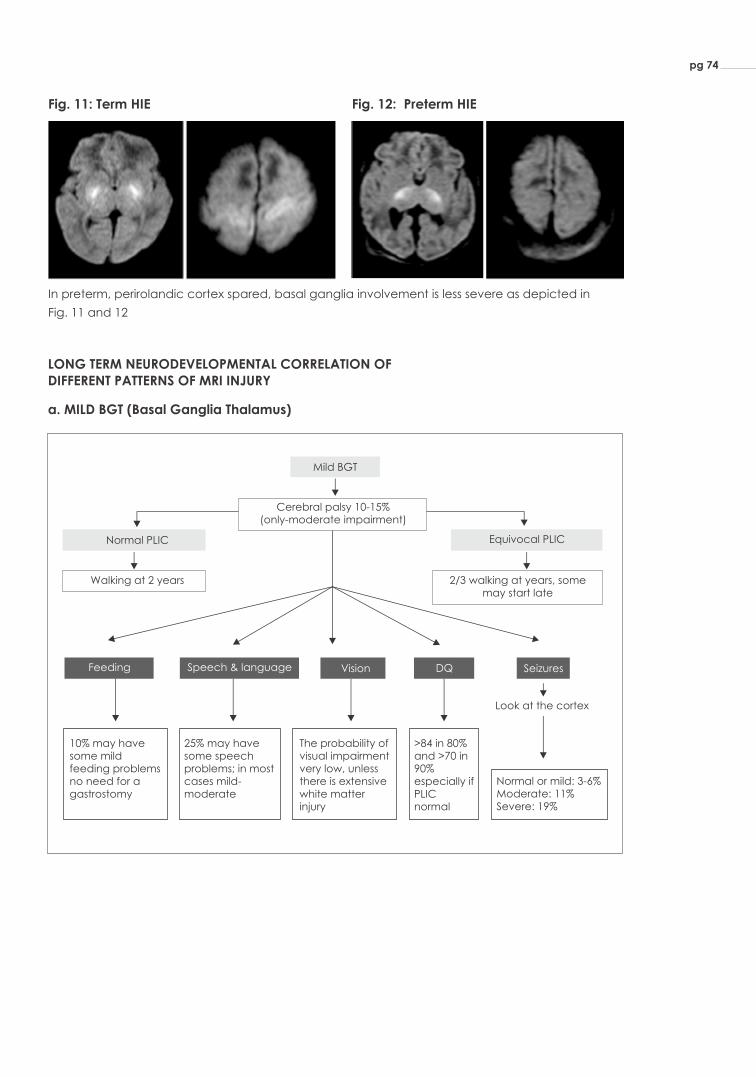
Fig. 11: Term HIE Fig. 12: Preterm HIE
In preterm, perirolandic cortex spared, basal ganglia involvement is less severe as depicted in
Fig. 11 and 12
LONG TERM NEURODEVELOPMENTAL CORRELATION OF DIFFERENT PATTERNS OF MRI INJURY
a. MILD BGT (Basal Ganglia Thalamus)
pg 74
Normal PLIC
Walking at 2 years
Equivocal PLIC
2/3 walking at years, somemay start late
Feeding Speech & language Vision DQ Seizures
10% may have some mild feeding problems no need for a gastrostomy
25% may have some speech problems; in most cases mild- moderate
The probability of visual impairment very low, unless there is extensive white matter injury
Normal or mild: 3-6% Moderate: 11% Severe: 19%
>84 in 80% and >70 in 90% especially if PLIC normal
Look at the cortex
Mild BGT
Cerebral palsy 10-15%(only-moderate impairment)
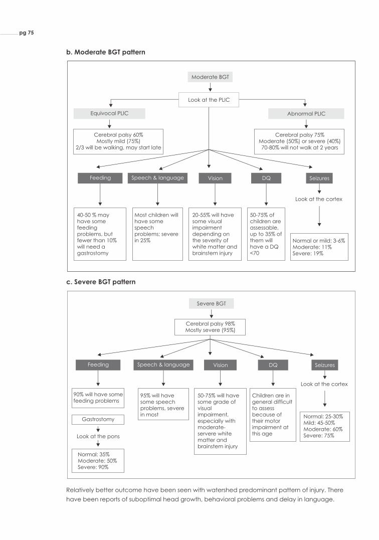
90% will have some feeding problems
95% will have some speech problems, severe in most
50-75% will have some grade of visual impairment, especially with moderate-servere white matter and brainstem injury
Children are in general difficult to assess because of their motor impairment at this age
Look at the cortex
Severe BGT
Cerebral palsy 98%Mostly severe (95%)
Normal: 25-30%Mild: 45-50% Moderate: 60% Severe: 75%
Gastrostomy
Look at the pons
Normal: 35%Moderate: 50% Severe: 90%
Feeding Speech & language Vision DQ Seizures
c. Severe BGT pattern
b. Moderate BGT pattern
Relatively better outcome have been seen with watershed predominant pattern of injury. There
have been reports of suboptimal head growth, behavioral problems and delay in language.
pg 75
Equivocal PLIC
40-50 % may have some feeding problems, but fewer than 10% will need a gastrostomy
Most children will have some speech problems; severe in 25%
20-55% will have some visual impairment depending on the severity of white matter and brainstem injury
50-75% of children are assessable, up to 35% of them will have a DQ <70
Look at the cortex
Moderate BGT
Look at the PLIC
Abnormal PLIC
Cerebral palsy 60% Mostly mild (75%)
2/3 will be walking, may start late
Cerebral palsy 75% Moderate (50%) or severe (40%) 70-80% will not walk at 2 years
Normal or mild: 3-6% Moderate: 11% Severe: 19%
Feeding Speech & language Vision DQ Seizures

MRS in TH
Concepts of MRS
MRS acquires signals from metabolites in predefined regions of brain. Regions to be
included are deep grey matter and cortical region. The signals are acquired from choline
(Cho), creatine (Cr), N-acetylaspartate (NAA) in normal status. Lactate peaks in Hypoxic
damage due to alteration in oxidative metabolism.
Marker
N-acetylaspartate (NAA) Neuron and their processes; Indicative of intact central nervous tissue
Membrane turnover & myelination
Brain metabolism
Anaerobic metabolism
Value of 0.62 indicative of poor neurodevelopmental outcome (De Vries 2001)
Value >0.67 predictive of death or severe CP at 2 years (Ancora 2013)
Value of > 0.29 in deep grey matter is indicative of poor neurodevelopmental outcome(Thayill 2010); Good marker for severity in sub-acute to chronic phase
Choline (Cho)
Creatine (Cr)
Lac/NAA ratio
Metabolite
Lactate
NAA/Cho
NAA/Cr ratio
Value of > 1 indicative of poor neurodevelopmental outcomeLac/Cho & Lac/Cr ratio
Fig. 13 Fig. 14
Fig. 13: MRS in occipital region shows lactate peak (superimposed with lipid peak), Choline levels
are low indicating disrupted myelin
Fig. 14: MRS line diagram showing different peaks. NAA - N acetylaspartate; Cr - Creatine; Lac -
Lactate; Cho - Choline
pg 76

pg 77
MRI IN INFANTS WITH HIE WHO HAVE UNDERGONE THERAPEUTIC HYPOTHERMIA (TH)
In babies undergoing Therapeutic Hypothermia, there are certain questions which need
to be answered:
Ÿ Q1. When do we obtain MRI?
Ÿ Q2. Is there any specific MRI pattern in these babies?
Ÿ Q3. Does TH reduce the MRI abnormalities in babies with asphyxia?
Ÿ Q4. Does TH alter the predictive value of MRI in HIE?
Q1. When do we obtain MRI?
Ans. Mean 8 days for T1, T2 changes - Obtain in 1-2 weeks, DW changes 3-5days -
Within 1 week; Interpretation of MRI should take into account the type and
timing of MRI.
Q2. Is there any specific MRI pattern in these babies?
Ans. No; Same BGT pattern with or without PLIC changes and cortical and sub-cortical
pattern occurs in infants undergoing TH. Smaller studies have shown that pseudo-
normalization of diffusion restriction changes occurred later.
Q3. Does TH reduce the MRI abnormalities in babies with asphyxia?
Ans. Yes; In TOBY trial, they found that among 131 infants treated with TH, less BGT
injury and fewer abnormalities of PLIC were identified.
Q4: Does TH alter the predictive value of MRI in HIE?
Ans. No; MRI retains the predictive value and abnormal MRI predicts adverse neuro-
developmental outcome. The pooled sensitivity and specificity for DW imaging is
58% and 89%, respectively. Sensitivity and specificity for T1/T2 changes done ≤ 1
week is 84% and 90%, respectively. If done ≤ 2 wk, the sensitivity and specificity is
98% and 76%, respectively.
CONCLUSION
Ÿ MRI imaging is the most sensitive imaging technique to detect early hypoxic injury
secondary to perinatal asphyxia.
Ÿ MRI is done between 5 and 14 days of life. Interpretation should take into
consideration the type of imaging and the timing. DW changes occurs within 24,
peaks in 3-4 days and pseudo-normalizes in 7 days, T1 and T2 changes are evident in
3 days and scan by 7 days continues to show new changes.
Ÿ MRI injury is less with TH. Both BGT and cortical/sub-cortical injury were observed less in
those undergoing cooling.
Ÿ MRI remains as a good predicting tool for the outcome in infants undergoing TH.
Ÿ MRS in deep grey matter region measuring the ratios of Lac/NAA, Lac/Cho & Lac/Cr
and NAA/Cr & NAA/Cho helps in predicting long term adverse outcome.

1. Barnette AR, Horbar JD, Soll RF, et al. Neuroimaging in the evaluation of
neonatal–encephalopathy. Pediatrics 2014;133:e150817.
2. Miller SP, Ramaswamy V, Michelson D, et al. Patterns of brain injury in term–neonatal
encephalopathy. J Pediatr 2005;146:45360.
3. Rutherford M, Counsell S, Allsop J, et al. Diffusion-weighted magnetic resonance imaging in
term perinatal brain injury: a comparison with site of lesion and time–from birth. Pediatrics
2004;114:100414.
4. Gano D, Chau V, Poskitt KJ, et al. Evolution of pattern of injury and quantitative MRI on days 1
and 3 in term newborns with hypoxic-ischemic encephalopathy.– Pediatr Res 2013;74:827.
5. signal in the internal capsule predicts poor neurodevelopmental outcome in infants with hypoxic-
ischemic encephalopathy. Pediatrics 1998; 102: 323-8
6. Thayyil S, Chandrasekaran M, Taylor A, et al. Cerebral magnetic resonance bio-markers in
neonatal encephalopathy: a meta-analysis. Pediatrics 2010;125:–e38295.
7. Hayes BC, Ryan S, McGarvey C, et al. Brain magnetic resonance imaging and outcome after
hypoxic ischaemic encephalopathy. J Matern Fetal Neonatal–Med 2016;29:77782.
8. Bednarek N, Mathur A, Inder T, et al. Impact of therapeutic hypothermia on MRI–diffusion
changes in neonatal encephalopathy. Neurology 2012;78:14207.
9. Bonifacio SL, Saporta A, Glass HC, et al. Therapeutic hypothermia for neonatalencephalopathy
results in improved micro-structure and metabolism in the deep–gray nuclei. AJNR Am J
Neuroradiol 2012;33:20505.
10. Roelants-Van Rijn AM, Van Der Grond J, De Vries LS, et al. Value of (1)h-MRS us-ing different echo
times in neonates with cerebral hypoxia-ischemia. Pediatr Res–2001;49:35662.
11. Alderliesten T, de Vries LS, Benders MJ, et al. MR imaging and outcome of term neonates with
perinatal asphyxia: value of diffusion-weighted MR imaging and (1)–H MR spectroscopy.
Radiology 2011;261:23542.
pg 78
BIBLIOGRAPHY

FOLLOW UP OF
COOLED BABIES
CHAPTER
13
With improved
survival of high
risk neonates,
follow-up
programs are
becoming as
important as the
initial intensive
care itself.
Dr. Nivedita M
With improved survival of high risk neonates, follow-up programs are
becoming as important as the initial intensive care itself. In India owing 1,2to the high incidence of perinatal asphyxia, survivors of neonatal
encephalopathy (NE) form a significant proportion of all patients
enrolled in any follow-up program. It has long being recognized that
infants suffering from HIE deserve long term follow-up. With the advent
of therapeutic hypothermia (TH), it has become even more important
to follow-up these infants, in order to know the long term effects of TH.
NEED FOR FOLLOW-UP
Neonates suffering from perinatal asphyxia and neonatal
encephalopathy are at an increased risk of neuro motor/sensory
impairments. The Cochrane meta-nalysis of 11 RCTs on TH has shown
significant reduction in the composite outcome of mortality or major
neurodevelopmental disability at 18 months of age. It also showed a
significant reduction in neurodevelopmental disability in survivors 3(RR 0.77 (95% CI 0.63 to 0.94). Though TH significantly reduces
neuromorbidity as compared to controls, yet among the surviving
infants a sizeable number suffer from varying degrees of
neurodisablity, which emphasizes the need for rigorous long term
follow-up of these children. Shankaran et al have published outcomes
following TH at 6-7 years of age, in which though TH did not result in
significant reduction in the combined outcome of death or an IQ
score below 70 at 6 to 7 years of age, it was still shown to reduce the
rate of death with no increase in the rates of a low IQ score or severe 4disability among survivors. Since TH is now standard practice in NICUs,
it is essential that long term neurodevelopmental outcome data
become available soon, to determine if the benefit of TH at 1.5-2 years
is indeed sustained beyond early childhood.

pg 80
Survivors of TH need to be followed-up in the same manner as any other high-risk neonate, till
long term data revealing any specific pattern of neurodevelopmental sequelae is
available.
DURATION OF FOLLOW-UP
By 18-24 months of age motor impairments can be confirmed and formal assessment of
cognitive and language function can be performed with good prediction to early school
age performance. Major neuroimpairment usually becomes overt in early childhood itself.
However survivors of NE though apparently normal in early childhood may develop subtle 6,7cognitive and behavioural abnormalities later on.
Like other high risk infants, it is necessary to follow up cases of TH till 8-10 years of age, if
possible throughout their school years, even if they appear to be normal in early 8,9childhood. At the least, they should be followed up till 18-24 months of corrected age (CA).
FREQUENCY OF FOLLOW-UP
The first visit may be planned at 2 weeks post discharge. Thereafter at 3, 6, 9, 12, 18 and 24
months or 2, 4, 8, 12, 18 and 24 months corrected age. As the child needs to come at 6, 10
and 14 weeks for immunization, the initial few visits may be combined with these. Then on, 10,11every 6 monthly or yearly till follow-up lasts. The 18-24 months visit is mandatory for formal
development assessment. In case the child develops any medical or neurodevelopmental
concerns, more frequent visits will be required.
AGE CORRECTION
Corrected age is used for growth and development assessment of preterm (<37 weeks) till
24 months. Currently TH is offered only to neonates 36 weeks and above.
4,5

Responsibility
Pediatricians /
neonatologists
Coordinator
Assessment of growth and screening for developmental delay
Management of inter-current illnesses
Immunization
Pediatric neurologist
Child psychologist
Ophthalmologist
Personnel
Management of neuro-disablities, seizures
Formal development/IQ assessment
Screening and management of behavioral disorders
Assessment of visual acuity
Screening and management of strabismus and refractive errors
OtorhinolaryngologistHearing assessment
Management of hearing impairment
Dietician Management of infants with failure to thrive
Speech therapistLanguage assessment
Management of oromotor dysfunction
Medical social worker/counsellor
Counselling
Arranging disability certification, free bus pass etc.
parents
Physiotherapist/Occupational therapist
Physiotherapy and rehabilitation
Special educator Special education
Table 1: Roles of each member of the follow-up team
Those administering tests of neurodevelopmental assessment, depending upon the test
used might require formal training and certification.
FOLLOW-UP DURING NICU STAY
Findings of a structured neuro exam needs to be documented daily during and at least until 121 week after TH, though it is often not possible to perform a complete examination due to
the infant's sickness, intubation and effect of sedatives and anti-seizure medications. Sarnat
staging and Thompson scoring are two such examination methods. Levene's classification is
a simplified system, shorter and easy to perform.
Though significant correlation exists between developmental status at 2 years and the
Sarnat grade and persistently abnormal neuro exam on day 3, the best predictability of the
outcome at 2 years is seen with the neurological examination at discharge (positive and
pg 81

negative predictive values of 86% and 72% respectively). In infants with moderate Sarnat
scores, abnormal outcome occurred in 25% if the discharge examination was normal and in
70% if it was abnormal. The time taken to establish oral feeding also correlated significantly 13with neurological outcome (r=0.40, p=0.008).
The role of cranial sonography, MRI and EEG have been described in other chapters in
this manual.
POST DISCHARGE FOLLOW-UP
FEEDING HISTORY
Infants with severe NE may suffer from significant oromotor disturbances leading to
difficulties in feeding. Neonates suffering from perinatal asphyxia may be born with low birth
weight or intrauterine growth restriction (IUGR), in which case adequate post natal feeding
has to be ensured. A detailed history of the child's feeding pattern must be elicited inclusive
of history of difficulty in sucking/chewing/swallowing, choking episodes, drooling, repeated
lower respiratory infections and failure to thrive. Those with oromotor dysfunction need
prompt referral to an occupational therapist or a speech and language pathologist.
MEDICAL PROBLEMS
14 Epilepsy has been reported to develop in 9-33% of HIE. Though every attempt must be
made to wean off all anti-convulsants by discharge, some infants may have to be
discharged on anti-convulsant medication. Details of any episode of seizure post discharge
must be obtained.
Apart from seizure disorder, cases of cerebral palsy suffer from a host of other medical
problems as well like hypertonia, drooling, recurrent respiratory infections, skeletal
deformities and contractures etc. Relevant history must be elicited and examination
performed. As parents are often uneducated, instructions regarding medications are
poorly understood. They must be requested to bring with them all the medications that the
child is on at every visit, in order to explain prescriptions clearly. Also this way the physician
knows for certain all the medications that the child is on, as drug interactions have to be
borne in mind when prescribing new drugs.
IMMUNIZATION
Immunization must proceed in survivors of HIE in the same manner as that of normal children,
including those with seizure disorder.
GROWTH
Weight, length/height and head circumference must be accurately checked at each visit.
Weight should be checked on an electronic weighing scale, length up to 2 years must be
checked with an infantometer and head circumference with a non-stretchable tape.
pg 82

Adductor angle
Popliteal angle
80°-100°
90°-20°
110°-160°
150°-170°
Dorsiflexion angle
60°-70°
60°-70°
60°-70°
60°-70°
Scarf sign
Elbow crosses midline
Elbow goes beyond
axillary line
0-3
4-6
7-9
10-12
40°-80°
70°-110°
110°-140°
140°-160°
Elbow does not cross
midline
Age
(months)
Growth must be plotted in the WHO child growth standards. For preterms Fenton growth
charts (http://www.ucalgary.ca/fenton/2013chart) may be used till 40 weeks CGA.
NEUROLOGICAL ASSESSMENT
Neurological examination must be done at every visit with special attention to muscle tone.
The NNF recommends neuro-motor assessment of high risk infants at least once during the 11first 6 months, once during the second six months and once yearly.
Skull shape must be noted, fontanelles and sutures must be palpated.
Amiel-Tison neurological assessment at term (ATNAT) is a systematic way of performing the
neurological examination. If the full exam is not possible, then a general neuro-exam with
measurement of AT angles for assessment of muscle tone may be done.
Table 2. Amiel –Tison angles
ATNAT at term age has been shown to have good agreement with neurological examination 15(k=0.83) and BSID scores (MDI, k=0.64; PDI, κ=0.74) at 12-15 months of age.
Angles smaller and larger than the normal indicate hypertonia and hypotonia respectively
pg 83
Measuring the AT angles
Check the angles when the infant is in a quiet alert state. Maintain the head in midline.
Adductor angle-With the infant in supine position, hold both lower limbs extended and
pull them apart gently as far as possible. Measure the angle formed between the two
legs. Look for asymmetry between the two sides.
Popliteal angle-Bring the thighs by the side of the abdomen and extend the knee.
Measure the angle formed between the leg and the thigh. Check both sides
simultaneously.
Ankle dorsiflexion-Check each side separately. Extend the knee with one hand and with
the other apply slow pressure on the sole to flex the foot. Measure the angle formed
between the dorsum of the foot and the anterior aspect of the leg. This is the 'slow
angle'. Repeat the manoeuver with rapid flexion of the foot, this gives the 'rapid angle'.
Normally both angles are equal. A difference of more than 10° is abnormal.
Scarf sign-Lift the infant's head to 45°, hold the wrist and try to pull the hand towards the
opposite shoulder.

pg 84
MOTOR SKILLS
Observation of spontaneous movements of the infant (Prechtl's assessment of general
movements) has been found to be predictive of future motor development. The first stage is
that of writhing general movements (GMs) which are present inutero to 6 to 9 postterm
weeks and are characterized by small to moderate amplitude and slow to moderate speed
movements. From 9 to 20 postterm weeks, writhing movements are replaced by fidgety
movements involving the neck, trunk and limbs. These are small-amplitude movements of
moderate speed and variable acceleration in all directions.
Abnormal GM has been found to correlate with lesions in MRI in term infants with HIE.
Cramped-synchronized GM, a type of abnormal rigid movements, lacking fluency with the
limbs and trunk contracting and relaxing almost simultaneously have been found to
correlate with central gray matter lesions in the MRI. They have been reported to be 100% 16specific and 68.7% sensitive for prediction of cerebral palsy.
Assessment of GMs may be done during an outpatient visit by video recording the infant's 16spontaneous movements during wakeful state for 10-15 minutes at 1 month and 3 months.
imited even with the use of assistive technology
DEVELOPMENT ASSESSMENT
DEVELOPMENT SCREENING TEST
A developmental screening test has to be done once within the first 6 months, once within 11the next 6 months and yearly thereafter.
DIAGNOSTIC DEVELOPMENT TEST
A formal assessment of development is to be done at corrected age of 12 months and
again between 18-24 months. If an assessment has to be done before 12 months, it is best
Other methods of motor examination are the Alberta Infant Motor Scale (AIMS), Test of
Infant Motor Performance (TIMP) and Peabody Developmental Motor Scales (PDMS). In a
case of cerebral palsy, assessment of severity of disability is to be done using the GMFCS (The
Gross Motor Function Classification System ). It classifies the functional status of the child into 85 levels.
1. Walks without restrictions; limitations in more advanced gross motor skills
2. Walks without assistive devices; limitations walking outdoors and in the community
3. Walks with assistive mobility devices; limitations walking outdoors and in the community
4. Self-mobility with limitations; children are transported or use power mobility outdoors
and in the community
5. Self-mobility is severely l
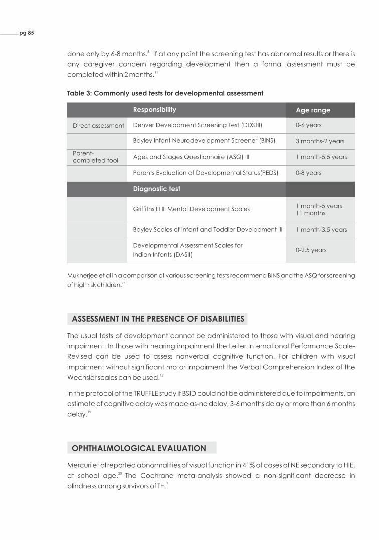
Table 3: Commonly used tests for developmental assessment
Mukherjee et al in a comparison of various screening tests recommend BINS and the ASQ for screening 17of high risk children.
Denver Development Screening Test (DDSTII) 0-6 years
Responsibility Age range
Direct assessment
Bayley Infant Neurodevelopment Screener (BINS)
Parent-completed tool
3 months-2 years
Ages and Stages Questionnaire (ASQ) III 1 month-5.5 years
Parents Evaluation of Developmental Status(PEDS) 0-8 years
Griffiths III III Mental Development Scales1 month-5 years 11 months
Diagnostic test
Bayley Scales of Infant and Toddler Development III 1 month-3.5 years
Developmental Assessment Scales for
Indian Infants (DASII)0-2.5 years
8done only by 6-8 months. If at any point the screening test has abnormal results or there is
any caregiver concern regarding development then a formal assessment must be 11completed within 2 months.
ASSESSMENT IN THE PRESENCE OF DISABILITIES
The usual tests of development cannot be administered to those with visual and hearing
impairment. In those with hearing impairment the Leiter International Performance Scale-
Revised can be used to assess nonverbal cognitive function. For children with visual
impairment without significant motor impairment the Verbal Comprehension Index of the 18Wechsler scales can be used.
In the protocol of the TRUFFLE study if BSID could not be administered due to impairments, an
estimate of cognitive delay was made as-no delay, 3-6 months delay or more than 6 months 19delay.
OPHTHALMOLOGICAL EVALUATION
Mercuri et al reported abnormalities of visual function in 41% of cases of NE secondary to HIE, 20 at school age. The Cochrane meta-analysis showed a non-significant decrease in
3blindness among survivors of TH.
pg 85

pg 86
Prior to discharge, red reflex must be checked in every neonate. At each visit visual
milestones must be elicited from the mother. In infants, clinical assessment of visual function
is mainly by fixation and following. One should also look for squint, roving eye movements
and nystagmus. With the infant lying down or seated on the mother's lap, a red wool, target
or any attractive toy can be used to check for fixation and following. The assessment must
be done binocularly and then monocularly. Care should be taken that the objects do not
make noise as the child may respond to the auditory rather than the visual signals. Visual
acuity can be formally assessed in young children using Cardiff cards or Teller Acuity Card.
Formal assessment by an ophthalmologist for refraction, strabismus and any other visual 11problems should be done at 1 year and annually thereafter till 5 years of age . Robertson et
al recommend early referral to a pediatric ophthalmologist in case of: HIE III, HIE II with
abnormal neurological examination or reduced visual awareness at hospital discharge and 9stroke associated with HIE.
HEARING EVALUATION
Of HIE survivors, up to 17% of those with and 6.3% of those without residual neurological 21deficits, have been found to have hearing loss. Incidence of permanent hearing loss has
22been reported to be 6% to 10% in follow-up cases of TH. The Cochrane meta-analysis of
seven studies failed to show any significant reduction in the incidence of sensorineural 3hearing loss requiring aid in neonates subjected to TH.
Hearing screening is to be done via Brainstem Auditory Evoked Responses (BAER/BERA), as
OAE misses auditory neuropathy. However if unavailable, then at least a screening OAE
must be done. Hearing screening is usually prior to discharge, in any case it must be 23completed before 1 month of age. Mietzsch et al in a pilot study in moderate to severe HIE
reported transient suppression of DPOAEs and prolongation of ABR waveforms in the first
week which normalized by the third week. They suggested that cooling might hasten
recovery of this transient auditory dysfunction. However this was only a pilot study with a 24 small sample size of 9 (4 cases and 5 controls). Further research will provide directions as to
whether screening in these infants should be delayed beyond the first week.
If the neonate fails the screening test, a diagnostic BERA has to be done before 3 months of 23age and appropriate intervention initiated before 6 months of age.
For those at high risk for hearing loss even if the initial screening test is passed, the NNF 11recommends a behavioral audiometry at 1 year of age. The American Academy of
Paediatrics recommends diagnostic audiologic testing between 24 and 30 months in the 25presence of risk factors. Perhaps it would be prudent to say that in follow-up cases of TH,
irrespective of the initial screen result, auditory milestones must be elicited at every visit
along with a clinical assessment of hearing and a repeat diagnostic test must be performed
between 1 to 2 years of age.

TFT SCREENING
Thyroid screening must be offered universally to all infants.
LANGUAGE ASSESSMENT
11The NNF recommends a language assessment between 1 and 2 years of age. Robertson et 9al recommend one between 3 and 5 years. Language assessment must include testing of
skills of expressive and receptive language, organization and grammar. Few of the tests
used are Receptive-Expressive Emergent Language Test, Peabody Picture Vocabulary Test
(PPVT), Linguistic Profile Test and Three Dimensional Language Acquisition Test (3D- LAT).
COGNITIVE ASSESSMENT
4Shankaran et al reported an IQ <70 in 27% of survivors of TH at 6-7 years of age. Survivors of
moderate encephalopathy have been found to have lower scores on school readiness test 7conducted at 5.5 years. Performance in intellectual, scholastic and neuropsychological
tests are uniformly poor in survivors of severe NE. Those with moderate NE show more varied
results with intelligence scores below those of children with mild NE and age-matched
peers, but within the normal range. They perform poorly in reading, spelling and 6mathematical skills. Cognitive dysfunction of low severity may not become apparent till the
child faces complex tasks, which will happen only at later school age. Due to these reasons,
cognitive assessment needs to be done during the child's schooling years.
Cognitive assessment should include assessment of intelligence, executive function,
sensorimotor functions (visual-motor precision, fine motor speed), visual spatial processes
(design-copying, visual closure) and memory and learning. IQ can be assessed from 3 years
of age. A battery of tests have to administered to the child at different ages (3-4 years, 6-7
years and 8-10 years of age )in order to complete a comprehensive assessment of cognitive 8functions.
In the Indian setting, extensive testing over several years would not be practicable. In our
country a practical approach might be to conduct an IQ test between 3-4 years and
repeat it at 8-10 years. IQ determined at 8 years predicts IQ at older age better than if 8 determined earlier. Like development assessment tests, the
al tests like NEPSY II (A developmental neuropsychological assessment)
are required in order to diagnose attention, memory, executive function and information
processing deficits.
re are a host of tests for assessing
intelligence. Worldwide the most commonly used tests are the Wechsler's tests. Wechsler's thPreschool and Primary Scale of Intelligence for young children (WPPSI, IV edition is
applicable from 2:6 – 7:7 years of age) and the Wechsler Intelligence Scale for Children th(WISC, V edition is for children aged 6:0–16:11 years) for older ones. Stanford-Binet
Intelligence Scales and Seguin Form Board (SFB) are other commonly used tests. Specific
neuropsychologic
pg 87

BEHAVIOURAL ASSESSMENT
Children suffering from moderate to severe NE are 5.9 times more likely to be diagnosed with 26an autism spectrum disorder as compared to controls at 5 years. Autism screening should
be conducted universally in all children between 18-24 months. Modified Checklist for
Autism in Toddlers (M-CHAT), Social Communication Questionnaire, Trivandrum Autism
Behavior Checklist are few of the tools available. Diagnosis can be established using
Childhood Autism Rating Scale (CARS), INCLEN diagnostic tool for ASD (INDT-ASD) or Indian 27Scale for Assessment of Autism (ISAA)
The Achenbach Child Behavior Check List (CBCL) is a good tool to identify behavioural
abnormalities based on parental perception of child's behaviour.
Assessment of functional status which would also reflect the quality of life can be measured
using the Vineland Adaptive Behavior Scales (VABS). Robertson et al recommends 9evaluation of adaptive behaviour at 3-5 years and the 8-10 years.
There is no consensus regarding the timing, frequency and tests for developmental
assessment in the follow-up of TH. If further research reveals a specific profile of
neurodevelopmental sequelae in this group of infants, that would guide the choice of tests
to be used. Till then each centre may design its protocol depending on the manpower,
patient load and funds available. The tests for development/behaviour assessment must 28 have a sensitivity and specificity of >/=70%. Some of the development assessment tools are
freely available, whereas others have to be purchased and are expensive. A reasonable
protocol is suggested in Table 3.
pg 88

2 weeks post discharge Med & Neuro exam
Screening BERA, thyroid screening if not done earlier
6 months
9 months
12 months
18-24 months
Med & Neuro exam
Clinical assessment of vision and hearing
Prechtl's assessment of general movements
Complete audiological evaluation if failed BERA
DDST II/BINS
Med & Neuro exam
DDST II/BINS
Med & Neuro exam
DDST II/BINS
1.5 months (immunization visit)
3 months
Med & Neuro exam
BSID III/DASII
Ophthalmological exam
Med & Neuro exam
BSID III/DASII
MCHAT
Behavioural audiometry
Ophthalmological exam
REELS III
Med & Neuro exam
DDST II/ASQ III
Ophthalmological exam (till 5 years)
WPPSI-IV
CBCL
VABS
WISC -V
CBCL
CBCL
Every year, after 2 years
3-4 years
8-10 years
The RBSK guidelines on examination and follow up of a child with suspected developmental delay are
easy to understand and implement. The website provides freely downloadable material
(http://nhm.gov.in/nrhm-components/rmnch-a/child-health-immunization/rashtriya-bal-swasthya-
karyakram-rbsk/2013-12-19-08-13-49.html)
pg 89

Detailed neurological examination must be performed and
documented at discharge.
Follow-up of cases of post-TH should be done in a structured manner.
Separate assessment of vision and hearing are mandatory.
Long term follow till at least till 8-10 years of age is needed.
KEY MESSAGE
HOW TO MAINTAIN GOOD FOLLOW-UP
8Follow-up rates must be at least 80% at the 18-24 months visit. In India most follow-up
programs face huge lost to-follow-up. Few measures that can help improve follow-up rates.
Enrol patients into the follow-up program before discharge itself. Prior to discharge counsel
the parents regarding the need for follow-up and inform them about the date, time and
venue of the first post-discharge visit.
Maintain a database of patient address and contact details. Collect back-up contact
numbers.
Make phone call or SMS reminders of the appointments. If patients miss an appointment,
call up and find out why and fix another date of visit.
Provide parents with the number of a contact person.
According to the NICHD network, if a single assessment alone is possible it should be
performed at 18-22 months corrected age. Therefore if the patient is unable to come for 8regular visit, request them to come for a single visit at 18-22 months corrected age.
Screening tools like Ages and Stages (ASQ) and Cognitive Adaptive Test/Clinical Linguistic
and Auditory Milestone Scale (CAT/CLAMS) may be administered over the phone.
All the above measures will be possible only if there exists adequate manpower and funds
dedicated to running the follow-up program.
pg 90
pg 91
1. National Neonatology Forum NNPD Network. National Neonatal-Perinatal Database Report
2002-2003. New Delhi, Jan 2005.
2. Dhinagar S, Srinivasan S, Bhat BV. Hypoxic-ischemic Encephalopathy: an analysis of perinatal
factors and outcome. Curr Pediatr Res.1999;3:37-41.
3. Jacobs SE, Berg M, Hunt R, Tarnow-Mordi WO, Inder TE, Davis PG. Cooling for newborns with
hypoxic ischaemic encephalopathy. Cochrane Database of Systematic Reviews 2013, Issue 1.
Art.No.:CD003311.DOI: 10.1002/14651858.CD003311.pub3.
4. Shankaran S, Pappas A, McDonald SA, Vohr BR, Hintz SR, Yolton K, et al. Childhood outcomes
after hypothermia for neonatal encephalopathy.NEJM.2012;366:2085–92.
5. Shankaran S, Woldt E, Koepke T, Bedard MP, Nandyal R. Acute neonatal morbidity and long-
term central nervous system sequelae of perinatal asphyxia in term infants. Early Hum Dev
1991;25:135-48.
6. Van Handel M, Swaab H, de Vries LS, Jongmans MJ. Long-term cognitive and behavioral
consequences of neonatal encephalopathy following perinatal asphyxia: a review. Eur J
Pediatr. 2007;166:645-54.
7. Robertson CM, Finer NNJ. Educational readiness of survivors of neonatal encephalopathy
associated with birth asphyxia at term. Dev Behav Pediatr.1988;9 298-306.
8. AAP.Follow-up care of high-risk infants.Pediatrics.2004,114 (Supplement 5) 1377-97.
9. Robertson CMT, Perlman M. Follow-up of the term infant after hypoxic-ischemic
encephalopathy. Paediatr Child Health.2006;11:278–82.
10. Kumar P, Sankar MJ, Sapra S, Agarwal R, Deorari A and Paul V. Follow up of high risk neonates.
AIIMS-NICU protocols 2008. (Internet). [cited 2017 Sept 25]. Available at
www.newbornwhocc.org.
11. Bhakoo O, Kumar P, Jain N, Thakre R, Murki S, S Venkataseshan. Follow up of high risk newborns.
In: Evidence based clinical practice guidelines.NNF. 2010.Pg 217-252
12. Ergenekon E. Therapeutic hypothermia in neonatal intensive care unit: Challenges and
practical points. J Clin Neonatol.2016;5:8-17.
13. Murray DM, Bala P, O'Connor CM, Ryan CA, Connolly S, Boylan GB. The predictive value of
early neurological examination in neonatal hypoxic-ischaemic encephalopathy and
neurodevelopmental outcome at 24 months. Dev Med Child Neurol.2010;52:e55-9.
14. Glass HC, Hong KJ, Rogers EE, Jeremy RJ, Bonifacio SL, Sullivan JE, Barkovich AJ, Ferriero DM.
Risk factors for epilepsy in children with neonatal encephalopathy. Pediatr Res. 2011;70:535-40.
15. Paro-Panjan D1, Neubauer D, Kodric J, Bratanic B. Amiel-Tison. Neurological Assessment at
term age: clinical application, correlation with other methods, and outcome at 12 to 15
months. Dev Med Child Neurol.2005;47:19-26.
16. Ferrari F, Todeschini A, Guidotti I, Martinez-Biarge M, Roversi MF, Berardi A et al. General
movements in full-term infants with perinatal asphyxia are related to Basal Ganglia and
thalamic lesions. J Pediatr.2011;158:904-11.
BIBLIOGRAPHY

pg 92
17. Mukherjee SB1, Aneja S, Krishnamurthy V, Srinivasan R. Incorporating developmental screening
and surveillance of young children in office practice. Indian Pediatr.2014;51:627-35.
18. Yin Foo R, Guppy M, Johnston LM. Intelligence assessments for children with cerebral palsy: a
systematic review.Dev Med Child Neurol.2013;55:911-8
19. Lees CC, Marlow N, van Wassenaer-Leemhuis A, Arabin B, M Bilardo CM, Brezinka C et al. 2
year neurodevelopmental and intermediate perinatal outcomes in infants with very preterm
fetal growth restriction (TRUFFLE): a randomised trial. Lancet.385;2162 – 72.
20. Mercuri E, Anker S, Guzzetta A, Barnett AL, Haataja L, Rutherford M et al. Visual function at
school age in children with neonatal encephalopathy and low Apgar scores. Arch Dis Child
Fetal Neonatal Ed. 2004; 89: F258-F262.
21. Jiang ZD. Long-term effect of perinatal and postnatal asphyxia on developing human auditory
brainstem responses: peripheral hearing loss. Int J Pediatr Otorhinolaryngol 1995; 33: 225-238
22. Stewart JE, Bentley J, Knorr A. Hearing Loss in Neonatal Intensive Care Unit Graduates. In: EC
Eichenwald, AR Hansen, CR Martin, AR Stark, editors. Cloherty& Stark's Manual of neonatal
care. 8th Edition.Wolters Kluwer.2017. Pg 994-999.
23. From National Consultation Meeting for Developing IAP Guidelines on Neurodevelopmental
Disorders under the aegis of IAP Childhood Disability Group and the Committee on Child
Development and Neurodevelopmental Disorders, Paul A, Prasad C, Kamath SS, Dalwai S, C
Nair MK, Pagarkar W. Consensus Statement of the Indian Academy of Pediatrics on Newborn
Hearing Screening. Indian Pediatr. 2017;54:647-651.
24. Mietzsch U, Parikh NA, Williams AL, Shankaran S, Lasky RE. Effects of hypoxic-ischemic
encephalopathy and whole-body hypothermia on neonatal auditory function: a pilot study.
Am J Perinatol.2008;25:435-41.
25. Allen D, BuzHarlor Jr, Charles Bower. Neonatal screening hearing assessment in infants and
children: recommendations beyond neonatal screening. Pediatrics 2009;124;1252-63.
26. Badawi N1, Dixon G, Felix JF, Keogh JM, Petterson B, Stanley FJ, Kurinczuk JJ. Autism following a
history of newborn encephalopathy: more than a coincidence? Dev Med Child Neurol.
2006;48:85-9.
27. National Consultation Meeting for Developing IAP Guidelines on Neuro Developmental
Disorders under the aegis of IAP Childhood Disability Group and the Committee on Child
Development and Neurodevelopmental Disorders, Dalwai S, Ahmed S, Udani V, Mundkur N,
Kamath SS, C Nair MK. Consensus Statement of the Indian Academy of Pediatrics on
Evaluation and Management of Autism Spectrum Disorder. Indian Pediatr.2017;54:385-393.
28. Carol Weitzman, Lynn Wegner and the Section on developmental and behavioral pediatrics,
Committee on psychosocial aspects of child and family health, Council on early childhood
and Society for developmental and behavioral pediatrics. Promoting Optimal Development:
Screening for Behavioral and Emotional problems.Pediatrics.2015;135;384
www.pluss.co.in [email protected]
There is sufficient evidence in support of therapeutic hypothermia in term
and late preterm newborns with hypoxic ischemic encephalopathy and
it reduces mortality without increasing major disability in survivors. We
have been practising therapeutic hypothermia for hypoxic neonates
since 2009 in our level III NICU. We have used cooling gel packs, phase
change material and servo controlled equipment both for service and ®research purposes. Phase change material - MiraCradle will be a good
investment for the Neonatologists and Pediatricians dealing with cases of
perinatal asphyxia. It is easy to use and an affordable equipment in
resource limited settings. However, the cooling procedure should be
undertaken with close monitoring of the neonate.
DR. B.VISHNU BHAT Professor and Head, Department of Pediatrics, Division of Neonatology, JIPMER, Pondicherry
MIRACLES SPEAK FOR THEMSELVES
®
Birth asphyxia is one of the largest contributors to neonatal mortality in the
developing countries and only approved and proven therapy which has
been shown to decreased mortality and morbidity in this condition is
therapeutic hypothermia or cooling the brain to reduce further damage.
Unfortunately, like most technologies, the servo controlled hypothermia
machines are outside the budget of most government and even private
hospitals in India and other developing parts of the world. We have had®MiraCradle since 2014 and have cooled more than 70 babies till now. I
feel this is one innovation which is totally "Made in India" and holds
promise as a low-cost device to help babies with perinatal asphyxia.
DR. SANJAY WAZIRConsultant Neonatologist, Cloudine Hospital, Gurgaon


![Therapeutic Hypothermia in Traumatic Brain Injurycdn.intechopen.com/pdfs/42406/InTech-Therapeutic... · 80 Therapeutic Hypothermia in Brain Injury hypothermia [13-50]. In addition,](https://img.pdfslide.us/doc/110x75/5e902d36c9c187069d5dbc10/therapeutic-hypothermia-in-traumatic-brain-80-therapeutic-hypothermia-in-brain-injury.jpg)