Embed Size (px)
Citation preview
MANUAL ON CRITICAL ISSUES IN
NANOTECHNOLOGY R&D MANAGEMENT
AN ASIA-PACIFIC PERSPECTIVE
CHAPTER 1
Nano-safety, Standardization and Certification
Prepared for
Asian and Pacific Centre for Transfer of Technology
of the United Nations – Economic and Social
Commission for Asia and the Pacific (UNESCAP)
By
Ashutosh Kumar and Alok Dhawan
This chapter was prepared by Mr. Ashutosh Kumar and Professor Alok Dhawan of the Institute
of Life Sciences, School of Science and Technology, Ahmedabad University, India, under a
consultancy assignment given by the Asian and Pacific Centre for Transfer of Technology
(APCTT).
Manual on Critical Issues in Nanotechnology R&D Management: An Asia-Pacific Perspective
Asian and Pacific Centre for Transfer of Technology (APCTT) of the
United Nations Economic and Social Commission for Asia and the Pacific (UNESCAP)
Copyright © APCTT-ESCAP 2013
All rights reserved
Disclaimer
Reference to dollars ($) are to United States dollars unless otherwise stated.
The designations employed and the presentation of the material in this manual do not imply the
expression of any opinion whatsoever on the part of the Secretariat of the United Nations concerning the
legal status of any country, territory, city or area, or of its authorities, or concerning the delimitation of its
frontiers or boundaries.
The designations employed and the presentations of material in the manual do not imply the endorsement
of any product, process or manufacturer by APCTT-ESCAP of the United Nations.
Where the designation “country or area” appears, it covers countries, territories, cities or areas.
Bibliographical and other references have, wherever possible, been verified. APCTT-ESCAP of the
United Nations bears no responsibility for the availability or functioning of URLs.
The views and opinions expressed in this manual are those of the contributors and do not necessarily
reflect the views of the United Nations.
The figures and estimates set forth in this manual are the responsibility of the contributors, and should not
necessarily be considered as reflecting the views or carrying the endorsement of the United Nations. Any
errors are the responsibility of the contributors.
Mention of firm names and commercial products does not imply the endorsement of the United Nations.
All material in this manual may be freely quoted, reprinted or reproduced in part or whole without change
for non-commercial purposes, provided that the manual is credited as the source and a voucher copy of
the publication containing the quotation or reprint is sent to APCTT-ESCAP of the United Nations. No
portion of this publication be reproduced for sale or mass publication without the express consent, in
writing, of APCTT-ESCAP.
This manual has been issued without formal editing.
Nano-safety, Standardization and Certification | 2
Table of Contents
1 Introduction 4
1.1 Application of nanotechnology 4
1.2 Scope 7
2 Environmental, health and safety impact of nanomaterials 8
2.1 Behaviour of ENMs in the environment (air, water and soil) and their
exposure to humans samples
9
2.2 Methodological and metrological approaches for the detection of
ENMs in environmental samples
11
2.3 Approaches and knowledge gaps in ecotoxicity studies 12
3 Social, ethical and legal issues of nanomaterials and
nanoproducts
13
4 Safe production, handling, use and disposal of nanomaterials –
risk assessment/analysis, risk monitoring/management
16
4.1 Safe production, handling and use of ENMs 17
4.2 Storage of nanoparticles 18
4.3 Disposal procedures 18
4.4 General approach to managing risks from nanoparticles 19
4.5 Safety precautions 19
5 Current regulatory landscape – nano-safety policies, risk
governance, regulatory and institutional mechanisms
20
6 Guideline for the best practices for testing, standardization and
certification of nanoproducts
22
6.1 Characterization 24
6.2 Agglomeration and aggregation 27
6.3 Bioavailability and uptake 28
6.4 Development and validation of standard operating procedure 29
6.5 Certification of nanoproducts
30
7 Summary 31
Acknowledgements 31
References
32
Nano-safety, Standardization and Certification | 3
List of Tables
List of Figures Figure 1 Human and environmental exposure paradigm of ENMs 7
Figure 2 Availability of engineered nanoparticles after interaction with
different environmental matrixes
10
Figure 3 Schematic for the possible routes of exposure to engineered
nanomaterials in humans
11
Figure 4 Risk assessment strategies for engineered nanomaterials 13
Figure 5 Participatory mode of risk prevention of engineered nanomaterials 15
Figure 6 Multipronged approach for hazard identification of engineered
nanomaterials
23
Table 1 Application of nanoparticles in medical technology 5
Table 2 Application of nanoparticles in food production 6
Table 3 Significance of measuring the physicochemical
properties of engineered nanomaterials
24
Table 4 Characterization techniques for nanoparticles 27
Nano-safety, Standardization and Certification | 4
Nano-safety, Standardization and Certification
1. Introduction
Nanoscience and nanotechnology has seen an exponential growth over the past decade. This is largely
due to the advances in nanomaterial synthesis, sophisticated and improved imaging/analysis tools and
funding from numerous agencies to pursue research and innovation in this emerging area.
Nanotechnology is a ‘converging technology’, which amalgamates various scientific disciplines, such as
physics, chemistry, information technology, medicine and biology for providing new and innovative
solutions. It is also referred to as ‘enabling technology’, since it opens new avenues in various disciplines
of science and technology. Nanotechnology is considered as the next logical step in science (Lehn, 2002).
This is due to the fact that size reduction leads to increased surface area imparting new optical, magnetic,
quantum properties to the material. These properties cannot be explained with the conventional assays
used to understand the biological effects. This has led to the development of a new branch of science to
unravel the uncertainties linked to engineered nanomaterials (ENMs). Due to their size, it is now well
established that ENMs exhibit unique physical and chemical properties different from those of the same
material in bulk form. Thus, engineered nanoparticles (ENPs) could be defined on the basis of length
scale, change in properties and new functionalities. The report by the Royal Society and Royal Academy
of Engineering (Royal Society, 2004) gives the following definitions of 'nanoscience' and
'nanotechnologies':
"Nanoscience is the study of phenomena and manipulation of materials at atomic, molecular and
macromolular scales, where the properties differ significantly from those at a larger scale". And,
"nanotechnologies are the design, characterisation, production and application of structures, devices and
systems by controlling shape and size at nanometre scale”.
Other definitions are more specific, such as by Nanoforum: Nanotechnology is made up of areas of
technology where dimensions and tolerances in the range of 0.1 nm to 100 nm play a critical role.
International Organization for Standardization (ISO) has defined it as follows:
Understanding and control of matter and processes at the nanoscale, typically, but not
exclusively, below 100 nanometres in one or more dimensions where the onset of size dependent
phenomena usually enables novel applications.
Utilizing the properties of nanoscale materials that differ from the properties of individual atoms,
molecules, and bulk matter, to create improved materials, devices, and systems that exploit these
new properties.
In conclusion, we can define nanotechnology, as the manipulation, precision placement, measurement,
modelling or manufacturing of sub-100 nanometre scale material where a size dependent modulation in
the physicochemical properties leads to novel functionalities.
1.1 Application of nanotechnology
Nanotechnology has found application in diverse sector such as energy, electronics, food and agriculture,
biomedical devices, imaging, bio-sensing and chips, high-density data to detecting DNA sequence,
environmental cleanup, house hold products, paints, consumer products and sports (PEN, 2013).
Nano-safety, Standardization and Certification | 5
In biomedical area, nanotechnology has been applied for development of colorimetric assay to measure
enzyme activity using bioconjugated gold nanoparticles and quantum dots; nanoscale sensors for
pathogen detection; nanodevices for disease diagnosis, and others (Fadeel and Garcia-Bennett, 2010;
Jyoti et al., 2010). It has also been used for rapid mapping the genetic information in DNA and RNA
molecules, including detection of mutations and measurement of expression levels. This technology uses
DNA microchip arrays that adapt some of the lithographic patterning technologies of the integrated
circuit industry. This microchip (nanofabricated structure) serves as molecular sieve to separate nucleic
acid according to size (Fadeel et al., 2007).
Nanomedicine is another important area which has revolutionized the health care sector by enhancing the
bioavailability of drugs and gene into the living cells through novel delivery systems. This has been
achieved due to the fact that surface functionalization of the nanoparticles permits conjugation with
insoluble chemicals, proteins, antibodies, DNA molecules and tracking dyes, thereby facilitating their
cellular internalization and detection (Gajewicz et al., 2012). The distinct advantage of such therapy has
been exploited in cancer and AIDS where the major drawback of therapeutics drugs is their side effects
and toxicity. The nanoformulation of anticancer and anti HIV drugs coupled with targeted delivery
systems has enabled the medical fraternity to administered far less amount of drugs with similar efficacy
in plasma levels thereby reducing the overall toxicity and hence increasing the lifespan and quality of life
of the patients. US FDA has approved 34 nanobased drug formulations for use in cancer, HIV,
cardiovascular disease and other patient as the benefit outweighs risk. In Asia including Indian context
liposomes based antibiotics are in market. This has reduced the drug burden in patients as well as their
side effects. More recently, silver nanoparticles, due to their antimicrobial properties, are being exploited
for developing wound dressings to avoid excessive use of antibiotics. DeMuth et al (2013) have come up
with a novel vaccine delivery system using multilayer polymer (Demuth et al., 2013). The efficacy and
speed of drug action in the human body can thereby be dramatically enhanced because of their higher
bioavailability and hybrid or synergistic properties. The development of new polymers and nanoparticles,
have improved the in vitro and in vivo transfection efficiencies that has made a significant impact on new
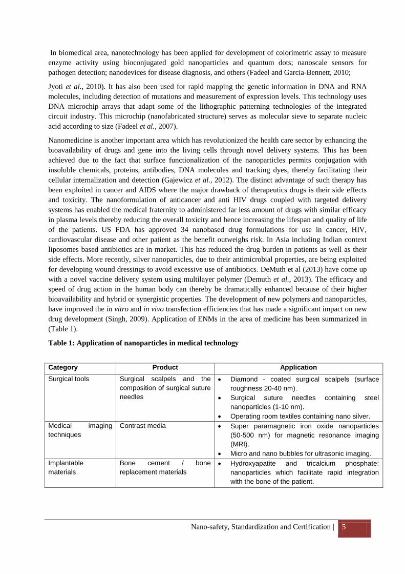
drug development (Singh, 2009). Application of ENMs in the area of medicine has been summarized in
(Table 1).
Table 1: Application of nanoparticles in medical technology
Category Product Application
Surgical tools Surgical scalpels and the
composition of surgical suture
needles
Diamond - coated surgical scalpels (surface
roughness 20-40 nm).
Surgical suture needles containing steel
nanoparticles (1-10 nm).
Operating room textiles containing nano silver.
Medical imaging
techniques
Contrast media Super paramagnetic iron oxide nanoparticles
(50-500 nm) for magnetic resonance imaging
(MRI).
Micro and nano bubbles for ultrasonic imaging.
Implantable
materials
Bone cement / bone
replacement materials
Hydroxyapatite and tricalcium phosphate:
nanoparticles which facilitate rapid integration
with the bone of the patient.
Nano-safety, Standardization and Certification | 6
In the area of agriculture and food production, nanotechnology is playing major role in improving the
product shelf-life, storage, processing and packaging (Maynard, 2007; Handy et al., 2008). This is being
achieved throughout the process of food processing, such as use of nano-sieves during industrial
processing, increasing food values by introducing nutrients in nano form into the product for increased
bioavailability (Table 2). Besides this, with the use of nanotechnology, healthy food could be developed
and introduced for preventive healthcare.
More than 1300 consumer products have already been released in the market, majority of these are
personal care products (PEN, 2013).
In the areas of sports, aviation, automobiles, construction etc. nanotechnology is being used to strengthen
the product by enhancing their quality and reducing the weight.
Nanotechnology has also helped in environmental cleanup of contaminated sites using different kind of
ENMs (PEN, 2013). This technology is also being used in house hold products such as air conditioner,
fridge, washing machine etc. to prevent microbial contamination.
Table: 2 Application of nanoparticles in food production
Surface coatings of
conventional implants with
ENMs
Joint prosthetics (hip, knee) with nano
hydroxyapatite coating.
Coronary stents with a diamond-like nano
composite coating made of ultrathin polymer.
Wound treatment Wound dressings Wound treatment products containing nano
crystalline silver particles which are used for
improved antibacterial and anti-fungal activity.
Biochips DNA/protein microarray chips lab-on-a-chip devices for molecular in vitro
diagnostics, point-of-care applications
Bio-sensors Bio-detection for the diagnosis of diabetes,
cancer, bacteria and viruses
Nano therapeutics Anticancer agents Heat therapy with super paramagnetic iron oxide
nanoparticles
Heat ablation with gold nanoparticles
Light therapy
Boron neutrons capture therapy.
Nanoparticles type Application Property/function
Colloidal metal nanoparticles Food additive Desired better gastro-intestinal uptake claimed
Metal oxide nanoparticles (silver, zinc oxide)
Food colorant Attractive and better representation
Packaging materials/ storage
Prevent from contaminant and extending shelf life
Equipment for food preparation
Cleaning of surfaces
Fridges, storage containers
Anti-bacterial coating of equipment for storage and handling of food
Water treatment/soil decontamination
Removal of contaminants /catalyse the metabolism of toxicant
Sprays Anti-bacterial
Nano-safety, Standardization and Certification | 7
ENM based products
Worker exposure
Raw material production
Consumer product
manufacturing
Consumer exposure
Consumer use
End of life
Industrial emission
Landfills, Incinerators
Consumer, worker and ecological exposure
There has been a significant impact on the global economy due to the advent of nanotechnology in
science and engineering since more products containing nanomaterials are moving from research and
development to industry.
1.2 Scope
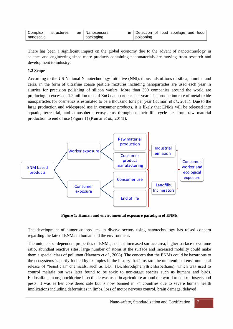
According to the US National Nanotechnology Initiative (NNI), thousands of tons of silica, alumina and
ceria, in the form of ultrafine coarse particle mixtures including nanoparticles are used each year in
slurries for precision polishing of silicon wafers. More than 300 companies around the world are
producing in excess of 1.2 million tons of ZnO nanoparticles per year. The production rate of metal oxide
nanoparticles for cosmetics is estimated to be a thousand tons per year (Kumari et al., 2011). Due to the
large production and widespread use in consumer products, it is likely that ENMs will be released into
aquatic, terrestrial, and atmospheric ecosystems throughout their life cycle i.e. from raw material
production to end of use (Figure 1) (Kumar et al., 2011f).
Figure 1: Human and environmental exposure paradigm of ENMs
The development of numerous products in diverse sectors using nanotechnology has raised concern
regarding the fate of ENMs in human and the environment.
The unique size-dependent properties of ENMs, such as increased surface area, higher surface-to-volume
ratio, abundant reactive sites, large number of atoms at the surface and increased mobility could make
them a special class of pollutant (Navarro et al., 2008). The concern that the ENMs could be hazardous to
the ecosystems is partly fuelled by examples in the history that illustrate the unintentional environmental
release of “beneficial” chemicals, such as DDT (Dichlorodiphenyltrichloroethane), which was used to
control malaria but was later found to be toxic to non-target species such as humans and birds.
Endosulfan, an organochlorine insecticide was used in agriculture around the world to control insects and
pests. It was earlier considered safe but is now banned in 74 countries due to severe human health
implications including deformities in limbs, loss of motor nervous control, brain damage, delayed
Complex structures on nanoscale
Nanosensors in packaging
Detection of food spoilage and food poisoning
Nano-safety, Standardization and Certification | 8
puberty, cancer and teratogenicity. The residues of these pesticides were persistent in the environment
and were detected in places where there were never used. This was due to the fact that they were
transferred through air and water globally.
Currently, ENMs are being incorporated into commercial products at a faster rate than the development of
knowledge and regulations to mitigate potential health and environmental impacts associated with their
manufacturing, application and disposal (Kumar et al., 2012). Variety of ENMs with different chemical
compositions, synthesized through different methods, differing in size, shape, surface coatings, etc. have
been shown to be genotoxic and cytotoxic in different models such as prokaryotes (Brayner, 2008;
Simon-Deckers et al., 2009; Kumar et al., 2011b; Kumar et al., 2011c; Kumar et al., 2011d), plants
(Kumari et al., 2011; Vajpayee et al., 2011), human cell lines (Sharma et al., 2009; Shukla et al., 2011a;
Shukla et al., 2011b; Sharma et al., 2012a), primary human cells (Sharma et al., 2011), in vivo (Wang et
al., 2008; Xie et al., 2011; Sharma et al., 2012c) and aquatic models (Allen et al., 2011; Fabrega et al.,
2011). There are several in vitro reports that have demonstrated the genotoxic, carcinogenic and apoptotic
properties of ENMs to human (Sharma et al., 2009; Shukla et al., 2011a; Sharma et al., 2012a). There is
considerable evidence that ENMs cause toxicity to bacteria which play a major role in maintaining the
homeostasis in human. Studies have shown that ENMs also adversely affect the microbes (Escherichia
coli, Pseudomonas aeruginosa and Streptococcus aureus) which are responsible for maintain the
environmental health. (Brayner et al., 2006; Wahab et al., 2010; Wu et al., 2010; Premanathan et al.,
2011) is also available. This also raises the possibility that the release of ENMs may be detrimental to
important bio-geochemical processes in soil such as carbon or nitrogen cycling. Therefore, organisms,
especially those that interact strongly with their immediate environment, are expected to be affected as a
result of their exposure to ENMs. It is also likely that the ENMs can directly interact with the food web at
different trophic levels and affect the ecological sustenance. The bio-magnification of ENMs across the
genera is also a big concern.
Humans get exposed to ENMs at various steps of its synthesis (laboratory), manufacture (industry), use
(consumer products, devices, medicines etc.) and the environment (through disposal). The lack of
regulatory guidelines, reference standards and certification processes for ENMs (from manufacture to
product development) is a major stumbling block in hazard identification through risk and exposure
assessment. This is compounded by the lack of equipment for accurate and sensitive measurement of
ENMs with respect to their number, mass and surface area in the environment. Hence, it is prudent to
address the issues of risks associated with ENMs and develop ethical, legal and regulatory framework to
mitigate their exposure.
Hence, the present document is intended to address the need for: (1) Environmental, health and safety
impact of nanomaterials; (2) Social, ethical and legal issues of nanomaterials and nanoproducts; (3) Safe
production, handling, use and disposal of nanomaterial – Risk assessment/analysis, Risk
monitoring/management; (4) Current regulatory landscape – nanosafety policies, risk governance,
regulatory and institutional mechanism; (5) Guideline for the best practices for testing, standardization
and certification of nanoproducts.
2. Environmental, health and safety impact of nanomaterials
The applications of nanotechnology in diverse areas will lead to their inadvertent release in surface and
sub-surface environments through landfills and other waste disposal methods. It is likely that some of
these ENMs may induce adverse/toxic effects in both lower and higher trophic organisms (Handy et al.,
2008; Kumar et al., 2011a). At the safety level, it is well known that the high surface area to volume ratio
of ENM leads to increased surface reactivity and associated risk. However, the mechanisms involved in
Nano-safety, Standardization and Certification | 9
reactivity and toxicity are not well understood yet. There is also a great deal of uncertainty about the
environmental fate, behaviour and bioavailability of ENMs in the ecosystem. Also, lack of reliable and
validated schemes for assessing the ecotoxicological risk is a big concern (Wen-Che Hou et al., 2013).
The major constraints in risk assessment of ENM are the lack of appropriate methods for characterization
in exposure media, bioavailability, mobility, biopersistance, and bioaccumulation (Farre et al., 2009;
Kumar et al., 2012). The impact of ENMs on various ecosystems will be significant because their
distribution depends on a number of factors such as Brownian motion, inertia, gravitational influences,
thermal influences, pH, and ionization. As the ENMs have high mobility, they can easily move in the air,
water and soil and can contaminate the flora and fauna. This may also result to the transfer of ENMs in
the food chain, leading to the creation of non-biodegradable pollutants (Mahapatra et al., 2013). Also,
ENMs can affect the bioavailability of the other toxicant/pollutant by facilitating their transportation
(Navarro et al., 2008). ENMs may also elicit a negative physical, chemical and biological impact on
different strata of the ecosystem (air, water and soil). Hence, to minimize the exposure to ENMs and
thereby its adverse effects on the environment and human health, it is imperative to consider the
following points: (a) the behaviour of ENMs in the environment (air, water, and soil) and their exposure
to humans (b) methodological and metrological approaches for the detection/quantification of ENMs in
environmental samples (c) approaches and knowledge gaps in ecotoxicity studies.
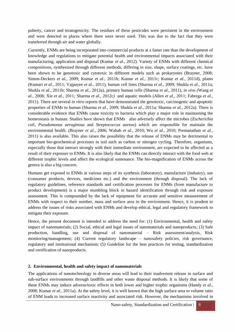
2.1 Behaviour of ENMs in the environment (air, water, and soil) and their exposure to humans
The behaviour and bioavailability of ENMs in freshwater/marine ecosystem depends on their interaction
with the aquatic colloids, such as natural organic matters (NOMs), humic substances, and salt ions
(Navarro et al., 2008). NOMs usually get adsorbed on the surface of the ENMs by different electrostatic,
hydrogen bonding and hydrophobic interactions, which affects their dispersity and bioavailability. NOMs
are classified into three major classes; (1) rigid biopolymers, such as polysaccharides and peptidoglycans
produced by phytoplankton or bacteria (2) fulvic compounds, mostly from terrestrial sources, originating
from the decomposition products of plants (3) flexible biopolymers, composed of aquagenic refractory
organic matter from a recombination of microbial degradation products (Buffle et al., 1998). ENMs in
aqueous suspension are dispersed due to the electrostatic and steric repulsion of the surface charge
(positive/negative) present on the particle. Apart from NOMs, salt ions, protein content, presence of
molecular clusters enable nucleation leading to agglomeration/aggregation thereby modulating the
bioavailability of ENMs in the environment (Figure 2). Also, the biomolecules such as proteins or
polymers present in the ecosystem form a layer over the ENMs, named as “corona” which plays
important role in their biological fate. It has also been shown that it is not only the ENMs alone but the
“corona” govern the properties of the “particle-plus-corona” compound in the biological system (Lynch
and Dawson, 2008; Elsaesser and Howard, 2011).
Nano-safety, Standardization and Certification | 10
NOMs
SI
Particle diffusion
coefficient
Particle
diameter
Rapid migration
and dispersion
ENPs
Environmental Matrix Bioavailability Scenarios
OM
ENPs
OM
OM
OM
OM
OM
SI
ENP ENP
ENP
ENP SI
SI SI
Increased
bioavailability
Decreased
bioavailability
ENPs
Increased
bioavailability
Figure 2: Availability of engineered nanoparticles after interaction with different
environmental matrixes
The behaviour of ENMs in air is majorly governed by diffusion, agglomeration and potential re-
suspension of aerosol from deposited nanomaterials. It is also reported that in traditional aerosol science,
particle size, inertia, gravitational and diffusion forces govern aerosol behaviour in the environment. As
the particle size decreases, diffusional forces become increasingly important and nanoscale particles are
thus likely to behave in a manner more alike to a gas or vapour (Aitken et al., 2004; 2008). Particle
diffusion coefficient is inversely proportional to the particle diameter. Particle with a high diffusion
coefficient such as ENMs have high mobility and mix rapidly in an aerosol. After their release in the
environment, atmospheric diffusion facilitates the ENMs to migrate rapidly from a higher to a lower
concentration, thus resulting in rapid dispersion and potential for particles to travel a great distance from
the source (Feliu and Fadeel, 2010).
European Commission’s Scientific Committee on Emerging and Newly Identified Health Risks
(SCENIHR) evaluated the risk assessment of products of nanotechnologies (SCENIHR, 2009). SCENIHR
evaluated the knowledge base on the release of ENMs into the environment and the subsequent exposure
to humans through inhalation. It was reported that “Examples of the exposure routes for ENMs via the
environment are inhalation by human and other air breathing species, and uptake by aquatic organisms
from water and sediments. Assessment of exposure concentrations of dispersed nanomaterials requires
detailed insight into the process that act on these materials in the environment. However, currently
available knowledge of these processes is insufficient to allow quantitative prediction of the
environmental fate of nanomaterials” (SCENIHR, 2009).
The critical questions in relation to ENM exposure are how much (intensity/concentration), how long
(duration/frequency) and how many (number). The main routes by which one can get exposed to the
ENMs are inhalation, ingestion and dermal (Figure 3). Inhalation is considered to be the primary route by
which air breathing species including humans get exposed to the ENMs suspended in air. Once these
ENMs are inhaled, they are likely to get deposited in different regions of the respiratory tract. However,
the location and the extent of the deposition depend on the particle size (Oberdorster et al., 2004).
Ingestion exposure of ENMs may arise through hand to mouth contact or by consuming contaminated
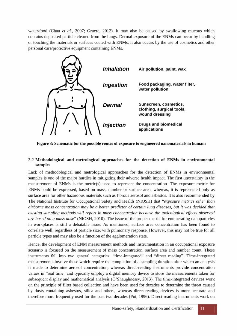
Nano-safety, Standardization and Certification | 11
water/food (Chau et al., 2007; Gruere, 2012). It may also be caused by swallowing mucous which
contains deposited particle cleared from the lungs. Dermal exposure of the ENMs can occur by handling
or touching the materials or surfaces coated with ENMs. It also occurs by the use of cosmetics and other
personal care/protective equipment containing ENMs.
Figure 3: Schematic for the possible routes of exposure to engineered nanomaterials in humans
2.2 Methodological and metrological approaches for the detection of ENMs in environmental
samples
Lack of methodological and metrological approaches for the detection of ENMs in environmental
samples is one of the major hurdles in mitigating their adverse health impact. The first uncertainty in the
measurement of ENMs is the metric(s) used to represent the concentration. The exposure metric for
ENMs could be expressed, based on mass, number or surface area, whereas, it is represented only as
surface area for other hazardous materials such as fibrous aerosol and asbestos. It is also recommended by
The National Institute for Occupational Safety and Health (NIOSH) that “exposure metrics other than
airborne mass concentration may be a better predictor of certain lung diseases, but it was decided that
existing sampling methods will report in mass concentration because the toxicological effects observed
are based on a mass dose” (NIOSH, 2010). The issue of the proper metric for enumerating nanoparticles
in workplaces is still a debatable issue. As mentioned, surface area concentration has been found to
correlate well, regardless of particle size, with pulmonary response. However, this may not be true for all
particle types and may also be a function of the agglomeration state.
Hence, the development of ENM measurement methods and instrumentation in an occupational exposure
scenario is focused on the measurement of mass concentration, surface area and number count. These
instruments fall into two general categories: “time-integrated” and “direct reading”. Time-integrated
measurements involve those which require the completion of a sampling duration after which an analysis
is made to determine aerosol concentration, whereas direct-reading instruments provide concentration
values in “real time” and typically employ a digital memory device to store the measurements taken for
subsequent display and mathematical analysis (O’Shaughnessy, 2013). The time-integrated devices work
on the principle of filter based collection and have been used for decades to determine the threat caused
by dusts containing asbestos, silica and others, whereas direct-reading devices is more accurate and
therefore more frequently used for the past two decades (Pui, 1996). Direct-reading instruments work on
Air pollution, paint, wax
Food packaging, water filter,
water pollution
Sunscreen, cosmetics,
clothing, surgical tools,
wound dressing
Drugs and biomedical
applications
Inhalation
Ingestion
Dermal
Injection
Nano-safety, Standardization and Certification | 12
optical particle counter (OPC), condensation particle counter (CPC), which measure a count
concentration, the surface area monitor (SAM), which measures surface area concentration, and the
aerosol photometer measures the mass concentration. An OPC provides a count concentration in the size
range of 300 – 20000 nm. This instrument sizes and count particles to allow for the determination of both
a number concentration and particle size distribution (Kulkarni et al., 2011). As a particle passes through
a viewing volume of the detector illuminated by a laser, it scatters light which is then detected by a photo
detector. The electrical pulses generated by the photo detector are converted to counts and the pulse
height is related to particle size.
These “time-integrated” and “direct reading” based instruments can only measure the ENMs from the air.
However, the major constraint in these instruments is that they can only measure ENMs of size ≥300nm.
Additionally, the detection of ENMs from the aquatic/colloidal system is a big challenge. Filtration and
centrifugation of large amount of the water or sediment and electron microscopy analysis of the pellet is
the only viable option for the qualitative and quantitative measurement of ENMs.
2.3 Approaches and knowledge gaps in ecotoxicity studies
The frequent release and interaction of ENMs with different components of the ecosystem, necessitates
the development of certain strategies to test the possible hazards of ENMs. The fate, behavior and
detection of different of ENMs in the ecosystem have been critically discussed above (1.2). It can be
inferred from the above discussion that interactions of cells with ENMs are dependent on their size,
shape, chemical composition, surface charge, surface structure, area, solubility and aggregation state.
Thus, it is essential to study these physiochemical properties of ENMs while assessing their biological
hazards.
Among these physiochemical characteristics, surface properties of the ENMs are the most important
factor that govern the stability and mobility of ENMs in aqueous suspension (Dhawan et al., 2009). The
agglomeration tendency of the ENMs is determined by the surface properties, which are mainly
dependent on temperature, ionic strength, pH, concentration, size and the solvent. However, it is difficult
to measure the surface properties of ENMs at nano to pico gram range due to the limitations of the
commercially available analytical instruments. On the other hand, the concentration of ENMs in
suspension is also a crucial step in designing the experiments, since the ENMs have the tendency to
agglomerate/aggregate which results in a change in their physicochemical properties, and hence modified
cellular concentration (Donaldson and Borm, 2004). Thus, the experimental design should also consider
the concentration induced aggregation effects of the ENMs.
It may also be speculated that at lower concentration range ENMs will tend to show less aggregation that
lead to increased uptake and response than that expected at high concentrations. However, different
surface modifications in ENMs (particle coating, dispersant /surfactant, sonication) stabilize the particles
and avoid agglomeration which may result in exacerbated biological response. The durability of surface
coating in cellular/biological environment and the effects of cellular metabolites on the ENMs are the
other key issues that need to be addressed in order to understand the adverse health effects of ENMs.
Other possible effects of ENMs uptake could be the interaction with other (toxic) substances and their
mobilisation and bioavailability.
The environmental fate, behavior and bioavailability of ENMs are not well understood; therefore their
persistence and the possible interaction/impact, bio magnification in food webs at different trophic levels
are of immediate concern. Hence, to study the ENMs effect in ecosystem, the study design should address
the ENMs interaction/impact directly with different trophic level organism as well as the perturbations
Nano-safety, Standardization and Certification | 13
Computational tools
Environmental toxicity
Experimental toxicity studies
Particle characterization
Particle synthesis
Workplace exposure
Particle UptakeEcotoxicology
ENPs
induced by the ENMs biomagnification (Kahru and Dubourguier, 2010). ENMs effect on other
toxicant/pollutant also needs to be examined, because the transportation of the contaminant could be
facilitated through their adsorption to ENMs which may have a negative impact on useful bacteria for
natural remediation and other water bodies (Navarro et al., 2008). The presence of impurities in the
ENMs also influence the toxicity, thus the purity of the ENMs should also be considered in the study
design. Elemental analysis using different analytical techniques could be helpful in analysing the purity of
ENMs (Nowack, 2009).
Some of the metal oxide nanoparticles are known to release ions in the aqueous suspension which could
alter the toxicity outcomes. Hence, the quantitation of soluble metal ions in the exposure medium is also a
prerequisite in nanotoxicology studies (Baun et al., 2008; Handy et al., 2008; Fairbrother and Fairbrother,
2009). Lack of reference materials, appropriate methods to monitor ENMs behaviour, dose dilemma and
exposure methods, ENM behaviour in environmental matrices, regulatory toxicology test methods are
certain other hurdles that need to be addressed properly. Therefore, prior to use the ENMs based
consumer products in daily life activities, it is important for nanotoxicology research to understand their
fate in environment, so that their undesirable effects can be avoided.
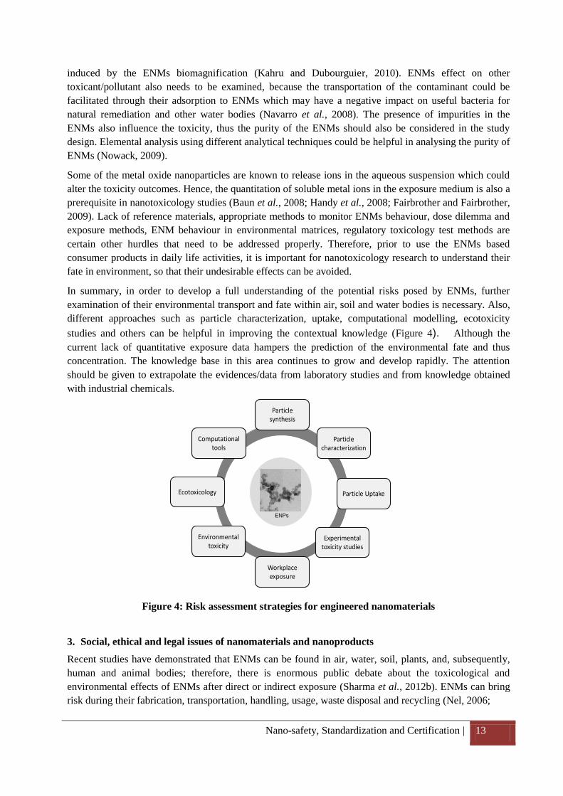
In summary, in order to develop a full understanding of the potential risks posed by ENMs, further
examination of their environmental transport and fate within air, soil and water bodies is necessary. Also,
different approaches such as particle characterization, uptake, computational modelling, ecotoxicity
studies and others can be helpful in improving the contextual knowledge (Figure 4). Although the
current lack of quantitative exposure data hampers the prediction of the environmental fate and thus
concentration. The knowledge base in this area continues to grow and develop rapidly. The attention
should be given to extrapolate the evidences/data from laboratory studies and from knowledge obtained
with industrial chemicals.
Figure 4: Risk assessment strategies for engineered nanomaterials
3. Social, ethical and legal issues of nanomaterials and nanoproducts
Recent studies have demonstrated that ENMs can be found in air, water, soil, plants, and, subsequently,
human and animal bodies; therefore, there is enormous public debate about the toxicological and
environmental effects of ENMs after direct or indirect exposure (Sharma et al., 2012b). ENMs can bring
risk during their fabrication, transportation, handling, usage, waste disposal and recycling (Nel, 2006;
Nano-safety, Standardization and Certification | 14
Stone and Donaldson, 2006; Oberdarster et al., 2007; Stebounova et al., 2012). Some ENMs can enter
into the body using a variety of routes, such as inhalation, ingestion, injection and through skin, and can
persist in the system for longer periods (Figure 1). Several kinds of sicknesses can be expected from
exposure to ENMs, including asthma, bronchitis, lung and liver cancer and others (Borm et al., 2006;
Wardak et al., 2008). Nanoethics is the area of ethics that relates to the study of nanotechnology and its
products, and provides guidelines for training, prohibition, and limitation in the use of these materials.
This ensures that the overall risk factors and public concerns can be minimized before the use of ENMs in
different applications.
The need of nanoethics can easily be linked with the development in nanotechnology and the doubts
about their misuse. The advent of biotechnology not only resulted into the beneficial products such as
transgenic plants and fruits, recombinant proteins, organ culture and many others but has raised some new
ethical issues that were not aroused previously. Examples of such ethical issues are pre-determination of
the sex of human offspring via various technical means, the development of recombinant protein, multi
drug resistance, creation of new forms of plant and animal life via r-DNA techniques, human reproductive
cloning via somatic cell nuclear transfer. There are strong disputes over the acceptability of such issues,
because of difference in the purpose of applications of these developed technologies.
Likewise, ENMs have several unique physiochemical properties which are getting exploited for the
development of novel materials/products with diverse application but also posed harmful effects to the
living organism due to the way they are manipulated on an atomic scale. They are also new materials
produced by entirely new manufacturing techniques. Hence, there are no specific rules and regulations to
cover their manufacturing processes. Also, the concerns about the health implications of ENMs have been
widely reported (Donaldson et al., 2004; Schins et al., 2004; Borm et al., 2006; Service, 2008).
Oberdorster et al. (2004) showed in animal experiments that inhaled ultrafine particles (smaller than
100nm) can be translocated from the olfactory nerve to the olfactory bulb in the brain (Oberdorster et al.,
2004). However, the significance of this study for humans still needs to be established. The translocation
of ENMs along nerve fibers could provide a portal of entry into the central nervous system. Thus the
effect of the inhaled ultrafine particles on central nervous system needs to be explored in future studies.
Also, the incorporation of ENMs in the sunscreen cream and other personal care products have been
questioned by different scientific groups, because of their ability to induce cytotoxic and genotoxic effects
after short term exposure (Hardman, 2006; Singh and Nalwa, 2007; Ahamed et al., 2008; Sharma et al.,
2009; Singh et al., 2009). Long term exposure studies are still necessary to understand the fate of ENMs
in the biological system. Carbon nanotubes (CNTs) have been extensively used in basic science research
and development worldwide because of their extraordinary physical, chemical, physicochemical and
biological properties. It is also reported that CNTs can induce genotoxicity, immunotoxicty, cytotoxicity
in human as well as ecotoxicity /environmental toxicity in the ecosystems (Dhawan et al., 2006; Singh et
al., 2006; Maynard, 2007). However, there are no defined rules and regulations regarding the
manufacturing and marketing of CNTs.
ENMs involve wider societal issues and pose several social challenges such as environmental pollution,
workplace exposure, water contamination, genetic alteration and carcinogenicity etc. Predicted adverse
consequence about different ENMs reiterates the need of nanoethics in research, development, production
and manufacturing as well as social, economical, moral, health, and other human applications. The
implication or knowledge of nanoethics will be very useful for training and protecting the academia
(undergraduate/graduate/research students), scientists, industries, policymakers and user for the health
and safety, social and philosophical, environmental, educational, and other legal issues associated with
the ENMs.
Nano-safety, Standardization and Certification | 15
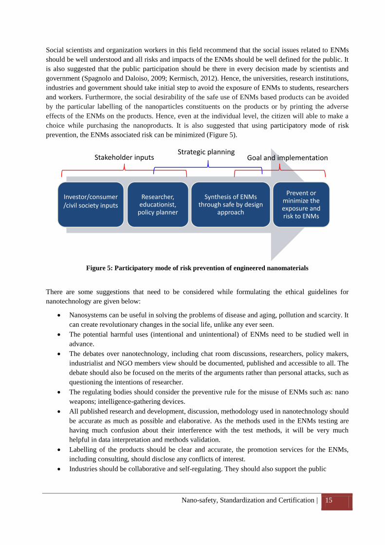
Investor/consumer /civil society inputs
Researcher, educationist,
policy planner
Synthesis of ENMs through safe by design
approach
Prevent or minimize the exposure and risk to ENMs
Stakeholder inputsStrategic planning
Goal and implementation
Social scientists and organization workers in this field recommend that the social issues related to ENMs
should be well understood and all risks and impacts of the ENMs should be well defined for the public. It
is also suggested that the public participation should be there in every decision made by scientists and
government (Spagnolo and Daloiso, 2009; Kermisch, 2012). Hence, the universities, research institutions,
industries and government should take initial step to avoid the exposure of ENMs to students, researchers
and workers. Furthermore, the social desirability of the safe use of ENMs based products can be avoided
by the particular labelling of the nanoparticles constituents on the products or by printing the adverse
effects of the ENMs on the products. Hence, even at the individual level, the citizen will able to make a
choice while purchasing the nanoproducts. It is also suggested that using participatory mode of risk
prevention, the ENMs associated risk can be minimized (Figure 5).
Figure 5: Participatory mode of risk prevention of engineered nanomaterials
There are some suggestions that need to be considered while formulating the ethical guidelines for
nanotechnology are given below:
Nanosystems can be useful in solving the problems of disease and aging, pollution and scarcity. It
can create revolutionary changes in the social life, unlike any ever seen.
The potential harmful uses (intentional and unintentional) of ENMs need to be studied well in
advance.
The debates over nanotechnology, including chat room discussions, researchers, policy makers,
industrialist and NGO members view should be documented, published and accessible to all. The
debate should also be focused on the merits of the arguments rather than personal attacks, such as
questioning the intentions of researcher.
The regulating bodies should consider the preventive rule for the misuse of ENMs such as: nano
weapons; intelligence-gathering devices.
All published research and development, discussion, methodology used in nanotechnology should
be accurate as much as possible and elaborative. As the methods used in the ENMs testing are
having much confusion about their interference with the test methods, it will be very much
helpful in data interpretation and methods validation.
Labelling of the products should be clear and accurate, the promotion services for the ENMs,
including consulting, should disclose any conflicts of interest.
Industries should be collaborative and self-regulating. They should also support the public
Nano-safety, Standardization and Certification | 16
awareness programme for the dissemination of science and reasonable legislation to deal with
legal and social issues associated with nanotechnology.
Scientists working in the field of material sciences in developing new ENMs should have a
compact training of ecology/ecotoxicology and public safety or they should consult someone for
the risk assessment of newly synthesized ENMs.
The research institutions, scientists, industries should be accountable for the fraudulent or
irresponsible misuse of the nanotechnology.
The research institutions, industries should have preventive measure to minimize the work place
exposure of the researcher and worker.
The research institutions, industries should have a proper storage and disposal guideline for
ENMs to avoid the contamination and risk.
4. Safe production, handling, use and disposal of nanomaterial – risk assessment/analysis, risk
monitoring/management
According to actual state of knowledge available with regard to the properties of ENMs, it is established
that the factors such as surface area, surface properties, elemental composition, tendency to aggregate, the
form of the particles and surface charge of ENM plays a critical role in their distribution through the
environment, ecosystem and human body. Due to having high mobility they may gain access into the
human body through skin (dermal exposure), lungs (inhalation) and gastrointestinal tract (ingestion).
Also, ENMs may penetrate deep into tissues through fine capillaries, readily travel throughout the body
and interact with organs, tissues, cells and/or sub-cellular structures. The pharmacokinetic studies show
that different types of nanoparticles can be found in various cells such as mitochondria (Li et al., 2003;
Xia et al., 2006), lipid vesicles (Penn et al., 2005), fibroblasts (Tian et al., 2006), nucleus (Chen and von
Mikecz, 2005; Shukla et al., 2011a; Shukla et al., 2011c) or macrophages (Yokoyama et al., 2005; Tian et
al., 2008). Moreover, in vivo and in vitro studies demonstrated that ENMs in contact with the cell surfaces
and cellular proteins may lead to:
i. formation of reactive oxygen species (ROS), which results in oxidative stress and inflammation,
leading to infection and exacerbation of asthma and chronic obstructive pulmonary disease (Nel,
2005; Oberdarster et al., 2005; Nel, 2006)
ii. DNA damage, lipid peroxidation of cellular membranes, resulting in cell damage (Sharma et al.,
2009; Shukla et al., 2011a)
iii. the up/down regulation of genes encoding a specific protein involved in inflammatory processes/
apoptosis/carcinogenicity (Cui et al., 2005; Dobrovolskaia et al., 2009)
Several in vitro and in vivo studies have also shown that ENMs can be cytotoxic (Thill et al., 2006;
Warheit et al., 2007; Kumar et al., 2011d), neurotoxic (Long et al., 2006; Win-Shwe and Fujimaki, 2011;
Wu et al., 2011), genotoxic (Singh et al., 2009; Xu et al., 2009; Shukla et al., 2011a), ecotoxic (Colvin,
2003; Vajpayee et al., 2011) or bactericidal (Brayner et al., 2006; Brayner, 2008; Kumar et al., 2011d;
Kumar et al., 2011e). The perturbations induced by ENMs in human and environment might significantly
affect ecological balance and the carrying capacity of the ecosystem. In general, ENMs tend to be toxic
due to chronic exposure, when a sufficient amount of an ENM has accumulated. In other words, the
potential adverse health effects of ENMs have been associated with dose, dimension, and durability.
However, reduction of particle's size to the nanoscale level results in display of unanticipated physical
and chemical properties that do not occur at the micro or macro scales (Auffan et al., 2009). According to
Oberdorster et al. (2005), particle size is not the only possible factor inducing toxicity, other factors such
Nano-safety, Standardization and Certification | 17
as size distribution, agglomeration state, shape, porosity, surface area, chemical composition, structure-
dependent electronic configuration, surface chemistry, surface charge, and crystal structure plays vital
role (Oberdarster et al., 2005) . It is still largely unknown that for a specific ENM, which
property/properties influence their toxicity. It is hard to generalize a common mechanism of the potential
toxicity of ENMs. Also, lack of current knowledge about the toxicity of ENMs, methods to assess ENMs
toxicity and the current safety data sheets do not adequately reflect the hazardous nature of ENMs. It is,
therefore, suggested that all ENMs should considered potentially hazardous unless sufficient information
to the contrary is obtained and should be treated as same as a radioactive substance. Therefore, a
comprehensive risk characterization (size, size distribution, agglomeration state, shape, porosity, surface
area, chemical composition, structure-dependent electronic configuration, surface chemistry, surface
charge, crystal structure, interaction with the DNA, protein cellular organelles, and others) should be
performed whenever a novel ENMs is designed/produced and then introduced into the market.
Additionally, the precaution should also be taken while handling, use, storage and disposal of ENM
containing products. Moreover, safety precautions while working with ENMs in laboratory/industry and
the general approach to managing risks from nanoparticles are also important to avoid the
exposure/contamination or to mitigate the exposure (Dhawan et al., 2011).
4.1 Safe production, handling and use of ENMs
As the adverse effects of ENMs have shown a close relationship with their size, atomic structure,
elemental composition and others, it is prudent to monitor the exposure assessment of ENMs at the level
of production, handling and use.
The preparation of ENMs at the laboratory and industrial scale offers several challenges as there is no
uniform process for the synthesis of ENMs. It varies considerably in different research institutions and
industrial scale laboratories. Sol gel technique, spray-drying process, microemulsion processing are few
of the commonly used methods. Sol gel technique is one of the most commonly used for the synthesis of
ENMs, due to its simplicity and flexibility in controlling the properties of the final products at various
stages (Brinker and Scherer, 1990; Fadeel and Garcia-Bennett, 2010). However, the disadvantage with
this method is in difficulties to control the kinetics of crystal growth precisely when large batches of
ENMs are prepared, which may lead to particle agglomeration and large particle size distributions.
Overall, this is a suitable method for laboratory-scale preparations.
Spray-drying process is another method for ENMs synthesis which involves spraying a homogenized
precursor solution composed of the inorganic compounds and relevant additives within a specially
designed chamber at temperatures at or above the boiling point of the solvent (Vasiliev et al., 2008). The
precursor solution is first atomized through a nozzle into droplets using flowing gas and then the droplet
is sprayed into a chamber through which a flow of hot air or nitrogen is introduced. This leads to the
quick evaporation of the droplets and the formation of the inorganic particle. The droplet size is the
limiting factor for the particle size and hence the type of nozzle and atomizer unit determines the
possibilities of using this technique for the production of ENMs.
There is a debate about the identification of the safe methods for the synthesis of the ENMs. Some of the
ENMs synthesized by the above mentioned methods are known to induce the perturbation in human and
environment, whereas few of them are nontoxic for the ecosystem. The probable reason for the conflict
could be the size limit of the ENMs synthesized by different methods as well as the internal properties of
the chemicals used in the precursor solution. Hence, we should adopt an approach that can be used to
produce the safe ENMs.
Nano-safety, Standardization and Certification | 18
4.2 Storage of nanoparticles
A suitable system for storing ENMs is one that:
minimizes the dangers to personnel
prevents the breaking of containers and contaminating the working environment
protects the ENMs from external contamination
These conditions may be achieved by dedicating specific areas and equipment for this purpose. The
storage cabinets must carry appropriate danger labels, and inside the doors, there should be lists showing
the contents, quantities, expiry dates of the products and the material safety data sheet of the nanoparticle.
Storage criteria should also take account of the potential incompatibility of chemically different products
(considering the fact that, in their dry state, ENMs constitute an explosion risk that is far greater than the
same materials with larger dimensions). The ENMs should be stored in suitable cabinets, separately,
according to their type, with proper labeling (Dhawan et al., 2011).
4.3 Disposal procedures
The waste management guidance for the disposal of hazardous materials applies to ENMs-bearing waste
streams (solid and liquid waste), including:
pure ENMs;
items contaminated with ENMs, such as containers, wipes, biological tissues, culture wares and
disposable personal protection equipment (PPE); and
liquid suspensions containing ENMs
A plan for storage and disposal of ENMs or ENMs contaminated waste should be developed, taking
account of the hazardous nature of the particles and the quantities involved. Any material that has come
into contact with dispersible manufactured ENMs should be considered as belonging to an ENM-bearing
waste stream. This includes PPE, wipes, blotters and other disposable laboratory materials used during
research activities. Material from ENMs-bearing waste streams should not be put into the regular waste or
down the drain. Equipments used during ENMs handling should be decontaminated before it is disposed
of or reused. Wastes (cleaning solutions, rinse waters, rags, disposable PPE) resulting from
decontamination should be treated as ENMs-bearing waste.
4.3.1 Storage of ENM waste prior to disposal
4.3.1.1 Storage in waste containers: Package ENM-bearing wastes in compatible container that is in
good condition and afford adequate containment to prevent the escape of the ENMs. Label the waste
container with a description of the waste and include available information characterizing known and
suspected properties.
4.3.1.2 Storage in plastic bags: Collect paper, wipes, PPE and other items with loose contamination in a
plastic bag or other sealable container stored in the laboratory hood. When the bag is full, close it and
carefully place it into a second plastic bag or other sealing container, avoiding outside contamination.
Take it out of the hood and label the outer bag with an appropriate waste label.
4.3.2 Disposal of nanoparticle waste
It is reasonable to assume all ENM waste as potentially hazardous. It can therefore be disposed of as
hazardous waste. The ENMs in solvent should be disposed by immobilizing them in agar/agarose made in
distilled water. All other solutions coming in contact with the ENMs should be collected in containers and
Nano-safety, Standardization and Certification | 19
disposed at the hazardous waste disposal site.
4.4 General approach to managing risks from nanoparticles
Treat ENMs/NPs as highly toxic till enough data is generated on the contrary. Following safety measures
may be undertaken to mitigate and manage the risks arising from handling of nanoparticles:
Designate the area where nanomaterials are to be used in the laboratory.
Instruct the personnel involved, about the specific physical properties of free nanoparticles, the
need for special measures, and potential long term effects of nanoparticles. Include relevant
information in the operating instructions. Furthermore, deny unauthorized persons access to the
relevant work areas. There should be a documentation of the training imparted with the signature
of the staff.
Perform activities in contained installations (laminar flow/ chemical hoods), wherever this is
possible. If this cannot be done, avoid the formation of dusts or aerosols.
Ensure clean work wear. Work wear must be stored separately.
Ensure the regular cleaning of workplaces.
Wear protective gloves, protection goggles with side protection and protective clothing
depending on substance properties.
Inside a laboratory, the ENMs will behave in a similar way to a gas; furthermore, if not
completely restricted, they will spread quickly and remain in the surrounding air for a long time.
Therefore, the specifications of control systems designed for ENMs, such as fume hoods, glove
box, and ventilation, should be like those typical to gases, rather than that of powders.
Appropriate containers properly labelled should be used for transporting bottles containing ENMs
safely, from the storage room to the testing laboratories.
4.5 Safety precautions
Laboratories and rooms, where nanoparticles are handled, must be labeled. In particular, when
nanomaterials are handled openly e.g. as dry powder, appropriate protective measures (lab coat,
gloves, respiratory mask) must be adopted.
Regular training for staff members should be implemented.
Staffs who work temporarily or for short periods of time have to be instructed, according to their
place of work and the tasks they have to undertake (once before commencement of work, further
training sessions 1 per year).
Cleaning of all working surfaces potentially contaminated with nanomaterials (e.g. glassware,
apparatus, exhaust hoods, support equipment) at the end of each day with a HEPA vacuum and/or
wet wiping. Do not dry sweep or use compressed air.
Trainees, doctoral students and scientific guests have to discuss their work with the head of the
laboratory, and obtain permission from their supervisor for tasks outside the regular working
hours.
Permanent employees should, in the framework of an informative session, be instructed about
new findings in connection with nanoscaled materials.
It can be assumed that nanoparticles in aqueous suspension, in solution or embedded in a solid
matrix (composite) or contained in completely tight vessels pose a minimal risk – low hazard!
Nanoparticles in free form, or as dry powder (during weighing) or even as aerosol pose a higher
risk and have to be dealt carefully and with a high degree of responsibility – high hazard! In such
Nano-safety, Standardization and Certification | 20
cases, additional precautions are to be taken: If possible, the work should be undertaken in a
separate room fitted with negative pressure. The chemicals should only be handled in a fume
hood or in a closed glove box to provide containment and avoid contaminant release.
In this case, mouth protection breathing mask and protective eye wear should be made
mandatory.
In nanoparticle laboratories, sufficient facilities (clothes racks, wardrobes) must be made
available so that the safety clothing in use can be deposited / stored inside the laboratory. It is not
permitted to wear safety clothing outside the laboratory (due to danger of contamination in
corridors, offices and to co-workers).
Laboratories, where nanoparticles are handled are to be marked (advisory signs: protective
clothing must be worn, limited entry: for trained staff only”) and are to be furnished with an
emergency plan.
In the case that nanoparticles are accidentally spilt, the work place must be cleaned immediately
with a damp towel. Under no circumstances may residual materials be blown off the surface
particularly in the case of metallic or explicitly toxic nanomaterials.
If it is suspected that even the smallest amounts of substances that may be potentially dangerous
for an unborn child and lactating mothers, it is advisable to forbid such women personnel from
carrying out any operations that entail handling these substances.
Personnel should be provided with suitable masks when there are NPs in the dry state, or in
aerosols.
Whenever possible NPs are to be used for in vivo experiments in animals housed in isolated
ventilated cages. Isolated cages should be assigned for in vivo experimentation to avoid any
transfer of material from one animal to the other, especially in case of dermal application.
Assessment should be made of whether, in addition to the danger characteristics already
indicated, it is possible to include an indication of the average quantities of products used, their
location, the loaded quantity, the loading date, and the name of the person who performed this.
Other important considerations for effective risk management of nanomaterial exposure include
fire and explosion control. Some studies indicate that nanomaterials may be more prone to
explosion and combustion than an equivalent mass concentration of larger particles.
Provide laundry service for contaminated work clothing.
Do not eat or drink in the areas where nanomaterials are handled.
5. Current regulatory landscape – nano-safety policies, risk governance, regulatory and
institutional mechanisms
Development of new technologies is usually associated with both benefits & risks, and nanotechnology
obeys the same rule. A lot of emphasis has been given to develop nanomaterial based products since last
decade. Various studies have underscored the potential risks and concerns associated with the ENM based
products. It has also underlined that ENMs based risks and concerns are not simple to identify or to
determine. Even if there were a clear-cut cause and effect connections, it is hard to predict the exact
reason of the effect and the extrapolation of the results toward their behaviour and fate in human and
environment. Hence a governance system which addresses the potential risks and concerns associated
with ENM, in a time manner, is of high importance.
It is also necessary to develop a clear idea of the risks and concerns associated with nanotechnology to
build a proper level of trust amongst stakeholders and the consumer. This will be helpful in differentiating
Nano-safety, Standardization and Certification | 21
the real and perceived risks associated with ENMs and will also define the risks and benefits graph for
using the nanotechnology based products. The lessons from previous emerging technologies (such as, the
use of genetically modified organisms; GMOs, asbestos, pesticides), where the information disseminated
by industry alone is often seen as biased, and as a consequence, it is perceived as unreliable. Interactive,
industry and research/ academic collaborative research, expert opinions, workers view and bidirectional
communication between the industries and public can be employed in gaining reliable data and consumer
confidence.
Currently there are many scientific uncertainties and regulatory challenges associated with the
nanotechnology. Different regulatory authorities of the nanotechnology using nations have a broad
consensus that as of now no new nanotechnology-specific regulatory framework is needed (Breggin et al.,
2009). In United States regulatory authority for nanomaterials and nanotechnology based products is
divided between several federal agencies. The Environmental Protection Agency (EPA) regulates any
chemical substances or pesticides that are, or contain, nanomaterials. The Food and Drug Administration
(FDA) considers the risks of nanomaterials used in drugs, medical devices, food, food additives and
cosmetics. The Occupational Health and Safety Administration (OSHA) deals with workplace safety
dimensions while the Consumer Product Safety Commission (CPSC) is concerned with protection against
risks from consumer products. Finally, the Department of Agriculture deals with food and feed safety
dimensions.
Later in 2000, US launched the National Nanotechnology Initiative (NNI) to coordinate the
nanotechnology-related research, development and policy activities of different federal agencies. They
pointed out many issues related to the ENMs based products and has taken a number of decisions in
response to the newly emerging risks. For example, in reaction to the marketing in 2006 of a washing
machine that uses nanosilver as an antimicrobial, the EPA decided to regulate such equipment as a
pesticide and to require registration accordingly (EPA, 2007b). Also, in 2008, EPA decided that carbon
nanotubes should be treated as new rather than existing chemicals under the Toxic Substances Control
Act (TSCA), with the consequence that stricter regulatory requirements apply, including premanufacture
notice (EPA, 2008). EPA and FDA have also examined the regulatory challenges that nanotechnologies
pose. FDA’s nanotechnology taskforce concluded in 2007 that nanomaterials are having unique health
risks and a number of uncertainties but the demand for the introduction of nano-specific labelling
requirements cannot be accepted because “the current science does not support finding that classes of
products with nanoscale materials necessarily present greater safety concerns than classes of products
without nanoscale materials” (FDA, 2007). Although, US regulatory agencies have also acknowledged
the knowledge gaps and scientific uncertainty with regard to nanomaterials risk. EPA, for example, has
identified research needs on the toxicology and ecotoxicology of nanomaterials and recommends bigger
collaboration with different research agencies and stakeholders (EPA, 2007a).
Like the US, European government also rely on the existing laws and regulations mostly at EU level, in
the fields of chemicals, food, cosmetics, drugs, etc. They have also opted for a sector and product-specific
regulatory approach, in contrast to its technology-focused regulatory system. As the nanomaterials enter
the market as chemical substances, the EU has formed a new chemicals law REACH (Registration,
Evaluation, Authorisation and Restriction of Chemicals), in 2007 to look the nanotechnology oversight in
Europe. Once REACH will be fully implemented, it will be one of the most advanced and comprehensive
chemicals laws in the world. European regulators have also taken the first regulatory decisions on specific
nanomaterials. The EU decided in 2008 not to exempt carbon and graphite from registration under
REACH due to safety concerns about certain carbon nanotubes (European Commission, 2008). In the
food safety area, the European Food Safety Authority (EFSA) has produced scientific assessments of the
safety of nanosilver for use in food supplements and of nanostructured silicon dioxide and titanium nitride
Nano-safety, Standardization and Certification | 22
in food contact materials. In both cases, EFSA pointed out the knowledge gaps that prevent in
determining the safety of nanosilver in food products (EFSA, 2007; EFSA, 2008b; EFSA, 2008a).
In India different government agencies, publicity funded research institutes, universities as well as private
academic or research institutes are involved in research & development as well as in formulation of
guideline for safe use of nanomaterials. Under the Ministry of Science and Technology, Government of
India, Department of Science and Technology (DST), Department of Biotechnology (DBT) and Council
of Scientific and Industrial Research (CSIR) are the primary agencies involved with nanotechnology.
DST is the nodal department for coordinating activities of nanoscience and technology in India through
the Nanoscience and Technology Mission (NSTM). DBT on the other hand is primarily involved in
promoting the field of biotechnology in India and therefore has been involved in the nanobiotechnology.
CSIR which is constituted by a network of 38 laboratories undertakes research in areas of scientific and
industrial importance and also supports R&D in the area of nanoscience and technology in its
laboratories. Other agencies supporting nanotechnology in India include Indian Council of Medical
Research (ICMR) under the Ministry of Health and Family Welfare for developing applications in the
context of health as well as Ministry of Renewable Energy that is encouraging nanomaterial research for
energy production and storage. Also, Department of Atomic Energy (DAE) and Defence Research and
Development Organisation (DRDO), a network of 50 laboratories under the Ministry of Defense have
also been sponsoring research in the area of nanoscience and technology. These agencies are involved in
development of the safe nanoparticles and are also investigating the potential risks associated with them
using the existing environmental health and safety (EHS) regulations.
It is now becoming now apparent that various international initiatives are being undertaken to address the
safety concerns of the nanoparticles using novel strategies. It can also be assumed that the regulatory
debate in nanotechnologies is also now well underway. International governance of nanotechnology risk
is still very much limited to scientific and technical standardization and coordination efforts by the
leading nanotechnology countries in the OECD and some other international forums. No deeper structures
for global governance of nanotechnology have been created despite the rapid globalization of
nanotechnologies.
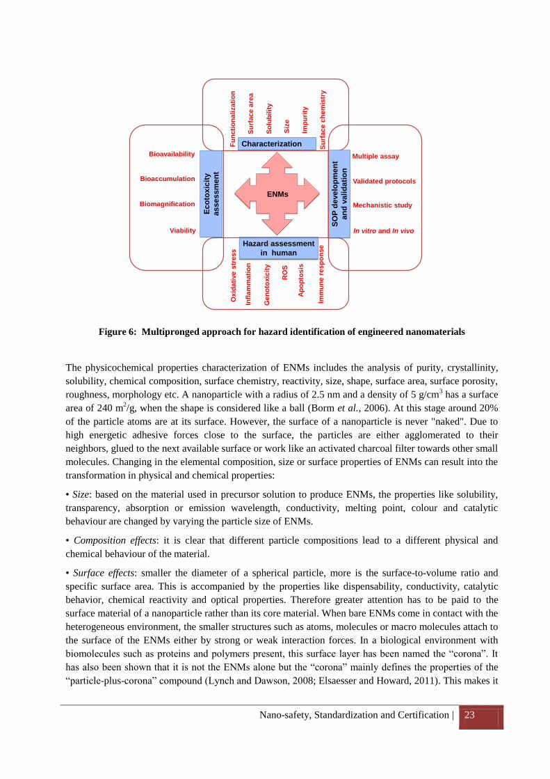
6. Guideline for the best practices for testing, standardization and certification of nanoproducts
It is now well established that the properties of ENMs are the combined function of their size, shape,
surface area, surface to volume ratio, chemical composition, solubility and others. Hence, to study the
ENMs’ effect in human and ecosystems, the study design should be multipronged, which address the
ENM characterization, validated protocols, hazard identification in human and environment (Figure 6). It
is also important to mention that surface properties of the ENMs affect their biological behaviour in the
ecosystem. In order to measure the risk/toxicological endpoints associated with the ENMs, the material
should needs to be fully understood and characterised. Otherwise, the possible risk/toxic effects cannot be
easily attributed to a certain property of the ENMs or even the ENM itself because, for example,
impurities and other components could be responsible (Dhawan and Sharma, 2010). Therefore, a critical
assessment of the biological behaviour of ENMs without a careful physicochemical characterization is not
meaningful. Apart from this the interference/interaction of ENMs with the testing methods/reagents also
creates the possibilities of wrong interpretation of the results (Howard, 2009; Stone et al., 2009). It has
also been reported that ENMs can bind with the active sites of the enzyme and made them inactive as well
as it can bind with the substrate and inhibit the binding sites of the enzyme (Kain et al., 2012). Hence, the
ENMs characterization and the activity testing should be done using array of methodologies.
Nano-safety, Standardization and Certification | 23
ENMs
Characterization
Hazard assessment
in human
Ap
op
tos
is
RO
S
Ox
idati
ve
str
es
s
Imm
un
e r
esp
on
se
Infl
am
mati
on
Ge
no
toxic
ity
Su
rfa
ce
ch
em
istr
y
Siz
e
Su
rface a
rea
Imp
uri
ty
Fu
ncti
on
alizati
on
So
lub
ilit
y
Eco
tox
icit
y
assessm
en
t
Bioavailability
Bioaccumulation
Biomagnification
Viability
SO
P d
eve
lop
men
t
an
d v
ali
da
tio
n
Multiple assay
Validated protocols
Mechanistic study
In vitro and In vivo
Figure 6: Multipronged approach for hazard identification of engineered nanomaterials
The physicochemical properties characterization of ENMs includes the analysis of purity, crystallinity,
solubility, chemical composition, surface chemistry, reactivity, size, shape, surface area, surface porosity,
roughness, morphology etc. A nanoparticle with a radius of 2.5 nm and a density of 5 g/cm3 has a surface
area of 240 m2/g, when the shape is considered like a ball (Borm et al., 2006). At this stage around 20%
of the particle atoms are at its surface. However, the surface of a nanoparticle is never "naked". Due to
high energetic adhesive forces close to the surface, the particles are either agglomerated to their
neighbors, glued to the next available surface or work like an activated charcoal filter towards other small
molecules. Changing in the elemental composition, size or surface properties of ENMs can result into the
transformation in physical and chemical properties:
• Size: based on the material used in precursor solution to produce ENMs, the properties like solubility,
transparency, absorption or emission wavelength, conductivity, melting point, colour and catalytic
behaviour are changed by varying the particle size of ENMs.
• Composition effects: it is clear that different particle compositions lead to a different physical and
chemical behaviour of the material.
• Surface effects: smaller the diameter of a spherical particle, more is the surface-to-volume ratio and
specific surface area. This is accompanied by the properties like dispensability, conductivity, catalytic
behavior, chemical reactivity and optical properties. Therefore greater attention has to be paid to the
surface material of a nanoparticle rather than its core material. When bare ENMs come in contact with the
heterogeneous environment, the smaller structures such as atoms, molecules or macro molecules attach to
the surface of the ENMs either by strong or weak interaction forces. In a biological environment with
biomolecules such as proteins and polymers present, this surface layer has been named the “corona”. It
has also been shown that it is not the ENMs alone but the “corona” mainly defines the properties of the
“particle-plus-corona” compound (Lynch and Dawson, 2008; Elsaesser and Howard, 2011). This makes it
Nano-safety, Standardization and Certification | 24
necessary to understand not only the behavior of ENMs but also the environment where they are going to
interact with biological system.
Hence, it can be summarized that to assess the risk/toxicity of ENMs, the primary criterion is to have full
knowledge of the ENMs to be tested. Considering the novel characteristics of ENMs, unlike their
chemical counterparts, it is imperative to undertake their comprehensive characterization prior to
risk/toxicity evaluation.
6.1 Characterization
The behavior and activity of ENMs is largely dependent on a number of physical and chemical properties.
Therefore, a complete characterization is essential for interpreting the results. The characterization of
ENMs should be carried out, to know about the specific physiochemical properties such as purity,
crystallinity, solubility, chemical composition, surface chemistry, reactivity, size, shape, surface area,
surface porosity, roughness, morphology etc (Table 3). Determination of the hydrodynamic size, size
distribution, zeta potential, dispersity and the concentration and time at which agglomeration occurs
should be done in the biological medium.
Table 3: Significance of measuring the physicochemical properties of engineered nanomaterials
S. No. Nanomaterials property
Significance
1. Size Nanomaterials possess a unique physicochemical property due to their size; it also affects the mobility and transport behaviour of the materials.
2. Shape Nanomaterials with different shapes (e.g. spherical, tubular, and cubical) have different affinities and accessibilities towards the cell wall. Toxicity of nanomaterials has also been reported due to their shape and size.
3. Structure The structure of the nanomaterials can influence the stability and behaviour of the ENMs (e.g. rutile and anatase are the possible crystal structures of TiO2 NPs).
4. Surface area As the size of nanomaterials reduces, the corresponding surface area increases leading to higher reactivity and sorption behaviour.
5. Agglomeration tendency
Agglomeration affects the surface properties of nanomaterials and their bio-availability to the cells.
6. Solubility Some of the nanomaterials are reported to produce ions in soluble form which may be toxic to the cells e.g. ZnO, CuO.
7. Elemental composition
Elemental composition shows as to whether the nanomaterials have contamination that may lead to false positive results or nanomaterials behaviour.
Nano-safety, Standardization and Certification | 25
8. Size distribution Size distribution of the nanomaterials gives an idea of the size range and helps in interpreting the results.
9. Surface charge and dispersity
Surface charge of the nanomaterials affects the particle solubility in suspension, whereas the dispersity of nanomaterials provides information about their tendency to agglomerate.
Different microscopic and spectroscopic techniques have been used to characterize the ENMs.
Microscopy-based methods include optical approaches, i.e. confocal microscopy, as well as electron and
scanning probe microscopy. The dimensions of ENMs are below the diffraction limit of visible light;
hence they are beyond the range of optical microscopy. However, near-field scanning optical microscopy
(NSOM) is a kind of scanning probe microscopy (SPM) technique that can achieve a spatial resolution of
50–100 nm through the use of a sub-wavelength diameter aperture. It is better than the conventional
optical microscopes to visualize the agglomeration of ENMs. The diffraction of light is also the limiting
factor for the conventional confocal microscopy. However, confocal laser scanning microscopy (CLSM)
has higher resolution (up to 200nm) hence the fluorescent ENMs (natural and labelled) can be observed.
Recently, for non-fluorescent particles, the reflection based study using confocal microscopy has been
reported to detect ENMs in cells (Lindfors et al., 2004; Van Dijk et al., 2005; Van Dijk et al., 2006;
Zucker et al., 2010).
Electron microscopy (scanning electron microscopy; SEM, transmission electron microscopy; TEM and
atomic force microscopy; AFM) is the most popular and extensively used technique to characterize the
ENMs. This technique not only gives visual image of the ENMs but also provides the information about
the properties such as size, state of aggregation, dispersion, structure and shape (Mavrocordatos et al.,
2004). In TEM, electrons are transmitted through a specimen; therefore the specimen needs to be well
distributed and spread on the grid (in case of materials) to get a good image, whereas in SEM, scattered
electrons are detected at the sample interface for imaging. Analytical tools, mostly spectroscopic, are
coupled with electron microscopes for additional elemental analysis. For example, energy dispersive X-
ray spectroscopy (EDS) when combined with SEM and TEM provides percentage elemental composition
of ENMs (Kumar et al., 2011c). Other analytical tools like electron energy loss spectroscopy (EELS)
when coupled with TEM, detect the elements based on the loss of energy of the incident electron through
the specimen (Mavrocordatos et al., 2004). Selected area electron diffraction (SAED) can also be
combined with TEM to provide information on crystalline properties of particles (Mavrocordatos et al.,
2004). Although electron microscopy is a very versatile tool for scientists in the area of nanotechnology,
it has certain limitations. A critical limitation is that TEM and SEM are operated under vacuum so, it is
difficult to analyse the liquid samples. The sample preparation steps of dehydration, cryo-fixation or
embedding usually lead to sample alteration and dehydration artifacts (Dhawan and Sharma, 2010).
Another disadvantage of the TEM is that the samples cannot be analysed twice or used for validation of
results. Further the charging effects caused by accumulation of static electric fields at the specimen due to
the electron irradiation create confusion during imaging (Tiede et al., 2008).
The atomic force microscopy (AFM) is also a kind of scanning probe microscope (SPM) which is a cost
effective instrument and has several advantages in the characterization of ENMs. The main advantage of
an AFM is that, it images sub-nanometer structures under ambient air and liquid dispersion, and provides
data about the size, shape, surface texture and roughness of the particles. In addition, multiple scanning of
the sample can also be done to get robust statistics. There are some limitations of AFM for ENMs
Nano-safety, Standardization and Certification | 26
visualization; generally the geometry of the probe is larger than the particles which lead to the over
estimation of the lateral dimensions of the nanoparticles.
It can be summarised that a combination of microscopic techniques, can be used to analyse the
nanoparticles for size, shape, size distribution etc. (Jose-Yacaman et al., 2001; Baatz et al., 2006;
Chuklanov et al., 2006). However, the analysis of the microscopic images is a crucial step because only
small amounts of samples can be analysed by microscopy which has an impact on the statistical
significance of the results. The average particle size of ENMs is a value that depends on the number of
particles counted and measured. As the ENMs in aqueous suspension have a tendency to agglomerate, it
is important to count and measure enough number of particles to obtain robust statistics on each size
fraction.
A wide range of spectroscopic techniques are available for the characterization of ENMs in suspension.
Some of the important techniques used for the characterization of ENMs based on the light scattering
property are static (SLS) and dynamic light-scattering (DLS) and small-angle neutron scattering (SANS).
Dynamic light scattering (DLS) or photon correlation spectroscopy (PCS) measures time dependent
fluctuations in scattering intensity of light produced by particles in Brownian motion and yields the size
of the particle by applying the Stokes–Einstein equation. DLS size of the nanoparticle is usually greater
than that measured by other techniques, like TEM, Brunauer–Emmett–Teller (BET), etc. DLS is
particularly very useful for sizing nanoparticles (based on intensity, volume and number) and determining
particle stability/aggregation state in suspensions with respect to time and medium. It is a quantitative
technique and gives the statistically relevant data as compared to TEM (Dhawan et al., 2009). Although
DLS provides fast, in situ and real-time sizing, it also has certain limitations. For example, interferences
can be caused by a range of materials such as dust particles and nanoparticle impurities which influences
the scattering intensity and skews the average hydrodynamic diameter towards the larger value. Also, the
intensity of the scattered light is proportional to the sixth power of the particle diameter that makes it very
sensitive to the presence of large particles and the data obtained from samples containing particles with
heterogeneous size distributions are difficult to interpret. DLS is considered an indispensable technique in
toxicity studies, as it provides valuable information pertaining to the zeta potential, polydispersity and
size range of the ENMs in the biological medium in which the organism is exposed.
Static light scattering, also known as multi-angle (laser) light-scattering [MAL(L)S], provides information
about the particle structure and together with dynamic light-scattering provides information about the
shape of the particle (Tiede et al., 2008). In small-angle neutron scattering (SANS), a beam of neutrons is
focussed on the sample, which can be solid (crystal, powder) or a suspension (aqueous, non-aqueous).
These neutrons interact with the nuclei of the atoms and get scattered due to changes in the refractive
index. The intensity of the scattered light gives information regarding the radius of gyration of a particle
using Guinier's equation.
Therefore, it can be inferred that a combination of analytical methods are required to detect and
characterize the nanoparticles in different matrices including air, soil, water and consumer products to
which human beings and ecosystems are likely to be exposed. Additionally, this will also provide the
broader idea related to the behaviour of the particles which will be helpful for the toxicological and risk
assessment of the nanoparticles. Different characterization techniques for ENPs as well as their merits &
demerits have been summarized in Table 4.
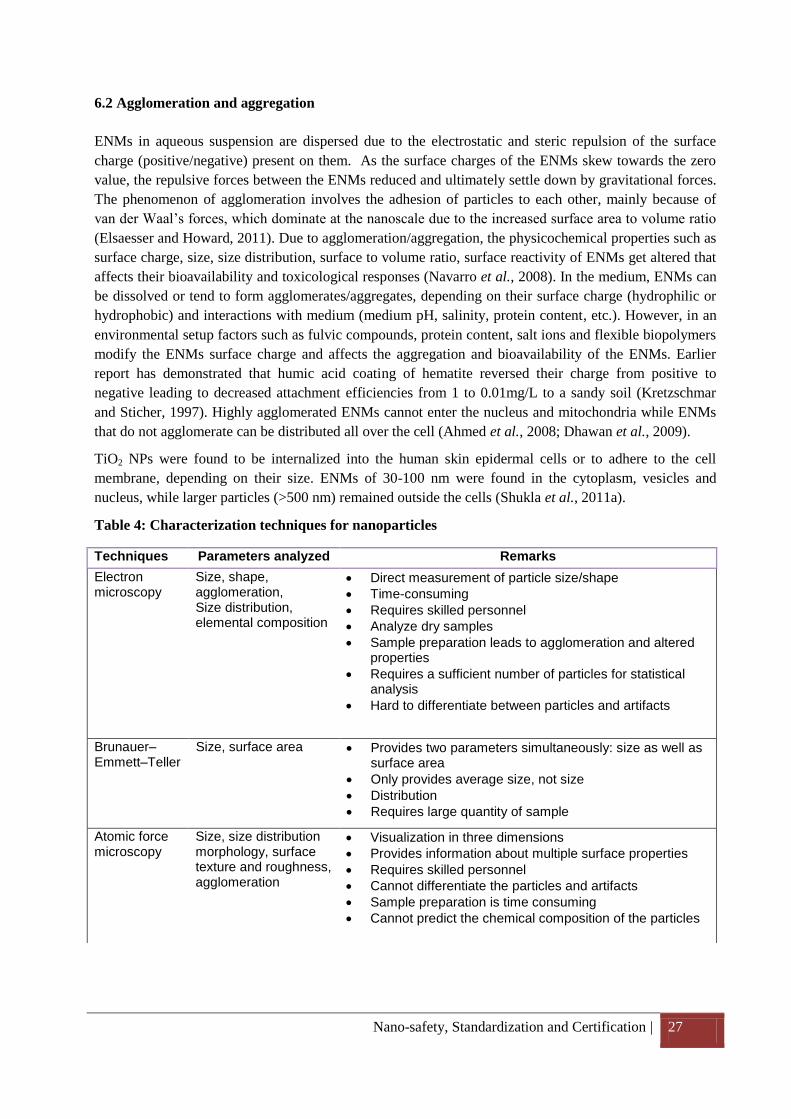
Nano-safety, Standardization and Certification | 27
6.2 Agglomeration and aggregation
ENMs in aqueous suspension are dispersed due to the electrostatic and steric repulsion of the surface
charge (positive/negative) present on them. As the surface charges of the ENMs skew towards the zero
value, the repulsive forces between the ENMs reduced and ultimately settle down by gravitational forces.
The phenomenon of agglomeration involves the adhesion of particles to each other, mainly because of
van der Waal’s forces, which dominate at the nanoscale due to the increased surface area to volume ratio
(Elsaesser and Howard, 2011). Due to agglomeration/aggregation, the physicochemical properties such as
surface charge, size, size distribution, surface to volume ratio, surface reactivity of ENMs get altered that
affects their bioavailability and toxicological responses (Navarro et al., 2008). In the medium, ENMs can
be dissolved or tend to form agglomerates/aggregates, depending on their surface charge (hydrophilic or
hydrophobic) and interactions with medium (medium pH, salinity, protein content, etc.). However, in an
environmental setup factors such as fulvic compounds, protein content, salt ions and flexible biopolymers
modify the ENMs surface charge and affects the aggregation and bioavailability of the ENMs. Earlier
report has demonstrated that humic acid coating of hematite reversed their charge from positive to
negative leading to decreased attachment efficiencies from 1 to 0.01mg/L to a sandy soil (Kretzschmar
and Sticher, 1997). Highly agglomerated ENMs cannot enter the nucleus and mitochondria while ENMs
that do not agglomerate can be distributed all over the cell (Ahmed et al., 2008; Dhawan et al., 2009).
TiO2 NPs were found to be internalized into the human skin epidermal cells or to adhere to the cell
membrane, depending on their size. ENMs of 30-100 nm were found in the cytoplasm, vesicles and
nucleus, while larger particles (>500 nm) remained outside the cells (Shukla et al., 2011a).
Table 4: Characterization techniques for nanoparticles
Techniques Parameters analyzed Remarks
Electron microscopy
Size, shape, agglomeration, Size distribution, elemental composition
Direct measurement of particle size/shape
Time-consuming
Requires skilled personnel
Analyze dry samples
Sample preparation leads to agglomeration and altered properties
Requires a sufficient number of particles for statistical analysis
Hard to differentiate between particles and artifacts
Brunauer–Emmett–Teller
Size, surface area Provides two parameters simultaneously: size as well as surface area
Only provides average size, not size
Distribution
Requires large quantity of sample
Atomic force microscopy
Size, size distribution morphology, surface texture and roughness, agglomeration
Visualization in three dimensions
Provides information about multiple surface properties
Requires skilled personnel
Cannot differentiate the particles and artifacts
Sample preparation is time consuming
Cannot predict the chemical composition of the particles
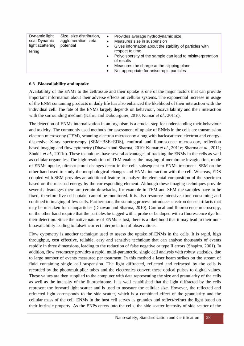
Nano-safety, Standardization and Certification | 28
6.3 Bioavailability and uptake
Availability of the ENMs to the cell/tissue and their uptake is one of the major factors that can provide
important information about their adverse effects on cellular systems. The exponential increase in usage
of the ENM containing products in daily life has also enhanced the likelihood of their interaction with the
individual cell. The fate of the ENMs largely depends on behaviour, bioavailability and their interaction
with the surrounding medium (Kahru and Dubourguier, 2010; Kumar et al., 2011c).
The detection of ENMs internalization in an organism is a crucial step for understanding their behaviour
and toxicity. The commonly used methods for assessment of uptake of ENMs in the cells are transmission
electron microscopy (TEM), scanning electron microscopy along with backscattered electron and energy-
dispersive X-ray spectroscopy (SEM+BSE+EDS), confocal and fluorescence microscopy, reflection
based imaging and flow cytometry (Dhawan and Sharma, 2010; Kumar et al., 2011e; Sharma et al., 2011;
Shukla et al., 2011c). These techniques have several advantages of tracking the ENMs in the cells as well
as cellular organelles. The high resolution of TEM enables the imaging of membrane invagination, mode
of ENMs uptake, ultrastructural changes occur in the cells subsequent to ENMs treatment. SEM on the
other hand used to study the morphological changes and ENMs interaction with the cell. Whereas, EDS
coupled with SEM provides an additional feature to analyze the elemental composition of the specimen
based on the released energy by the corresponding element. Although these imaging techniques provide
several advantages there are certain drawbacks, for example in TEM and SEM the samples have to be
fixed, therefore live cell uptake cannot be monitored. It is also resource intensive, time consuming and
confined to imaging of few cells. Furthermore, the staining process introduces electron dense artifacts that
may be mistaken for nanoparticles (Dhawan and Sharma, 2010). Confocal and fluorescence microscopy,
on the other hand require that the particles be tagged with a probe or be doped with a fluorescence dye for
their detection. Since the native nature of ENMs is lost, there is a likelihood that it may lead to their non-
bioavailability leading to false/incorrect interpretation of observations.
Flow cytometry is another technique used to assess the uptake of ENMs in the cells. It is rapid, high
throughput, cost effective, reliable, easy and sensitive technique that can analyse thousands of events
rapidly in three dimensions, leading to the reduction of false negative or type II errors (Shapiro, 2001). In
addition, flow cytometry provides a rapid, multi-parametric, single cell analysis with robust statistics, due
to large number of events measured per treatment. In this method a laser beam strikes on the stream of
fluid containing single cell suspension. The light diffracted, reflected and refracted by the cells is
recorded by the photomultiplier tubes and the electronics convert these optical pulses to digital values.
These values are then supplied to the computer with data representing the size and granularity of the cells
as well as the intensity of the fluorochrome. It is well established that the light diffracted by the cells
represent the forward light scatter and is used to measure the cellular size. However, the reflected and
refracted light corresponds to the side scatter, which is a combined effect of the granularity and the
cellular mass of the cell. ENMs in the host cell serves as granules and reflect/refract the light based on
their intrinsic property. As the ENPs enters into the cells, the side scatter intensity of side scatter of the
Dynamic light scat Dynamic light scattering
tering
Size, size distribution, agglomeration, zeta potential
Provides average hydrodynamic size
Measures size in suspension
Gives information about the stability of particles with respect to time
Polydispersity of the sample can lead to misinterpretation of results
Measures the charge at the slipping plane
Not appropriate for anisotropic particles
Nano-safety, Standardization and Certification | 29
cell increases proportionately to the concentration of the ENMs. However, a fluorescent particle can give
an increased signal of side scatter as well as the fluorochrome intensity in a dose dependent manner.
It is also reported that ENMs are having their own scattering phenomenon, hence the interference of the
ENMs scattering phenomenon with the scattering phenomenon of interrogated cells cannot be ruled out.
6.4 Development and validation of standard operating procedure
The development of the methodologies for the assessment of risk associated with the ENMs is in early
stage. Most of the studies concerning to ENMs risk have been carried out using the classical in vitro
toxicity test methods established for chemicals. However, these established methods cannot be used for
assessing the toxicity of ENMs, as ENMs display several unique physicochemical properties. Due to
these properties, ENMs interfere with normal test systems, and this interference has been well
documented in the literatures (Monteiro-Riviere and Inman, 2006; Doak et al., 2009; Kroll et al., 2009;
Monteiro-Riviere et al., 2009; Song et al., 2010; Kain et al., 2012). Examples of such properties include:
high surface area, leading to increased adsorption capacity; different optical properties that interfere with
fluorescence or visible light absorption detection systems; increased catalytic activity due to enhanced
surface energy; and magnetic properties that make them redox active and thus interfere with methods
based on redox reactions (reference). Single-walled carbon nanotubes (SWCNTs) interact with a variety
of indicator dyes employed in commonly used cytotoxicity assays, such as 3-(4,5-dimethylthiazole-2-yl)-
2,5-biphenyl tetrazolium bromide (MTT), 2-(4-iodophenyl)- 3-(4-nitrophenyl)-5-(2,4-disulfophenyl)-2H
tetrazolium, monosodium salt (WST-1), Coomassie blue, alamar blue and neutral red. It is also suggested
that these nanotubes bind with formazan crystals and stabilize their chemical structure, meaning that these
crystals cannot be solubilized. The same phenomenon can be observed with carbon nanotubes where the
unusual rope-like structure of these ENMs binds with the MTT dye and leads to the false positive result
(Worle-Knirsch et al., 2006). The high adsorptive capacities of ENMs have also been reported to interfere
with annexin V/PI binding and ELISA tests for cytokine responses (Monteiro-Riviere and Inman, 2006).
Another example for the ENMs interference with the test system is detection of oxidative DNA damage
with formamidopyrimidine DNA glycosylase (FPG) enzyme. The presence of ENMs in the nucleoid has
been reported and their possibility to induce additional DNA damage during the assay has been discussed
extensively (Karlsson et al., 2004; Stone et al., 2009; Karlsson, 2010). The presence of ENMs close to
DNA during the comet assay also increases the probability for the interaction of ENMs with FPG. It has
recently been shown that the incubation of the ENMs and ions with FPG enzyme leads to the total loss of
the ability of the enzyme to detect oxidatively damaged DNA in the comet assay (Kain et al., 2012). This
disturbance is most likely due to the binding of ions to the SH groups at the active site (Kain et al., 2012).
Another possible reason could be the physical hindrance by NPs, which prevent the enzyme action at the
damaged DNA site.
Hence, it is important to standardize the methods for the physicochemical characterization of
nanoparticles; development of appropriate methods/protocols for hazard assessment; safe production,
handling, use and disposal methods; methodological and metrological approaches for the detection,
bioavailability and uptake; agglomeration and aggregation state; reference materials; modelling and
simulations and many more to overcome the limitations of current hazard and risk assessment schemes. It
is also recommended to analyze the interaction/interference properties of ENMs prior to the beginning of
the experiments. United Kingdom is playing a key role in leading the development of nanotechnology
standards through its national committee NTI/1 "Nanotechnologies". They established the technical
committee (TC; ISO TC 229) in June 2004 with the motto to (a) formulate a UK strategy for
standardization in nanotechnologies through a broad consultation with relevant stakeholders (b) ensure
Nano-safety, Standardization and Certification | 30
the UK view is given due consideration within the European Union, CEN, ISO and IEC (c) develop and
support formal standards and other standardization documents in the area of nanotechnologies and to
promote their use by industry and other stakeholders (d) ensure due consideration of the need for
standards and standardization is given by UK nanotechnology networks and organisations, and to
coordinate activities and actions in this area (BSI, ; ISO/TC229). Funding agencies such as the European
Union; Department of Science and Technology, Department of Biotechnology, Council of Scientific and
Industrial Research, Government of India, have focussed on the development & validation of SOPs for
ENM preparation and testing as well as their life cycle assessment and fate in ecosystems. India is also
participating in the European Union Seventh Framework Programme (FP7/2007-2013) under the project
“Development of reference methods for hazard identification, risk assessment and LCA of engineered
nanomaterials (NanoValid)”. These characterization and standardization process will contribute to a
better mechanistic understanding of the behaviour of nanoparticles in various test media, physiological
solutions and environmental matrices. This can also be helpful in developing reference material as well as
the test schemes for risk management. The first certified reference material was developed by the Institute
for Reference Materials and Measurements, Joint Research Centre. The material was prepared with
colloidal silica of a spherical diameter of 20nm. Certification of the material was based on a global
interlaboratory comparison in which 34 laboratories participated with various analytical methods (DLS,
CLS, EM, SAXS, ELS; (Braun et al., 2012).
To maximize the outcomes of the initiatives taken by EU, a NanoSafety Cluster (NanoSafetyCluster,
2013) has been formed between the existing 29 projects of FP6 and FP7 programmes (NANOMMUNE,
NanoTEST, NANODEVICE, NanoFATE, MARINA, QNnao NanoValid, NanoLinen, NewIndigo project
etc.) addressing all aspects of nanosafety including toxicology, ecotoxicology, exposure assessment,
mechanisms of interaction, risk assessment and standardisation.
6.5 Certification of nanoproducts
Certification plays an important role in getting the confidence of the consumer in commercial sector. It
also provides an evidence of the existence of agreements between manufacturers and national
accreditation testing and certification organisations. With the increasing use of ENM based products in
the market, nanocertification will be important to confirm the applicability and technologies to this
developing sector. It will also provide a guarantee and adequate quality of nanoproducts both at
manufacturing & application stage, and encourage the manufacturers to develop quality products for
consumer acceptance. Besides this, the certification system will also promote a favourable public opinion
concerning the reliability and safety of nanoproducts and nanotechnologies.
Initially in September 2005, the UK Micro and NanoTechnology Network (MNT), has initiated a MNT
Quality Mark for firms involved in the nanotechnology industry. The primary objective of the MNT
Quality Mark was the development and implementation of best practice in nanotechnologies and to set a
minimum standard of performance and achievement (UKMNT).
Later, in Taiwan, a National Science Council (NSC) was established to develop a “Nano-product
Certification System Plan” with the coordination of Industrial Development Bureau (IDB), Ministry of
Economic Affairs (MOEA), and Centre for Measurement Standards (CMS). The goal of NSC was to
promote the development of nano-industries starting with the different aspects of the nanotechnology and
industry. Additionally, the development of nanoMark certification in Taiwan enhances the overall
enterprise competitiveness and already 34 companies with 1,215 products have passed the nanoMark
certification. From the viewpoint of economy, granting the nanoMark would encourage manufacturers to
produce quality products for a sustainable operation (nanoMark).
Nano-safety, Standardization and Certification | 31
Further, RUSNANO was established in March 2011 by Russian Corporation of Nanotechnologies, with a
mission is to develop the Russian nanotechnology industry through co-investment in nanotechnology
projects with substantial economic potential or social benefit. They developed a voluntary certification
system (VCS) called “Nanocertifica” for nanoindustry products. It is registered with the Federal Agency
on Technical Regulation and Metrology for the quality management systems of companies operating in
the nanoindustry under the provision of ISO 9000 and ISO 14000 (RUSNANO).
Recently, National Nanotechnology Center (NANOTEC), National Science and Technology
Development Agency, Thailand have also launched the NanoQ mark to set industrial standards for
nanotechnology related products (NanoQ, 2013).
It is apparent that different countries are realizing the potential of nanotech based products where
standardization leading to certification will play a key role in quality assurance. This will also build
confidence amongst the manufacturers and consumers alike regarding the widespread acceptability of
nanotech based products.
7. Summary
The increasing use of engineered nanomaterials in various applications has increased the likelihood of
their possible interaction with human and the environment. Humans get exposed to ENMs at various steps
of its synthesis (laboratory), manufacture (industry), use (consumer products, devices, medicines etc.).
Also, there is an almost complete lack of data on bioaccumulation, bioconcentration and biodegradation
of ENMs in environmentally relevant species. Currently, the rate of ENM incorporation in commercial
products is much faster than the development of regulation and knowledge to mitigate their potential
adverse effects. This can be attributed to the lack of regulatory guidelines, reference standards and
certification processes for ENMs (from manufacture to product development). This is compounded by the
lack of suitable models, problems in experimental protocols and appropriate study design.
Hence, the knowledge regarding the fate and impact of ENMs on human and the environment; social,
ethical and legal issues; safe production, handling, use and disposal; nanosafety policies, risk governance
and the guidelines for the best practices for testing, standardization and certification of nanoproducts
could be helpful in mitigating the risk associated with ENMs.
Acknowledgements
Funding received from the Council of Scientific and Industrial Research, New Delhi (NanoSHE;BSC-
0112); Department of Science and Technology, Government of India under the Nanomission programme
(DST-NSTI grant No. SR/S5/NM-01/2007) and UKIERI-DST (grant No. IND/CONT/E/11-12/217) and
from the Department of Biotechnology, Government of India under the NewINDIGO Scheme for
NanoLINEN project is gratefully acknowledged. Funding from the European Union Seventh Framework
Programme (FP7/200 7-2013) under grant agreement No. 263147 (NanoValid - Development of
reference methods for hazard identification, risk assessment and LCA of engineered nanomaterials) is
also acknowledged.
Nano-safety, Standardization and Certification | 32
References
Ahamed, M., Karns, M., Goodson, M., Rowe, J., Hussain, S. M., Schlager, J. J., and Hong, Y. (2008).
DNA damage response to different surface chemistry of silver nanoparticles in mammalian cells.
Toxicology and Applied Pharmacology 233, 404-410.
Ahmed, M., Ramadan, W., Rambhu, D., and Shakeel, F. (2008). Potential of nanoemulsions for
intravenous delivery of rifampicin. Die Pharmazie 63, 806-811.
Aitken, R. J., Hankin, S. M., Lang Tran, C., Donaldson, K., Stone, V., Cumpson, P., Johnstone, J.,
Chaudhry, Q., Cash, S., and Garrod, J. (2008). A multidisciplinary approach to the identification of
reference materials for engineered nanoparticle toxicology. Nanotoxicology 2, 71-78.
Aitken, R. J., Koopman, P., and Lewis, S. E. (2004). Seeds of concern. Nature 432, 48-52.
Allen, H. J., Impellitteri, C. A., Macke, D. A., Heckman, J. L., Poynton, H. C., Lazorchak, J. M.,
Govindaswamy, S., Roose, D. L., and Nadagouda, M. N. (2011). Effects from filtration, capping agents,
and presence/absence of food on the toxicity of silver nanoparticles to Daphnia magna. Environmental
toxicology and chemistry / SETAC 29, 2742-2750.
Auffan, M., Rose, J., Wiesner, M. R., and Bottero, J. Y. (2009). Chemical stability of metallic
nanoparticles: a parameter controlling their potential cellular toxicity in vitro. Environ Pollut 157, 1127-
1133.
Baatz, M., Arini, N., Schape, A., Binnig, G., and Linssen, B. (2006). Object-oriented image analysis for
high content screening: detailed quantification of cells and sub cellular structures with the Cellenger
software. Cytometry A 69, 652-658.
Baun, A., Hartmann, N. B., Grieger, K., and Kusk, K. O. (2008). Ecotoxicity of engineered nanoparticles
to aquatic invertebrates: A brief review and recommendations for future toxicity testing. Ecotoxicology
(London, England) 17, 387-395.
Borm, P. J., Robbins, D., Haubold, S., Kuhlbusch, T., Fissan, H., Donaldson, K., Schins, R., Stone, V.,
Kreyling, W., Lademann, J., Krutmann, J., Warheit, D., and Oberdorster, E. (2006). The potential risks of
nanomaterials: a review carried out for ECETOC. Particle and Fibre Toxicology 3, 11.
Braun, A., Kestens, V., Franks, K., Roebben, G., Lamberty, A., and Linsinger, T. P. J. (2012). A new
certified reference material for size analysis of nanoparticles. Journal of Nanoparticle Research 14, 1021-
1033.
Brayner, R. (2008). The toxicological impact of nanoparticles. Nano today 3, 48-55.
Brayner, R., Ferrari-Iliou, R., Brivois, N., Djediat, S., Benedetti, M. F., and Fievet, F. (2006).
Toxicological impact studies based on Escherichia coli bacteria in ultrafine ZnO nanoparticles colloidal
medium. Nano letters 6, 866-870.
Breggin, L., Robert, F., Nico, J., John, P., and Read, P. (2009). Securing the promise of nanotechnologies:
Towards transatlantic regulatory cooperation. Report. London: Chatham House.
Brinker, C. J., and Scherer, G. (1990). The Physics and Chemistry of Sol–Gel Processing,. Elsevier, The
Netherlands.
Nano-safety, Standardization and Certification | 33
BSI http://shop.bsigroup.com/en/Browse-By-Subject/Nanotechnology/BSI-Committee-for-
Nanotechnologies/.
Buffle, J., Wilkinson, K., Stoll, S., Filella, M., and Zhang, J. (1998). A generalized description of aquatic
colloidal interactions: The three-colloidal component approach. Environmental Science and Technology
32, 2887–2899.
Chau, C., Wu, S., and Yen, G. (2007). The development of regulations for food nanotechnology. Trends
in Food Science and Technology. 18, 269–280.
Chen, M., and von Mikecz, A. (2005). Formation of nucleoplasmic protein aggregates impairs nuclear
function in response to SiO2 nanoparticles. Exp Cell Res 305, 51-62.
Chuklanov, A., Ziganshina, S., and Bukharaev, A. (2006). Computer program for the grain analysis of
AFM images of nanoparticles placed on a rough surface. Surf Interface Analysis 38, 679–681.
Colvin, V. L. (2003). The potential environmental impact of engineered nanomaterials. Nature
Biotechnology 21, 1166-1170.
Cui, D., Tian, F., Ozkan, C. S., Wang, M., and Gao, H. (2005). Effect of single wall carbon nanotubes on
human HEK293 cells. Toxicol Lett 155, 73-85.
Demuth, P. C., Min, Y., Huang, B., Kramer, J. A., Miller, A. D., Barouch, D. H., Hammond, P. T., and
Irvine, D. J. (2013). Polymer multilayer tattooing for enhanced DNA vaccination. Nature materials,
doi:10.1038/nmat3550.
Dhawan, A., Shanker, R., Das, M., and Gupta, K. C. (2011). Guidance for safe handling of nanomaterials.
J Biomed Nanotechnol 7, 218-224.
Dhawan, A., and Sharma, V. (2010). Toxicity assessment of nanomaterials: Methods and challanges.
Analatical and Bioanalytical Chemistry 398, 589-605.
Dhawan, A., Sharma, V., and Parmar, D. (2009). Nanomaterials: A challenge for toxicologists.
Nanotoxicology 3, 1-9.
Dhawan, A., Taurozzi, J. S., Pandey, A. K., Shan, W., Miller, S. M., Hashsham, S. A., and Tarabara, V.
V. (2006). Stable colloidal dispersions of C60 fullerenes in water: Evidence for genotoxicity.
Environmental Science and Technology 40, 7394-7401.
Doak, S. H., Griffiths, S. M., Manshian, B., Singh, N., Williams, P. M., Brown, A. P., and Jenkins, G. J.
S. (2009). Confounding experimental considerations in nanogenotoxicology. Mutagenesis 24, 285-293.
Dobrovolskaia, M. A., Germolec, D. R., and Weaver, J. L. (2009). Evaluation of nanoparticle
immunotoxicity. Nat Nanotechnol 4, 411-414.
Donaldson, K., and Borm, P. (2004). Particle and Fibre Toxicology, a new journal to meet a real need.
Particle and Fibre Toxicology 1, 1.
Donaldson, K., Stone, V., Tran, C. L., Kreyling, W., and Borm, P. J. A. (2004). Nanotoxicology.
Occupational and Environmental Medicine 61, 727-728.
EFSA (2007). Opinion of the scientiac panel on food additives, flavourings, processing aids and materials
in contact with food (AFC) on a request related to a 14th list of substances for food contact materials. .
EFSA Journal 5, 452–454.
EFSA (2008a). Scientiac opinion of the panel on food contact materials, enzymes, fla- vourings and
processing Aids (CEF). EFSA Journal 6, 888-890.
Nano-safety, Standardization and Certification | 34
EFSA (2008b). Scientific opinion: Inability to assess the safety of a silver hydrosol added for nutritional
purposes as a source of silver in food supplements and the bioavailability of silver from this source based
on the supporting dossier. EFSA Journal 6, 884.
Elsaesser, A., and Howard, C. V. (2011). Toxicology of nanoparticles. Advanced Drug Delivery Reviews
64, 129-137.
EPA (2007a). Nanotechnology White Paper. EPA 100/B-07/001.
EPA (2007b). Pesticide registration; Clarification for ion-generating equipment. Federal Register
72(183).
EPA (2008). Toxic substances control act inventory status of carbon nanotubes. Federal Register
73(212).
European Commission (2008). Commission Regulation (EC) No. 987/2008 Official Journal of the
European Union L268/14.
Fabrega, J., Luoma, S. N., Tyler, C. R., Galloway, T. S., and Lead, J. R. (2011). Silver nanoparticles:
behaviour and effects in the aquatic environment. Environment International 37, 517-531.
Fadeel, B., and Garcia-Bennett, A. E. (2010). Better safe than sorry: Understanding the toxicological
properties of inorganic nanoparticles manufactured for biomedical applications. Advanced Drug Delivery
Reviews 62, 362-374.
Fadeel, B., Kagan, V., Krug, H., Shvedova, A., Svartengren, M., Tran, L., and Wiklund, L. (2007).
There's plenty of room at the forum: Potential risks and safety assessment of engineered nanomaterials.
Nanotoxicology 1, 73-84.
Fairbrother, A., and Fairbrother, J. R. (2009). Are environmental regulations keeping up with innovation?
A case study of the nanotechnology industry. Ecotoxicology and Environmental Safety 72, 1327-1330.
Farre, M., Gajda-Schrantz, K., Kantiani, L., and Barcelo, D. (2009). Ecotoxicity and analysis of
nanomaterials in the aquatic environment. Analytical and Bioanalytical Chemistry 393, 81-95.
FDA (2007). Nanotechnology. A report of the U.S. food and drug administration nanotechnology task
force.
Feliu, N., and Fadeel, B. (2010). Nanotoxicology: no small matter. Nanoscale 2, 2514-2520.
Gajewicz, A., Rasulev, B., Dinadayalane, T. C., Urbaszek, P., Puzyn, T., Leszczynska, D., and
Leszczynski, J. (2012). Advancing risk assessment of engineered nanomaterials: application of
computational approaches. Advanced Drug Delivery Reviews 64, 1663-1693.
Gruere, G. P. (2012). Implications of nanotechnology growth in food and agriculture in OECD countries.
Food Policy 37, 191–198.
Handy, R. D., Owen, R., and Valsami-Jones, E. (2008). The ecotoxicology of nanoparticles and
nanomaterials: current status, knowledge gaps, challenges, and future needs. Ecotoxicology (London,
England) 17, 315-325.
Hardman, R. (2006). A toxicologic review of quantum dots: Toxicity depends on physicochemical and
environmental factors. Environmental health perspectives 114, 165-172.
Howard, A. G. (2009). On the challenge of quantifying man-made nanoparticles in the aquatic
environment. J Environ Monit 12, 135-142.
Nano-safety, Standardization and Certification | 35
ISO/TC229
http://www.iso.org/iso/iso_catalogue/catalogue_tc/catalogue_tc_browse.htm?commid=381983&publishe
d=on&includesc=true.
Jose-Yacaman, M., Marin-Almazo, M., and Ascencio, J. (2001). High resolution TEM studies on
palladium nanoparticles. Journal of Molecular Catalysis A 173, 61–74.
Jyoti, A., Pandey, P., Singh, S. P., Jain, S. K., and Shanker, R. (2010). Colorimetric detection of nucleic
acid signature of shiga toxin producing Escherichia coli using gold nanoparticles. Journal of Nanoscience
and Nanotechnology 10, 4154-4158.
Kahru, A., and Dubourguier, H. C. (2010). From ecotoxicology to nanoecotoxicology. Toxicology 269,
105-119.
Kain, J., Karlsson, H. L., and Moller, L. (2012). DNA damage induced by micro- and nanoparticles--
interaction with FPG influences the detection of DNA oxidation in the comet assay. Mutagenesis 27, 491-
500.
Karlsson, H. L. (2010). The comet assay in nanotoxicology research. Analytical and Bioanalytical
Chemistry 398, 651-666.
Karlsson, H. L., Nygren, J., and Moller, L. (2004). Genotoxicity of airborne particulate matter: the role of
cell-particle interaction and of substances with adduct-forming and oxidizing capacity. Mutation
Research 565, 1-10.
Kermisch, C. (2012). Do new ethical issues arise at each stage of nanotechnological development.
Nanoethics 6, 29–37.
Kretzschmar, R., and Sticher, H. (1997). Transport of humic-coated iron oxide colloids in a sandy soil:
influence of Ca2+ and trace metals. Environmental Science and Technology 31, 3497–3504.
Kroll, A., Pillukat, M. H., Hahn, D., and Schnekenburger, J. (2009). Current in vitro methods in
nanoparticle risk assessment: Limitations and challenges. European Journal of Pharmaceutics and
Biopharmaceutics 72, 370-377.
Kulkarni, P., Willeke, K., and Baron, P. A. (2011). Aerosol Measurement: Principles, Techniques, and
Applications. Wiley, New York.
Kumar, A., Dhawan, A., and Shanker, R. (2011a). The need for novel approaches in ecotoxicity of
engineered nanomaterials. J Biomed Nanotechnol 7, 79-80.
Kumar, A., Pandey, A. K., Shanker, R., and dhawan, A. (2012). Microorganism: A varsatile model for
toxicity assessment of engineered nanomaterials. Book Name :- Nano-Antimicrobials: Progress and
Prospects, Publisher:- Springer Heidelberg Dordrecht London New York Edited by Nicola Cioffi,
Mahendra Rai, 497-524.
Kumar, A., Pandey, A. K., Singh, S. S., Shanker, R., and Dhawan, A. (2011b). Cellular response to metal
oxide nanoparticles in bacteria. J Biomed Nanotechnol 7, 102-103.
Kumar, A., Pandey, A. K., Singh, S. S., Shanker, R., and Dhawan, A. (2011c). Cellular uptake and
mutagenic potential of metal oxide nanoparticles in bacterial cells. Chemosphere 83, 1124-1132.
Kumar, A., Pandey, A. K., Singh, S. S., Shanker, R., and Dhawan, A. (2011d). Engineered ZnO and
TiO(2) nanoparticles induce oxidative stress and DNA damage leading to reduced viability of Escherichia
coli. Free radical biology & medicine 51, 1872-1881.
Nano-safety, Standardization and Certification | 36
Kumar, A., Pandey, A. K., Singh, S. S., Shanker, R., and Dhawan, A. (2011e). A flow cytometric method
to assess nanoparticle uptake in bacteria. Cytometry A 79A, 707-712.
Kumar, A., Shanker, R., and Dhawan, A. (2011f). The need for novel approaches in ecotoxicity of
engineered nanomaterials. Journal of Biomedical Nanotechnology 7 79–80.
Kumari, M., Khan, S. S., Pakrashi, S., Mukherjee, A., and Chandrasekaran, N. (2011). Cytogenetic and
genotoxic effects of zinc oxide nanoparticles on root cells of Allium cepa. Journal of Hazardous
Materials 190, 613-621.
Lehn, J. M. (2002). Towards self organization and complex matter. Science 295, 2400-2403.
Li, N., Sioutas, C., Cho, A., Schmitz, D., Misra, C., Sempf, J., Wang, M., Oberley, T., Froines, J., and
Nel, A. (2003). Ultrafine particulate pollutants induce oxidative stress and mitochondrial damage.
Environmental Health Perspectives 111, 455-460.
Lindfors, K., Kalkbrenner, T., Stoller, P., and Sandoghdar, V. (2004). Detection and spectroscopy of gold
nanoparticles using supercontinuum white light confocal microscopy. Physical Review Letters 93,
037401.
Long, T. C., Saleh, N., Tilton, R. D., Lowry, G. V., and Veronesi, B. (2006). Titanium dioxide (P25)
produces reactive oxygen species in immortalized brain microglia (BV2): implications for nanoparticle
neurotoxicity. Environmental Science & Technology 40, 4346-4352.
Lynch, I., and Dawson, K. A. (2008). Protein-nanoparticle interactions. Nano Today 3, 40-47.
Mahapatra, I., Clark, J., Dobson, P. J., Owenc, R., and Lead, J. R. (2013). Potential environmental
implications of nano-enabled medical applications: critical review. Environmental Science: Processes &
Impacts 15, 123–144.
Mavrocordatos, D., Pronk, W., and Boiler, M. (2004). Analysis of environmental particles by atomic
force microscopy, scanning and transmission electron microscopy. Water Sci Technol 50, 9-18.
Maynard, A. D. (2007). Nanotechnology: the next big thing, or much ado about nothing? The Annals of
occupational hygiene 51, 1-12.
Monteiro-Riviere, N., and Inman, A. (2006). Challenges for assessing carbon nanomaterial toxicity to the
skin. Carbon 44, 1070–1078.
Monteiro-Riviere, N. A., Inman, A. O., and Zhang, L. W. (2009). Limitations and relative utility of
screening assays to assess engineered nanoparticle toxicity in a human cell line. Toxicol Appl Pharmacol
234, 222-235.
nanoMark http://proj3.moeaidb.gov.tw/nanomark/Eng/.
NanoQ (2013). http://www.nanotec.or.th/en/?p=4014.
NanoSafetyCluster (2013). http://www.nanosafetycluster.eu/eu-nanosafety-cluster-projects/seventh-
framework-programme-projects/ennsatox.html.
Navarro, E., Baun, A., Behra, R., Hartmann, N. B., Filser, J., Miao, A. J., Quigg, A., Santschi, P. H., and
Sigg, L. (2008). Environmental behavior and ecotoxicity of engineered nanoparticles to algae, plants, and
fungi. Ecotoxicology (London, England) 17, 372-386.
Nel, A. (2005). Atmosphere air pollution-related illness: effects of particles. Science 308, 804-806.
Nel, A., Xia, T., Madler, L., Li, N. (2006). Toxic potential of materials at the nanolevel. Science 311,
622-627.
Nano-safety, Standardization and Certification | 37
NIOSH (2010). National Institute for Occupational Safety and Health, Occupational exposure to carbon
nanotubes and nanofibers, current intelligence bulletin, Department of Health and Human Services,
Centers for Disease Control and Prevention.
Nowack, B. (2009). The behavior and effects of nanoparticles in the environment. Environ Pollut 157,
1063-1064.
O’Shaughnessy, P. T. (2013). Occupational health risk to nanoparticulate exposure. Environmental
Science: Processes & Impacts 15, 49-62.
Oberdarster, G., Oberdarster, E., and Oberdarster, J. (2005). Nanotoxicology: An emerging discipline
evolving from studies of ultrafine particles. Environmental Health Perspectives 113, 823-839.
Oberdarster, G., Stone, V., and Donaldson, K. (2007). Toxicology of nanoparticles: A historical
perspective. Nanotoxicology 1, 2-25.
Oberdorster, G., Sharp, Z., Atudorei, V., Elder, A., Gelein, R., Kreyling, W., and Cox, C. (2004).
Translocation of inhaled ultrafine particles to the brain. Inhalation Toxicology 16, 437-445.
PEN (2013). http://www.nanotechproject.org/inventories/remediation_map/.
PEN (2013). Project of the Emerging Nanotechnologies (PEN) Available at PEN
"http://www.nanotechproject.org/inventories/consumer/browse/products/."
Penn, A., Murphy, G., Barker, S., Henk, W., and Penn, L. (2005). Combustion-derived ultrafine particles
transport organic toxicants to target respiratory cells. Environmental Health Perspectives 113, 956-963.
Premanathan, M., Karthikeyan, K., Jeyasubramanian, K., and Manivannan, G. (2011). Selective toxicity
of ZnO nanoparticles toward Gram-positive bacteria and cancer cells by apoptosis through lipid
peroxidation. Nanomedicine 7, 184-192.
Pui, D. Y. H. (1996). Direct-reading Instrumentation for workplace aerosol measurements. The Analyst
121, 1215–1224.
Royal, S. (2004). The Royal society and the Royal Academy of engineers: Nanoscience and
nanotechnologies:oppurtunities and uncertainties.
RUSNANO http://eng.thesaurus.rusnano.com/wiki/article3122.
SCENIHR (2009). (European Commission Scientific Committee on Emerging and Newly Identified
Health Risks) Risk Assessment of Products of Nanotechnologies.
Schins, R. P., Lightbody, J. H., Borm, P. J., Shi, T., Donaldson, K., and Stone, V. (2004). Inflammatory
effects of coarse and fine particulate matter in relation to chemical and biological constituents. Toxicol
Appl Pharmacol 195, 1-11.
Service, R. F. (2008). Science policy: Report faults U.S. strategy for nanotoxicology research. Science
322, 1779.
Shapiro, H. M. (2001). Optical measurements in cytometry: Light scattering, extinction, absorption, and
fluorescence. Methods in Cell Biology, Vol 63 63, 107-129.
Sharma, V., Anderson, D., and Dhawan, A. (2012a). Zinc oxide nanoparticles induce oxidative DNA
damage and ROS-triggered mitochondria mediated apoptosis in human liver cells (HepG2). Apoptosis 17,
852-870.
Nano-safety, Standardization and Certification | 38
Sharma, V., Kumar, A., and Dhawan, A. (2012b). Nanomaterials: Exposure, effects and toxicity
assessment. Proceedings of the National Academy of Sciences, India Section B: Biological Sciences 82, 3-
11.
Sharma, V., Shukla, R. K., Saxena, N., Parmar, D., Das, M., and Dhawan, A. (2009). DNA damaging
potential of zinc oxide nanoparticles in human epidermal cells. Toxicology Letters 185, 211-218.
Sharma, V., Singh, P., Pandey, A. K., and Dhawan, A. (2012c). Induction of oxidative stress, DNA
damage and apoptosis in mouse liver after sub-acute oral exposure to zinc oxide nanoparticles. Mutation
Research 745, 84-91.
Sharma, V., Singh, S. K., Anderson, D., Tobin, D. J., and Dhawan, A. (2011). Zinc oxide nanoparticle
induced genotoxicity in primary human epidermal keratinocytes Journal of Nanoscience and
Nanotechnology 11, 3782-3788.
Shukla, R. K., Kumar, A., Gurbani, D., Pandey, A. K., Singh, S., and Dhawan, A. (2011a). TiO(2)
nanoparticles induce oxidative DNA damage and apoptosis in human liver cells. Nanotoxicology 7, 48-
60.
Shukla, R. K., Kumar, A., Pandey, A. K., Singh, S. S., and Dhawan, A. (2011b). Titanium Dioxide
Nanoparticles Induce Oxidative Stress-Mediated Apoptosis in Human Keratinocyte Cells. Journal of
Biomedical Nanotechnology 7, 100–101.
Shukla, R. K., Sharma, V., Pandey, A. K., Singh, S., Sultana, S., and Dhawan, A. (2011c). ROS-mediated
genotoxicity induced by titanium dioxide nanoparticles in human epidermal cells. Toxicol In Vitro 25,
231-241.
Simon-Deckers, A., Loo, S., Mayne-L'Hermite, M., Herlin-Boime, N., Menguy, N., Reynaud, C., Gouget,
B., and Carriere, M. (2009). Size-, Composition- and Shape-Dependent Toxicological Impact of Metal
Oxide Nanoparticles and Carbon Nanotubes toward Bacteria. Environmental Science & Technology 43,
8423-8429.
Singh, N. (2009). Conference scene - Nanotoxicology: Health and environmental impacts. Nanomedicine
4, 385-390.
Singh, N., Manshian, B., Jenkins, G. J. S., Griffiths, S. M., Williams, P. M., Maffeis, T. G. G., Wright, C.
J., and Doak, S. H. (2009). NanoGenotoxicology: The DNA damaging potential of engineered
nanomaterials. Biomaterials 30, 3891-3914.
Singh, R., Pantarotto, D., Lacerda, L., Pastorin, G., Klumpp, C., Prato, M., Bianco, A., and Kostarelos, K.
(2006). Tissue biodistribution and blood clearance rates of intravenously administered carbon nanotube
radiotracers. Proceedings of the National Academy of Sciences of the United States of America 103, 3357-
3362.
Singh, S., and Nalwa, H. S. (2007). Nanotechnology and health safety--toxicity and risk assessments of
nanostructured materials on human health. Journal of Nanoscience and Nanotechnology 7, 3048-3070.
Song, M. M., Song, W. J., Bi, H., Wang, J., Wu, W. L., Sun, J., and Yu, M. (2010). Cytotoxicity and
cellular uptake of iron nanowires. Biomaterials 31, 1509-1517.
Spagnolo, A. G., and Daloiso, V. (2009). Outlining ethical issues in nanotechnologies. Bioethics 23, 394-
402.
Stebounova, L. V., Morgan, H., Grassian, V. H., and Brenner, S. (2012). Health and safety implications
of occupational exposure to engineered nanomaterials. Wiley Interdisciplinary Reviews 4, 310-321.
Nano-safety, Standardization and Certification | 39
Stone, V., and Donaldson, K. (2006). Nanotoxicology: signs of stress. Nature Nanotechnology 1, 23-24.
Stone, V., Johnston, H., and Schins, R. P. (2009). Development of in vitro systems for nanotoxicology:
methodological considerations. Critical Reviews in Toxicology 39, 613-626.
Thill, A., Zeyons, O., Spalla, O., Chauvat, F., Rose, J., Auffan, M., and Flank, A. M. (2006). Cytotoxicity
of CeO2 nanoparticles for Escherichia coli. Physico-chemical insight of the cytotoxicity mechanism.
Environmental Science and Technology 40, 6151-6156.
Tian, F., Cui, D., Schwarz, H., Estrada, G. G., and Kobayashi, H. (2006). Cytotoxicity of single-wall
carbon nanotubes on human fibroblasts. Toxicol In Vitro 20, 1202-1212.
Tian, F., Prina-Mello, A., Estrada, G., Beyerle, A., Möller, W., Schulz, H., Kreyling, W., and Stoeger,
T. (2008). A novel assay for the quantification of internalized nanoparticles in macrophages.
Nanotoxicology 2, 232-242.
Tiede, K., Boxall, A. B., Tear, S. P., Lewis, J., David, H., and Hassellov, M. (2008). Detection and
characterization of engineered nanoparticles in food and the environment. Food Additives &
Contaminants 25, 795-821.
UKMNT http://www.innovateuk.org/deliveringinnovation/micronanotechnologycentres.ashx.
Vajpayee, P., Khatoon, I., Patel, C. B., Singh, G., Gupta, K. C., and Shanker, R. (2011). Adverse effects
of chromium oxide nano-particles on seed germination and growth in Triticum aestivum L. J Biomed
Nanotechnol 7, 205-206.
Van Dijk, M. A., Lippitz, M., and Orrit, M. (2005). Far-field optical microscopy of single metal
nanoparticles. Accounts of Chemical Research 38, 594-601.
Van Dijk, M. A., Tchebotareva, A. L., Orrit, M., Lippitz, M., Berciaud, S., Lasne, D., Cognet, L., and
Lounis, B. (2006). Absorption and scattering microscopy of single metal nanoparticles. Phys Chem Chem
Phys 8, 3486-3495.
Vasiliev, P. O., Faure, B., Ng, J. B. S., and Bergström, L. (2008). Colloidal aspects relating to direct
incorporation of TiO2 nanoparticles into mesoporous spheres by an aerosol-assisted process. Journal of
Colloid and Interface Science 319, 144–151.
Wahab, R., Mishra, A., Yun, S. I., Kim, Y. S., and Shin, H. S. (2010). Antibacterial activity of ZnO
nanoparticles prepared via non-hydrolytic solution route. Applied Microbiology and Biotechnology 87,
1917-1925.
Wang, B., Feng, W., Wang, M., Wang, T., Gu, Y., Zhu, M., Ouyang, H., Shi, J., Zhang, F., and Zhao, Y.
(2008). Acute toxicological impact of nano- and submicro-scaled zinc oxide powder on healthy adult
mice. Journal of Nanoparticle Research 10 263-276.
Wardak, A., Gorman, M. E., Swami, N., and Deshpande, S. (2008). Identification of risks in the life cycle
of nanotechnology-based products. Journal of Industrial Ecology 12, 435-448.
Warheit, D. B., Webb, T. R., Reed, K. L., Frerichs, S., and Sayes, C. M. (2007). Pulmonary toxicity study
in rats with three forms of ultrafine-TiO2 particles: differential responses related to surface properties.
Toxicology 230, 90-104.
Wen-Che Hou, Westerhoff, P., and Posner, J. D. (2013). Biological accumulation of engineered
nanomaterials: a review of current knowledge. Environmental Science: Processes & Impacts 15, 103-122.
Win-Shwe, T. T., and Fujimaki, H. (2011). Nanoparticles and neurotoxicity. International Journal of
Molecular Sciences 12, 6267-6280.
Nano-safety, Standardization and Certification | 40
Worle-Knirsch, J., Pulskamp, K., and Krug, H. (2006). Oops they did it again! Carbon nanotubes hoax
scientists in viability assays. Nano Letters 6, 1261–1268.
Wu, B., Wang, Y., Lee, Y. H., Horst, A., Wang, Z., Chen, D. R., Sureshkumar, R., and Tang, Y. J.
(2010). Comparative eco-toxicities of nano-ZnO particles under aquatic and aerosol exposure modes.
Environmental Science & Technology 44, 1484-1489.
Wu, J., Wang, C., Sun, J., and Xue, Y. (2011). Neurotoxicity of silica nanoparticles: brain localization
and dopaminergic neurons damage pathways. ACS Nano 5, 4476-4489.
Xia, T., Kovochich, M., Brant, J., Hotze, M., Sempf, J., Oberley, T., Sioutas, C., Yeh, J. I., Wiesner, M.
R., and Nel, A. E. (2006). Comparison of the abilities of ambient and manufactured nanoparticles to
induce cellular toxicity according to an oxidative stress paradigm. Nano Letters 6, 1794-1807.
Xie, G., Wang, C., Sun, J., and Zhong, G. (2011). Tissue distribution and excretion of intravenously
administered titanium dioxide nanoparticles. Toxicol Letters 205, 55-61.
Xu, A., Chai, Y., Nohmi, T., and Hei, T. K. (2009). Genotoxic responses to titanium dioxide
nanoparticles and fullerene in gpt delta transgenic MEF cells. Particle and Fibre Toxicology 6.
Yokoyama, A., Sato, Y., Nodasaka, Y., Yamamoto, S., Kawasaki, T., Shindoh, M., Kohgo, T., Akasaka,
T., Uo, M., Watari, F., and Tohji, K. (2005). Biological behavior of hat-stacked carbon nanofibers in the
subcutaneous tissue in rats. Nano Letters 5, 157-161.
Zucker, R. M., Massaro, E. J., Sanders, K. M., Degn, L. L., and Boyes, W. K. (2010). Detection of TiO2
nanoparticles in cells by flow cytometry. Cytometry A 77, 677-685.