Embed Size (px)
Citation preview
Manual of Antimicrobial Stewardship
(1st Edition)
The Government of Japan
Ministry of Health, Labour and Welfare
Health Service Bureau
Tuberculosis and Infectious Diseases Control Division

Table of Contents
1. Introduction .......................................................................................... 1
(1) Background ....................................................................................................................... 1
(2) Purpose of the Manual ................................................................................................. 1
(3) Target Readers ................................................................................................................. 2
(4) Target Patient Populations .......................................................................................... 2
(5) Manual Development Processes ................................................................................ 2
2. General Principles................................................................................ 4
(1) What is Antimicrobial Stewardship? ...................................................................... 4
(2) Indications for Antimicrobials ................................................................................... 4
(3) Inappropriate and Unnecessary Use of Antimicrobials .................................. 4
(4) Miscellaneous ................................................................................................................... 5
3. Acute Respiratory Tract Infection (ARTI) ........................................ 6
(1) What is acute respiratory tract infection? ............................................................ 6
(2) Epidemiology of ARTI .................................................................................................. 6
(3) Diagnosis and Differential Diagnoses of ARTI ................................................... 7
(4) Treatment of ARTI ...................................................................................................... 11
(5) Explanations to Patient and Family Education ............................................... 16
4. Acute Diarrhea ................................................................................... 20
(1) What is Acute Diarrhea? .......................................................................................... 20
(2) Epidemiology of Acute Diarrhea ........................................................................... 20
(3) Diagnosis and Differential Diagnoses of Acute Diarrhea ............................. 20
(4) Treatment of Acute Diarrhea .................................................................................. 22
(5) Patient and Family Education ................................................................................ 26
5. Appendix ............................................................................................. 29
(1) To Better Understand Antimicrobial Stewardship ......................................... 29
(2) What is Delayed Antibiotics Prescription? ........................................................ 32
(3) Check list for Clinical Management of ARTI and Acute Diarrhea ......... 33
6. Reference ............................................................................................ 35
Introduction
Manual of Antimicrobial Stewardship (1st edition) 1
1. Introduction (1) Background
Antimicrobials*1
are of paramount importance in today’s health care and have
contributed greatly to the treatment of infectious diseases and reduction in morbidity
and mortality.1 On the other hand, antimicrobials can cause adverse effects and
therefore need to be used in an appropriate manner.1 As a result of misuse of
antimicrobials, antimicrobial resistance (AMR) has been recently recognized as a
major global public health threat.1 Without appropriate measures, it is estimated that
there will be 10 million deaths per year due to organisms with AMR in 2050.2,3
The
development of new antimicrobial agents has been stagnant since the 1980s, while
AMR has posed significant threats to public health.1 There is a concern, therefore,
that without appropriate antimicrobial use today, effective antimicrobial agents may
run out in the future.4 This situation must be averted; and antimicrobial stewardship is
an important strategy to combat AMR.
A global action plan on AMR was adopted at the World Health Assembly in May
2015, and was followed by a national action plan on AMR adopted by the
Government of Japan in April 2016.1 Antimicrobial stewardship has been set as one
of the important strategies and needs to be promoted among all stakeholders including
medical professionals and patients in everyday practice.1
A study on antimicrobial consumption in Japan based on sales data showed Japan
consumed 15.8 Defined Daily Doses (DDDs)*2 per 1,000 inhabitants per day in 2013
and oral antimicrobial agents accounted for 92.4% of the total consumption.5
Compared to other countries, Japan consumed a relatively higher proportion of oral
third-generation cephalosporins, fluoroquinolones and macrolides.1,5
Little is known
about the misuse of antimicrobials in Japan, but for example, a report from the USA
showed about 30% of total antimicrobial use was inappropriate.6 Therefore, it is
reasonably assumed that a certain proportion of antimicrobial use in Japan is also not
appropriate and this needs to be addressed in Japan.
This manual aims to promote antimicrobial stewardship by providing clear
guidance to improve the clinical management of selected infectious diseases.
(2) Purpose of the Manual
The purpose of this manual is to improve the clinical management of
infectious diseases, leading to a reduction in inappropriate and unnecessary use of
antimicrobial agents without causing harm to patients. Japan’s national plan on AMR
sets “Reduce antimicrobial use per day per 1,000 inhabitants in 2020 to two-thirds of
the level in 2013” as one of the outcome indices,1 and it is noted that those outcome
indices should be achieved through promoting appropriate infectious disease practice.
*1 There are multiple relevant terminologies with different definitions. However, in reality, the following terms
are often used interchangeably by the general public in Japan to mean drugs effective against bacteria:
antimicrobial agents, antimicrobials: antimicrobial agents, or antimicrobials, are active against
microorganisms, which are generally categorized into bacteria, fungi, viruses and parasites. These are the
general term for drugs to treat and prevent infectious diseases. They contain antibacterial agents,
antifungal agents, antiviral agents and antiparasitic agents.
antibacterial agents: antimicrobial agents that are active against bacteria.
antibiotics: informally defined as an agent that is derived from bacterial sources to inhibit and control cell
activities of microorganisms
antibiotic agents: another term for drugs that use the antibacterial action of antibiotics *2 DDD: DDD stands for Defined Daily Dose. It represents the average dose for an adult when an antimicrobial
agent is used for its main disease indication. The World Health Organization provides the DDD for each agent.
Introduction
Manual of Antimicrobial Stewardship (1st edition) 2
(3) Target Readers
This manual is intended for medical professionals, particularly physicians who
examine, prescribe for, and counsel patients in an outpatient setting. The manual does
not provide appropriate antimicrobial use in an inpatient setting. Topics that are
controversial even among experts were excluded from the manual. Seeking expert
consultation and referring to academic literatures are encouraged for topics beyond
the scope of the manual, such as alternative recommendations for those with a
penicillin allergy.
As noted above, a large proportion of antimicrobial consumption in Japan is
explained by oral antimicrobial agents and, presumably, a substantial share of the oral
third-generation cephalosporins, fluoroquinolones and macrolides are prescribed in
outpatient settings. Therefore, the manual is structured so as to help medical
professionals distinguish the outpatient clinical situations where antimicrobial agents
are indicated from those where they are not. Moreover, the manual is expected to be
helpful to other medical professionals who are not directly involved with
antimicrobial prescription, and it is highly recommended that all who are involved in
health care including patients read the manual in order to fully promote antimicrobial
stewardship.
(4) Target Patient Populations
The indications for antimicrobial use in outpatient settings are relatively limited
since many clinical entities such as acute respiratory tract infections (ARTI) and acute
diarrhea do not require antimicrobials. In order to promote optimal use of specific
antimicrobial agents, the latter half of the manual focuses on the clinical management
of ARTI and acute diarrhea because it is believed that antimicrobials are often
unnecessarily prescribed for these two common conditions based on the available
evidence regarding misuse of antimicrobials and the type of antimicrobial agents
commonly prescribed in Japan.5,6
The target subjects of the manual are healthy,
immunocompetent adult and pediatric (school aged children and above) patients.
Infants are excluded as particular attention to the age-specific pathophysiology is
often required for infants.
The Summary of Product Characteristics (SPC) of each medication needs to be
referred to for appropriate prescription with the right dose and frequency.
In the appendix, the manual contains relevant documents to support clinical
practice according to the recommendations given within.
(5) Manual Development Processes
While major clinical guidelines developed by the Japanese Association for
Infectious Diseases (JAID), Japanese Society of Chemotherapy (JSC), Japanese
Society for Pediatric Infectious Diseases (JSPID), Oto-Rhino-Laryngological Society
of Japan, Japanese Rhinologic Society, the US Centers for Disease Control and
Prevention (CDC), American College of Physicians (ACP), Infectious Diseases
Society of America (IDSA), American Academy of Pediatrics (AAP), European
Society of Clinical Microbiology and Infectious Diseases (ESCMID), National
Institute for Health and Care Excellence (NICE) and others were referred to, a review
of the literature on ARTI was made for meta-analyses, systematic reviews and
randomized clinical trials in order to formulate recommendations based on the latest
Introduction
Manual of Antimicrobial Stewardship (1st edition) 3
scientific evidence. Cochrane Library, PubMed and Ichushi (Japan Medical Abstracts
Society) were used as search websites for articles published as of December 31, 2016.
“Acute bronchitis” OR “respiratory tract infection” OR “pharyngitis” OR
“rhinosinusitis” OR “the common cold” as Medical Subject Headings (MeSH) terms
were used for English articles while “acute bronchitis” OR “respiratory tract infection”
OR “pharyngitis” OR “rhinosinusitis” OR “common cold” were used for Japanese
articles.
For acute diarrhea, while major clinical guidelines developed by JAID/JSC,
IDSA, the American College of Gastroenterology (ACG), World Gastroenterology
Organization (WGO) and others were referred to, a similar search strategy was
adopted with the search terms of “diarrhea” and (“acute disease” OR “infectious
diarrhea” OR “dysentery” OR “acute gastroenteritis”) as MeSH terms for English
articles, and “gastroenteritis” OR “acute diarrhea” for Japanese articles.
Of note, the patient population of the literature review was limited to
immunocompetent adult or pediatric patients without chronic lung disease for ARTI,
and immunocompetent adult or pediatric patients without chronic bowel disease for
acute diarrhea.
General principal
Manual of Antimicrobial Stewardship (1st edition) 4
2. General Principles (1) What is Antimicrobial Stewardship?
Antimicrobial stewardship*3
is a concept involving measures and interventions
taken in order to improve optimal antimicrobial use.7 Antimicrobial stewardship aims
to help determine indications for antimicrobials and optimal antimicrobial regimens
with the right route, dose, frequency and duration, leading to improving patients’
outcomes and the minimization of adverse events caused by antimicrobials.
The activities reported in the literature include prospective audits with direct
feedback to those who prescribe antimicrobials, limited access to particular
antimicrobial agents with preauthorization, education and promotion for optimal
antimicrobial use, facility-specific guideline development for de-escalation of
antimicrobials and treatment guidance, change from intravenous to oral regimens, use
of rapid diagnostics, and interventions to delay antimicrobial use.7–9
In actual clinical
settings, the above activities are utilized singly or in combination. Which activities are
chosen should be determined by the clinical setting (inpatient vs. outpatient) and
resources available at individual health care facilities.10
(2) Indications for Antimicrobials
In general, antimicrobial use is indicated when an infectious disease for which
antimicrobial use is the standard treatment has been diagnosed or is strongly
suspected. Antimicrobial use needs to be minimized for other situations, and every
physician should know the indications for antimicrobials depending on his or her
clinical setting, as even a bacterial infection may not necessarily require
antimicrobials and may be self-limiting.
Patients should adhere to prescriptions of antimicrobials given by physicians. The
remaining antimicrobials should be discarded when a physician gives an instruction to
stop taking them before the originally intended duration is up.
Also, patients should be referred to an appropriate health care facility in a timely
manner in case it is difficult to manage them in an outpatient setting. While preparing
for patient referral, physicians are encouraged to obtain appropriate microbiological
work-ups such as multiple sets of blood cultures and a gram stain and culture of
sputum and/or urine prior to empiric antimicrobial treatment in order to diagnose an
infectious disease without compromising the culture results.
(3) Inappropriate and Unnecessary Use of Antimicrobials
In this manual, the situations where antimicrobial use is not appropriate are
divided into “unnecessary use” and “inappropriate use.” “Unnecessary use” is when
antimicrobials are used when they are unnecessary. “Inappropriate use” is when
antimicrobial selection, dosage and/or duration are not within the standardized usage.
It is noted that saving and taking antimicrobials from prior prescriptions based on
patients’ own judgements can compromise the diagnosis of an infectious disease and
even harm patients due to adverse events and overdose. Therefore, patients should
refrain from such behavior while physicians should instruct patients not to engage
such use of antimicrobials.
*3 Frequently referred to as ‘Antimicrobial Stewardship’
General principal
Manual of Antimicrobial Stewardship (1st edition) 5
(4) Miscellaneous
Prevention of infectious diseases contributes to a reduction in antimicrobial use
through reduced infectious disease burden with antimicrobial indications. The
following are considered preventive against ARTI and acute diarrhea.
(i) Hand hygiene
Hand hygiene is proven to prevent the spread of microorganisms including viruses
that cause ARTI and acute diarrhea, and, in particular, is reported to be effective
against the spread of ARTI from pediatric patients11
and to reduce the incidence of
acute diarrhea.12
Alcohol-based hand rub, and soap and water are the two major ways
of performing hand hygiene, and soap and water is recommended when hands are
(visibly) contaminated with nasal discharge, sputum, vomitus or stools.13
Soap and
water is also indicated to manage acute diarrhea caused by norovirus.14
(ii) Vaccination
There are several vaccines available to prevent ARTI and acute diarrhea in Japan.
They include influenza vaccine, pertussis-containing vaccine (given as combination
DPT-IPV vaccines including diphtheria, pertussis, tetanus and inactivated polio
vaccine components), measles and rubella (MR) vaccine, pneumococcal vaccine and
Haemophilus influenza type b (Hib) vaccine for ARTI, and rotavirus vaccine for acute
diarrhea. In Japan, DPT-IPV vaccines, MR vaccine, 13-valenet pneumococcal
conjugate vaccine and Hib vaccine are given to children as routine vaccination, 23-
valent pneumococcal polysaccharide vaccine and influenza vaccine are given to the
elderly as routine vaccination, and rotavirus and influenza virus vaccine for the non-
elderly are given as voluntary vaccination.15
(iii) Cough etiquette
Cough etiquette is recommended to prevent person-to-person transmission of
microorganisms that cause ARTI.16
The following are specifically recommended:
- Wear a mask when coughing and sneezing
- If a mask is not worn, use a tissue or upper arm to cover coughs and sneezes, and
turn face away from others
- Discard tissues contaminated with nasal discharge and/or sputum, and clean hands
immediately
(iv) Gargling
Evidence of throat gargling is scarce in the literature. In a randomized controlled
trial conducted in Japan, comparisons were made among three groups, that is, usual
care (control), water gargling, and iodine gargling, and the water gargling group had
significantly lower incidence of ARTI than the control group.17
However, the study
was non-blind and the external validity of the study was difficult to assess.
Additionally, a randomized controlled trial to assess the effectiveness of vitamin D
and gargling to prevent ARTI showed no apparent effectiveness of gargling.18
Given
these findings, the effectiveness of gargling is still being debated.
Acute Respiratory Tract Infection
Manual of Antimicrobial Stewardship (1st edition) 6
3. Acute Respiratory Tract Infection (ARTI) (1) What is acute respiratory tract infection?
Acute respiratory tract infection (ARTI) includes acute upper respiratory tract
infection and acute lower respiratory tract infection (acute uncomplicated bronchitis).
Terminologies such as “flu,” “flu-like syndrome” and “common cold” are commonly
used.19,20
The word “flu” is used in many ways, referring to “acute upper respiratory
infection” in a narrow sense and “acute upper and lower respiratory infection” in a
wide sense,21
and patients report as “flu” even when they do not have respiratory tract
symptoms.22,23
It is important to determine whether a patient’s clinical presentation
suggests ARTI or not when he or she complains, “I’ve got the flu.”
(2) Epidemiology of ARTI
A patient census report conducted by the Ministry of Health, Labour and Welfare
(MHLW) in October 2014 estimated that there were 195 patients presenting with
acute upper respiratory tract infection per 100,000 populations per day.24
Also, a study
conducted in the USA in the 1960s showed the numbers of ARTI episodes per year
were three to seven times among the age group of below 10 years, two to three times
among the age group of 10 to 39, and one to two times among the age group of 40 and
above,25
and a recent nation-wide report in Australia showed there was a linear
correlation between age and predicted incidence of ARTI and the predicted incidence
decreased as age increased.26
A cohort study following 419 people aged 65 and above who received home
health care in Japan showed there were 13 cases diagnosed as “common cold” among
229 fever episodes in a year.27
Therefore, the question, “Does this clinical
presentation really constitute ARTI?” must be carefully assessed when an elderly
patient complains of “common cold.” About 90% of the pathogens involved in ARTI
are viruses such as rhinovirus and coronavirus.25,28
The pathogens are rarely bacteria,
including group A streptococcus (GAS), a pathogen of acute pharyngitis, and
Mycoplasma pneumoniae and Chlamydia pneumoniae, pathogens of acute
bronchitis.25,28
When elderly patients with chronic cardiac and/or respiratory illness are infected
with viruses of ARTI pathogens, dyspnea is more commonly seen among them,
leading to more frequent hospitalizations.29,30
It is noted that among infants, symptoms and signs of ARTI are difficult to assess
and age-specific conditions such as croup syndrome and bronchiolitis are included in
ARTI, making the categorization suggested in this manual less applicable.
Furthermore, fever among infants requires particular attention to bacteremia and
urinary tract infection as important differential diagnoses.31
Therefore, “pediatric”
patients in this manual refers to school aged children and above unless otherwise
specified.
The epidemiology of ARTI among school aged children and above is generally
similar to that of adults,32,33
but among pediatric patients caution is required with
respect to bacterial infections secondary to ARTI, pneumonia caused by Mycoplasma
pneumoniae,34,35
diagnosis of GAS (described below),36
and age-specific adverse
effects due to medications.37
Acute Respiratory Tract Infection
Manual of Antimicrobial Stewardship (1st edition) 7
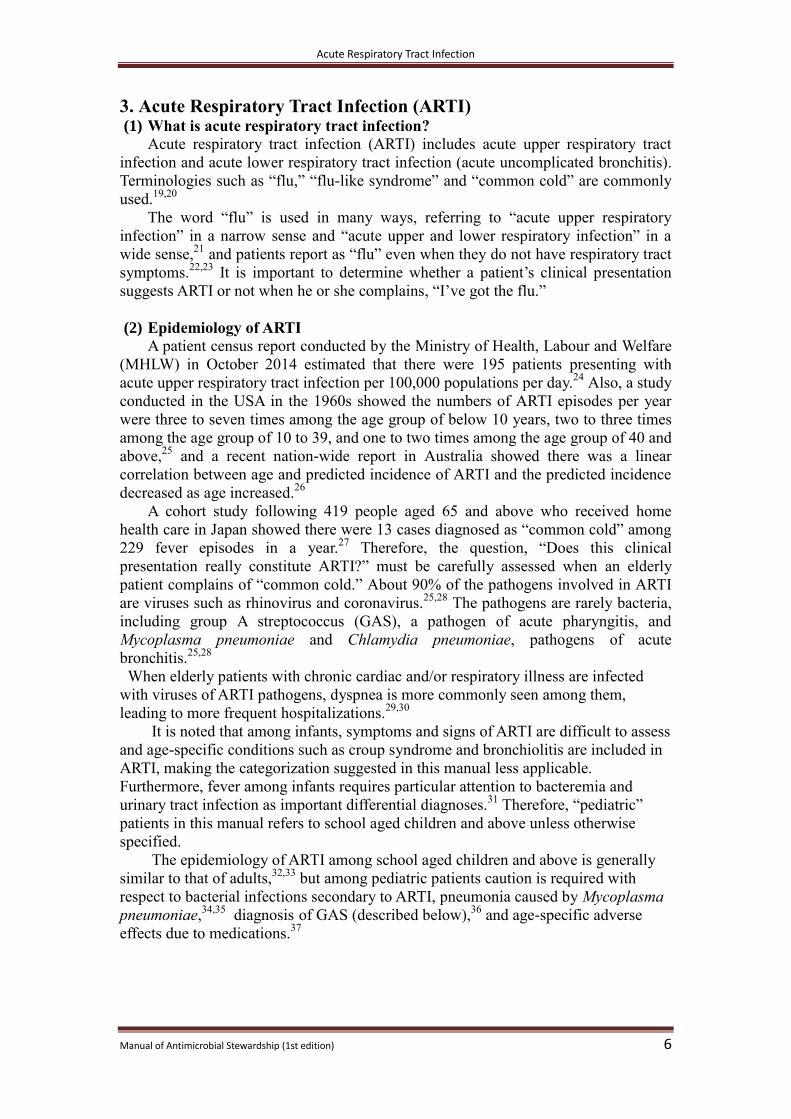
Figure 1. Concept and classification of acute respiratory tract infection in this
manual *4
(3) Diagnosis and Differential Diagnoses of ARTI
ACP provides a classification of ARTI and can be used as a tool to differentiate
between those who require antimicrobials and those who don’t.21,38–40
This classifies
ARTI into common cold (nonspecific upper respiratory infection), acute
rhinosinusitis, pharyngitis and acute bronchitis, according to nasal symptoms
(rhinorrhea and nasal congestion), throat symptoms (sore throat) and lower respiratory
symptoms (cough and sputum production) as the three major types of symptoms
(Table 1). This manual follows this classification. Of note, management of pneumonia
is beyond the scope of this manual.
Table 1. Classification of acute respiratory tract infection – modified from
References 21 and 39
Classification Rhinorrhea/
Nasal Congestion
Sore Throat Cough/Sputum
Production
Common Cold △ △ △
Acute Rhinosinusitis ◎ × ×
Pharyngitis × ◎ ×
Acute Bronchitis × × ◎
◎ as major symptoms, △ as concurrent but not prominent symptoms, × as mild
symptoms or no symptoms
*4 The definitions of “Common Cold,” “Acute Rhinosinusitis,” “Pharyngitis” and “Acute Bronchitis” from Ann
Intern Med. 2016;164:425-34 are applied to four different classifications of ARTI in this manual.
患者にとっての「かぜ」
Patients’ various complaints of “flu”
Common Cold Pharyngitis
Acute Rhinosinusitis
Acute Bronchitis
Acute Respiratory Tract Infection
Acute Respiratory Tract Infection
Manual of Antimicrobial Stewardship (1st edition) 8
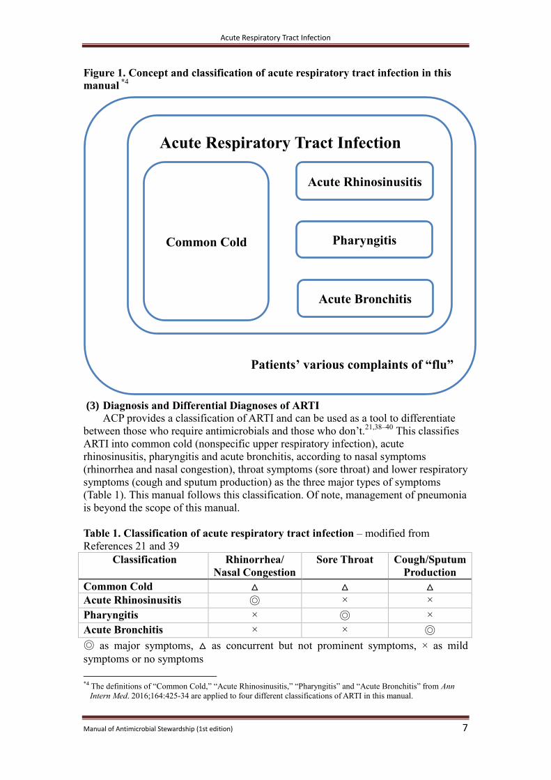
Figure 2. Visual image of acute respiratory tract infection classification
(i) Common cold
In this manual, the common cold is acute upper respiratory viral illness with
three major types of symptoms co-existent “simultaneously” and “to the same extent”
regardless of fever (Table 1). Nonspecific upper respiratory infection is classified as
common cold in this manual.
Patients with the common cold typically present with mild fever, malaise and
sore throat, followed by rhinorrhea and nasal congestion, and further followed by
cough and sputum production. The peak of the symptoms occurs around 3 days after
the onset of the symptoms, and the illness is relieved after 7 to 10 days.41
Cough due
to common cold may last for about 3 weeks but a prolonged cough does not
necessarily suggest a secondary bacterial infection which requires antimicrobials.41
In
contrast, persistent progression of the illness beyond its natural course and onset of
worsening symptoms after initial improvement may suggest a secondary bacterial
infection.40
It is noted that influenza, for which an anti-viral agent may be indicated,
causes relatively severe constitutional symptoms such as high fever, muscle ache and
joint pain, and cough is more frequently observed and its onset is earlier compared to
the common cold. A rapid influenza diagnostic test is also available if the diagnosis is
in question.42–44
(ii) Acute rhinosinusitis
In this manual, acute rhinosinusitis is classified as a type of ARTI with
sneezing, rhinorrhea and nasal congestion dominant, with or without fever. Sinusitis is
mostly accompanied by inflammation of nasal cavities and is preceded by rhinitis.
The term “rhinosinusitis” has lately replaced “sinusitis.”45
Less than 2% of acute viral upper respiratory infections have been reported to
be complicated by acute bacterial sinusitis.46,47
Color of nasal discharge is not helpful
to differentiate between viral and bacterial infections,48
but double-sickening
(worsening symptoms following an illness that was initially improving) may be
suggestive of bacterial infections.40,49
Acute Respiratory Tract Infection
Manual of Antimicrobial Stewardship (1st edition) 9
(iii) Pharyngitis
Pharyngitis is classified in this manual as a type of ARTI with sore throat
dominant. For the sake of the manual, tonsillitis is included in pharyngitis. Most of
the pathogens are viruses, and GAS, an indication for antibacterial agents, constitutes
10% of the pathogens among adult cases of pharyngitis.36,50,51
On the other hand,
researchers in Japan reported about 30% of adult cases of pharyngitis in the age group
of 20 to 59 years old and 17% of pediatric cases tested positive for GAS.52,53
In
general, pharyngitis caused by GAS is common among school aged children and
above while it is relatively rare among infants,36,50,54
but GAS growth from throat
culture does not necessarily represent a true pathogen, and more than 20% of
asymptomatic children may be carriers of GAS.55
Although group C and G
streptococci and Fusobacterium have been recently identified as a possible pathogen
for pharyngitis in Europe and America, little data exists for the epidemiology of those
organisms in Japan.56-64
The Centor score and McIsaac score, a modified Centor score with age
adjustment, are known to support diagnosis of GAS pharyngitis (Table 2).65,66
Recommendations on the use of rapid diagnostic tests for GAS and antibacterial
treatment based on Centor and/or McIsaac scores vary. 36,40,67,68
ACP/CDC and
ESCMID suggest rapid diagnostic tests may be unnecessary when the Centor score is
2 or below.40,67
Rapid diagnostic tests, however, may be considered for high-risk
populations for GAS infection such as those with recent and close exposure to GAS
patients, even if the Centor score is 2 or below.69
When antibacterial treatment was
limited only to those tested positive for GAS rapid diagnostic test or culture,
unnecessary antibacterial use was reduced65
and cost effectiveness was improved.70
Conversely, among pediatric patients, only 68% of those with a Centor score
of 4 tested positive for GAS.71
Therefore, over-diagnosis may occur if only the Centor
score or McIsaac score is used to diagnose GAS pharyngitis among children:
laboratory tests are required for more accurate diagnoses.
Table 2. McIsaac score – created from References 65 and 66
・Fever of 38°C or above: 1
・Absence of cough: 1
・Swollen, tender anterior cervical nodes: 1
・Tonsillar swelling or exudate: 1
・Age of 3 to 14: 1, 15 to 44: 0, 45 and above : -1
Differential diagnoses of pharyngitis include infectious mononucleosis (IM)
caused by Epstein–Barr virus (EBV), cytomegalovirus (CMV), human
immunodeficiency virus (HIV), rubella virus and toxoplasma, but IM can’t be ruled
out by Centor/McIsaac scores alone as the scores are often high among patients with
IM.72
Posterior cervical and/or auricular adenopathy, and splenomegaly are specific
findings among patients with IM,73
and lymphocyte dominance in peripheral blood
test with a lymphocyte-white blood cell count ratio higher than 0.35 is also helpful to
diagnose IM. 74
Differential diagnoses of pharyngitis also include epiglottitis, deep neck
abscess (peri-tonsillar abscess, retropharyngeal abscess and Ludwig angina, etc.) and
Lemierre syndrome. Therefore, “red flag” signs and symptoms*5
such as the worst
*5 “Red flag” (dangerous symptoms) refers to symptoms that should be properly diagnosed or treated without fail
Acute Respiratory Tract Infection
Manual of Antimicrobial Stewardship (1st edition) 10
throat pain ever, trismus, drooling, tripod position and stridor should be taken
seriously as possible indications of these high-risk illnesses, and arrangements for
emergency airway management should be made.75,76
In particular, pediatric patients
with these conditions may cry as a result of medical examination of oral cavity, blood
test and X-rays, which may lead to airway obstruction. Therefore, when these
conditions are suspected, such stressful examinations and tests should be avoided and
urgent transfer to a higher level of care is required for potential emergency airway
management.68
Furthermore, “sore throat” without odynophagia or abnormal clinical findings
in the pharynx and tonsils may suggest referred pain to the neck as well as acute
myocardial infarction, sub-arachnoid hemorrhage, cervical artery dissection or
vertebral artery dissection.75,76
(iv) Acute bronchitis
Acute bronchitis is classified as a type of ARTI with cough dominant, with or
without fever and sputum production. It is not unusual that cough due to ARTI lasts
for 2 to 3 weeks. The mean duration of cough due to ARTI was reported to be 17.8
days. 77 *6
More than 90% of the pathogens of acute bronchitis are viruses and the
remaining 5 to 10% are Bordetella pertussis, Mycoplasma pneumoniae and
Chlamydia pneumoniae, and so forth,40,78
but purulence and color of sputum are not
helpful in differentiating bacterial infection.40
Of note, for healthy, immunocompetent
adults less than 70 years of age, X-ray is generally not indicated when neither
abnormal vital signs (body temperature ≥ 38°C, pulse ≥ 100/min and respiratory rate
≥ 24) nor abnormal lung examination is found.40
Pertussis, because of few specific clinical findings, is difficult to accurately
diagnose in a clinical setting.79
Vomiting after cough episodes and inspiratory whoop
make diagnosis of pertussis a little more likely.79
A serum test for pertussis, that is,
anti-Bordetella pertussis toxin (PT) antibody, is difficult to utilize in an actual clinical
setting due to the long turn-around time.80,81
However, polymerase chain reaction
(PCR) utilizing loop-mediated isothermal amplification (LAMP) to detect B. pertussis
from a posterior pharynx swab, which was approved to be covered by insurance in
November 2016 in Japan, had the sensitivity and specificity of 76.2% to 96.6% and
94.1% to 99.5%, respectively, compared to real-time PCR as a reference standard.82,83
Thus, during epidemics of pertussis cases, laboratory tests may be considered for
diagnosis of pertussis if severe cough persists or respiratory symptoms develop after
exposure to patients with pertussis.
Differential diagnoses of acute bronchitis also include tuberculosis. If cough
lasts for 2 to 3 weeks, tuberculosis needs to be ruled out as the incidence remains high
in Japan.
Among pediatric patients, acute rhinosinusitis is a differential diagnosis when
productive cough persists for longer than 2 weeks,49
and 10% of school aged children
and above infected with Mycoplasma pneumoniae may subsequently develop
pneumonia.35
In addition, a guideline by the Japanese Society of Pediatric
Pulmonology (JSPP)/JSPID describes pertussis as a differential diagnosis for pediatric
patients aged 1 and above with cough lasting longer than a week, and defines the
clinical diagnosis of pertussis for those aged 1 and above as at least one of the
following being met: characteristic inspiratory whoop, episodic prolonged coughing
in medical practice.
*6 It varies between 15.3 and 28.6 days, depending on the study.
Acute Respiratory Tract Infection
Manual of Antimicrobial Stewardship (1st edition) 11
spells, vomiting after coughing, and dyspnea.84
Therefore, follow-up over time is one
of the keys to successful management.
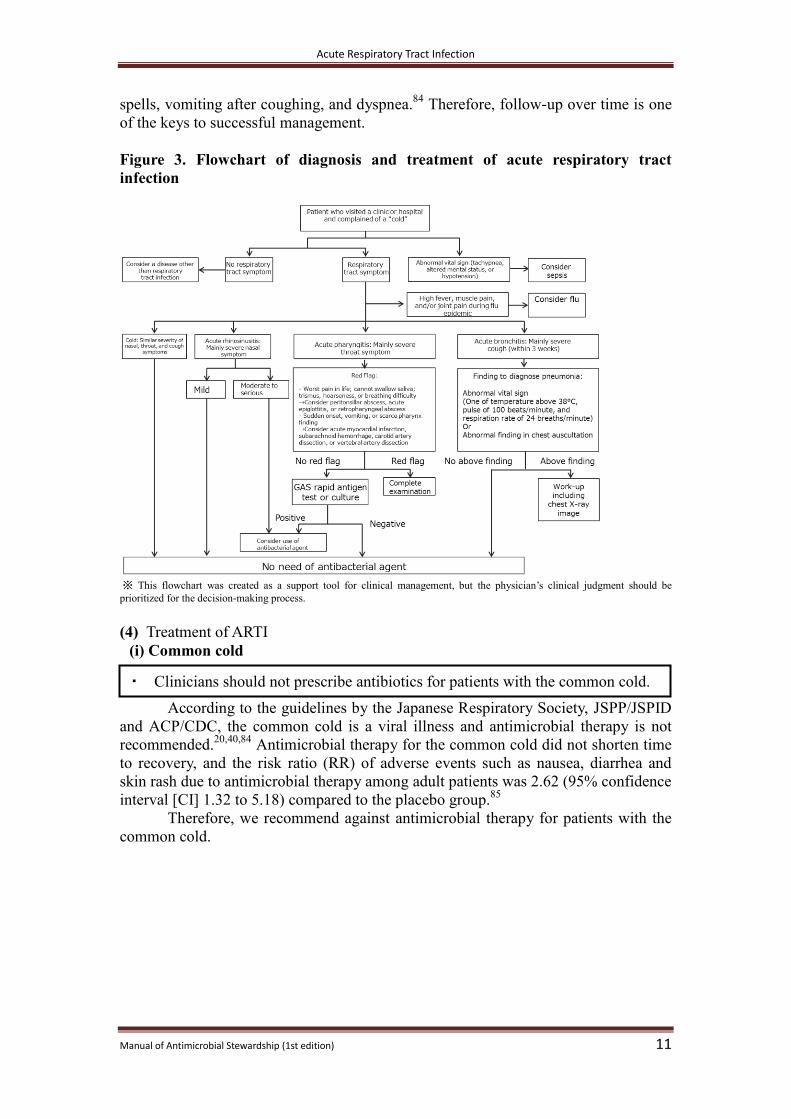
Figure 3. Flowchart of diagnosis and treatment of acute respiratory tract
infection
※ This flowchart was created as a support tool for clinical management, but the physician’s clinical judgment should be
prioritized for the decision-making process.
(4) Treatment of ARTI
(i) Common cold
According to the guidelines by the Japanese Respiratory Society, JSPP/JSPID
and ACP/CDC, the common cold is a viral illness and antimicrobial therapy is not
recommended.20,40,84
Antimicrobial therapy for the common cold did not shorten time
to recovery, and the risk ratio (RR) of adverse events such as nausea, diarrhea and
skin rash due to antimicrobial therapy among adult patients was 2.62 (95% confidence
interval [CI] 1.32 to 5.18) compared to the placebo group.85
Therefore, we recommend against antimicrobial therapy for patients with the
common cold.
・ Clinicians should not prescribe antibiotics for patients with the common cold.
Acute Respiratory Tract Infection
Manual of Antimicrobial Stewardship (1st edition) 12
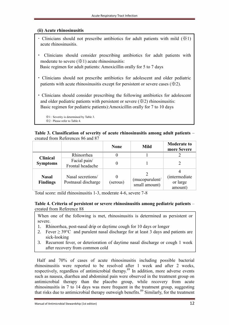
(ii) Acute rhinosinusitis
Table 3. Classification of severity of acute rhinosinusitis among adult patients –
created from References 86 and 87
None Mild Moderate to
more Severe
Clinical
Symptoms
Rhinorrhea 0 1 2
Facial pain/
Frontal headache 0 1 2
Nasal
Findings
Nasal secretions/
Postnasal discharge
0
(serous)
2
(mucopurulent/
small amount)
4
(intermediate
or large
amount)
Total score: mild rhinosinusitis 1-3, moderate 4-6, severe 7-8
Table 4. Criteria of persistent or severe rhinosinusitis among pediatric patients –
created from Reference 88
Half and 70% of cases of acute rhinosinusitis including possible bacterial
rhinosinusitis were reported to be resolved after 1 week and after 2 weeks,
respectively, regardless of antimicrobial therapy.89
In addition, more adverse events
such as nausea, diarrhea and abdominal pain were observed in the treatment group on
antimicrobial therapy than the placebo group, while recovery from acute
rhinosinusitis in 7 to 14 days was more frequent in the treatment group, suggesting
that risks due to antimicrobial therapy outweigh benefits.89
Similarly, for the treatment
When one of the following is met, rhinosinusitis is determined as persistent or
severe. 1. Rhinorrhea, post-nasal drip or daytime cough for 10 days or longer
2. Fever ≥ 39°C and purulent nasal discharge for at least 3 days and patients are
sick-looking
3. Recurrent fever, or deterioration of daytime nasal discharge or cough 1 week
after recovery from common cold
・ Clinicians should not prescribe antibiotics for adult patients with mild (※1)
acute rhinosinusitis.
・ Clinicians should consider prescribing antibiotics for adult patients with
moderate to severe (※1) acute rhinosinusitis:
Basic regimen for adult patients: Amoxicillin orally for 5 to 7 days
・ Clinicians should not prescribe antibiotics for adolescent and older pediatric
patients with acute rhinosinusitis except for persistent or severe cases (※2).
・ Clinicians should consider prescribing the following antibiotics for adolescent
and older pediatric patients with persistent or severe (※2) rhinosinusitis:
Basic regimen for pediatric patients):Amoxicillin orally for 7 to 10 days
※1: Severity is determined by Table 3.
※2: Please refer to Table 4.
Acute Respiratory Tract Infection
Manual of Antimicrobial Stewardship (1st edition) 13
of acute rhinorrhea with symptoms shorter than 10 days, no clear benefit of
antimicrobial therapy was observed over the placebo group, regardless of gross
appearance of nasal discharge, and the risk ratio of adverse events for acute purulent
rhinitis on antimicrobial therapy was 1.46 (95% CI 1.10 to 1.94) compared to the
placebo group.85
According to the ACP/CDC guideline, indications of antimicrobial therapy for
acute rhinosinusitis are limited to cases with symptoms lasting longer than 10 days,
severe cases (fever, ≥ 39°C and purulent nasal discharge or facial pain lasting for at
least 3 days) and cases of double-sickening (worsening symptoms following a typical
viral illness that lasted 5 days and was initially improving).40
In addition, JAID/JSC
and the guidelines by the Japanese Rhinologic Society recommend watchful waiting
without antimicrobial therapy rather than antimicrobial therapy for mild cases of acute
rhinosinusitis with a score of 1 to 3 as shown in Table 3.68,86,87
Accordingly, we recommend against antimicrobial therapy for adult patients
with mild acute rhinosinusitis.
For pediatric patients, a guideline by AAP lists the following as indications of
antimicrobial therapy for acute rhinosinusitis: (1) Nasal discharge or daytime cough
or both > 10 days; (2) Fever ≥ 39°C and purulent nasal discharge for at least 3 days;
(3) Worsening or new onset of nasal discharge, daytime cough or fever after initial
improvement. Otherwise, watchful waiting without antimicrobial therapy is
recommended.88
Therefore, we recommend against antimicrobial therapy for pediatric patients
with acute rhinosinusitis except in persistent, severe and worsening cases as
mentioned above.
No systematic review or randomized control trial has proven that
cephalosporins or macrolides are more effective in treatment of acute rhinosinusitis
than amoxicillin or amoxicillin/clavulanate,90,91
and guidelines by the American
Academy of Otolaryngology – Head and Neck Surgery (AAO-HNS) and ACP/CDC
recommend amoxicillin as a first-line option when a decision is made to treat
moderate to severe acute rhinosinusitis with antimicrobial therapy.40,91
The
recommended regimen is oral amoxicillin 500mg*7
three times daily for 5 to 7 days.40
AAO-HNS also suggests amoxicillin/clavulanate if concern for bacterial resistance is
high or the first-line treatment response is poor. The regimen recommended by
ACP/CDC is oral amoxicillin 500mg and clavulanate 125mg three times daily for 5 to
7 days.40
The recommended duration of antimicrobial therapy used to be 10 to 14
days83
, but a recent study showed short-term treatment (3 to 7 days) was not inferior
in treatment effect to long-term treatment (6 to 10 days). Rather, the treatment effect
between the 5-day treatment group and the 10-day treatment group was similar, and
fewer adverse events were observed in the 5-day treatment group.92
In Japan, amoxicillin is not approved to treat rhinosinusitis under the
Pharmaceutical Affairs Law, but according to a reference by the Health Insurance
Claims Review and Reimbursement Services, in general, “claims can be accepted
when amoxicillin is prescribed for acute sinusitis.” The drug package insert of
amoxicillin states, for infections other than Helicobacter pylori infection, “The usual
dosage for oral administration is 250mg of amoxicillin hydrate three or four times
daily. The dosage may be adjusted according to the patient’s age and symptoms,”
though the description is not specific to acute rhinosinusitis.
*7 In this manual, the dosages are described by ingredient amount (titer), not by formulation amount.
Acute Respiratory Tract Infection
Manual of Antimicrobial Stewardship (1st edition) 14
Thus, we recommend antimicrobial therapy for adult patients with moderate to
severe acute rhinosinusitis and, if a decision is made to treat with an antimicrobial
agent, we suggest oral amoxicillin for 5 to 7 days be selected as the first-line regimen.
While guidelines developed abroad recommend tetracyclines and
fluoroquinolones as alternatives if an adult patient is allergic to β lactams,49,91
it has
been reported that resistance of Streptococcus pneumoniae, the major pathogen of
bacterial rhinosinusitis, to tetracyclines is high in Japan,93
and referral to a specialist
may be considered.
For pediatric patients, the drug package insert of amoxicillin states: “The usual
dosage for oral administration is 20 to 40mg/kg daily in three to four divided doses.
The dosage may be adjusted according to the patient’s age and symptoms provided
that the daily dosage should not exceed 90mg/kg of amoxicillin hydrate.” Also, a
couple of guidelines recommend amoxicillin as the first-line regimen for acute
rhinosinusitis.68,86,88
Thus, we recommend antimicrobial therapy for pediatric patients with acute
rhinosinusitis only when the illness is severe or persistent as shown in Table 4, and if
a decision is made to treat with an antimicrobial agent, we suggest oral amoxicillin for
7 to 10 days be selected as the first-line regimen.
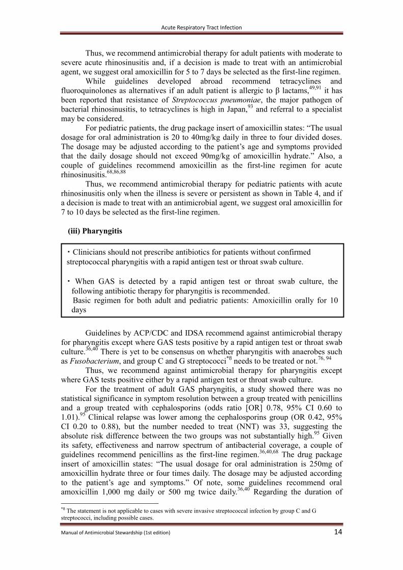
(iii) Pharyngitis
Guidelines by ACP/CDC and IDSA recommend against antimicrobial therapy
for pharyngitis except where GAS tests positive by a rapid antigen test or throat swab
culture.36,40
There is yet to be consensus on whether pharyngitis with anaerobes such
as Fusobacterium, and group C and G streptococci*8
needs to be treated or not.76, 94
Thus, we recommend against antimicrobial therapy for pharyngitis except
where GAS tests positive either by a rapid antigen test or throat swab culture.
For the treatment of adult GAS pharyngitis, a study showed there was no
statistical significance in symptom resolution between a group treated with penicillins
and a group treated with cephalosporins (odds ratio [OR] 0.78, 95% CI 0.60 to
1.01).95
Clinical relapse was lower among the cephalosporins group (OR 0.42, 95%
CI 0.20 to 0.88), but the number needed to treat (NNT) was 33, suggesting the
absolute risk difference between the two groups was not substantially high.95
Given
its safety, effectiveness and narrow spectrum of antibacterial coverage, a couple of
guidelines recommend penicillins as the first-line regimen.36,40,68
The drug package
insert of amoxicillin states: “The usual dosage for oral administration is 250mg of
amoxicillin hydrate three or four times daily. The dosage may be adjusted according
to the patient’s age and symptoms.” Of note, some guidelines recommend oral
amoxicillin 1,000 mg daily or 500 mg twice daily.36,40
Regarding the duration of
*8 The statement is not applicable to cases with severe invasive streptococcal infection by group C and G
streptococci, including possible cases.
・ Clinicians should not prescribe antibiotics for patients without confirmed
streptococcal pharyngitis with a rapid antigen test or throat swab culture.
・ When GAS is detected by a rapid antigen test or throat swab culture, the
following antibiotic therapy for pharyngitis is recommended.
Basic regimen for both adult and pediatric patients: Amoxicillin orally for 10
days
Acute Respiratory Tract Infection
Manual of Antimicrobial Stewardship (1st edition) 15
antimicrobial therapy, the evidence to support short-term therapy has been scarce and
the guidelines in the USA and Europe recommend a 10-day course.36,67
According to the IDSA guideline, cephalexin, a first-generation cephalosporin
is recommended for those with mild penicillin allergy, and clindamycin is
recommended for those with severe penicillin allergy: history of anaphylaxis and
severe drug rash. 36
In Japan, cephalexin and clindamycin are approved to treat pharyngitis under
the Pharmaceutical Affairs Law. The drug package insert of cephalexin states: “For
adults and children with a body weight of ≥ 20 kg, the usual dosage for oral
administration is 250 mg of cephalexin every six hours. For severe cases, or cases
with bacteria growth of low susceptibility, the dosage is given as 500 mg orally every
six hours. The dosage may be adjusted according to the patient’s age, body weight
and symptoms.” That of clindamycin states: “For adults, the dosages for oral
administration are 150mg every six hours in usual cases and 300mg every eight hours
in severe cases. For children, the dosages for oral administration are 15 mg/kg daily in
three to four divided doses in usual cases and 20 mg/kg daily in three to four divided
doses in severe cases. The dosage may be adjusted according to the patient’s age,
body weight and symptoms.” The IDSA guideline recommends cephalexin 500mg
orally twice daily for those with a mild penicillin allergy and clindamycin 300mg
orally three times daily for those with a severe penicillin allergy.36
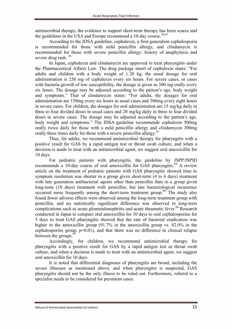
Thus, for adults, we recommend antimicrobial therapy for pharyngitis with a
positive result for GAS by a rapid antigen test or throat swab culture, and when a
decision is made to treat with an antimicrobial agent, we suggest oral amoxicillin for
10 days.
For pediatric patients with pharyngitis, the guideline by JSPP/JSPID
recommends a 10-day course of oral amoxicillin for GAS pharyngitis.84
A review
article on the treatment of pediatric patients with GAS pharyngitis showed time to
symptom resolution was shorter in a group given short-term (4 to 6 days) treatment
with late generation antibacterial agents other than penicillin than in a group given
long-term (10 days) treatment with penicillin, but late bacteriological recurrence
occurred more frequently among the short-term treatment group.96
The study also
found fewer adverse effects were observed among the long-term treatment group with
penicillin, and no statistically significant difference was observed in long-term
complications such as acute glomerulonephritis and acute rheumatic fever.96
Research
conducted in Japan to compare oral amoxicillin for 10 days to oral cephalosporins for
5 days to treat GAS pharyngitis showed that the rate of bacterial eradication was
higher in the amoxicillin group (91.7% in the amoxicillin group vs. 82.0% in the
cephalosporins group, p=0.01), and that there was no difference in clinical relapse
between the groups.97
Accordingly, for children, we recommend antimicrobial therapy for
pharyngitis with a positive result for GAS by a rapid antigen test or throat swab
culture, and when a decision is made to treat with an antimicrobial agent, we suggest
oral amoxicillin for 10 days.
It is noted that differential diagnoses of pharyngitis are broad, including the
severe illnesses as mentioned above, and when pharyngitis is suspected, GAS
pharyngitis should not be the only illness to be ruled out. Furthermore, referral to a
specialist needs to be considered for persistent cases.
Acute Respiratory Tract Infection
Manual of Antimicrobial Stewardship (1st edition) 16
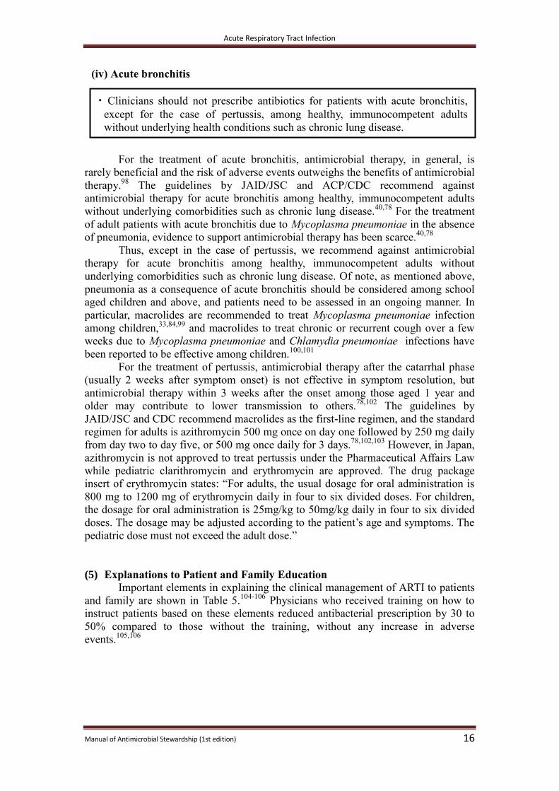
(iv) Acute bronchitis
For the treatment of acute bronchitis, antimicrobial therapy, in general, is
rarely beneficial and the risk of adverse events outweighs the benefits of antimicrobial
therapy.98
The guidelines by JAID/JSC and ACP/CDC recommend against
antimicrobial therapy for acute bronchitis among healthy, immunocompetent adults
without underlying comorbidities such as chronic lung disease.40,78
For the treatment
of adult patients with acute bronchitis due to Mycoplasma pneumoniae in the absence
of pneumonia, evidence to support antimicrobial therapy has been scarce.40,78
Thus, except in the case of pertussis, we recommend against antimicrobial
therapy for acute bronchitis among healthy, immunocompetent adults without
underlying comorbidities such as chronic lung disease. Of note, as mentioned above,
pneumonia as a consequence of acute bronchitis should be considered among school
aged children and above, and patients need to be assessed in an ongoing manner. In
particular, macrolides are recommended to treat Mycoplasma pneumoniae infection
among children,33,84,99
and macrolides to treat chronic or recurrent cough over a few
weeks due to Mycoplasma pneumoniae and Chlamydia pneumoniae infections have
been reported to be effective among children.100,101
For the treatment of pertussis, antimicrobial therapy after the catarrhal phase
(usually 2 weeks after symptom onset) is not effective in symptom resolution, but
antimicrobial therapy within 3 weeks after the onset among those aged 1 year and
older may contribute to lower transmission to others.78,102
The guidelines by
JAID/JSC and CDC recommend macrolides as the first-line regimen, and the standard
regimen for adults is azithromycin 500 mg once on day one followed by 250 mg daily
from day two to day five, or 500 mg once daily for 3 days.78,102,103
However, in Japan,
azithromycin is not approved to treat pertussis under the Pharmaceutical Affairs Law
while pediatric clarithromycin and erythromycin are approved. The drug package
insert of erythromycin states: “For adults, the usual dosage for oral administration is
800 mg to 1200 mg of erythromycin daily in four to six divided doses. For children,
the dosage for oral administration is 25mg/kg to 50mg/kg daily in four to six divided
doses. The dosage may be adjusted according to the patient’s age and symptoms. The
pediatric dose must not exceed the adult dose.”
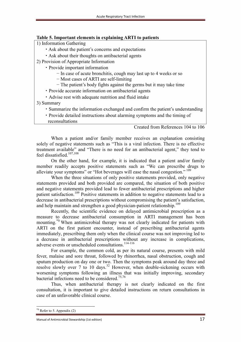
(5) Explanations to Patient and Family Education
Important elements in explaining the clinical management of ARTI to patients
and family are shown in Table 5.104-106
Physicians who received training on how to
instruct patients based on these elements reduced antibacterial prescription by 30 to
50% compared to those without the training, without any increase in adverse
events.105,106
・ Clinicians should not prescribe antibiotics for patients with acute bronchitis,
except for the case of pertussis, among healthy, immunocompetent adults
without underlying health conditions such as chronic lung disease.
Acute Respiratory Tract Infection
Manual of Antimicrobial Stewardship (1st edition) 17
Table 5. Important elements in explaining ARTI to patients
1) Information Gathering
・ Ask about the patient’s concerns and expectations
・ Ask about their thoughts on antibacterial agents
2) Provision of Appropriate Information
・ Provide important information
− In case of acute bronchitis, cough may last up to 4 weeks or so
− Most cases of ARTI are self-limiting
− The patient’s body fights against the germs but it may take time
・ Provide accurate information on antibacterial agents
・ Advise rest with adequate nutrition and fluid intake
3) Summary
・ Summarize the information exchanged and confirm the patient’s understanding
・ Provide detailed instructions about alarming symptoms and the timing of
reconsultations
Created from References 104 to 106
When a patient and/or family member receives an explanation consisting
solely of negative statements such as “This is a viral infection. There is no effective
treatment available” and “There is no need for an antibacterial agent,” they tend to
feel dissatisfied.107,108
On the other hand, for example, it is indicated that a patient and/or family
member readily accepts positive statements such as “We can prescribe drugs to
alleviate your symptoms” or “Hot beverages will ease the nasal congestion.” 109
When the three situations of only positive statements provided, only negative
statements provided and both provided are compared, the situation of both positive
and negative statements provided lead to fewer antibacterial prescriptions and higher
patient satisfaction.109
Positive statements in addition to negative statements lead to a
decrease in antibacterial prescriptions without compromising the patient’s satisfaction,
and help maintain and strengthen a good physician-patient relationship.109
Recently, the scientific evidence on delayed antimicrobial prescription as a
measure to decrease antibacterial consumption in ARTI management has been
mounting.*9
When antimicrobial therapy was not clearly indicated for patients with
ARTI on the first patient encounter, instead of prescribing antibacterial agents
immediately, prescribing them only when the clinical course was not improving led to
a decrease in antibacterial prescriptions without any increase in complications,
adverse events or unscheduled consultations.114-116
For example, the common cold, as per its natural course, presents with mild
fever, malaise and sore throat, followed by rhinorrhea, nasal obstruction, cough and
sputum production on day one or two. Then the symptoms peak around day three and
resolve slowly over 7 to 10 days.32
However, when double-sickening occurs with
worsening symptoms following an illness that was initially improving, secondary
bacterial infections need to be considered.75,76
Thus, when antibacterial therapy is not clearly indicated on the first
consultation, it is important to give detailed instructions on return consultations in
case of an unfavorable clinical course.
*9 Refer to 5. Appendix (2)
Acute Respiratory Tract Infection
Manual of Antimicrobial Stewardship (1st edition) 18
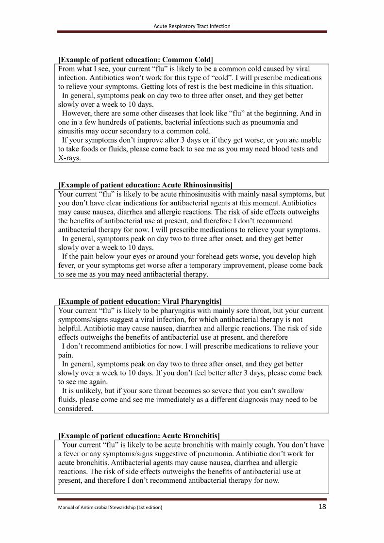
[Example of patient education: Common Cold]
From what I see, your current “flu” is likely to be a common cold caused by viral
infection. Antibiotics won’t work for this type of “cold”. I will prescribe medications
to relieve your symptoms. Getting lots of rest is the best medicine in this situation.
In general, symptoms peak on day two to three after onset, and they get better
slowly over a week to 10 days.
However, there are some other diseases that look like “flu” at the beginning. And in
one in a few hundreds of patients, bacterial infections such as pneumonia and
sinusitis may occur secondary to a common cold.
If your symptoms don’t improve after 3 days or if they get worse, or you are unable
to take foods or fluids, please come back to see me as you may need blood tests and
X-rays.
[Example of patient education: Acute Rhinosinusitis]
Your current “flu” is likely to be acute rhinosinusitis with mainly nasal symptoms, but
you don’t have clear indications for antibacterial agents at this moment. Antibiotics
may cause nausea, diarrhea and allergic reactions. The risk of side effects outweighs
the benefits of antibacterial use at present, and therefore I don’t recommend
antibacterial therapy for now. I will prescribe medications to relieve your symptoms.
In general, symptoms peak on day two to three after onset, and they get better
slowly over a week to 10 days.
If the pain below your eyes or around your forehead gets worse, you develop high
fever, or your symptoms get worse after a temporary improvement, please come back
to see me as you may need antibacterial therapy.
[Example of patient education: Viral Pharyngitis]
Your current “flu” is likely to be pharyngitis with mainly sore throat, but your current
symptoms/signs suggest a viral infection, for which antibacterial therapy is not
helpful. Antibiotic may cause nausea, diarrhea and allergic reactions. The risk of side
effects outweighs the benefits of antibacterial use at present, and therefore
I don’t recommend antibiotics for now. I will prescribe medications to relieve your
pain.
In general, symptoms peak on day two to three after onset, and they get better
slowly over a week to 10 days. If you don’t feel better after 3 days, please come back
to see me again.
It is unlikely, but if your sore throat becomes so severe that you can’t swallow
fluids, please come and see me immediately as a different diagnosis may need to be
considered.
[Example of patient education: Acute Bronchitis]
Your current “flu” is likely to be acute bronchitis with mainly cough. You don’t have
a fever or any symptoms/signs suggestive of pneumonia. Antibiotic don’t work for
acute bronchitis. Antibacterial agents may cause nausea, diarrhea and allergic
reactions. The risk of side effects outweighs the benefits of antibacterial use at
present, and therefore I don’t recommend antibacterial therapy for now.
Acute Respiratory Tract Infection
Manual of Antimicrobial Stewardship (1st edition) 19
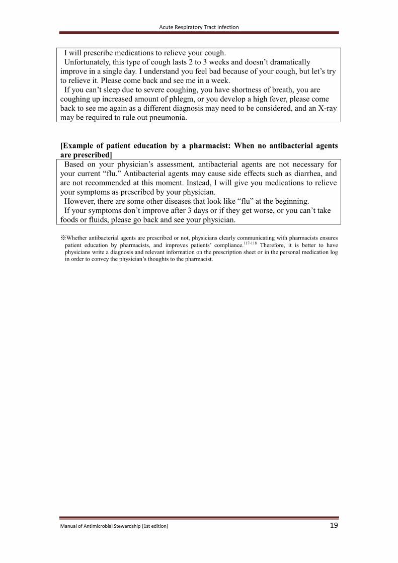
I will prescribe medications to relieve your cough.
Unfortunately, this type of cough lasts 2 to 3 weeks and doesn’t dramatically
improve in a single day. I understand you feel bad because of your cough, but let’s try
to relieve it. Please come back and see me in a week.
If you can’t sleep due to severe coughing, you have shortness of breath, you are
coughing up increased amount of phlegm, or you develop a high fever, please come
back to see me again as a different diagnosis may need to be considered, and an X-ray
may be required to rule out pneumonia.
[Example of patient education by a pharmacist: When no antibacterial agents
are prescribed]
Based on your physician’s assessment, antibacterial agents are not necessary for
your current “flu.” Antibacterial agents may cause side effects such as diarrhea, and
are not recommended at this moment. Instead, I will give you medications to relieve
your symptoms as prescribed by your physician.
However, there are some other diseases that look like “flu” at the beginning.
If your symptoms don’t improve after 3 days or if they get worse, or you can’t take
foods or fluids, please go back and see your physician.
※Whether antibacterial agents are prescribed or not, physicians clearly communicating with pharmacists ensures
patient education by pharmacists, and improves patients’ compliance.117-118 Therefore, it is better to have
physicians write a diagnosis and relevant information on the prescription sheet or in the personal medication log
in order to convey the physician’s thoughts to the pharmacist.
Acute Diarrhea
Manual of Antimicrobial Stewardship (1st edition) 20
4. Acute Diarrhea (1) What is Acute Diarrhea?
Acute diarrhea is defined as the passage of unusually loose or watery stools,
usually at least three or more times above baseline in a 24-hour period, lasting less
than 14 days.119-120
More than 90% of acute diarrhea is caused by infections while the
remaining 10% results from drug-induced, toxic, ischemic or other non-infectious
causes, and diarrhea may be one of multiple symptoms of these systemic illness.121
Acute infectious diarrhea may be associated with nausea, vomiting, abdominal pain,
abdominal distention, fever, bloody stool and tenesmus.120
Acute infectious diarrhea
is referred to as “gastroenteritis” and “enteritis” and vomiting may be the dominant
symptom with diarrhea less prominent.120
(2) Epidemiology of Acute Diarrhea
A patient census report conducted by MHLW in October 2014, when it was
not the peak season for diarrheal diseases, estimated that there were 24 patients
presenting with intestinal infectious diseases*10
per 100,000 populations per day.24
The etiology of acute diarrhea is mostly viral infections,120
such as norovirus
and rotavirus.122
In Japan, voluntary vaccination for rotavirus started in 2011, and the
incidence of rotavirus diarrhea has been decreasing.123
Bacteria that can cause acute diarrhea include non-typhoidal Salmonella spp.,
Campylobacter spp., enterohemorrhagic Escherichia coli (EHEC), and Vibrio spp.,124
while enterotoxigenic E. coli (ETEC), Campylobacter spp. and, rarely, Shigella spp.
and V. cholerae are pathogens that can be found in travelers returning from abroad.125
Clostridium difficile is also in differential if a patient has recent exposure to
antibacterial agents.126
It is notable that typhoid fever and paratyphoid fever rarely
cause diarrhea.127
(3) Diagnosis and Differential Diagnoses of Acute Diarrhea
Information needed to identify the etiology of acute diarrhea includes onset,
associated symptoms such as fever, abdominal pain and presence of bloody diarrhea,
history of food/fluid intake, travel history, antimicrobial use, immune status and sick
contact.124
In particular, if vomiting is dominant, viral illness and food poisoning due
to toxins are more likely.128
In an outbreak, incubation periods of 14 hours and longer
(typically, 24 to 48 hours) suggest viral illness, and incubation periods of two to seven
hours suggests food poisoning. The difference may be useful to differential
diagnosis.128
Nausea and vomiting may occur when an illness is not associated with the
gastrointestinal system such as with acute myocardial infarction, intracranial
pathology, sepsis, electrolyte imbalance and drug-induced illness.129,130
Since a study
showed about 30% of those who were hospitalized under diagnosis of “acute
gastroenteritis” had etiologies outside the gastrointestinal (GI) system, diagnosing
“acute gastroenteritis” only relying on patients’ symptoms without ruling out critical
conditions should be avoided.
During history taking, it is important to consider the characteristics (watery or
bloody) and severity of the diarrhea. Particularly, returning travelers (especially from
developing countries) who develop severe bloody diarrhea with total disability and
*10 “Intestinal infectious diseases” represent A00 to A09 according to ICD10.
Acute Diarrhea
Manual of Antimicrobial Stewardship (1st edition) 21
body temperature of ≥ 38°C or watery diarrhea with resultant moderate physical
disability with onset of 1 week after travel may have bacterial enteritis, such as
typhoid fever, Non-typhoidal Salmonella enteritis, Campylobacter enteritis and
ETEC, or amebic dysentery.120,132
Therefore, laboratory tests and antibacterial therapy
need to be considered in consultation with experts in travel medicine and infectious
diseases.
Among children, acute diarrhea is mostly caused by viral infections.133
Viral
acute diarrhea often starts with vomiting, followed by mild to moderate peri-umbilical
pain and tenderness, watery diarrhea without blood, no fever (or mild fever), no
severe abdominal pain, and sick contact. On the other hand, differential diagnoses of
bloody diarrhea include EHEC, intussusception, Meckel’s diverticulum and upper GI
bleeding.134,135
(i) Acute diarrhea due to viruses
Acute diarrhea due to viral infections includes rotavirus, and norovirus in
adults.124,128
Food exposure to bivalves that are poorly cooked and contaminated with
norovirus is well known as a mode of transmission of norovirus infection, but human
to human transmission is not rare.136
The incubation period of norovirus infection is
generally half a day to 2 days. The illness often starts with vomiting, followed by
watery diarrhea.137
Vomiting and diarrhea usually resolve within a day and within 2 to
3 days, respectively, but the symptoms may persist over 7 days to 10 days.138,139
Fever
is often absent or, if any, resolves within 2 days,138
so if fever lasts longer than 2 days,
a different etiology other than viral infection needs to be considered.
A rapid antigen test for norovirus*11
is approved under the Pharmaceutical
Affairs Law, and its sensitivity has improved up to 87.4 to 93.1% recently.140-143
However, during the peak season of norovirus infections, a negative rapid antigen test
does not rule out norovirus for those with typical acute diarrhea because of high pre-
test probability and the routine test for every diarrheal patient is therefore not
considered useful. From an infection control standpoint, regardless of the etiology,
vomit and excreta must be handled as infectious materials, and a stand-alone result of
negative antigen testing should not result in negligence of the control measures.
Of note, for children, the rapid antigen test for norovirus is approved for those aged
less than 3 years old.
(ii) Acute diarrhea due to bacteria
Those with acute diarrhea due to bacteria tend to develop severe abdominal
pain, high fever (≥ 38°C), bloody stool, bloody mucous stool and tenesmus more
often than those with acute diarrhea due to viruses. Patients’ signs and symptoms,
however, are not always helpful in identifying the etiology, and food/fluid
consumption history and incubation period may be useful to some extent as shown in
Table 6.138,144,145
Acute diarrhea due to bacteria among adults is often self-limiting, and
therefore the benefit of identifying the etiology through routine laboratory tests for all
adult patients including mild cases may be limited. On the other hand, for moderate to
severe cases, cases involving persistent diarrhea, and cases where antimicrobial
therapy is going to be given, laboratory tests such as stool culture may be preferable
in order to identify the etiology.110
For children, it is rare to require urgent laboratory tests including stool culture,
*11 As of March 2016, the approval is limited to those aged 3 and younger, those aged 65 and older, those with
malignancies, post-transplant patients and those on antineoplastic agents and immunosuppressants.
Acute Diarrhea
Manual of Antimicrobial Stewardship (1st edition) 22
and indications for such tests include cases involving severe abdominal pain or
bloody stool, cases of possible EHEC complicated by hemolytic uremic syndrome
(HUS), and immunocompromised patients.146
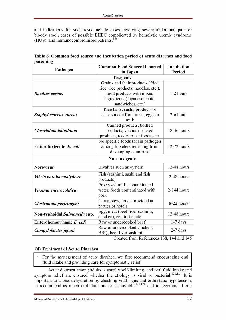
Table 6. Common food source and incubation period of acute diarrhea and food
poisoning
Pathogen Common Food Source Reported
in Japan
Incubation
Period
Toxigenic
Bacillus cereus
Grains and their products (fried
rice, rice products, noodles, etc.),
food products with mixed
ingredients (Japanese bento,
sandwiches, etc.)
1-2 hours
Staphylococcus aureus
Rice balls, sushi, products or
snacks made from meat, eggs or
milk
2-6 hours
Clostridium botulinum
Canned products, bottled
products, vacuum-packed
products, ready-to-eat foods, etc.
18-36 hours
Enterotoxigenic E. coli
No specific foods (Main pathogen
among travelers returning from
developing countries)
12-72 hours
Non-toxigenic
Norovirus Bivalves such as oysters 12-48 hours
Vibrio parahaemolyticus Fish (sashimi, sushi and fish
products) 2-48 hours
Yersinia enterocolitica
Processed milk, contaminated
water, foods contaminated with
pork
2-144 hours
Clostridium perfringens Curry, stew, foods provided at
parties or hotels 8-22 hours
Non-typhoidal Salmonella spp. Egg, meat (beef liver sashimi,
chicken), eel, turtle, etc. 12-48 hours
Enterohemorrhagic E. coli Raw or undercooked beef 1-7 days
Campylobacter jejuni Raw or undercooked chicken,
BBQ, beef liver sashimi 2-7 days
Created from References 138, 144 and 145
(4) Treatment of Acute Diarrhea
Acute diarrhea among adults is usually self-limiting, and oral fluid intake and
symptom relief are ensured whether the etiology is viral or bacterial.120,124
It is
important to assess dehydration by checking vital signs and orthostatic hypotension,
to recommend as much oral fluid intake as possible,120,124
and to recommend oral
For the management of acute diarrhea, we first recommend encouraging oral
fluid intake and providing care for symptomatic relief.
Acute Diarrhea
Manual of Antimicrobial Stewardship (1st edition) 23
fluid containing sugar, sodium and potassium. For severely dehydrated infants and the
elderly, oral rehydration solution (ORS) is recommended, but for adults fruit juice and
sports drinks are mostly sufficient, though fluids with little sodium may necessitate
additional sodium intake.120,147
According to the guidelines by JAID/JSC and ACG, antibacterial therapy is
not recommended except in severe cases and those involving travelers returning from
abroad (traveler’s diarrhea).120,124
JAID/JSC suggest antibacterial therapy for the
following situations:124
・ Suspected bacteremia such as hypotension and shivering
・ Cases with severe diarrhea and/or shock that require hospitalization for
rehydration
・ High risk of bacteremia (HIV with low CD4 count, cell-mediated
immunosuppression due to steroids and immunosuppressants)
・ High risk of complications (age of 50 years and older, artificial graft/valve,
artificial joints)
・ Return travelers
Caring for dehydration is also crucial for the management of acute diarrhea among
children.134
Thus, for the management of acute diarrhea, we first recommend encouraging oral
fluid intake and providing care for symptomatic relief.
We suggest referring to the academic literatures for guidance on the detailed
management of severe cases and traveler’s diarrhea.
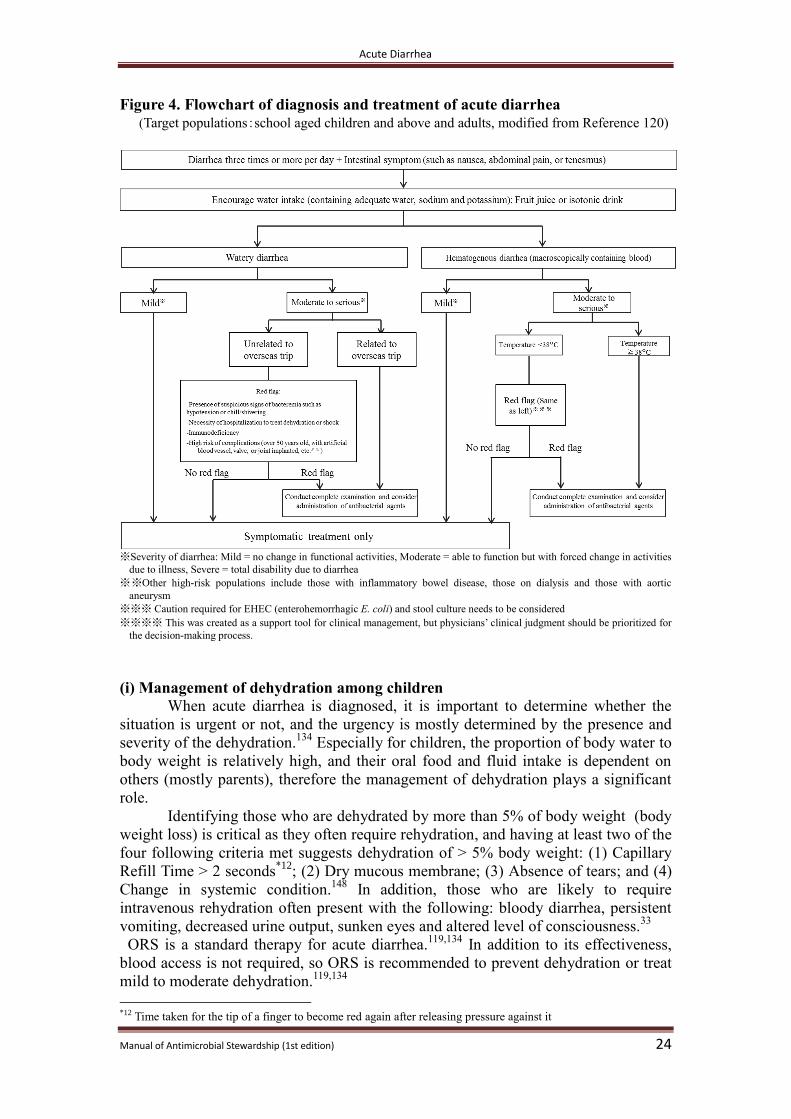
The process of the diagnosis and management of acute diarrhea is shown in
Figure 4.
Acute Diarrhea
Manual of Antimicrobial Stewardship (1st edition) 24
Figure 4. Flowchart of diagnosis and treatment of acute diarrhea (Target populations:school aged children and above and adults, modified from Reference 120)
※Severity of diarrhea: Mild = no change in functional activities, Moderate = able to function but with forced change in activities
due to illness, Severe = total disability due to diarrhea
※※Other high-risk populations include those with inflammatory bowel disease, those on dialysis and those with aortic
aneurysm
※※※ Caution required for EHEC (enterohemorrhagic E. coli) and stool culture needs to be considered
※※※※ This was created as a support tool for clinical management, but physicians’ clinical judgment should be prioritized for
the decision-making process.
(i) Management of dehydration among children
When acute diarrhea is diagnosed, it is important to determine whether the
situation is urgent or not, and the urgency is mostly determined by the presence and
severity of the dehydration.134
Especially for children, the proportion of body water to
body weight is relatively high, and their oral food and fluid intake is dependent on
others (mostly parents), therefore the management of dehydration plays a significant
role.
Identifying those who are dehydrated by more than 5% of body weight (body
weight loss) is critical as they often require rehydration, and having at least two of the
four following criteria met suggests dehydration of > 5% body weight: (1) Capillary
Refill Time > 2 seconds*12
; (2) Dry mucous membrane; (3) Absence of tears; and (4)
Change in systemic condition.148
In addition, those who are likely to require
intravenous rehydration often present with the following: bloody diarrhea, persistent
vomiting, decreased urine output, sunken eyes and altered level of consciousness.33
ORS is a standard therapy for acute diarrhea.119,134
In addition to its effectiveness,
blood access is not required, so ORS is recommended to prevent dehydration or treat
mild to moderate dehydration.119,134
*12 Time taken for the tip of a finger to become red again after releasing pressure against it
Acute Diarrhea
Manual of Antimicrobial Stewardship (1st edition) 25
In practice, ORS should be given at an early stage (within three to four hours
after the onset of dehydration), and the amount given should be increased gradually
from one full teaspoon and adjusted every two to four hours until it equals the amount
lost (50ml/kg to 100ml/kg for mild to moderate dehydration).134
Of note, evidence on anti-emesis for vomiting and antidiarrheals for diarrhea
is scarce and neither is recommended.134
(ii) Indications of antibacterial therapy for children with acute diarrhea
Most of the pathogens causing acute diarrhea in children are viruses.
Therefore, antibacterial therapy is not only ineffective, but also disrupts gut flora,
leading to microbial substitution, and its use is not recommended.124,134
Even if the
cause of acute diarrhea is considered bacterial, most are self-limiting and antibacterial
therapy is not required.124,134
Of note, the guidelines developed in other countries limit
indications of stool culture and antibacterial therapy to situations where the systemic
illness is severe, Non-typhoidal Salmonella spp. or Campylobacter spp. is suspected
among immunocompromised patients, and so forth.134,149
(iii)Non-typhoidal Salmonella gastroenteritis
Even if non-typhoidal Salmonella spp. is identified as a pathogen, antibacterial
therapy for non-typhoidal Salmonella spp. among healthy adults without
comorbidities does not shorten time to relief of symptoms such as diarrhea and fever,
but rather prolongs colonization.150
Therefore, in this manual, we recommend against
antibacterial therapy for mild non-typhoidal Salmonella infection in otherwise healthy
patients.
It is noted that the following are risk factors of severe non-typhoidal Salmonella
infection, and therefore are indications of antibacterial therapy:151
・ Age younger than 3 months or 65 years and older
・ Use of steroids or immunosuppressants
・ Inflammatory bowel disease
・ Hemodialysis
・ Hemoglobinopathy such as sickle cell disease
・ Abdominal aneurysm
・ Prosthetic heart valve
According to the JAID/JSC guideline, when antibacterial therapy is
considered for non-typhoidal Salmonella infections, oral levofloxacin for 3 to 7 days
as the first-line treatment and, in the setting of low susceptibility to fluoroquinolones
or allergy to fluoroquinolones, intravenous ceftriaxone or oral azithromycin for 3 to 7
days as the second-line treatment are recommended.124
(iv) Campylobacter enteritis
・ We recommend against antibacterial therapy for mild* Campylobacter enteritis
in otherwise healthy patients. * Mild = no change in functional activities
・ We recommend against antibacterial therapy for mild* non-typhoidal Salmonella
gastroenteritis among otherwise healthy patients. * Mild = no change in functional activities
Acute Diarrhea
Manual of Antimicrobial Stewardship (1st edition) 26
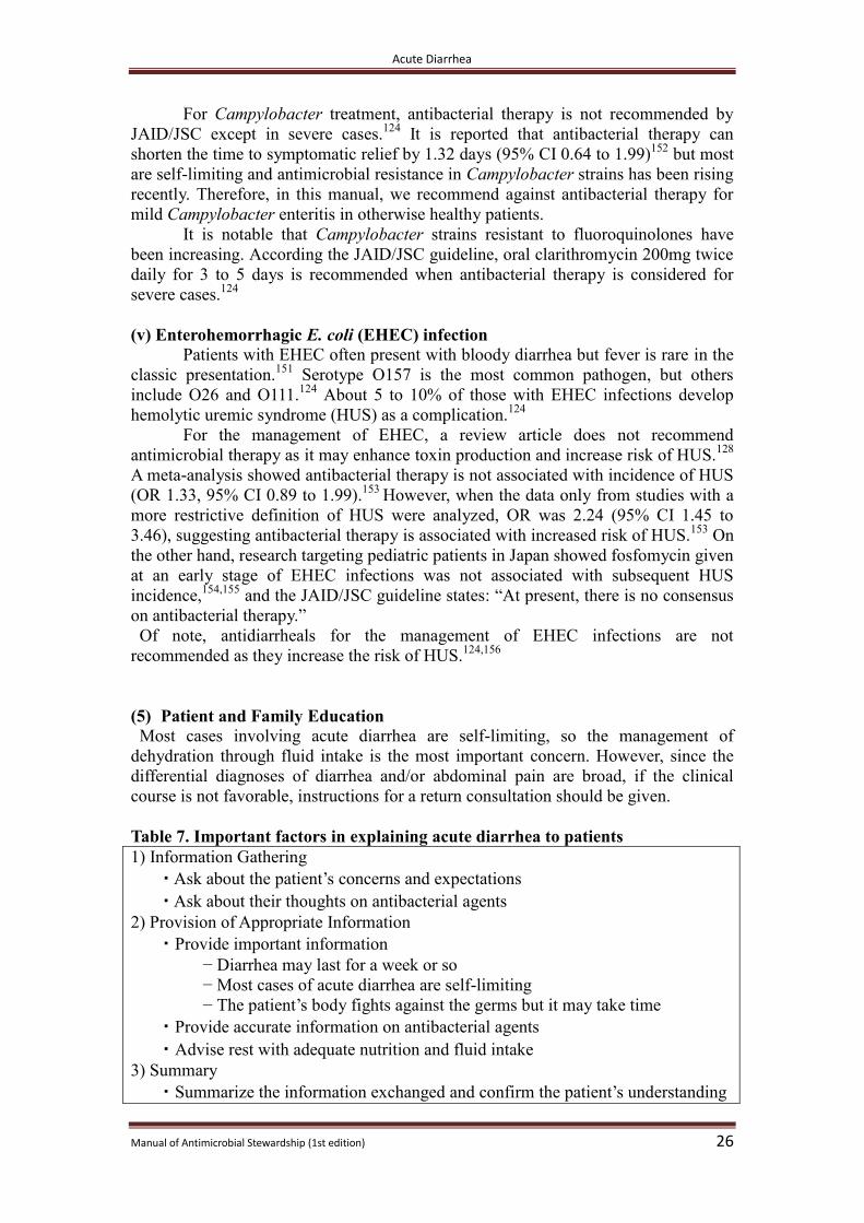
For Campylobacter treatment, antibacterial therapy is not recommended by
JAID/JSC except in severe cases.124
It is reported that antibacterial therapy can
shorten the time to symptomatic relief by 1.32 days (95% CI 0.64 to 1.99)152
but most
are self-limiting and antimicrobial resistance in Campylobacter strains has been rising
recently. Therefore, in this manual, we recommend against antibacterial therapy for
mild Campylobacter enteritis in otherwise healthy patients.
It is notable that Campylobacter strains resistant to fluoroquinolones have
been increasing. According the JAID/JSC guideline, oral clarithromycin 200mg twice
daily for 3 to 5 days is recommended when antibacterial therapy is considered for
severe cases.124
(v) Enterohemorrhagic E. coli (EHEC) infection
Patients with EHEC often present with bloody diarrhea but fever is rare in the
classic presentation.151
Serotype O157 is the most common pathogen, but others
include O26 and O111.124
About 5 to 10% of those with EHEC infections develop
hemolytic uremic syndrome (HUS) as a complication.124
For the management of EHEC, a review article does not recommend
antimicrobial therapy as it may enhance toxin production and increase risk of HUS.128
A meta-analysis showed antibacterial therapy is not associated with incidence of HUS
(OR 1.33, 95% CI 0.89 to 1.99).153
However, when the data only from studies with a
more restrictive definition of HUS were analyzed, OR was 2.24 (95% CI 1.45 to
3.46), suggesting antibacterial therapy is associated with increased risk of HUS.153
On
the other hand, research targeting pediatric patients in Japan showed fosfomycin given
at an early stage of EHEC infections was not associated with subsequent HUS
incidence,154,155
and the JAID/JSC guideline states: “At present, there is no consensus
on antibacterial therapy.”
Of note, antidiarrheals for the management of EHEC infections are not
recommended as they increase the risk of HUS.124,156
(5) Patient and Family Education
Most cases involving acute diarrhea are self-limiting, so the management of
dehydration through fluid intake is the most important concern. However, since the
differential diagnoses of diarrhea and/or abdominal pain are broad, if the clinical
course is not favorable, instructions for a return consultation should be given.
Table 7. Important factors in explaining acute diarrhea to patients
1) Information Gathering
・ Ask about the patient’s concerns and expectations
・ Ask about their thoughts on antibacterial agents
2) Provision of Appropriate Information
・ Provide important information
− Diarrhea may last for a week or so
− Most cases of acute diarrhea are self-limiting
− The patient’s body fights against the germs but it may take time
・ Provide accurate information on antibacterial agents
・ Advise rest with adequate nutrition and fluid intake
3) Summary
・ Summarize the information exchanged and confirm the patient’s understanding
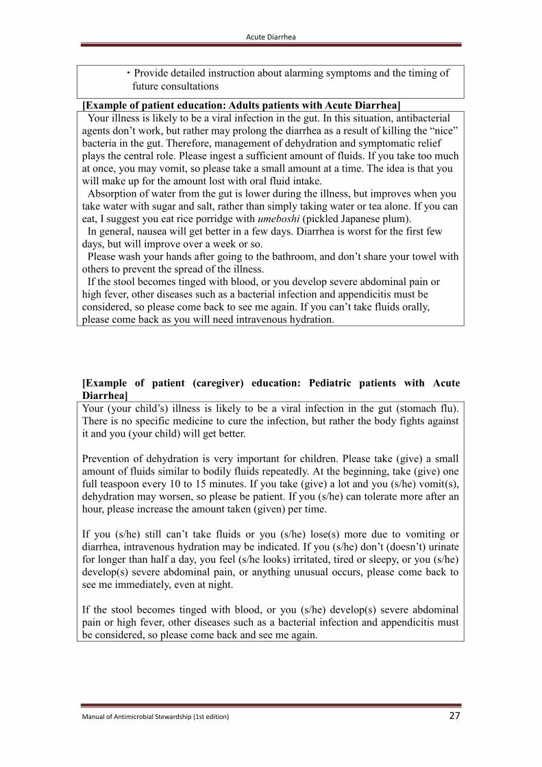
Acute Diarrhea
Manual of Antimicrobial Stewardship (1st edition) 27
・ Provide detailed instruction about alarming symptoms and the timing of
future consultations
[Example of patient education: Adults patients with Acute Diarrhea]
Your illness is likely to be a viral infection in the gut. In this situation, antibacterial
agents don’t work, but rather may prolong the diarrhea as a result of killing the “nice”
bacteria in the gut. Therefore, management of dehydration and symptomatic relief
plays the central role. Please ingest a sufficient amount of fluids. If you take too much
at once, you may vomit, so please take a small amount at a time. The idea is that you
will make up for the amount lost with oral fluid intake.
Absorption of water from the gut is lower during the illness, but improves when you
take water with sugar and salt, rather than simply taking water or tea alone. If you can
eat, I suggest you eat rice porridge with umeboshi (pickled Japanese plum).
In general, nausea will get better in a few days. Diarrhea is worst for the first few
days, but will improve over a week or so.
Please wash your hands after going to the bathroom, and don’t share your towel with
others to prevent the spread of the illness.
If the stool becomes tinged with blood, or you develop severe abdominal pain or
high fever, other diseases such as a bacterial infection and appendicitis must be
considered, so please come back to see me again. If you can’t take fluids orally,
please come back as you will need intravenous hydration.
[Example of patient (caregiver) education: Pediatric patients with Acute
Diarrhea]
Your (your child’s) illness is likely to be a viral infection in the gut (stomach flu).
There is no specific medicine to cure the infection, but rather the body fights against
it and you (your child) will get better.
Prevention of dehydration is very important for children. Please take (give) a small
amount of fluids similar to bodily fluids repeatedly. At the beginning, take (give) one
full teaspoon every 10 to 15 minutes. If you take (give) a lot and you (s/he) vomit(s),
dehydration may worsen, so please be patient. If you (s/he) can tolerate more after an
hour, please increase the amount taken (given) per time.
If you (s/he) still can’t take fluids or you (s/he) lose(s) more due to vomiting or
diarrhea, intravenous hydration may be indicated. If you (s/he) don’t (doesn’t) urinate
for longer than half a day, you feel (s/he looks) irritated, tired or sleepy, or you (s/he)
develop(s) severe abdominal pain, or anything unusual occurs, please come back to
see me immediately, even at night.
If the stool becomes tinged with blood, or you (s/he) develop(s) severe abdominal
pain or high fever, other diseases such as a bacterial infection and appendicitis must
be considered, so please come back and see me again.
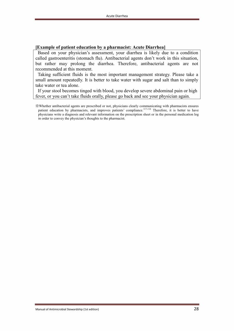
Acute Diarrhea
Manual of Antimicrobial Stewardship (1st edition) 28
[Example of patient education by a pharmacist: Acute Diarrhea]
Based on your physician’s assessment, your diarrhea is likely due to a condition
called gastroenteritis (stomach flu). Antibacterial agents don’t work in this situation,
but rather may prolong the diarrhea. Therefore, antibacterial agents are not
recommended at this moment.
Taking sufficient fluids is the most important management strategy. Please take a
small amount repeatedly. It is better to take water with sugar and salt than to simply
take water or tea alone.
If your stool becomes tinged with blood, you develop severe abdominal pain or high
fever, or you can’t take fluids orally, please go back and see your physician again.
※Whether antibacterial agents are prescribed or not, physicians clearly communicating with pharmacists ensures
patient education by pharmacists, and improves patients’ compliance.117-118 Therefore, it is better to have
physicians write a diagnosis and relevant information on the prescription sheet or in the personal medication log
in order to convey the physician’s thoughts to the pharmacist.
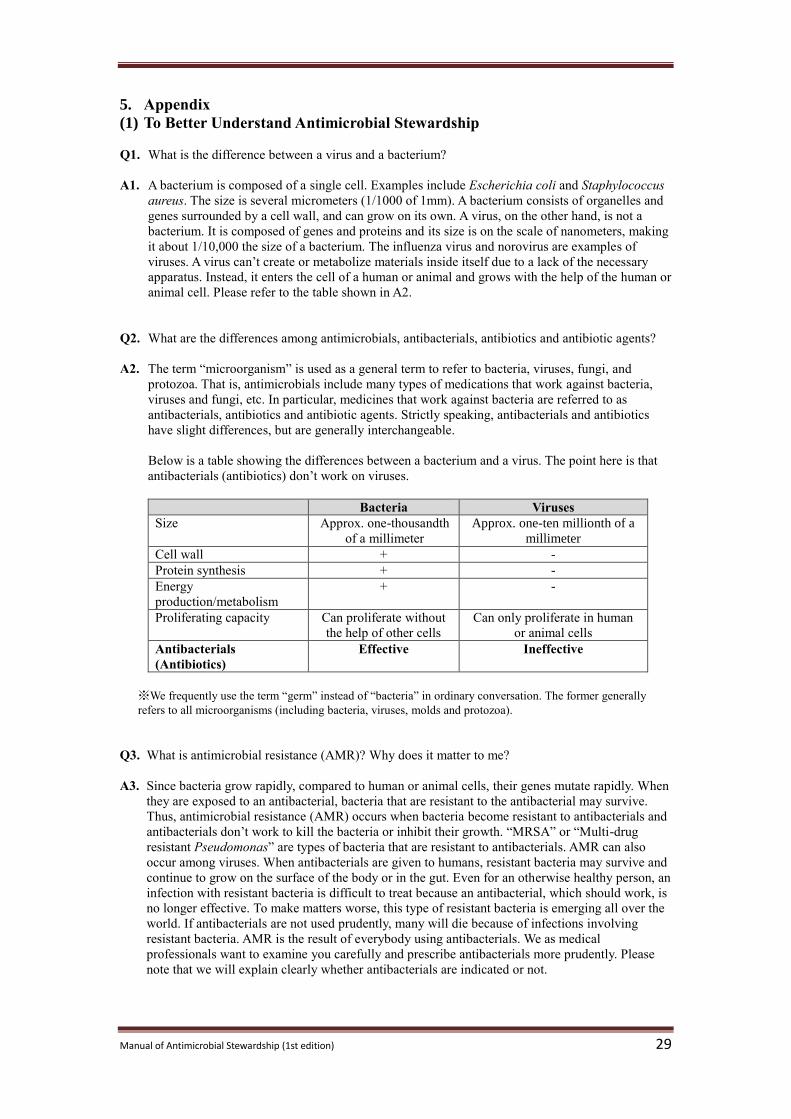
Manual of Antimicrobial Stewardship (1st edition) 29
5. Appendix
(1) To Better Understand Antimicrobial Stewardship
Q1. What is the difference between a virus and a bacterium?
A1. A bacterium is composed of a single cell. Examples include Escherichia coli and Staphylococcus
aureus. The size is several micrometers (1/1000 of 1mm). A bacterium consists of organelles and
genes surrounded by a cell wall, and can grow on its own. A virus, on the other hand, is not a
bacterium. It is composed of genes and proteins and its size is on the scale of nanometers, making
it about 1/10,000 the size of a bacterium. The influenza virus and norovirus are examples of
viruses. A virus can’t create or metabolize materials inside itself due to a lack of the necessary
apparatus. Instead, it enters the cell of a human or animal and grows with the help of the human or
animal cell. Please refer to the table shown in A2.
Q2. What are the differences among antimicrobials, antibacterials, antibiotics and antibiotic agents?
A2. The term “microorganism” is used as a general term to refer to bacteria, viruses, fungi, and
protozoa. That is, antimicrobials include many types of medications that work against bacteria,
viruses and fungi, etc. In particular, medicines that work against bacteria are referred to as
antibacterials, antibiotics and antibiotic agents. Strictly speaking, antibacterials and antibiotics
have slight differences, but are generally interchangeable.
Below is a table showing the differences between a bacterium and a virus. The point here is that
antibacterials (antibiotics) don’t work on viruses.
Bacteria Viruses
Size Approx. one-thousandth
of a millimeter
Approx. one-ten millionth of a
millimeter
Cell wall + -
Protein synthesis + -
Energy
production/metabolism
+ -
Proliferating capacity Can proliferate without
the help of other cells
Can only proliferate in human
or animal cells
Antibacterials
(Antibiotics)
Effective Ineffective
※We frequently use the term “germ” instead of “bacteria” in ordinary conversation. The former generally
refers to all microorganisms (including bacteria, viruses, molds and protozoa).
Q3. What is antimicrobial resistance (AMR)? Why does it matter to me?
A3. Since bacteria grow rapidly, compared to human or animal cells, their genes mutate rapidly. When
they are exposed to an antibacterial, bacteria that are resistant to the antibacterial may survive.
Thus, antimicrobial resistance (AMR) occurs when bacteria become resistant to antibacterials and
antibacterials don’t work to kill the bacteria or inhibit their growth. “MRSA” or “Multi-drug
resistant Pseudomonas” are types of bacteria that are resistant to antibacterials. AMR can also
occur among viruses. When antibacterials are given to humans, resistant bacteria may survive and
continue to grow on the surface of the body or in the gut. Even for an otherwise healthy person, an
infection with resistant bacteria is difficult to treat because an antibacterial, which should work, is
no longer effective. To make matters worse, this type of resistant bacteria is emerging all over the
world. If antibacterials are not used prudently, many will die because of infections involving
resistant bacteria. AMR is the result of everybody using antibacterials. We as medical
professionals want to examine you carefully and prescribe antibacterials more prudently. Please
note that we will explain clearly whether antibacterials are indicated or not.
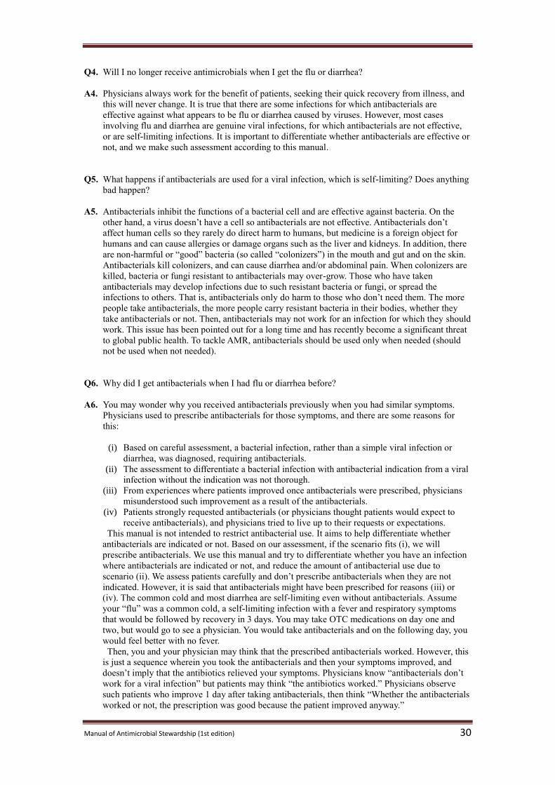
Manual of Antimicrobial Stewardship (1st edition) 30
Q4. Will I no longer receive antimicrobials when I get the flu or diarrhea?
A4. Physicians always work for the benefit of patients, seeking their quick recovery from illness, and
this will never change. It is true that there are some infections for which antibacterials are
effective against what appears to be flu or diarrhea caused by viruses. However, most cases
involving flu and diarrhea are genuine viral infections, for which antibacterials are not effective,
or are self-limiting infections. It is important to differentiate whether antibacterials are effective or
not, and we make such assessment according to this manual.
Q5. What happens if antibacterials are used for a viral infection, which is self-limiting? Does anything
bad happen?
A5. Antibacterials inhibit the functions of a bacterial cell and are effective against bacteria. On the
other hand, a virus doesn’t have a cell so antibacterials are not effective. Antibacterials don’t
affect human cells so they rarely do direct harm to humans, but medicine is a foreign object for
humans and can cause allergies or damage organs such as the liver and kidneys. In addition, there
are non-harmful or “good” bacteria (so called “colonizers”) in the mouth and gut and on the skin.
Antibacterials kill colonizers, and can cause diarrhea and/or abdominal pain. When colonizers are
killed, bacteria or fungi resistant to antibacterials may over-grow. Those who have taken
antibacterials may develop infections due to such resistant bacteria or fungi, or spread the
infections to others. That is, antibacterials only do harm to those who don’t need them. The more
people take antibacterials, the more people carry resistant bacteria in their bodies, whether they
take antibacterials or not. Then, antibacterials may not work for an infection for which they should
work. This issue has been pointed out for a long time and has recently become a significant threat
to global public health. To tackle AMR, antibacterials should be used only when needed (should
not be used when not needed).
Q6. Why did I get antibacterials when I had flu or diarrhea before?
A6. You may wonder why you received antibacterials previously when you had similar symptoms.
Physicians used to prescribe antibacterials for those symptoms, and there are some reasons for
this:
(i) Based on careful assessment, a bacterial infection, rather than a simple viral infection or
diarrhea, was diagnosed, requiring antibacterials.
(ii) The assessment to differentiate a bacterial infection with antibacterial indication from a viral
infection without the indication was not thorough.
(iii) From experiences where patients improved once antibacterials were prescribed, physicians
misunderstood such improvement as a result of the antibacterials.
(iv) Patients strongly requested antibacterials (or physicians thought patients would expect to
receive antibacterials), and physicians tried to live up to their requests or expectations.
This manual is not intended to restrict antibacterial use. It aims to help differentiate whether
antibacterials are indicated or not. Based on our assessment, if the scenario fits (i), we will
prescribe antibacterials. We use this manual and try to differentiate whether you have an infection
where antibacterials are indicated or not, and reduce the amount of antibacterial use due to
scenario (ii). We assess patients carefully and don’t prescribe antibacterials when they are not
indicated. However, it is said that antibacterials might have been prescribed for reasons (iii) or
(iv). The common cold and most diarrhea are self-limiting even without antibacterials. Assume
your “flu” was a common cold, a self-limiting infection with a fever and respiratory symptoms
that would be followed by recovery in 3 days. You may take OTC medications on day one and
two, but would go to see a physician. You would take antibacterials and on the following day, you
would feel better with no fever.
Then, you and your physician may think that the prescribed antibacterials worked. However, this
is just a sequence wherein you took the antibacterials and then your symptoms improved, and
doesn’t imply that the antibiotics relieved your symptoms. Physicians know “antibacterials don’t
work for a viral infection” but patients may think “the antibiotics worked.” Physicians observe
such patients who improve 1 day after taking antibacterials, then think “Whether the antibacterials
worked or not, the prescription was good because the patient improved anyway.”
Manual of Antimicrobial Stewardship (1st edition) 31
These repeated experiences may have led physicians to think patients would be happy with
antibacterials. On rare occasions, patients say “Please give me antibiotics this time because I felt
better quickly with them last time.” Physicians value patients’ satisfaction and therefore might
have prescribed antibacterials because the patient expressed or the physician intuited such
expectations.
Q7. Will you not prescribe antibacterials for the flu or diarrhea?
A7. This manual does not instruct physicians to either prescribe or not prescribe antibacterials for the
flu or diarrhea. When you have the flu or diarrhea, this manual helps physicians assess if
antibacterials are indicated or not, and, if not indicated, this manual recommends against
antibacterial therapy. Please ask us if you feel concerned about not having antibacterials
prescribed. We will explain how we have assessed you and how we have reached a diagnosis.
As a result of a series of experiences and behaviors enacted between physicians and patients,
antimicrobials have been misused and AMR has become a public threat. Previously, physicians
might have prescribed antibacterials based on the idea that “antibacterials can be prescribed
because at least they do no harm.” However, this is not the case. Using this manual, we try to
differentiate whether antibacterials are indicated or not, and prescribe them only when really
needed. Otherwise, AMR will continue to be a threat, and antibacterials may not work when they
are supposed to work. In fact, we are already experiencing this to a certain extent.
As physicians, we always wish our patients a quick recovery. We will prescribe antibacterials for
a bacterial infection for which they are indicated. We will try not to miss those infections. On the
other hand, we will not prescribe antibacterials when we are certain that they are not indicated.
We hope you understand that this approach will eventually help when you get a bacterial infection
and you have effective antibacterials available.
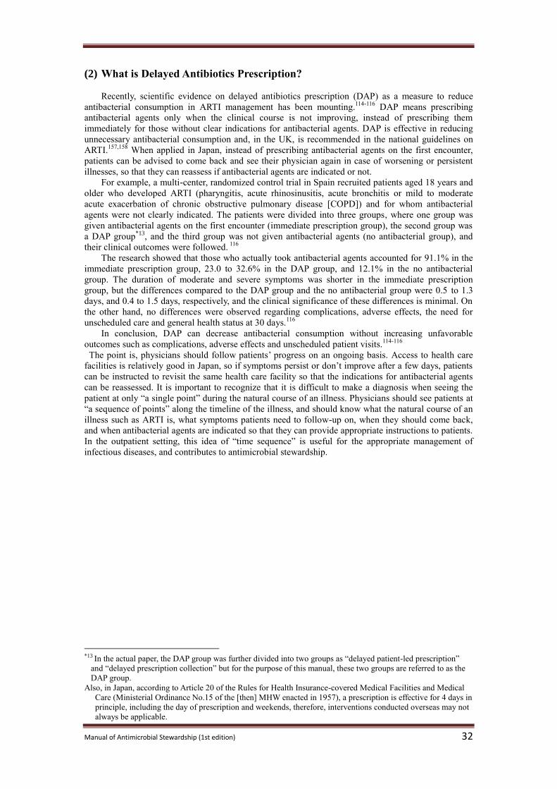
Manual of Antimicrobial Stewardship (1st edition) 32
(2) What is Delayed Antibiotics Prescription?
Recently, scientific evidence on delayed antibiotics prescription (DAP) as a measure to reduce
antibacterial consumption in ARTI management has been mounting.114-116
DAP means prescribing
antibacterial agents only when the clinical course is not improving, instead of prescribing them
immediately for those without clear indications for antibacterial agents. DAP is effective in reducing
unnecessary antibacterial consumption and, in the UK, is recommended in the national guidelines on
ARTI.157,158
When applied in Japan, instead of prescribing antibacterial agents on the first encounter,
patients can be advised to come back and see their physician again in case of worsening or persistent
illnesses, so that they can reassess if antibacterial agents are indicated or not.
For example, a multi-center, randomized control trial in Spain recruited patients aged 18 years and
older who developed ARTI (pharyngitis, acute rhinosinusitis, acute bronchitis or mild to moderate
acute exacerbation of chronic obstructive pulmonary disease [COPD]) and for whom antibacterial
agents were not clearly indicated. The patients were divided into three groups, where one group was
given antibacterial agents on the first encounter (immediate prescription group), the second group was
a DAP group*13
, and the third group was not given antibacterial agents (no antibacterial group), and
their clinical outcomes were followed. 116
The research showed that those who actually took antibacterial agents accounted for 91.1% in the
immediate prescription group, 23.0 to 32.6% in the DAP group, and 12.1% in the no antibacterial
group. The duration of moderate and severe symptoms was shorter in the immediate prescription
group, but the differences compared to the DAP group and the no antibacterial group were 0.5 to 1.3
days, and 0.4 to 1.5 days, respectively, and the clinical significance of these differences is minimal. On
the other hand, no differences were observed regarding complications, adverse effects, the need for
unscheduled care and general health status at 30 days.116
In conclusion, DAP can decrease antibacterial consumption without increasing unfavorable
outcomes such as complications, adverse effects and unscheduled patient visits.114-116
The point is, physicians should follow patients’ progress on an ongoing basis. Access to health care
facilities is relatively good in Japan, so if symptoms persist or don’t improve after a few days, patients
can be instructed to revisit the same health care facility so that the indications for antibacterial agents
can be reassessed. It is important to recognize that it is difficult to make a diagnosis when seeing the
patient at only “a single point” during the natural course of an illness. Physicians should see patients at
“a sequence of points” along the timeline of the illness, and should know what the natural course of an
illness such as ARTI is, what symptoms patients need to follow-up on, when they should come back,
and when antibacterial agents are indicated so that they can provide appropriate instructions to patients.
In the outpatient setting, this idea of “time sequence” is useful for the appropriate management of
infectious diseases, and contributes to antimicrobial stewardship.
*13 In the actual paper, the DAP group was further divided into two groups as “delayed patient-led prescription”
and “delayed prescription collection” but for the purpose of this manual, these two groups are referred to as the
DAP group.
Also, in Japan, according to Article 20 of the Rules for Health Insurance-covered Medical Facilities and Medical
Care (Ministerial Ordinance No.15 of the [then] MHW enacted in 1957), a prescription is effective for 4 days in
principle, including the day of prescription and weekends, therefore, interventions conducted overseas may not
always be applicable.
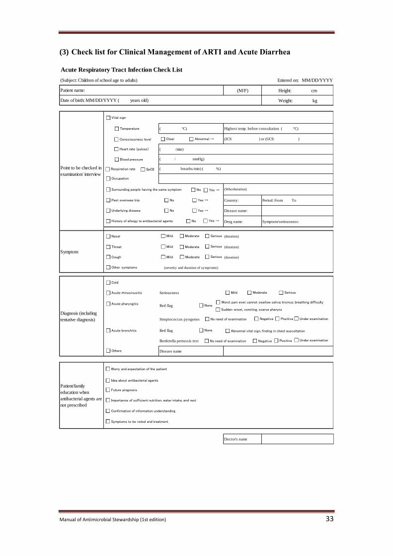
Manual of Antimicrobial Stewardship (1st edition) 33
(3) Check list for Clinical Management of ARTI and Acute Diarrhea
Acute Respiratory Tract Infection Check List
(Subject: Children of school age to adults) Entered on: MM/DD/YYYY
(M/F) Height: cm
Weight: kg
( °C)
Country: Period: From To
Disease name:
Drug name: Symptom/seriousness:
(duration)
(duration)
(duration)
(severity and duration of symptoms)
Seriousness
Streptococcus pyogenes
Red flag
Bordetella pertussis test
Disease name
Doctor's name
Patient name:
Date of birth: MM/DD/YYYY ( years old)
(Who/duration)
Symptom
(JCS ) or (GCS: )
( /min)
Point to be checked in
examination/ interview
Highest temp. before consultation ( °C)
( / mmHg)
( breaths/min) ( %)
Patient/family
education when
antibacterial agents are
not prescribed
Diagnosis (including
tentative diagnosis)
Red flag
Vital sign
Blood pressure
Respiration rate SpO2
Past overseas trip
Nasal
Throat
Negative
Cough
Worry and expectation of the patient
Mild
Confirmation of information understanding
Importance of sufficient nutrition, water intake, and rest
Future prognosis
Worst pain ever; cannot swallow saliva; trismus; breathing difficulty
Underlying disease
Yes →
Cold
Acute rhinosinusitis
No
Symptoms to be noted and treatment
Idea about antibacterial agents
Acute pharyngitis
Acute bronchitis
Serious
Positive Under examinationNo need of examination
Negative Positive Under examinationNo need of examination
No Yes →
Others
Moderate
Surrounding people having the same symptom Yes →No
Mild SeriousModerate
Mild SeriousModerate
Mild SeriousModerate
Other symptoms
Temperature
Sudden onset, vomiting, scarce pharynx None
Abnormal vital sign, finding in chest auscultationNone
Heart rate (pulses)
Consciousness level Clear Abnormal →
History of allergy to antibacterial agents No Yes →
Occupation
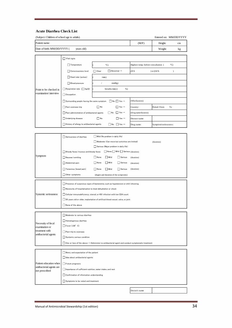
Manual of Antimicrobial Stewardship (1st edition) 34
Acute Diarrhea Check List
(Subject: Children of school age to adults) Entered on: MM/DD/YYYY
(M/F) Height: cm
Weight: kg
( °C)
Country: Period: From To
Disease name:
Drug name: Symptom/seriousness:
(duration)
(duration)
(duration)
(duration)
(duration)
(degree and duration of the symptoms)
Doctor's name
Patient name:
Date of birth: MM/DD/YYYY ( years old)
Point to be checked in
examination/ interview
Highest temp. before consultation ( °C)
( / mmHg)
( breaths/min) ( %)
(Who/duration)
(Drug name/duration)
( /min)
(JCS ) or (GCS: )
Systemic seriousness
Symptom
Patient education when
antibacterial agents are
not prescribed
Necessity of fecal
examination or
treatment with
antibacterial agents
Vital signs
Blood pressure
Respiration rate SpO2
Past overseas trip
Nausea/vomiting
Abdominal pain
Bloody feces/mucous and bloody feces
Worry and expectation of the patient
Confirmation of information understanding
Importance of sufficient nutrition, water intake, and rest
Future prognosis
Yes →
Hematogenous diarrhea
No
Symptoms to be noted and treatment
Idea about antibacterial agents
Systemic serious condition
No Yes →
Moderate to serious diarrhea
Surrounding people having the same symptom Yes →No
Mild (No problem in daily life)
Serious (Major problem in daily life)
Moderate (Can move but activities are limited)
None SeriousMild
None SeriousMild
Tenesmus (bowel pain)
Temperature
Seriousness of diarrhea
None SeriousMild
Occupation
Past administration of antibacterial agents Yes →No
None SeriousMild
Other symptoms
50 years old or older, implantation of artificial blood vessel, valve, or joint
None of the above
Presence of suspicious signs of bacteremia, such as hypotension or chill/shivering
Necessity of hospitalization to treat dehydration or shock
Cellular immunodeficiency, steroid, or HIV infection with low CD4 count
Fever (≥38°C)
Past trip to overseas
One or less of the above -> Administer no antibacterial agent and conduct symptomatic treatment
Heart rate (pulses)
Consciousness level Clear Abnormal →
Underlying disease
History of allergy to antibacterial agents No Yes →
Reference
Manual of Antimicrobial Stewardship (1st edition) 35
6. Reference
1. National Action Plan on Antimicrobial Resistance (AMR). 2016-2020. Tokyo: Cabinet Secretariat;
2016.
2. Ardal C, Outterson K, Hoffman SJ, et al. International cooperation to improve access to and
sustain effectiveness of antimicrobials. Lancet. 2016;387(10015):296-307.
3. The Review on Antimicrobial Resistance. Tackling Drug-Resistant Infections Globally: Final
Report and Recommendations. https://amr-review.org/Publications.html. Last accessed on
2/13/2017.
4. Arias CA, Murray BE. Antibiotic-resistant bugs in the 21st century - a clinical super-challenge. N
Engl J Med. 2009;360(5):439-443.
5. Muraki Y, Yagi T, Tsuji Y, et al. Japanese antimicrobial consumption surveillance: First report on
oral and parenteral antimicrobial consumption in Japan (2009-2013). J Glob Antimicrob Resist.
2016;7:19-23.
6. Fleming-Dutra KE, Hersh AL, Shapiro DJ, et al. Prevalence of Inappropriate Antibiotic
Prescriptions Among US Ambulatory Care Visits, 2010-2011. JAMA. 2016;315(17):1864-1873.
7. Barlam TF, Cosgrove SE, Abbo LM, et al. Implementing an Antibiotic Stewardship Program:
Guidelines by the Infectious Diseases Society of America and the Society for Healthcare
Epidemiology of America. Clin Infect Dis. 2016;62(10):e51-77.
8. Arnold SR, Straus SE. Interventions to improve antibiotic prescribing practices in ambulatory
care. Cochrane Database Syst Rev. 2005;(4):CD003539.
9. Drekonja DM, Filice GA, Greer N, et al. Antimicrobial stewardship in outpatient settings: a
systematic review. Infect Control Hosp Epidemiol. 2015;36(2):142-152.
10. Johannsson B, Beekmann SE, Srinivasan A, Hersh AL, Laxminarayan R, Polgreen PM. Improving
antimicrobial stewardship: the evolution of programmatic strategies and barriers. Infect Control
Hosp Epidemiol. 2011;32(4):367-374.
11. Jefferson T, Del Mar CB, Dooley L, et al. Physical interventions to interrupt or reduce the spread
of respiratory viruses. Cochrane Database Syst Rev. 2011;(7):CD006207.
12. Ejemot-Nwadiaro RI, Ehiri JE, Arikpo D, Meremikwu MM, Critchley JA. Hand washing
promotion for preventing diarrhoea. Cochrane Database Syst Rev. 2015;(9):CD004265.
13. Centers for Disease Control and Prevention. Norovirus | Preventing Norovirus Infection.
https://www.cdc.gov/norovirus/preventing-infection.html. Last accessed on 2/13/2017.
Tuberculosis and Infectious Disease Control Division, Health Service Bureau, Ministry of
Health, Labour and Welfare. Q&A-Norovirus infection http://www.mhlw.go.jp/file/06-
Seisakujouhou-11130500-Shokuhinanzenbu/0000129187.pdf. Last accessed on 2/13/2017.
15. National Institute of Infectious Disease. Routine/Voluntary Immunization Schedule in Japan.
http://www.nih.go.jp/niid/images/vaccine/schedule/2016/JP20161001.png. Published 2016. Last
accessed on 2/13/2017.
16. Tuberculosis and Infectious Disease Control Division, Health Service Bureau, Ministry of Health,
Labour and Welfare. Influenza-Q&A. http://www.mhlw.go.jp/bunya/kenkou/kekkaku-
kansenshou01/qa.html. Published 2016. Last accessed on 2/13/2017.
17. Satomura K, Kitamura T, Kawamura T, et al. Prevention of upper respiratory tract infections by
gargling: a randomized trial. Am J Prev Med. 2005;29(4):302-307.
18. Goodall EC, Granados AC, Luinstra K, et al. Vitamin D3 and gargling for the prevention of upper
Reference
Manual of Antimicrobial Stewardship (1st edition) 36
respiratory tract infections: a randomized controlled trial. BMC Infect Dis. 2014;14:273.
19. Kaji M. Influenza and common cold [in Japanese]. Tokyo: Nanzando, 2003. Print.
20. The Japanese Respiratory Society. The Japanese Respiratory Society guidelines for the
management of respiratory tract infection [in Japanese]. Tokyo: The Japanese Respiratory
Society; 2003.
21. Matsumura Y, et al. Common cold/acute respiratory infection -Standard terminology and
appropriate use of antimicrobial agent- Questionnaire survey (1) [in Japanese]. Naika Senmoni
Kaishi. 2003;15:217-221.
22. Kaji M. Common cold in daily clinical practice [in Japanese]. Rinsho to Kenkyu. 1994;71:1-3.
23. Tanaka Y. Classification of common cold and differential diagnosis [in Japanese]. Konnichi no
chiryou. 2005;13:17-21.
24. Statistics and Information Policy, Ministry of Health, Labour and Welfare. Patient survey 2014 [in
Japanese]. Tokyo: Kousei Tokei Kyoukai; 2016.
http://www.mhlw.go.jp/toukei/saikin/hw/kanja/14/dl/toukei.pdf. Last accessed on 2/13/2017.
25. Monto AS, Ullman BM. Acute respiratory illness in an American community. The Tecumseh
study. JAMA. 1974;227(2):164-169.
26. Chen Y, Williams E, Kirk M. Risk factors for acute respiratory infection in the Australian
community. PloS One. 2014;9(7):e101440.
27. Yokobayashi K, Matsushima M, Watanabe T, Fujinuma Y, Tazuma S. Prospective cohort study of
fever incidence and risk in elderly persons living at home. BMJ Open. 2014;4(7):e004998.
28. Nicholson KG, Kent J, Hammersley V, Cancio E. Acute viral infections of upper respiratory tract
in elderly people living in the community: comparative, prospective, population based study of
disease burden. BMJ. 1997;315(7115):1060-1064.
29. Graat JM, Schouten EG, Heijnen M-LA, et al. A prospective, community-based study on virologic
assessment among elderly people with and without symptoms of acute respiratory infection. J Clin
Epidemiol. 2003;56(12):1218-1223.
30. Falsey AR, Walsh EE, Hayden FG. Rhinovirus and coronavirus infection-associated
hospitalizations among older adults. J Infect Dis. 2002;185(9):1338-1341.
31. National Institute for Health and Care Excellence (NICE). Fever in under 5s: assessment and
initial management | Clinical guideline. https://www.nice.org.uk/guidance/cg160/chapter/1-
Recommendations#clinical-assessment-of-children-with-fever. Last accessed on 2/13/2017.
32. Heikkinen T, Järvinen A. The common cold. Lancet. 2003;361(9351):51-59.
33. Kliegman R, Stanton B, Geme J, Schor N. Nelson Textbook of Pediatrics. 20th edition.
Philadelphia, PA: Elsevier; 2015.
34. Keith T, Saxena S, Murray J, Sharland M. Risk-benefit analysis of restricting antimicrobial
prescribing in children: what do we really know? Curr Opin Infect Dis. 2010;23(3):242-248.
35. American Academy of Pediatrics, Committee on Infectious Diseases, Kimberlin DW, Brady MT,
Jackson MA, Long SS. Red Book: 2015 Report of the Committee on Infectious Diseases. 2015.
36. Shulman ST, Bisno AL, Clegg HW, et al. Clinical practice guideline for the diagnosis and
management of group A streptococcal pharyngitis: 2012 update by the Infectious Diseases Society
of America. Clin Infect Dis. 2012;55(10):1279-1282.
Reference
Manual of Antimicrobial Stewardship (1st edition) 37
37. Kearns GL, Abdel-Rahman SM, Alander SW, Blowey DL, Leeder JS, Kauffman RE.
Developmental pharmacology - drug disposition, action, and therapy in infants and children. N
Engl J Med. 2003;349(12):1157-1167.
38. Gonzales R, Bartlett JG, Besser RE, et al. Principles of appropriate antibiotic use for treatment of
acute respiratory tract infections in adults: background, specific aims, and methods. Ann Intern
Med. 2001;134(6):479-486.
39. The Japanese Society of Internal Medicine-Board certification committee. Common disease book:
Differential diagnosis and explanation to patients in daily clinical practice. Tokyo: The Japanese
Society of Internal Medicine; 2013. Print.
40. Harris AM, Hicks LA, Qaseem A, High Value Care Task Force of the American College of
Physicians and for the Centers for Disease Control and Prevention. Appropriate Antibiotic Use for
Acute Respiratory Tract Infection in Adults: Advice for High-Value Care From the American
College of Physicians and the Centers for Disease Control and Prevention. Ann Intern Med.
2016;164(6):425-434.
41. Gwaltney JM, Hendley JO, Simon G, Jordan WS. Rhinovirus infections in an industrial
population. II. Characteristics of illness and antibody response. JAMA. 1967;202(6):494-500.
42. Monto AS, Gravenstein S, Elliott M, Colopy M, Schweinle J. Clinical signs and symptoms
predicting influenza infection. Arch Intern Med. 2000;160(21):3243-3247.
43. Ebell MH, Afonso AM, Gonzales R, Stein J, Genton B, Senn N. Development and validation of a
clinical decision rule for the diagnosis of influenza. J Am Board Fam Med. 2012;25(1):55-62.
44. Chartrand C, Leeflang MMG, Minion J, Brewer T, Pai M. Accuracy of rapid influenza diagnostic
tests: a meta-analysis. Ann Intern Med. 2012;156(7):500-511.
45. Meltzer EO, Hamilos DL, Hadley JA, et al. Rhinosinusitis: establishing definitions for clinical
research and patient care. J Allergy Clin Immunol. 2004;114(6 Suppl):155-212.
46. Berg O, Carenfelt C, Rystedt G, Anggård A. Occurrence of asymptomatic sinusitis in common
cold and other acute ENT-infections. Rhinology. 1986;24(3):223-225.
47. Dingle J, Badger G, Jordan WJ. Illness in the Home: A Study of 25,000 Illnesses in a Group of
Cleveland Families. OH: Western Reserve Univ Pr; 1964.
48. Lacroix JS, Ricchetti A, Lew D, et al. Symptoms and clinical and radiological signs predicting the
presence of pathogenic bacteria in acute rhinosinusitis. Acta Otolaryngol (Stockh).
2002;122(2):192-196.
49. Chow AW, Benninger MS, Brook I, et al. IDSA clinical practice guideline for acute bacterial
rhinosinusitis in children and adults. Clin Infect Dis. 2012;54(8):e72-e112.
50. Bisno AL. Acute pharyngitis. N Engl J Med. 2001;344(3):205-211.
51. Suzuki K, Kurono Y, Kobayashi T, et al. The 4th
national surveillance of clinically isolated bacteria
in otolaryngology field [in Japanese]. Nihon Jibi Kansensyou kennkyuukaishi. 2008;26:15-26.
52. Suzuki K, Kurono Y, Ikeda K, et al. Nationwide surveillance of 6 otorhinolaryngological
infectious diseases and antimicrobial susceptibility pattern in the isolated pathogens in Japan. J
Infect Chemother. 2015;21(7):483-491.
53. Takeuchi H, Fukazawa M, Yoshida H, et al. Role of viruses and bacteria associated with
tonsilopharyngitis [in Japanese]. Nihon syounikagakkai zashi .2009;113(4):694-700.
54. Bisno AL, Gerber MA, Gwaltney JM, Kaplan EL, Schwartz RH. Practice Guidelines for the
Reference
Manual of Antimicrobial Stewardship (1st edition) 38
Diagnosis and Management of Group A Streptococcal Pharyngitis. Clin Infect Dis.
2002;35(2):113-125.
55. Tanz RR, Shulman ST. Chronic pharyngeal carriage of group A streptococci. Pediatr Infect Dis J.
2007;26(2):175-176.
56. Aliyu SH, Marriott RK, Curran MD, Parmar S, Bentley N, Brown NM, Brazier JS, Ludlam H.
Real-time PCR investigation into the importance of Fusobacterium necrophorum as a cause of
acute pharyngitis in general practice. J Med Microbiol. 2004;53(Pt 10):1029-1035.
57. Batty A, Wren MW. Prevalence of Fusobacterium necrophorum and other upper respiratory tract
pathogens isolated from throat swabs. Br J Biomed Sci. 2005;62(2):66-70.
58. Amess JA, O'Neill W, Giollariabhaigh CN, Dytrych JK. A six-month audit of the isolation of
Fusobacterium necrophorum from patients with sore throat in a district general hospital. Br J
Biomed Sci. 2007;64(2):63-65.
59. Centor RM, Geiger P, Waites KB. Fusobacterium necrophorum bacteremic tonsillitis: 2 Cases and
a review of the literature. Anaerobe 2010;16(6):626-628.
60. Shah M, Centor RM, Jennings M. Severe acute pharyngitis caused by group C streptococcus. J
Gen Intern Med. 2007;22(2):272-274.
61. Centor RM. Expand the pharyngitis paradigm for adolescents and young adults. Ann Intern Med.
2009;151(11):812-815.
62. Jensen A, Hansen TM, Bank S, Kristensen LH, Prag J. Fusobacterium necrophorum tonsillitis: an
important cause of tonsillitis in adolescents and young adults. Clin Microbiol Infect.
2015;21(3):266.e1-266.e3.
63. Hedin K, Bieber L, Lindh M, Sundqvist M. The aetiology of pharyngotonsillitis in adolescents
and adults - Fusobacterium necrophorum is commonly found. Clin Microbiol Infect.
2015;21(3):263.e1-263.e7.
64. Centor RM, Atkinson TP, Ratliff AE, Xiao L, Crabb DM, Estrada CA, Faircloth MB, Oestreich L,
Hatchett J, Khalife W, Waites KB. The Clinical Presentation of Fusobacterium-Positive and
Streptococcal-Positive Pharyngitis in a University Health Clinic: A Cross-sectional Study. Ann
Intern Med. 2015;162(4):241-247.
65. McIsaac WJ, Kellner JD, Aufricht P, Vanjaka A, Low DE. Empirical validation of guidelines for
the management of pharyngitis in children and adults. JAMA. 2004;291(13):1587-1595.
66. McIsaac WJ, Goel V, To T, Low DE. The validity of a sore throat score in family practice. Can
Med Assoc J. 2000;163(7):811-815.
67. ESCMID Sore Throat Guideline Group, Pelucchi C, Grigoryan L, et al. Guideline for the
management of acute sore throat. Clin Microbiol Infect. 2012;18 Suppl 1:1-28.
68. JAID/JSC infectious disease treatment guide/guideline development committee. JAID/JSC
infectious disease treatment guide 2014 [in Japanese].Tokyo: Life Science Publishing; 2014. Print.
69. McGinn TG, Deluca J, Ahlawat SK, Mobo BH, Wisnivesky JP. Validation and modification of
streptococcal pharyngitis clinical prediction rules. Mayo Clin Proc. 2003;78(3):289-293.
70. Humair J-P, Revaz SA, Bovier P, Stalder H. Management of acute pharyngitis in adults: reliability
of rapid streptococcal tests and clinical findings. Arch Intern Med. 2006;166(6):640-644.
71. Fine AM, Nizet V, Mandl KD. Large-scale validation of the Centor and McIsaac scores to predict
group A streptococcal pharyngitis. Arch Intern Med. 2012;172(11):847-852.
Reference
Manual of Antimicrobial Stewardship (1st edition) 39
72. Llor C, Hernández M, Hernández S, Martínez T, Gómez FF. Validity of a point-of-care based on
heterophile antibody detection for the diagnosis of infectious mononucleosis in primary care. Eur
J Gen Pract. 2012;18(1):15-21.
73. Ebell MH, Call M, Shinholser J, Gardner J. Does This Patient Have Infectious Mononucleosis?:
The Rational Clinical Examination Systematic Review. JAMA. 2016;315(14):1502-1509.
74. Wolf DM, Friedrichs I, Toma AG. Lymphocyte-white blood cell count ratio: a quickly available
screening tool to differentiate acute purulent tonsillitis from glandular fever. Arch Otolaryngol
Head Neck Surg. 2007;133(1):61-64.
75. Kishida N. How to see "common cold." Rule out critical diseases! [in Japanese]. Tokyo: Igaku-
Shoin; 2012. Print.
76. Yamamoto S. A manual of practice of "cold". 2nd
edition [in Japanese]. Tokyo: Japan Medical
Journal; 2017. Print.
77. Ebell MH, Lundgren J, Youngpairoj S. How long does a cough last? Comparing patients’
expectations with data from a systematic review of the literature. Ann Fam Med. 2013;11(1):5-13.
78. JAID/JSC infectious disease treatment guide/guideline development committee. Respiratory
infection working group. JAID/JSC infectious disease treatment guideline-respiratory infection
[in Japanese]. Japanese Journal of Chemotherapy. 2014;62:1-109.
79. Cornia PB, Hersh AL, Lipsky BA, Newman TB, Gonzales R. Does this coughing adolescent or
adult patient have pertussis? JAMA. 2010;304(8):890-896.
80. de Melker HE, Versteegh FG, Conyn-Van Spaendonck MA, et al. Specificity and sensitivity of
high levels of immunoglobulin G antibodies against pertussis toxin in a single serum sample for
diagnosis of infection with Bordetella pertussis. J Clin Microbiol. 2000;38(2):800-806.
81. Yih WK, Lett SM, des Vignes FN, Garrison KM, Sipe PL, Marchant CD. The increasing incidence
of pertussis in Massachusetts adolescents and adults, 1989-1998. J Infect Dis. 2000;182(5):1409-
1416.
82. Torkaman MRA, Kamachi K, Nikbin VS, Lotfi MN, Shahcheraghi F. Comparison of loop-
mediated isothermal amplification and real-time PCR for detecting Bordetella pertussis. J Med
Microbiol. 2015;64(Pt 4):463-465.
83. Brotons P, de Paz HD, Esteva C, Latorre I, Muñoz-Almagro C. Validation of a loop-mediated
isothermal amplification assay for rapid diagnosis of pertussis infection in nasopharyngeal
samples. Expert Rev Mol Diagn. 2016;16(1):125-130.
84. Pediatric respiratory infection practice guideline development committee. Pediatric respiratory
infection practice guideline 2017 [in Japanese]. Tokyo: Kyowa Kikaku; 2016.
85. Kenealy T, Arroll B. Antibiotics for the common cold and acute purulent rhinitis. Cochrane
Database Syst Rev. 2013;(6):CD000247.
86. Japanese Rhinologic Society. Acute sinusitis practice guideline [in Japanese]. Japanese Journal
of Rhinology. 2010;49(2):143-198.
87. Yamanaka N, Iino Y, Uno Y, et al. Practical guideline for management of acute rhinosinusitis in
Japan. Auris Nasus Larynx. 2015;42(1):1-7.
88. Wald ER, Applegate KE, Bordley C, et al. Clinical practice guideline for the diagnosis and
management of acute bacterial sinusitis in children aged 1 to 18 years. Pediatrics.
2013;132(1):e262-e280.
Reference
Manual of Antimicrobial Stewardship (1st edition) 40
89. Lemiengre MB, van Driel ML, Merenstein D, Young J, De Sutter AIM. Antibiotics for clinically
diagnosed acute rhinosinusitis in adults. Cochrane Database Syst Rev. 2012;10:CD006089.
90. Ahovuo-Saloranta A, Rautakorpi U-M, Borisenko OV, Liira H, Williams JW, Mäkelä M.
Antibiotics for acute maxillary sinusitis in adults. Cochrane Database Syst Rev.
2014;(2):CD000243.
91. Rosenfeld RM, Piccirillo JF, Chandrasekhar SS, et al. Clinical practice guideline (update): adult
sinusitis. Otolaryngol--Head Neck Surg. 2015;152(2 Suppl):S1-S39.
92. Falagas ME, Karageorgopoulos DE, Grammatikos AP, Matthaiou DK. Effectiveness and safety of
short vs. long duration of antibiotic therapy for acute bacterial sinusitis: a meta-analysis of
randomized trials. Br J Clin Pharmacol. 2009;67(2):161-171.
93. Suzuki K, Nishimaki K, Okuyama K, et al. Trends in antimicrobial susceptibility of Streptococcus
pneumoniae in the Tohoku district of Japan: a longitudinal analysis from 1998 to 2007. Tohoku J
Exp Med. 2010;220(1):47-57.
94. Ikebe T. Streptococcal toxic shock syndrome [in Japanese]. severe invasive streptococcal
infection Infectious Disease Weekly Report Japan. 2002;4(46):12-14.
95. van Driel ML, De Sutter AI, Habraken H, Thorning S, Christiaens T. Different antibiotic
treatments for group A streptococcal pharyngitis. Cochrane Database Syst Rev.
2016;9:CD004406.
96. Altamimi S, Khalil A, Khalaiwi KA, Milner RA, Pusic MV, Al Othman MA. Short-term late-
generation antibiotics versus longer term penicillin for acute streptococcal pharyngitis in children.
Cochrane Database Syst Rev. 2012;(8):CD004872.
97. Shimizu H, Saito M, Atsumi M, et al. The Eradication Rate and Recurrence Rate between 10-day
Penicillin and 5-day Cephalosporin Therapies for Treating Pharyngitis Caused by Group A
Streptococcus [in Japanese]. The Journal of Japan Pediatric Society. 2013;117(10):1569-1573.
98. Smith SM, Fahey T, Smucny J, Becker LA. Antibiotics for acute bronchitis. Cochrane Database
Syst Rev. 2014;(3):CD000245.
99. Cherry J, Demmler-Harrison GJ, Kaplan SL, Steinbach WJ, Hotez P. Feigin and Cherry’s
Textbook of Pediatric Infectious Diseases. 7th edition. PA: Saunders; 2013.
100. Esposito S, Bosis S, Faelli N, et al. Role of atypical bacteria and azithromycin therapy for children
with recurrent respiratory tract infections. Pediatr Infect Dis J. 2005;24(5):438-444.
101. Velissariou IM, Papadopoulos NG, Giannaki M, Tsolia M, Saxoni-Papageorgiou P, Kafetzis DA.
Mycoplasma pneumoniae and Chlamydia pneumoniae chronic cough in children: efficacy of
clarithromycin. Int J Antimicrob Agents. 2005;26(2):179-180.
102. Tiwari T, Murphy TV, Moran J, National Immunization Program, CDC. Recommended
antimicrobial agents for the treatment and postexposure prophylaxis of pertussis: 2005 CDC
Guidelines. MMWR Recomm Rep. 2005;54(RR-14):1-16.
103. Altunaiji S, Kukuruzovic R, Curtis N, Massie J. Antibiotics for whooping cough (pertussis).
Cochrane Database Syst Rev. 2007;(3):CD004404.
104. Cals JWL, Hopstaken RM, Butler CC, Hood K, Severens JL, Dinant G-J. Improving management
of patients with acute cough by C-reactive protein point of care testing and communication
training (IMPAC3T): study protocol of a cluster randomised controlled trial. BMC Fam Pract.
2007;8:15.
105. Cals JWL, Butler CC, Hopstaken RM, Hood K, Dinant G-J. Effect of point of care testing for C
Reference
Manual of Antimicrobial Stewardship (1st edition) 41
reactive protein and training in communication skills on antibiotic use in lower respiratory tract
infections: cluster randomised trial. BMJ. 2009;338:b1374.
106. Little P, Stuart B, Francis N, et al. Effects of internet-based training on antibiotic prescribing rates
for acute respiratory-tract infections: a multinational, cluster, randomised, factorial, controlled
trial. Lancet. 2013;382(9899):1175-1182.
107. Mangione-Smith R, Elliott MN, Stivers T, McDonald LL, Heritage J. Ruling out the need for
antibiotics: are we sending the right message? Arch Pediatr Adolesc Med. 2006;160(9):945-952.
108. Cabral C, Ingram J, Hay AD, Horwood J, TARGET team. “They just say everything’s a virus”--
parent’s judgment of the credibility of clinician communication in primary care consultations for
respiratory tract infections in children: a qualitative study. Patient Educ Couns. 2014;95(2):248-
253.
109. Mangione-Smith R, Zhou C, Robinson JD, Taylor JA, Elliott MN, Heritage J. Communication
practices and antibiotic use for acute respiratory tract infections in children. Ann Fam Med.
2015;13(3):221-227.
110. Paul IM, Beiler J, McMonagle A, Shaffer ML, Duda L, Berlin CM. Effect of honey,
dextromethorphan, and no treatment on nocturnal cough and sleep quality for coughing children
and their parents. Arch Pediatr Adolesc Med. 2007;161(12):1140-1146.
111. Cohen HA, Rozen J, Kristal H, et al. Effect of honey on nocturnal cough and sleep quality: a
double-blind, randomized, placebo-controlled study. Pediatrics. 2012;130(3):465-471.
112. Raeessi M-A, Aslani J, Gharaie H, Karimi Zarchi A-A, Raeessi N, Assari S. Honey with Coffee: A
new finding in the treatment of Persistent Postinfectious Cough. Iran J Otorhinolaryngol.
2011;23(2):1-8.
113. Raeessi MA, Aslani J, Raeessi N, Gharaie H, Karimi Zarchi AA, Raeessi F. Honey plus coffee
versus systemic steroid in the treatment of persistent post-infectious cough: a randomised
controlled trial. Prim Care Respir J. 2013;22(3):325-330.
114. Spurling GKP, Del Mar CB, Dooley L, Foxlee R, Farley R. Delayed antibiotics for respiratory
infections. Cochrane Database Syst Rev. 2013;(4):CD004417.
115. Little P, Moore M, Kelly J, et al. Delayed antibiotic prescribing strategies for respiratory tract
infections in primary care: pragmatic, factorial, randomised controlled trial. BMJ.
2014;348:g1606.
116. de la Poza Abad M, Mas Dalmau G, Moreno Bakedano M, et al. Prescription Strategies in Acute
Uncomplicated Respiratory Infections: A Randomized Clinical Trial. JAMA Intern Med.
2016;176(1):21-29.
117. Kawazoe H, Ueno M, Sumikawa S, et al. Evaluation of bidirectional sharing of patient
information for S-1 between hospitals and community pharmacies using a modified prescription
format [in Japanese]. Japanese Journal of Pharmaceutical Health Care and Science.
2014;40(8):441-448.
118. Sakaguchi K, Fujiwara D, Yamaguchi Y, et al. The impact and issue of a prescription with
laboratory results on pharmacy practice [in Japanese]. Nihon byouin Yakuzaishikai zashi
2016;52(9):1131-1135.
119. World Health Organization. The treatment of diarrhoea: A manual for physicians and other senior
health workers. http://www.who.int/maternal_child_adolescent/documents/9241593180/en/. Last
accessed on 2/13/2017.
120. Riddle MS, DuPont HL, Connor BA. ACG Clinical Guideline: Diagnosis, Treatment, and
Prevention of Acute Diarrheal Infections in Adults. Am J Gastroenterol. 2016;111(5):602-622.
Reference
Manual of Antimicrobial Stewardship (1st edition) 42
121. Kasper DL, Fauci AS, Hauser SL, Longo DL, Jameson JL, Loscalzo J. Harrison's Principles of
Internal Medicine 19th
edition. New York: McGraw-Hill Professional; 2015.
122. National Institute of Infectious Disease. Infectious gastroenteritis. Infectious Disease Weekly
Report Japan. 2017;19(1):7-8.
123. National Institute of Infectious Disease, Tuberculosis and Infectious Disease Control Division,
Health Service Bureau, Ministry of Health, Labour and Welfare. Report of rotavirus gastroenteritis
incidence after implementation of rotavirus vaccination [in Japanese]. Infectious Agents
Surveillance Report. 2015;36(7):145-146.
124. JAID/JSC infectious disease treatment guide/guideline development committee-intestinal
infection working group. JAID/JSC infection treatment guideline 2015-intestinal infection [in
Japanese]. The Journal of the Japanese Association for Infectious Diseases. 2016;90(1):31-65.
125. Steffen R, Hill DR, DuPont HL. Traveler’s diarrhea: a clinical review. JAMA. 2015;313(1):71-80.
126. Cohen SH, Gerding DN, Johnson S, et al. Clinical Practice Guidelines for Clostridium difficile
Infection in Adults: 2010 Update by the Society for Healthcare Epidemiology of America (SHEA)
and the Infectious Diseases Society of America (IDSA). Infect Control Hosp Epidemiol.
2010;31(5):431-455.
127. Beeching N, Gill G. Lecture Notes: Tropical Medicine. 7th edition. West Sussex: Wiley-
Blackwell; 2014.
128. DuPont HL. Acute infectious diarrhea in immunocompetent adults. N Engl J Med.
2014;370(16):1532-1540.
129. Scorza K, Williams A, Phillips JD, Shaw J. Evaluation of nausea and vomiting. Am Fam
Physician. 2007;76(1):76-84.
130. Fontanarosa PB, Kaeberlein FJ, Gerson LW, Thomson RB. Difficulty in predicting bacteremia in
elderly emergency patients. Ann Emerg Med. 1992;21(7):842-848.
131. Felton JM, Harries AD, Beeching NJ, Rogerson SJ, Nye FJ. Acute gastroenteritis: the need to
remember alternative diagnoses. Postgrad Med J. 1990;66(782):1037-1039.
132. Kollaritsch H, Paulke-Korinek M, Wiedermann U. Traveler’s Diarrhea. Infect Dis Clin North Am.
2012;26(3):691-706.
133. Vernacchio L, Vezina RM, Mitchell AA, Lesko SM, Plaut AG, Acheson DWK. Diarrhea in
American infants and young children in the community setting: incidence, clinical presentation
and microbiology. Pediatr Infect Dis J. 2006;25(1):2-7.
134. King CK, Glass R, Bresee JS, Duggan C, Centers for Disease Control and Prevention. Managing
acute gastroenteritis among children: oral rehydration, maintenance, and nutritional therapy.
MMWR Recomm Rep. 2003;52(RR-16):1-16.
135. Talan D, Moran GJ, Newdow M, et al. Etiology of bloody diarrhea among patients presenting to
United States emergency departments: prevalence of Escherichia coli O157:H7 and other
enteropathogens. Clin Infect Dis. 2001;32(4):573-580.
136. Division of Viral Diseases, National Center for Immunization and Respiratory Diseases, Centers
for Disease Control and Prevention. Updated norovirus outbreak management and disease
prevention guidelines. MMWR Recomm Rep. 2011;60(RR-3):1-18.
137. Glass RI, Parashar UD, Estes MK. Norovirus gastroenteritis. N Engl J Med. 2009;361(18):1776-
1785.
Reference
Manual of Antimicrobial Stewardship (1st edition) 43
138. Rockx B, De Wit M, Vennema H, et al. Natural history of human calicivirus infection: a
prospective cohort study. Clin Infect Dis. 2002;35(3):246-253.
139. Lopman BA, Reacher MH, Vipond IB, Sarangi J, Brown DWG. Clinical manifestation of
norovirus gastroenteritis in health care settings. Clin Infect Dis. 2004;39(3):318-324.
140. Tanaka T. Norovirus antigen rapid diagnostic tool "Quick Navi <TM> "-The evaluation of Noro
[in Japanese]. Igaku to Yakugaku. 2012;68(6):1033-1039.
141. Yamazaki T, Yui I, Morishima N, et al. Usefulness of a newly developed immunochromatographic
assay kit for the detection of norovirus in stool specimens [in Japanese]. The Journal of the
Japanese Association for Infectious Diseases. 2013;87(1):27-32.
142. Watabe K, Musashi Y, Suzuki C, et al. Clinical evaluation of Norovirus rapid diagnostic kit using
immunochromatography [in Japanese]. Igaku to Yakugaku. 2014;71(10):1917-1926.
143. Yamazaki T. Comparison of performance between stool and rectal swab specimens for the
norovirus rapid-diagnostic kit. The Journal of the Japanese Association for Infectious Diseases.
2016;90(1):92-95.
144. Kelly P. Infectious diarrhoea. Medicine (Baltimore). 2011;39(4):201-206.
145. Cabinet Office Food Safety Comission of Japan. Factsheet. https://www.fsc.go.jp/factsheets/. Last
accessed on 2/13/2017.
146. National Institute for Health and Care Excellence (NICE). Diarrhoea and vomiting caused by
gastroenteritis in under 5s: diagnosis and management | Guidance and guidelines.
https://www.nice.org.uk/guidance/cg84?unlid=6082725412017216194549. Last accessed on
2/13/2017.
147. Caeiro JP, DuPont HL, Albrecht H, Ericsson CD. Oral rehydration therapy plus loperamide versus
loperamide alone in the treatment of traveler’s diarrhea. Clin Infect Dis. 1999;28(6):1286-1289.
148. Simel D, Rennie D. The Rational Clinical Examination: Evidence-Based Clinical Diagnosis. 1st
edition. New York: McGraw-Hill Education / Medical; 2008.
149. Farthing M, Salam MA, Lindberg G, et al. Acute diarrhea in adults and children: a global
perspective. J Clin Gastroenterol. 2013;47(1):12-20.
150. Onwuezobe IA, Oshun PO, Odigwe CC. Antimicrobials for treating symptomatic non-typhoidal
Salmonella infection. Cochrane Database Syst Rev. 2012;11:CD001167.
151. DuPont HL. Bacterial diarrhea. N Engl J Med. 2009;361(16):1560-1569.
152. Ternhag A, Asikainen T, Giesecke J, Ekdahl K. A meta-analysis on the effects of antibiotic
treatment on duration of symptoms caused by infection with Campylobacter species. Clin Infect
Dis. 2007;44(5):696-700.
153. Freedman SB, Xie J, Neufeld MS, et al. Shiga Toxin-Producing Escherichia coli Infection,
Antibiotics, and Risk of Developing Hemolytic Uremic Syndrome: A Meta-analysis. Clin Infect
Dis. 2016;62(10):1251-1258.
154. Ikeda K, Ida O, Kimoto K, Takatorige T, Nakanishi N, Tatara K. Effect of early fosfomycin
treatment on prevention of hemolytic uremic syndrome accompanying Escherichia coli O157:H7
infection. Clin Nephrol. 1999;52(6):357-362.
155. Tajiri H, Nishi J, Ushijima K, et al. A role for fosfomycin treatment in children for prevention of
haemolytic-uraemic syndrome accompanying Shiga toxin-producing Escherichia coli infection. Int
J Antimicrob Agents. 2015;46(5):586-589.
Reference
Manual of Antimicrobial Stewardship (1st edition) 44
156. Bell BP, Griffin PM, Lozano P, Christie DL, Kobayashi JM, Tarr PI. Predictors of hemolytic
uremic syndrome in children during a large outbreak of Escherichia coli O157:H7 infections.
Pediatrics. 1997;100(1):E12.
157. National Institute for Health and Care Excellence (NICE). Respiratory tract infections (self-
limiting): prescribing antibiotics | Clinical guideline. 2008.
https://www.nice.org.uk/guidance/cg69?unlid=345604181201722763. Last accessed on 2/13/2017.
158. Ryves R, Eyles C, Moore M, McDermott L, Little P, Leydon GM. Understanding the delayed
prescribing of antibiotics for respiratory tract infection in primary care: a qualitative analysis. BMJ
Open. 2016;6(11):e011882.
Administrative background of this manual
Following the national action plan on AMR adopted on April 5, 2016, the first meeting of a committee on AMR
discussed the need for a working group on antimicrobial stewardship (chaired by Haruo Watanabe, December 5,
2016). This led to the establishment of the working group, and the group made a draft of this manual after a series
of meetings (chaired by Norio Ohmagari, first time on December 19, 2016, second time on January 30, 2017, third
time on February 21, 2017). Later, the draft was further discussed and approved at the second meeting of the
committee (March 6, 2017) and at the 20th meeting of an advisory board of infectious diseases (chaired by Ichiro
Kurane, March 27, 2017). The manual was officially published on June 1, 2017.
Working group on antimicrobial stewardship
Norio Ohmagari, M.D., M.Sc., Ph.D. * AMR Clinical Reference Center, National Center for Global
Health and Medicine Hospital
Shingaku Kaneko, M.T. The Japanese Society for Evidence-based Infection Control
Takashi Kitahara, Ph.D. Department of Hospital Pharmacy, Nagasaki University Hospital
Yoshiaki Gu, M.D., M.P.H., Ph.D. AMR Clinical Reference Center, National Center for Global
Health and Medicine Hospital
Fumie Sakamoto, B.S.N., M.P.H., C.I.C. QI Center, St. Luke's International Hospital
Yasuharu Tokuda, M.D., M.P.H. Muribushi Project for Okinawa Residency Programs
Yoshiro Hayashi, M.D., Ph.D. Department of Intensive Care Medicine, Kameda Medical Center
Hitoshi Honda, M.D., Ph.D. Division of Infectious Diseases, Tokyo Metropolitan Tama
Medical Center
Isao Miyairi, M.D. Division of Infectious Diseases, National Center for Child Health
and Development
Shungo Yamamoto, M.D., DTM&H, Dr.P.H. Integrated Clinical Education Center, Kyoto University Hospital
(*Chair)
Additional contributors (attended second and third meetings)
Masashi Kasai, M.D. Division of Infectious Diseases, Kobe Children’s Hospital
Yuho Horikoshi, M.D. Division of Infectious Diseases, Department of Pediatrics, Tokyo
Metropolitan Children's Medical Center
Secretariat (Tuberculosis and Infectious Diseases Control Division, Health Service Bureau, Ministry of Health,
Labour and Welfare)
Kazunari Asanuma, M.D., Ph.D.; Hiroyuki Noda, M.D., Ph.D.; Shunji Takakura, M.D., Ph.D.; Yushi Matsuura
M.D.; Hiroki Saito, M.D.,M.P.H.; Satoru Maeda; and Kyoko Shinkai
(As of March 27, 2017)
The English version of this manual was edited by
Hiroki Saito, M.D.M.P.H.; Kazuaki Jindai M.D., M.P.H.; Rieko Takahashi, M.D., M.P.H.; Shunji Takakura, M.D.,
Ph.D.; and Hiroyuki Noda, M.D., Ph.D. All are affiliated with Tuberculosis and Infectious Diseases Control
Division, Health Service Bureau, Ministry of Health, Labour and Welfare.
Manual of Antimicrobial Stewardship (1st Edition)
Published on June 1, 2017
Publisher: Government of Japan, Ministry of Health, Labour and Welfare, Health Service Bureau,
Tuberculosis and Infectious Diseases Control Division
1-2-2, Kasumigaseki, Chiyoda-ku, Tokyo, Japan (zip code: 100-8916)
Manual of Antimicrobial Stewardship (1st Edition) edited by the Tuberculosis and Infectious Diseases
Control Division, Health Service Bureau, Ministry of Health, Labour and Welfare, Tokyo:
Tuberculosis and Infectious Diseases Control Division, Health Service Bureau, Ministry of Health,
Labour and Welfare; 2017.
Suggested citation: Tuberculosis and Infectious Diseases Control Division, Health Service Bureau,
Ministry of Health, Labour and Welfare. Manual of Antimicrobial Stewardship (1st Edition). Tokyo:
Tuberculosis and Infectious Diseases Control Division, Health Service Bureau, Ministry of Health,
Labour and Welfare; 2017.
To download full copy of the manual, please visit:
http://www.mhlw.go.jp/file/06-Seisakujouhou-10900000-Kenkoukyoku/0000166612.pdf (Japanese)
http://www.mhlw.go.jp/file/06-Seisakujouhou-10900000-Kenkoukyoku/0000193504.pdf (English)