Embed Size (px)
Citation preview
Manfred Tillich, MDBradley B. Hill, MDDavid S. Paik, MSKerstin Petz, RTSandy Napel, PhDChristopher K. Zarins, MDGeoffrey D. Rubin, MD
Index terms:Aneurysm, aortic, 89.73, 98.73Aorta, CT, 89.12912, 89.12915Aorta, grafts and prostheses,
89.1268, 98.1268Aorta, interventional procedures,
89.1268
Published online: July 19, 200110.1148/radiol.2202000950
Radiology 2001; 220:475–483
Abbreviations:AAA 5 abdominal aortic aneurysmIUW 5 intravascular US withdrawalMCL 5 median centerline lengthMLC 5 median luminal centerlineSP 5 shortest path
1 From the Departments of Radiology(M.T., D.S.P., K.P., S.N., G.D.R.) and Vas-cular Surgery (B.B.H., C.K.Z.), StanfordUniversity School of Medicine, S-072B,300 Pasteur Dr, Stanford, CA 94305-5105. Received November 21, 2000;revision requested January 4, 2001; fi-nal revision received February 9; ac-cepted February 26. Address corre-spondence to G.D.R. (e-mail: [email protected]).© RSNA, 2001
Author contributions:Guarantors of integrity of entire study,M.T., G.D.R.; study concepts and de-sign, G.D.R.; literature research, M.T.,G.D.R.; clinical studies, B.B.H., C.K.Z.,G.D.R.; data acquisition, M.T., K.P.,G.D.R.; data analysis/interpretation,M.T., G.D.R.; statistical analysis, M.T.;manuscript preparation, M.T., G.D.R.;manuscript definition of intellectualcontent, D.S.P., S.N., G.D.R.; manu-script editing, M.T., G.D.R.; manu-script revision/review, B.B.H., D.S.P.,S.N., C.K.Z., G.D.R.; manuscript finalversion approval, G.D.R.
Prediction of AortoiliacStent-Graft Length:Comparison of MeasurementMethods1
PURPOSE: To determine the accuracy of helical computed tomography (CT),projectional angiography derived from CT angiography, and intravascular ultra-sonographic withdrawal (IUW) length measurements for predicting appropriateaortoiliac stent-graft length.
MATERIALS AND METHODS: Helical CT data from 33 patients were analyzedbefore and after endovascular repair of abdominal aortic aneurysm (Aneuryx graft,n 5 31; Excluder graft, n 5 2). The aortoiliac length of the median luminal centerline(MLC) and the shortest path (SP) that remained at least one common iliac arterialradius away from the vessel wall were calculated. Conventional angiographic mea-surements were simulated from CT data as the length of the three-dimensional MLCprojected onto four standard viewing planes. These predeployment lengths andIUW length, available in 24 patients, were compared with the aortoiliac arteriallength after stent-graft deployment.
RESULTS: The mean error values of SP, MLC, the maximum projected MLC, andIUW were 22.1 mm 6 4.6 (SD) (P 5 .013), 9.8 mm 6 6.8 (P , .001), 25.2 mm 67.8 (P , .001), and 214.1 mm 6 9.3 (P , .001), respectively. The preproceduralprediction of the postprocedural aortoiliac length with the SP was significantly moreaccurate than that with the MLC (P , .001), maximum projected MLC (P , .001),and IUW (P , .001).
CONCLUSION: The shortest aortoiliac path length maintaining at least one radiusdistance from the vessel wall most accurately enabled stent-graft length predictionfor 31 AneuRx and two Excluder stent-grafts.
The current standard treatment for abdominal aortic aneurysm (AAA) in patients withoutsubstantial comorbid disease, with preclusion of laparotomy, is elective open surgicalrepair, which has a low overall risk (1.4%–6.5% mortality rate) (1). However, the risk ofperioperative death from surgical AAA repair is considerably higher (5.7%–31.0%) inpatients with a comorbid medical condition such as severe cardiovascular, pulmonary, orrenal disease (1–3). To reduce the surgical risk in patients with comorbid medical condi-tions, less invasive repair methods have been considered. Treatment of AAA with trans-femoral intraarterial deployment of an endovascular stent-graft is becoming a valuablealternative to surgical repair (4–6).
The planning of endovascular repair of AAA puts greater requirements on preoperativeimaging because it must provide accurate information on the morphologic structure andquantitative dimensions of the arterial segments involved. Selection of the appropriatestent-graft diameter and length is a key factor in minimizing the most common compli-cations after endovascular repair of AAA: endoleak, branch occlusion, and, rarely, graftthrombosis. Excessively long stent-grafts with an unsupported body may kink or fold,whereas completely supported stent-grafts may cover the orifices of major side branches.If the stent-graft is too short, there is a risk of endoleak or, in rare cases, deployment intothe aneurysmal sac (6).
Two-dimensional measurements of computed tomographic (CT) data on the basis of
Vascular and Interventional Radiology
475
transverse and craniocaudal dimensionshave a potential for substantial measure-ment error in three-dimensional struc-tures (7,8). Because conventional arte-riography is a projectional technique,overlap and parallax limit it for deter-mining appropriate stent-graft length(8,9). Intravascular ultrasonography (US)can provide useful information on aorticwall characteristics and facilitate evalua-tion of atheromatous and calcified iliacarterial plaques (10,11). However, aorticlength measurements based on intravascu-lar US catheter withdrawal may not enableaccurate prediction of the length requiredfor an endovascular stent-graft (10). Quan-titative volumetric analysis of helical CTscans has been proposed as a more accuratemethod for planning endovascular repairof AAA (8,12).
The purpose of this investigation wasto determine the accuracy of helical CT,projectional angiography derived fromCT angiography, and intravascular USwithdrawal (IUW) length measurementsfor predicting appropriate aortoiliac stent-graft length. This was accomplished by(a) developing a hypothesis of the coursethat aortoiliac arterial stent-grafts tend tofollow within an aneurysmal lumen, (b)translating our hypothesis into a predic-tive measurement of aortic luminal length,and (c) testing our hypothesis by comparingthe new measurement method against cur-rently used methods for predicting aorticstent-graft length before deployment.
MATERIALS AND METHODS
Determination of Stent-GraftCourse
Helical CT data obtained before and 2days after deployment of an aortoiliacstent-graft for the treatment of infrarenalAAA were analyzed in 10 consecutive pa-tients. The mean age of the 10 patients(eight men and two women) was 71 years(age range, 63–81 years), and the meananeurysmal diameter was 58 mm (range,48–79 mm). All aneurysmal diameterswere preoperatively measured from dou-ble-oblique helical CT reformations ob-tained perpendicular to the wall of theaorta at its point of maximal dilation.Three stent-graft types were representedin this group: four aortobiiliac stent-grafts (AneuRx; Medtronic, Santa Rosa,Calif), four polyester-covered modified Z-stents (made at Stanford University)in an aortouniiliac configuration, andtwo tube stent-grafts (EVT, Menlo Park,Calif). For bifurcated stent-grafts, we as-sessed the primary stent-graft limb only,which corresponded to the component
of the two-component bifurcated systemthat contains the device bifurcation andreceives the secondarily placed contralat-eral iliac arterial limb.
To determine the deviation in the flowlumen that results after stent-graft de-ployment, we assumed that the outerwall of the aorta does not change within2 days after stent-graft deployment. Wetherefore devised the following methodfor referencing the center of the flow lu-men relative to the outer wall of the aortaon pre- and postdeployment CT scansand for subsequently measuring the dis-placement of the central axis of the flowlumen (Fig 1).
Ellipses were fit to the outer aortic walland outer border of the patent lumen ofthe aortic aneurysm at every centimeteralong its length. The center of each el-lipse pair thus indicated the center of theaorta and the aortic lumen on each of themeasured transverse sections. The el-lipses were fit by hand (G.D.R.) by usingan ellipse-drawing tool at a CT worksta-tion (Advantage Windows; GE MedicalSystems, Milwaukee, Wis) that allowed in-teractive control of the angle and length ofthe major and minor axes of the ellipse. Aline segment was subsequently tracedthat joined the centers of each ellipsepair on pre- and postdeployment scans.The transverse displacement (D) of theluminal center between pre- and postde-ployment scans relative to the outer aor-tic wall was thus defined by the lengths(L1 5 before deployment, L2 5 after de-ployment) and angles relative to the CTtable (u1 5 before deployment, u2 5 afterdeployment) and calculated trigonomet-rically as
D 5 =[L1 sin(u2 2 u1)]2
1 [L2 2 L1 cos(u2 2 u1)]2.
The maximum displacement of the post-deployment flow luminal center relativeto the predeployment flow luminal cen-ter was used as an index of overall flowchannel displacement for each patient.
The average maximum displacementof the flow lumen after stent-graft de-ployment was 8.2 mm 6 3.8 (SD) (range,4.8–14.1 mm), indicating that the stent-graft did not follow the MLC. Visual in-spection of the luminal displacementsuggested that the stent-graft tended tofollow as straight a course as possible,thus deviating the flow lumen toward thelesser or inner curve of the aorta.
Patient Population
Thirty-three patients (30 men and threewomen; mean age, 67 years; age range,
45–87 years) with infrarenal AAA under-went helical CT before and after endovas-cular stent-graft deployment. Only pa-tients without postdeployment endoleaksor other stent-graft–related complicationswere included. The mean intervals be-tween pre- and postdeployment helicalCT relative to stent-graft deploymentwere 28 days 6 15 and 4 days 6 2, respec-tively. All imaging was performed as partof routine clinical care. A fusiform aneu-rysm was present in 31 patients; a saccu-lar aneurysm, in two patients. All accessoryrenal arteries arose above the aneurysmalsac and were thus spared at stent-graftdeployment.
Two types of endovascular stent-graftwere used: An AneuRx device was de-ployed in 31 patients, and an Excluderstent-graft (Gore, Flagstaff, Ariz) was de-ployed in two patients. A bifurcated graftwas used in 32 patients; a tube graft, inone patient. All devices were composedof a self-expanding nitinol skeleton cov-ered with woven polyester graft material.
The primary limb of the AneuRx stent-grafts and the entirety of the Excluderstent-grafts were inserted by way of sur-gical femoral arteriotomy over an extra-stiff guide wire within a 22-F introducersheath for the AneuRx device and withinan 18-F sheath for the Excluder device.For bifurcated grafts, the contralateralfemoral artery was punctured after pri-mary limb deployment, and the second-ary limb was inserted through a 16-F in-troducer sheath for the AneuRx graft andthrough a 12-F sheath for the Excluderstent-graft.
Helical CT data were obtained by usingone of two CT scanners (HiSpeed Advan-tage, GE Medical Systems; or SomatomPlus 4, Siemens, Iselin, NJ). CT angiogra-phy was performed to image from theceliac origin to the bifurcation of thefemoral arteries in a single acquisition. Adetailed description of this protocol waspublished previously (13).
Measurement of Aortoiliac LuminalLength
To identify the median luminal center-line (MLC) of the aortoiliac arterial lu-men, pre- and postdeployment imagedata were transferred to a computer work-station (O2; Silicon Graphics, MountainView, Calif) with an R5000 processorchip (180-MHz IP 32 processor) with 256MB of RAM. Linear interpolation was per-formed to create isotropic voxels. Thecontrast material–enhanced flow chan-nel was extracted with three-dimensionalregion growing (14,15). Two authors (K.P.
476 z Radiology z August 2001 Tillich et al

and M.T.) manually selected three pointswithin the supraceliac aorta and bilateralfemoral arteries, respectively. The mediancenterline of the contrast-enhanced lumenwas computed between these points by
means of a median axis transform,which used a morphologic operationthat thinned the segmented flow lumenfrom the outside in. What remained afterthis “erosion” was a set of connected
points that defined the MLC through theaorta and iliac arteries (Fig 2) (12). Subse-quently, orthonormal cross-sections ofthe aortoiliac flow lumen were automat-ically created at every millimeter alongthe path, as described in reference 12.
Visualization was by means of an inter-active graph of the result that allowed theuser to drag a cursor to any point alongthe graph. The corresponding point wasshown in a three-dimensional modelcontaining a point-cloud representationof the surface of the segmented volume,the path, the current position along thepath, and the orthonormal cross-sectionthrough the current position. The originof the most inferior renal artery was in-dicated with a small localized peak, and adistinct reduction in mean diameter in-dicated the origin of the external iliacartery on the graph. Two mouse clickswere used to select these positions on thegraph. The automatically displayed cor-responding orthonormal sections at eachlocation were viewed to confirm the re-nal arterial and external iliac arterial ori-gins, and the median centerline lengths(MCLs) of the infrarenal aorta and com-mon iliac artery were thus established asthe distance between the two points onthe graph. This graphic confirmation ofMLC origin and terminus identificationallowed measurement subjectivity to beminimized (Fig 3). The mean luminal di-ameter orthonormal to the median cen-terline and the curvature of the center-line were determined at every millimeteralong the path, as described in reference12, to assess the relationship of luminaldiameter and curvature with the accu-racy of length measurements.
On the basis of our observation thatthe primary limb of stent-grafts tends tofollow the shortest path (SP) through theaortoiliac lumen, we developed a com-puter algorithm to automatically calcu-late this path and its length as a means ofaccurately predicting the appropriatelength of the primary stent-graft compo-nent by using predeployment anatomicstructures (Fig 2). The SP was calculatedautomatically by using an algorithm thatcreates a path simulating a taut stringthrough the aortic lumen, constrained tolie a distance, R, from the vessel wall. Theonly user interactions were selection ofthe most inferior renal arterial origin andcommon iliac arterial bifurcation withtwo mouse clicks, as described for MLCmeasurement, and selection of the radiusof the stent-graft to be deployed. Thepurpose of the latter constraint was toaccount for the fact that the center of thestent-graft lumen will lie not against the
Figure 1. Top: Schematic representation of a transverse section of the pre- and postdeploymentaorta. C 5 center of outer aortic wall, C1 5 center of predeployment flow lumen, C2 5 center ofpostdeployment flow lumen, L1 5 distance between C and C1, L2 5 distance between C and C2,u1 5 angle of L1 relative to CT table, and u2 5 angle of L2 relative to CT table. The displacement(D) of the flow lumen after stent-graft deployment was calculated as shown. Transverse CTsections obtained at the same level within an aortic aneurysm before (bottom left) and after(bottom right) stent-graft repair. An ellipse (E) with center (C) conforms to the outer aortic wallon both images. An ellipse (E1) with center C1 conforms to the wall of the flow lumen beforedeployment, and an ellipse (E2) with center C2 conforms to the wall of the flow lumen afterdeployment. In this example, L1 5 6 mm, L2 5 12 mm, and u2 2 u1 5 13°, resulting in 6.3-mmflow lumen displacement after endovascular repair.
Volume 220 z Number 2 Prediction of Aortoiliac Stent-Graft Length z 477

edge of the aortic flow lumen but rather adistance, R, equal to the radius of thestent-graft, so that the wall of the stent-graft may rest against the inner curve ofthe aortic lumen.
On the basis of the assumption that thestent-graft diameter would approximatethe diameter of the anticipated distal fix-ation site, we specifically designated the Rvalue to be equal to the radius of theanticipated distal fixation site of the pri-mary stent-graft component. This corre-sponded to the distal common iliac ar-tery or distal abdominal aorta just beforeits bifurcation in patients receiving AneuRxor aortic tube stent-grafts, respectively. Mea-surements were made relative to theseabsolute anatomic landmarks for concor-dant comparison of pre- and postdeploy-ment scans. Whereas in practice this wouldprovide an upper limit for stent-graftlength, once the SP has been created, asurgeon or radiologist can refine deviceselection by assessing the SP to any de-sired point along the iliac artery thatseems most appropriate for distal fixation.
The computer algorithm enabled auto-matic identification of the shortest lumi-nal path by using the following proce-dure: Starting at the origin of the MLCpath, the algorithm continuously stepsone voxel closer to its goal, the farthestvisible voxel along the path. Visibilitybetween two voxels is defined as the con-dition of having a line segment betweenthe two voxels that does not exit thelumen. However, to keep this path atleast R away from the wall, a differentneighboring voxel is chosen if the nextvoxel would be less than R away from thewall. In such a case, among the neighbor-ing voxels that are closer to the goal, theone farthest away from the wall is cho-sen. The algorithm finishes when the lastvoxel on the path is reached. Finally, thesame smoothing algorithm that was ap-plied to the MLC paths is applied to theSP to smooth out the stair-steps that re-sult from using integer coordinate voxelcenters.
To simulate a commonly used digitalsubtraction angiographic measurementtechnique in which line segments aremanually connected through the opaci-fied arterial lumen on a workstation, wemeasured the two-dimensional length ofthe three-dimensional median centerlineprojected through four directions: an-teroposterior, 45°; left anterior oblique,45°; right anterior oblique; and lateral.These measurements were designated asprojected MLC lengths. Measurementswere performed by two authors (M.T. andK.P.) together. The maximum projected
MLC was defined as the longest of theprojected MLC measurements among thefour projections in each patient.
IUW measurements (n 5 24) were ob-tained during a planning conventionalarteriographic study performed within30 days before stent-graft deployment in10 patients and at the time of stent-graftdeployment in 14 patients. These mea-surements were part of routine patientexamination before stent-graft deploy-ment. The vascular surgeon (B.B.H.) whomeasured lengths was blinded to the pre-deployment CT measurements and stent-graft length and diameter. A 6.2-F 10–20-MHz intravascular US transducer andcatheter assembly (Sonicath; Boston Sci-entific, Nantucket, Mass) was advancedover a 0.035-inch guide wire through an8-F sheath to the supraceliac aorta andconnected to a US scanner (CVIS; BostonScientific). The tip of the intravascular UScatheter was positioned at the inferioraspect of the lower main renal artery. Amarker was placed on the catheter as itexited the groin. The catheter was with-
drawn until it reached the bifurcation ofthe common iliac artery ipsilateral to theside of anticipated primary componentdeployment. A second marker was placedon the catheter as it exited the groin, andthe distance between the two markerswas measured. These measurements weredesignated as IUW lengths.
Comparison of MeasurementMethods
To determine the accuracy with whichseven predeployment measurement tech-niques enabled prediction of stent-graftlength, MLC, SP, projected MLC througheach of four projections, and IUW mea-surements were compared with the lengthof the flow lumen through the primarycomponent after the entire stent-graftwas deployed. The MLC length of thestent-graft was measured on postdeploy-ment CT angiograms from the most infe-rior main renal artery to the commoniliac artery bifurcation of the primary siteof stent-graft deployment and estab-
Figure 2. Left: CT scan data from a 56-year-old man with an AAA prior to stent-graft deploy-ment, with three-dimensional representation of the contrast-enhanced lumen by using a point-cloud rendering. A single transverse reconstruction in the middle of the aneurysm is displayedwith the aortic aneurysm (A), the inferior vena cava (C), and a lumbar vertebral body (S). Themedian centerline of the aortoiliac arterial flow lumen is indicated, as is the computer-derived SP,and is 173 mm from the most inferior renal artery to the right common iliac arterial bifurcation.The SP was selected to maintain a distance of at least 7 mm from the luminal wall because theright common iliac arterial diameter was 14 mm and measured 137 mm, a 36-mm difference.Right: CT scan of the same patient as in the image at left 1 day after deployment of an AneuRxarterial stent-graft. The aneurysm around the stent-graft has thrombosed. The primary stent-graftlimb (SG1) and the secondarily positioned contralateral limb (SG2) also are indicated. The centralaxis (median centerline) of SG1 is indicated by the white line through the point cloud of theresidual aortoiliac arterial lumen. Note the similarity between the position of this path and thatof the SP on the image at left. The length of this path served as the reference standard againstwhich all other measurements were compared and was 139 mm long, indicating a 2-mmunderestimation with the predeployment SP length measurement versus a 34-mm overestima-tion with the predeployment median centerline.
478 z Radiology z August 2001 Tillich et al

lished as the reference standard. M.T. andK.P. performed the measurements to-gether, and M.T. performed the compar-isons.
Finally, the MLC length of the stent-graft was measured and compared with themanufacturer’s stated stent-graft length asfurther indication of the accuracy of thelength measurements. One caveat to thismeasurement is the statement by the de-vice manufacturers that the nominal de-vice length may not correspond to thepostdeployment length.
Differences between predeploymentmeasurements and the reference stan-dard were calculated for each patient.The predeployment measurement meth-ods were compared relative to the refer-ence standard by using one-way analysisof variance. Subsequently, Duncan mul-tiple-range testing was applied to deter-mine if significant differences existedbetween the individual predeployment
measurement methods. A P value of lessthan .05 was considered to indicate a sig-nificant difference. Pearson correlationwas used to assess the effect of the max-imum diameter of the AAA and the meanaortoiliac curvature on the percentages ofdifference between pre- and postdeploy-ment length measurements.
RESULTS
Comparison of the postdeployment MLCstent-graft length and the manufacturer-stated stent-graft length revealed a meandifference of 20.6 mm 6 3. All measure-ments were within the manufacturers’tolerance for stent-graft length, 5 mm.
To assess the accuracy of stent-graftlength prediction, the mean reference stan-dard aortoiliac arterial length was 197mm 6 18. The mean predeployment aor-toiliac SP, MLC, maximum projected
MLC, and IUW were 195 mm 6 18, 207mm 6 20, 192 mm 6 19, and 182 mm 619, respectively, resulting in mean differ-ences relative to the reference standard of22.1 mm 6 4.6 (P 5 .013), 9.8 mm 6 6.8(P , .001), 25.2 mm 6 7.8 (P , .001),and 214.1 mm 6 9.3 (P , .001) for thefour measurements, respectively (Fig 4).Analysis of variance results indicated thatthese measurement methods were signif-icantly different from each other (P ,.001). Duncan testing produced four sig-nificantly different groups of measure-ments. These four groups, in order of de-scending accuracy, were SP, MLC, maximumprojected MLC, and IUW together withthe four individual projected MLC mea-surements (ie, anteroposterior, lateral,and bilateral 45° oblique).
The performance of the various mea-surement methods relative to varyingmeasurement tolerances from within 5,7, 10, 12, and 15 mm of the referencestandard result is presented graphicallyin Figure 5. This indicates a substantialadvantage for SP measurements relative toall other measurement methods within atolerance of 12 mm from truth. The perfor-mance of MLC approximated that of SPwhen tolerance was increased to 15 mm;however, both remained significantly bet-ter than projected MLC and IUW measure-ments. Examples of the performance of SPrelative to that of MLC are illustrated inFigures 2 and 6.
In two patients, the SP resulted in un-derestimation of the postdeployment re-sult by 17 and 12 mm (Fig 7). Imageanalysis revealed that stent-grafts con-formed to the greatest aortic curve inthese two patients.
To determine if MLC measurementsare more reliable for some aortoiliac ge-ometries, we correlated MLC measure-ments with the aortic luminal diameterand aortic tortuosity, quantified as themaximum curvature measured along theaortic MLC (12). The Pearson correlationsof the percentage difference between pre-and postdeployment MLCs with maxi-mum diameters of the AAAs and themean aortoiliac curvatures were 0.21 and0.26, respectively; this indicated eitherpoor correlation between the size andcurvature of the aortic lumen and the useof the MLC in predicting postdeploy-ment luminal length or a sample size in-sufficient to establish a significant corre-lation. Although smaller (greater muralthrombus) and straighter aortic luminamay ultimately emerge as morphologicfeatures conferring favorable stent-graftlength predictions with MLC, our data donot establish this relationship.
Figure 3. (a) Plot of the mean orthonormal luminal diameter versusposition along the median centerline of the aorta and iliac arteries.This plot was automatically generated after the median centerline wasidentified. A cursor was moved along the plot, and correspondingtransverse sections were displayed dynamically for exact identifica-tion of the origin and terminus of the distance measurements.(b, d) Branch points (B, D) were displayed as local peaks on the curvewhen the branches originated parallel to the orthonormal plane(renal arteries [arrows in b]) or as transitions in luminal diameterwhen oriented longitudinally (common to external [long arrow in d]and internal [short arrow in d] iliac arteries). (c) The maximumdiameter on the plot (C) corresponds to a maximum mean luminaldiameter of 33 mm, even though the outer wall of the aneurysm wasmore than 55 mm in diameter.
Volume 220 z Number 2 Prediction of Aortoiliac Stent-Graft Length z 479

The difference between the minimumand maximum aortoiliac projected MLCmeasurements among the four projec-tions tested was 2–27 mm, with a meanof 14 mm 6 7 (Fig 8). The maximumprojected MLC was obtained from an an-teroposterior projection in two patients,from a 45° left anterior oblique projec-tion in 13 patients, from a 45° right an-terior oblique projection in eight pa-tients, and from a lateral projection in 10patients. No single projection enabledprediction of postdeployment aortoiliacarterial length as well as maximum pro-jected MLC did (Fig 4).
DISCUSSION
On the basis of our analysis of aortic lu-minal displacement after stent-graft de-ployment, we predicted that an algo-rithm that could be used to calculate theSP could enable prediction of the stent-graft course through the aortic lumenmore accurately than could traditionalmeasurement methods. Our subsequentanalysis in 33 additional patients estab-lished that the SP was significantly moreaccurate than were the MLC, projectedMLC, and IUW for predicting luminallength after stent-graft deployment. Al-though the description of the algorithmfor calculating the SP through the aor-toiliac lumen may seem complex, it iscritical for the clinician to understandthat the implementation of this methodis highly automated, which requires thatthe user merely identify the origin andterminus of the stent-graft, as well as theanticipated device diameter. With thisinformation, the computer automaticallycalculates the path and path length.
Although validation of the clinicalutility of the aforementioned measure-ment technique awaits prospective appli-cation in patients prior to stent-graftdeployment, with device length determi-nation based on the calculated SP, ourdata suggest that this measurementmethod has merit. Accurate stent-graftsizing may minimize complications re-lated to inadvertently occluded aortoiliacbranches, shorten the duration of the de-ployment procedure, and obviate expen-sive extension grafts when primary de-vices are too short for aneurysmal exclusion.
Although the SP measurement was su-perior to the other measurement meth-ods in general, there were two patients inwhom the SP resulted in underestimationof the postdeployment luminal lengthbecause the stent-graft followed thegreater curve of the AAA. We were unable
to identify any unique morphologic fea-tures associated with these aneurysms toexplain the unusual course of the stent-graft; however, one possible explanationmight relate to the use of excess longitu-dinal force on the delivery system at thetime of deployment to make an oversized
device fit without occluding the internaliliac artery. In fact, this was confirmed inone of the two patients, in whom sub-stantial longitudinal force was requiredto fit a stent-graft with a proximal endinadvertently deployed farther distal tothe most inferior renal artery than was
Figure 4. Graph shows a comparison of measurement methods and their absolute errors forpredeployment aortoiliac lengths prediction relative to postdeployment lengths. Mean error isindicated by the black bar, with values expressed in millimeters to the right. The gray boxindicates SD, and the whiskers indicate the highest and lowest error values. AP 5 anteroposterior,LAO 5 left anterior oblique, Lat 5 lateral, Max 5 maximum, PMLC 5 projected MLC, and RAO 5right anterior oblique.
Figure 5. Graph shows percentage of all measurements for each measurement method (PMLC 5maximum projected MLC) that are within varying tolerances of measurement accuracy indicatedas being within 5, 7, 10, 12, or 15 mm of the luminal length after stent-graft deployment. For SP,MLC, and projected MLC, n 5 33 patients; for IUW, n 5 24 patients.
480 z Radiology z August 2001 Tillich et al

intended into the aortoiliac lumen with-out occluding the internal iliac artery.Although we did not assess the effect ofatheromatous and calcified plaques ofthe iliac arteries on the course of a stent-graft, its evaluation may be important infurther refining the preprocedural predic-tion of device course.
MCL has been proposed as a preferredmethod for measuring aneurysmal lengthprior to stent-graft repair (8,12,16). Toour knowledge, these measurements havenot been formally validated in vivo. Inthe current study, the MLC enabled pre-diction of postdeployment luminal lengthin 33%–82% of patients within 5–15 mmof truth, respectively, with overestima-tion of stent-graft length by a mean of 10mm. This decrement in accuracy relative
to SP was likely because the stent-graftsthat we examined tended to follow theshortest distance through the lumenfrom proximal to distal fixation, with amean MLC displacement of more than 8mm between pre- and postdeploymentmeasurements.
Recent software developments have al-lowed angiographers to measure dis-tances along the curved course of opaci-fied arterial lumina on digital images.Although a seemingly compelling tool formeasuring luminal lengths, the method ishighly limited by foreshortening of thecomplex arterial lumen once projectedonto a two-dimensional display (17). Tounderstand this phenomenon quantita-tively, we simulated this measurementmethod by projecting the three-dimen-
sional MLC from CT through four com-monly acquired projections. Becauseoverestimation of the true MLC is geo-metrically impossible on the basis ofmeasurement of a two-dimensional pro-jection, we hypothesized that the maxi-mum projected MLC would be closest totruth. In fact, we found that the maxi-mum projected MLC enabled predictionof postdeployment length in 36%–94%of patients within 5–15 mm of truth, re-spectively, resulting in underestimationof length by a mean of 8 mm in 27 pa-tients and overestimation of length by amean of 8 mm in six patients.
It is interesting to note that the viewangle that provided the most accurateprojected MLC was not consistent amongthe cases studied; therefore, one cannotrely on a specific projection to providethe least foreshortened representation ofthe arterial lumen. However, on the basisof the results of the current study, theanteroposterior projection is the least re-liable for measuring projected luminallength, since it provided maximum aor-toiliac length in only two patients. Thisobservation is likely attributable to theanteroposterior curvature of the iliac ar-teries, which caused foreshortening inthe frontal plane.
Rotational angiography, which en-ables acquisition of multiple projectionsabout an axis of rotation, may facilitatefinding a projection that minimizes lu-minal foreshortening; however, to ourknowledge, the measurement of true three-dimensional paths has not been demon-strated. The degree of underestimationresulting from projected MLC measure-ments was less than might have beenexpected because of the compensatoryoverestimation of lengths that follow theluminal centerline. Our measurement ofprojected MLC does not fully representthe limitations of length measurementsfrom true projectional arteriographic data,since we did not simulate additional er-rors caused by magnification and paral-lax. Parallax error has been shown to in-fluence length measurements to a highdegree; even within a small fluoroscopicfield it can exceed 4 cm (18).
It is interesting to note that aortoiliacarterial length measurements based onprojectional arteriography have been re-ported to correspond well to hand-drawnMCLs obtained at CT (8). However, intube-graft placement candidates, angio-graphic length measurements exceededMCLs, whereas in bifurcated graft place-ment candidates, the opposite was true.The authors (8) attributed this finding toarching of the catheter in the aneurysmal
Figure 6. Helical CT angiographic data obtained before (A, C) andafter (B, D) deployment of a bifurcated stent-graft in a 74-year-oldman with AAA. A, Fifteen-degree right anterior oblique view of thesegmented predeployment lumen shows the luminal SP (straightarrow) and the MLC (curved arrow) on the primary site of stent-graftdeployment. B, Fifteen-degree right anterior oblique view of the post-deployment lumen of the device shows the MLC in the primary limbof the stent-graft. C, Transverse predeployment helical CT sectionthrough the AAA. ● 5 luminal SP position within the flow lumen, and1 5 MLC position. D, Postdeployment transverse helical CT sectionthrough the AAA. ● 5 MLC position in the primary limb of thebifurcated stent-graft. There is a high degree of concordance betweenthe luminal SP of the predeployment data and the position of theprimary limb of the stent-graft in the AAA. The SP and postdeploy-ment measurements were identical in this case; however, the prede-ployment MLC was 11 mm longer than the postdeployment length.
Volume 220 z Number 2 Prediction of Aortoiliac Stent-Graft Length z 481

sac, whereas shortcuts of the catheter inthe curves of the tortuous iliac arteriescompensated for length overestimation.An important limitation of that study (8)was the absence of a reference standardresult; the investigators did not correlatepredeployment aortoiliac lengths withpostdeployment lengths, substantially lim-iting the utility of their results. In addi-tion to lesser accuracy in predicting stent-graft length, conventional arteriography islimited because it may not demonstratethe extent of aneurysmal disease and ab-normal vessel wall changes (9,10,19).
An even more recent advance thatovercomes the limitations of projection,parallax, and magnification is the use of
graduated guide wires and catheters tomeasure luminal lengths. Unfortunately,these catheters were not routinely used atthe time of patient imaging for thisstudy; therefore, we cannot ascertain theaccuracy of measurements made withthis technique relative to those madewith the other techniques assessed. Nev-ertheless, one may glean insight into theperformance of this method by examin-ing our results for IUW, which shouldprovide similar measurements to thosederived with graduated catheters if it issafe to assume that the course of the
graduated catheter within the arterial lu-men is similar to that of an intravascularUS catheter.
Aortoiliac IUW measurements enabledprediction of postdeployment length in13%–63% of patients within 5–15 mm oftruth, resulting in underestimation oflength by a mean of 15 mm in 23 pa-tients and overestimation of length by 3mm in one patient. This underestimationtendency was likely due to the tendencyof the intravascular US catheter to followthe SP through the aorta and iliac arteriesbut did not compensate for the stent-
Figure 7. Helical CT angiographic data obtained before (A, C) andafter (B, D) deployment of a bifurcated stent-graft in a 66-year-oldman with AAA. A, Forty-five-degree right anterior oblique view of thepredeployment lumen shows the luminal SP (straight arrow) and theMLC (curved arrow) at the primary site of stent-graft deployment. B,Forty-five-degree right anterior oblique view of the postdeploymentlumen of the device shows the MLC in the primary limb of thestent-graft. The course of the primary limb is closer to that of themedian centerline path than to the luminal SP. C, Predeploymenttransverse helical CT section through the AAA. ● 5 luminal SP posi-tion within the flow lumen, and 1 5 MLC position. D, Postde-ployment transverse helical CT section obtained through the AAA.● 5 MLC position within the primary limb of the bifurcated stent-graft. The shortest luminal length resulted in 17-mm underestima-tion of the postdeployment length because the stent-graft took anunusual course along the greater aortic curve. This patient, along withanother in whom the SP was used to underestimate the postdeploy-ment length by 12 mm, were the only two patients whose predeploy-ment length predictions were not within 7 mm of the postdeploy-ment length.
Figure 8. Helical CT angiographic data obtained before deploymentof a bifurcated stent-graft in an 82-year-old man with AAA. A, Frontal;B, 45° right anterior oblique; C, 45° left anterior oblique; and D,lateral MLC projections obtained before deployment are shown. Lu-minal lengths vary substantially. Absolute errors of the measuredlengths were 213, 227, 0, and 213 mm for the four view angles,respectively.
482 z Radiology z August 2001 Tillich et al
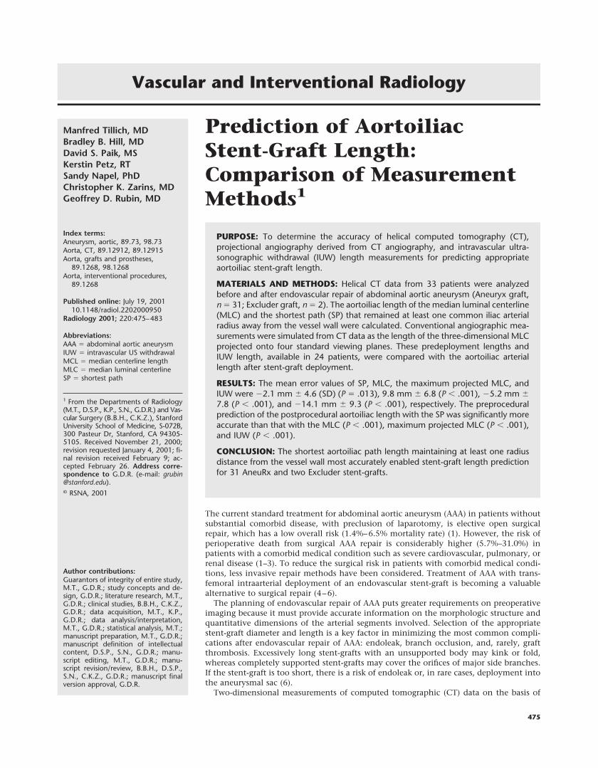
graft radius (Fig 9). These findings areconsistent with the results of an in vitrostudy of AAA (10), in which investigatorsobserved that the distance between themajor side branches and aortic bifurca-tion was larger for external measure-ments than for those obtained with in-travascular US. Our findings furthersupport the conclusion that stent-graftsizing with IUW is inaccurate (17,20).
An important limitation of this studyis that our results cannot be generalizedto all stent-grafts. Although the SP en-abled accurate prediction of the appro-priate stent-graft length for 31 AneuRxand two Excluder stent-grafts, further in-vestigations are required to establish thegeneralizability of these results to otherdevices. It is possible that stent-graftsmight be developed that do not followthe SP. Finally, the clinical utility of SPmeasurement cannot be determined un-til the outcomes of a cohort of patientswith AAAs treated with stent-grafts pro-spectively sized on the basis of prede-ployment SP measurements can be stud-ied.
In conclusion, our study findings dem-onstrated that for the AneuRx and Ex-cluder devices studied, the shortest aor-toiliac path length that maintained atleast one common iliac arterial radius
from the vessel wall enabled predictionof stent-graft length significantly moreaccurately than did MLC, maximum pro-jectional arteriography, and IUW mea-surements. By assuming accurate stent-graft delivery, this potentially allows for atolerance of 5 mm at the proximal anddistal fixation sites.
References1. Ernst CB. Abdominal aortic aneurysm.
N Engl J Med 1993; 328:1167–1172.2. Brown OW, Hollier LH, Pairolero PC, et
al. Abdominal aortic aneurysm and coro-nary artery disease. Arch Surg 1981; 116:1484–1488.
3. Hollier LH, Reigel MM, Kazmier FJ, Pai-rolero PC, Cherry KJ, Hallett JW. Conven-tional repair of abdominal aortic aneu-rysm in the high-risk patient: a plea forabandonment of nonresective treatment.J Vasc Surg 1986; 3:712–717.
4. Parodi JC, Criado FJ, Barone HD, Schon-holz C, Queral LA. Endoluminal aorticaneurysm repair using a balloon-expand-able stent-graft device: a progress report.Ann Vasc Surg 1994; 8:523–529.
5. Blum U, Langer M, Spillner G, et al. Ab-dominal aortic aneurysms: preliminarytechnical and clinical results with trans-femoral placement of endovascular self-expanding stent-grafts. Radiology 1996;198:25–31.
6. Dorffner R, Thurnher S, Polterauer P,Kretschmer G, Lammer J. Treatment ofabdominal aortic aneurysms with trans-femoral placement of stent-grafts: com-
plications and secondary radiologic inter-vention. Radiology 1997; 204:79–86.
7. Moritz JD, Rotermund S, Keating DP,Oestmann JW. Infrarenal abdominal aor-tic aneurysms: implications of CT evalu-ation of size and configuration for place-ment of endovascular aortic grafts.Radiology 1996; 198:463–466.
8. Broeders IA, Blankensteijn JD, Olree M,Mali W, Eikelboom BC. Preoperative siz-ing of grafts for transfemoral endovascu-lar aneurysm management: a prospectivecomparative study of spiral CT angiogra-phy, arteriography, and conventional CTimaging. J Endovasc Surg 1997; 4:252–261.
9. Beebe HG, Jackson T, Pigott JP. Aortic an-eurysm morphology for planning endo-vascular aortic grafts: limitations of con-ventional imaging methods. J EndovascSurg 1995; 2:139–148.
10. van Essen JA, Gussenhoven EJ, van derLugt A, et al. Accurate assessment ofabdominal aortic aneurysm with intra-vascular ultrasonography scanning: vali-dation with computed tomographic an-giography. J Vasc Surg 1999; 29:631–638.
11. White RA, Donayre C, Walot I, et al. Pre-liminary clinical outcome and imagingcriterion for endovascular prosthesis de-velopment in high-risk patients whohave aortoiliac and traumatic arterial le-sions. J Vasc Surg 1996; 24:556–571.
12. Rubin GD, Paik DS, Johnston PC, Napel S.Measurement of the aorta and its brancheswith helical CT. Radiology 1998; 206:823–829.
13. Rubin GD, Silverman SG. Helical CT ofthe retroperitoneum. Radiol Clin NorthAm 1995; 33:903–932.
14. Lorensen WE, Cline HE. Marching cubes:a high-resolution 3D surface constructionsystem. Comput Graph 1987; 21:163–169.
15. Paik DS, Beaulieu CF, Jeffrey RB, RubinGD, Napel S. Automated flight path plan-ning for virtual endoscopy. Med Phys1998; 25:629–637.
16. Balm R, Kaatee R, Blankensteijn JD, MaliWPTM, Eikelboom BC. CT-angiographyof abdominal aortic aneurysms after trans-femoral endovascular aneurysm manage-ment. Eur J Vasc Endovasc Surg 1996; 12:182–188.
17. Gottwik MG, Siebes M, Bahawar H, et al.Quantitative angiographic assessment ofcoronary stenoses: problems and pitfalls.Z Kardiol 1983; 72(suppl 3):111–115.
18. Beebe HG. Imaging modalities for aorticendografting. J Endovasc Surg 1997; 4:111–123.
19. Fox AD, Whitely MS, Murphy P, Budd JS,Horrocks M. Comparison of magnetic res-onance imaging measurements of ab-dominal aortic aneurysms with measure-ments obtained by other imaging techniquesand intraoperative measurements: possi-ble implications for endovascular graft-ing. J Vasc Surg 1996; 24:632–638.
20. Fuessi RT, Mintz GS, Pichard AD, et al. Invivo validation of intravascular ultra-sonography length measurements using amotorized transducer pullback system.Am J Cardiol 1996; 77:1115–1118.
Figure 9. Helical CT images obtained with an intravascular US cath-eter (arrows) within the lumen of an aneurysmal aorta in a 72-year-old man. Left: Transverse reconstruction. Right: 20-mm-thick sagittalmaximum intensity projection image. The catheter was inserted intothe right femoral artery. These views demonstrate why IUW measure-ments tend to result in underestimation of the length of the path thatthe stent-graft will follow, since the catheter tends to lie against thewall of the aortic lumen and take a straighter course through the aortathan would a stent-graft, which has a substantially larger diameterand thus must lie in a position placing its median axis at least oneradius away from the luminal wall.
Volume 220 z Number 2 Prediction of Aortoiliac Stent-Graft Length z 483