Embed Size (px)
Citation preview
Rajeev Vohra et al
28
Management of Open Injuries of the Foot: Current Concepts1Rajeev Vohra, 2Avtar Singh, 3Kanwar K Singh, 4Inderdeep Singh, 5Navjot Singh, 6Abhishek Bansal
ABSTRACT
Open fractures of the foot are rare, and a few surgeons see the whole spectrum of these injuries. When confronted with open injuries of the foot, the decision to salvage the foot needs to be taken after thorough assessment of bony and soft tissue injuries and the associated life-threatening injuries. Foot and ankle surgeon, plastic surgeon, and vascular surgeon must work as a team to provide a pain-free functional and plantigrade foot that fits into a conventional shoe or a brace.
Aggressive debridement and wound management remains the cornerstone of modern surgical treatment. If debridement and wound management can convert a contaminated traumatic wound into a clean surgical wound, which can be adequately covered within 7 to 10 days, then definitive skeletal fixation is advised. In the presence of severe soft tissue and bony injuries, the reconstruction needs to be staged and planned to match the unique personality of the patient and injury.
Amputation can be a positive step toward decreasing the overall morbidity in the presence of severe injury and poor host biology. Despite appropriate treatment, the prognosis of the patient with severe open foot injuries remains guarded.
Keywords: Foot, Fractures, Open.
How to cite this article: Vohra R, Singh A, Singh KK, Singh I, Singh N, Bansal A. Management of Open Injuries of the Foot: Current Concepts. J Foot Ankle Surg (Asia-Pacific) 2016;3(1): 28-40.
Source of support: Nil
Conflict of interest: None
INTROduCTION
Open fractures of the foot are rare and a few surgeons see the whole spectrum of the injuries.1 When confronted with compound injuries of the foot, a correct judgment is required to decide whether to proceed with an immediate amputation or begin the steps needed for foot salvage. Several graded scores, such as Mangled Extremity Sever-ity Score,2 Limb Salvage Index,3 and Predictive Salvage
JFAS (AP)
Review aRticle
1,3Senior Consultant, 2Senior Consultant and Head, 4-6Senior Resident1-6Department of Orthopaedics, Amandeep Hospital, Amritsar Punjab, India
Corresponding Author: Rajeev Vohra, Senior Consultant Department of Orthopaedics, Amandeep Hospital, Amritsar Punjab, India, Phone: +919814536372, e-mail: [email protected]
10.5005/jp-journals-10040-1045
Index3 have attempted to facilitate this decision. Unfor-tunately, the clinical usefulness of these scoring systems in predicting amputation has not been validated, and these scores should be cautiously used by a surgeon to decide the fate of the lower extremity.4 However, certain factors that influence outcome and possible amputation in patients with severe foot and ankle injury (Table 1) need to be considered.5
Patients sustaining open fractures of the foot often have concomitant injuries, which need evaluation using advanced trauma life support principles. About 10 to 17% of patients with a severely traumatized limb have associated life-threatening injuries.6,7 At times in a life-threatening situation, a probably salvageable foot needs to be amputated to save the life.8 However, the decision to salvage the foot needs to be taken with caution. Protracted limb salvage may demonstrate only technical advances, leaving the patient physically, emotionally, and psychologically in ruin.9-11 Numerous studies have demonstrated better functional results in severely open foot injuries treated by amputation, particularly when the
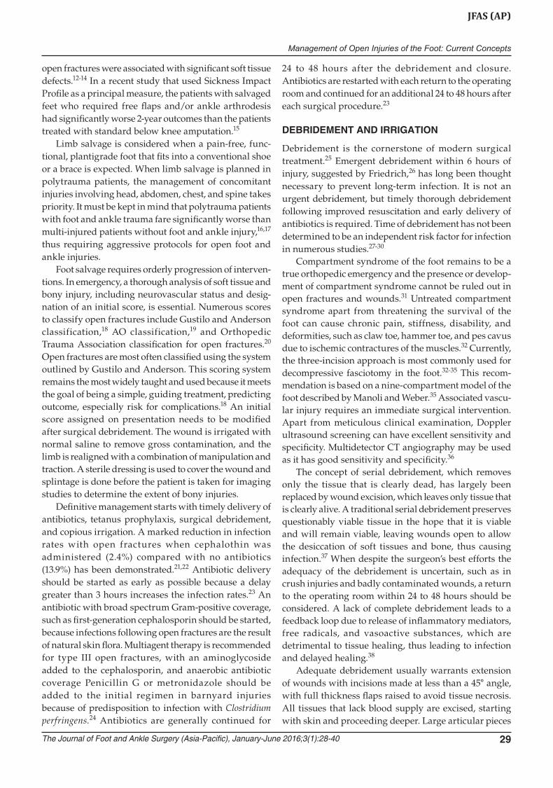
Table 1: Factors that influence outcome and possible amputation in patients with severe foot and ankle injury
Duration and severity of limb ischemiaPatient agePresence of shockEnergy of the injuryDegree of contamination (soil)Nerve disruptionOpen or closed injury (Gustilo open-fracture grading system)Fracture grade, type, level(s)Delay in fracture fixationElevated compartment pressuresLevel and type of arterial injuryDelay of revascularizationInjury severity score/associated injuriesComorbid medical conditions (diabetes mellitus, immune-compromised patients)Transport time, use of pneumatic anti-shock garmentExperience of the receiving hospital (trauma center vs community hospital)Steroid useMalnutritionPremature wound closureDelayed soft-tissue coverageOperating room time greater than 2 hoursMultiple wound exposures outside the operating room
Management of Open Injuries of the Foot: Current Concepts
The Journal of Foot and Ankle Surgery (Asia-Pacific), January-June 2016;3(1):28-40 29
JFAs (AP)
open fractures were associated with significant soft tissue defects.12-14 In a recent study that used Sickness Impact Profile as a principal measure, the patients with salvaged feet who required free flaps and/or ankle arthrodesis had significantly worse 2-year outcomes than the patients treated with standard below knee amputation.15
Limb salvage is considered when a pain-free, func-tional, plantigrade foot that fits into a conventional shoe or a brace is expected. When limb salvage is planned in polytrauma patients, the management of concomitant injuries involving head, abdomen, chest, and spine takes priority. It must be kept in mind that polytrauma patients with foot and ankle trauma fare significantly worse than multi-injured patients without foot and ankle injury,16,17 thus requiring aggressive protocols for open foot and ankle injuries.
Foot salvage requires orderly progression of interven-tions. In emergency, a thorough analysis of soft tissue and bony injury, including neurovascular status and desig-nation of an initial score, is essential. Numerous scores to classify open fractures include Gustilo and Anderson classification,18 AO classification,19 and Orthopedic Trauma Association classification for open fractures.20 Open fractures are most often classified using the system outlined by Gustilo and Anderson. This scoring system remains the most widely taught and used because it meets the goal of being a simple, guiding treatment, predicting outcome, especially risk for complications.18 An initial score assigned on presentation needs to be modified after surgical debridement. The wound is irrigated with normal saline to remove gross contamination, and the limb is realigned with a combination of manipulation and traction. A sterile dressing is used to cover the wound and splintage is done before the patient is taken for imaging studies to determine the extent of bony injuries.
Definitive management starts with timely delivery of antibiotics, tetanus prophylaxis, surgical debridement, and copious irrigation. A marked reduction in infection rates with open fractures when cephalothin was administered (2.4%) compared with no antibiotics (13.9%) has been demonstrated.21,22 Antibiotic delivery should be started as early as possible because a delay greater than 3 hours increases the infection rates.23 An antibiotic with broad spectrum Gram-positive coverage, such as first-generation cephalosporin should be started, because infections following open fractures are the result of natural skin flora. Multiagent therapy is recommended for type III open fractures, with an aminoglycoside added to the cephalosporin, and anaerobic antibiotic coverage Penicillin G or metronidazole should be added to the initial regimen in barnyard injuries because of predisposition to infection with Clostridium perfringens.24 Antibiotics are generally continued for
24 to 48 hours after the debridement and closure. Antibiotics are restarted with each return to the operating room and continued for an additional 24 to 48 hours after each surgical procedure.23
dEBRIdEMENT ANd IRRIGATION
Debridement is the cornerstone of modern surgical treatment.25 Emergent debridement within 6 hours of injury, suggested by Friedrich,26 has long been thought necessary to prevent long-term infection. It is not an urgent debridement, but timely thorough debridement following improved resuscitation and early delivery of antibiotics is required. Time of debridement has not been determined to be an independent risk factor for infection in numerous studies.27-30
Compartment syndrome of the foot remains to be a true orthopedic emergency and the presence or develop-ment of compartment syndrome cannot be ruled out in open fractures and wounds.31 Untreated compartment syndrome apart from threatening the survival of the foot can cause chronic pain, stiffness, disability, and deformities, such as claw toe, hammer toe, and pes cavus due to ischemic contractures of the muscles.32 Currently, the three-incision approach is most commonly used for decompressive fasciotomy in the foot.32-35 This recom-mendation is based on a nine-compartment model of the foot described by Manoli and Weber.35 Associated vascu-lar injury requires an immediate surgical intervention. Apart from meticulous clinical examination, Doppler ultrasound screening can have excellent sensitivity and specificity. Multidetector CT angiography may be used as it has good sensitivity and specificity.36
The concept of serial debridement, which removes only the tissue that is clearly dead, has largely been replaced by wound excision, which leaves only tissue that is clearly alive. A traditional serial debridement preserves questionably viable tissue in the hope that it is viable and will remain viable, leaving wounds open to allow the desiccation of soft tissues and bone, thus causing infection.37 When despite the surgeon’s best efforts the adequacy of the debridement is uncertain, such as in crush injuries and badly contaminated wounds, a return to the operating room within 24 to 48 hours should be considered. A lack of complete debridement leads to a feedback loop due to release of inflammatory mediators, free radicals, and vasoactive substances, which are detrimental to tissue healing, thus leading to infection and delayed healing.38
Adequate debridement usually warrants extension of wounds with incisions made at less than a 45° angle, with full thickness flaps raised to avoid tissue necrosis. All tissues that lack blood supply are excised, starting with skin and proceeding deeper. Large articular pieces
Rajeev Vohra et al
30
necessary for joint stability are usually retained and cleaned with copious amount of saline. Tourniquet may be required to control the bleeding that obscures the surgical field and places vital structures at risk. Furthermore, bleeding from adjacent live tissue may make it appear that devitalized tissue is bleeding and therefore alive. On the contrary, tourniquet can cause tissue hypoxia and hinder the assessment of bleeding from wound margins. After thorough debridement, tourniquet can be released or it can be briefly released and then re-inflated. This staged release of the tourniquet allows viability of all the structures to be examined and wound excision to proceed without torrential bleeding obscuring the surgeon’s view.
After surgical debridement, irrigation is performed to prevent infection and promote healing by cleansing the wound of foreign matter, microscopic pathogens, and toxic substances that may inhibit healing. The recommendations for volume of irrigation solution are not evidence based. Available in vitro and animal studies indicate that increasing the irrigation volume improves removal of foreign material up to a point, after which there is a plateau effect.39 One empirical protocol for irrigation is use of 3 liters for Gustilo type I fractures, 6 liters for Gustilo type II fractures, and 9 liters for Gustilo type III fractures.40 It is important to actively wash all parts of the wound, including cavities and recesses, and not simply flood a particular area with solution.
A wide variety of irrigation solutions, such as water, saline antiseptics, antibiotics, chelating agents, and soaps have been proposed, but normal saline remains to be the most commonly used. Topical antibiotics in the irrigation fluid are toxic to local tissues and do not provide any significant benefit.40 High-pressure pulsatile lavage can cause additional soft tissue trauma, driving the contaminants deeper into the wound, damaging the superficial neurovascular structure, and causing impaired healing.41-43 For these reasons, many surgeons prefer low-pressure pulsatile lavage or irrigation with bulb syringe.
After thorough debridement and irrigation, reassess-ment and re-scoring is done. If it does not seem feasible to provide a functional limb, amputation should be
considered as a positive step towards minimizing overall morbidity in severe injuries and not as a failure of treat-ment. Once the final decision to salvage the foot is taken, it is necessary to stabilize the bony injuries.
SKELETAL STABILIZATION
Skeletal stabilization is done with an aim of obtaining and maintaining anatomic reduction. The individual hardware recommendations for skeletal stabilization depend on the location of the injury (hindfoot, midfoot, or forefoot). In most circumstances, K-wires or a combination of K-wires and external fixator is used for temporary fixation, but in selective cases stabilization with a fixator may serve as a definitive treatment.44-47 At times when an immediate definitive fixation is feasible, fixation devices need to be chosen according to the merits of the situation.
Realignment of bone and joint surfaces decreases abnormal soft tissue motion and irritation and edema, which increase the efficacy of cellular and humoral defenses, thus decreasing the infection rate.48,49 Skeletal stabilization helps in early mobility and rehabilitation of the patient and thereby improve pulmonary status and decrease incidence of venous congestion and thrombosis.50-53 Early joint mobility also improves cartilage nutrition and decreases joint stiffness.54,55
After skeletal stabilization, compound foot injuries require early durable soft tissue coverage to reduce infection and fibrosis.
WOuNd MANAGEMENT
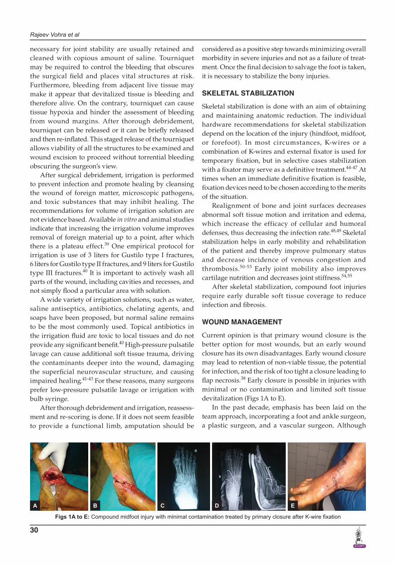
Current opinion is that primary wound closure is the better option for most wounds, but an early wound closure has its own disadvantages. Early wound closure may lead to retention of non-viable tissue, the potential for infection, and the risk of too tight a closure leading to flap necrosis.38 Early closure is possible in injuries with minimal or no contamination and limited soft tissue devitalization (Figs 1A to E).
In the past decade, emphasis has been laid on the team approach, incorporating a foot and ankle surgeon, a plastic surgeon, and a vascular surgeon. Although
Figs 1A to E: Compound midfoot injury with minimal contamination treated by primary closure after K-wire fixation
A B C D E
Management of Open Injuries of the Foot: Current Concepts
The Journal of Foot and Ankle Surgery (Asia-Pacific), January-June 2016;3(1):28-40 31
JFAs (AP)
expeditious wound coverage is associated with lower rates of infection, the timing of coverage is debatable. In his landmark study, Godina56 advocated coverage within 72 hours of injury to achieve the lowest rates of infection. It is essential to do soft tissue coverage as early as possible, preferably within 7 to 10 days.38
Wound dressings are required till the wound is amenable to coverage or secondary closure. Wound dressings should be capable of absorbing exudates, pre-vent bacterial contamination, avoid further trauma to a wound, and promote healing. Modern dressings, such as some form of hydrogel and alginate or thin covers, such as polymer or silicone have a limited role in acute trauma setting. Antimicrobial dressings are sometimes used to control infection. Hemostatic dressings can be applied directly to hemorrhagic wounds to stop the bleeding immediately. Biological dressings composed of allograft skin, xenograft skin, and collagen matrices are being used more and more in acute trauma. These temporary dressings work in concert with the natural healing process until the wound is prepared to accept definitive coverage.57
The advent of negative pressure wound systems has revolutionized our ability to treat soft tissue defects. Currently, these systems are commonly used as a dressing and for promoting healing. Vacuum-assisted closure allows the evacuation of interstitial fluids that accumulate in post-traumatic wounds. These fluids contain inhibitory factors that suppress the formation of fibroblasts, vascular endothelial cells, and keratinocytes, which are crucial for wound healing.58-60 Negative pressure wound therapy reduces the frequency of dressing changes and prevents wound desiccation that occurs so often if the conventional dressings are neglected.61 It enhances the wound contraction by secondary intention that can result in spontaneous healing of small defects or allows use of only an autogenous skin graft.62 In the presence of large wounds, negative pressure wound therapy is a bridge to the definitive soft tissue coverage by flaps.63,64 Vacuum-assisted closure has been shown to be effective at reducing bacterial counts,61 so it can be used as an adjunct therapy in cases of failed primary closure or infected cases after debridement.
Bead pouch technique is another method commonly used to control the infection.65,66 In this technique, antibiotic beads are made by mixing polymethylmethacrylate with an appropriate antibiotic, and the beads are packed into open or dead space and then the wound is either closed or covered with a suitable dressing. The most commonly used antibiotics are tobramycin and vancomycin. The majority of the drug is eluted over the first 24 hours; however, elution may occur in small doses for as long as 90 days.65,67 Levels which are well above the therapeutic
range and have little effect on osteoblast replication can be achieved in wound serum.68-70
SOFT TISSuE RECONSTRuCTION
After wound management, coverage options range from basic to complex and include delayed primary closure, healing by secondary intention, skin grafting, local flap coverage, and distant tissue transfer.71 The concept of a reconstructive ladder originally used for complex orbitofacial defects was taken over by orthopedic surgeons to reconstruct bone and soft tissue defects.72-75 According to this rigid ladder (Table 2), the simplest technique should be explored first before proceeding to the next rung of ladder. The problem with this rigid approach is that a wound amenable to a less complex method may not provide the long-term success that can be achieved by a more complex technique. Currently, the use of the procedure that has the best chance of success is recommended.76
The soft tissue coverage of a particular region of the foot needs to be done keeping in view the specific requirements. Hidalgo and Shaw77 and other investiga-tors78 divided the foot into discrete zones according to the requirements of each region. It has been emphasized that any flap selected must meet the functional and esthetic demands of the given zone, with bulk or contour not impeding the use of shoe wear and proper ambulation.79
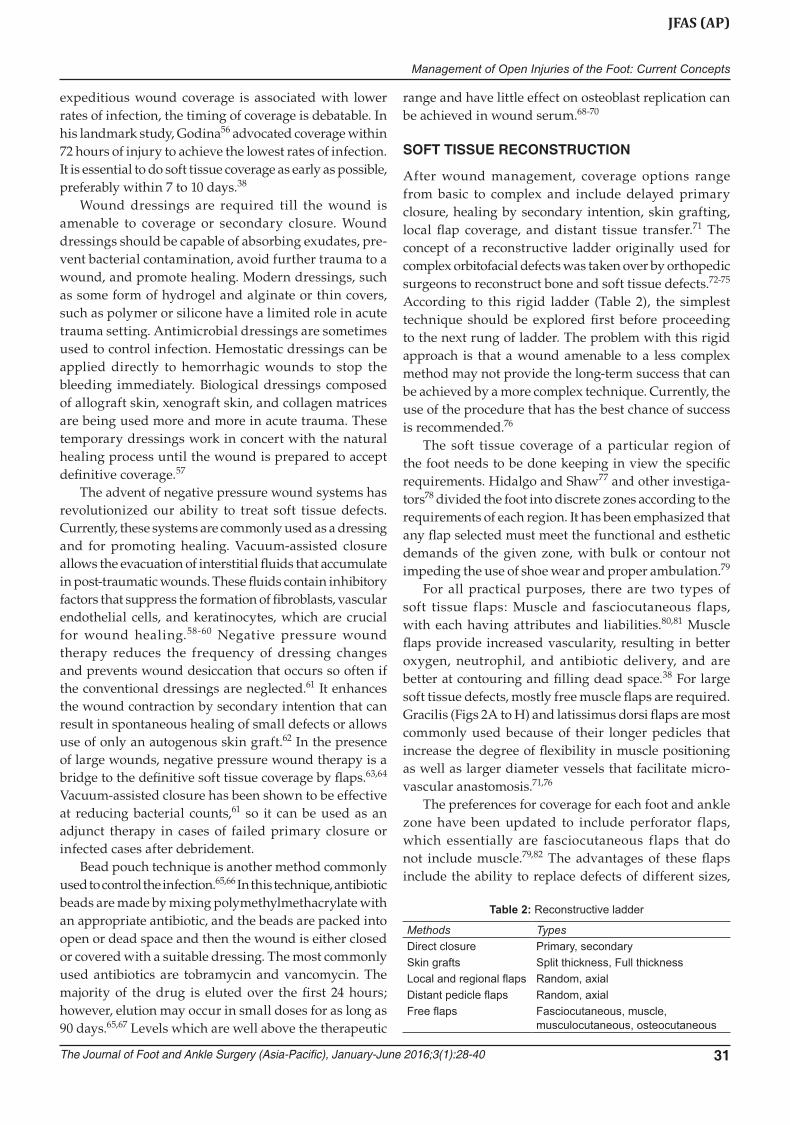
For all practical purposes, there are two types of soft tissue flaps: Muscle and fasciocutaneous flaps, with each having attributes and liabilities.80,81 Muscle flaps provide increased vascularity, resulting in better oxygen, neutrophil, and antibiotic delivery, and are better at contouring and filling dead space.38 For large soft tissue defects, mostly free muscle flaps are required. Gracilis (Figs 2A to H) and latissimus dorsi flaps are most commonly used because of their longer pedicles that increase the degree of flexibility in muscle positioning as well as larger diameter vessels that facilitate micro-vascular anastomosis.71,76
The preferences for coverage for each foot and ankle zone have been updated to include perforator flaps, which essentially are fasciocutaneous flaps that do not include muscle.79,82 The advantages of these flaps include the ability to replace defects of different sizes,
Table 2: Reconstructive ladder
Methods TypesDirect closure Primary, secondarySkin grafts Split thickness, Full thicknessLocal and regional flaps Random, axialDistant pedicle flaps Random, axialFree flaps Fasciocutaneous, muscle,
musculocutaneous, osteocutaneous
Rajeev Vohra et al
32
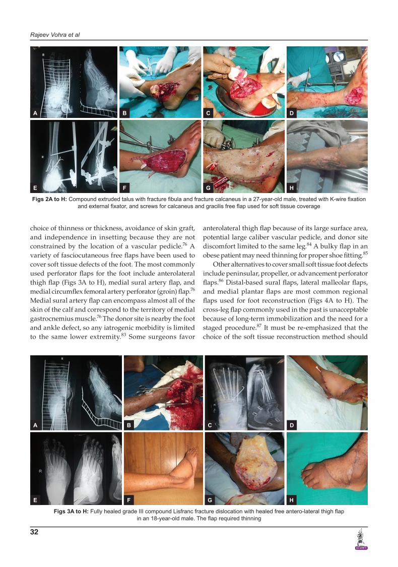
choice of thinness or thickness, avoidance of skin graft, and independence in insetting because they are not constrained by the location of a vascular pedicle.76 A variety of fasciocutaneous free flaps have been used to cover soft tissue defects of the foot. The most commonly used perforator flaps for the foot include anterolateral thigh flap (Figs 3A to H), medial sural artery flap, and medial circumflex femoral artery perforator (groin) flap.76 Medial sural artery flap can encompass almost all of the skin of the calf and correspond to the territory of medial gastrocnemius muscle.76 The donor site is nearby the foot and ankle defect, so any iatrogenic morbidity is limited to the same lower extremity.83 Some surgeons favor
anterolateral thigh flap because of its large surface area, potential large caliber vascular pedicle, and donor site discomfort limited to the same leg.84 A bulky flap in an obese patient may need thinning for proper shoe fitting.85
Other alternatives to cover small soft tissue foot defects include peninsular, propeller, or advancement perforator flaps.86 Distal-based sural flaps, lateral malleolar flaps, and medial plantar flaps are most common regional flaps used for foot reconstruction (Figs 4A to H). The cross-leg flap commonly used in the past is unacceptable because of long-term immobilization and the need for a staged procedure.87 It must be re-emphasized that the choice of the soft tissue reconstruction method should
Figs 2A to H: Compound extruded talus with fracture fibula and fracture calcaneus in a 27-year-old male, treated with K-wire fixation and external fixator, and screws for calcaneus and gracilis free flap used for soft tissue coverage
A
E
B
F
C
G
D
H
A
E
B
F
C
G
D
H
Figs 3A to H: Fully healed grade III compound Lisfranc fracture dislocation with healed free antero-lateral thigh flap in an 18-year-old male. The flap required thinning
Management of Open Injuries of the Foot: Current Concepts
The Journal of Foot and Ankle Surgery (Asia-Pacific), January-June 2016;3(1):28-40 33
JFAs (AP)
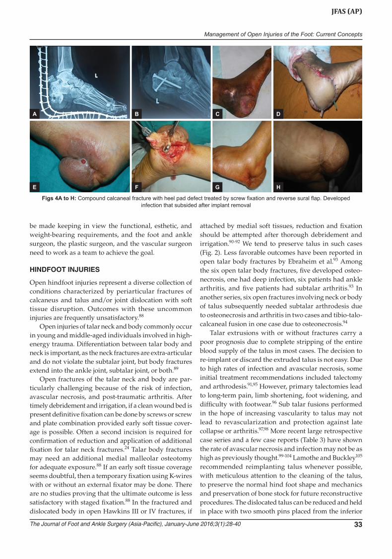
Figs 4A to H: Compound calcaneal fracture with heel pad defect treated by screw fixation and reverse sural flap. Developed infection that subsided after implant removal
A
E
B
F
C
G
D
H
be made keeping in view the functional, esthetic, and weight-bearing requirements, and the foot and ankle surgeon, the plastic surgeon, and the vascular surgeon need to work as a team to achieve the goal.
HINdFOOT INJuRIES
Open hindfoot injuries represent a diverse collection of conditions characterized by periarticular fractures of calcaneus and talus and/or joint dislocation with soft tissue disruption. Outcomes with these uncommon injuries are frequently unsatisfactory.88
Open injuries of talar neck and body commonly occur in young and middle-aged individuals involved in high-energy trauma. Differentiation between talar body and neck is important, as the neck fractures are extra-articular and do not violate the subtalar joint, but body fractures extend into the ankle joint, subtalar joint, or both.89
Open fractures of the talar neck and body are par-ticularly challenging because of the risk of infection, avascular necrosis, and post-traumatic arthritis. After timely debridement and irrigation, if a clean wound bed is present definitive fixation can be done by screws or screw and plate combination provided early soft tissue cover-age is possible. Often a second incision is required for confirmation of reduction and application of additional fixation for talar neck fractures.24 Talar body fractures may need an additional medial malleolar osteotomy for adequate exposure.88 If an early soft tissue coverage seems doubtful, then a temporary fixation using K-wires with or without an external fixator may be done. There are no studies proving that the ultimate outcome is less satisfactory with staged fixation.88 In the fractured and dislocated body in open Hawkins III or IV fractures, if
attached by medial soft tissues, reduction and fixation should be attempted after thorough debridement and irrigation.90-92 We tend to preserve talus in such cases (Fig. 2). Less favorable outcomes have been reported in open talar body fractures by Ebraheim et al.93 Among the six open talar body fractures, five developed osteo-necrosis, one had deep infection, six patients had ankle arthritis, and five patients had subtalar arthritis.93 In another series, six open fractures involving neck or body of talus subsequently needed subtalar arthrodesis due to osteonecrosis and arthritis in two cases and tibio-talo-calcaneal fusion in one case due to osteonecrosis.94
Talar extrusions with or without fractures carry a poor prognosis due to complete stripping of the entire blood supply of the talus in most cases. The decision to re-implant or discard the extruded talus is not easy. Due to high rates of infection and avascular necrosis, some initial treatment recommendations included talectomy and arthrodesis.91,95 However, primary talectomies lead to long-term pain, limb shortening, foot widening, and difficulty with footwear.96 Sub talar fusions performed in the hope of increasing vascularity to talus may not lead to revascularization and protection against late collapse or arthritis.97,98 More recent large retrospective case series and a few case reports (Table 3) have shown the rate of avascular necrosis and infection may not be as high as previously thought.99-104 Lamothe and Buckley105 recommended reimplanting talus whenever possible, with meticulous attention to the cleaning of the talus, to preserve the normal hind foot shape and mechanics and preservation of bone stock for future reconstructive procedures. The dislocated talus can be reduced and held in place with two smooth pins placed from the inferior
Rajeev Vohra et al
34
aspect of the calcaneus, through the talus and into the inferior aspect of the tibia, and an external fixation may be added (Fig. 2). We recommend reimplantation of talus after thorough cleaning with pulsatile lavage and soaking in antibiotic solution.
The dislocation between calcaneus and talus involves both subtalar and talocalcaneal joint and is referred to as subtalar or peritalar dislocation. Open injuries make up approximately 20 to 45% of all peritalar dislocations.106-109 Medial dislocations predominate among closed injuries, but in open injuries lateral dislocations are more common.106-111 Currently, there is no consensus regarding the treatment of open subtalar dislocations. However, like any other open fracture, irrigation and debridement of the joint and soft tissues is performed urgently.
The majority of reductions performed for open subtalar dislocations are unstable because of severe soft tissue injury or associated intra-articular fractures. Stability can be achieved by smooth pin fixation across the dislocated joint or through fixation of articular fractures. An external fixator has also been advocated to maintain reduction in unstable joints.112,113 Although most open medial dislocations are amenable to delayed primary closure or skin grafting, lateral dislocations require a myocutaneous free flap in 30% cases.110 Outcomes following open peritalar injuries are often unsatisfactory due to associated fractures osteochondral injuries, nerve involvement, and tendon injuries.106,114
Open fractures of the calcaneus comprise 0.8 to 10% of all calcaneus fractures.115 They are generally associated with high complications, such as impaired wound healing, deep infection, and osteomyelitis. Folk et al reported wound complications in 72% of open fractures treated operatively.116 In a retrospective study of 36 open calcaneal fractures, Siebert et al117 reported more than a 60% complication rate and found that the complication
rate was 100% when immediate internal fixation was attempted. Heier et al118 reported an overall 37% infection rate and a 19% deep infection rate in a series of 43 open calcaneal fractures, out of which 25% were treated with internal fixation and 25% with primary arthrodesis.
Recently, better outcomes have been reported in various series (Table 4).119-124 In one study, unsatisfactory outcomes were reported in the presence of plantar wound and severe comminution.120 In another study, factors predictive of a less satisfactory outcome included wound > 5 cm in length, presence of a neurovascular injury, the need for free tissue transfer, and the presence of heel pad avulsion.125 If the avulsed heel pads do not survive, then flap coverage may be required (Fig. 3). Based on a limited number of studies, with conflicting results in a small number of patients, there is insufficient evidence (grade I recommendation) to support one form of treatment over the other in the management of open calcaneal fractures.126 In open calcaneal fractures, the severity and location of the soft tissue damage and extent of the comminution and articular damage dictates the treatment.
In low-velocity open fractures with a medial skin split, the so-called “susper lesions” are caused by landing on an everted and externally rotated heel and can be treated by aggressive debridement and early internal fixation through a lateral approach.127 Thornton et al treated 31 open intra-articular fractures in 29 patients with standard open reduction and internal fixation techniques when the medial wound was < 4 cm and could be closed and remained stable, staying off antibiotics. Percutaneous wire fixation was recommended for wound > 4 cm or unstable wounds.121 Mehta et al showed good results in 14 patients with grade II or IIIa open calcaneal fractures treated with debridement and temporary percutaneous fixation within 8 hours of presentation and plating through a lateral approach after an average of 18 days.122
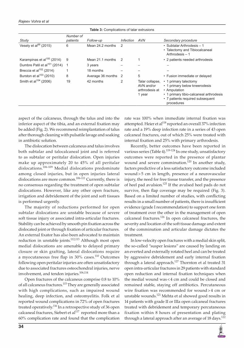
Table 3: Complications of talar extrusions
StudyNumber of patients Follow-up Infection AVN Secondary procedure
Vesely et al99 (2015) 6 Mean 24.2 months 2 • Subtalar Arthrodesis – 1• Talectomy and Tibiocalcaneal
Arthrodesis – 1Karampinas et al100 (2014) 9 Mean 21.1 months 2 1 • 2 patients needed arthrodesisDumbre Patil et al101 (2014) 1 3 years – – –Breccia et al102 (2014) 1 18 months – – –Burston et al103 (2010) 8 Average 36 months 2 5 • Fusion immediate or delayedSmith et al104 (2006) 19 42 months 2 Talar collapse,
AVN and/or arthrodesis at 1 year
• 1 primary talectomy• 1 primary below kneerodesis• Amputation• 1 primary tibio-calcaneal arthrodesis• 7 patients required subsequent
procedures
Management of Open Injuries of the Foot: Current Concepts
The Journal of Foot and Ankle Surgery (Asia-Pacific), January-June 2016;3(1):28-40 35
JFAs (AP)
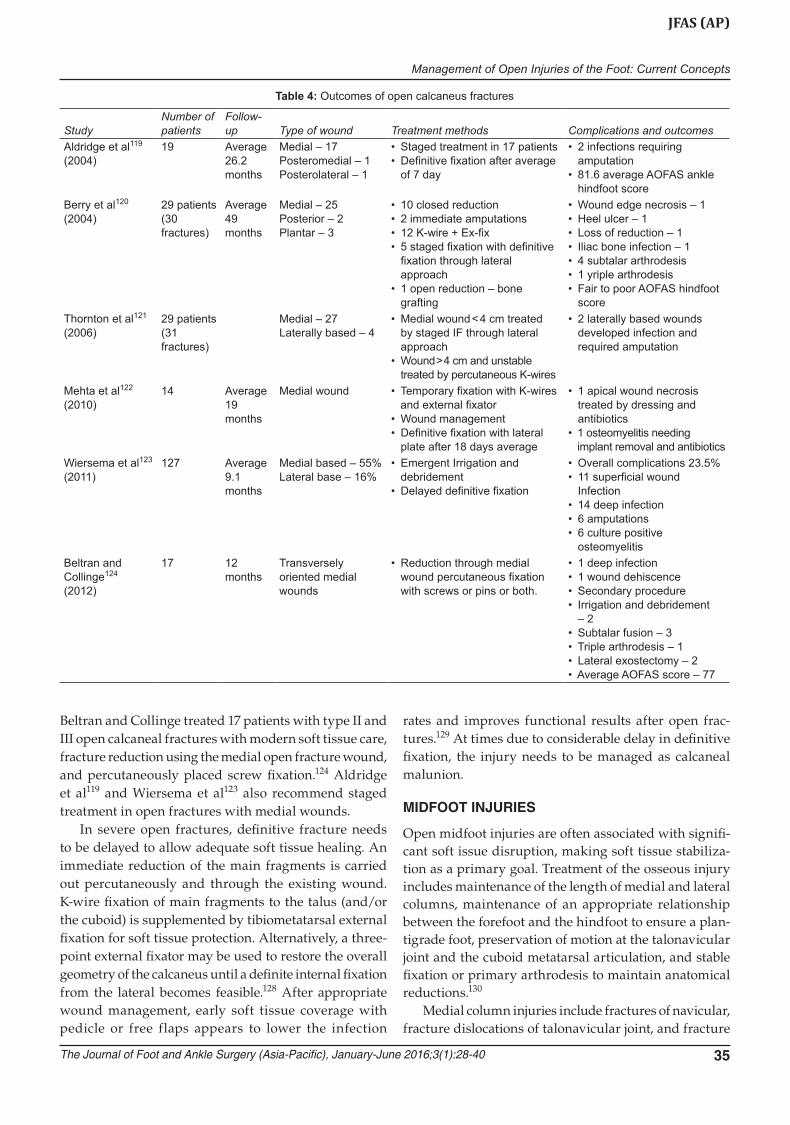
Beltran and Collinge treated 17 patients with type II and III open calcaneal fractures with modern soft tissue care, fracture reduction using the medial open fracture wound, and percutaneously placed screw fixation.124 Aldridge et al119 and Wiersema et al123 also recommend staged treatment in open fractures with medial wounds.
In severe open fractures, definitive fracture needs to be delayed to allow adequate soft tissue healing. An immediate reduction of the main fragments is carried out percutaneously and through the existing wound. K-wire fixation of main fragments to the talus (and/or the cuboid) is supplemented by tibiometatarsal external fixation for soft tissue protection. Alternatively, a three-point external fixator may be used to restore the overall geometry of the calcaneus until a definite internal fixation from the lateral becomes feasible.128 After appropriate wound management, early soft tissue coverage with pedicle or free flaps appears to lower the infection
rates and improves functional results after open frac- tures.129 At times due to considerable delay in definitive fixation, the injury needs to be managed as calcaneal malunion.
MIdFOOT INJuRIES
Open midfoot injuries are often associated with signifi-cant soft issue disruption, making soft tissue stabiliza-tion as a primary goal. Treatment of the osseous injury includes maintenance of the length of medial and lateral columns, maintenance of an appropriate relationship between the forefoot and the hindfoot to ensure a plan-tigrade foot, preservation of motion at the talonavicular joint and the cuboid metatarsal articulation, and stable fixation or primary arthrodesis to maintain anatomical reductions.130
Medial column injuries include fractures of navicular, fracture dislocations of talonavicular joint, and fracture
Table 4: Outcomes of open calcaneus fractures
StudyNumber of patients
Follow-up Type of wound Treatment methods Complications and outcomes
Aldridge et al119 (2004)
19 Average 26.2 months
Medial – 17Posteromedial – 1Posterolateral – 1
• Staged treatment in 17 patients• Definitive fixation after average
of 7 day
• 2 infections requiring amputation
• 81.6 average AOFAS ankle hindfoot score
Berry et al120 (2004)
29 patients (30 fractures)
Average 49 months
Medial – 25Posterior – 2Plantar – 3
• 10 closed reduction• 2 immediate amputations• 12 K-wire + Ex-fix• 5 staged fixation with definitive
fixation through lateral approach
• 1 open reduction – bone grafting
• Wound edge necrosis – 1• Heel ulcer – 1• Loss of reduction – 1• Iliac bone infection – 1• 4 subtalar arthrodesis• 1 yriple arthrodesis• Fair to poor AOFAS hindfoot
scoreThornton et al121 (2006)
29 patients (31 fractures)
Medial – 27Laterally based – 4
• Medial wound < 4 cm treated by staged IF through lateral approach
• Wound > 4 cm and unstable treated by percutaneous K-wires
• 2 laterally based wounds developed infection and required amputation
Mehta et al122 (2010)
14 Average 19 months
Medial wound • Temporary fixation with K-wires and external fixator
• Wound management• Definitive fixation with lateral
plate after 18 days average
• 1 apical wound necrosis treated by dressing and antibiotics
• 1 osteomyelitis needing implant removal and antibiotics
Wiersema et al123 (2011)
127 Average 9.1 months
Medial based – 55%Lateral base – 16%
• Emergent Irrigation and debridement
• Delayed definitive fixation
• Overall complications 23.5%• 11 superficial wound
Infection• 14 deep infection• 6 amputations• 6 culture positive
osteomyelitisBeltran and Collinge124 (2012)
17 12 months
Transversely oriented medial wounds
• Reduction through medial wound percutaneous fixation with screws or pins or both.
• 1 deep infection• 1 wound dehiscence• Secondary procedure• Irrigation and debridement
– 2• Subtalar fusion – 3• Triple arthrodesis – 1• Lateral exostectomy – 2• Average AOFAS score – 77
Rajeev Vohra et al
36
dislocations of naviculocuneiform joint. Trauma to the talonavicular joint can result in deleterious changes in pedal motion because talonavicular joint accounts for most hindfoot motion.131 Every attempt at salvaging this joint should be undertaken before primary arthrodesis is considered.
Comminution of navicular leads to loss of mechanical advantage of the posterior tibial tendon and frank collapse of the medial column.132 The mainstay of acute navicular crushing injuries is reduction with bridging external fixation to keep the midfoot out of length. If the soft tissue allows, acute treatment entails early open reduction and internal fixation with screws, K-wires, and bridge plating if necessary from the talus to the first metatarsal, which can be removed once consolidation has occurred.133
Open injuries of cuboid commonly result from direct crush injury or forced abduction (nutcracker injury). The crushing of cuboid invariably leads to shortening of the lateral column, which causes painful flatfoot deformity.134 The lateral column can be kept out of length by open reduction or internal fixation and bridge plating if the soft tissue conditions permit. In the presence of gross instability and severe soft tissue injury, early stabiliza-tion with an external fixator allows the soft tissues to settle before definitive fixation. Whatever the method chosen, it is essential to preserve the gliding motion of peroneus longus in its groove on the plantar surface of the cuboid.134,135
Most cuboid fractures are often associated with other injuries of the foot, and in a series 76% of the cuboid fractures were associated with Lisfranc or Chopart injuries.136 One study showed that nutcracker cuboid fractures cannot occur in isolation and are stressed due to attention to the associated foot injuries.137 In massive crush injuries, the length of both medial and lateral columns needs to be maintained. The presence of global comminution may warrant primary fusions.
The intercuneiform joints and the naviculocuneiform joints have little or no essential movements in the normal foot and can be primarily arthrodesed with minimal functional loss,130 but Chopart joint should be fused as a last resort.
FOREFOOT INJuRIES
Open Lisfranc injuries typically present with significant displacement and instability. Apart from non-anatomic alignment, other factors, such as energy of the injury, cartilage damage, and soft tissue injuries can compromise the final outcome.138 Stable and satisfactory interim provisional reduction is essential, to prevent further soft tissue damage, and can be achieved by K-wires with or
without external fixator. Malunited foot fractures are difficult or impossible to reduce anatomically after soft tissue swelling has resolved and too much time has lapsed.139 After adequate soft tissue coverage, definitive fixation with screws and/or plates may be required, and at times K-wires may serve as definite fixation (Fig. 4). In a study, 77% spontaneous fusion rate was reported using open fracture protocols, with multiple debridements and multiple K-wire fixation.140
Primary fixation may be done provided early soft tissue coverage can be achieved after adequate debride-ment.141 A consensus has largely been reached that the fusion of the lateral column should be avoided.142-144 However, the choice between internal fixation and primary arthrodesis for medial and middle columns is still controversial. A systematic review of 6 studies with 193 patients comparing primary arthrodesis and open reduction and internal fixation found satisfactory and equivalent results in both groups. Mean American Orthopedic Foot and Ankle Society scores of 72.5 and 88.0 were reported for open reduction and internal fixa-tion and primary arthrodesis groups respectively.145 The injuries with significant cartilage blowout often require primary arthrodesis.
Open fractures of the metatarsals and phalanges and dislocations of the metarsophalangeal joints and inter-phalangeal can be stabilized with K-wires, placed either longitudinally or in crossed configuration.44,45 Dorsal or plantar displacement of the metatarsal heads needs to be avoided, to prevent abnormal weight bearing on the foot, which may lead to development of callosities. Crushing injury often leads to a significant loss of motion of the small joints of the toes. Additional dissection contributes to more loss of motion as well as the potential for additional wound problems. A review of 23 open metatarsal fractures in 10 patients found that injuries with minimal soft tissue damage had improved outcomes compared with those with Gustilo-type IIIb injuries.146
CONCLuSION
Prompt decision needs to be taken, whether to proceed with an immediate amputation or begin the steps for foot salvage. When foot salvage is planned, soft tissue management is of paramount importance in the outcome of the patients with foot and ankle trauma. Expeditious wound coverage and early restoration of skeletal anatomy can dramatically decrease complication rates and improve ultimate outcomes.
ACKNOWLEdGMENT
Authors would like to thank Mr. Sanjiv Kumar for providing writing assistance in this manuscript.
Management of Open Injuries of the Foot: Current Concepts
The Journal of Foot and Ankle Surgery (Asia-Pacific), January-June 2016;3(1):28-40 37
JFAs (AP)
REFERENCES
1. Court-Brown C, Honeyman C, Bugler K, McQueen M. The spectrum of open fractures of the foot in adults. Foot Ankle Int 2013 Mar;34(3):323-328.
2. Helfet DL, Howey T, Sanders R, Johansen K. Limb salvage ver-sus amputation: preliminary results of the mangled extrem-ity severity score. Clin Orthop Relat Res 1990 Jul;(256):80-86.
3. Bonanni F, Rhodes M, Lucke JF. The futility of predictive scoring of mangled lower extremities. J Trauma 1993 Jan;34(1): 99-104.
4. Bosse MJ, MacKenzie EJ, Kellam JF, Burgess AR, Webb LX, Swiontkowski MF, Sanders RW, Jones AL, McAndrew MP, Patterson BM, et al. A prospective evaluation of the clinical utility of the lower extremity injury severity scores. J Bone Joint Surg Am 2001 Jan;83-A(1):3-14.
5. Baumhauer JF, Manoli A II. Principles of management of severely traumatized foot and ankle. Instr Course Lect 2002;51: 159-167.
6. Edwards CC, Simmons SC, Browner BD, Weigel MC. Severe open tibial fractures: results treating 202 injuries with external fixation. Clin Orthop Relat Res 1988 May;(230): 98-115.
7. Lange RH, Bach AW, Hansen ST Jr, Johansen KH. Open tibial fractures with associated vascular injuries: prognosis for limb salvage. J Trauma 1985 Mar;25(3):203-208.
8. Wolinsky PR, Webb LX, Harvey EJ, Tejwani NC. The mangled limb: Salvage versus amputation. Inst Course Lect 2011;60: 27-34.
9. Bondurant FJ, Cotler HB, Buckle R, Miller-Crotchett P, Browner BD. The medical and economic impact of severely injured lower extremities. J Trauma 1988 Aug;28(8):1270-1273.
10. Hansen ST Jr. Overview of the severely traumatized lower limb: reconstruction versus amputation. Clin Orthop Relat Res 1989 Jun;(243):17-19.
11. Lange RH. Limb reconstruction versus amputation decision making in massive lower extremity trauma. Clin Orthop Relat Res 1989;(243):92-99.
12. McGuigan FX, Forsberg JA, Andersen RC. Foot and ankle reconstruction after blast injuries. Foot Ankle Clin 2006 Mar;11(1):165-182.
13. Hansen ST Jr. Salvage or amputation after complex foot and ankle trauma. Orthop Clin North Am 2001 Jan;32(1):181-186.
14. Ferreira RC, Sakata MA, Costa MT, Frizzo GG, Santin RA. Long-term results of salvage surgery in severely injured feet. Foot Ankle Int 2010 Feb;31(2):113-123.
15. Ellington JK, Bosse MJ, Castillo RC, MacKenzie EJ. The mangled foot and ankle: results from a 2-year prospective study. J Orthop Trauma 2013 Jan;27(1):43-48.
16. Turchin DC, Schemitsch EH, McKee MD, Waddell JP. Do foot injuries significantly affect the functional outcome of multiple injured patients? J Orthop Trauma 1999 Jan;13(1):1-4.
17. Tran T, Thordarson D. Functional outcome of multiple inured patients with associated foot injury. Foot Ankle Int 2002 Apr;23(4):340-343.
18. Gustilo RB, Anderson JT. Prevention of infection in the treatment of one thousand and twenty-five open fractures of long bones: retrospective and prospective analysis. J Bone Joint Surg Am 1976 Jun;58(4):453-458.
19. Volgas DA. Classification systems. In: Volgas DA, Harder Y, editors. Manual of soft tissue management in orthopaedic trauma. New York (NY): Thieme; 2011. p. 62-77.
20. Orthopaedic Trauma Association: Open Fracture Study Group. A new classification scheme for open fractures. J Orthop Trauma 2010 Aug;24(8):457-465.
21. Patzakis MJ, Harvey JP Jr, Ivler D. The role of antibiotics in the management of open fractures. J Bone Joint Surg Am 1974 Apr;56(3):532-541.
22. Ostermann PA, Seligson D, Henry SL. Local antibiotic therapy for severe open fractures: a review of 1085 consecutive cases. J Bone Joint Surg Br 1995 Jan;77(1):93-97.
23. Patzakis MJ, Wilkins J. Factors influencing infection rate in open fracture wounds. Clin Orthop Relat Res 1989 Jun;(243):36-40.
24. Sexton SE. Open fractures of the foot and ankle. J Clin Podiatr Med Surg 2014 Oct;31(4):461-486.
25. Teasdall RD. Principles of debridement. In: Volgas DA, Harder Y, editors. Manual of soft tissue management in orthopaedic trauma. New York (NY): Thieme; 2011. p. 87-91.
26. Friedrich PL. Die Aseptische Versorgung frischer Wunden. Arch Klin Chir 1898;57:288-310.
27. Khatod M, Botte MJ, Hoyt DB, Meyer RS, Smith JM, Akeson WH. Outcomes in open tibia fractures: relationship between delay in treatment and infection. J Trauma 2003 Nov;55(5):949-954.
28. Spencer J, Smith A, Woods D. The effect of time delay on infection in open long bone fractures: a 5-year prospective audit from a district general hospital. Ann R Coll Surg Engl 2004 Mar;86(2):108-112.
29. Charalambous CP, Siddique I, Zenios M, Roberts S, Samarji R, Paul A, Hirst P. Early versus delayed surgical treatment of open tibial fractures: effects on rates of infection and need of secondary surgical procedures to promote bone union. Injury 2005 May;36(5):656-661.
30. Noumi T, Yokoyama K, Ohtsuka H, Nakamura K, Itoman M. Intramedullary nailing for open fractures of the femoral shaft: evaluation of contributing factors on deep infection and nonunion using multivariate analysis. Injury 2005 Sep;36(9): 1085-1093.
31. Myerson M. Diagnosis and treatment of compartment syndrome of the foot. Orthopedics 1990 Jul;13(7):711-717.
32. Dodd A, Le I. Foot compartment syndrome: diagnosis and management. J Am Acad Orthop Surg 2013 Nov;21(11):657-664.
33. Fulkerson E, Razi A, Tejwani N. Review: acute compartment syndrome of the foot. Foot Ankle Int 2003 Feb;24(2):180-187.
34. Middleton S, Clasper J. Compartment syndrome of the foot- implications for military surgeons. J R Army Med Corps 2010 Dec;156(4):241-244.
35. Manoli A II, Weber TG. Fasciotomy of the foot: An anatomical study with special reference to release of the calcaneal compartment. Foot Ankle 1990 Apr;10(5):267-275.
36. Crist BD, Ferguson T, Murtha YM, Lee MA. Surgical timing of treating injured extremities: An evolving concept of urgency. Instr Course Lect 2013;62:17-28.
37. Gupta A, Shatford RA, Wolff TW, Tsai TM, Schecker LR, Levin LS. Treatment of the severely injured upper extremity. Instr Course Lect 2000;49:377-396.
38. Tejwani NC, Webb LX, Harvey EJ, Wolinsky PR. Soft tissue management after trauma: Initial management and wound coverage. Instr Course Lect 2011;60:15-25.
39. Svoboda SJ, Bice TG, Gooden HA, Brooks DE, Thomas DB, Wenke JC. Comparison of bulb syringe and pulse lavage irrigation with use of bioluminescent musculoskeletal wound model. J Bone Joint Surg Am 2006 Oct;88(10):2167-2174.
Rajeev Vohra et al
38
40. Anglen JO. Wound irrigation in musculoskeletal injury. J Am Acad Orthop Surg 2001 Jul-Aug;9(4):219-226.
41. Dirschl DR, Duff GP, Dahners LE, Edin M, Rahn BA, Miclau T. High pulsatile lavage irrigation of intra-articular fractures: effects on fracture healing. J Orthop Trauma 1998 Sep-Oct;12(7):460-463.
42. Bhandari M, Adili A, Lachowski RJ. High pressure pulsatile lavage of contaminated human tibiae: an in vitro study. J Orthop Trauma 1998 Sep-Oct;12(7):479-484.
43. Bhandari M, Schemitsch EH, Adili A, Lachowski R, Shaughnessy SG. High and low pressure pulsatile lavage of contaminated tibia fractures: an in vitro study of bacterial adherence and bone damage. J Orthop Trauma 1999 Nov;13(8):526-533.
44. DeLee JC. Fractures and dislocations of the foot. In: Mann RA, Coughlin MJ, editors. Surgery of the foot and ankle. 6th ed. Vol. 2. St Louis (MO): Mosby-Year Book; 1993. p. 1465-1703.
45. Shields NN, Valdez RR, Brennan MJ, Johnson EE, Gould JS. Metatarsal fractures and dislocations and Lisfranc’s fracture-dislocations. In: Gould JS, editor. Operative foot surgery. Philadelphia (PA): WB Saunders; 1994. p. 399-420.
46. Besch L, Radke B, Mueller M, Daniels-Wredenhagen M, Varoga D, Hilgert RE, Mathiak G, Oehlert K, Seekamp A. Dynamic and functional gait analysis of severely displaced intra-articular calcaneus fractures treated with a hinged external fixator or internal stabilization J Foot Ankle Surg 2008 Jan-Feb;47(1):19-25.
47. Chandran P, Puttaswamaiah R, Dhillon MS, Gill SS. Manage-ment of complex open fracture injuries of the midfoot with external fixation. J Foot Ankle Surg 2006 Sep-Oct;45(5): 308-315.
48. Rittmann WW, Schibli M, Matter P, Allgower M. Open fractures: long term results in 200 consecutive cases. Clin Orthop Relat Res 1979 Jan-Feb;(138):132-140.
49. Wray JB. Factors in the pathogenesis of non-union. J Bone Joint Surg Am 1965 Jan;47:168-173.
50. Gustilo RB, Merkow RL, Templeman D. The management of open fractures. J Bone Joint Surg Am 1990 Feb;72(2):299-304.
51. Chapman MW. Role of bone stability in open fractures. Instr Course Lect 1982;31:75-87.
52. Gustilo RB. Management of open fractures and complications. Instr Course Lect 1982;31:64-75.
53. Allgower M, Border JR. Management of open fractures in the multiple trauma patient. World J Surg 1983 Jan;7(1):88-95.
54. Mitchell N, Shepard N. Healing of articular cartilage in intra-articular fractures in rabbits. J Bone Joint Surg Am 1980;62(4): 628-634.
55. Salter RB, Simmonds DF, Malcolm BW, Rumble EJ, MacMichael D, Clements ND. The biological effect of continuous passive motion on the healing of full thickness defects in articular cartilage: an experimental investigation in the rabbit. J Bone Joint Surg Am 1980 Dec;62(8):1232-1251.
56. Godina M. Early microsurgical reconstruction of complex trauma of the extremities. Plast Reconstr Surg 1986 Sep;78(3): 285-292.
57. Volgas DA. Dressing. In: Volgas DA, Harder Y, editors. Manual of soft tissue management in orthopaedic trauma. New York (NY): Thieme; 2011. p. 109-113.
58. Bucalo B, Eaglstein WH, Falanga V. Inhibition of cell proliferation by chronic wound fluid. Wound Repair Regen 1993 Jul;1(3):181-186.
59. Falanga V. Growth factors and chronic wounds: The need to understand the microenvironment. J Dermatol 1992 Nov;19(11): 667-672.
60. Grinnell F, Ho CH, Wysocki A. Degradation of fibronectin and vitronectin in chronic wound fluid: analysis by cell blotting, immunoblotting, and cell adhesion assays. J Invest Dermatol 1992 Apr;98(4):410-416.
61. Hou Z, Irgit K, Strohecker KA, Matzko ME, Wingert NC, DeSantis JG, Smith WR. Delayed flap reconstruction vacuum-assisted closure management of the open IIIB tibial fracture. J Trauma 2011 Dec;71(6):1705-1708.
62. Ullman Y, Fodor L, Ramon Y, Soudry M, Lerner A. The revised reconstructive ladder and its applications for high energy injuries to the extremities. Ann Plast Surg 2006 Apr;56(4):401-405.
63. Medina ND, Kovach SJ III, Levin LS. An evidence-based approach to lower extremity acute trauma. Plast Reconst Surg 2011 Feb;127(2):926-931.
64. Hallock GG. Evidence-based medicine: Lower extremity trauma. Plast Reconstr Surg 2013 Dec;132(6):1733-1741.
65. DeCoster TA, Bozorgnia S. Antibiotic beads. J Am Acad Orthop Surg 2008 Nov;16(11):674-678.
66. Kent ME, Rapp RP, Smith KM. Antibiotic beads and osteo-myelitis: here today, What’s coming tomorrow? Orthopedics 2006 Jul;29(7):599-603.
67. Anglen JO, Watson JT. Musculoskeletal infection associated with skeletal trauma. In: Stannard JP, Schmidt AH, Kregor PJ, editors. Surgical treatment of orthopaedic trauma. 1st ed. New York (NY): Thieme Publishers; 2007. p. 20-43.
68. Eckman JB Jr, Henry SL, Mangino PD, Seligson D. Wound and serum levels of tobramycin-impregnated polymethylmeth-acrylate beads in compound fractures. Clin Ortop Relat Res 1988 Dec;(237):213-215.
69. Miclau T, Edin ML, Lester GE, Lindsay RW, Dahners LE. Bone toxicity of locally applied aminoglycosides. J Orthop Trauma 1995;9(5):401-406.
70. Edin ML, Miclau T, Lester GE, Lindsay RW, Dahners LE. Effect of cefazolin and vancomycin on osteoblasts in vitro. Clin Ortho Relat Res 1996 Dec;(333):245-251.
71. Friedrich JB, Katollik LI, Hanel DP. Reconstruction of soft tissue injury associated with lower extremity fracture. J Am Acad Orthop Surg 2011 Feb;19(2):81-90.
72. Levin PS, Ellis DS, Stewart WB, Toth BA. Orbital exenteration. The reconstructive ladder. Opthal Plast Reconstr Surg 1991;7(2): 84-92.
73. Lineaweaver WC. Microsurgery and the reconstructive ladder. Microsurgery 2005;25(3):185-186.
74. Levin LS. The reconstructive ladder. an orthoplastic approach. Orthop Clin North Am 1993 Jul;24(3):393-409.
75. Levin LS, Condit DP. Combined injuries – soft tissue management. Clin Orthop Relat Res 1996 Jun;(327):172-181.
76. Hallock GG. The mangled foot and ankle-soft tissue salvage techniques. Clin Podiatr Med Surg 2014 Oct;31(4):565-576.
77. Hidalgo DA, Shaw WW. Reconstruction of foot injuries. Clin Plast Surg 1986 Oct;13(4):663-680.
78. Hallock GG. Cutaneous coverage for the difficult wound of the foot. Contemp Orthop 1988;16:19-30.
79. Medina ND, Kovach SJ III, Levin LS. An evidence based approach to lower extremity acute trauma. Plastic Reconstr Surg 2011 Feb;127(2):926-931.
80. Hallock GG. Utility of both muscle and fascia flaps in severe lower extremity trauma. J Trauma 2000 May;48(5):913-917.
Management of Open Injuries of the Foot: Current Concepts
The Journal of Foot and Ankle Surgery (Asia-Pacific), January-June 2016;3(1):28-40 39
JFAs (AP)
81. Hallock GG. In an era of perforator flaps are muscle flaps passé? Plast Reconstr Surg 2009 Apr;123(4):1357-1363.
82. Hallock GG. Evidence-based medicine: lower extremity acute trauma. Plast Reconstr Surg 2013 Dec;132(6):1733-1741.
83. Hallock GG. The medial sural medial gastrocnemius perfora-tor free flap: an ideal prone position skin flap. Ann Plast Surg 2004 Feb;52(2):184-187.
84. Wei FC, Jain V, Celik N, Chen HC, Chuang DC, Lin CH. Have we found an ideal soft tissue flap? An experience with 672 anterolateral thigh flaps. Plast Reconstr Surg 2002 Jun;109(7):2219-2226.
85. Hong JP, Chung IW. The superficial fascia as a new plane of elevation for anterolateral thigh flaps. Ann Plast Surg 2013 Feb;70(2):192-195.
86. Hallock GG. A paradigm shift in flap selection protocols for the zones of lower extremities using perforator flaps. J Reconstr Microsurg 2013 May;29(4):233-240.
87. Hong JP, Kim EK. Sole reconstruction using anterolateral thigh perforator free flaps. Plast Reconstr Surg 2007 Jan;119(1): 186-193.
88. Lawerence SJ, Singhal M. Open hindfoot injuries. J Am Acad Orthop Surg 2007 Jun;15(6):367-376.
89. Inokuchi S, Ogawa K, Usami N. Classification of fractures of the talus: clear differentiation between neck and body fractures. Foot Ankle Int 1996 Dec;17(12):748-750.
90. Grob D, Simpson LA, Weber BG, Bray T. Operative treatment of displaced talus fractures. Clin Orthop Relat Res 1985 Oct;(199):88-96.
91. Marsh JL, Saltzman CL, Iverson M, Shapiro DS. Major open injuries of talus. J Orthop Trauma 1995;9(5):371-376.
92. Szyszkowitz R, Reschauer R, Seggl W. Eighty-five talus fractures treated by ORIF with five to eight years of follow up study of 69 patients. Clin Othop Relat Res 1985 Oct;(199):97-107.
93. Ebraheim NA, Patil V, Owens C, Kandimalla Y. Clinical outcome of fractures of the talar body. Int Orthop 2008 Dec;32(6): 773-777.
94. Ohl X, Harisboure A, Hemery X, Dehoux E. Long term follow-up after surgical treatment of talar fractures. Twenty cases with an average follow-up of 7.5 years. Int Orthop 2011 Jan;35(1):93-99.
95. Detenbeck LC, Kelly PJ. Total dislocation of talus. J Bone Joint Surg Am 1969 Mar;51(2):283-288.
96. Higgins TF, Baumgaertner MR. Diagnosis and treatment of fractures of the talus: a comprehensive review of the literature. Foot Ankle Int 1999 Sep;20(9):595-605.
97. Hawkins LG. Fractures of the neck of the talus. J Bone Joint Surg Am 1970 Jul;52(5):991-1002.
98. Pennal GF. Fractures of the talus, Clin Orthop Relat Res 1963;30:53-63.
99. Vesely R, Kocis J, Kelbel M. Open talar dislocations. Acta Chir Orthop Traumatol Cech 2015;82(1):80-83.
100. Karampinas PK, Kavroudakis E, Polyzois V, Vlamis J, Pneumaticos S. Open talar dislocations without associated fractures J Foot Ankle Surg 2014 Jun:20(2):100-104.
101. Dumbre Patil SS, Abane SR, Dumbre Patil VS, Nande PN. Open fracture dislocation of talus with extrusion: a case report. Foot Ankle Spec 2014 Oct;7(5):427-431.
102. Breccia M, Peruzzi M, Cerbarano L, Galli M. Treatment and outcome of open dislocation of the ankle with complete talar extrusion: a case report. Foot (Edinb) 2014 Jun;24(2):89-93.
103. Burston JL, Isseneger P, Zellweger R. Open talus dislocation: Clinical and functional outcomes: a case series. J Tauma 2010 Jun;68(6):1453-1458.
104. Smith CS, Nork SE, Sangeorzan B. The extruded talus: results of implantation. J Bone Joint Surg Am 2006 Nov;88(11): 2418-2424.
105. Lamothe JM, Buckley RE. Talus fractures: a current concepts review of diagnoses, treatments and outcomes. Acta Chir Orthop Traumatol Cech 2012;79(2):97-106.
106. Merchan EC. Subtalar dislocations: Long term follow-up of 39 cases. Injury 1992;23(2):97-100.
107. Elgafy H, Ebraheim NA, Tile M, Stephen D, Kase J. Fractures of the talus: experience of two level I trauma centers. Foot Ankle Int 2000 Dec;21(12):1023-1029.
108. Bibbo C, Anderson RB, Davis WH. Injury characteristics and clinical outcome of subtalar dislocations: a clinical and radiographic analysis of 25 cases. Foot Ankle Int 2003 Feb;24(2):158-163.
109. Wagner R, Blattert TR, Weckbach A. Talar dislocations. Injury 2004 Sep;35 (Suppl 2):SB36-SB45.
110. Hawkins LG. Fracture of the neck of the talus. J Bone Joint Surg Am 1970 Jul;52(5):991-1002.
111. Juliano PJ, Dabbah M, Harris TG. Talar neck fractures. Foot Ankle Clin 2004 Dec;9(4):723-736.
112. Jungbluth P, Wild M, Hakimi M, Gehrmann S, Djurisic M, Windolf J, Muhr J, Kalicke T. Isolated subtalar dislocation. J Bone Joint Surg Am 2010 Apr;92(4):890-894.
113. Milenkovic S, Radenkovic M, Mitkovic M. Open subtalar dislocation treated by distractional external fixation. J Orthop Trauma 2004 Oct;18(9):638-640.
114. Goldner JL, Poletti SC, Gates HS III, Richardson WJ. Severe open subtalar dislocations. Long term results. J Bone Joint Surg Am 1995 Jul;77(7):1075-1079.
115. Sanders WR, Clare MP. Fractures of the calcaneus. In: Bucholoz RW, Heckman JD, Court-Brown CM, editors. Rockwood and Green’s fractures in adults. 6th ed. Vol. 55. Philadelphia (PA): Lippincot Williams & Wilkins; 2006. p. 2293-2336.
116. Folk JW, Starr AJ, Early JS. Early wound complications of operative treatment of calcaneus fractures: analysis of 190 fractures. J Orthop Trauma 1999 Jul;13(5):369-372.
117. Siebert CH, Hansen M, Wolter D. Follow-up evaluation of open intra-articular fractures of calcaneus. Arch Orthop Trauma Surg 1998;117(8):442-447.
118. Heier KA, Infante AF, Walling AK, Sanders RW. Open fractures of the calcaneus: soft-tissue injury determines outcome. J Bone Joint Surg Am 2003 Dec;85-A(12):2276-2282.
119. Aldridge JM 3rd, Easely M, Nunley JA. Open calcaneal fractures: results of operative treatment. J Orthop Trauma 2004 Jan;18(1):7-11.
120. Berry GK, Stevens DG, Kreder HJ, McKee M, Schemitsch E, Stephen DJ. Open fractures of the calcaneus: a review of treat-ment and outcome. J Orthop Trauma 2004 Apr;18(4):202-206.
121. Thornton SJ, Cheleuitte D, Ptaszek AJ, Early JS. Treatment of open intra-articular fractures: evaluation of treatment protocol based on wound location and size. J Foot Ankle Int 2006 May;27(5):317-323.
122. Mehta S, Mirza AJ, Dunbar RP, Barei DP, Benirschke SK. A staged treatment plan for the management of type II and type IIIA open calcaneus fractures. J Orthop Trauma 2010 Mar;24(3):142-147.
123. Wiersema B, Brokaw D, Weber T, Psaradellis T, Panero C, Weber C, Musapatika D. Complications associated with open calcaneus fractures. Foot Ankle Int 2011 Nov;32(11):1052-1057.
124. Beltran MJ, Collinge CA. Outcomes of high-grade open calcaneal fractures managed with open reduction via the
Rajeev Vohra et al
40
medial wound and percutaneous screw fixation. J Orthop Trauma 2012 Nov;26(11):662-670.
125. Lawrence SJ, Grau GF. Evaluation and treatment of open calcaneal fractures: a retrospective view. Orthopedics 2003 Jun;26(6):621-626.
126. Epstein N, Chandran S, Chou L. Current concepts review: Intra articular fractures of calcaneus. Foot Ank Int 2012 Jan;33(1):79-86.
127. Eeckhoudt SV, Govaers K. The “susper” lesion, a specific entity in open calcaneal fractures. Acta Orthop Belg 2003 Jun;69(3):275-279.
128. Rammelt S, Zwipp H. Calcaneus fractures. J Trauma 2006 Jul;8(3):197-212.
129. Rammelt S, Zwipp H. Fractures of the calcaneus: Current treatment strategies. Acta Chir Orthop Traumatol Cech 2014; 81(3):177-196.
130. Pinney SJ, Sangeorzan BJ. Fractures of tarsal bones. Orthop Clin North Am 2001 Jan;32(1):21-33.
131. Astion DJ, Deland JT, Otis JC, Kenneally S. Motion of the hindfoot after simulated arthrodesis. J Bone Joint Surg Am 1997 Feb;79(2):241-246.
132. DiDomenico LA, Thomas ZM. Midfoot crush injuries. Clin Podiatr Med Surg 2014 Oct;31(4):493-508.
133. Makawana NK, Liefland MR. Injuries of the midfoot. Curr Orthop 2005;19:231-242.
134. Mihalich RM, Early JS. Management of cuboid crush injuries. Foot Ankle Clin 2006 Mar;11(1):121-126.
135. Swords MP, Schramski M, Switzer K, Nemec S. Chopart fractures and dislocations. Foot Ankle Clin 2008 Dec;13(4): 679-693.
136. Richter M, Wippermann B, Krettek C, Schratt HE, Hufner T, Therman H. Fractures and fracture dislocations of the midfoot: Occurrence, causes, and long-term results. Foot Ankle Int 2001 May;22(5):392-398.
137. Sharma S, Dhillon MS, Sharma G, John R. Nutcracker cuboid fractures are never isolated injuries. J Foot Ankle Surg (Asia-Pacific) 2014 Jan-Jun;1(1):9-11.
138. Watson TS, Shurnas PS, Denker J. Treatment of Lisfranc joint injury: Current concepts. J Am Acad Orthop Surg 2010 Dec;18(12):718-728.
139. Benirschke SK, Meinberg EG, Anderson SA, Jones CB, Cole PA. Fractures and dislocations of the midfoot: Lisfranc and Chopart injuries. Instr Course Lect 2013;62:79-91.
140. Nithyananth M, Booplan PR, Titus VT, Sundararaj GD, Lee VN. Long term outcome of high energy open lisfranc injuries: A retrospective study. J Trauma 2011 Mar;70(3): 710-716.
141. Sanli I, Hermus J, Poeze M. Primary internal fixation and soft tissue reconstruction in the treatment for an open lisfranc fracture-dislocation. Musculoskelet Surg 2012 Jun;96(1):59-62.
142. Henning JA, Jones CB, Sietsema DL, Bohay DR, Anderson JG. Open reduction internal fixation versus primary arthrodesis for Lisfranc injuries: a prospective randomized study. Foot Ankle Int 2009 Oct;30(10):913-922.
143. Ly TV, Coetzee JC. Treatment of primary ligamentous Lisfranc joint injuries: primary arthrodesis compared with open reduction. A prospective randomized study. J Bone Joint Surg Am 2006 Mar;88(3):514-520.
144. Mulier T, Reynders P, Dereymaeker G, Broos P. Severe Lisfranc injuries: Primary arthrodesis or ORIF? Foot Ankle Int 2002 Oct;23(10):902-905.
145. Sheibani-Rad S, Coetzee JC, Giveans MR, Di Giovanni C. Arthrodesis versus ORIF for lisfranc fractures. Orthopaedics 2012 Jun;35(6):e868-e873.
146. Hoxie S, Turner NS III, Strickland J, Jackofsky D. Clinical course of open metatarsal fractures. Orthopedics 2007 Aug;30(8): 662-665.