Embed Size (px)
Citation preview
Management of Carotid Artery Stenosis
Corneliu T. Vulpe M.D.Downstate Medical Center
May, 2006
Case Presentation
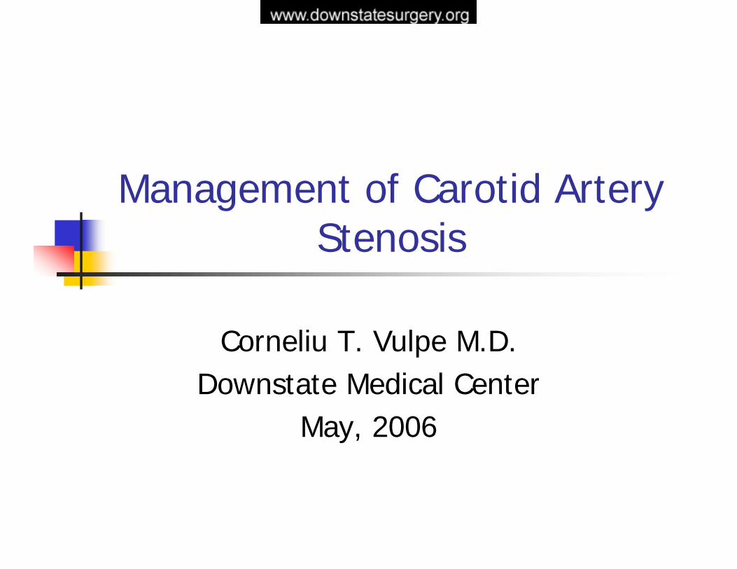
60 year old femaleDM, HTN, COPD, asthmaEthanol, smokerNow with lethargyNo focal neurologic deficitsAdmitted to MICU for DKA
Case Presentation
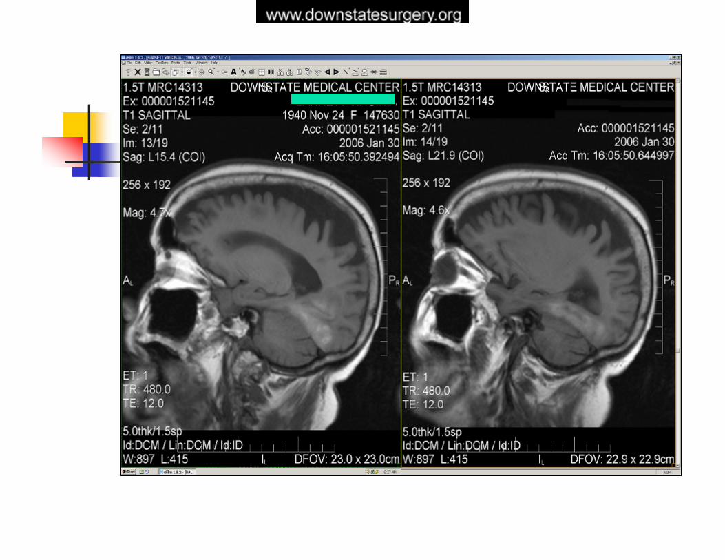
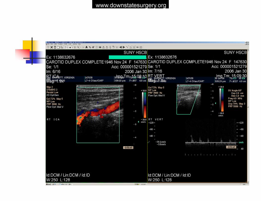
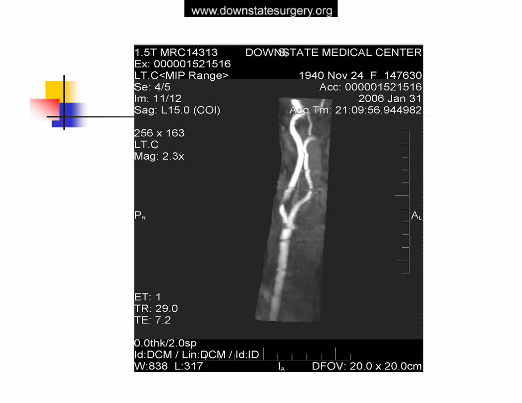
CT head – left occipital infarct with hemorrhagic component, no mass efect, subacuteConfirmed with MRICarotid duplex – left ICA stenosis90-99%Confirmed with MRA
Case Presentation
Summary:
- patient with symptomatic severe left carotid artery stenosis without large infarctions, neurologically stable
Case Presentation
10 days after admission the patient underwent CEA (cleared by Cardiology, Neurology, Neurosurgery)Shunt usedA Dacron patch was usedPOD#3 – fever spike – phlebitis left forearm – iv abx and warm compressesWBC 14
Case Presentation
POD#5 – left neck cellulittis, no drainage, then swellingCellulitis subsided with iv abxPseodoaneurysm ruled out with duplexPossible hematomaPOD#7 – 30 cc pus drained
Case Presentation
The patient underwent reexploration100cc pus drainedShunt usedDacron patch removedSaphenous vein patch angioplastyBilateral forearm thrombosed veins excisedMRSA treated, discharged POD#16
Management of Carotid Artery Stenosis
Overview
StrokeClinical presentation and work-upCEA historyCurrent indications Operative managementComplicationsOngoing issues
Stroke
Third leading cause of death in US50% survivors alive after 5 years25% survivors will have a second neurologicevent, leading to death >50%Substantial morbidity – 18% unable to return to work, 4% require total custodial care$10 billion health care cost anually
Stroke Risk Factors
Hypertension - the single most important modifiable risk factor for ischemic strokecigarette smokingsickle cell diseasetransient ischemic attack (TIA)asymptomatic carotid stenosiscardiac diseases - atrial fibrillation, infective endocarditis, mitral stenosis, and recent large myocardial infarction

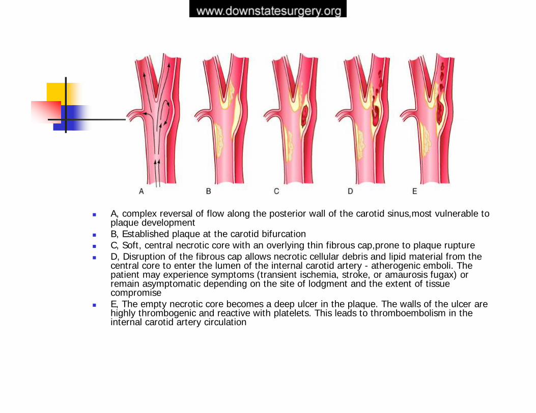
A, complex reversal of flow along the posterior wall of the carotid sinus,most vulnerable to plaque developmentB, Established plaque at the carotid bifurcationC, Soft, central necrotic core with an overlying thin fibrous cap,prone to plaque ruptureD, Disruption of the fibrous cap allows necrotic cellular debris and lipid material from the central core to enter the lumen of the internal carotid artery - atherogenic emboli. The patient may experience symptoms (transient ischemia, stroke, or amaurosis fugax) or remain asymptomatic depending on the site of lodgment and the extent of tissue compromiseE, The empty necrotic core becomes a deep ulcer in the plaque. The walls of the ulcer are highly thrombogenic and reactive with platelets. This leads to thromboembolism in the internal carotid artery circulation
Clinical PresentationTIAs are defined as brief episodes of focal loss of brain function due to ischemia that can usually be localized to that portion of the brain supplied by one vascular system (left or right carotid or vertebrobasilar) , lasting less than 24 hours TIAs commonly last 2 to 15 minutes and are rapid in onset (no symptoms to maximal symptoms in < 5 minutes and usually in < 2 minutes
Left carotid system TIAs manifest as (1) motor dysfunction (dysarthria, weakness, paralysis, or clumsiness of the right extremities and/or face); (2) loss of vision in the left eye (amaurosis fugax ,(3) sensory symptoms (numbness, including loss of sensation or paresthesiainvolving the right upper and/or lower extremity and/or face); and (4) aphasia (language disturbance)
Right carotid system TIAs produce similar symptoms on the opposite side, except that aphasia occurs only when the right hemisphere is dominant for speech (left-handed individual)
Clinical PresentationVertebrobasilar system TIAs are characterized by the rapid onset of
(1) motor dysfunction (weakness, paralysis, or clumsiness) of any combination of upper and lower extremities and face (left and/or right)(2) sensory symptoms (loss of sensation, numbness, or paresthesiainvolving the left, right, or both sides)(3) loss of vision in one or both homonymous visual fields(4) loss of balance, vertigo, unsteadiness or disequilibrium, diplopia, or dysarthria
These last symptoms are characteristic but are not considered as a TIA when any of these symptoms are alone
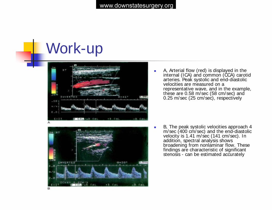
Work-upA, Arterial flow (red) is displayed in the internal (ICA) and common (CCA) carotid arteries. Peak systolic and end-diastolic velocities are measured on a representative wave, and in the example, these are 0.58 m/sec (58 cm/sec) and 0.25 m/sec (25 cm/sec), respectively
B, The peak systolic velocities approach 4 m/sec (400 cm/sec) and the end-diastolic velocity is 1.41 m/sec (141 cm/sec). In addition, spectral analysis shows broadening from nonlaminar flow. These findings are characteristic of significant stenosis - can be estimated accurately
Work-up
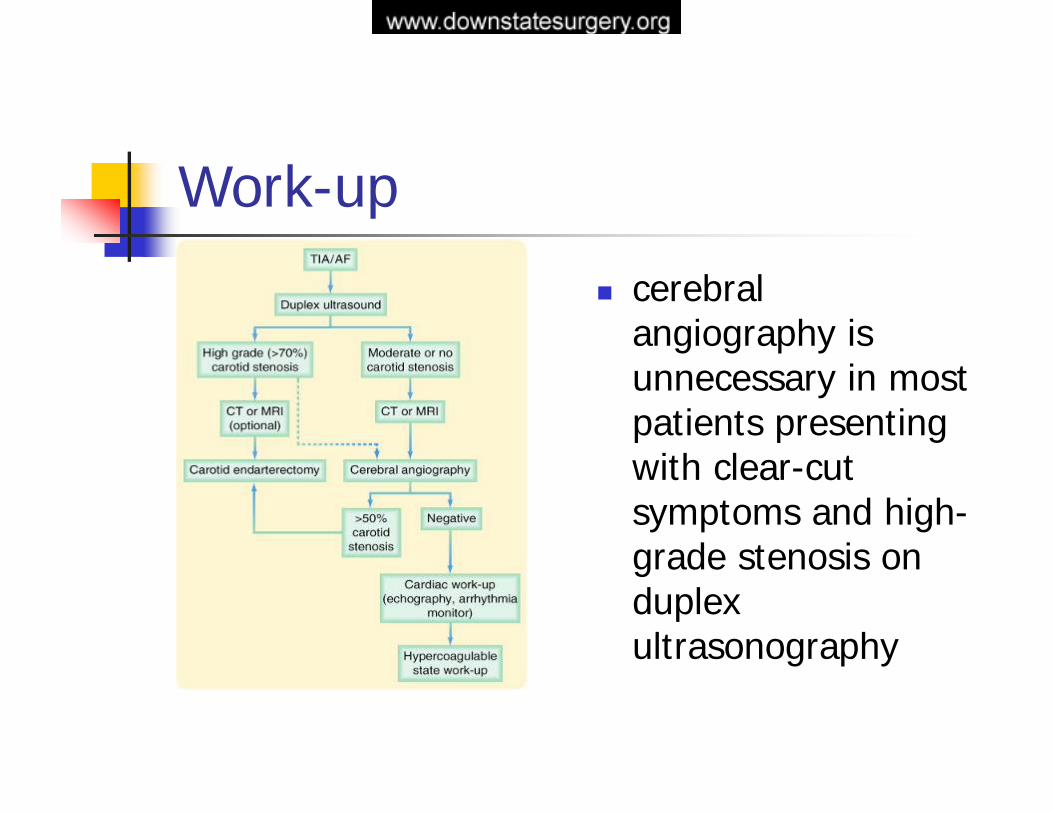
cerebral angiography is unnecessary in most patients presenting with clear-cut symptoms and high-grade stenosis on duplex ultrasonography
CEA History1950s, Fisher - predilection for atheroma to occur at the carotid bifurcation in the neckthe internal carotid artery distal to the bifurcation and the intracranial vessels were usually free of diseaseimportant cause of strokes but also suggested the possible form of therapy to prevent stroke. 1951 - Carrea, Mollins, and Murphy performed the first successful surgical reconstruction of the carotid artery in Buenos Aires1953 – DeBakey - the first successful carotid endarterectomy1954 - Eastcott, Pickering, and Robb – the case was a woman who had recurrent TIAs associated with stenosis of the left carotid bifurcation. She underwent resection of the bifurcation with restoration of blood flow by anastomosis of the internal carotid artery to the common carotid artery. The patient was completely relieved of symptoms, and the operation dramatically demonstrated that removal of carotid bifurcation atherosclerosis could halt TIAs and, presumably, prevent strokes
CEA
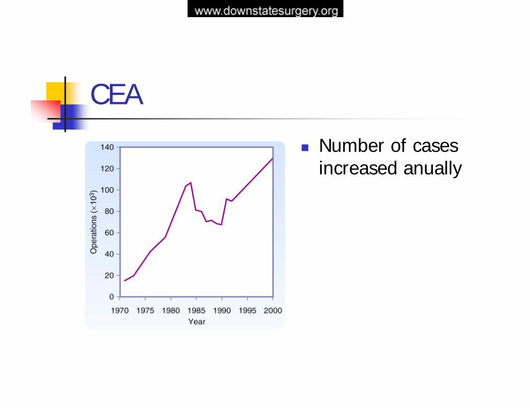
Number of cases increased anually
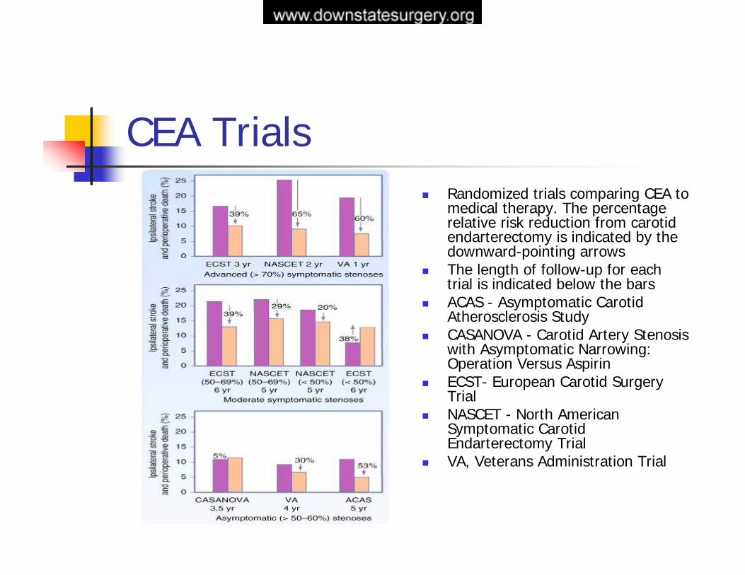
CEA TrialsRandomized trials comparing CEA to medical therapy. The percentage relative risk reduction from carotid endarterectomy is indicated by the downward-pointing arrowsThe length of follow-up for each trial is indicated below the barsACAS - Asymptomatic Carotid Atherosclerosis StudyCASANOVA - Carotid Artery Stenosiswith Asymptomatic Narrowing: Operation Versus AspirinECST- European Carotid Surgery TrialNASCET - North American Symptomatic Carotid Endarterectomy TrialVA, Veterans Administration Trial
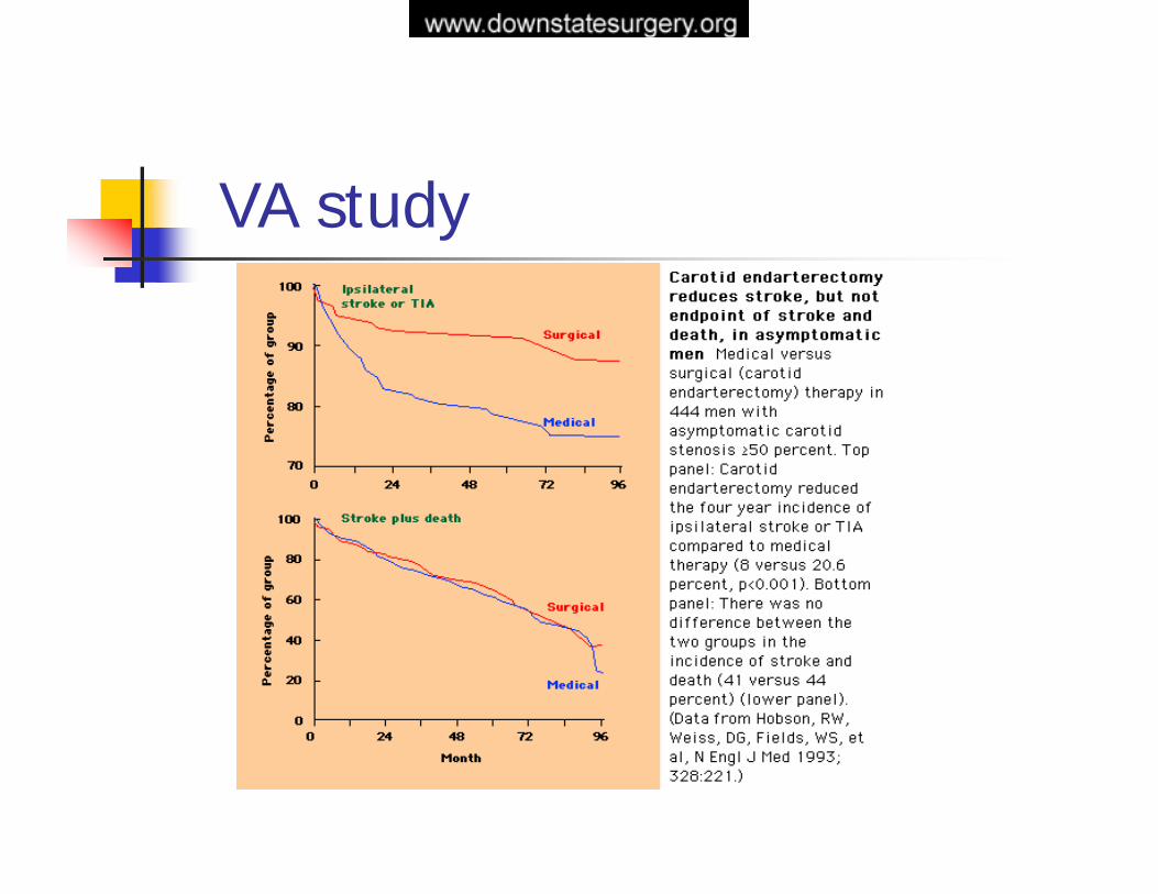
VA study
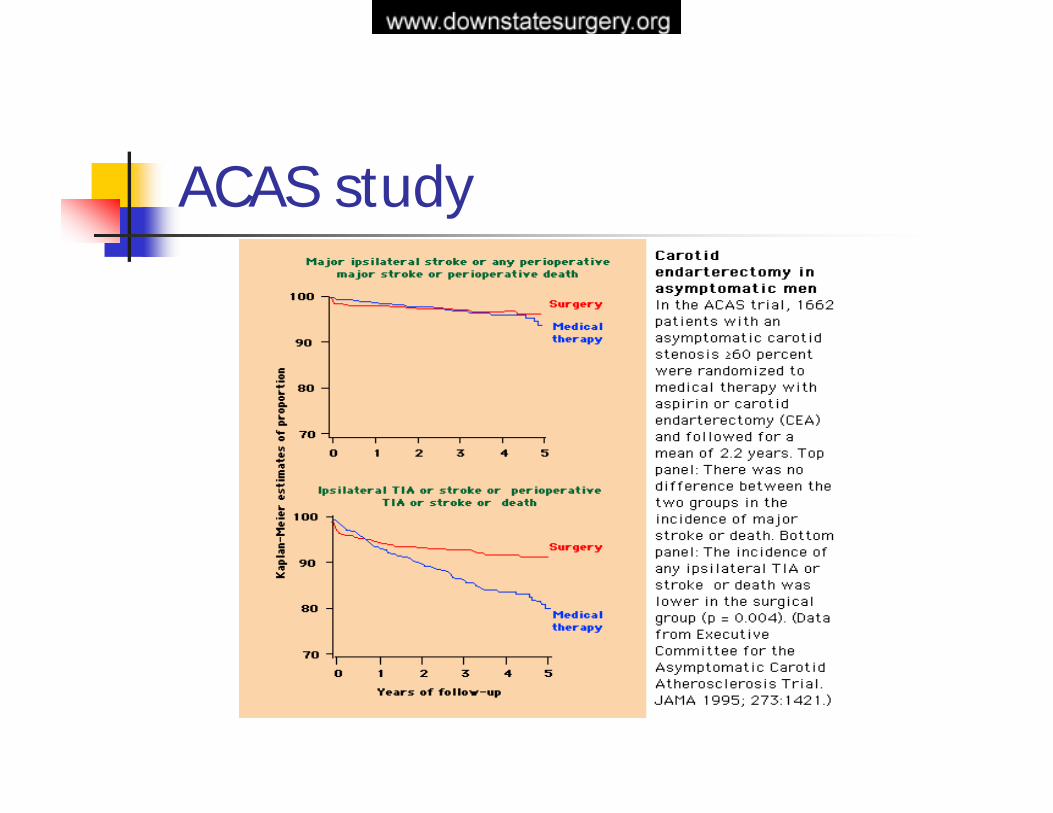
ACAS study
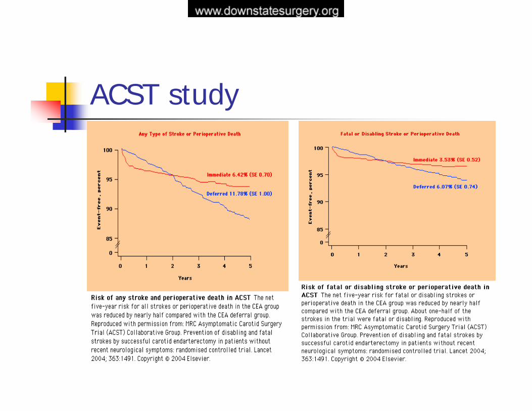
ACST study
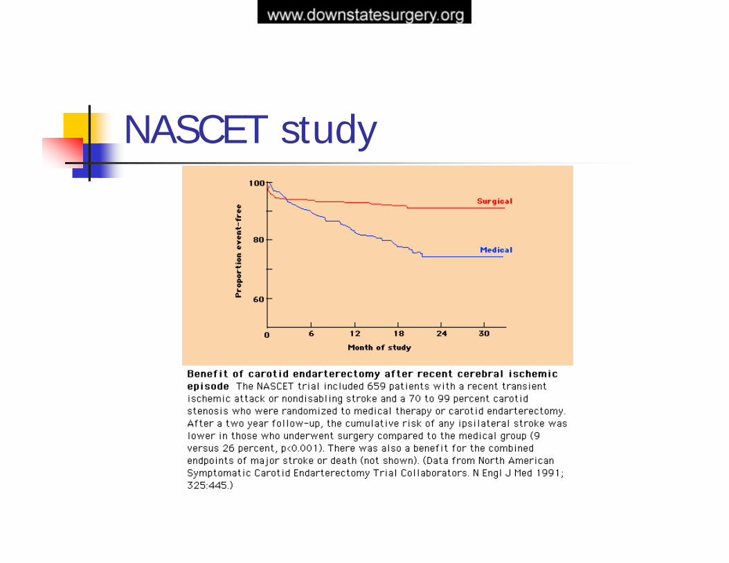
NASCET study
Current Indicationscarotid stenosis of 50% or greater with ipsilateral TIAs, amaurosis fugax, a reversible neurologic deficit, or small stroke and in selected cases of recurrent, symptomatic carotid stenosisPatients with lesser degrees of symptomatic stenosis if they have failed medical therapy (have ongoing symptoms), particularly if there is evidence of ulceration of the lesion or if contralateral occlusion is present.progressive stroke, progressive retinal ischemia, acute carotid occlusion, global cerebral ischemia caused by multiple large-vessel occlusive disease, and in certain cases of symptomatic carotid dissection and true or false aneurysm
Current IndicationsThe indications for endarterectomy in asymptomatic patients remain less clear cutACAS demonstrated significant benefit for all patients randomized to operation with 60% to 99% carotid stenosesit is likely that those with advanced stenoses benefited most. Because the benefit-to-risk ratio in asymptomatic patients is much less than that of symptomatic patients, it is appropriate to reserve carotid endarterectomy only for good risk, asymptomatic patients with advanced stenosesthe presence of ulceration or contralateral occlusion may lower the threshold for recommending operation
Contraindicationsvertebrobasilar distribution TIAsmulti-infarct dementiapatients with severe neurologic deficitsevidence of intracranial hemorrhage or large infarctsuncontrolled congestive heart failurerecent myocardial infarctionunstable anginaDementiaadvanced malignancyuncertain diagnosis
Preoperative Evaluation
HistoryEKGCardiac cathSwan-GanzASA, Plavix, HeparinControl HTN, DM
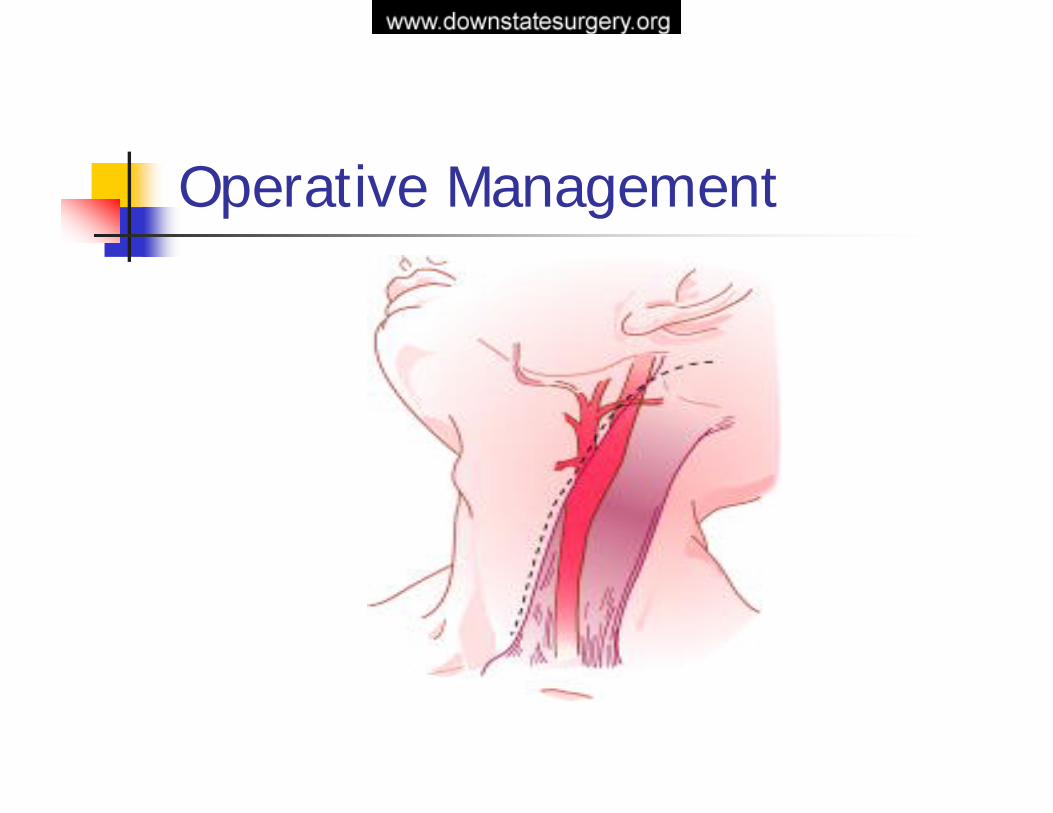
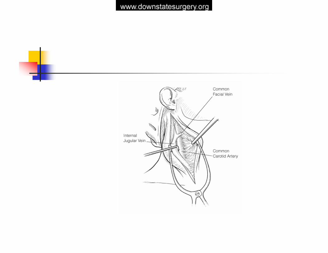
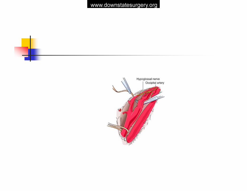
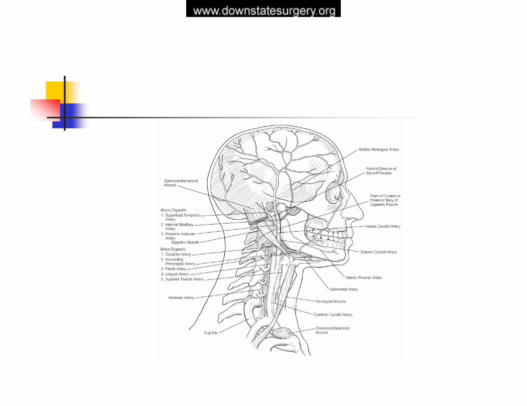
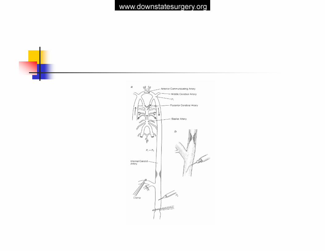
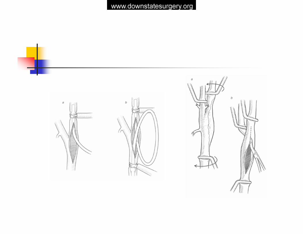
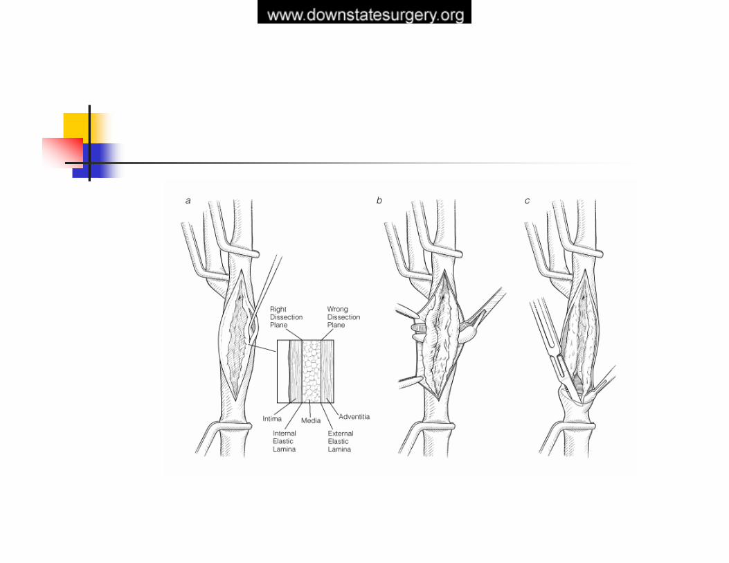
Operative Management
Postoperative ComplicationsStroke or TIA within first 12 hours postop – heparinization and reexploration; 12-24 hrs – CT scanHyperperfusion syndrome and intracerebral hematoma - 0.3-1% - paralysis of autoregulation due to chronic ischemia –ipsilateral headache, seizures, postictal paralysis – angiogram. Risk factors : high-grade (>70%) stenosis; poor collateral hemispheric flow; contralateral carotid occlusion; evidence of chronic ipsilateral hypoperfusion; preoperataive and postoperative hypertension; preexisting ipsilateral cerebral infarction; preoperative anticoagulation or antiplatelet therapyIntracranial hemorrhage - 0.5% to 0.7% of patients undergoing CEA and may account for up to 20% of perioperative strokes
Postoperative ComplicationsBP instability - 1/2-1/3 patientslimited o first 12h; NTG drip to maintain SBP around 140, Dopamine preferred for hypotensionWound hematomas 1.4 -3 % - combination antiplateletRupture saphenous patch – 0.5% 1-7 days postop – use veins no smaller than 4-5 mm in diameter. Risk of stroke and death 48%
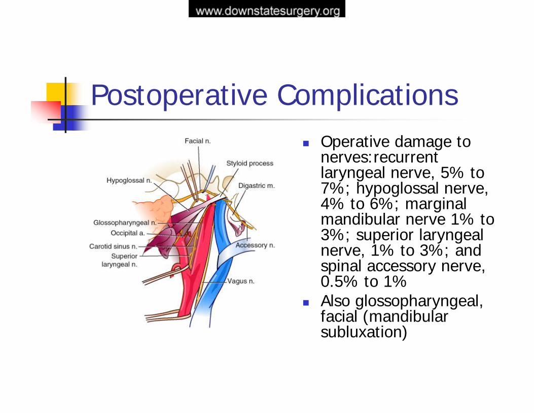
Postoperative ComplicationsOperative damage to nerves:recurrentlaryngeal nerve, 5% to 7%; hypoglossal nerve, 4% to 6%; marginal mandibular nerve 1% to 3%; superior laryngeal nerve, 1% to 3%; and spinal accessory nerve, 0.5% to 1%Also glossopharyngeal, facial (mandibularsubluxation)
Ongoing IssuesSurgical expertise and training - carotid endarterectomy should be performed with low morbidity and mortality in selected patients with appropriate symptoms and that the limits of perioperative morbidity and mortality should be categorized by clinical presentation.The combined morbidity and mortality of the procedure should not exceed 3% for asymptomatic patients, 5% for TIAs, and 7% for ischemic stroke. In addition, the 30-day mortality rate from all causes related to endarterectomy should not exceed 2%
Increasing the cost/benefit ratio - patients have been observed in an intensive care unit for 12 to 24 hours after the operation. Only 10% to 20% of patients required this expensive monitoring. Predictors of the need for intensive care unit observation include preoperative history of hypertension, myocardial infarction, arrhythmia, recent stroke, and chronic renal failure duplex ultrasonography alone or in combination with magnetic resonance angiography (MRA) and the elimination of contrast angiography in the preoperative work-up of patients undergoing endarterectomy - 0.5% to 1% incidence of major neurologic complications, puncture site complications 5% of patients, contrast-induced renal dysfunction in 1% to 5%
Indications for adjunctive arteriography
1. Discrepancy among the history, physical examination, duplex scan, and CT scan 2. Patients presenting with vertebrobasilar symptoms, since they often have proximal
brachiocephalic disease 3. Patients suspected of proximal disease involving branches of the aortic arch (patients with
unequal arm blood pressures or duplex ultrasonographic evidence of abnormal flow characteristics in the proximal common carotid arteries)
4. Patients presenting with focal cerebrovascular symptoms and a stenosis in the 40% to 59% (moderate) range according to duplex criteria (this is the range where even slight overestimation or underestimation may inaccurately categorize the patient)
5. Patients with duplex findings suggestive of distal internal carotid artery or carotid siphon disease
6. Patients with duplex evidence of total carotid occlusion in the presence of ongoing ipsilateralhemispheric symptoms (patients may have near-total occlusion or a “string sign”)
7. Patients with contralateral carotid occlusion or severe carotid stenosis since ipsilateral duplex results are often overestimated because of increased ipsilateral flow velocities
8. Patients with nonatherosclerotic disease such as fibromuscular dysplasia and patients with recurrent carotid stenosis because plaque morphology and extent of disease are sometimes unusual in these patients
9. Patients with duplex scans that are equivocal or of poor quality
Ongoing Issues
Recurrent carotid stenosis -10% in the first year after primary endarterectomy, 3% in the second year, and 2% in the third year. Long-term risk has been estimated to be approximately 1% per year. Symptomatic recurrent carotid disease occurs in about 0.6% to 3% of patients after endarterectomy. Asymptomatic lesions occur with a much greater frequency (7% to 49%)Systemic factors that have been associated with the development of
recurrent disease include female sex, continued smoking after endarterectomy, hypercholesterolemia, diabetes mellitus, hypertension, young age at original endarterectomy, and associated severe atherosclerotic diseasethe mean risk of stroke with reoperation is approximately 4%, with a
death rate of approximately 1.2% and cranial nerve injury of approximately 12%
Ongoing IssuesClosure technique of carotid arteriotomyVein patch - increasing operative time- patch rupture- false aneurysm formation, thromboembolism stemming from the dilated aneurysmalreconstructed bifurcationDacron or other prosthetic material
-potential for infection is present - can lead to catastrophic complications
In men, the use of vein patch closure does not significantly reduce the long-term follow-up incidence of recurrent carotid disease.However, in women, who have a higher incidence of recurrent carotid stenosis, vein patch closure significantly reduces the incidence of thislong-term complication
Myers SI, Valentine RJ, Chervu A, et al: Saphenous vein patch versus primary closure for carotid endarterectomy: Long-term assessment of a randomized prospective study. J Vasc Surg 19:15–22, 1994
Ongoing IssuesLocal vs. general anesthesiaCarotid shunt and monitoring - only 10% to 15% of patients who are intolerant of temporary carotid clamping benefit from an internal shunt. Halsey JH Jr: Risks and benefits of shunting in carotid endarterectomy. The International TranscranialDoppler Collaborators. Stroke 23:1583–1587, 1992.
Harada RN, Comerota AJ, Good GM, et al: Stump pressure, electroencephalographic changes, and the contralateral carotid artery: Another look at selective shunting. Am J Surg 170:148–153, 1995.
Imparato AM, Ramirez A, Riles T, et al: Cerebral protection in carotid surgery. Arch Surg 117:1073–1078, 1982.
Sundt TM Jr, Sharbrough FW, Piepgras DG, et al: Correlation of cerebral blood flow and electroencephalographic changes during carotid endarterectomy: With results of surgery and hemodynamics of cerebral ischemia. Mayo Clin Proc 56:533–543, 1981monitoring neurologic status during temporary carotid occlusion in an awake patient under local anesthesia, measurement of internal carotid artery back pressure (“stump pressure” of <50 mm Hg is the generally accepted criterion for need for shunt placement), isotopic regional blood flow measurements, transcranial Doppler monitoring, somatosensory evoked potential monitoring, and EEG monitoring
Ongoing IssuesTiming of operation after stroke
- 4 to 6 weeks in patients diagnosed with acute stroke, regardless of its severity, for fear of clinical deterioration associated with conversion of a bland infarct into a hemorrhagic one
- an early operation without waiting 4 to 6 weeks is safe in patients with minor, nondisabling stroke
- Gasecki AP, Ferguson GG, Eliasziw M, et al: Early endarterectomy for severe carotid artery stenosis after a nondisabling stroke: Results from the North American Symptomatic Carotid Endarterectomy Trial. J Vasc Surg 20:288–295, 1994.
Piotrowski JJ, Bernhard VM, Rubin JR, et al: Timing of carotid endarterectomy after acute stroke. J Vasc Surg 11:45–52, 1990
- On the other hand, a higher incidence of perioperative stroke has been reported in patients undergoing operation within 5 to 6 weeks after presenting with stroke
- a compelling reason for not delaying the operation is that patients may be placed at risk for recurrent stroke during the waiting period, particularly in circumstances where the stenosis is advanced or preocclusive
Ongoing IssuesSimultaneous CEA and CABG
The incidence of hemodynamically significant carotid stenosis in screening studies of patients undergoing coronary artery bypass is 5% to 11% Although many centers have reported favorable experiences in combined carotid endarterectomy and coronary artery bypass procedures performed simultaneously, others point out that the overall stroke and death rate with this approach is higher than with either procedure alone
Hertzer NR, Loop FD, Beven EG, et al: Surgical staging for simultaneous coronary and carotid disease: A study including prospective
randomization. J Vasc Surg 9:455–463, 1989
Simultaneous operation - precarious coronary artery disease such as unstable angina or high-grade left main lesions who have symptomatic high-grade carotid stenoses, bilateral high-grade asymptomatic stenoses, or ipsilateral advanced, asymptomatic stenosis and contralateral occlusion
Ongoing IssuesEversion CEA
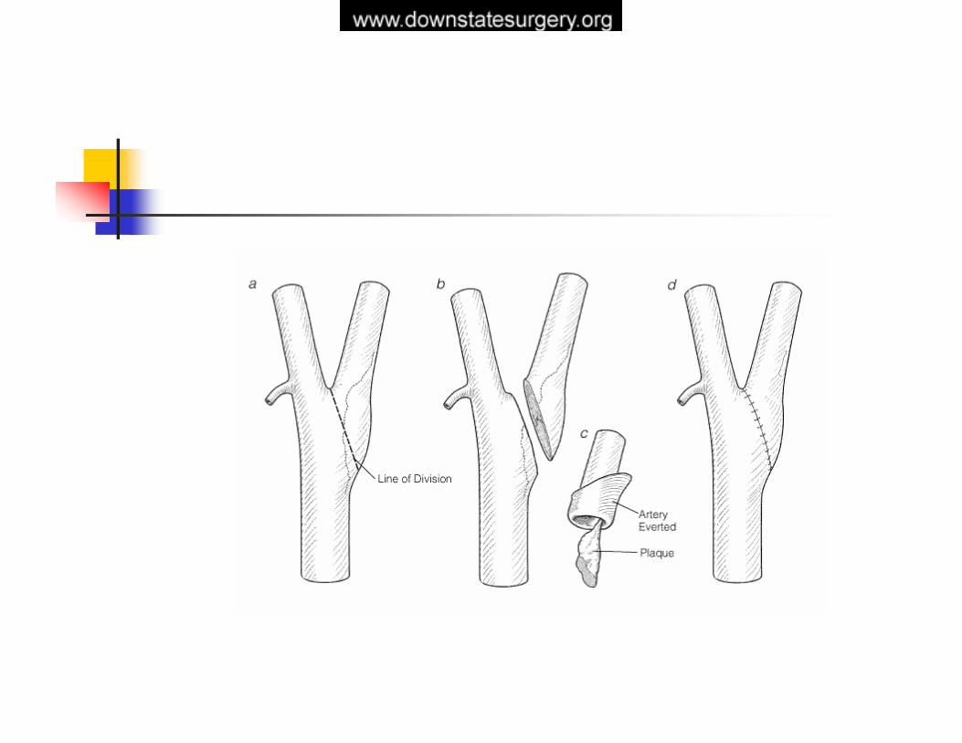
introduced in the late 1950s division of the common carotid artery below the bifurcation and eversionendarterectomy of both the external and internal carotid arteriesrecent modifications of the technique involve transection of the internal carotid artery at the level of the bifurcation and reimplantation of the internal carotid artery after endarterectomy into the common carotid artery simplicity, faster operating times, ease of correction of elongated and tortuous internal carotid arteries and, possibly, a lower rate of carotid restenosisdifficulty in shunting, the possibility of incomplete removal of distal intimal flaps, difficulties in obtaining complete endarterectomy of the external and common carotid arteries when these are extensively involved with the disease, and frequent need for extensive distal mobilization of the internal carotid artery with a higher rate of cranial nerve injury in some series Randomized studies to date demonstrate no differences in the major outcomes of stroke, death, and recurrent stenosis
Cao P, De Rango P, Zannetti S: Eversion versus conventional carotid endarterectomy: A systematic review. Eur J Vasc Endovasc Surg23:195–201, 2002
Ongoing Issues
Carotid angioplasty/ stent placementtechnical success rate of 97% to 98% and a stroke and death rate of 0% to 7.1%
Gray WA, White HJ Jr, Barrett DM, et al: Carotid stenting and endarterectomy: A clinical and cost comparison of revascularization strategies. Stroke
33:1063–1070, 2002
Cerebral protection devices that capture atherothrombotic debris at the time of angioplasty and stent deployment reduce the overall rate of periprocedural neurologic deficits by 40% to 50%
Kastrup A, Groschel K, Krapf H, et al: Early outcome of carotid angioplasty and stenting with and without
cerebral protection devices: A systematic review of the literature. Stroke 34:813–819, 2003
Carotid Angioplasty and Stenting
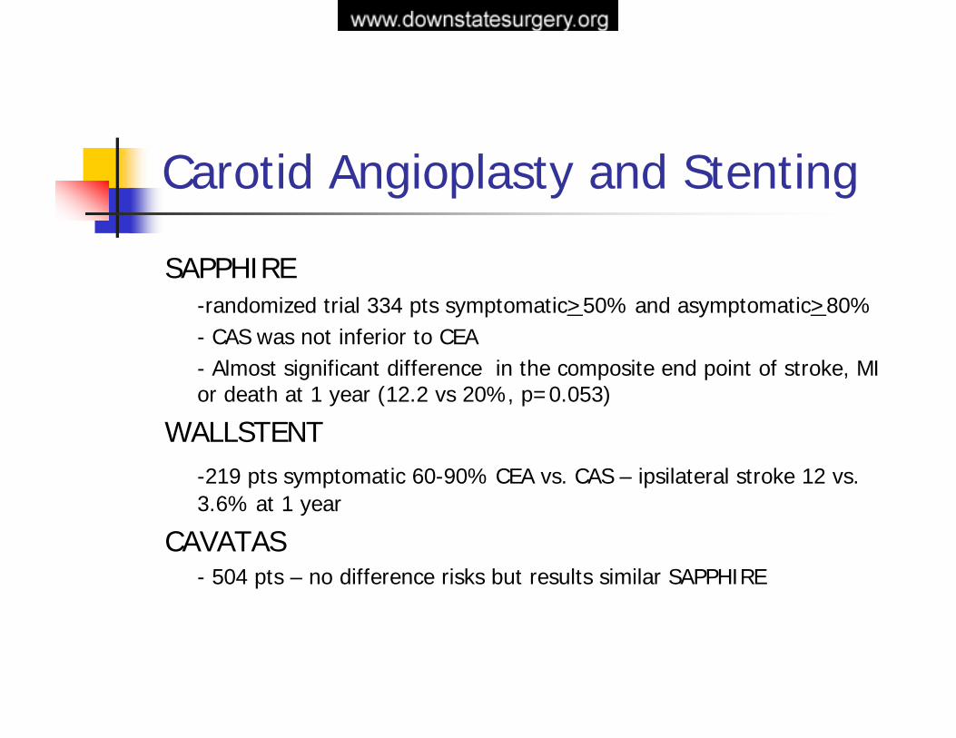
SAPPHIRE-randomized trial 334 pts symptomatic>50% and asymptomatic>80%- CAS was not inferior to CEA- Almost significant difference in the composite end point of stroke, MI or death at 1 year (12.2 vs 20%, p=0.053)
WALLSTENT-219 pts symptomatic 60-90% CEA vs. CAS – ipsilateral stroke 12 vs. 3.6% at 1 year
CAVATAS- 504 pts – no difference risks but results similar SAPPHIRE
Carotid Angioplasty and Stenting
Ongoing trialsCRESTSPACECAVATAS-2