Embed Size (px)
Citation preview
Article
Mammalian Innate Immune Response to a
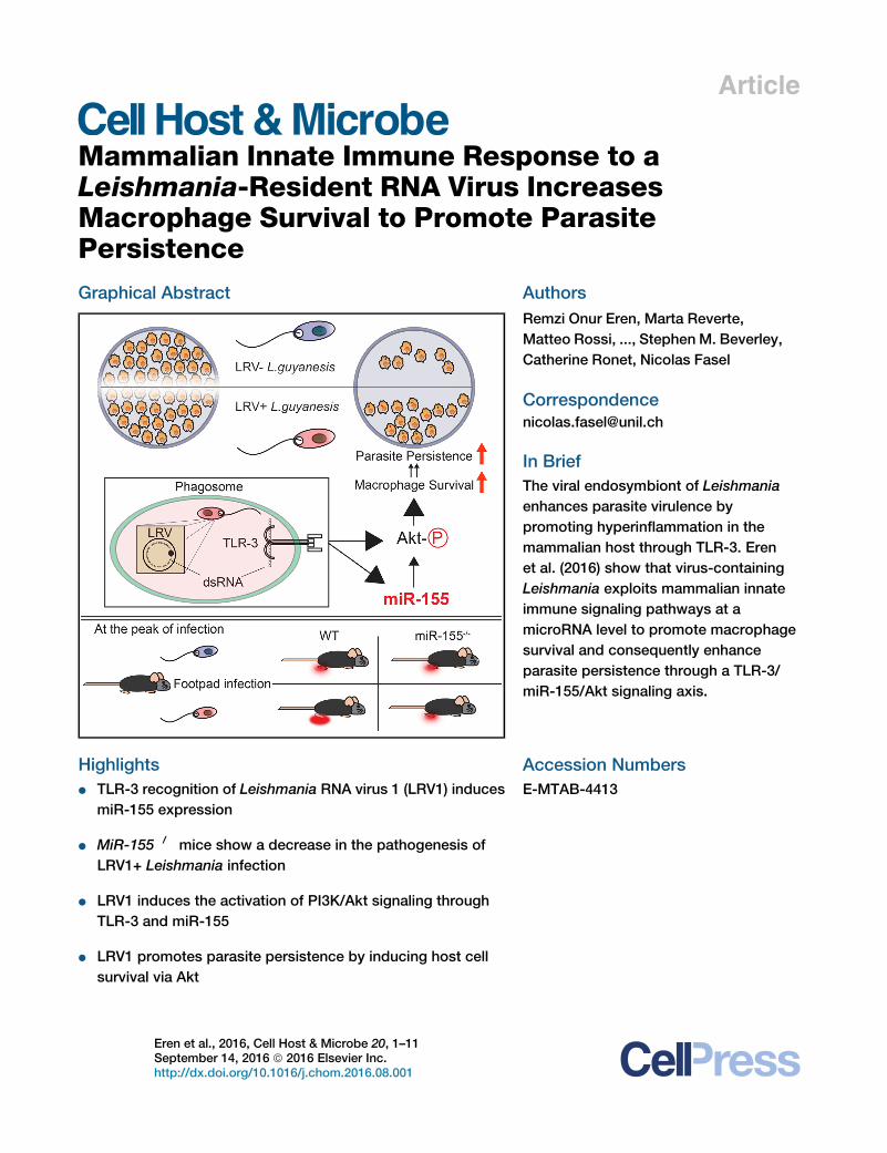
Leishmania-Resident RNA Virus IncreasesMacrophage Survival to Promote ParasitePersistenceGraphical Abstract
Highlights
d TLR-3 recognition of Leishmania RNA virus 1 (LRV1) induces
miR-155 expression
d MiR-155�/� mice show a decrease in the pathogenesis of
LRV1+ Leishmania infection
d LRV1 induces the activation of PI3K/Akt signaling through
TLR-3 and miR-155
d LRV1 promotes parasite persistence by inducing host cell
survival via Akt
Eren et al., 2016, Cell Host & Microbe 20, 1–11September 14, 2016 ª 2016 Elsevier Inc.http://dx.doi.org/10.1016/j.chom.2016.08.001
Authors
Remzi Onur Eren, Marta Reverte,
Matteo Rossi, ..., Stephen M. Beverley,
Catherine Ronet, Nicolas Fasel
In Brief
The viral endosymbiont of Leishmania
enhances parasite virulence by
promoting hyperinflammation in the
mammalian host through TLR-3. Eren
et al. (2016) show that virus-containing
Leishmania exploits mammalian innate
immune signaling pathways at a
microRNA level to promote macrophage
survival and consequently enhance
parasite persistence through a TLR-3/
miR-155/Akt signaling axis.
Accession Numbers
E-MTAB-4413

Please cite this article in press as: Eren et al., Mammalian Innate Immune Response to a Leishmania-Resident RNA Virus Increases Macrophage Sur-vival to Promote Parasite Persistence, Cell Host & Microbe (2016), http://dx.doi.org/10.1016/j.chom.2016.08.001
Cell Host & Microbe
Article
Mammalian Innate Immune Responseto a Leishmania-Resident RNA Virus IncreasesMacrophage Survival to Promote Parasite PersistenceRemzi Onur Eren,1 Marta Reverte,1 Matteo Rossi,1 Mary-Anne Hartley,1 Patrik Castiglioni,1 Florence Prevel,1
Ricardo Martin,1 Chantal Desponds,1 Lon-Fye Lye,2 Stefan K. Drexler,1,4 Walter Reith,3 Stephen M. Beverley,2
Catherine Ronet,1 and Nicolas Fasel1,5,*1Department of Biochemistry, University of Lausanne, 1066 Epalinges, Switzerland2Department of Molecular Microbiology, School of Medicine, Washington University, St. Louis, MO 63110, USA3Department of Pathology and Immunology, University of Geneva, 1211 Geneva, Switzerland4Present address: Biozentrum, University of Basel, 4056 Basel, Switzerland5Lead Contact*Correspondence: [email protected]
http://dx.doi.org/10.1016/j.chom.2016.08.001
SUMMARY
Some strains of the protozoan parasite Leishmaniaguyanensis (L.g) harbor a viral endosymbiont calledLeishmania RNA virus 1 (LRV1). LRV1 recognitionby TLR-3 increases parasite burden and lesionswelling in vivo. However, the mechanisms by whichanti-viral innate immune responses affect parasiticinfection are largely unknown. Upon investigatingthe mammalian host’s response to LRV1, we foundthat miR-155 was singularly and strongly upregu-lated in macrophages infected with LRV1+ L.gwhen compared to LRV1� L.g. LRV1-driven miR-155 expression was dependent on TLR-3/TRIFsignaling. Furthermore, LRV1-induced TLR-3 activa-tion promoted parasite persistence by enhancingmacrophage survival through Akt activation in amanner partially dependent on miR-155. Pharmaco-logical inhibition of Akt resulted in a decrease inLRV1-mediated macrophage survival and conse-quently decreased parasite persistence. Consistentwith these data, miR-155-deficient mice showed adrastic decrease in LRV1-induced disease severity,and lesional macrophages from these mice dis-played reduced levels of Akt phosphorylation.
INTRODUCTION
Macrophages are host cells for several obligate intracellular pro-
tozoan parasites such as Leishmania spp., Trypanosoma cruzi,
and Toxoplasma gondii (Sacks and Sher, 2002). Upon infection,
macrophages shape the early phases of immunity by sensing
pathogen-associated molecular patterns (PAMPs) through path-
ogen recognition receptors (PRRs). The family of Toll-like recep-
tors (TLRs) is one of the most intensively studied classes of
PRRs. The engagement of TLRs with their cognate ligands leads
to a cascade of events including the recruitment of various
Cell Ho
adaptor molecules such as myeloid differentiation primary
response gene 88 (MyD88), MyD88 adaptor-like, TIR-domain-
containing adaptor-inducing IFN-b (TRIF), and the TRIF-related
adaptor molecule. The MyD88 adaptor protein is involved in
the signaling of all TLRs except the double-stranded RNA
(dsRNA) receptor TLR-3, which exclusively recruits TRIF. Both
MyD88-dependent and -independent TLR pathways transduce
signals through conserved inflammatory signaling pathways,
inducing Interleukin-6 (IL-6) and tumor necrosis factor-a
(TNF-a) secretion. TLR stimulation also promotes the activation
of phosphatidylinositol 3-kinase (PI3K)/Akt. In addition to these
pathways, TRIF-dependent TLR signaling induces the phos-
phorylation of interferon response element 3 (IRF-3), leading
to the transcription of IFN-b (Rakoff-Nahoum and Medzhitov,
2009).
TLR activation of innate cells is involved in the development of
parasiticidal immunity via the presentation of parasitic antigens
to T cells (Iwasaki and Medzhitov, 2004). Antigen-activated
CD4+ T cells are amajor source of IFN-g, which is a key mediator
of anti-Leishmania effector genes in macrophages (Bogdan
et al., 2000). Excessive TLR simulation, however, can be detri-
mental to the host in chronic infectious diseases, leading to pro-
gressive tissue damage and fatal systemic disorders. For
example, TLR-3 recognition of an endosymbiotic dsRNA virus
within Leishmania guyanensis (L.g) increases the virulence of
the virus’s microbial host and induces a hyper-inflammatory
response by exploiting the innate recognition of Leishmania’s
mammalian host (Ives et al., 2011).
In addition to initiating inflammation and adaptive immunity,
TLRs are known to be involved in macrophage survival
(Lombardo et al., 2007). Certain bacterial species hinder TLR-
4-mediated cell-survival factors to induce macrophage
apoptosis, proposing amechanism for bacterial immune evasion
(Hsu et al., 2004; Park et al., 2005). Several viral species are re-
ported to extend host cell viability by inducing the PI3K/Akt
signaling pathway to achieve latent and/or chronic infection
(Cooray, 2004). Similarly, intracellular protozoan parasites can
promote host cell survival by activating pro-survival signaling
pathways or by interrupting host cell apoptotic machinery
(Heussler et al., 2001). For example, infection of a murine
st & Microbe 20, 1–11, September 14, 2016 ª 2016 Elsevier Inc. 1

Please cite this article in press as: Eren et al., Mammalian Innate Immune Response to a Leishmania-Resident RNA Virus Increases Macrophage Sur-vival to Promote Parasite Persistence, Cell Host & Microbe (2016), http://dx.doi.org/10.1016/j.chom.2016.08.001
macrophage cell line with Leishmania spp. induces PI3K/Akt
signaling and grants protection against apoptosis (Ruhland
et al., 2007). Taken together, the subtle equilibrium between
the death and survival of innate cells has a determining impact
on the outcome of microbial infections.
Akt, a member of the protein kinase B family, is a serine thre-
onine kinase that regulates numerous substrates involved in cell
survival, cellular growth, and metabolism. For instance, TLR-3-
induced endogenous phosphorylation of Akt in primary murine
macrophages leads to the degradation of Forkhead box O3
(Foxo3a), a transcription factor that regulates genes involved in
cell death (Litvak et al., 2012). Furthermore, Akt positively regu-
lates the mammalian target of rapamaycin 1 (mTOR1), which
plays a key role in cell survival and growth by modulating
mRNA translation and many cellular processes in innate cells
(Weichhart et al., 2015). The activating serine 2448 phosphoryla-
tion of mTOR1 by Akt leads to the phosphorylation of ribosomal
protein S6 kinase (S6K), which promotes mRNA translation
(Huang and Houghton, 2003).
Another important component of TLR stimulation is the
expression of small non-coding RNAs, named microRNAs,
which regulate genes at the post-transcriptional level, culmi-
nating in the degradation of the transcript or the inhibition of
translation (Valencia-Sanchez et al., 2006). In macrophages, mi-
croRNA-155 (miR-155) is the only microRNA that is significantly
upregulated in response to polyinosinic:polycytidylic acid (poly
I:C), a synthetic dsRNA ligand for TLR-3 (O’Connell et al.,
2007). MiR-155 is also classified as an oncogenic microRNA.
The overexpression of miR-155 in mice causes a myeloid prolif-
erative disorder, which is at least partially due to Akt-mediated
cell survival and proliferation (O’Connell et al., 2008). This indi-
cates the oncogenic potential of a TLR-induced microRNA.
In this study, we asked which microRNAs were modulated in
macrophages in the presence of L.g endosymbiont Leishmania
RNA virus 1 (LRV1), and investigated how and by which mecha-
nism(s) LRV1 contributed to increased pathology.
RESULTS
MiR-155 Is the Uniquely Expressed MicroRNA due toLRV1/TLR-3/TRIF SignalingTo determine the impact of LRV1 on the miRNA expression pro-
file, murine bone-marrow macrophages (BMMs) were incubated
with LRV1+ L.g or LRV1� L.g parasites for 10 hr. AmiRNAmicro-
array analysis was then performed on total RNA. Out of 1,179
miRNAs, miR-155 was the only miRNA significantly upregulated
by LRV1+ L.g infection in comparison to LRV� L.g (Figure 1A). To
verify this result, isolated RNA from LRV1+ L.g- or LRV1� L.g-in-
fected macrophages was reverse transcribed into complemen-
tary DNA (cDNA) using a stem-loop primer approach (Hurley
et al., 2012). cDNAs were then used to quantify mature miR-
155 by real-time PCR (RT-PCR). Consistent with the microRNA
microarray data, miR-155 was drastically upregulated in
response to poly I:C treatment and LRV1+ L.g infection
(Figure 1B). These findings demonstrated that the dsRNA
endosymbiont within L.g altered the miRNAome profile of host
macrophages and induced the expression of miR-155.
MiR-155 is located in the third exon of the non-protein-codingB
cell-integrated cluster (BIC) gene (Tam, 2001). To examine the ki-
2 Cell Host & Microbe 20, 1–11, September 14, 2016
netics ofmiR-155 induction in BMMs following various stimuli, the
expression of both BIC mRNA and the mature form of miR-155
wasmonitored for 24 hr post-infection using RT-PCR. In the pres-
enceofLRV1orpoly I:C,miR-155and theBICgeneweredetected
after 2 hr, with a peak at 10 hr post-infection (Figures 1C and 1D).
MiR-155 expression stabilized after 10 hr post-incubation, while
the transcript level of BIC was strongly downregulated in macro-
phages incubatedwith LRV1+ L.g and poly I:C after 24 hr (Figures
1C and 1D). Upon LRV1� L.g infection, BIC remained almost
undetectable during the 24 hr time course (Figure 1D). In miR-
155�/��deficientmice, a b-galactosidase reporter gene replaces
the third exon of BIC, allowing the measurement of BIC promoter
activity (Thai et al., 2007). LRV1+ L.g- and LRV1� L.g-infected
miR-155�/� BMMs were lysed, and cell lysates were tested for
b-galactosidase by immunoblotting (Figure 1E). We observed
that the b-galactosidase level peaked after 10 hr incubation with
LRV1+ L.g or poly I:C. Comparatively, LRV1� L.g infection
induced residual b-galactosidase expression (Figure 1E).
The LRV1dsRNAgenome is an agonist of TLR-3,which signals
through TRIF adaptor protein (Ives et al., 2011). To investigate
whether LRV1 promotesmiR-155 expression through the TLR-3/
TRIF signaling pathway, BMMs derived from WT, TLR3�/�, andTRIFDLPS2 were infected with LRV1+ L.g or LRV1� L.g. Poly I:C
treatment was used as a positive control. Both LRV1 and poly
I:C-induced miR-155 expression required functional TLR-3 and
TRIF molecules, since the ablation of either TLR-3 or TRIF abro-
gated miR-155 expression (Figure 1F). These data indicated that
the TLR-3/TRIF signaling pathway initiated the expression of
miR-155 upon sensing LRV1 or synthetic dsRNA.
LRV1-Mediated miR-155 Expression Confers IncreasedParasite Burden and Severity of Pathology in MiceThe role of miR-155 in the development of disease pathology
was evaluated in vivo by infecting both WT and miR-155�/�
mice in the footpad. A drastic decrease in footpad swelling
and reduced parasite burden at the peak of infection was
observed in miR-155�/� mice infected with LRV1+ L.g (Figures
2A and 2B). In the case of LRV1� L.g infection, no significant dif-
ference in disease pathology was detected between WT and
miR-155�/�mice (Figures 2A and 2B). These findings suggested
that LRV1-mediated host miR-155 expression played a crucial
role in the survival of LRV1+ L.g parasites in mice.
Since miR-155 has also been shown to be required for the
normal function of B cells and antibody secretion (Rodriguez
et al., 2007; Thai et al., 2007), we decided to determine the
impact of B cells on L.g infection. WT mice and mice lacking
mature B cells (Jh�/�) were infected with LRV1+ L.g. Infected
WT and B cell-deficient mice displayed similar footpad swelling
and parasitemia profiles (Figures S1A and S1B). We additionally
examined B cell responses of miR-155-deficient mice infected
with LRV1+ L.g by measuring total serum anti-LRV1 capsid
immunoglobulin G (IgG) titers. Infected miR-155�/� mice pro-
duced reduced titers of anti-LRV1 capsid IgG compared to in-
fected WT mice (Figure S1C). These data suggested that B cells
and the antibody response were not involved in the pathology of
Leishmania infection, and were not essential for the LRV1+ L.g
parasite resistant phenotype of miR-155�/� mice.
It has also been reported that in in vitro differentiation condi-
tions, CD4+ T cells lacking miR-155 secrete more IL-4 than WT

A
C
E F
D
B Figure 1. LRV1-Induced miR-155 Expres-
sion through TLR-3/TRIF Signaling
(A) MicroRNA microarray analysis was performed
on total RNA extracted from WT BMMs infected
with LRV1+ L.g and LRV1� L.g for 10 hr. Scatter-
plot shows log10-transformed normalized aver-
aged intensity values (n = 4) for eachmiRNA probe.
(B) A portion of WT BMMs generated in (A) were
treated with medium or poly I:C (2 mg/ml). RT-PCR
was used to measure the abundance of the
miR-155 in extracted RNA. The numbers represent
relative miR-155 expression as fold induction over
the medium-treated condition subsequent to
normalization with miR-16.
(C and D) Kinetic quantification of miR-16-
normalized miR-155 (C) and L32-normalized BIC
(D) expression in WT BMMs incubated with
medium, poly I:C (2 mg/ml), LRV1+ L.g, or LRV1�L.g using RT-PCR.
(E) b-galactosidase western blot analysis of
miR-155�/� BMMs infected with LRV1+ L.g (+) or
LRV1� L.g (�) at indicated time points. Cells
treated with medium (as negative control) or with
poly I:C (2 mg/ml) (as positive control) were also
blotted for b-galactosidase.
(F) The relative miR-155 expression levels of WT,
TLR-3�/�, and TRIFDLPS2 macrophages incubated
with medium, poly I:C (2 mg/ml), LRV1+ L.g, or
LRV1� L.g.
Data are presented as mean ± SEM from the pool
of three independent experiments (C, D, and F).
Representative western blots were shown from
three independent experiments (E). p values shown
were calculated by unpaired Student’s t test.
**p < 0.01 and ***p < 0.001.
Please cite this article in press as: Eren et al., Mammalian Innate Immune Response to a Leishmania-Resident RNA Virus Increases Macrophage Sur-vival to Promote Parasite Persistence, Cell Host & Microbe (2016), http://dx.doi.org/10.1016/j.chom.2016.08.001
CD4+ T cells, but similar levels of IFN-g (Rodriguez et al., 2007).
IFN-g, which is predominantly secreted by CD4+ T cells, aug-
ments macrophage parasiticidal activity, while IL-4 suppresses
it (Sacks and Sher, 2002). Therefore, we evaluated the role of
CD4+ T cells, which are required for controlling Leishmania
parasites in our model of infection. To determine the impact of
miR-155 on IL-4- and IFN-g-secreting CD4+ T cells during L.g
infection, at the peak of infection we used intracellular flow
cytometry analysis on the popliteal lymph node cells from WT
and miR-155�/� mice infected either with LRV1+ L.g or LRV1�L.g. Our results showed that the number and percentages of
IFN-g- and IL-4-secreting CD4+ T cells in LRV1+ L.g-infected
mice were similar to those found in LRV1� L.g-infected mice
(Figures 2C and S2). These data indicated that the reduced
disease progression in miR-155�/� mice in response to LRV1+
L.g did not arise from an IFN-g /IL-4 imbalance.
To further investigate the role of T cells in LRV1+ L.g infection,
WT andmiR-155�/� mice were injected with anti-CD4-depleting
antibody 5 days prior to infection and on a weekly basis until
6 weeks post-infection (Figure S3A). We verified the efficiency
Cell Host
of the antibody-mediated depletion using
flow cytometry (Figures S3B and S3C).
Interestingly, CD4 depletion caused a
significant decrease in footpad swelling
at 2–3 weeks post-infection in both
LRV1+ L.g-infected WT and miR-155�/� mice (Figure 2D). We
also observed that the resolution of footpad swelling was
impaired in both LRV1+ L.g-infected WT and miR-155�/� mice
after week 5 (Figure 2D). These data indicated that CD4+ cells
contributed to lesion development early in LRV1+ L.g infection
in a miR-155-independent manner and were required for the
LRV1+ L.g infection resolution.
We analyzed parasite load in CD4-depleted WT and
miR-155�/� mice infected with LRV1+ L.g. We found that
CD4 depletion had no effect on parasite burden in WT
mice, but led to a significant increase in parasite load at the
peak of infection in LRV1+ L.g-infected miR-155�/� mice (Fig-
ure 2E). These data suggested that CD4+ T cells were required
for control of LRV1+ L.g during the earlier phases of infection
in miR-155�/� mice, but not in WT mice, which probably have
a compensatory source of IFN-g other than CD4+ T cells in this
time frame.
To better understand the possible impact of IFN-g on the resis-
tant phenotype ofmiR-155�/� mice against LRV1+ L.g infection,
we crossedmiR-155�/�micewithmice lacking IFN-g to generate
& Microbe 20, 1–11, September 14, 2016 3

A
C
E F
D
B
Figure 2. MiR-155–/– Mice Infected with LRV1+ L.g Parasites Had Decreased Disease Pathology when Compared to WT Mice
(A and B) Hind footpads of WT and MiR-155�/� mice were infected with 1 3 106 LRV1+ L.g or LRV1� L.g.
(A) The graph displays the weekly measurement of footpad swelling.
(B) Parasite burden was determined at 3 weeks after infection by bioluminescence imaging.
(C) Popliteal lymph nodes were collected fromWT andmiR-155mice infected with LRV1+ L.g or LRV1� L.g on the footpad after 3 weeks of infection. Intracellular
FACS analysis was performed using primary ex vivo lymphocytes extracted and stimulated with PMA/ionomycin to measure IL-4 and IFN-g levels in CD4+ T cells
(see also Figure S2).
(D and E) Mice were intraperitoneally injected with CD4-depleting antibody 5 days prior to LRV1+ L.g infection and then on a weekly basis for 6 weeks. A group of
mice was treated with PBS as a negative control (see also Figures S3 and S4).
(D) Footpad swelling of LRV1+ L.g-infected WT and MiR-155�/� mice.
(E) Parasite load was measured by bioluminescence 4 weeks post-infection.
(F) The measurement of footpad swelling in WT, MiR-155�/�, IFN-g, and IFN-gxmiR-155 DKO mice following of LRV1+ L.g infection.
Data show mean ± SD from representative experiments (n = 4–5 mice) of two (C–E) or three (A, B, and F) independent experiments. Each dot in the scatter blot
represents a footpad (B and E). Statistical significance is calculated using two-way ANOVA analysis with Bonferonni’s post test (A and D) and unpaired Student’s
t test (B and E). Not significant (NS), *p < 0.05, and ***p < 0.001. See also Figure S1.
Please cite this article in press as: Eren et al., Mammalian Innate Immune Response to a Leishmania-Resident RNA Virus Increases Macrophage Sur-vival to Promote Parasite Persistence, Cell Host & Microbe (2016), http://dx.doi.org/10.1016/j.chom.2016.08.001
IFN-gxmiR-155 double-knockout (DKO) mice. We then infected
these mice with LRV1+ L.g parasites. Our data showed that the
lesion size of infected IFN-g�/� and DKO mice was similar, and
was greater than that of their IFN-g sufficient counterparts (Fig-
ure 2F). These findings showed that IFN-g is essential for the
clearance of L.g both in WT andmiR-155�/� mice.
Macrophages Lacking miR-155 Do Not Exhibit AnyChanges in the LRV1-Mediated Hyper-inflammatoryResponseTLR-3 recognition of LRV1 induces a hyper-inflammatory res-
ponse in macrophages, leading to the secretion of pro-inflam-
4 Cell Host & Microbe 20, 1–11, September 14, 2016
matory cytokines and an increase in infection severity in mice
(Ives et al., 2011). We thus tested whether a deficiency in
miR-155 expression could affect the LRV1-mediated pro-inflam-
matory cytokine profile in macrophages. BMMs were treated
with poly I:C or infected with either LRV1+ L.g or LRV1� L.g.
The supernatants were assayed for IL-6 and TNF-a. As shown
previously, WT BMMs secreted IL-6 and TNF-a after 24 hr in
the presence of poly I:C or LRV1+ L.g (Figure 3A). The TLR-3
activation of miR-155�/� BMMs yielded similar cytokine profiles
to those found in WT macrophages, providing evidence that
miR-155 did not play a role in the inflammatory response
to LRV1.
A
CB
D E
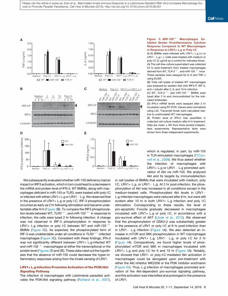
Figure 3. MiR-155–/– Macrophages Ex-
hibited Similar Proinflammatory Cytokine
Response Compared to WT Macrophages
in Response to LRV1+ L.g or Poly I:C
(A–E) BMMs were infected with LRV1+ L.g (+) or
LRV1� L.g (�). Cells were treated with medium or
poly I:C (2 mg/ml) as a control for indicated times.
(A) The cell-free culture supernatant was collected
24 hr post-treatment from treated macrophages
derived fromWT, TLR-3�/�, andmiR-155�/�mice.
These samples were assayed for IL-6 and TNF-a
using ELISA.
(B) Total cell lysate of treated WT macrophages
was analyzed by western blot with IRF3-P, IRF-3,
and g-tubulin after 2, 6, and 10 hr infection.
(C) WT, TLR-3�/�, and miR-155�/� BMMs were
lysed after 2 hr and immunoblotted for the indi-
cated antibodies.
(D) IFN-b mRNA levels were assayed after 2 hr
incubation using RT-PCR. Values were normalized
using L32. Transcript levels were calculated rela-
tive to unstimulated WT macrophages.
(E) Protein level of IFN-b was quantified in
collected cell culture medium after 6 hr treatment.
Data are mean ± SD from three pooled indepen-
dent experiments. Representative blots were
shown from three independent experiments.
Please cite this article in press as: Eren et al., Mammalian Innate Immune Response to a Leishmania-Resident RNA Virus Increases Macrophage Sur-vival to Promote Parasite Persistence, Cell Host & Microbe (2016), http://dx.doi.org/10.1016/j.chom.2016.08.001
Wesubsequently evaluatedwhethermiR-155deficiencyhadan
impact on IRF3activation,which in turncould lead toadecrease in
the mRNA and protein level of IFN-b. WT BMMs, along with mac-
rophages deficient in miR-155 or TLR3, were treated with poly I:C
or infectedwith either LRV1+ L.g or LRV1� L.g.We observed that
in the presence of LRV1+ L.g or poly I:C, IRF-3 phosphorylation
occurred as early as 2 hr following stimulation and became unde-
tectable after 6 hr (Figure 3B). To compare the IRF3 phosphoryla-
tion levels between WT, TLR3�/�, andmiR-155�/� in response to
infection, the cells were lysed 2 hr following infection. A change
was not observed in IRF-3 phosphorylation in response to
LRV1+ L.g infection or poly I:C between WT and miR-155�/�
BMMs (Figure 3C). As expected, the phosphorylated form of
IRF-3 was undetectable under all conditions in TLR3�/�-infectedmacrophages (Figure 3C). Consistent with these findings, IFN-b
was not significantly different between LRV1+ L.g-infected WT
and miR-155�/� macrophages at either the transcriptional or the
protein level (Figures 3D and 3E). These data ruled out the hypoth-
esis that the absence of miR-155 could decrease the hyper-in-
flammatory responses arising from the innate sensing of LRV1.
LRV1+ L.g Infection Promotes Activation of the PI3K/AktSignaling PathwayThe infection of macrophages with Leishmania parasites acti-
vates the PI3K/Akt signaling pathway (Ruhland et al., 2007),
Cell Host &
which is regulated, in part, by miR-155
in TLR-stimulated macrophages (O’Con-
nell et al., 2009). We thus asked whether
the infection of macrophages with
LRV1+ L.g or LRV1� L.g promoted acti-
vation of Akt via miR-155. We analyzed
Akt and its targets by immunodetection
in cell lysates of BMMs that were incubated with medium, poly
I:C, LRV1+ L.g, or LRV1� L.g. At 2 hr post-infection, the phos-
phorylation of Akt was increased in all conditions except in the
medium-treated cells. Phosphorylated Akt levels in LRV1�L.g-infected macrophages were reduced after 6 hr, but were still
evident after 10 hr in both LRV1+ L.g infection and poly I:C
stimulation. Corresponding to these results, the level of
pro-apoptotic Foxo3a gradually decreased in macrophages
incubated with LRV1+ L.g or poly I:C, in accordance with a
pro-survival effect of AKT (Litvak et al., 2012). We observed
that the phosphorylation of GSK3-b was substantially greater
in the presence of LRV1 or poly I:C at 6 hr post-infection than
in LRV1� L.g infection (Figure 4A). We also detected an in-
crease in mTOR and S6K phosphorylation in WT macrophages
incubated with LRV1+ L.g, LRV1� L.g, or poly I:C for 2 hr
(Figure 4A). Comparatively, we found higher levels of phos-
phorylated mTOR and S6K in macrophages incubated with
LRV1+ L.g and poly I:C for 6 and 10 hr (Figure 4A). Notably,
we showed that LRV1- or poly-I:C-mediated Akt activation in
macrophages could be abrogated upon pre-treatment with
either the Akt inhibitor MK2206 or the PI3K inhibitor wortmanin
(Figure S4). Thus, L.g infection of macrophages promoted acti-
vation of the Akt-dependent pro-survival signaling pathway,
and this activation was intensified and prolonged in the presence
of LRV1.
Microbe 20, 1–11, September 14, 2016 5

A
B
C D
Figure 4. MiR-155 Partially Contributed to
TLR-3-Dependent Akt Activation
(A and B) Macrophages incubated with medium,
poly I:C, LRV1+ L.g (+), or LRV1� L.g (�) for indi-
cated time(s). Cells were lysed and total protein
lysates were immunoblotted for Foxo3a, GSK3-b,
phosho-GSK3-b (Ser9), Akt, phosho-Akt (T308),
S6K, phosho-S6K (Thr389), mTOR, phosho-mTOR
(Ser2448), and g-tubulin.
(A)Whole-cell lysate ofWTmacrophages after 2, 6,
and 10 hr post-treatment were immunoblotted for
the indicated proteins.
(B) Macrophages derived from WT, TLR-3�/�, andmiR-155�/� mice were incubated with indicated
conditions for 10 hr, and whole-cell lysates of
indicated samples were subjected to western blot
analysis with the indicated antibodies.
(C) Cell lysates described in (B) were immuno-
blotted for Akt, phosho-Akt (T308), and g-tubulin.
The levels of Akt (Thr308) phosphorylation were
quantified by densitometric analysis. The data
were normalized with total Akt and shown as fold
increase of normalized Akt phosphorylation over
the Akt (T308)/Akt ratio from medium-treated
macrophages.
(D) Flow cytometry analysis of the mean fluores-
cence intensity (MFI) of phosphorylated Akt (T308)
in CD45+ CD11b+ CD11c� F4/80+ lesional mac-
rophages from LRV1+ L.g- or LRV1� L.g-infected
WT and miR-155�/� mice at 4 weeks post-infec-
tion. See also Figure S6.
Representative blots (A–B) and quantification (C)
from at least three independent experiments are
shown. The graph (D) shows pooled data from two
independent experiments (n = 2–5 mice). Data are
expressed as mean ± SD. Unpaired Student’s
t test was used to measure statistical significance.
Not significant (NS) and *p < 0.05. See also Figures
S3 and S4.
Please cite this article in press as: Eren et al., Mammalian Innate Immune Response to a Leishmania-Resident RNA Virus Increases Macrophage Sur-vival to Promote Parasite Persistence, Cell Host & Microbe (2016), http://dx.doi.org/10.1016/j.chom.2016.08.001
Since LRV1+ L.g infection elicited an increase in Akt
phosphorylation in macrophages 10 hr post-infection, we
investigated the role of miR-155 and TLR-3 in LRV1-mediated
Akt activation. Protein levels of phosphorylated Akt and its tar-
gets were evaluated in WT, miR-155�/�, and TLR3�/� macro-
phage lysates at 10 hr post-infection. It was observed that
LRV1-mediated Akt activation and Akt-dependent modulation
of targets relied on TLR-3, and that miR-155 was partly
involved in LRV1-mediated Akt activation (Figures 4B and
4C). Since miR-155 deficiency in TLR-stimulated macrophages
6 Cell Host & Microbe 20, 1–11, September 14, 2016
results in an approximately 1.5-fold in-
crease in the protein levels of the Src
homology 2 domain-containing inositol-
5-phosphatase 1 (SHIP1), a negative
regulator of the PI3K/Akt signaling
pathway (O’Connell et al., 2009), we
investigated the changes in the SHIP1
protein level in WT, miR-155�/�, and
TLR3�/� macrophages in response to
LRV1 at 24 hr post-infection. As previ-
ously reported, we observed a 1.5-fold
increase in the protein level of SHIP1 in
LRV1+ L.g-infected miR-155-deficient macrophages over their
WT counterparts (Figure S5A and S5B), indicating that LRV1-
mediated miR-155 expression could induce Akt activation by
modulating SHIP1 protein levels.
To determine whether miR-155 contributed to Akt acti-
vation in infected animals, we performed flow cytometry
analysis to measure phosphorylated Akt levels in lesional
macrophages from LRV1+ L.g or LRV1� L.g-infected WT
and miR-155�/�-infected mice at 4 weeks post-infection
(Figure S6). We found that lesional macrophages from

A
B
Figure 5. TLR-3-Induced Macrophage Sur-
vival in an Akt-Dependent Manner
(A and B) WT (A) andmiR-155�/� (B) macrophages
were seeded in 96-well plates and were pre-
cultured with DMSO orMK2206 for 1 hr. Cells were
treated with a series of three 10-fold dilutions of
TLR ligands, and were fixed after 48 hr. Macro-
phages were stained with DAPI (nucleus) and
phalloidin (cytoskeleton), and were analyzed with a
high-content microscope. Images are in phalloidin
channel and display an entire 96-well plate. Each
square (352 mM 3 352 mM) represents a well
composed of 49 (7 3 7) pictures taken by 403
lens. The fold increase in cell number was calcu-
lated by normalizing sample cell counts to the cell
numbers within DMSO-treated wells.
Data represent mean ± SD from two independent
experiments with three biological replicates. See
also Table S1.
Please cite this article in press as: Eren et al., Mammalian Innate Immune Response to a Leishmania-Resident RNA Virus Increases Macrophage Sur-vival to Promote Parasite Persistence, Cell Host & Microbe (2016), http://dx.doi.org/10.1016/j.chom.2016.08.001
infected miR-155�/� mice displayed a significant reduction
in phosphorylated Akt levels compared to WT infected
mice (Figure 4D), whereas no significant difference was
observed in LRV� L.g-infected WT or miR-155�/� mice.
Taken together, our results indicated that miR-155 was
involved in the TLR-3 mediated activation of the pro-survival
PI3K/Akt signaling pathway in macrophages infected with
LRV1+ L.g.
Cell Host &
TLR-3-Mediated Cell Survival IsDependent on TLR LigandConcentration and Akt ActivationStudies using knockout animal models
have shown that Leishmania molecules
interact with several TLRs; however, cur-
rent knowledge on the identity and func-
tion of these molecules and their cognate
TLRs is limited (Ives et al., 2014). Our re-
sults demonstrated that LRV1 drastically
enhanced the L.g-mediated activation of
the pro-survival signaling pathway Akt,
suggesting that the innate recognition of
L.g through other TLRs could induce the
activation of Akt. Thus, we raised the
question of how TLR-mediated Akt acti-
vation affected the cellular fitness of mac-
rophages in terms of cell survival. To this
end, we performed high-content micro-
scope analysis of WT and miR-155�/�
macrophages pretreated with DMSO or
the allosteric Akt inhibitor MK2206. We
then incubated the macrophages with
various TLR- and nucleotide-binding olig-
omerization domain-containing protein
(NOD) ligands for 48 hr. We found that
the ligation of certain TLRs, such as
TLR-1/2 and TLR-5, did not promote cell
survival (Figures 5A and 5B). In contrast,
TLR-2/6, TLR-4, and TLR-7 ligation
promoted macrophage survival. This ef-
fect was not, however, diminished by
MK2206 pre-treatment at higher concentrations of TLR ligand
(Figures 5A and 5B ). TLR-3 binding induced cell survival in a
dose- and Akt-dependent manner, while Akt-dependent cell sur-
vival in the presence of TLR-2 and TLR-9 ligands was only
observed at the highest agonist concentration. The fold change
in WT macrophage numbers was similar to the fold change in
miR-155 macrophage numbers in all conditions, except in mac-
rophages treated with the TLR-9 ligand ODN 2006 (Figures 5A
Microbe 20, 1–11, September 14, 2016 7

A B
C
Figure 6. LRV1 Exploited the TLR-3/miR-
155/Akt Pathway to Promote the Persis-
tence of Its Microbial Host
(A–C) WT, TLR-3�/�, and miR-155�/� macro-
phages were infected with promastigotes of
LRV1+ or LRV1� L.g at 1 MOI for 48 hr. Cells were
fixed and processed for high-content analysis by
staining with DAPI and phalloidin.
(A) A representative composite image of a well
containing 7 3 7 pictures with 352 mM 3 352 mM
images from each condition were displayed in
phalloidin channel.
(B) Relative macrophage number was quan-
tified by normalizing cell numbers of the samples
to the cell count of the medium-treated WT
macrophages.
(C) Parasite load in macrophages described in (B)
were quantified.
Mean ± SD was calculated from two independent
experiments with three biological replicates in
triplicate. Data were analyzed using unpaired
Student’s t test. Not significant (NS), *p < 0.05,
**p < 0.01, and ***p < 0.001.
Please cite this article in press as: Eren et al., Mammalian Innate Immune Response to a Leishmania-Resident RNA Virus Increases Macrophage Sur-vival to Promote Parasite Persistence, Cell Host & Microbe (2016), http://dx.doi.org/10.1016/j.chom.2016.08.001
and 5B). Taken together, these findings showed that the engage-
ment of certain TLRs promoted macrophage survival in an Akt-
dependent or -independent manner and that this effect varied
depending on the TLR-ligand concentration.
LRV1+ L.g-Induced Macrophage Survival through theTLR-3/Akt Axis Is Partially Dependent on miR-155Our results demonstrated that TLR-3 stimulation promoted
macrophage survival through the Akt signaling pathway. To
determine whether LRV1-mediated Akt activation promoted
parasite persistence by inducing macrophage survival through
miR-155 or TLR-3, BMMs were pre-incubated with DMSO or
MK2206 and then infected with a multiplicity of infection (MOI)
of 1 of L.g parasites either containing LRV1 or not. We found
that the infection of WT and miR-155�/� macrophages with 1
MOI of LRV1+ L.g increased macrophage survival. This effect
was reversed by MK2206 pre-treatment in WT BMMs, but not
in miR-155�/� and TLR-3�/� BMMs (Figures 6A and 6B). Con-
firming our previous work (Zangger et al., 2014), the presence
of LRV1 within parasites did not affect the parasite number per
macrophage, but did affect infected macrophage survival (Fig-
ure 6C). Taken together, these results indicated that LRV1
dsRNA genome ligation to TLR-3 promoted macrophage sur-
vival, and therefore parasite persistence, by activating the Akt
signaling pathway in a partially miR-155-dependent manner.
DISCUSSION
Our results demonstrated that LRV1 was able to exploit the TLR-
3/miR-155/Akt signaling axis to enhance macrophage survival
and persistence of intracellular Leishmania parasites. Interest-
ingly, the miR-155/Akt signaling pathway is known to cause
myeloid proliferative disorder in both humans and mice (O’Con-
nell et al., 2008; Xue et al., 2014). However, a deficiency in the
myeloid compartment is not observed inmiR-155�/�mice under
basal conditions (Rodriguez et al., 2007), suggesting that miR-
8 Cell Host & Microbe 20, 1–11, September 14, 2016
155 function requires induction. It is known that miR-155 is the
only microRNA upregulated upon TLR-3 ligation in macro-
phages, and also that other TLR receptor agonists also promote
miR-155 expression (O’Connell et al., 2007). Concurrently, we
found that miR-155 was the only microRNA among 1,179 miR-
NAs which was significantly upregulated in the presence of
LRV1 in a TLR-3/TRIF-dependent manner. We observed that
LRV1� L.g infection only weakly induced miR-155 and BIC
expression, suggesting that the innate recognition of parasites
promoted only a slight increase in miR-155 expression through
PRRs other than TLR-3.We found that micewhichwere deficient
in miR-155 had significantly reduced disease pathology when in-
fected with LRV1+ L.g, but not when infected with LRV1� L.g.
These results indicated that LRV1 used host miR-155 as a
persistence mechanism, a situation evocative of certain herpes-
viruses which exert their virulence via the pathogenic overex-
pression of miR-155 from either viral or host origin (Gottwein
et al., 2007; Linnstaedt et al., 2010; Zhao et al., 2011). In this re-
gard, whether other protozoan parasites containing viruses
belonging to the Totiviridae family can subvert the host immune
system through TLR-3/miR-155 remains to be determined.
MiR-155 deficiency in mice was shown to be protective
against T cell- and B cell-driven autoimmune disorders such as
rheumatoid arthritis (Bluml et al., 2011; Kurowska-Stolarska
et al., 2011), experimental autoimmune encephalomyelitis
(O’Connell et al., 2010), and systemic lupus erythematous (Thai
et al., 2013). Interestingly, however, in serum or autoantibody
transfer arthritis models, which are predominantly driven by
innate cells, the severity of joint inflammation is not different be-
tween WT and miR-155�/� mice (Bluml et al., 2011; Kurowska-
Stolarska et al., 2011). In addition, WT and miR-155�/� macro-
phages secrete similar levels of TNF-a after in vitro stimulation
with auto-antibody immune complexes (Kurowska-Stolarska
et al., 2011). Other studies show that miR-155 impacts the pro-
inflammatory cytokine profile of LPS-stimulated macrophages
(Androulidaki et al., 2009) and other cell types (Tili et al., 2007)

Please cite this article in press as: Eren et al., Mammalian Innate Immune Response to a Leishmania-Resident RNA Virus Increases Macrophage Sur-vival to Promote Parasite Persistence, Cell Host & Microbe (2016), http://dx.doi.org/10.1016/j.chom.2016.08.001
at the protein level by transfecting a microRNA mimic or antag-
onist. However, we did not observe a difference in the pro-in-
flammatory cytokine profile between WT and miR-155�/� pri-
mary murine macrophages in response to poly I:C or LRV1+
L.g infection, either in terms of the secretion of pro-inflammatory
cytokines (TNF-a, IL-6, and IFN-b), the expression of IFN-b
mRNA, or the phosphorylation of IRF-3. These results might be
explained by the difference in stimuli, but it should be noted
that transfecting either microRNA mimics or antagonists could
cause an increase in the expression of endogenous microRNA
targets due to the overexpression-mediated saturation of the
intracellular protein complex which guides microRNAs to their
target mRNA (Khan et al., 2009).
We found that the IFN-g- and IL-4-secreting CD4+ T cell
numbers in LRV1+ L.g-infected WT mice were not different
from LRV1+ L.g-infected miR-155�/� mice at the peak of infec-
tion, although it was previously shown that CD4+ T cells of
miR-155-deficient mice secrete more IL-4 and similar levels of
IFN-g under in vitro differentiation conditions (Rodriguez et al.,
2007). However, the IFN-g and/or IL-4 secretion of ex vivo WT
andmiR-155CD4+ T cells was found to be similar in different bio-
logical settings (Kurowska-Stolarska et al., 2011; O’Connell
et al., 2010), suggesting that miR-155 affects CD4+ T cell plas-
ticity in a condition-specific manner. Interestingly, we showed
that CD4+ cells mediated footpad swelling in mice infected
with LRV1+ L.g through miR-155-independent mechanisms.
Thus, further studies are required to understand the role of
CD4+ T cell lineages in thismiR155-independent LRV1-mediated
disease pathology.
We demonstrated that miR-155�/� mice, unlike WT mice,
required CD4+ T cells to control parasite growth in the early
phases of infection. Further, it is known that miR-155 deficiency
impairs the effector function of other-IFN-g-producing cells,
such as NK cells (Trotta et al., 2012), which can play a compen-
satory role in the restriction of parasite growth in the absence of
CD4+ T cells in the early phases of infection (Scharton and Scott,
1993). In our study, we found that both WT andmiR-155�/�mice
were unable to control LRV1+ L.g infection in the absence of IFN-
g mediated immune pressure, suggesting that IFN-g is abso-
lutely essential to infection control in both WT and miR-155�/�
mice.
Several reports have proposed that the infection of macro-
phages with Leishmania spp. promotes parasite persistence by
enhancing macrophage survival (Akarid et al., 2004; Moore
and Matlashewski, 1994; Moore et al., 1994; Ruhland et al.,
2007). Addressing the question on how the innate recognition
of LRV1 modifies host cells to induce the survival of LRV1’s mi-
crobial host, we found that LRV1 induced the phosphorylation of
the pro-survival protein kinase Akt through TLR-3 in a partially
miR-155-dependent manner. Consistent with a previous study,
our findings supported the observation that miR-155 can modu-
late the PI3K/Akt signaling pathway by regulating SHIP1 protein
expression (O’Connell et al., 2009). Functionally, our data
showed that neither the LRV1 status of L.g nor the deficiency
of TLR-3 and miR-155 in macrophages had an impact on the
number of parasites per macrophage in vitro. We cannot, how-
ever, exclude the involvement of TLR-3-expressing phagocytic
cells, other than macrophages, which can act as host cells for
Leishmania parasites, in the miR-155-deficiency-mediated pro-
tection against LRV1+ L.g infection in mice. Nonetheless, our
findings revealed a virulence mechanism in LRV1+ L.g infection
whereby LRV1 conferred a survival advantage to its parasite
host by promoting the survival of infected macrophages, which
are the definitive host cells for Leishmania, through a TLR3/
miR-155/Akt signaling circuit. These results highlighted the
potential clinical importance of oncogenic kinases during leish-
maniasis, and identified several potential therapeutic targets to
treat and prevent the disfiguring complications of cutaneous
leishmaniasis.
EXPERIMENTAL PROCEDURES
Animals
All animal protocols described in this report were approved by the Swiss
Federal Veterinary Office (SFVO), under the authorization numbers 2113.1
and 2113.2. Animal handling and experimental procedures were undertaken
with strict adherence to the ethical guidelines set out by the SFVO and under
inspection by the Department of Security and Environment of the State of
Vaud, Switzerland. Details are given in the Supplemental Experimental
Procedures.
Parasite Culture
Parasites were cultured at 26�C in complete Schneider’smediumwith L-gluta-
mine (Sigma-Aldrich) and were supplemented with 1% penicillin/streptomycin
(Amimed) and 20% fetal calf serum (FCS) (PAA). The parasites were passaged
in culture for not more than five passages and isolated frommouse footpads to
maintain their virulence. Details are given in the Supplemental Experimental
Procedures.
Macrophage Infection
The femurs and tibias of naive mice were washed with medium containing
1% P/S to extract cells, which were differentiated into BMMs for 6 days us-
ing complete DMEM supplemented with L-929-conditioned media (EACC
cell line, Sigma-Aldrich) at 37�C. BMMs were seeded on culture plates
and incubated overnight. BMMs were infected with stationary-phase
LRV1+ or LRV1� L.g at MOI of 10 parasite per macrophage unless
otherwise indicated. BMMs were also treated with poly I:C (Invivogen) at
2 mg/ml or pretreated with DMSO (Sigma-Aldrich) or 5 mM MK2206
(Apexbio) for 1 hr.
MicroRNA Microarray
Four independent experiments were performed. In each experiment, macro-
phages derived from two individual C57BL/6 mice were infected with LRV1+
or LRV1� L.g, or lysed with RNAzol RT (Mrcgene) after 10 hr post-infection.
As a control, macrophages were treated with medium or poly I:C for post-array
verification by RT-PCR. Total RNA was isolated from samples, and microRNA
microarray (Agilent mouse miRbase v18.0) was performed following the man-
ufacturer’s instructions. Details are given in the Supplemental Experimental
Procedures.
The Quantification of MicroRNA and mRNA Using Quantitative
Real-Time PCR
The miR-16 and miR-155 levels were quantified with stem-loop RT-PCR. The
abundance of mRNA and pre-miRNA was quantified with RT-PCR using
Taqman probe or SYBR-Green-based (Roche) detection. The results were
normalized against 60S ribosomal protein L32 (L32) for mRNA and miR-16
for microRNA. Details are given in the Supplemental Experimental Procedures.
Mice Infection and the Quantification of Parasite Burden by
Bioluminescence
Age-matchedmice were infected with 13 106 stationary Leishmania parasites
in both hind footpads. Change in footpad thicknesswasmonitored on aweekly
basis. As described previously (Ives et al., 2011), parasite burden was quanti-
fied in mice by intra-peritoneally injecting D-Luciferin sodium salt (Regis Tech-
nologies) in 13 PBS at a concentration of 150 mg/kg. The images were
Cell Host & Microbe 20, 1–11, September 14, 2016 9

Please cite this article in press as: Eren et al., Mammalian Innate Immune Response to a Leishmania-Resident RNA Virus Increases Macrophage Sur-vival to Promote Parasite Persistence, Cell Host & Microbe (2016), http://dx.doi.org/10.1016/j.chom.2016.08.001
acquired and analyzed using a Xenogen Lumina II imaging system (IVIS) and
Living Image software. Mice were anesthetized with isoflurane during image
acquisition. Oval regions of interest (ROI) were set on the footpads and the
tail to determine the parasite burden and the background, respectively. Biolu-
minescent signals were expressed in units of photons per second (P/s).
Flow Cytometry Analysis
Popliteal lymph nodes (PLNs) or footpad lesions (FLs) were harvested from in-
fected mice at the peak of infection. A single-cell suspension was prepared
from FLs and PLNswith or without collagenase and DNaseI treatment, respec-
tively. Cells isolated from PLNs were counted before PMA and ionomycin
in vitro stimulation. Extracellular staining was performed using anti-mouse
CD4 and CD8 antibodies. Cells were intracellularly stained for IFN-g and IL-
4 cytokines using an intracellular fixation and permeabilization buffer set,
following the manufacturer’s instructions (eBioscience). Cells isolated from
FLs were fixed and permeabilized as described before (Krutzik and Nolan,
2003), and subsequently stained with CD45, CD11b, CD11c, F4/80, and
p-Akt (T308) antibodies. Samples were acquired in BD FACSVerse or LSR II
flow cytometry and were analyzed using FlowJo software. Details are given
in the Supplemental Experimental Procedures.
In Vivo Depletion of CD4 Cells
Anti-CD4 GK1.5 monoclonal antibody was purified from hybridoma (GK1.5;
ATCC) culture supernatant using affinity chromatography on HiTrap Protein
G columns (Amersham-Pharmacia). Mice were intraperitoneally injected
with 500 mg anti-CD4 GK1.5 antibody 5 days prior to infection and on a
weekly basis until 7 weeks post- infection. The splenocytes of mice were har-
vested to verify the efficiency of antibody-mediated depletion using flow
cytometry.
The Quantification of Cytokine Concentration in In Vitro Culture
The concentrations of TNF-a (eBioscience), IL-6 (eBioscience), and IFN-b
(PBL, Interferon Source) in collected supernatants from treated macrophages
were determined using enzyme-linked immuno-sorbent assay (ELISA)
following the manufacturer’s instructions. The plates (Nunc-Immuno) were
read on a Synergy HT Multi-Mode Plate Reader (Biotek Instruments). Wave-
length correction and background signals were subtracted from the absor-
bance values.
Western Blot Analysis
Cells were lysed using cell lysis buffer containing protease/phoshotase
inhibitors. Cell lysates were run on SDS-PAGE, transferred to nitrocellulose
membrane, and analyzed by western blotting using antibodies specific for
the indicated antigens. Details are given in the Supplemental Experimental
Procedures.
High-Content Microscopy
BMMs were counted with Vi-Cell (Beckman Coulter), then seeded on 96-well
tissue-culture-treated clear-bottom plates (Falcon). After overnight incubation,
cells were treated with DMSO (Sigma-Aldrich) or 5 mM MK2206 (ApeXBio) for
1 hr. Subsequent to a washing step, BMMs were treated with ligands in Multi-
TLR array (Invivogen) medium, poly I:C (2 mg/ml), LRV1+ L.g, or LRV1� L.g for
48 hr. Cells were fixedwith freshly made 3.7%PFA in 13 PBS (pH 7.4), stained
with DAPI (Molecular Probes) and Alexa 488-Phalloidin (Molecular Probes)
then washed with 13 PBS using a Biotek MultioFlo FX plate washer. Forty-
nine images (73 7 square, 0.12 mm2) were acquired from each well using Im-
ageXpress Micro XLS. Cell and parasite numbers were counted using an auto-
mated software program (MetaExpress).
Statistical Analysis
All graphs were generated in GraphPad Prism. Unpaired Student’s t tests and
repeated-measure two-way ANOVA tests were performed using Microsoft
Excel and GraphPad Prism, respectively. We considered p < 0.05 and p <
0.001 for unpaired Student’s t test and repeated-measure two-way ANOVA
test with Bonferroni’s post test to be statistically significant, respectively. NS
stands for a non-significant statistical difference. p values are shown as * for
p < 0.05, ** for p < 0.01, and *** for p < 0.001.
10 Cell Host & Microbe 20, 1–11, September 14, 2016
ACCESSION NUMBERS
The ArrayExpress accession number for the microRNA microarray is
E-MTAB-4413.
SUPPLEMENTAL INFORMATION
Supplemental Information includes Figures S1–S6, Table S1, and Supple-
mental Experimental Procedures and can be found with this article online at
http://dx.doi.org/10.1016/j.chom.2016.08.001.
AUTHOR CONTRIBUTIONS
Conceptualization, R.O.E. and N.F.; Methodology, R.O.E. and N.F.; Investiga-
tion,M. Reverte, M. Rossi, M.-A.H., P.C., F.P., R.M., C.H., S.K.D., C.R., R.O.E.,
and N.F.; Resources, L.-F.L., W.R., and S.M.B.; Writing – Original Draft, R.O.E.
and N.F.; Writing – Review & Editing, W.R., S.M.B., C.R., R.O.E., and N.F.;
Funding Acquisition, M.-A.H., C.R., S.M.B., and N.F.
ACKNOWLEDGMENTS
We thank S. Hickerson and K. Owens for assistance with generation and
handling of luciferase-expressing L.g. We thank K. Harshman and F.C. Barras
(Center of Integrative Genomics, UNIL) for the microRNA microarray experi-
ment.We thank D.Monreau andC.U. Eren for assistancewith the high-content
microscopy and S. Masina for critical reading of the manuscript. We thank the
NCCR Geneva Access platform for providing the equipment for the high-con-
tent microscope experiments. This work is funded by grants from the Swiss
National fund for research (FNRS 310030-153204 and IZRJZ3_164176,
N.F.), the Institute for Arthritis Research (iAR), the COST action (CM1307 SE-
FRI: C14.0070, N.F.), the Pierre Mercier Foundation (M.A.H. and C.R.), and
the NIH (R56AI099364 and R01AI029646, S.M.B.).
Received: February 9, 2016
Revised: July 13, 2016
Accepted: August 5, 2016
Published: September 1, 2016
REFERENCES
Akarid, K., Arnoult, D., Micic-Polianski, J., Sif, J., Estaquier, J., and Ameisen,
J.C. (2004). Leishmania major-mediated prevention of programmed cell death
induction in infected macrophages is associated with the repression of mito-
chondrial release of cytochrome c. J. Leukoc. Biol. 76, 95–103.
Androulidaki, A., Iliopoulos, D., Arranz, A., Doxaki, C., Schworer, S.,
Zacharioudaki, V., Margioris, A.N., Tsichlis, P.N., and Tsatsanis, C. (2009).
The kinase Akt1 controls macrophage response to lipopolysaccharide by
regulating microRNAs. Immunity 31, 220–231.
Bluml, S., Bonelli, M., Niederreiter, B., Puchner, A., Mayr, G., Hayer, S.,
Koenders, M.I., van den Berg, W.B., Smolen, J., and Redlich, K. (2011).
Essential role of microRNA-155 in the pathogenesis of autoimmune arthritis
in mice. Arthritis Rheum. 63, 1281–1288.
Bogdan, C., Rollinghoff, M., and Diefenbach, A. (2000). Reactive oxygen and
reactive nitrogen intermediates in innate and specific immunity. Curr. Opin.
Immunol. 12, 64–76.
Cooray, S. (2004). The pivotal role of phosphatidylinositol 3-kinase-Akt signal
transduction in virus survival. J. Gen. Virol. 85, 1065–1076.
Gottwein, E., Mukherjee, N., Sachse, C., Frenzel, C., Majoros, W.H., Chi, J.T.,
Braich, R., Manoharan, M., Soutschek, J., Ohler, U., and Cullen, B.R. (2007). A
viral microRNA functions as an orthologue of cellular miR-155. Nature 450,
1096–1099.
Heussler, V.T., Kuenzi, P., and Rottenberg, S. (2001). Inhibition of apoptosis by
intracellular protozoan parasites. Int. J. Parasitol. 31, 1166–1176.
Hsu, L.C., Park, J.M., Zhang, K., Luo, J.L., Maeda, S., Kaufman, R.J.,
Eckmann, L., Guiney, D.G., and Karin, M. (2004). The protein kinase PKR is

Please cite this article in press as: Eren et al., Mammalian Innate Immune Response to a Leishmania-Resident RNA Virus Increases Macrophage Sur-vival to Promote Parasite Persistence, Cell Host & Microbe (2016), http://dx.doi.org/10.1016/j.chom.2016.08.001
required for macrophage apoptosis after activation of Toll-like receptor 4.
Nature 428, 341–345.
Huang, S., and Houghton, P.J. (2003). Targeting mTOR signaling for cancer
therapy. Curr. Opin. Pharmacol. 3, 371–377.
Hurley, J., Roberts, D., Bond, A., Keys, D., and Chen, C. (2012). Stem-loop RT-
qPCR for microRNA expression profiling. Methods Mol. Biol. 822, 33–52.
Ives, A., Ronet, C., Prevel, F., Ruzzante, G., Fuertes-Marraco, S., Schutz, F.,
Zangger, H., Revaz-Breton, M., Lye, L.F., Hickerson, S.M., et al. (2011).
Leishmania RNA virus controls the severity of mucocutaneous leishmaniasis.
Science 331, 775–778.
Ives, A., Masina, S., Castiglioni, P., Prevel, F., Revaz-Breton, M., Hartley, M.A.,
Launois, P., Fasel, N., and Ronet, C. (2014). MyD88 and TLR9 dependent im-
mune responsesmediate resistance to Leishmania guyanensis infections, irre-
spective of Leishmania RNA virus burden. PLoS ONE 9, e96766.
Iwasaki, A., and Medzhitov, R. (2004). Toll-like receptor control of the adaptive
immune responses. Nat. Immunol. 5, 987–995.
Khan, A.A., Betel, D., Miller, M.L., Sander, C., Leslie, C.S., and Marks, D.S.
(2009). Transfection of small RNAs globally perturbs gene regulation by endog-
enous microRNAs. Nat. Biotechnol. 27, 549–555.
Krutzik, P.O., and Nolan, G.P. (2003). Intracellular phospho-protein staining
techniques for flow cytometry: monitoring single cell signaling events.
Cytometry A 55, 61–70.
Kurowska-Stolarska, M., Alivernini, S., Ballantine, L.E., Asquith, D.L., Millar,
N.L., Gilchrist, D.S., Reilly, J., Ierna, M., Fraser, A.R., Stolarski, B., et al.
(2011). MicroRNA-155 as a proinflammatory regulator in clinical and experi-
mental arthritis. Proc. Natl. Acad. Sci. USA 108, 11193–11198.
Linnstaedt, S.D., Gottwein, E., Skalsky, R.L., Luftig, M.A., and Cullen, B.R.
(2010). Virally induced cellular microRNA miR-155 plays a key role in B-cell
immortalization by Epstein-Barr virus. J. Virol. 84, 11670–11678.
Litvak, V., Ratushny, A.V., Lampano, A.E., Schmitz, F., Huang, A.C., Raman,
A., Rust, A.G., Bergthaler, A., Aitchison, J.D., and Aderem, A. (2012). A
FOXO3-IRF7 gene regulatory circuit limits inflammatory sequelae of antiviral
responses. Nature 490, 421–425.
Lombardo, E., Alvarez-Barrientos, A., Maroto, B., Bosca, L., and Knaus, U.G.
(2007). TLR4-mediated survival of macrophages is MyD88 dependent and re-
quires TNF-alpha autocrine signalling. J. Immunol. 178, 3731–3739.
Moore, K.J., andMatlashewski, G. (1994). Intracellular infection by Leishmania
donovani inhibits macrophage apoptosis. J. Immunol. 152, 2930–2937.
Moore, K.J., Turco, S.J., and Matlashewski, G. (1994). Leishmania donovani
infection enhances macrophage viability in the absence of exogenous growth
factor. J. Leukoc. Biol. 55, 91–98.
O’Connell, R.M., Taganov, K.D., Boldin, M.P., Cheng, G., and Baltimore, D.
(2007). MicroRNA-155 is induced during the macrophage inflammatory
response. Proc. Natl. Acad. Sci. USA 104, 1604–1609.
O’Connell, R.M., Rao, D.S., Chaudhuri, A.A., Boldin, M.P., Taganov, K.D.,
Nicoll, J., Paquette, R.L., and Baltimore, D. (2008). Sustained expression of
microRNA-155 in hematopoietic stem cells causes a myeloproliferative disor-
der. J. Exp. Med. 205, 585–594.
O’Connell, R.M., Chaudhuri, A.A., Rao, D.S., and Baltimore, D. (2009). Inositol
phosphatase SHIP1 is a primary target of miR-155. Proc. Natl. Acad. Sci. USA
106, 7113–7118.
O’Connell, R.M., Kahn, D., Gibson,W.S., Round, J.L., Scholz, R.L., Chaudhuri,
A.A., Kahn, M.E., Rao, D.S., and Baltimore, D. (2010). MicroRNA-155 pro-
motes autoimmune inflammation by enhancing inflammatory T cell develop-
ment. Immunity 33, 607–619.
Park, J.M., Greten, F.R., Wong, A., Westrick, R.J., Arthur, J.S., Otsu, K.,
Hoffmann, A., Montminy, M., and Karin, M. (2005). Signaling pathways and
genes that inhibit pathogen-induced macrophage apoptosis–CREB and NF-
kappaB as key regulators. Immunity 23, 319–329.
Rakoff-Nahoum, S., and Medzhitov, R. (2009). Toll-like receptors and cancer.
Nat. Rev. Cancer 9, 57–63.
Rodriguez, A., Vigorito, E., Clare, S., Warren, M.V., Couttet, P., Soond, D.R.,
van Dongen, S., Grocock, R.J., Das, P.P., Miska, E.A., et al. (2007).
Requirement of bic/microRNA-155 for normal immune function. Science
316, 608–611.
Ruhland, A., Leal, N., and Kima, P.E. (2007). Leishmania promastigotes acti-
vate PI3K/Akt signalling to confer host cell resistance to apoptosis. Cell.
Microbiol. 9, 84–96.
Sacks, D., and Sher, A. (2002). Evasion of innate immunity by parasitic proto-
zoa. Nat. Immunol. 3, 1041–1047.
Scharton, T.M., and Scott, P. (1993). Natural killer cells are a source of inter-
feron gamma that drives differentiation of CD4+ T cell subsets and induces
early resistance to Leishmania major in mice. J. Exp. Med. 178, 567–577.
Tam, W. (2001). Identification and characterization of human BIC, a gene on
chromosome 21 that encodes a noncoding RNA. Gene 274, 157–167.
Thai, T.H., Calado, D.P., Casola, S., Ansel, K.M., Xiao, C., Xue, Y., Murphy, A.,
Frendewey, D., Valenzuela, D., Kutok, J.L., et al. (2007). Regulation of the
germinal center response by microRNA-155. Science 316, 604–608.
Thai, T.H., Patterson, H.C., Pham, D.H., Kis-Toth, K., Kaminski, D.A., and
Tsokos, G.C. (2013). Deletion of microRNA-155 reduces autoantibody re-
sponses and alleviates lupus-like disease in the Fas(lpr) mouse. Proc. Natl.
Acad. Sci. USA 110, 20194–20199.
Tili, E., Michaille, J.J., Cimino, A., Costinean, S., Dumitru, C.D., Adair, B.,
Fabbri, M., Alder, H., Liu, C.G., Calin, G.A., and Croce, C.M. (2007).
Modulation of miR-155 and miR-125b levels following lipopolysaccharide/
TNF-alpha stimulation and their possible roles in regulating the response to
endotoxin shock. J. Immunol. 179, 5082–5089.
Trotta, R., Chen, L., Ciarlariello, D., Josyula, S., Mao, C., Costinean, S., Yu, L.,
Butchar, J.P., Tridandapani, S., Croce, C.M., and Caligiuri, M.A. (2012). miR-
155 regulates IFN-g production in natural killer cells. Blood 119, 3478–3485.
Valencia-Sanchez,M.A., Liu, J., Hannon, G.J., and Parker, R. (2006). Control of
translation and mRNA degradation by miRNAs and siRNAs. Genes Dev. 20,
515–524.
Weichhart, T., Hengstschlager, M., and Linke, M. (2015). Regulation of innate
immune cell function by mTOR. Nat. Rev. Immunol. 15, 599–614.
Xue, H., Hua, L.M., Guo, M., and Luo, J.M. (2014). SHIP1 is targeted by miR-
155 in acute myeloid leukemia. Oncol. Rep. 32, 2253–2259.
Zangger, H., Hailu, A., Desponds, C., Lye, L.F., Akopyants, N.S., Dobson, D.E.,
Ronet, C., Ghalib, H., Beverley, S.M., and Fasel, N. (2014). Leishmania ae-
thiopica field isolates bearing an endosymbiontic dsRNA virus induce pro-in-
flammatory cytokine response. PLoS Negl. Trop. Dis. 8, e2836.
Zhao, Y., Xu, H., Yao, Y., Smith, L.P., Kgosana, L., Green, J., Petherbridge, L.,
Baigent, S.J., and Nair, V. (2011). Critical role of the virus-encoded microRNA-
155 ortholog in the induction of Marek’s disease lymphomas. PLoS Pathog. 7,
e1001305.
Cell Host & Microbe 20, 1–11, September 14, 2016 11