Embed Size (px)
Citation preview
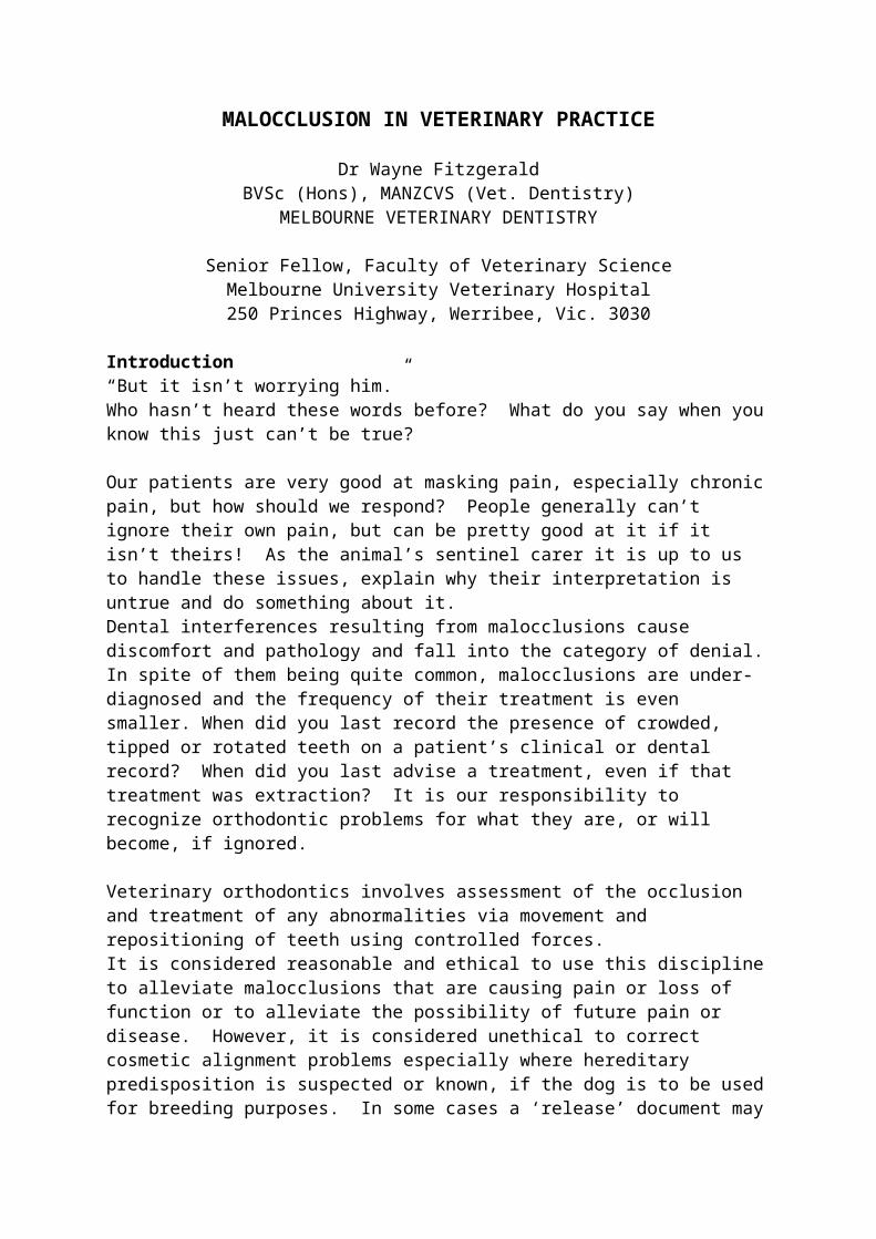
MALOCCLUSION IN VETERINARY PRACTICE
Dr Wayne FitzgeraldBVSc (Hons), MANZCVS (Vet. Dentistry)MELBOURNE VETERINARY DENTISTRY
Senior Fellow, Faculty of Veterinary ScienceMelbourne University Veterinary Hospital250 Princes Highway, Werribee, Vic. 3030
Introduction“But it isn’t worrying him.” Who hasn’t heard these words before? What do you say when you know this just can’t be true?
Our patients are very good at masking pain, especially chronic pain, but how should we respond? People generally can’t ignore their own pain, but can be pretty good at it if it isn’t theirs! As the animal’s sentinel carer it is up to us to handle these issues, explain why their interpretation is untrue and do something about it.Dental interferences resulting from malocclusions cause discomfort and pathology and fall into the category of denial. In spite of them being quite common, malocclusions are under-diagnosed and the frequency of their treatment is even smaller. When did you last record the presence of crowded, tipped or rotated teeth on a patient’s clinical or dental record? When did you last advise a treatment, even if that treatment was extraction? It is our responsibility to recognize orthodontic problems for what they are, or will become, if ignored.
Veterinary orthodontics involves assessment of the occlusion and treatment of any abnormalities via movement and repositioning of teeth using controlled forces.It is considered reasonable and ethical to use this discipline to alleviate malocclusions that are causing pain or loss of function or to alleviate the possibility of future pain or disease. However, it is considered unethical to correct cosmetic alignment problems especially where hereditary predisposition is suspected or known, if the dog is to be used for breeding purposes. In some cases a ‘release’ document may be sought from the client. It is our responsibility for us, as practitioners, to ensure that breeders are aware of these legal and ethical considerations.
“In people, the main reason for treating malocclusion is cosmetic; in dogs and cats however, the reason for performing orthodontic correction is to enhance function and prevent disease.” (Jan Bellows, 1999)
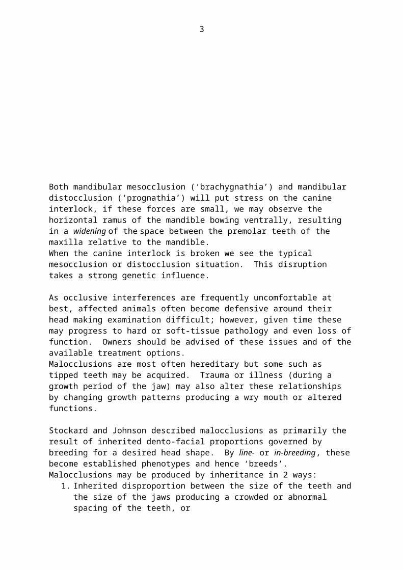
The initial evaluation is a consideration of the symmetry of the head, the incisor relationship, the canine interlock and the number of teeth. The normal head type in feral dogs is the mesocephalic head (refer figure 1). Teeth should not be rotated or misplaced and the line of the teeth should form a smooth, symmetrical curve. The most reliable reference point in the mouth is the relationship of the canine teeth and the maxillary third (or corner) incisor (figure 2). The premolar relationship should be such that the large cusp of the mandibular forth premolar should divide the space between the maxillary third and forth premolars. In fact the premolars
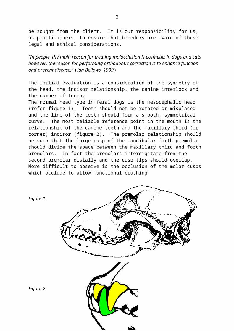
interdigitate from the second premolar distally and the cusp tips should overlap. More difficult to observe is the occlusion of the molar cusps which occlude to allow functional crushing.
Figure 1.
Figure 2.
Both mandibular mesocclusion (‘brachygnathia’) and mandibular distocclusion (‘prognathia’) will put stress on the canine interlock, if these forces are small, we may observe the horizontal ramus of the mandible bowing ventrally, resulting in a widening of the space between the premolar teeth of the maxilla relative to the mandible.When the canine interlock is broken we see the typical mesocclusion or distocclusion situation. This disruption takes a strong genetic influence.
As occlusive interferences are frequently uncomfortable at best, affected animals often become defensive around their head making examination difficult; however, given time these may progress to hard or soft-tissue pathology and even loss of function. Owners should be advised of these issues and of the available treatment options. Malocclusions are most often hereditary but some such as tipped teeth may be acquired. Trauma or illness (during a growth period of the jaw) may also alter these relationships by changing growth patterns producing a wry mouth or altered functions.
2
Stockard and Johnson described malocclusions as primarily the result of inherited dento-facial proportions governed by breeding for a desired head shape. By line- or in-breeding, these become established phenotypes and hence ‘breeds’. Malocclusions may be produced by inheritance in 2 ways:
1. Inherited disproportion between the size of the teeth and the size of the jaws producing a crowded or abnormal spacing of the teeth, or
2. Inherited disproportion between the size or shape of the upper and lower jaws causing improper occlusal relationships.
Genetic isolation and uniformity as seen in wild dogs, rarely produce instances of malocclusion. When this group carries the same genetic information for tooth and jaw size, there is little possibility of individuals inheriting discordant characteristics. Genes that introduce disturbances into the masticatory system would tend to be eliminated from the population. The ‘typical’ specimen has a normal scissor occlusion and tooth/jaw size discrepancies are infrequent, as each tends to have the same jaw relationship (i.e. mesocephalic).
When out-breeding between distinct breeds occurs, malocclusions may develop. Stockard demonstrated this genetic problem by crossbreeding dogs and recording their body structure. He believed that malocclusions occurred in crossbred dogs more from jaw length or width discrepancies than from tooth/jaw size imbalances. However, he did not examine the miniature dog breeds where the latter is common. His research confirmed that “independent inheritance of facial characteristics is a major cause of malocclusion and the rapid increase in malocclusion accompanying urbanization was probably the result of increased out-breeding.” i.e. breeding between dissimilar dogs such as Basset Hounds and English Bulldogs.
Many small and medium-sized dogs carry the gene for achrondroplasia (a deficient growth of cartilage resulting in short legs and an under-developed mid-face. Achrondroplasia is an autosomal dominant trait with variable penetrance; therefore the trait will be expressed more dramatically in some breeds or individuals than others. Stockard explained that some malocclusions were based not on inherited jaw size but to the extent that achrondroplasia was expressed in those animals and severe malocclusions could be developed by crossing morphologically different breeds.Orthodontic research has shown that the simple belief that malocclusion is the result of independent inheritance of dental and facial characteristics is not completely correct but the precise role of heredity as an aetiologic agent has not been clarified.
Consequences often arising from malocclusions: Soft tissue trauma typically results from teeth crowns impinging on gingival or
palatal tissues such as when the crown of a ‘base narrow’ mandibular canine impinges onto the palate. Besides being painful, the resulting depression frequently impacts with debris and food and further inflammation results. This occurs commonly palatal to the maxillary canine and may lead to deep periodontal pockets in this area.
Trauma to bone usually follows as a progression of the above situation. If we use the base narrow example, necrosis of the palatal mucus membrane may occur exposing the bone. An oro-nasal fistula may follow.
3
Trauma to the teeth, seen as abnormal wear patterns, often follows dental ‘interference’. This is most commonly seen as grooves in maloccluding canine teeth.
Tooth loss is a common sequel from periodontitis secondary to malocclusions as it may contribute to food and plaque being retained in areas that are out of normal function making it harder for the animal or owner to care for the teeth.
Chronic pain may lead to changes in personality, behaviour or training. Severe malocclusions may compromise oral function such as excursion
restrictions when only a few teeth meet. This may mean difficulty in mastication and adaptive alterations in swallowing.
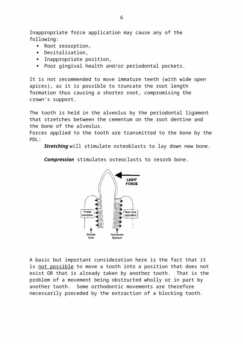
Orthodontic principles:The periodontal ligament (PDL) holds the tooth in the alveolus stretching between the cementum of the root dentine and the bone of the alveolus (‘bundle bone’ tissue). If we apply a force to the tooth, it will be transmitted to the bone by this periodontal ligament: Stretching will stimulate osteoblasts to lay down new bone while compression stimulates osteoclasts to resorb bone. Of critical concern is the magnitude of the forces we apply. If it exceeds the capillary pressure (of the PDL), then we can cause cell death and necrosis. In orthodontic procedures, we aim to move teeth, not to remove them! Inappropriate force application may cause any of the following:
Root resorption, Devitalisation, Inappropriate position, Poor gingival health and/or periodontal pockets.
It is not recommended to move immature teeth (with wide open apices), as it is possible to truncate the root length formation thus causing a shorter root, compromising the crown’s support.
The tooth is held in the alveolus by the periodontal ligament that stretches between the cementum on the root dentine and the bone of the alveolus. Forces applied to the tooth are transmitted to the bone by the PDL:
Stretching will stimulate osteoblasts to lay down new bone. Compression stimulates osteoclasts to resorb bone.
4
A basic but important consideration here is the fact that it is not possible to move a tooth into a position that does not exist OR that is already taken by another tooth. That is the problem of a movement being obstructed wholly or in part by another tooth. Some orthodontic movements are therefore necessarily preceded by the extraction of a blocking tooth.
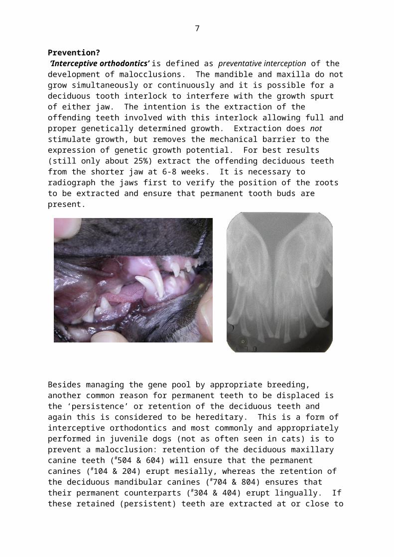
Prevention? ‘Interceptive orthodontics’ is defined as preventative interception of the development of malocclusions. The mandible and maxilla do not grow simultaneously or continuously and it is possible for a deciduous tooth interlock to interfere with the growth spurt of either jaw. The intention is the extraction of the offending teeth involved with this interlock allowing full and proper genetically determined growth. Extraction does not stimulate growth, but removes the mechanical barrier to the expression of genetic growth potential. For best results (still only about 25%) extract the offending deciduous teeth from the shorter jaw at 6-8 weeks. It is necessary to radiograph the jaws first to verify the position of the roots to be extracted and ensure that permanent tooth buds are present.
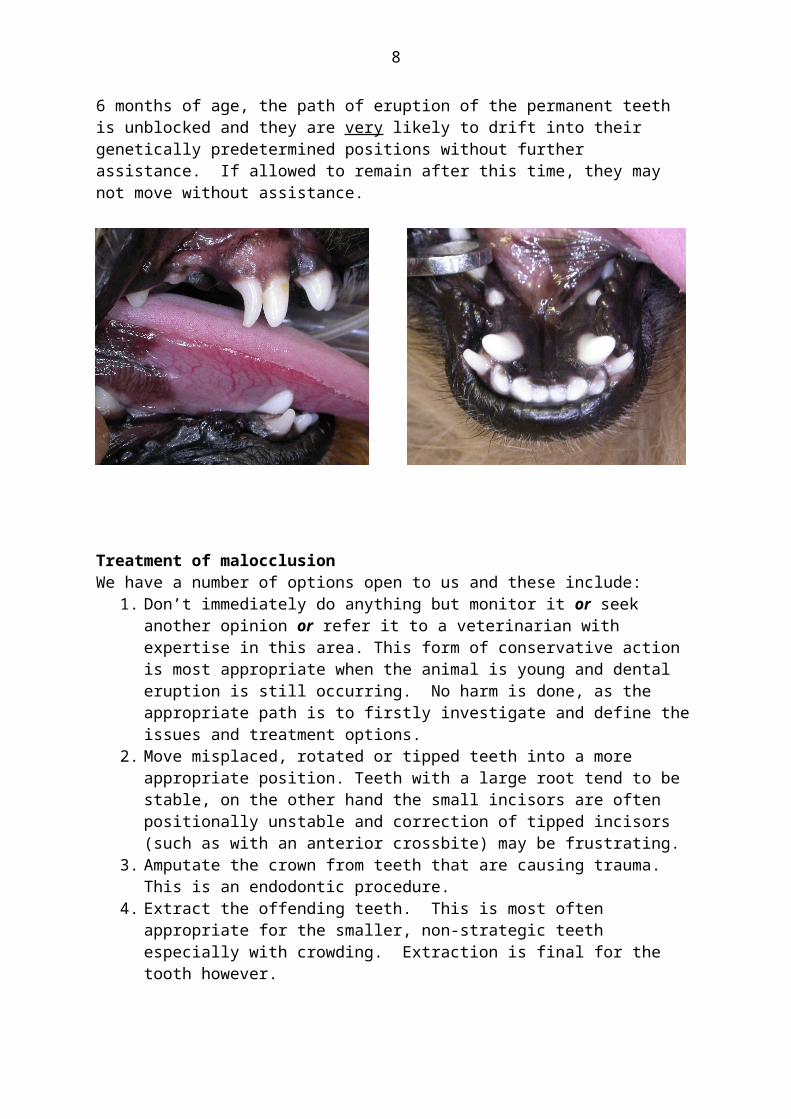
Besides managing the gene pool by appropriate breeding, another common reason for permanent teeth to be displaced is the ‘persistence’ or retention of the deciduous teeth and again this is considered to be hereditary. This is a form of interceptive orthodontics and most commonly and appropriately performed in juvenile dogs (not as often seen in cats) is to prevent a malocclusion: retention of the deciduous maxillary canine teeth (#504 & 604) will ensure that the permanent canines (#104 & 204) erupt mesially, whereas the retention of the deciduous mandibular canines (#704 & 804) ensures that their permanent counterparts (#304 & 404) erupt lingually. If these retained (persistent) teeth are extracted at or close to 6 months of age, the path of eruption of the permanent teeth is unblocked and they are very likely to drift into their genetically predetermined positions without further assistance. If allowed to remain after this time, they may not move without assistance.
5
Treatment of malocclusionWe have a number of options open to us and these include:
1. Don’t immediately do anything but monitor it or seek another opinion or refer it to a veterinarian with expertise in this area. This form of conservative action is most appropriate when the animal is young and dental eruption is still occurring. No harm is done, as the appropriate path is to firstly investigate and define the issues and treatment options.
2. Move misplaced, rotated or tipped teeth into a more appropriate position. Teeth with a large root tend to be stable, on the other hand the small incisors are often positionally unstable and correction of tipped incisors (such as with an anterior crossbite) may be frustrating.
3. Amputate the crown from teeth that are causing trauma. This is an endodontic procedure.
4. Extract the offending teeth. This is most often appropriate for the smaller, non-strategic teeth especially with crowding. Extraction is final for the tooth however.
Orthodontic adjustments may be described as: Rotation Tipping Body movement Crown height adjustment
Rotation of premolars is frequently encountered in the brachycephalic breeds because of crowding. Early extraction of affected premolars may be appropriate to allow room for the remaining 3 premolar teeth within the arcade and lessen the future likelihood of periodontal disease problems.
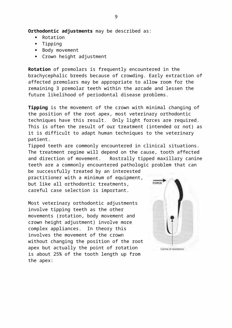
Tipping is the movement of the crown with minimal changing of the position of the root apex, most veterinary orthodontic techniques have this result. Only light forces are required. This is often the result of our treatment (intended or not) as it is difficult to adapt human techniques to the veterinary patient.
6
Tipped teeth are commonly encountered in clinical situations. The treatment regime will depend on the cause, tooth affected and direction of movement. Rostrally tipped maxillary canine teeth are a commonly encountered pathologic problem that can be successfully treated by an interested practitioner with a minimum of equipment, but like all orthodontic treatments, careful case selection is important.
Most veterinary orthodontic adjustments involve tipping teeth as the other movements (rotation, body movement and crown height adjustment) involve more complex appliances. In theory this involves the movement of the crown without changing the position of the root apex but actually the point of rotation is about 25% of the tooth length up from the apex:
Body movement such as crown height adjustment is more difficult to perform and requires brackets, plates, square wire etc. This is the preferred movement when moving teeth rostrally or caudally and results in a more stable tooth. However, these movements are unlikely to be performed by the veterinary practitioner.Adjusting crown height involves moving the whole tooth either into or out of the alveolar socket, it is slower, as it acts in opposition to the periodontal ligament fibres and if moved too quickly the tooth may loosen and exfoliate!
With orthodontic treatments, consider:Firstly:
Define the problem/s and the aims of the treatment. Ensure the client understands, as their expectations are often not in accordance with yours.
Determine the owner’s compliance and willingness to perform home-care and revisits.
The animal’s temperament and behaviour relates to its level of cooperation. Our patients frequently attempt to undo our work and can damage appliances.
When to time the treatment/s?Secondly:
Accurately describe the starting point (documentation may include casts and impressions, photographs, radiographs and charts) and measure and document the results.
Cost in dollars, time and effort. Most appliances are custom made for the patient. Presence of coexisting problems such as periodontal disease or root ankylosis. The practitioner’s level of expertise and equipment. Keep the treatment time to a minimum. Oral hygiene is important and design appliances for minimum discomfort and
irritation.Iatrogenic Damage: 4 possible areas:
1. The appliance itself can irritate and cause soft tissue damage.
7
2. Debris and food will impact around the appliance.3. Any foreign object positioned at or below the gingival margin will cause
irritation …In response to these, we try to keep the treatment time to a minimum, maintain good oral hygiene and design appliances for a minimum of discomfort and irritation.
4. Application of improper forces.
Three classes of force are recognized. 1. Continuous forces; in practice these usually gradually diminish between
adjustments.2. Interrupted forces; these reduce to zero between adjustments.3. Intermittent forces are usually associated with appliances such as the bite plane
plates.
Anchorage is the ‘platform’ from which we exert a force. The anchor should (will) only move a minimal amount. In choosing ‘anchor’ teeth consider their resistance value; this will be related to the number of roots, their size and length (total surface area) and the health of the supporting tissues.
Retention period is the time in which we hold the moved teeth to allow them to gain stability in their new positions. The failure rate will be high if this is not adequate. Some appliances, such as the bite planes, will often double as retention devices. However, patient or owner compliance may influence this time and in practical terms often limit it to about 4 weeks.
Documentation of these cases is very important; we must be able to describe and document the starting points and any/all results for professional, clinical and legal reasons.Photographs are useful and now with the advent of digital photography, we have immediate confirmation that the record is satisfactory plus the low unit cost gives it advantages over conventional film. Oral radiographs are mandatory in the defining of the problems and also to monitor progress and /or success.Impressions and stone casts are necessary for the manufacture of some appliances and are also unequivocal documentation.Charting of all dentistry cases is now accepted as a part of the procedure and conventions should be followed when describing findings.
The American Veterinary Dental College’s Nomenclature Committee defines classifications and terminology that we follow:
Normal Occlusion:Class 0. Scissor bite as seen in the wild canids and some domestic breeds. The lower incisors occlude on the cingulum on the palatal surface of the upper incisors. The arches are symmetrical and the maxillary premolars are buccal to the mandibular premolars.
The Malocclusions:Class 1. Normal occlusion with one or more misplaced or rotated teeth. For example:
8
Crowded or rotated teeth: Toy dog breeds (small jaws) very often have crowding of their incisors whereby the brachycephalic breeds generally have crowding of their premolars.Anterior (rostral) crossbite: A commonly seen condition where one or more upper incisors are displaced lingual to the lowers and the rest of the teeth occlude normally. May result from retained deciduous teeth; considered to be inherited.Caudal (posterior) crossbite: Upper premolars and molars are lingual to lowers. Seen in some dolichocephalic breeds such as the Borzoi. Attempted corrections generally fail and it is presumed to be an inherited disorder.‘Base narrow’ or lingually displaced mandibular canine teeth: One or both mandibular canine teeth are displaced lingually with the tip/s occluding on the hard palate. Considered to be genetic in origin. Rostrally displaced maxillary canines: Generally considered to be genetic. Seen sometimes in a severe form in Shelties, this is called: ‘lance canine’.
Class II. Where the mandibular premolars / molars are positioned caudal to the normal relationship. Correctly called mandibular distocclusion. May be referred to as ‘overshot’ in lay terms.
Class III. The mandibular premolars / molars are rostral (mesial) to the normal.This is known as mandibular mesocclusion (‘undershot’ in lay terms). A ‘level bite’ is a form of class 3 malocclusion.
Class IV or ‘Unclassified’. Wry bite. Caused by a difference in length of the two maxillae and mandibles resulting in an asymmetrical head. Reported to be genetic in origin.
“Dental” (Class I) malocclusions may not always be hereditary. “Bony” (Class II, III & IV) malocclusions are considered to be hereditary.
Commonly used veterinary orthodontic appliances:
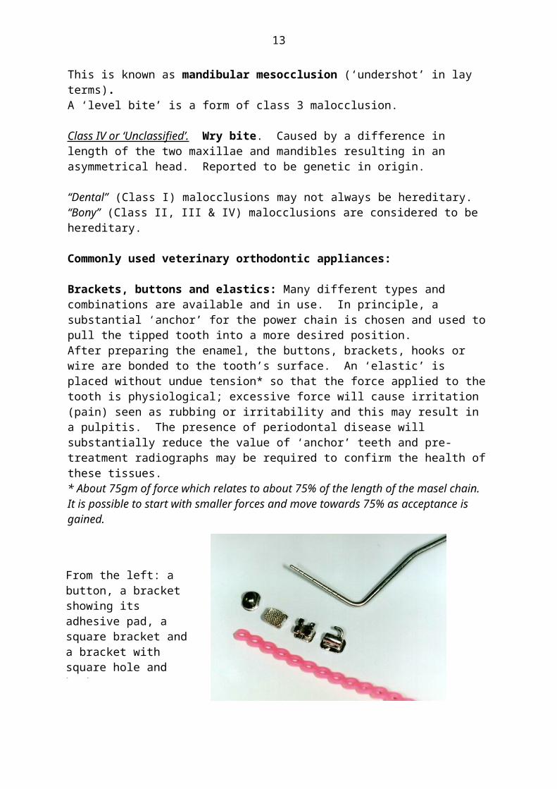
Brackets, buttons and elastics: Many different types and combinations are available and in use. In principle, a substantial ‘anchor’ for the power chain is chosen and used to pull the tipped tooth into a more desired position.After preparing the enamel, the buttons, brackets, hooks or wire are bonded to the tooth’s surface. An ‘elastic’ is placed without undue tension* so that the force applied to the tooth is physiological; excessive force will cause irritation (pain) seen as rubbing or irritability and this may result in a pulpitis. The presence of periodontal disease will substantially reduce the value of ‘anchor’ teeth and pre-treatment radiographs may be required to confirm the health of these tissues.* About 75gm of force which relates to about 75% of the length of the masel chain. It is possible to start with smaller forces and move towards 75% as acceptance is gained.
9
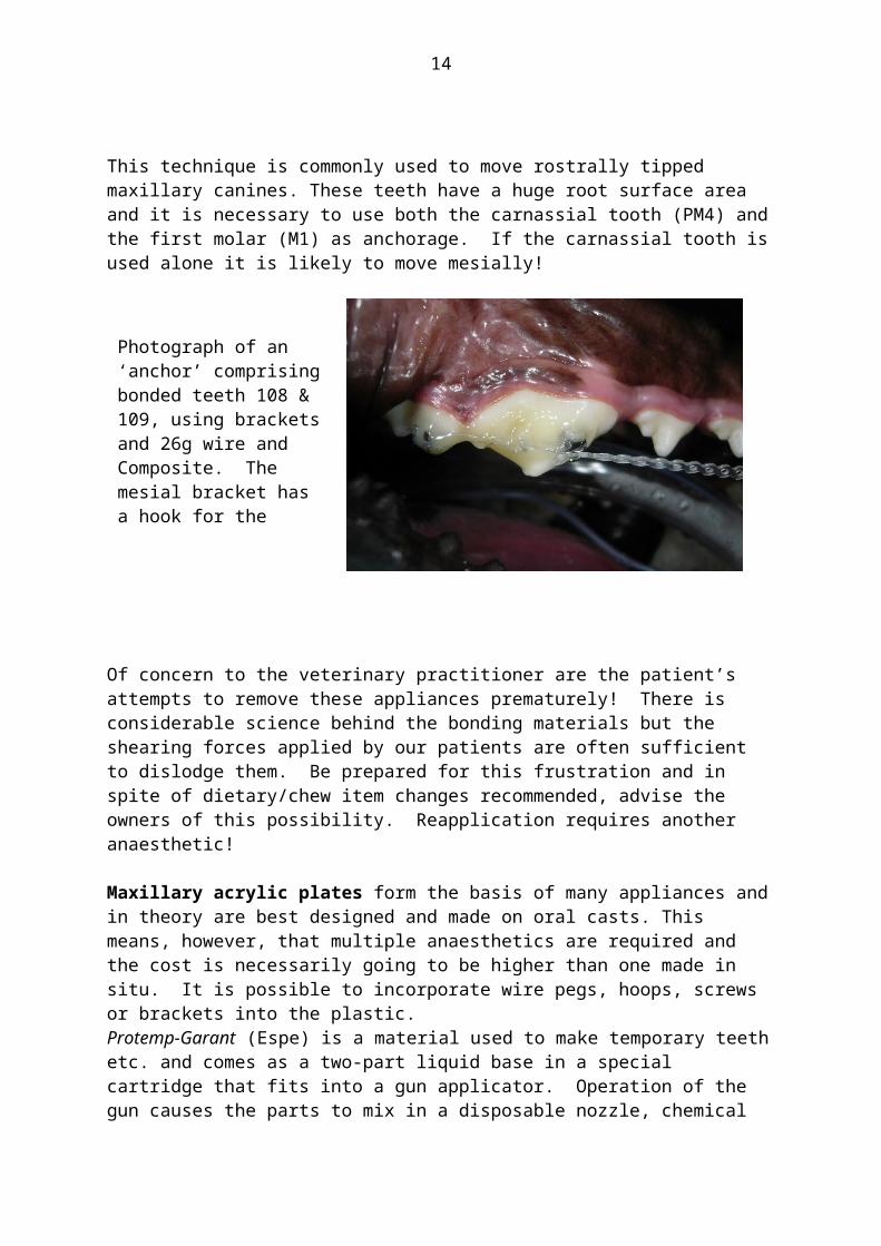
This technique is commonly used to move rostrally tipped maxillary canines. These teeth have a huge root surface area and it is necessary to use both the carnassial tooth (PM4) and the first molar (M1) as anchorage. If the carnassial tooth is used alone it is likely to move mesially!
Of concern to the veterinary practitioner are the patient’s attempts to remove these appliances prematurely! There is considerable science behind the bonding materials but the shearing forces applied by our patients are often sufficient to dislodge them. Be prepared for this frustration and in spite of dietary/chew item changes recommended, advise the owners of this possibility. Reapplication requires another anaesthetic!
Maxillary acrylic plates form the basis of many appliances and in theory are best designed and made on oral casts. This means, however, that multiple anaesthetics are required and the cost is necessarily going to be higher than one made in situ. It is possible to incorporate wire pegs, hoops, screws or brackets into the plastic. Protemp-Garant (Espe) is a material used to make temporary teeth etc. and comes as a two-part liquid base in a special cartridge that fits into a gun applicator. Operation of the gun causes the parts to mix in a disposable nozzle, chemical cure occurs in less than 5 minutes. It is non-exothermic, will (weakly) bond to etched enamel, has some elastic properties (non-brittle) and is simple to use and apply.
Photograph of an ‘anchor’ comprising bonded teeth 108 & 109, using brackets and 26g wire and Composite. The mesial bracket has a hook for the masel chain.
From the left: a button, a bracket showing its adhesive pad, a square bracket and a bracket with square hole and hook.Pink, closed loop masel chain is present also.
10
Another alternative is ‘Triad’* an advanced, visible light cured dental base resin material available in small sheets. It has excellent dimensional stability and polishes to a high lustre using conventional methods and materials. It can be used in the construction of these plates but requires some practise in its use to get good results.
* Triad. Light pink sheets (x6). Code: 135161 Supplier: Densply Australia
204-206 Gipps StreetAbbotsford, Vic. 3067(03) 9417-1777
Historically, ‘blue-tack’ or wax was used as a ‘dam’ to form up a methylmethacrylate resin plate. The serious problem with this material was its curing exothermic properties; its manufacture method also meant that it cured with sharp edges and this had to be addressed in the mouth. Composites can also be used but are too brittle and breakage is more frequent. They are also more expensive in this role; we do use composites to bond the holding wires onto the supporting teeth however.After the plate has cured, a Goldie (or acrylic) bur on a slow speed handpiece is used to shape the planes on the appliance. The angle of the plane is critical as if it is flatter than 450 the force will tend to be compressive and not tipping …i.e. the tooth will not move down the plane. These plates are generally left in-situ after the movement has been completed as they then act as retention devices.
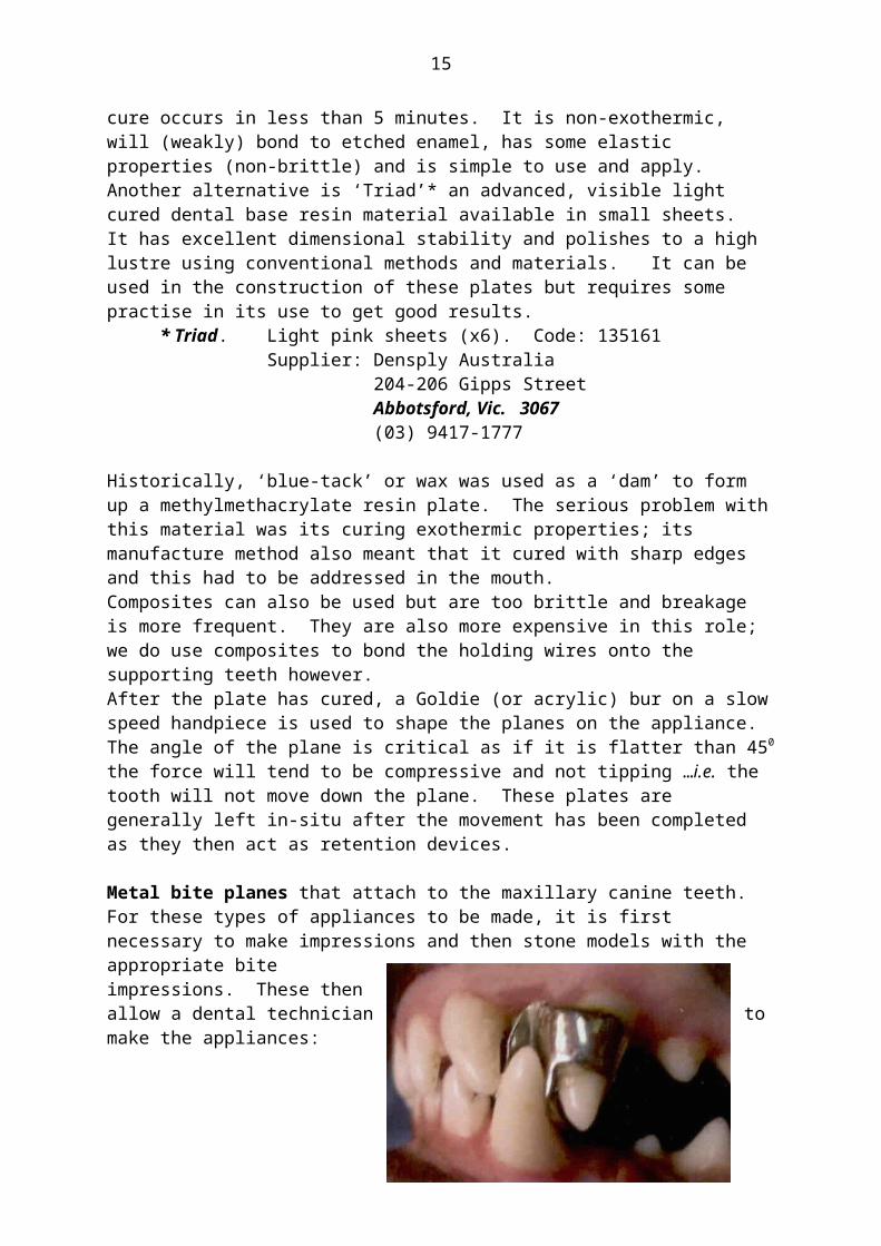
Metal bite planes that attach to the maxillary canine teeth.For these types of appliances to be made, it is first necessary to make impressions and then stone models with the appropriate bite impressions. These then allow a dental technician to make the appliances:
Once fitted (cemented onto the canine teeth), they are fixed and cannot be adjusted. Removal can also be more difficult than with the acrylic plates made insitu as the supporting wires are easily cut to remove the plate.
Mandibular acrylic unit: These are difficult to make removable and can irritate the patient significantly and therefore are of limited use to us however they may be used for the movement of lower incisors if used as a base for hooks etc.
Brackets and wire: Brackets are attached to the tooth like buttons but are designed with hooks, slots, round or square holes etc. for specific applications. These are not made for veterinary use in particular so we select from the vast numbers designed for the form of the human tooth. Wires likewise come in different metals, grades of
11
springiness, sizes and shapes. The general practitioner is unlikely to use these appliances, as they require an extensive material and equipment inventory. ‘Square’ wires can be fitted to square holes or slots in brackets to bodily move teeth; they may or may not be fixed to the bracket with elastic bands. Arch bars, for instance, are made from these materials. The arch bar is an appliance that has been used for moving incisor teeth as in anterior crossbite. In my experience, this is a fairly fragile appliance and our patients seem to take delight in debonding the brackets from the surface of the teeth. The available bonding surface area of incisor teeth is small, as are their roots that make them positionally unstable. Success in moving them may be achieved but retention may be more challenging!
How does this all work in practise?In practise, we are commonly going to be presented with the following malocclusion types:
Anterior crossbite. The arch bar and variations are used to correct these but commonly they are not traumatic, but are generally just cosmetic. Breeders and owners that show will more than likely ask you to correct them. Besides the ethics involved, a serious concern here is that these small teeth are easily moved but just as easily move back! Permanent retention in their ‘new’ position is a problem. It is my advice that these be noted but that as orthodontic movement is not rewarding due to reoccurrence, you don’t attempt to correct them.
Rostrally tipped maxillary canine teeth, either bi- or unilateral. This malocclusion often closes the diastema between the canine and the 3rd incisor thus not allowing the mandibular canine to erupt into its proper occlusion, i.e. becoming ‘base narrow’. If the result is a traumatic occlusion, then treatment is advised: carefully assess the dental relationships and if it is considered that distal movement is possible, then the most useful appliance is the manufacture of a hook on the target canine tooth. I usually make this canine hook out of 22g s/s orthopaedic wire, bonded on with a flowable composite; position the hook coronial to provide appropriate vector traction forces. The anchor: using a flowable composite, bond a hooked bracket onto the 4th premolar’s mesio-buccal surface, follow this with a circlage of 26g s/s wire wrapped around the 1st molar and brought back to the bracket …cover this well with flowable composite to ensure stability. The shearing chewing forces here will show up any weaknesses! A closed loop masel chain is then placed between the 2 hooks and
Arch bar made from brackets and square spring wire with elastic loops. Tooth 101 is under tension, the others are stable.
12
cut off; then stretched about 3-4 loops. If there is excessive face rubbing or irritation, it may be too tight and it should be relaxed. The masel chain should be replaced weekly and its tension adjusted.This is about the most fragile appliance we (vets) use in general practice. The advice to the client is to a) take away chew toys and b) soften the diet.
Linguoversed mandibular canine teeth (‘base narrow) may also be uni- or bilateral. Most of these will be traumatic (to the palate but sometimes the teeth themselves) and require treatment: orthodontic movement, crown height reduction (vital pulpotomy) or extraction (= ‘amputation’ and as such consider this advice carefully).There are a number of plane devices that can be considered, including metal planes made externally on stone models and fitted at a later time, these require multiple anaesthetics and once made, are not amenable to adjustment.The maxillary plate made insitu is the most common appliance used; the planes are made into the material of the plate and must be bilateral to avoid moving the mandible itself! An advantage of these plates is that they are relatively robust. Once the plate has been finished, the dog will have an open mouth as the crowns of the mandibular canine teeth sit at the tops of the planes; this reduces as the teeth tip into the desired positions. As long as the movement path has no obstructions, this movement can happen relatively quickly (often in a few weeks) but effectively the intermittent forces being applied (biting forces) are up to the dog.
Of consideration with these appliances is the build-up of material (food etc) around the teeth and between the plate and the palate. Oral
Typical bite plane maxillary plate; note the open mouth at this time and that tooth 303 was a blockage and has been extracted.
Demonstration model for clients: shows the hook on the canine and the anchor (108 & 109).There have been minor changes to this technique since this model was made.
13
hygiene should be attended to at the weekly checks and the palatitis that develops is transient and resolves quickly once the plate is removed.
DiscussionOwners often don’t see malocclusions or the problems they cause as the affected structures are out of sight and as the patient often masks its discomfort so that it appears ‘pain free’. We may detect them during a visit but considering the apparent health of the patient, we can have problems convincing owners of their significance, however this can be largely addressed if we use visual examples to further support our advice.In the past, treatments have often been confined almost entirely to extraction; however there are other options including orthodontics that are preferable especially when the teeth involved are strategic.
14
Further reading:Orthodontics – Malocclusions and AKC Standards for all breeds. Bellows, J. DVM; Dip.Am.VDC, Dip AmBVP. 13th Annual Vet. Dental Forum, Baltimore, USA. Oct.1999.
Common Dental Procedures. Lobprise, HB & Wiggs, RB. AAHA Press, 2000; pp.98-109
Small Animal Oral Medicine & Surgery. Bojrab, MJ. & Tholen, M. Lea & Febiger, Philadelphia. 1990; Ch.9.
Small Animal Dentistry. Harvey, CE. & Emily, PP. Mosby, Philadelphia. 1993; Ch.8.
Genetic and endocrine basis for differences in form and behaviour. Stockard, CR & Johnson AL. Am. Anat. Memoir 19, Philadephelia, 1941; Wistar Institute
The Dog: His Varied Biological Makeup and Its Relationship to Orthopaedic Diseases. Riser, WH. AAHA publication. 1985.
Breed Predispositions for Orthodontic and Other Dental Conditions in the Dog. (1998) Mary S. Aller. 12th Annual Vet. Dental Forum.
British Small Animal Vet.Assoc. Manual of Small Animal Dentistry 2nd edn. (1995)Ed. Crossley, DA. & Penman, S.
Occlusion / Orthodontics. (1998) Lobprise, HB. 12th Annual Vet. Dental Forum.
Orthodontics. (1998) Legrandre, L. 12th Annual Vet. Dental Forum.
Orthodontic Correction of Lingually Displaced canine Teeth in a Young Dog using Light-cured Acrylic Resin. (1996) Hale, FA. J.Vet.Dent. Vol.13 No.2.
Orthodontics for the Dog. (1986) Ross, DL. The Vet. Clinics of N.America. pp.939-966.
Small Animal Dentistry, Mosby (1993) Harvey, CE. & Emily, PP.
The Angle Classification System of Malocclusion: Is it Appropriate for use in Veterinary Dentistry? (1992) Hennet, PR., Harvey, CE. & Emily, PP. J.Vet.Dent. Vol.9 No.3.
Veterinary Dental Techniques for the Small Animal Practitioner. 2nd edn. (1998) Holstrom, SE., Frost, P. and Eisner, ER. WB.Saunders Company
15