Embed Size (px)
Citation preview

MALNUTRITION RECOGNITION GUIDE

Two factors in the table below must be present for a malnutrition diagnosis.
Acute Illness or Injury Chronic Illness Social or Environmental Factors
Moderate Protein Calorie Malnutrition
Severe Protein Calorie Malnutrition
Moderate Protein Calorie Malnutrition
Severe Protein Calorie Malnutrition
Moderate Protein Calorie Malnutrition
Severe Protein Calorie Malnutrition
Energy Intake <75% of EEE >7 days
≤50 % of EEE >5 days
<75% of EEE ≥1 month
<75% of EEE ≥1 month
<75% of EEE ≥3 months
≤50% of EEE ≥1 month
Weight Loss
1–2% 1 week
5% 1 month
7.5% 3 months
>2% 1 week
>5% 1 month
>7.5% 3 months
5% 1 month
7.5% 3 months
10% 6 months
20% 1 year
>5% 1 months
>7.5% 3 months
>10% 6 months
>20% 1 year
>5% 1 month
>7.5% 3 months
>10% 6 months
>20% 1 year
> 5% 1 month
>7.5% 3 months
>10% 6 months
> 20% 1 year
Body Fat Loss Mild Moderate Mild Severe Mild Severe
Muscle Mass Wasting
Mild Moderate Mild Severe Mild Severe
Fluid (Edema) Mild Moderate to Severe
Mild Moderate to Severe
Mild Moderate to Severe
Hand Grip Strength
N/A Measurably Reduced
N/A Measurably Reduced
N/A Measurably Reduced
EEE: Estimated energy expenditure N/A: Not applicable Reference: Academy of Nutrition and Dietetics & American Society of Parenteral and Enteral Nutrition Clinical Characteristics Malnutrition 2011.
These materials were developed by the Malnutrition Quality Improvement (MQii), a project of the Academy of Nutrition and Dietetics, Avalere Health, and other stakeholders who provided guidance and expertise through a collaborative partnership. Support provided by Abbott. © 2020. All rights reserved.
3
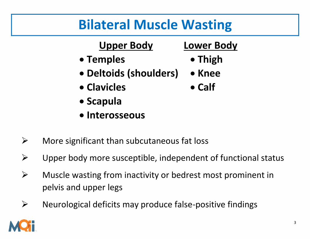
Bilateral Muscle Wasting
Upper Body Lower Body
• Temples • Thigh
• Deltoids (shoulders) • Knee
• Clavicles • Calf
• Scapula
• Interosseous
More significant than subcutaneous fat loss
Upper body more susceptible, independent of functional status
Muscle wasting from inactivity or bedrest most prominent in
pelvis and upper legs
Neurological deficits may produce false-positive findings

4
Bilateral Muscle Wasting (continued)
Knee
Thigh
Calf
Interosseous
Scapula
Temple
Deltoid
Clavicle

5
Bilateral Muscle Wasting: Temples
• Look at patients straight on
and have them turn their
head from side to side
• Inspect for “scooping” or
hollowing of the temporal
region
• Such signs indicate wasting
of the temporalis muscle Photo used with permission. University of California, San Diego. Available at:
http://meded.ucsd.edu/clinicalimg/head_temporal_wasting2.htm. Accessed March 1, 2016.

6
Bilateral Muscle Wasting: Deltoids
Normal
Severe
Inspect straight on with patients’ arms at
side and look for:
• “Squaring” of the shoulders
• Loss of roundness at junction of
shoulder and neck
• Loss of deltoid muscle at junction of
shoulder and arm
• Acromion process may protrude
vs.

7
Bilateral Muscle Wasting: Clavicles
• Inspect for prominence of bone
• Clavicle less prominent for women
• Indicates wasting of pectoral and
deltoid muscles
Normal
Moderate
Severe
8
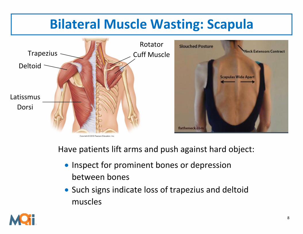
Bilateral Muscle Wasting: Scapula
Have patients lift arms and push against hard object:
• Inspect for prominent bones or depression
between bones
• Such signs indicate loss of trapezius and deltoid
muscles
Deltoid
Trapezius Rotator
Cuff Muscle
Latissmus
Dorsi

9
Bilateral Muscle Wasting: Interosseous
• Engage muscle by pressing forefinger
and middle finger against thumb pad.
While engaged, palpate interosseous
between forefinger and thumb.
• If unable to engage the muscle, place
palm face down with fingers together.
Have the patient adduct and abduct
the thumb to assess the interosseous
muscle.
• For well-nourished patients, the
interosseous muscle will bulge with
good tone (ie, bounce back) as the
thumb is adducted.
Interosseous
muscles

10
Subcutaneous Fat Loss
Inspect and palpate areas where adipose tissue is
normally present. Look for:
• Subjective impressions of loss of fat stores
• Loss of fullness, loose or hanging skin, or hollow
appearance
Note: Age-related loss of subcutaneous tissue
may confound findings
11
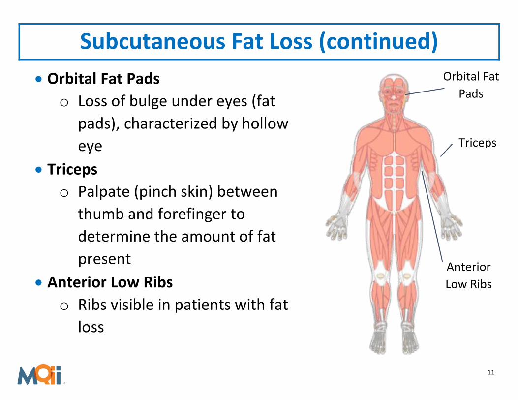
Subcutaneous Fat Loss (continued)
• Orbital Fat Pads
o Loss of bulge under eyes (fat
pads), characterized by hollow
eye
• Triceps
o Palpate (pinch skin) between
thumb and forefinger to
determine the amount of fat
present
• Anterior Low Ribs
o Ribs visible in patients with fat
loss
Orbital Fat
Pads
Triceps
Anterior
Low Ribs

12
Subcutaneous Fat Loss: Orbital Fat Pads
Subcutaneous Fat Loss: Triceps
Slightly bulged fat pads
Normal Mild-Moderate Severe
Slightly dark circle, somewhat tired look
Hollow and sunken look, dark circles, loose skin
Ample fat tissue
between folds of skin
Normal Mild-Moderate Severe
Slightly loose skin; Fingers almost touch when pinching skin between
fingers
Loose skin, very little space between skin folds

13
Fluid Status
• Edema
o Dependent areas
o Ankles, sacrum
• Ascites
o Abdomen
• Dehydration
o Orbital area
o Skin
Orbital Fat
Pads
Skin
Abdomen
Ankle

14
Fluid Status: Edema
• Inspect for swelling in contour
of leg, ankle, or foot
• Palpate by gently squeezing top
of foot, ankle, or front of lower
leg, or by gently pressing skin in
sacral area
• Note if an impression is left
Ankles
Sacrum
Dependent Areas

15
Fluid Status: Ascites
• Stand at foot of bed, look up
toward patient’s head, and
observe contours of
abdomen
• Global abdominal
enlargement is usually cause
by air, fluid, or fat
16
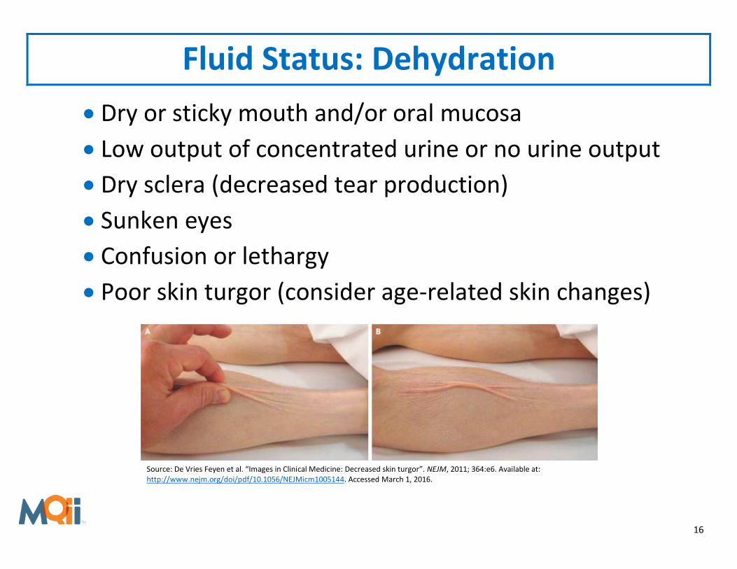
Fluid Status: Dehydration
• Dry or sticky mouth and/or oral mucosa
• Low output of concentrated urine or no urine output
• Dry sclera (decreased tear production)
• Sunken eyes
• Confusion or lethargy
• Poor skin turgor (consider age-related skin changes)
Source: De Vries Feyen et al. “Images in Clinical Medicine: Decreased skin turgor”. NEJM, 2011; 364:e6. Available at: http://www.nejm.org/doi/pdf/10.1056/NEJMicm1005144. Accessed March 1, 2016.

17
Protein-Energy Malnutrition (PEM)
Look for signs of physical PEM, which include:
• Pitting edema
• Dry, flaky, scaly, cracked, bruised, or
bleeding skin
• Dull, brittle, and loose hair
• Ridged, cracked, spoon-shaped, or pale nails
18
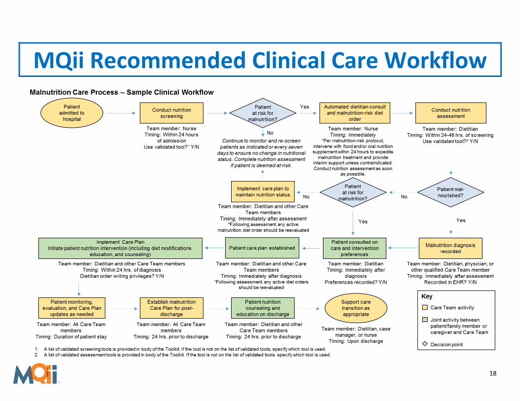
MQii Recommended Clinical Care Workflow