Embed Size (px)
Citation preview

Prelab exercise #7 REPRODUCTIVE SYSTEM Page 1
MALE REPRODUCTIVE SYSTEM Slide 186 This slide is good for studying the general structure of the human testis. Identify the following: tunica albuginea, seminiferous tubules, interstitial CT and interstitial (Leydig) cells. Reserve detailed study of the seminiferous epithelium for the next slide. In the H&E slide you will see the mediastinum testis with its epithelium-lined channels, the rete testis, into which the seminiferous tubules lead. The Masson stained section provides a good view of the interstitial CT, blood vessels, and interstitial (Leydig) cells. In the latter, look for the crystals of Reinke which are peculiar to humans but of unknown significance. What hormone is produced by the Leydig cells and what hormone stimulates its synthesis and secretion? Slide 187 This thin plastic-embedded section of the testis shows the seminiferous tubules and all stages of spermatogenesis in the seminiferous epithelium. With the aid of textbook illustrations and using high magnification identify the following: Sertoli cells with their large pale nuclei and prominent nucleoli, spermatogonia, primary spermatocytes, spermatids, spermatozoa. You are unlikely to find any secondary spermatocytes. Why? Of what does the blood-testis barrier consist? What hormone is produced by Sertoli cells and what hormone stimulates its production? Slide 185 Observe the lining epithelium of the epididymis. It is of the pseudostratified columnar type and consists of two distinct cell types: tall principal cells covered with stereocilia and smaller, basal cells which rest against the basal lamina. What is the difference between a stereocilium and an ordinary cilium (kinetocilium)? In the lumen of the tubules there are some mature spermatozoa as well as cellular debris. In the stroma, note connective tissue, smooth muscles, and blood vessels. What functions are served by the epididymis? Slide 40 This slide shows the same features of the epididymis as the last slide. The stereocilia are particularly nicely demonstrated.

Prelab exercise #7 REPRODUCTIVE SYSTEM Page 2
Slide 188 The ductus deferens (picture to the right) is a thick-walled tube consisting of three concentric layers: mucosa, muscularis, and adventitia. The lumen of the organ is relatively small. The mucosa is composed of a pseudostratified columnar epithelium with stereocilia (similar to the ductus epididymis) and a thin lamina propria rich in elastic fibers, which generally causes the mucosa to form longitudinal folds. The muscularis is very thick and consists of three layers of smooth muscle. The inner and outer layers have a longitudinal orientation, the intermediate layer circular. The adventitia is continuous with the CT of the spermatic cord. Slide 189 This is a thick, but good quality section of the prostate stained with trichrome. The prostate is a compound tubuloalveolar gland. The Masson stain shows the well-developed fibromuscular stroma of this organ. The secretory epithelium is usually of the simple columnar or pseudostratified columnar type. Note the distinct boundary lines between the cells. You will have no difficulty finding the intraluminal lamellated concretions (corpora amylacea), which are a characteristic feature of this gland in older males. Slide 191 Now examine the H&E appearance of the prostate gland. The smooth muscle content of the fibromuscular stroma that characterizes the prostate is not so obvious as in the Masson stained section, but with more careful examination, the smooth muscle component of the stroma is apparent. Corpora amylacea can be seen in the slide of human prostate taken from an older individual, but these concretions are not a distinctive feature in younger prostate. Be sure that you can identify prostate even in the absence of the corpora amylacea.

Prelab exercise #7 REPRODUCTIVE SYSTEM Page 3
Slide 190 Note the three cylinders of erectile tissue in this section of human penis: the two corpora cavernosa and the single corpus spongiosum containing the penile urethra. Various tunicas are present, the tunica albuginea representing one. The dorsal portion of the outer connective tissue contains numerous blood vessels and nerves.
FEMALE REPRODUCTIVE SYSTEM Slide 173 This thin section of the ovary shows well the general features of ovarian follicles. Identify the surface (“germinal”) epithelium, tunica albuginea, hilum, cortex, and medulla. The surface epithelium would be continuous with the mesothelium of the mesovarium at the top of the section. Follow the branches of the ovarian artery as they extend from the hilum (at the top), through the medulla and into the cortex. Compare the stroma of the medulla with that of the cortex. The cortical stroma consists of whorls of densely packed fibroblast-like cells. The medullary stroma is more like loose fibroelastic connective tissue with some smooth muscles cells. Look for examples of each of the following types of ovarian follicles (see figure, below): primordial, unilaminar primary, multilaminar primary, secondary, mature (Graafian). What hormones are secreted by the follicular cells? Identify the following follicular features: 1˚ oocyte, zona pellucida, granulosa cells, theca interna, theca externa. There is no corpus luteum in this section, but you should find many corpora albicantes and atretic follicles.

Prelab exercise #7 REPRODUCTIVE SYSTEM Page 4
Slide 174 The slide of monkey ovary also shows a variety of follicles in the cortex. Slide 175 This slide is useful for showing the size and appearance of a recently formed corpus luteum (“corpus hemorrhagicum”) occupying the center of the section of a human ovary. Note the predominance of the granulosa lutein. The theca lutein cells are easily distinguished in this section as well; they are the more darkly stained cell population immediately outside of the granulosa lutein cells; they also penetrate their way into the corpus luteum with the perivascular connective tissue.

Prelab exercise #7 REPRODUCTIVE SYSTEM Page 5
Slide 176 This slide of monkey uterine tube shows a cross-section of the ampulla of the oviduct. The wall consists of highly folded mucosa, a muscularis (inner circular, outer longitudinal layers of smooth muscle and a serosal layer. Look for the mesosalpinx. Study the mucosa at high magnification. The epithelial layer varies between simple columnar and pseudostratified columnar and is composed mostly of ciliated cells with some interspersed non-ciliated secretory (peg) cells. The lamina propria consists of loose fibroelastic connective tissue. (see figure right). Slide 178 This is a slide of a human uterus with endometrium in the proliferative (follicular) stage of the menstrual cycle (days 5-14). This slide includes endometrium and part of the myometrium. Note that a single layer of tall columnar epithelium covers the luminal surface of the endometrium and extends down to line the tubular glands. The glands are, in turn, surrounded by endometrial stroma. In the proliferative stage the glands are relatively straight and lined by an epithelium that is uniform and shows no cytoplasmic signs of secretion. Numerous mitotic figures can be seen in the glandular epithelium. Most of the endometrium consists of loose connective tissue, mainly fibroblasts, and blood vessels. The stromal cells are uniform, although some of them show mitotic figures. A few lymphocytes can be seen. The endometrial zone adjacent to the myometrium has a greater density of cells and constitutes the stratum basale. This stratum is not sloughed off during
Prelab exercise #7 REPRODUCTIVE SYSTEM Page 6
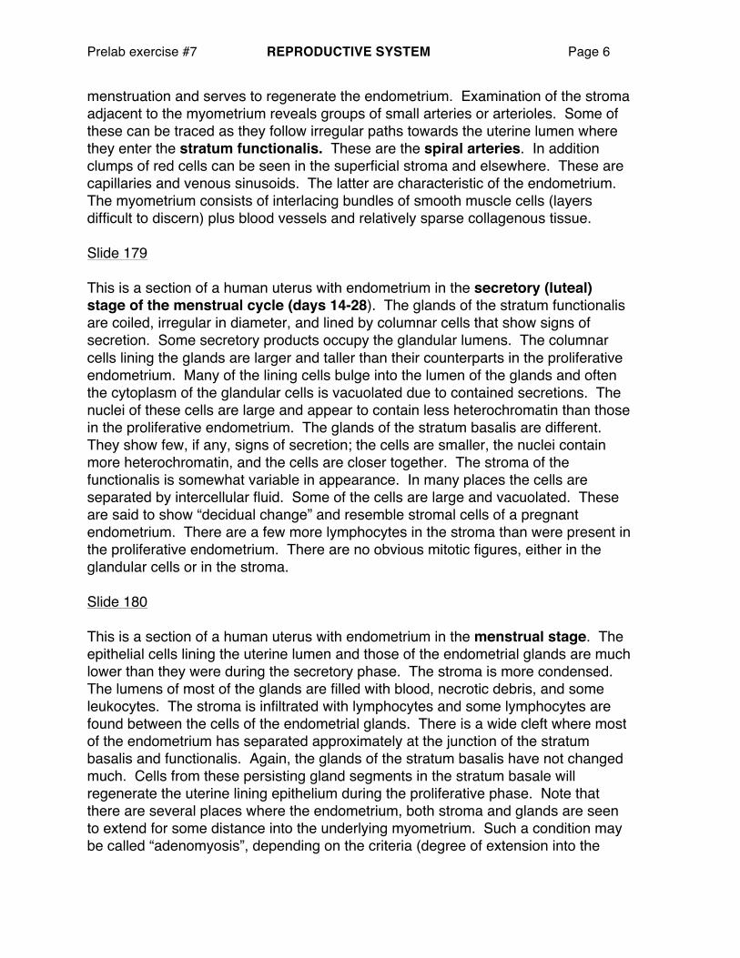
menstruation and serves to regenerate the endometrium. Examination of the stroma adjacent to the myometrium reveals groups of small arteries or arterioles. Some of these can be traced as they follow irregular paths towards the uterine lumen where they enter the stratum functionalis. These are the spiral arteries. In addition clumps of red cells can be seen in the superficial stroma and elsewhere. These are capillaries and venous sinusoids. The latter are characteristic of the endometrium. The myometrium consists of interlacing bundles of smooth muscle cells (layers difficult to discern) plus blood vessels and relatively sparse collagenous tissue. Slide 179 This is a section of a human uterus with endometrium in the secretory (luteal) stage of the menstrual cycle (days 14-28). The glands of the stratum functionalis are coiled, irregular in diameter, and lined by columnar cells that show signs of secretion. Some secretory products occupy the glandular lumens. The columnar cells lining the glands are larger and taller than their counterparts in the proliferative endometrium. Many of the lining cells bulge into the lumen of the glands and often the cytoplasm of the glandular cells is vacuolated due to contained secretions. The nuclei of these cells are large and appear to contain less heterochromatin than those in the proliferative endometrium. The glands of the stratum basalis are different. They show few, if any, signs of secretion; the cells are smaller, the nuclei contain more heterochromatin, and the cells are closer together. The stroma of the functionalis is somewhat variable in appearance. In many places the cells are separated by intercellular fluid. Some of the cells are large and vacuolated. These are said to show “decidual change” and resemble stromal cells of a pregnant endometrium. There are a few more lymphocytes in the stroma than were present in the proliferative endometrium. There are no obvious mitotic figures, either in the glandular cells or in the stroma. Slide 180 This is a section of a human uterus with endometrium in the menstrual stage. The epithelial cells lining the uterine lumen and those of the endometrial glands are much lower than they were during the secretory phase. The stroma is more condensed. The lumens of most of the glands are filled with blood, necrotic debris, and some leukocytes. The stroma is infiltrated with lymphocytes and some lymphocytes are found between the cells of the endometrial glands. There is a wide cleft where most of the endometrium has separated approximately at the junction of the stratum basalis and functionalis. Again, the glands of the stratum basalis have not changed much. Cells from these persisting gland segments in the stratum basale will regenerate the uterine lining epithelium during the proliferative phase. Note that there are several places where the endometrium, both stroma and glands are seen to extend for some distance into the underlying myometrium. Such a condition may be called “adenomyosis”, depending on the criteria (degree of extension into the

Prelab exercise #7 REPRODUCTIVE SYSTEM Page 7
myometrium, presence or absence of inflammatory response, etc.) used for diagnosis. It may or may not produce symptoms. In some studies this has been found in up to 15-20 percent of the female population. Slide 181 This is a slide of the Cervix and vagina of a monkey (the lumen would be on the bottom of the slide). Note the nonkeratinized stratified squamous epithelium of the vagina and vaginal portion of the cervix (“ectocervix”) on the left. Find the well developed cervical mucous glands and the abrupt change of stratified squamous to simple columnar epithelium near the external os of the cervical canal, the junction between endocervix and ectocervix. This region of epithelial transition is clinically important since it is here that many carcinomas of the cervix develop. Slide 54 This Masson stained section of the human uterine ectocervix demonstrates nicely the dominant connective tissue composition of this specialized portion of the organ. Unlike the body of the uterus whose myometrial wall is comprised almost entirely of smooth muscle tissue with just a bit of intervening connective tissue, the cervix is dominated by dense irregular connective tissue (stained greenish-blue here) with scattered smooth muscle cells (stained reddish). How does the tissue composition of the cervix reflect its function? What is cervical “ripening” during the process of parturition? Slide 184 This is a side of the human vagina. The vaginal wall consists of three principle layers: mucosa, muscle and adventitia. The vaginal mucosa, which may be thrown into folds (rugae), is lined by a stratified squamous, nonkeratinizing epithelium, the thickness of which may vary with the reproductive cycle. The epithelial cells are rich in glycogen (thus, the “empty” appearance of these cells in routine histological preps), which serves as a metabolic substrate for the commensal bacteria of the vagina. The epithelium is supported by a loose to moderately dense fibroelastic connective tissue stroma with abundant venous and lymphatic vessels. Note the absence of mucous glands in the vaginal wall. The muscle layer consists predominantly of smooth muscle fibers, which run in spiral-like and longitudinal fashion through the wall. Skeletal muscle fibers from the perineal musculature may also blend with the wall, though not in this particular section.
Prelab exercise #7 REPRODUCTIVE SYSTEM Page 8
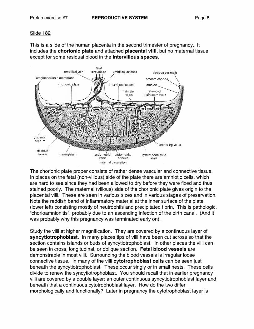
Slide 182 This is a slide of the human placenta in the second trimester of pregnancy. It includes the chorionic plate and attached placental villi, but no maternal tissue except for some residual blood in the intervillous spaces.
The chorionic plate proper consists of rather dense vascular and connective tissue. In places on the fetal (non-villous) side of the plate there are amniotic cells, which are hard to see since they had been allowed to dry before they were fixed and thus stained poorly. The maternal (villous) side of the chorionic plate gives origin to the placental villi. These are seen in various sizes and in various stages of preservation. Note the reddish band of inflammatory material at the inner surface of the plate (lower left) consisting mostly of neutrophils and precipitated fibrin. This is pathologic, “chorioamnionitis”, probably due to an ascending infection of the birth canal. (And it was probably why this pregnancy was terminated early on). Study the villi at higher magnification. They are covered by a continuous layer of syncytiotrophoblast. In many places tips of villi have been cut across so that the section contains islands or buds of syncytiotrophoblast. In other places the villi can be seen in cross, longitudinal, or oblique section. Fetal blood vessels are demonstrable in most villi. Surrounding the blood vessels is irregular loose connective tissue. In many of the villi cytotrophoblast cells can be seen just beneath the syncytiotrophoblast. These occur singly or in small nests. These cells divide to renew the syncytiotrophoblast. You should recall that in earlier pregnancy villi are covered by a double layer: an outer continuous syncytiotrophoblast layer and beneath that a continuous cytotrophoblast layer. How do the two differ morphologically and functionally? Later in pregnancy the cytotrophoblast layer is

Prelab exercise #7 REPRODUCTIVE SYSTEM Page 9
almost completely lost; only a few scattered cells or nests of cells remain in the term placenta. The CT within the villi contains a population of macrophages called Hofbauer cells. In addition to their customary role as part of the immune system, these cells may help to maintain villous homeostasis by affecting water regulation and nutrient/waste transport. Slide 183 This is an excellent section of a delivered term human placenta and worth careful study. Identify the chorionic plate (better named “chorioamnionic plate” since it consists of fused chorion and amniotic membranes). An intact amniotic membrane covers the fetal surface (it is continuous with amniotic covering of the umbilical cord that attaches to the chorionic plate). At the fetal surface find the origin of stem villi and, extending into the intervillous space, their branches: terminal villi and anchoring villi. Note that the outer covering of the villi is at this stage almost exclusively syncytiotrophoblast. In the smallest terminal villi note the proximity of fetal capillaries to the syncytiotrophoblast. Of what would the minimal hemochorial placental barrier consist? At the side opposite the chorionic plate in this section there remains attached some decidua basalis (maternal tissue) consisting of large decidual cells and abundant eosinophilic fibrin material (so-called Nitabuch’s layer). Note the attachment of anchoring villi to this layer. Where does detachment of the placenta from the uterine wall usually occur at parturition?
Cross section of placental villus, early pregnancy (≈ 300X)
1. CT stroma with fetal b.v. & nucleated RBCs
2. Hofbauer cell 3. Cytotrophoblast 4. Syncytiotrophoblast