Embed Size (px)
Citation preview
Malaria: the clinical basics
Cristin Weekley, BA, & D. Scott Smith, MD Stanford University
February 2013
Prepared as part of an education project of the
Global Health Education Consortium
and collaborating partners
Page 2 Page 2
Learning Overview (Clinical)
1. What is malaria?
2. Malaria Parasites
3. Life Cycle
4. Transmission
5. Who is at Risk?
6. Symptoms
7. Diagnosis
8. Treatment
IMPORTANT NOTE: This module provides information about diagnosis and treatment that is current as of
January 2013. However, treatments can vary widely depending on predominant vectors, drug resistance
levels, patient conditions, drug availability, local practice norms, and of course, the advent of new
treatments. You should always consult current treatment protocols and local authorities before providing
treatment and not regard this module as a definitive treatment guide.
Page 3 Page 3
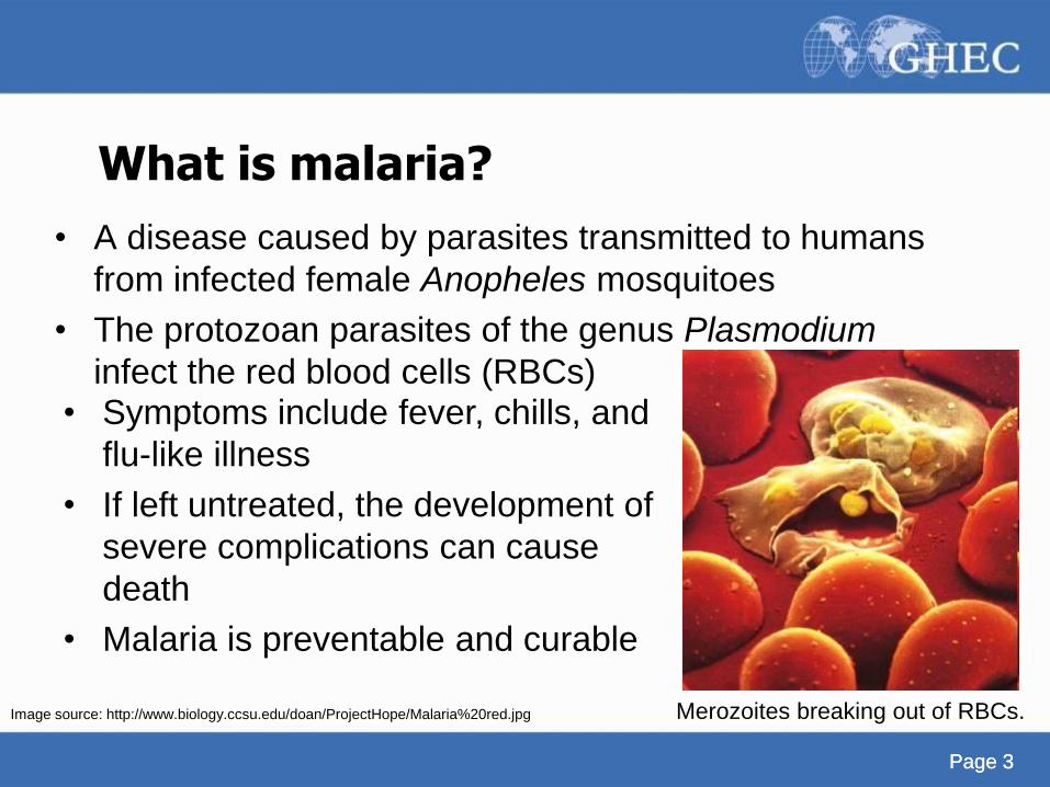
What is malaria?
• A disease caused by parasites transmitted to humans
from infected female Anopheles mosquitoes
• The protozoan parasites of the genus Plasmodium
infect the red blood cells (RBCs)
• Symptoms include fever, chills, and
flu-like illness
• If left untreated, the development of
severe complications can cause
death
• Malaria is preventable and curable
Merozoites breaking out of RBCs. Image source: http://www.biology.ccsu.edu/doan/ProjectHope/Malaria%20red.jpg
Page 4
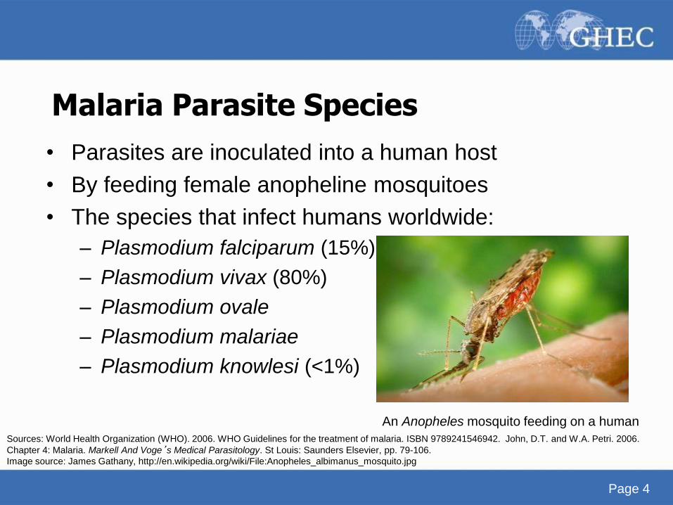
• Parasites are inoculated into a human host
• By feeding female anopheline mosquitoes
• The species that infect humans worldwide:
– Plasmodium falciparum (15%)
– Plasmodium vivax (80%)
– Plasmodium ovale
– Plasmodium malariae
– Plasmodium knowlesi (<1%)
An Anopheles mosquito feeding on a human
Malaria Parasite Species
Sources: World Health Organization (WHO). 2006. WHO Guidelines for the treatment of malaria. ISBN 9789241546942. John, D.T. and W.A. Petri. 2006.
Chapter 4: Malaria. Markell And Voge’s Medical Parasitology. St Louis: Saunders Elsevier, pp. 79-106.
Image source: James Gathany, http://en.wikipedia.org/wiki/File:Anopheles_albimanus_mosquito.jpg
Page 5
Malaria Parasites Species Plasmodium falciparum
• Almost entirely confined to tropics and subtropics
• Accounts for 15% of malaria infection
• Clinically and morphologically different from other
malarias
– Gametocytes are elongate/sausage-shaped
– Schizogony usually does not take place in
peripheral blood of falciparum malaria
– P. falciparum attacks all stages of red blood cells
Source: John, D.T. and W.A. Petri. 2006. Chapter 4: Malaria. Markell And Voge’s Medical Parasitology. St Louis: Saunders Elsevier, pp. 79-106.
Image source: http://www.plosmedicine.org/article/info:doi/10.1371/journal.pmed.1000048
Page 6
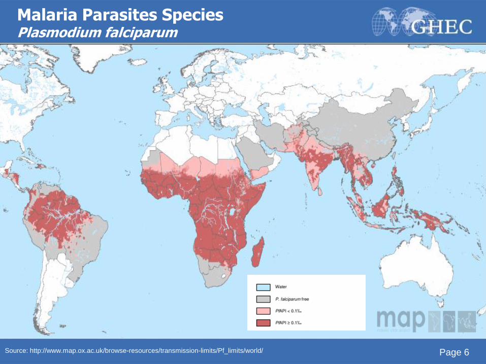
Malaria Parasites Species Plasmodium falciparum
Source: http://www.map.ox.ac.uk/browse-resources/transmission-limits/Pf_limits/world/
Page 7
Malaria Parasites Species Plasmodium vivax
• ‘Vivax’ = vigorous (Latin)
• Accounts for 80% of malaria infection
• Predominant malaria parasite in the world
– Only malarial parasite whose range extends into
temperate regions
– Seldom causes severe disease
Sources: John, D.T. and W.A. Petri. 2006. Chapter 4: Malaria. Markell And Voge’s Medical Parasitology. St Louis: Saunders Elsevier, pp. 79-106. Rosenthal, P.
2008. Knol. http://knol.google.com/k/philip-rosenthal/malaria/dtqBgoqt/MRpmSg#
Page 8
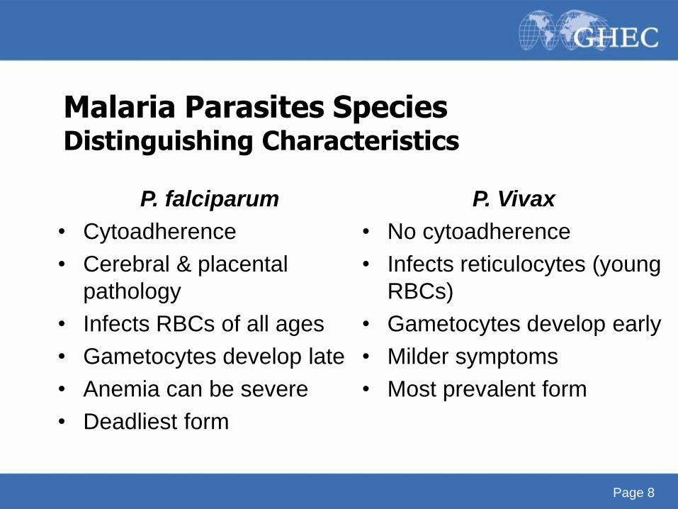
Malaria Parasites Species Distinguishing Characteristics
P. falciparum
• Cytoadherence
• Cerebral & placental
pathology
• Infects RBCs of all ages
• Gametocytes develop late
• Anemia can be severe
• Deadliest form
P. Vivax
• No cytoadherence
• Infects reticulocytes (young
RBCs)
• Gametocytes develop early
• Milder symptoms
• Most prevalent form
Page 9
Malaria Parasites Species Plasmodium ovale
• Only been known since 1922
• Widely distributed in tropical Africa, especially West
African coast
• Main distinctive morphologic feature is ovoid shape of
many of infected RBCs
• Other features:
– Parasite not as ameboid as P. vivax
– Nuclei in all stages are larger
Source: John, D.T. and W.A. Petri. 2006. Chapter 4: Malaria. Markell And Voge’s Medical Parasitology. St Louis: Saunders Elsevier, pp. 79-106.
Page 10
Malaria Parasites Species Plasmodium malariae
• Is rarer than P. vivax or P. falciparum
• Occurs primarily in subtropical and temperate areas
where other malaria spp. are found
• Distinguishing characteristics:
– Asexual cycle lasts 24 hours longer than other spp.
– Infected cell is not enlarged
Source: John, D.T. and W.A. Petri. 2006. Chapter 4: Malaria. Markell And Voge’s Medical Parasitology. St Louis: Saunders Elsevier, pp. 79-106.
Page 11
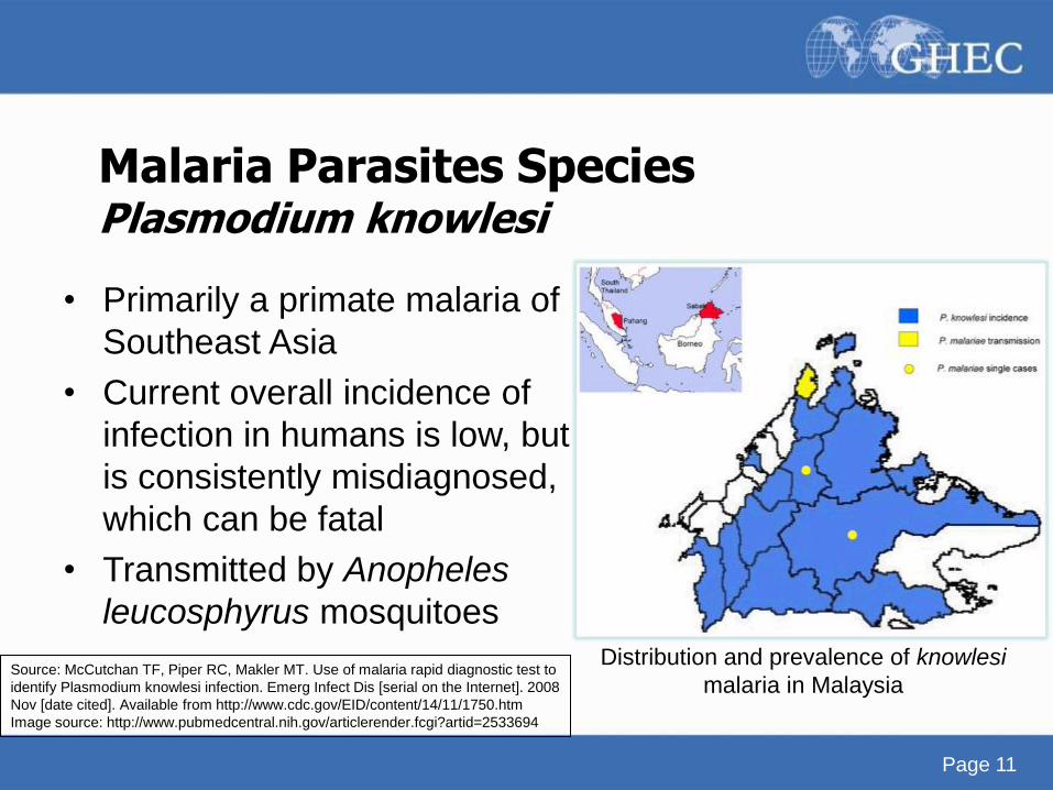
Malaria Parasites Species Plasmodium knowlesi
• Primarily a primate malaria of
Southeast Asia
• Current overall incidence of
infection in humans is low, but
is consistently misdiagnosed,
which can be fatal
• Transmitted by Anopheles
leucosphyrus mosquitoes
Distribution and prevalence of knowlesi
malaria in Malaysia Source: McCutchan TF, Piper RC, Makler MT. Use of malaria rapid diagnostic test to
identify Plasmodium knowlesi infection. Emerg Infect Dis [serial on the Internet]. 2008
Nov [date cited]. Available from http://www.cdc.gov/EID/content/14/11/1750.htm
Image source: http://www.pubmedcentral.nih.gov/articlerender.fcgi?artid=2533694
Page 12
Life Cycle Vocabulary
• Trophozoite: the feeding stage of a protozoan parasite
(intracellular)
• Schizogony: the process of asexual reproduction in
which the nucleus undergoes multiple divisions prior to
cell division
Page 13
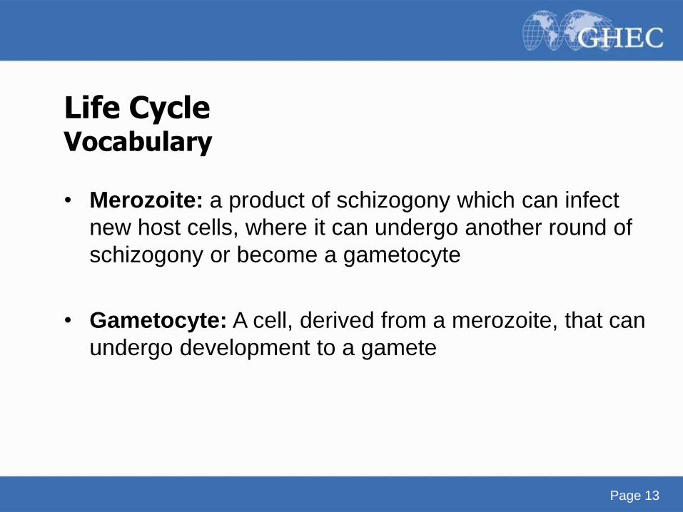
Life Cycle Vocabulary
• Merozoite: a product of schizogony which can infect
new host cells, where it can undergo another round of
schizogony or become a gametocyte
• Gametocyte: A cell, derived from a merozoite, that can
undergo development to a gamete
Page 14
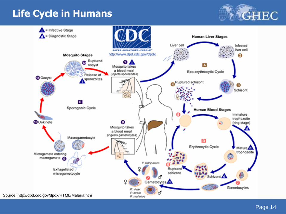
Life Cycle in Humans
Source: http://dpd.cdc.gov/dpdx/HTML/Malaria.htm
Page 15
Notes on the Life Cycle in Humans
The malaria parasite life cycle involves two hosts. During a blood meal, a malaria-infected female
Anopheles mosquito inoculates sporozoites into the human host (1). Sporozoites infect liver cells
(2) and mature into schizonts (3), which rupture and release merozoites (4). (Of note, in P. vivax
and P. ovale a dormant stage [hypnozoites] can persist in the liver and cause relapses by invading
the bloodstream weeks, or even years later.) After this initial replication in the liver (exo-
erythrocytic schizogony (A)), the parasites undergo asexual multiplication in the erythrocytes
(erythrocytic schizogony (B)). Merozoites infect red blood cells (5). The ring stage trophozoites
mature into schizonts, which rupture releasing merozoites (6). Some parasites differentiate into
sexual erythrocytic stages (gametocytes) (7). Blood stage parasites are responsible for the clinical
manifestations of the disease.
The gametocytes, male (microgametocytes) and female (macrogametocytes), are ingested by an
Anopheles mosquito during a blood meal (8). The parasites’multiplication in the mosquito is
known as the sporogonic cycle (C). While in the mosquito's stomach, the microgametes penetrate
the macrogametes generating zygotes (9). The zygotes in turn become motile and elongated
(ookinetes) (10) which invade the midgut wall of the mosquito where they develop into oocysts
(11). The oocysts grow, rupture, and release sporozoites (12), which make their way to the
mosquito's salivary glands. Inoculation of the sporozoites into a new human host perpetuates the
malaria life cycle (1).
Source: http://dpd.cdc.gov/dpdx/HTML/Malaria.htm
Page 16
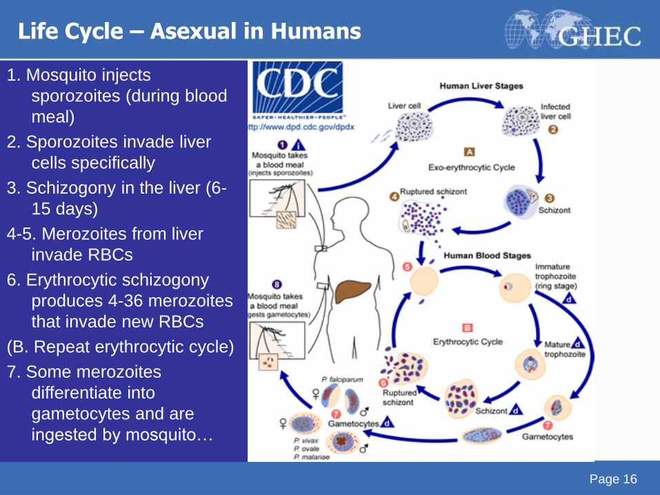
Life Cycle – Asexual in Humans
1. Mosquito injects
sporozoites (during blood
meal)
2. Sporozoites invade liver
cells specifically
3. Schizogony in the liver (6-
15 days)
4-5. Merozoites from liver
invade RBCs
6. Erythrocytic schizogony
produces 4-36 merozoites
that invade new RBCs
(B. Repeat erythrocytic cycle)
7. Some merozoites
differentiate into
gametocytes and are
ingested by mosquito…
Page 17 Page 17
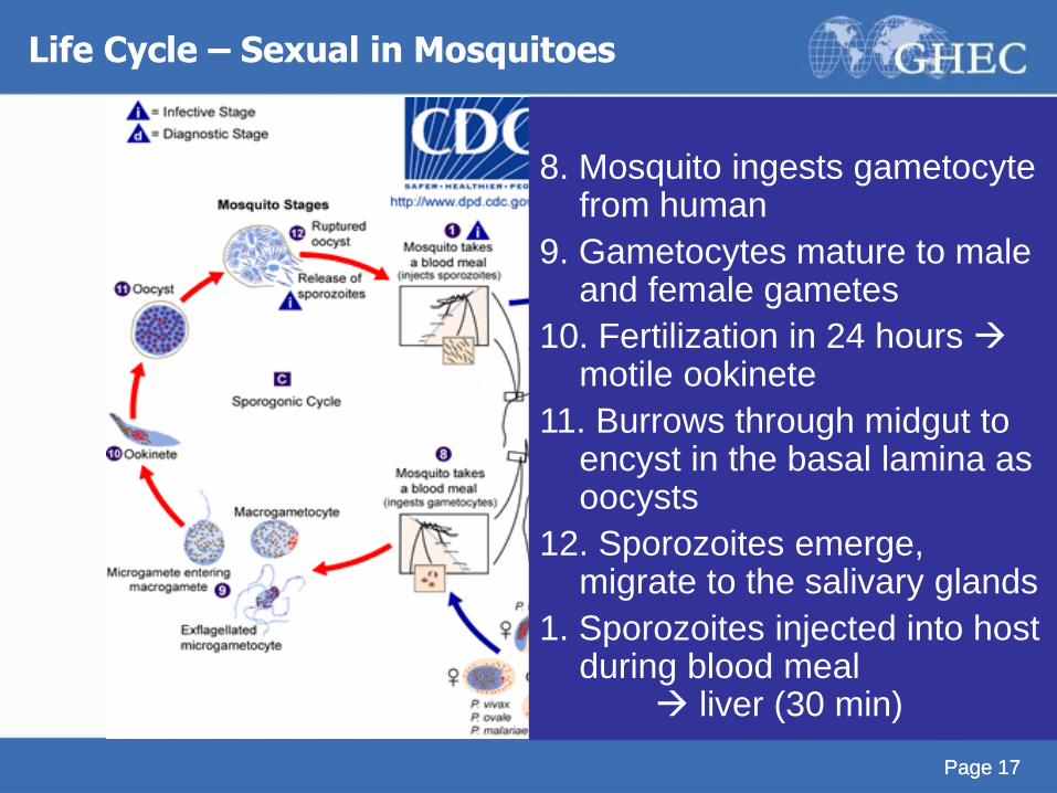
Life Cycle – Sexual in Mosquitoes
8. Mosquito ingests gametocyte from human
9. Gametocytes mature to male and female gametes
10. Fertilization in 24 hours motile ookinete
11. Burrows through midgut to encyst in the basal lamina as oocysts
12. Sporozoites emerge, migrate to the salivary glands
1. Sporozoites injected into host during blood meal liver (30 min)
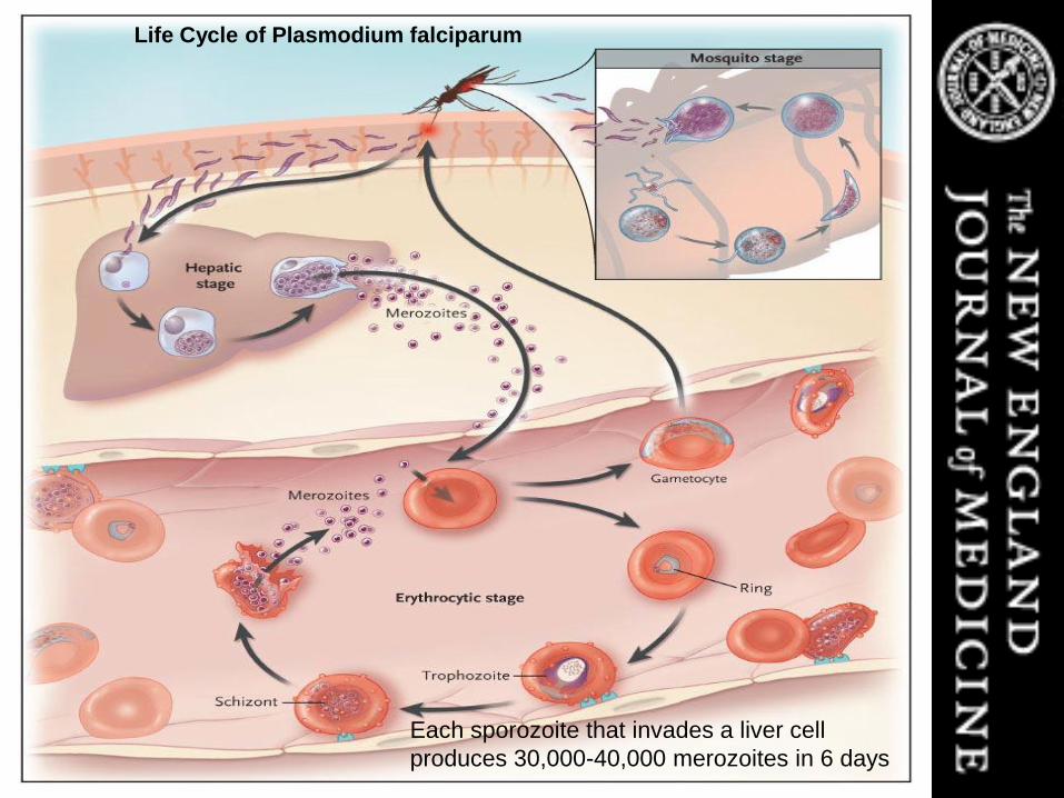
Life Cycle of Plasmodium falciparum
Each sporozoite that invades a liver cell
produces 30,000-40,000 merozoites in 6 days
Page 19
Notes on the figure: Life Cycle of Plasmodium falciparum Elements that are important for the pathogenesis of severe malaria are
shown. Erythrocytes containing P. falciparum in mature intraerythrocytic stages (trophozoites and schizonts) adhere to vascular endothelium, thereby avoiding clearance by the spleen. High numbers of circulating parasites and elaboration of host and parasite factors in the vasculature of various organs lead to the manifestations of severe malaria.
Sources: Rosenthal P. N Engl J Med 2008;358:1829-1836; http://www.cdc.gov/malaria/facts.htm

Page 20
• After they are released
into the blood, each
merozoite invades a red
blood cell, where it
produces 8-24 daughter
cells in 48 hours
• Daughter cells released
into the blood and are
ingested by a feeding
mosquito Male gametocyte of Plasmodium falciparum (yellow)
surrounded by RBCs
Source: http://www.cdc.gov/malaria/facts.htm
Image source: www.jhsph.edu/bin/n/v/22malaria.jpg
After a single sporozoite (the parasite form inoculated by the female mosquito) of P. falciparum invades a liver cell, the
parasite grows in 6 days and produces 30,000-40,000 daughter cells (merozoites) which are released into the blood when the
liver cell ruptures. In the blood, after a single merozoite invades a red blood cell, the parasite grows in 48 hours and produces
8-24 daughter cells, which are released into the blood when the red blood cell ruptures.
Transmission – Within Humans
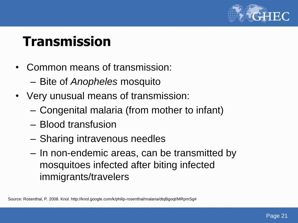
Transmission
• Common means of transmission:
– Bite of Anopheles mosquito
• Very unusual means of transmission:
– Congenital malaria (from mother to infant)
– Blood transfusion
– Sharing intravenous needles
– In non-endemic areas, can be transmitted by
mosquitoes infected after biting infected
immigrants/travelers
Page 21
Source: Rosenthal, P. 2008. Knol. http://knol.google.com/k/philip-rosenthal/malaria/dtqBgoqt/MRpmSg#
Transmission
• Even in tropical and subtropical areas, transmission will NOT occur:
– At high altitudes (>2500m)
– During cooler seasons in some areas
– In deserts (excluding the oases)
– In some islands in the Pacific Ocean, which have no local Anopheles species capable of transmitting malaria
– In some countries where transmission has been interrupted through successful control
Page 22
Page 23
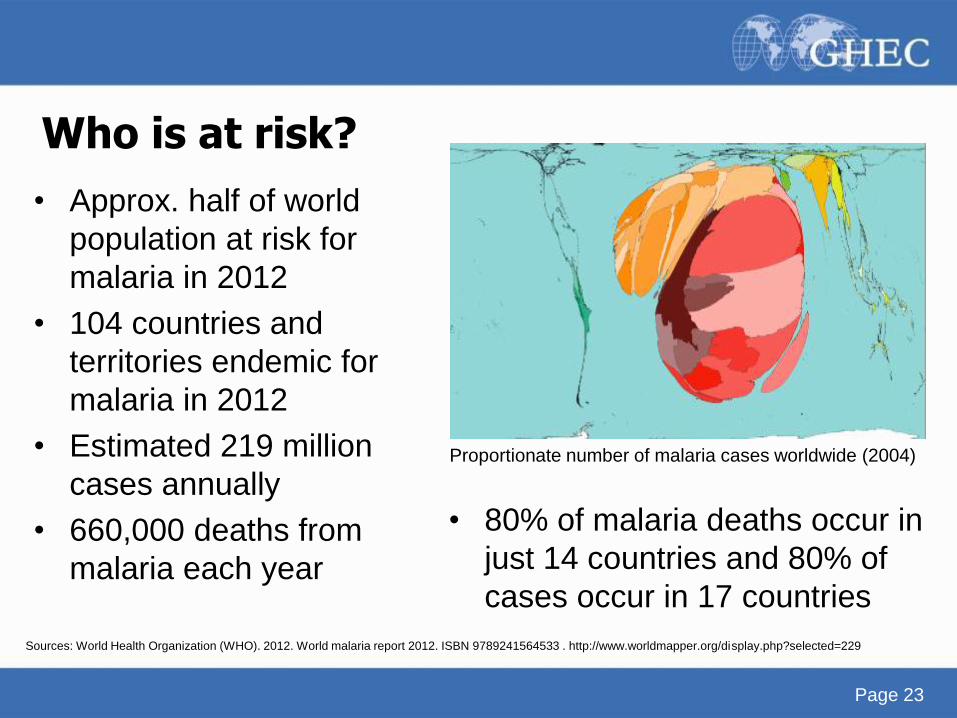
• Approx. half of world
population at risk for
malaria in 2012
• 104 countries and
territories endemic for
malaria in 2012
• Estimated 219 million
cases annually
• 660,000 deaths from
malaria each year
• 80% of malaria deaths occur in
just 14 countries and 80% of
cases occur in 17 countries
Proportionate number of malaria cases worldwide (2004)
Who is at risk?
Sources: World Health Organization (WHO). 2012. World malaria report 2012. ISBN 9789241564533 . http://www.worldmapper.org/display.php?selected=229
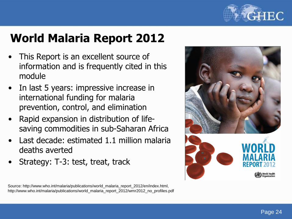
World Malaria Report 2012
• This Report is an excellent source of information and is frequently cited in this module
• In last 5 years: impressive increase in international funding for malaria prevention, control, and elimination
• Rapid expansion in distribution of life-saving commodities in sub-Saharan Africa
• Last decade: estimated 1.1 million malaria deaths averted
• Strategy: T-3: test, treat, track
Source: http://www.who.int/malaria/publications/world_malaria_report_2012/en/index.html,
http://www.who.int/malaria/publications/world_malaria_report_2012/wmr2012_no_profiles.pdf
Page 24

Page 25
Changes in malaria incidence and mortality Approximately half of countries with ongoing malaria transmission are on track to meet the World Health Assembly (WHA) and RBM target:
to achieve a 75% reduction in malaria cases by 2015, compared to levels in 2000. While 50 countries are on track to reach the target,
progress in more than a third of countries cannot be assessed due to limitations in their reported data. Further progress towards
international malaria targets depends on achieving substantial gains in the highest burden countries (36). Of 99 countries with ongoing
malaria transmission, 58 submitted sufficiently complete and consistent data on malaria cases between 2000 and 2011 to enable an
assessment of trends to be made. Based on these reported data, 50 countries, including 9 countries in the African Region, are on track to
meet the WHA and RBM target to reduce malaria case incidence by 75% by 2015. A further 4 countries are projected to achieve reductions
of between 50% and 75%. Malaria case incidence increased in 3 countries of the Region of the Americas (37). Of the 104 endemic
countries in 2012, 79 countries are classified as being in the malaria control phase, 10 are in the pre-elimination phase, 10 are in
elimination phase. Another 5 countries without ongoing transmission are classified in the prevention of re-introduction phase (38). There
were an estimated 219 million cases of malaria (range 154–289 million) and 660,000 deaths (range 610,000–971,000) in 2010. The
estimates for 2010 have been updated since they were first published in the World Malaria Report 2011 after a process of country
consultation. Country level malaria estimates available for 2010 show that 80% of estimated malaria deaths occur in just 14 countries and
approximately 80% of estimated cases occur in 17 countries. Together, the Democratic Republic of the Congo and Nigeria account for over
40% of the estimated total of malaria deaths globally. The Democratic Republic of the Congo, India and Nigeria account for 40% of
estimated malaria cases (39). Malaria is strongly associated with poverty. Estimated malaria mortality rates are highest in countries with a
lower GNI per capita. Countries with higher proportions of their population living in poverty (less than US$ 1.25 per person per day) have
higher mortality rates from malaria. Within countries, parasite prevalence rates in children are highest among poorer populations and in
rural areas (40). Progress in reducing malaria case incidence and mortality rates has been faster in countries with lower numbers of cases
and deaths. Nonetheless, greater numbers of cases and deaths are estimated to have been averted between 2001 and 2010 in countries
which had the highest malaria burdens in 2000. If the malaria incidence and mortality rates estimated for 2000 had remained unchanged
over the decade, 274 million more cases and 1.1 million more deaths would have occurred between 2001 and 2010. The majority of cases
averted (52%) and lives saved (58%) are in the 10 countries which had the highest estimated malaria burdens in 2000. Such estimations
indicate that malaria programmes are having their greatest impact where the burden is highest (41). There are many inherent uncertainties
in any approach to producing estimates of malaria case incidence and mortality, and in analyses based on these estimates. The global
malaria community needs to increase its efforts to support malaria endemic countries in improving diagnostic testing, surveillance, vital
registration, and routine health information systems, so that accurate information on malaria morbidity and mortality can be obtained.
Sources: World Health Organization (WHO). 2012. World malaria report 2012. ISBN 9789241564533 . http://www.worldmapper.org/display.php?selected=229
Page 26
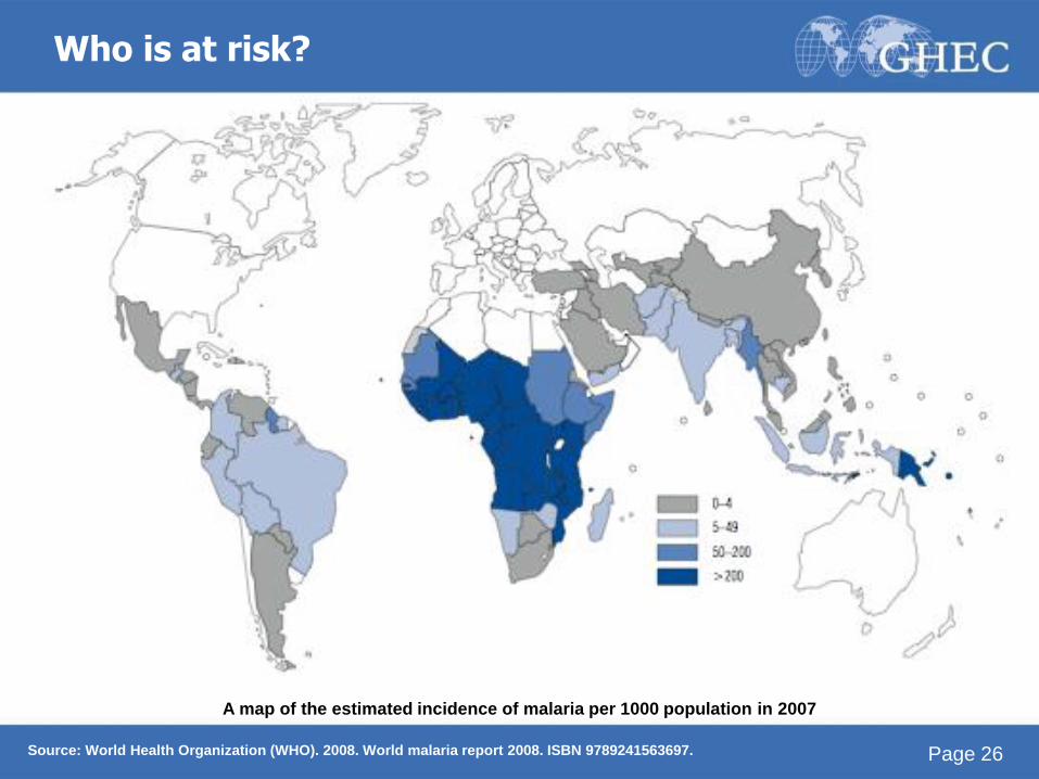
Who is at risk?
Source: World Health Organization (WHO). 2008. World malaria report 2008. ISBN 9789241563697.
A map of the estimated incidence of malaria per 1000 population in 2007
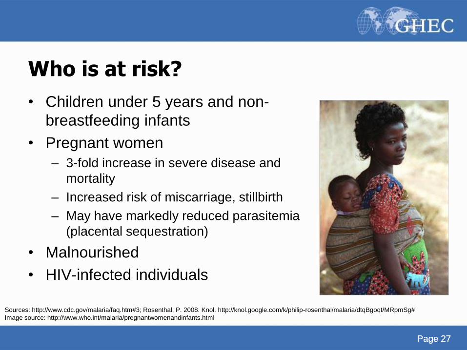
Page 27 Page 27
Who is at risk?
• Children under 5 years and non-
breastfeeding infants
• Pregnant women
– 3-fold increase in severe disease and
mortality
– Increased risk of miscarriage, stillbirth
– May have markedly reduced parasitemia
(placental sequestration)
• Malnourished
• HIV-infected individuals
Sources: http://www.cdc.gov/malaria/faq.htm#3; Rosenthal, P. 2008. Knol. http://knol.google.com/k/philip-rosenthal/malaria/dtqBgoqt/MRpmSg#
Image source: http://www.who.int/malaria/pregnantwomenandinfants.html
Page 28
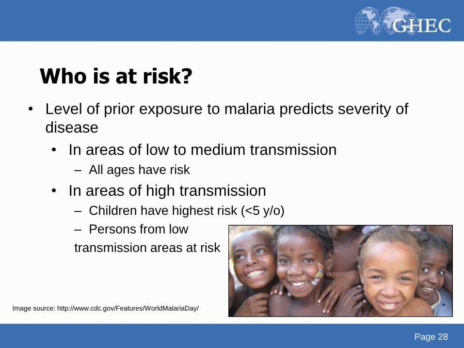
Who is at risk?
• Level of prior exposure to malaria predicts severity of
disease
• In areas of low to medium transmission
– All ages have risk
• In areas of high transmission
– Children have highest risk (<5 y/o)
– Persons from low
transmission areas at risk
Image source: http://www.cdc.gov/Features/WorldMalariaDay/
Page 29
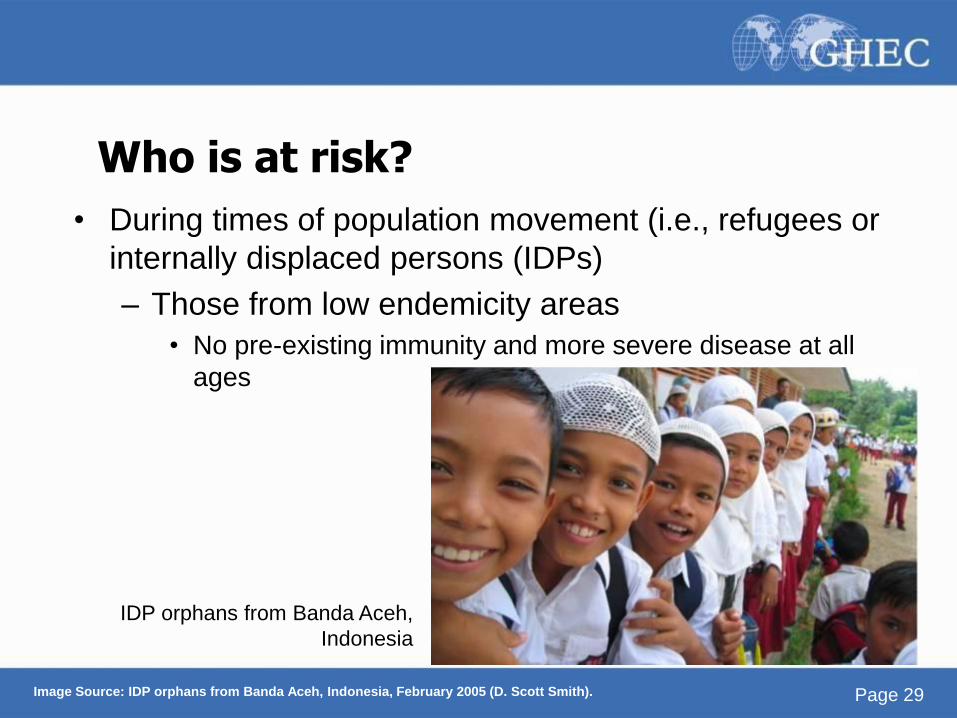
• During times of population movement (i.e., refugees or
internally displaced persons (IDPs)
– Those from low endemicity areas
• No pre-existing immunity and more severe disease at all
ages
Who is at risk?
IDP orphans from Banda Aceh,
Indonesia
Image Source: IDP orphans from Banda Aceh, Indonesia, February 2005 (D. Scott Smith).
Page 30 Page 30
Who is at risk? Protective Factors
• Biological characteristics and behavior traits can
influence risk of developing malaria
• Newborns are protected by maternal antibodies
– Antibodies decrease with time, explaining the
vulnerability in children who have stopped
breastfeeding
Source: http://www.cdc.gov/malaria/distribution_epi/human_epidemiology.htm
Page 31 Page 31
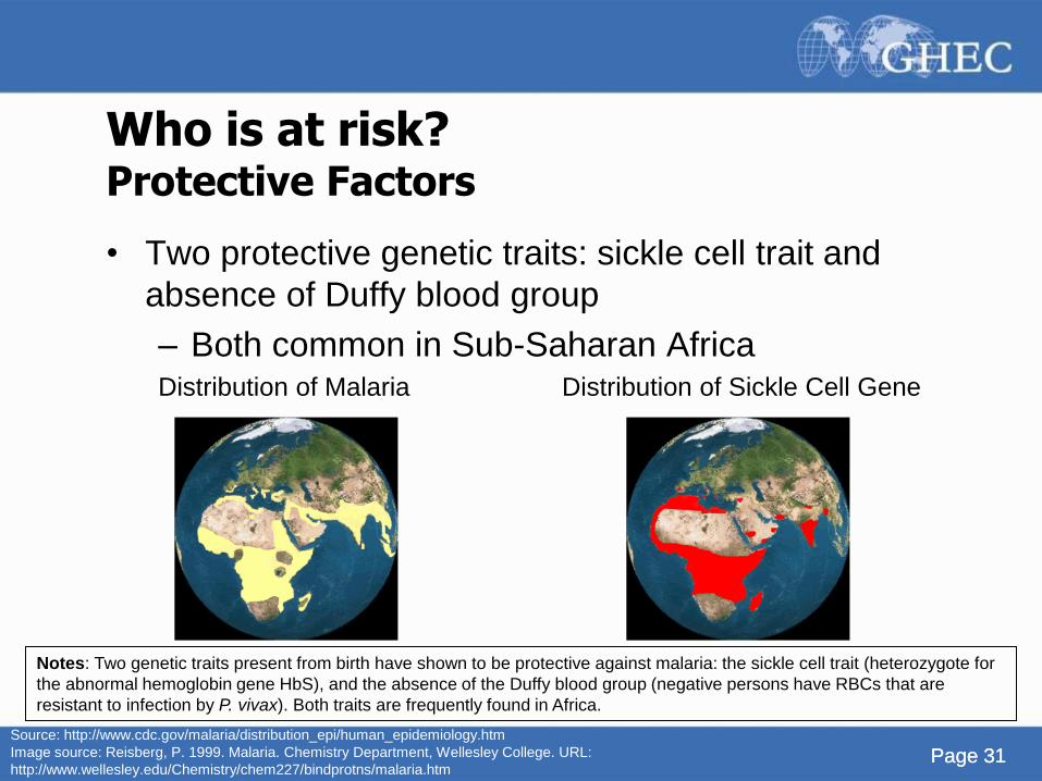
Who is at risk? Protective Factors
• Two protective genetic traits: sickle cell trait and
absence of Duffy blood group
– Both common in Sub-Saharan Africa Distribution of Malaria Distribution of Sickle Cell Gene
Source: http://www.cdc.gov/malaria/distribution_epi/human_epidemiology.htm
Image source: Reisberg, P. 1999. Malaria. Chemistry Department, Wellesley College. URL:
http://www.wellesley.edu/Chemistry/chem227/bindprotns/malaria.htm
Notes: Two genetic traits present from birth have shown to be protective against malaria: the sickle cell trait (heterozygote for
the abnormal hemoglobin gene HbS), and the absence of the Duffy blood group (negative persons have RBCs that are
resistant to infection by P. vivax). Both traits are frequently found in Africa.
Page 32
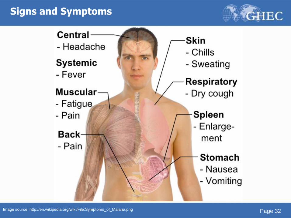
Signs and Symptoms
Image source: http://en.wikipedia.org/wiki/File:Symptoms_of_Malaria.png
Page 33 Page 33
Symptoms
• 7 to 30 days after an infective bite, symptoms appear
• The blood stage parasites are what cause the
symptoms of malaria
• Infected patients are categorized as having either:
– Uncomplicated malaria
– Severe malaria
Source: John, D.T. and W.A. Petri. 2006. Chapter 4: Malaria. Markell And Voge’s Medical Parasitology. St Louis: Saunders Elsevier, pp. 79-106.
Page 34
• Initial symptoms non-specific
• Headache
• Muscle aches
• Nausea, vomiting
• Then malarial paroxysms begin
• Shaking chill (10-15 min)
• High fever (typically 10 h; up to 36 h)
• Cycle repeats every 36-72 hours (species specific)
• Primary attack lasts 2-24 weeks (spp. specific)
Symptoms Uncomplicated Malaria
Source: John, D.T. and W.A. Petri. 2006. Chapter 4: Malaria. Markell And Voge’s Medical Parasitology. St Louis: Saunders Elsevier, pp. 79-106.
Page 35
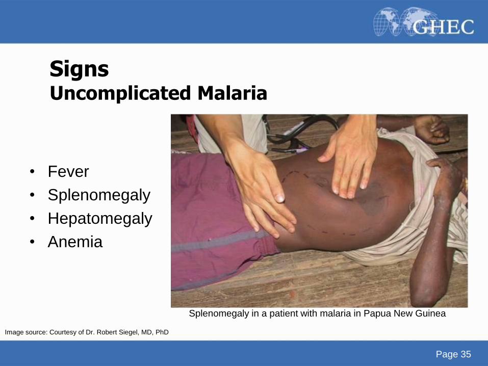
Signs Uncomplicated Malaria
• Fever
• Splenomegaly
• Hepatomegaly
• Anemia
Splenomegaly in a patient with malaria in Papua New Guinea
Image source: Courtesy of Dr. Robert Siegel, MD, PhD
Page 36
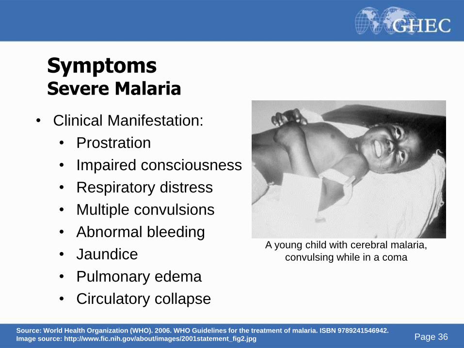
Symptoms Severe Malaria
• Clinical Manifestation:
• Prostration
• Impaired consciousness
• Respiratory distress
• Multiple convulsions
• Abnormal bleeding
• Jaundice
• Pulmonary edema
• Circulatory collapse
A young child with cerebral malaria,
convulsing while in a coma
Source: World Health Organization (WHO). 2006. WHO Guidelines for the treatment of malaria. ISBN 9789241546942.
Image source: http://www.fic.nih.gov/about/images/2001statement_fig2.jpg
Page 37 Page 37
Symptoms Severe Malaria
• Is nearly always due to P. falciparum infection
• Complications can include:
– Cerebral malaria (neurologic abnormalities), severe
anemia due to hemolysis, pulminary edema/acute
respiratory distress syndrome, abnormalities in blood
coagulation and thrombocytopenia, cardiovascular
collapse, shock
• Most often occurs in persons with absent or decreased
malaria immunity
Source: http://www.cdc.gov/malaria/disease.htm
Page 38 Page 38
Symptoms
• Infected individuals can also be aysmptomatic
• Anti-malarial drugs taken for prophylaxis can delay the
onset of symptoms by weeks or months
• In P. vivax and P. ovale infections, patients having
recovered from the first illness may suffer additional,
“relapse” attacks, after months or years without symptoms
• This is due to the reactivation of their dormant liver stage
(hypnozoites)
Source: John, D.T. and W.A. Petri. 2006. Chapter 4: Malaria. Markell And Voge’s Medical Parasitology. St Louis: Saunders Elsevier, pp. 79-106.;
http://www.cdc.gov/malaria/disease.htm
Page 39
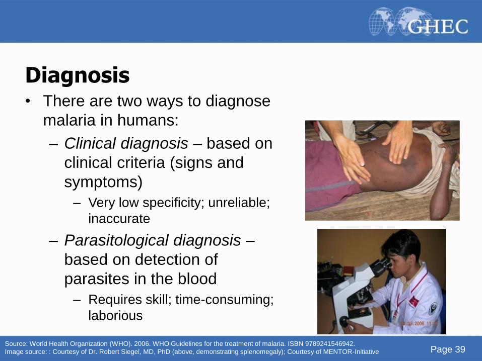
Diagnosis • There are two ways to diagnose
malaria in humans:
– Clinical diagnosis – based on
clinical criteria (signs and
symptoms)
– Very low specificity; unreliable;
inaccurate
– Parasitological diagnosis –
based on detection of
parasites in the blood
– Requires skill; time-consuming;
laborious
Source: World Health Organization (WHO). 2006. WHO Guidelines for the treatment of malaria. ISBN 9789241546942.
Image source: : Courtesy of Dr. Robert Siegel, MD, PhD (above, demonstrating splenomegaly); Courtesy of MENTOR-Initiative
Page 40
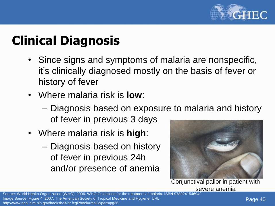
Clinical Diagnosis
• Since signs and symptoms of malaria are nonspecific,
it’s clinically diagnosed mostly on the basis of fever or
history of fever
• Where malaria risk is low:
– Diagnosis based on exposure to malaria and history
of fever in previous 3 days
• Where malaria risk is high:
– Diagnosis based on history
of fever in previous 24h
and/or presence of anemia
Conjunctival pallor in patient with
severe anemia Source: World Health Organization (WHO). 2006. WHO Guidelines for the treatment of malaria. ISBN 9789241546942.
Image Source: Figure 4. 2007. The American Society of Tropical Medicine and Hygiene. URL:
http://www.ncbi.nlm.nih.gov/bookshelf/br.fcgi?book=mal3&part=pg36
Page 41
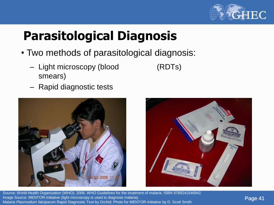
Parasitological Diagnosis
– Light microscopy (blood
smears)
– Rapid diagnostic tests
(RDTs)
Page 41
• Two methods of parasitological diagnosis:
Source: World Health Organization (WHO). 2006. WHO Guidelines for the treatment of malaria. ISBN 9789241546942.
Image Source: MENTOR-Initiative (light microscopy is used to diagnose malaria).
Malaria Plasmodium falciparum Rapid Diagnostic Test by Orchid; Photo for MENTOR-Initiative by D. Scott Smith
Page 42 Page 42 Page 42
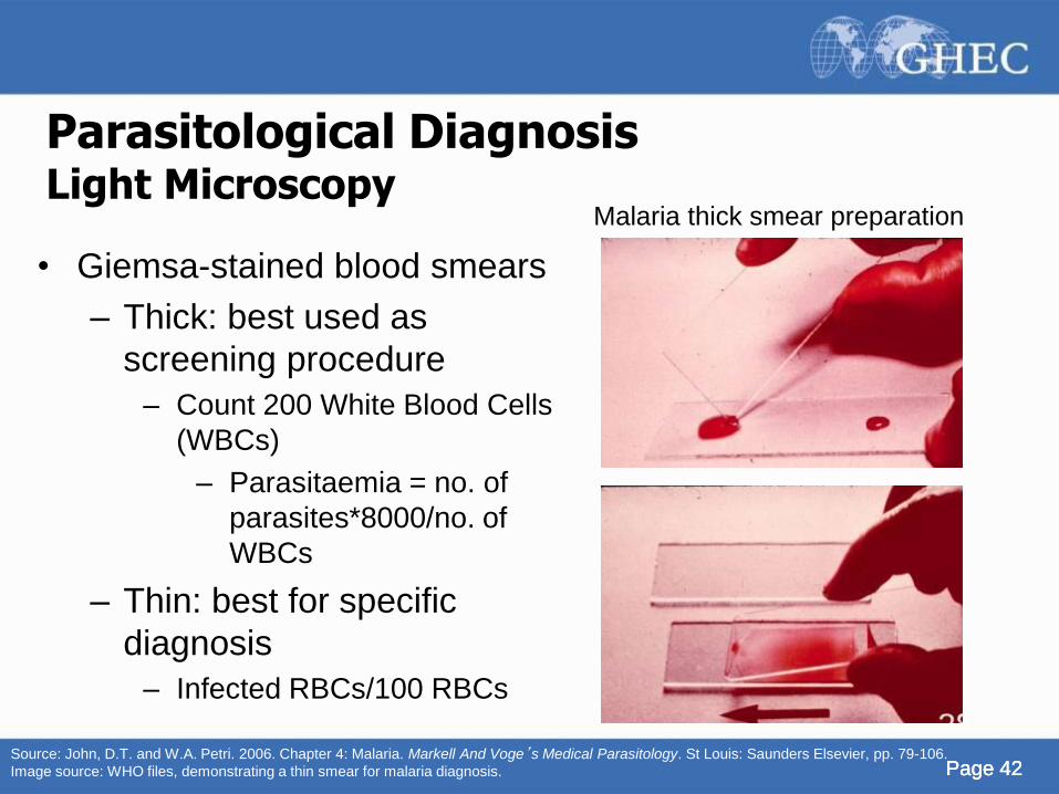
Parasitological Diagnosis Light Microscopy
• Giemsa-stained blood smears
– Thick: best used as
screening procedure
– Count 200 White Blood Cells
(WBCs)
– Parasitaemia = no. of
parasites*8000/no. of
WBCs
– Thin: best for specific
diagnosis
– Infected RBCs/100 RBCs
Malaria thick smear preparation
Source: John, D.T. and W.A. Petri. 2006. Chapter 4: Malaria. Markell And Voge’s Medical Parasitology. St Louis: Saunders Elsevier, pp. 79-106.
Image source: WHO files, demonstrating a thin smear for malaria diagnosis.
Page 43
• Blood smears allow identification of the
infecting species
– Important because treatment varies
depending on Plasmodium species
Parasitological Diagnosis Light Microscopy
Source: John, D.T. and W.A. Petri. 2006. Chapter 4: Malaria. Markell And Voge’s Medical Parasitology. St Louis: Saunders Elsevier, pp. 79-106.
Page 44
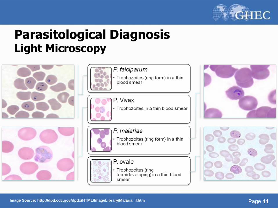
Parasitological Diagnosis Light Microscopy
Page 44 Image Source: http://dpd.cdc.gov/dpdx/HTML/ImageLibrary/Malaria_il.htm
Page 45
Parasitological Diagnosis Rapid Diagnostic Tests
• Developed as a way to quickly identify malaria in the field
• Simpler to perform than microscopic methods
• However, RDTs show positive results up to 14 days after
effective treatment of malaria because remnants of the
parasite remain
• Thus, do not use RDTs for follow-up of suspected
treatment failures
Source: Rosenthal, P. 2008. Knol. http://knol.google.com/k/philip-rosenthal/malaria/dtqBgoqt/MRpmSg#
Page 46
Parasitological Diagnosis Rapid Diagnostic Tests
• Offer sensitivity/specificity close to or above that of high-
quality blood smear analysis in the hands of expert
microscopists
• At parasite densities above 100 parasites/microliter of
blood, RDTs can detect P. falciparum with a sensitivity of
>90%
• This is generally higher than the sensitivity normally
achieved with skilled microscopy
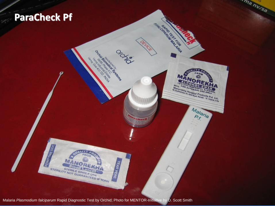
ParaCheck Pf
Malaria Plasmodium falciparum Rapid Diagnostic Test by Orchid; Photo for MENTOR-Initiative by D. Scott Smith
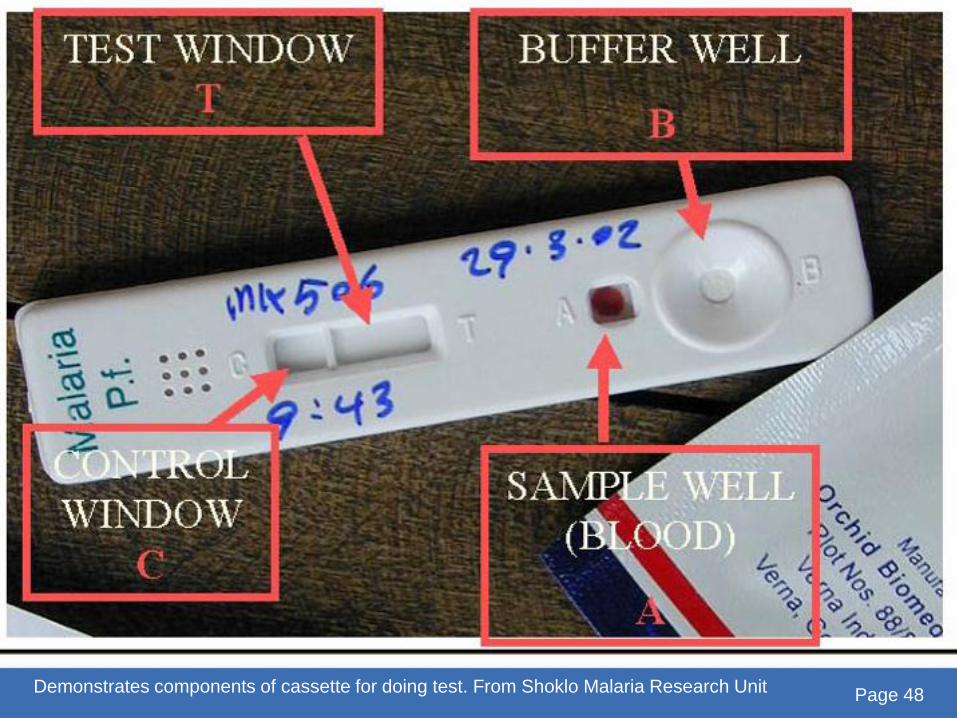
Demonstrates components of cassette for doing test. From Shoklo Malaria Research Unit Page 48
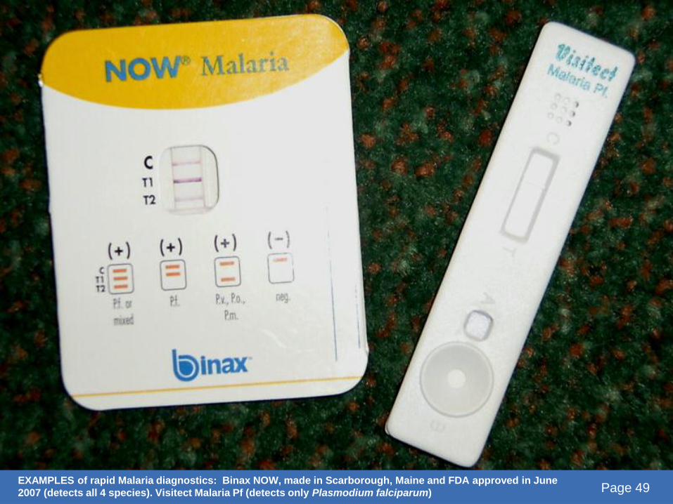
EXAMPLES of rapid Malaria diagnostics: Binax NOW, made in Scarborough, Maine and FDA approved in June
2007 (detects all 4 species). Visitect Malaria Pf (detects only Plasmodium falciparum) Page 49
Page 50 Page 50
Treatment
• Should commence as soon as possible
• The treatment depends on:
– species of infecting parasite
– area where the infection was acquired and its drug-
resistance status
– clinical status of the patient
– any accompanying illness or condition, pregnancy,
and drug allergies
Page 51 Page 51 Page 51
Treatment
• Treatment should be based on laboratory-confirmed
diagnosis where possible
• Different kinds of treatment:
– Suppressive therapy (chemoprophylaxis)
– Clinical cure
– Radical cure
Sources: World Health Organization (WHO). 2008. World malaria report 2008. ISBN 9789241563697.; John, D.T. and W.A. Petri. 2006.
Chapter 4: Malaria. Markell And Voge’s Medical Parasitology. St Louis: Saunders Elsevier, pp. 79-106.
Notes:
•Suppressive therapy: attempts to destroy parasites as they enter bloodstream with small doses of drugs effective against
erythrocytic stages
•Clinical cure: larger doses of drugs to eliminate large numbers of erythrocytic parasites present in a clinical attack
•Radical cure: implies elimination of not only bloodstream infection but also the tissues stages in liver as well; patient may still
be infectious after this because of gametocytes remaining in circulation blood or hypnozoites in liver that aren’t killed by drugs
Page 52 Page 52
Treatment
• Determination of infecting species important because:
– P. falciparum can cause rapidly progressive severe
illness or death while other species rarely cause severe
illness
– P. vivax and P. ovale require treatment for hypnozoite
forms that remain dormant in liver
– P. falciparum and P. vivax have different drug
resistance patterns in different regions
Page 53 Page 53
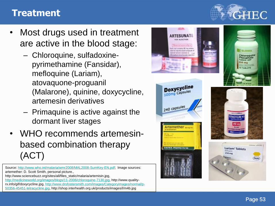
Treatment
• Most drugs used in treatment
are active in the blood stage:
– Chloroquine, sulfadoxine-
pyrimethamine (Fansidar),
mefloquine (Lariam),
atovaquone-proguanil
(Malarone), quinine, doxycycline,
artemesin derivatives
– Primaquine is active against the
dormant liver stages
• WHO recommends artemesin-
based combination therapy
(ACT) Source: http://www.who.int/malaria/wmr2008/MAL2008-SumKey-EN.pdf; Image sources:
artemether: D. Scott Smith, personal picture.,
http://www.sciencebuzz.org/sites/all/files_static/malaria/artemisin.jpg,
http://medicineworld.org/images/blogs/11-2006/chloroquine-7130.jpg, http://www.quality-
rx.info/gif/doxycycline.jpg, http://www.drsfostersmith.com/images/Categoryimages/normal/p-
50356-45451-tetracycline.jpg, http://shop.interhealth.org.uk/products/images/l/m4b.jpg
Page 54
Treatment Notes
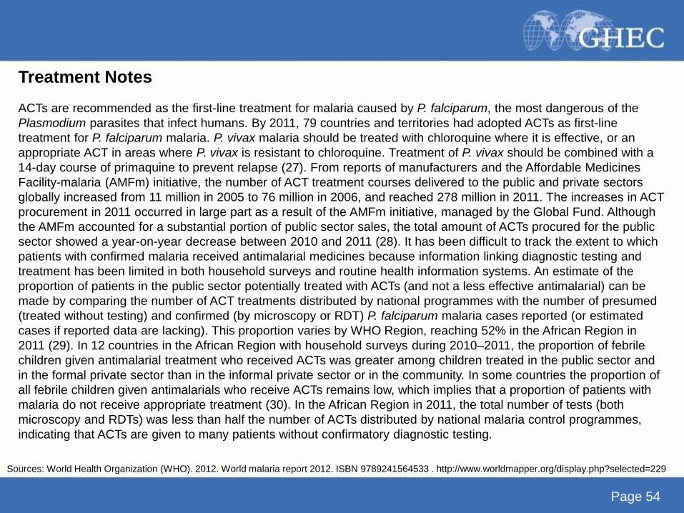
ACTs are recommended as the first-line treatment for malaria caused by P. falciparum, the most dangerous of the
Plasmodium parasites that infect humans. By 2011, 79 countries and territories had adopted ACTs as first-line
treatment for P. falciparum malaria. P. vivax malaria should be treated with chloroquine where it is effective, or an
appropriate ACT in areas where P. vivax is resistant to chloroquine. Treatment of P. vivax should be combined with a
14-day course of primaquine to prevent relapse (27). From reports of manufacturers and the Affordable Medicines
Facility-malaria (AMFm) initiative, the number of ACT treatment courses delivered to the public and private sectors
globally increased from 11 million in 2005 to 76 million in 2006, and reached 278 million in 2011. The increases in ACT
procurement in 2011 occurred in large part as a result of the AMFm initiative, managed by the Global Fund. Although
the AMFm accounted for a substantial portion of public sector sales, the total amount of ACTs procured for the public
sector showed a year-on-year decrease between 2010 and 2011 (28). It has been difficult to track the extent to which
patients with confirmed malaria received antimalarial medicines because information linking diagnostic testing and
treatment has been limited in both household surveys and routine health information systems. An estimate of the
proportion of patients in the public sector potentially treated with ACTs (and not a less effective antimalarial) can be
made by comparing the number of ACT treatments distributed by national programmes with the number of presumed
(treated without testing) and confirmed (by microscopy or RDT) P. falciparum malaria cases reported (or estimated
cases if reported data are lacking). This proportion varies by WHO Region, reaching 52% in the African Region in
2011 (29). In 12 countries in the African Region with household surveys during 2010–2011, the proportion of febrile
children given antimalarial treatment who received ACTs was greater among children treated in the public sector and
in the formal private sector than in the informal private sector or in the community. In some countries the proportion of
all febrile children given antimalarials who receive ACTs remains low, which implies that a proportion of patients with
malaria do not receive appropriate treatment (30). In the African Region in 2011, the total number of tests (both
microscopy and RDTs) was less than half the number of ACTs distributed by national malaria control programmes,
indicating that ACTs are given to many patients without confirmatory diagnostic testing.
Sources: World Health Organization (WHO). 2012. World malaria report 2012. ISBN 9789241564533 . http://www.worldmapper.org/display.php?selected=229
Page 55
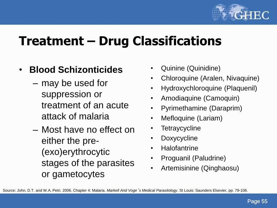
Treatment – Drug Classifications
• Blood Schizonticides
– may be used for
suppression or
treatment of an acute
attack of malaria
– Most have no effect on
either the pre-
(exo)erythrocytic
stages of the parasites
or gametocytes
Page 55
• Quinine (Quinidine)
• Chloroquine (Aralen, Nivaquine)
• Hydroxychloroquine (Plaquenil)
• Amodiaquine (Camoquin)
• Pyrimethamine (Daraprim)
• Mefloquine (Lariam)
• Tetraycycline
• Doxycycline
• Halofantrine
• Proguanil (Paludrine)
• Artemisinine (Qinghaosu)
Source: John, D.T. and W.A. Petri. 2006. Chapter 4: Malaria. Markell And Voge’s Medical Parasitology. St Louis: Saunders Elsevier, pp. 79-106.
Page 56
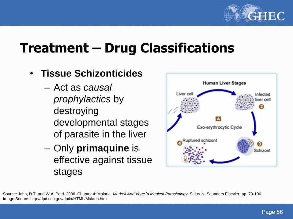
Treatment – Drug Classifications
• Tissue Schizonticides
– Act as causal
prophylactics by
destroying
developmental stages
of parasite in the liver
– Only primaquine is
effective against tissue
stages
Page 56
Source: John, D.T. and W.A. Petri. 2006. Chapter 4: Malaria. Markell And Voge’s Medical Parasitology. St Louis: Saunders Elsevier, pp. 79-106.
Image Source: http://dpd.cdc.gov/dpdx/HTML/Malaria.htm
Page 57
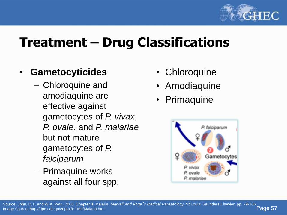
Treatment – Drug Classifications
• Gametocyticides
– Chloroquine and
amodiaquine are
effective against
gametocytes of P. vivax,
P. ovale, and P. malariae
but not mature
gametocytes of P.
falciparum
– Primaquine works
against all four spp.
Page 57
• Chloroquine
• Amodiaquine
• Primaquine
Source: John, D.T. and W.A. Petri. 2006. Chapter 4: Malaria. Markell And Voge’s Medical Parasitology. St Louis: Saunders Elsevier, pp. 79-106.
Image Source: http://dpd.cdc.gov/dpdx/HTML/Malaria.htm
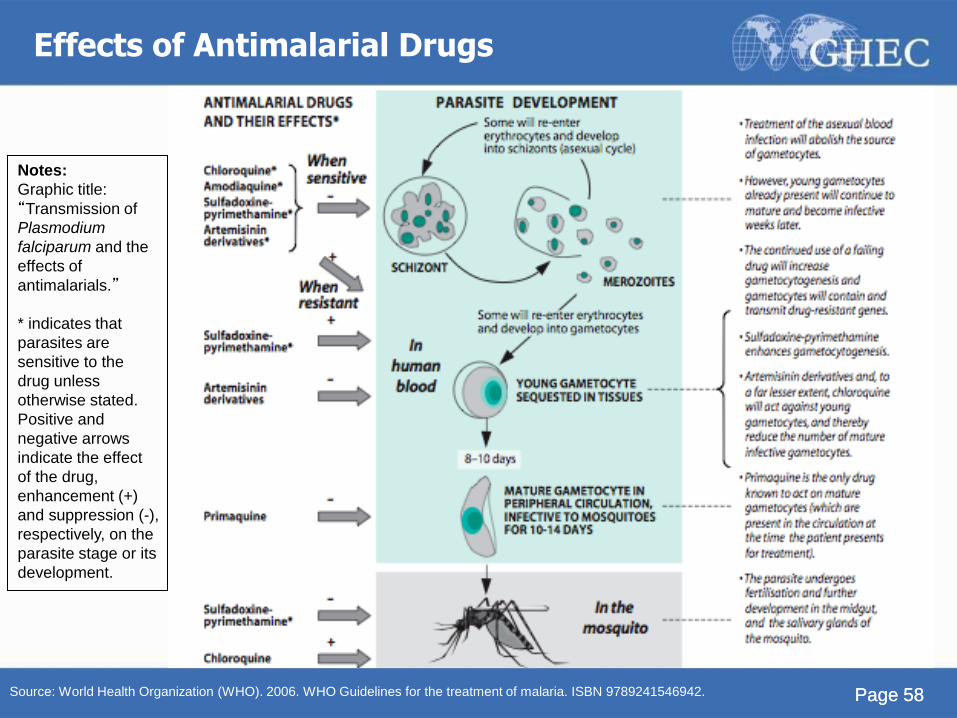
Page 58 Page 58 Source: World Health Organization (WHO). 2006. WHO Guidelines for the treatment of malaria. ISBN 9789241546942.
Notes:
Graphic title:
“Transmission of
Plasmodium
falciparum and the
effects of
antimalarials.”
* indicates that
parasites are
sensitive to the
drug unless
otherwise stated.
Positive and
negative arrows
indicate the effect
of the drug,
enhancement (+)
and suppression (-),
respectively, on the
parasite stage or its
development.
Effects of Antimalarial Drugs
Page 59
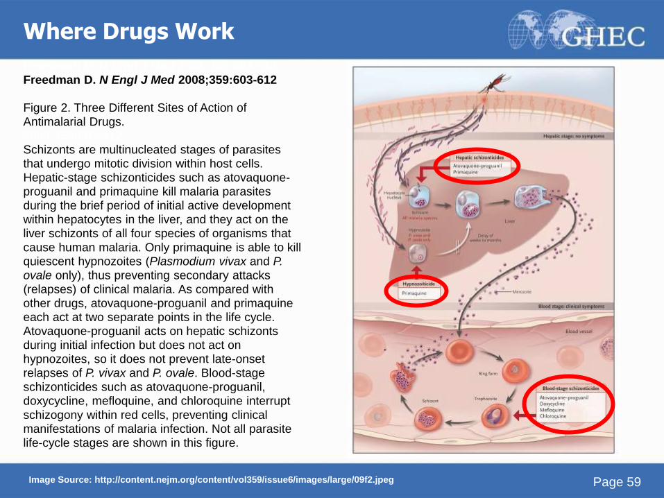
Where Drugs Work
Image Source: http://content.nejm.org/content/vol359/issue6/images/large/09f2.jpeg
Freedman D. N Engl J Med 2008;359:603-612
Freedman D. N Engl J Med 2008;359:603-612 Figure 2. Three Different Sites of Action of Antimalarial Drugs. 2008;359:603-612 Schizonts are multinucleated stages of parasites that undergo mitotic division within host cells. Hepatic-stage schizonticides such as atovaquone-proguanil and primaquine kill malaria parasites during the brief period of initial active development within hepatocytes in the liver, and they act on the liver schizonts of all four species of organisms that
cause human malaria. Only primaquine is able to kill quiescent hypnozoites (Plasmodium vivax and P. ovale only), thus preventing secondary attacks (relapses) of clinical malaria. As compared with other drugs, atovaquone-proguanil and primaquine each act at two separate points in the life cycle. Atovaquone-proguanil acts on hepatic schizonts during initial infection but does not act on hypnozoites, so it does not prevent late-onset relapses of P. vivax and P. ovale. Blood-stage schizonticides such as atovaquone-proguanil, doxycycline, mefloquine, and chloroquine interrupt
schizogony within red cells, preventing clinical manifestations of malaria infection. Not all parasite life-cycle stages are shown in this figure.
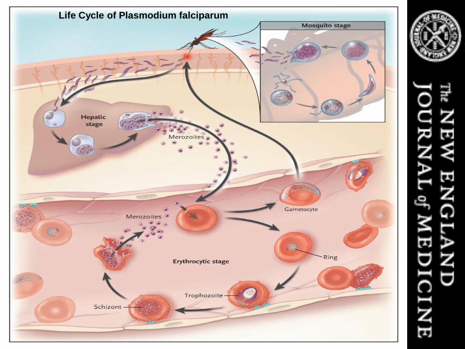
Life Cycle of Plasmodium falciparum
Page 61
Notes on Figure of the Life Cycle of Plasmodium falciparum
Elements that are important for the pathogenesis of severe
malaria are shown. Erythrocytes containing P. falciparum in mature intraerythrocytic stages (trophozoites and schizonts) adhere to vascular endothelium, thereby avoiding clearance by the spleen. High numbers of circulating parasites and elaboration of host and parasite factors in the vasculature of various organs lead to the manifestations of severe malaria.
R Med 2008;358:1829-1836
Source: Rosenthal P. N Engl J Med 2008;358:1829-1836
Page 62 Page 62
Drug Resistance
• Drug resistance has been confirmed in P. falciparum
and P. vivax
• P. falciparum is usually completely resistant to
chloroquine
• Resistance in P. falciparum has been documented with
nearly all other drugs, though this is less widespread
• P. vivax resistant to chloroquine in SE Asia, India, and
S. America
Source: http://www.cdc.gov/malaria/drug_resistance.htm
Page 63 Page 63
Summary (1)
• Malaria is a protozoan parasite with 5 species that affect
humans
• Malaria transmission involves a mosquito vector of the
Anopheles species
• Children under 5 years old and pregnant women are most
at risk for malaria
• Sub-saharan Africa is the most affected region in the world,
accounting for 90% of deaths from falciparum malaria.
Page 64 Page 64
Summary (2)
• Symptoms of malaria include fever, but later, coma and
prostration are seen in severe malaria
• Diagnosis is made by microscopy looking at blood smears,
but also with Rapid Diagnostic Tests (RDTs)
• Treatment is critical in terms of timing, kind of drug and
administration with respect to the specifics of the patient
and the local resistance patterns.
• Understanding the biology and lifecycle of malaria lends
insight into strategy for diagnosis, treatment and prevention
(see previous module)
Page 65
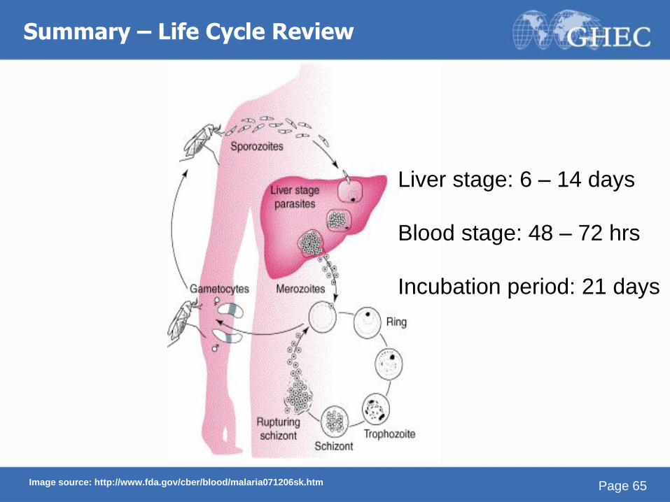
Liver stage: 6 – 14 days
Blood stage: 48 – 72 hrs
Incubation period: 21 days
Summary – Life Cycle Review
Image source: http://www.fda.gov/cber/blood/malaria071206sk.htm
Page 66
Credits
• Cristin Weekley, BA Stanford University; MPH
student at UC Berkeley School of Public Health
• D. Scott Smith, MD, MSc, DTM&H Chief of Infectious Disease & Geographic Medicine, Kaiser
Permanente, Redwood City, California
Adjunct Assistant Clinical Professor
Dept. of Human Biology and
Dept. of Medical Microbiology & Immunology
Stanford University Medical School
The Global Health Education Consortium and the Consortium of
Universities for Global Health gratefully acknowledge the support
provided for developing teaching modules from the:
Margaret Kendrick Blodgett Foundation
The Josiah Macy, Jr. Foundation
Arnold P. Gold Foundation
This work is licensed under a Creative Commons Attribution-Noncommercial-No Derivative Works 3.0 United
States License.
Page 67