Embed Size (px)
Citation preview

Tao et al., Sci. Transl. Med. 11, eaan4479 (2019) 2 January 2019
S C I E N C E T R A N S L A T I O N A L M E D I C I N E | R E S E A R C H A R T I C L E
1 of 13
M A L A R I A
A saliva-based rapid test to quantify the infectious subclinical malaria parasite reservoirDingyin Tao1,2*†, Brent McGill1,2†, Timothy Hamerly1,2,3†, Tamaki Kobayashi2,4‡, Prachi Khare3‡, Amanda Dziedzic1‡, Tomasz Leski5, Andrew Holtz6, Bruce Shull7, Anne E. Jedlicka1, Andrew Walzer7, Paul D. Slowey8, Christopher C. Slowey8, Sandrine E. Nsango9,10, David A. Stenger5, Mike Chaponda11, Modest Mulenga11, Kathryn H. Jacobsen12, David J. Sullivan1, Sadie J. Ryan13, Rashid Ansumana14, William J. Moss2,4, Isabelle Morlais9, Rhoel R. Dinglasan1,2,3§
A large proportion of ongoing malaria parasite transmission is attributed to low-density subclinical infections not readily detected by available rapid diagnostic tests (RDTs) or microscopy. Plasmodium falciparum gametocyte carriage is subclinical, but gametocytemic individuals comprise the parasite reservoir that leads to infection of mosquitoes and local transmission. Effective detection and quantification of these carriers can help advance ma-laria elimination strategies. However, no point-of-need (PON) RDTs for gametocyte detection exist, much less one that can perform noninvasive sampling of saliva outside a clinical setting. Here, we report on the discovery of 35 parasite markers from which we selected a single candidate for use in a PON RDT. We performed a cross-sectional, multi-omics study of saliva from 364 children with subclinical infection in Cameroon and Zambia and produced a prototype saliva-based PON lateral flow immunoassay test for P. falciparum gametocyte carriers. The test is capa-ble of identifying submicroscopic carriage in both clinical and nonclinical settings and is compatible with archived saliva samples.
INTRODUCTIONMalaria kills ~500,000 children each year, mostly children under the age of 5 in sub-Saharan Africa (1). Although malaria control efforts are having a substantial impact on disease-related deaths, parasite transmission remains a major issue. Low-density subclinical infec-tion with Plasmodium falciparum among defined population sub-sets has been shown to be an important but cryptic facet of malaria transmission. Typically, these individuals are not readily detected by currently available point-of-need (PON) rapid diagnostic tests (RDTs) or microscopy (2–5), and molecular methods not readily available to hospitals, clinics, or other PON sites such as households, schools, or apothecaries are required for detection and quantifica-tion of parasite carriage. The sexual stage gametocytes do not cause disease, but gametocytemic individuals with asexual stages can event-
ually progress to clinical malaria, especially younger children. This subclinical gametocytemic population represents the parasite res-ervoir that drives local malaria transmission through mosquitoes. However, in both microscopy-detectable and submicroscopic sub-clinical infections, gametocytes are coincident with low-density asexual stages (5) and can contribute to up to 80% of the infectious reservoir (5). It is now well recognized that effective detection of these carriers, as a whole, is crucial to advancing malaria control and elimination strategies (6–10), especially in the context of low- transmission and pre-elimination settings. However, no point-of-care or PON diagnostic for gametocyte detection exists (11).
The most commonly used sensitive and heat-stable blood-based RDT in sub-Saharan Africa is dependent on antibody detection of P. falciparum histidine-rich protein 2 (HRP2). Recently, in response to the growing concern that parasites lacking the pfhrp2/pfhrp3 genes are resulting in false-negative HRP2-RDT results and children be-ing left untreated, the World Health Organization (WHO) has en-couraged the scientific community to identify new target biomarkers of the parasite that allow for the sensitive detection of carriage (12). To address this critical scientific gap and obstacle to malaria elimi-nation and eradication, we developed a noninvasive diagnostic amena-ble for epidemiological surveillance programs and mass screening campaigns, both of which go beyond the limitation of a clinical setting.
The use of blood for screening campaigns substantially increases the inherent risk to both the screener and the individual (patient), and complicates the logistics, training requirements, and efficiency of the surveillance effort. Recently, it has been shown that parasite or pathogen biomarkers can enter into other biofluids, specifically urine and saliva (13–15). Stage V, mature gametocytes circulate in the bloodstream but can also be sequestered in capillary beds (16–18). The oral cavity is highly vascularized with numerous capillary loops, and there is a dynamic transfer of serum transudate proteins from the vasculature as part of the gingival crevicular fluid (GCF) into the oral fluid (a mixture of saliva and GCF) in both healthy and
1W. Harry Feinstone Department of Molecular Microbiology and Immunology, Johns Hopkins Bloomberg School of Public Health, Baltimore, MD 21205, USA. 2Johns Hopkins Malaria Research Institute, Baltimore, MD 21205, USA. 3Emerging Patho-gens Institute and Department of Infectious Diseases & Immunology, College of Veterinary Medicine, University of Florida, Gainesville, FL 32611, USA. 4Department of Epidemiology, Johns Hopkins Bloomberg School of Public Health, Baltimore, MD 21205, USA. 5United States Naval Research Laboratory (NRL), Center for Biomolec-ular Science and Engineering, Washington, DC 20375, USA. 6College of Science, George Mason University, Fairfax, VA 22030, USA. 7Thermo Fisher Scientific, Fremont, CA 94538, USA. 8Oasis Diagnostics, Vancouver, WA 98686, USA. 9Laboratoire de Recherche sur le Paludisme, Institut de Recherche pour le Développement– Organisation de Coordination et de Coopération pour la Lutte Contre les Grandes Endémies en Afrique Centrale (IRD-OCEAC), Yaoundé, Cameroon. 10Faculty of Medicine and Pharmaceutical Sciences, University of Douala, PO Box 2701, Douala, Cameroon. 11Tropical Disease Research Centre, Ndola, Zambia. 12College of Health and Human Services, George Mason University, Fairfax, VA 22030, USA. 13Emerging Pathogens Institute and Department of Geography, College of Liberal Arts and Sci-ences, University of Florida, Gainesville, FL 32611, USA. 14Mercy Hospital Research Laboratory, Kulanda Town, Bo, Sierra Leone.*Present address: National Center for Advancing Translational Sciences, National Institutes of Health, Rockville, MD 20850, USA.†These authors contributed equally to this work.‡These authors contributed equally to this work.§Corresponding author. Email: [email protected]
Copyright © 2019 The Authors, some rights reserved; exclusive licensee American Association for the Advancement of Science. No claim to original U.S. Government Works. Distributed under a Creative Commons Attribution License 4.0 (CC BY).
by guest on April 7, 2020
http://stm.sciencem
ag.org/D
ownloaded from

Tao et al., Sci. Transl. Med. 11, eaan4479 (2019) 2 January 2019
S C I E N C E T R A N S L A T I O N A L M E D I C I N E | R E S E A R C H A R T I C L E
2 of 13
diseased individuals (19). Here, we refer to oral fluid collection as saliva collection. We tested the hypothesis that if mature gametocytes potentially aggregate in gum capillary beds, then we can expect to sample gametocyte-derived proteins in the saliva of children infected with subclinical parasite densities in malaria-endemic countries such as Cameroon and Zambia.
Here, we report the identification of 35 P. falciparum proteins from carriers with subclinical parasitic infection, representing vari-ous stages of the parasite life cycle including stage V gametocytes, and a marker that is specific to mature female gametocytes. We per-formed a competitive profiling study using a liquid chromatography– multiple reaction monitoring (LC-MRM) mass spectrometry (MS) approach to estimate the prevalence of the candidate marker in the saliva of children with subclinical infection in Cameroon and Zambia and compared to microscopy and polymerase chain reaction (PCR) analyses of matched blood samples. Last, we developed a prototype antibody-based lateral flow immunoassay (LFIA) rapid test that further validated the presence of the marker in saliva from children with subclinical infection, revealing an underappreciated amount of subclinical carriage in these two malaria-endemic countries.
RESULTSProtein marker discovery in the saliva of children with subclinical parasitic infectionWe collected ethanol-stabilized saliva (5 ml) from 12 children (ages 5 to 12) in Mfou, Cameroon, who had subclinical parasitemia. Us-ing a simple saliva collection strategy and LC-MS/MS approach, we generated a P. falciparum candidate protein marker list (Table 1 and tables S1 and S2). The proteins can be divided into several classes, including cytosolic “housekeeping” proteins that are conserved be-tween asexual and gametocyte stages of P. falciparum (20), and those that are specific to asexual stages alone (21, 22). We noted that 22 of the proteins have been previously identified as highly enriched in stage V mature gametocytes (19, 23).
Of the 35 proteins that we detected in pooled saliva from the children, PF3D7_1218800 was the most abundant in individual sa-liva samples from children who were found to be gametocyte posi-tive by blood film microscopy (Mascot ions scores; Table 1 and table S2). We had previously characterized PF3D7_1218800 as a female- specific stage V gametocyte marker (23) and corrected its current annotation to Plasmodium sexual stage protein 17 (PSSP17). We selected PSSP17 as a candidate gametocyte marker and developed an LC-MRM MS workflow to detect PSSP17 in saliva (2 ml) from children with subclinical infection to estimate the prevalence of marker carriage in children. The 2-ml volume was selected because there was no a priori information on the potential variation in the abundance of any parasite protein in the saliva of individuals with subclinical infection.
We returned to Mfou, Cameroon, and sampled saliva from 307 children with subclinical infection (5 to 16 years of age, Fig. 1) at-tending six primary schools. In parallel, we sampled saliva from 42 children with subclinical malaria infection (under 16 years of age; table S3A) at home in Nchelenge District, Zambia (table S4). Three adults with subclinical infection (teachers from three different schools: Publique d’Ekali II, Publique de Metet, and Ecole Catholique de Nkilzok) were included in the blinded samples before analyses by LC-MRM MS, giving rise to a total of 310 samples from Cameroon during the second cross-sectional study. The total number of saliva
samples collected and analyzed for both the initial and follow-up studies was 364. For our comparative profiling study, we acquired matched blood samples for blood film, RDT, and quantitative PCR (qPCR) analysis in Cameroon and matched blood samples for RDT and dried blood spot (DBS) cataloging and downstream PCR anal-ysis in Zambia. These matched samples were important for com-paring the sensitivity of current RDTs and orthogonal molecular detection techniques with MRM MS.
Cross-sectional MS-based profiling of PSSP17 in salivaThe blinded LC-MRM analyses of 364 stabilized samples identified 364 valid spectral data (Fig. 2A). Of the 364, we observed a high prevalence (85.3%, 310/364) of PSSP17 in the 5 to 16 years old age group in both Cameroon and Zambia, using a peak area ratio (PAR) of >0.01, with an average original peptide peak signal/noise (S/N) ratio of >10 (Fig. 2B). Using a more rigid cutoff of PAR > 0.02, with the average original peptide peak S/N > 15, the prevalence of car-riage was 66.2% (241/364). In Cameroon, previous analyses of gametocyte carriage assessed by microscopy suggested that gameto-cytemic carriage in the same age group of children is about 6%, at a detection limit of ~8 gametocytes/l of blood (24, 25). A summary of the unblinded comparative profiling study conducted in Cameroon and Zambia suggests, as expected, that the LC-MRM analyses showed strong agreement with microscopy and molecular assays for the detection of either female gametocytes (pfs25 transcripts) or the presence of Plasmodium blood-stage parasites (asexual or ga-metocyte) via Plasmodium 18S ribosomal RNA (rRNA) from whole blood or cytochrome B (cytB) gene from DBS (Fig. 2C). For samples from Cameroon, we verified indirectly the presence of gametocytes by quantifying the gametocyte-specific transcript pfs25 in a subset of 100 matched blood samples. Within this set were samples that were negative and positive by microscopy (submicroscopic carriage of gametocytes). The qPCR analyses confirmed gametocyte carriage in those individuals whose saliva samples were positive for PSSP17 by LC-MRM (table S5), with the exception of two samples: A042 and A081. It is unclear whether the RNA extraction procedure contrib-uted to detection failure or whether gametocyte density was below the level of detection in our system. For those cases from Zambia where RDT detection failed (14/42), both PCR and LC-MRM anal-yses indicated parasite carriage. On the basis of our cytB nested PCR results, about 67% (28/42) were positive for parasite carriage (not gametocyte alone), whereas MRM confirmed the presence of ga-metocytes in 62% (26/42) of these samples, of which 67% (28/42) were also PCR positive.
We further analyzed a subset of samples (n = 93) with matched qPCR and LC-MRM data (Fig. 2D). We noted discrepancies in the estimated protein abundance (represented by PAR) and qPCR for samples B364, C022, and N235 (Fig. 2D, red arrows). For these three samples, despite the relatively lower gametocyte abundance as estimated by qPCR, PSSP17 appeared to be abundant in saliva. This is especially true for sample N235, where the pfs25 qPCR estimated ~0.12 gametocytes/l of blood, yet the protein marker was easily detected and measured in saliva. Conversely, five samples showed a marked inverse relationship between PAR and gametocytes/l of blood estimates: A048, A265, A471, B020, and D070. The comparison in PSSP17/parasite detection approaches for the remaining avail-able samples (n = 195) is shown in table S3B.
To test the relationship between PAR and gametocyte abundance [gametocytes/l of blood (GAM)] for all data included in Fig. 2D,
by guest on April 7, 2020
http://stm.sciencem
ag.org/D
ownloaded from

Tao et al., Sci. Transl. Med. 11, eaan4479 (2019) 2 January 2019
S C I E N C E T R A N S L A T I O N A L M E D I C I N E | R E S E A R C H A R T I C L E
3 of 13
Table 1. The most abundant P. falciparum proteins identified in children aged 5 to 16 years with subclinical parasitemia in Cameroon. A total of 35 proteins were identified by LC-MS/MS with a Mascot ions score of ≥25 (table S1). Of the 35, the 19 proteins below were considered the most abundant. Score indicates the Mascot ions scores, as described previously (23). The P. falciparum female gametocyte-specific biomarker, indicated in bold, was found to have the highest Mascot ions score in our pooled sample. Mr, molecular mass in kilodaltons (kDa).
Gene ID Description Mr (kDa) Annotated Gene Ontology
MS* Mascot ions score
PF3D7_0401900 Acyl-CoA synthetase 110.8 Long-chain fatty acid–CoA ligase activity
A 28
PF3D7_0422300 Alpha tubulin 2 49.7 GTP binding, GTPase activity, structural
molecule
A 31
PF3D7_0507800 Conserved Plasmodium protein
177.4 None A, G 40
PF3D7_0509400 RNA polymerase I 340.7 DNA binding, DNA-directed RNA polymerase activity
G 47
PF3D7_0610400 Histone H3 15.5 DNA binding A 534
PF3D7_0705500 Inositol-phosphate phosphatase, putative
330.7 None G 31
PF3D7_0906100 Developmental protein, putative
21.9 None A, G 37
PF3D7_1102400 Flavoprotein, putative 78.5 None A 27
PF3D7_1134700 DNA-directed RNA polymerase 1, subunit
2, putative
175.5 DNA binding, DNA-directed RNA
polymerase activity, ribonucleoside binding
G 37
PF3D7_1215100 Conserved Plasmodium protein
113.0 ATP binding, actin binding, calmodulin
binding, motor activity
IG 27
PF3D7_1216900 DNA binding chaperone, putative
111.1 DNA binding, heat shock protein binding
A 36
PF3D7_1218800 Conserved Plasmodium sexual
stage protein, putative
39.6 None G 949
PF3D7_1235700 ATP synthase subunit beta, mitochondrial
58.4 Hydrogen ion transporting ATP synthase activity,
rotational mechanism
A 116
PF3D7_1313500 Conserved Plasmodium membrane protein
209.1 Hydrolase activity, triglyceride lipase
activity
G 42
PF3D7_1319200 Conserved Plasmodium protein
103.9 Flavin adenine dinucleotide binding
G 33
PF3D7_1325900 Conserved Plasmodium protein
326.2 ATP binding, actin binding, calmodulin
binding, motor activity
G 34
PF3D7_1337500 Conserved Plasmodium protein
393.3 Calcium ion binding, receptor activity
IG 33
PF3D7_1353000 Tryptophan-rich antigen, pseudogene
96.3 None A 49
PF3D7_1411400 Plastid replication-repair enzyme
235.8 3′-5′ Exonuclease activity, ATP binding,
DNA binding, DNA helicase/polymerase
activity
G 31
*MS evidence for mature gametocytes (G), immature gametocytes (IG), and asexual stages (A), based on the following published references (20, 23).
by guest on April 7, 2020
http://stm.sciencem
ag.org/D
ownloaded from
Tao et al., Sci. Transl. Med. 11, eaan4479 (2019) 2 January 2019
S C I E N C E T R A N S L A T I O N A L M E D I C I N E | R E S E A R C H A R T I C L E
4 of 13
we first converted the PAR into binary positive and negative findings, using the LC-MRM cutoff value of 0.01 to define negative results. We then recoded GAM similarly. As expected, given the biology of gametocyte development, a 2 test revealed no dependence of PAR on GAM (2 = 5.95 × 10⁻³¹, P = 1.00). For the raw continuous data, Shapiro-Wilk tests of normality were rejected for both PAR and GAM; thus, we used a nonparametric paired Wilcoxon signed-rank test with continuity correction (V = 36, P < 1.0 × 10⁻¹⁶). On the ba-sis of these results, we reject the hypothesis that similarly ranked scores of the measures are the same. These data are visualized in fig. S1. These two tests tell us that there is no detectable connection or dependency between the PAR and GAM, as measured in this study.
Development of a PSSP17 LFIATo further confirm that MRM detection did not represent false pos-itives or false negatives (due to the loss of its diagnostic potential during sample processing or because gametocyte abundance was below the limit of LC-MRM detection), we developed an orthogonal assay. Our prototype saliva-based LFIA test for PSSP17 uses LFIA strips, which contain a generic test line (streptavidin) and a control line [anti-mouse immunoglobulin G (IgG)] (Fig. 3A). We produced two high-affinity, murine monoclonal IgG antibodies to recombi-nant PSSP17, 10E2.B7 and 27C9.B5, for capture and detection, re-spectively. To complement the features of the LFIA strips and to allow for sensitive detection of the anticipated low amounts of cir-culating PSSP17, the monoclonal antibody (mAb) 10E2.B7 was biotinylated, and 27C9.B5 was conjugated to a Europium chelate (EuChelate) microparticle (26). EuChelate is readily excitable by a handheld ultraviolet light-emitting diode flashlight and exhibits
visible fluorescence in normal lighting without requiring instru-mentation. We selected the initial limit of detection (LOD) for our generic rapid assay device (gRAD) platform as 50 pg/ml recombi-nant PSSP17 using laboratory spiked-in saliva assays (Fig. 3B). We tested the LFIA on human saliva samples from carriers with sub-clinical infection (as determined by microscopy) and demonstrated that the initial LOD for our LFIA strips was comparable to that of microscopy. Despite several replicate runs, we did not detect a false- positive signal using naïve human saliva spiked with a nontarget parasite protein, such as recombinant P. falciparum histidine-rich protein II (PfHRP2; ItG strain), or with a mixed trophozoite-schizont lysate. We tested the LFIA on human saliva samples from carriers with subclinical infection (as determined by microscopy) and demon-strated a comparable LOD. Our spiked assessment light microscopy (LM) estimates demonstrated an LOD range for the LFIA as ~1 to 16 gametocytes/l of blood. This initial comparison was limited to those blood films where gametocytes and/or trophozoites were de-tectable by LM. The speed of the LFIA is dependent on the abun-dance of the biomarker in saliva, ranging from 3 to 5 min to as much as 30 min (Table 2). Drying the strips greatly improved the signal for samples that had low gametocyte density in the blood.
Comparative profiling of PSSP17 LFIA, microscopy, LC-MRM, and PCRWe selected initially samples from Cameroon that were either mi-croscopy negative/LC-MRM positive/qPCR positive or microscopy negative/LC-MRM negative/qPCR positive (Fig. 3C). Several LC-MRM–positive samples, which were microscopy negative (A053, B357, B364, C007, C017, C020, C022, and C037), were positive by
Fig. 1. Study design of the cross-sectional studies of subclinical malaria parasite carriage among children in Cameroon and Zambia. Primary schools in the catch-ment area in Mfou, Cameroon, where screening and sampling performed are listed. School children between the ages of 5 to 16 were enrolled in the study. For Zambia, children <16 years of age who were present in households were sampled. The sampling strategy in Zambia dovetailed on an ongoing Southern Africa International Center of Excellence for Malaria Research (ICEMR) program project in Nchelenge, Zambia, which was conducted in partnership with the Tropical Diseases Research Centre (TDRC) of Zambia. Informed consent for all enrolled children was provided by a parent/guardian. If a child was found to be positive for malaria parasites by blood smear, they were referred for treatment according to the National Malaria Control Program guidelines. *Children who were not approved by their parent or guardian to partici-pate were not included during the screen, and these children were not included in the total number of samples that were used for downstream molecular analyses.
by guest on April 7, 2020
http://stm.sciencem
ag.org/D
ownloaded from

Tao et al., Sci. Transl. Med. 11, eaan4479 (2019) 2 January 2019
S C I E N C E T R A N S L A T I O N A L M E D I C I N E | R E S E A R C H A R T I C L E
5 of 13
LFIA. Each one of these samples was found to be positive for ga-metocyte carriage by pfs25 qPCR (table S5). We also noted that two replicate LC-MRM–negative samples (A048/A048b) that were ana-lyzed independently to exclude potential experimental variability were positive by microscopy and qPCR. This is not surprising due to the extensive sample preparation that is needed for LC-MRM analysis, akin to what has been observed in the field for qPCR analyses. The LFIA was positive at the test line for both samples A048/A048b, demonstrating reproducible sampling from the same individual. Sample D492 was negative by both LC-MRM and microscopy, but was positive by LFIA and qPCR for pfs25 (117 gametocytes/l).
Considering the observed discrepancies in orthogonal detection approaches (Fig. 2D), we selected a second set of 10 samples where protein transcript discrepancies were obvious (Fig. 3D). Although most of the samples fell into one category (positive by smear for asexuals but negative for gametocytes, and positive by qPCR for asexuals and gametocytes), the selected samples represent the ex-treme outliers in our sample set. We further partitioned the LFIA detection data for all LC-MRM–positive saliva samples by micros-copy (gametocyte versus trophozoite positivity) and PCR (pfs25 or 18S rRNA) results to identify instances where PSSP17 was detected by LFIA, but the sample was pfs25 negative. Although LC-MRM was sufficiently sensitive in detecting PSSP17 in individual saliva samples, the LFIA readout (brightness of the test line signal) was not directly correlated with PARs. However, LFIA and LC-MRM positivity as a whole were in agreement. It is important to note that the LFIA prototype has not been fully optimized and the intrinsic
differences in the detection approach do not immediately prescribe a direct correlation. We also observed that the LFIA can detect PSSP17 in the saliva of an individual with submicroscopic parasitemia (C084, C100, C112, and D517) and in individuals with no detect-able gametocytes by microscopy (all samples). Several of the LFIA signals were weak (A193, C100, C112, and C117); despite the gener-al agreement of LFIA and LC-MRM results, these samples point out issues with the prototype setup. For example, PSSP17 was present in case C100, with a PAR = 0.06038; this sample would have been pre-dicted qualitatively to have a strong signal if compared to sample C024.
The analyses of 100 samples with matching microscopy (includ-ing the 10 in Fig. 3D), PCR, LC-MRM, and LFIA data are shown in Table 2. In this set of samples, we noted that some LC-MRM–negative samples (B345, C075, D062, and D487) showed disagreement between LFIA and LC-MRM data. B345, C075, and D062 were pos-itive by LFIA, despite PAR estimates considered subjectively “nega-tive”, due to the poor confidence measurement by LC-MRM. D487 is a case example where both LFIA and LC-MRM were negative despite molecular detection of gametocytes/trophozoites. Tropho-zoites were identified in the blood smear from D487 but not ga-metocytes, indicating that this individual is considered a clear, false negative. There were seven samples that were also found to be neg-ative by LFIA: B124, B360, B381, C106, C357, D041, and D054. Of these, B124, B381, and D054 were positive for trophozoites by microscopy, indicating that these individuals would be otherwise missed using the current prototype PSSP17 LFIA. B360, C106, C357, and D041 were all submicroscopic (microscope negative) but PCR
Fig. 2. Saliva and matched blood sample collection, stabilization, and analyses. (A) Photograph and diagram of saliva collection (drool method) and stabilization for transport for two different collection periods from schools (Cameroon) and households (Zambia). The total number of saliva samples is 364. Saliva samples were stabilized for transport and analysis by LC-MS/MS for marker discovery. The prevalence of the marker PSSP17 in the saliva of children was then measured by LC-MRM MS. (B) Table summary of the LC-MRM results and estimated prevalence of marker carriage across all samples collected from both Cameroon and Zambia. (C) Table summary data of samples comparing the LC-MRM results with the reference molecular method of qPCR and PCR for pfs25, 18S rRNA gene in Cameroon, and cytB in Zambia, respectively. (D) Comparison of estimated female gametocytes/l of blood (as determined by pfs25 mRNA qPCR, left y axis, blue bars) and PSSP17 (PF3D7_1218800) protein expression (as determined by PAR, right y axis, gray bars) for a subset of 93 samples with paired qPCR (pfs25) and PAR data available. Sample case codes are indicated on the x axis. Red arrows refer to examples of discordant results, as described in the main text.
by guest on April 7, 2020
http://stm.sciencem
ag.org/D
ownloaded from

Tao et al., Sci. Transl. Med. 11, eaan4479 (2019) 2 January 2019
S C I E N C E T R A N S L A T I O N A L M E D I C I N E | R E S E A R C H A R T I C L E
6 of 13
positive for both gametocytes and tro-phozoites, and again considered false neg-atives. B381 had a high PAR and was positive by microscopy for trophozoites but negative by PCR (18S rRNA). It is unclear why the LFIA failed to detect PSSP17 in this individual given the high PAR and general agreement between LC- MRM and the LFIA. Given the variabil-ity observed in terms of signal, we were not surprised to note that the estimated time to readout for each test also varied extensively. The LC-MRM did not pre-dict either test line positivity or speed to read out on the LFIA.
The discovery of PSSP17 in the sali-va of children with subclinical parasite-mias suggested that the marker is likely secreted into the GCF, because, to our knowledge, the presence of whole, intact gametocyte/trophozoites in saliva has never been reported. This is not surpris-ing, given that saliva is a hypotonic solu-tion. Using the LFIA, we determined whether PSSP17 is secreted into culture medium by mature, stage V P. falciparum gametocytes. We found that, in repli-cate tests, PSSP17 was detectable in ax-enic, filtered medium harvested from a day 18 gametocyte culture as compared to controls (filtered complete parasite culture medium) (Fig. 4A). Filtration assured that material from lysed stage V gametocytes would not contribute to a positive signal. As expected, axenic, fil-tered culture medium from asexual blood- stage culture, with both trophozoites and early schizonts, did not produce a positive signal because PSSP17 is ga-metocyte specific. Culture medium is changed daily in standard gametocyte culture protocols, and therefore, our data suggest that PSSP17 would have been secreted only during the preced-ing 24-hour period before harvest. Fil-tration itself is not necessary, and the data indicate that any material from lysed gametocytes did not result in an en-hanced signal. The nature of the PSSP17 present in the culture medium remains unknown, but it is less likely to repre-sent a single, soluble molecular species and may present as a surface-exposed protein on a lipid carrier or as a stabi-lized protein aggregate.
We had observed that samples from Cameroon that were positive for gametocytes/ trophozoites by microscopy or molec-ular detection generally agreed with LFIA
Fig. 3. Prototype LFIA RDT for PSSP17 (PF3D7_1218800). (A) Schematic of the gRAD lateral flow platform and the capture and detection of PSSP17 by EuChelate microparticle-conjugated mAbs. (B) Images of LFIA gRAD strips to estimate the LOD of recombinant PSSP17 in spiked-in assays using naïve human saliva as matrix. (C) Initial analyses of frozen, stabilized samples from Cameroon (n = 12). Samples were randomly selected and, upon unblinding, were found to be either positive or negative by MS (LC-MRM), microscopy, or qPCR. A048 and A048b are two independent saliva samples collected from the same child to show consistency in detection by the LFIA. Positive (+) control: ga-metocyte lysate spiked into naïve saliva; negative (−) control: irrelevant asexual parasite lysate spiked into naïve saliva. Images on the left side of the figure are gRAD platform strips that were captured by mobile phone camera. (D) A sec-ond subset of independent Cameroon samples (n = 10), positive by LC-MRM, was selected and compared across the same categories as in (C). Microscopy was subdivided into gametocyte versus asexual trophozoite positivity/negativity, and qPCR/PCR analyses were subdivided into gametocyte-specific transcript (pfs25) or a transcript present in both trophozoites and gametocytes (18S rRNA). For (B) to (D), 10 l of saliva containing either recombinant PSSP17 or en-dogenous marker was used with each test. C, control line; T, test line. Vertical dotted line: Demarcation of the end of the sample loading pad of the gRAD strip.
by guest on April 7, 2020
http://stm.sciencem
ag.org/D
ownloaded from

Tao et al., Sci. Transl. Med. 11, eaan4479 (2019) 2 January 2019
S C I E N C E T R A N S L A T I O N A L M E D I C I N E | R E S E A R C H A R T I C L E
7 of 13
Table 2. Comparative microscopy, qPCR/PCR, LC-MRM, and LFIA for 100 samples collected from children with subclinical parasitemia from Cameroon. Time to readout for LFIA test line positivity and a maximum of 15 min for negative tests are included (right column).
Case code AgeMicroscopy qPCR/PCR PSSP17
Gametocyte Trophozoite Pfs25 18S rRNA LC-MRM LFIA Time to readout
A115 12 − − + + 0.05765 + 15
A120 11 − + + + 0.03222 + 15
A187 8 − + + + 0.01779 + 10
A193* 7 − + + + 0.02164 +/− 15
A225 10 − + + + 0.04005 + >20
A227 11 − + + + 0.02615 + 10
A268 10 − + + + 0.04036 + 15
A270 10 − + + + 0.04381 + 10
A278 11 − + + + 0.01951 + 15
A279 12 − + + + 0.02216 + 10
A290 10 − + + + 0.05321 + >20
A447 8 − + + + 0.03030 + 15
A450 7 − + + + 0.03537 + >20
A452 10 − + + + 0.01364 + 15
A460 8 − + + + 0.03212 + >20
A474 9 − + + + 0.01039 ++ 5
A475 8 − + + + 0.01483 + 10
A493 10 − + + + 0.01932 ++ 5
B006 10 − − + + 0.02097 + 10
B007 10 − + + + 0.01784 + >20
B023 9 + + + + 0.05046 + >20
B024 13 − − + + 0.04245 ++ 5
B026 6 − + + + 0.08123 + 15
B028 7 + + + + 0.06404 ++ 5
B029 10 − + + + 0.08174 + 15
B034 9 + + + + 0.04904 + 5
B043 13 − + + + 0.01549 + 10
B046 13 − + + + 0.06638 + 10
B092 8 + + + + 0.07328 + 5
B096 5 + + + + 0.03484 + 15
B115 8 − + + + 0.05615 + 10
B124 6 − + + + 0.01231 − 15
B126 7 − + + + 0.09663 + 15
B128 10 − + + + 0.07039 ++ 5
B145 7 − + + − 0.02850 ++ 5
B336 10 − − + + 0.07140 ++ 5
B345 8 − + + + 0.00521 ++ 5
B349 8 − − + + 0.04474 + 10
B350 14 − − + − 0.04074 + 15
B360 8 − − + + 0.02313 − 15
B380 12 − − + + 0.04243 ++ 5
B381 12 − + + − 0.06924 − 15
continued on next page
by guest on April 7, 2020
http://stm.sciencem
ag.org/D
ownloaded from

Tao et al., Sci. Transl. Med. 11, eaan4479 (2019) 2 January 2019
S C I E N C E T R A N S L A T I O N A L M E D I C I N E | R E S E A R C H A R T I C L E
8 of 13
B390 11 − − + + 0.03683 + 10
B392 16 − + + + 0.04583 + 15
C001 10 − + + + 0.03759 + 10
C009 10 − + + + 0.07004 + 5
C010 13 − + + + 0.08945 ++ 5
C024* 12 − + + + 0.06621 ++ 5
C026* 10 − + + + 0.04700 + 15
C046* 12 − + + + 0.02633 + >20
C075 9 − + + + 0.01026 + 10
C080 12 − − + + 0.01808 ++ 5
C084* 8 − − + + 0.04231 + >20
C095* 10 − − + + 0.01362 ++ 5
C100* 10 − − + + 0.06038 +/− 10
C106 11 − − + + 0.02973 − 15
C107 8 − − + − 0.02309 + 10
C112* 10 − − + + 0.01808 +/− 15
C117* 12 − − + + 0.10120 +/− 15
C123 8 − + + + 0.01950 ++ 5
C334 7 − + + + 0.04649 + 5
C337 6 − + + + 0.04951 + 10
C339 7 − + + + 0.03356 + 15
C340 7 − + + + 0.02086 + 10
C341 6 + + + + 0.01425 + >20
C357 5 − − + + 0.05616 − 15
C358 8 − − + + 0.02162 + 15
C363 8 − + + + 0.04584 + 5
D030 11 − + + + 0.03905 ++ 5
D033* 13 − + + + 0.03652 ++ 5
D040 10 − − + + 0.03692 ++ 5
D041 12 − − + + 0.01479 − 15
D046 7 − − + + 0.05459 + 5
D049 9 − − + + 0.02713 + >20
D053 12 − − + + 0.04751 + 10
D054 6 − + + + 0.01723 − 15
D057 8 − − + + 0.02074 + 10
D062 9 − + + − 0.00967 + >20
D155 10 − − + + 0.03353 + 15
D446 9 − − + + 0.02517 + 10
D447 8 − − + + 0.04891 + 15
D449 9 − − + + 0.01917 + >20
D453 9 − + + + 0.04835 + 10
D456 9 − + + + 0.01120 + 15
D459 13 − + + + 0.04252 + 10
Case Code AgeMicroscopy qPCR/PCR PSSP17
Gametocyte Trophozoite Pfs25 18S rRNA LC-MRM LFIA Time to readout
continued on next page
by guest on April 7, 2020
http://stm.sciencem
ag.org/D
ownloaded from

Tao et al., Sci. Transl. Med. 11, eaan4479 (2019) 2 January 2019
S C I E N C E T R A N S L A T I O N A L M E D I C I N E | R E S E A R C H A R T I C L E
9 of 13
results (Fig. 3, C and D). In comparison with microscopy detection of gametocytes (the reference standard), the PSSP17 LFIA had an estimated sensitivity of 100% [95% confidence interval (CI), 59 to 100]; compared to microscopy detection of trophozoites, sensitivity was 92% (95% CI, 82 to 97). Considering molecular detection for pfs25 or 18S rRNA as reference standards, the estimated test sensi-tivities are 92% (95% CI, 85 to 97) and 91% (95% CI, 84 to 96), re-spectively. Given that PSSP17 is primarily secreted by gametocytes, we estimated the LOD of the LFIA to be ~0.7 gametocytes/l (Fig. 4B) blood equivalents, as determined by microscopy or quantitative reverse transcription PCR for pfs25 transcript (table S5). These data suggest that our LFIA is potentially approaching the reported detec-tion sensitivity of pfs25 qPCR of about 0.02 gametocytes/l (5), but in its current form, the prototype LFIA cannot reach this LOD. Here, we were limited in our comparative analysis: We are left with attempts to correlate estimated gametocyte densities in blood (pfs25 qPCR) with the detection of a different gametocyte-specific protein marker in saliva (MRM and LFIA). The absolute quantification of PSSP17 in human saliva has not been performed to date, and such a study performed in the context of a comprehensive biomarker validation project is important in delineating the true LOD of this PON test.
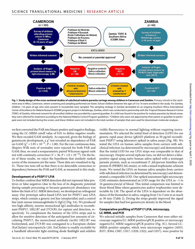
Considering that gametocytes tend to be concomitantly present with asexual blood stages in symptomatic individuals and gameto-cytes derive from a preceding asexual blood-stage population, we had originally hypothesized that the LFIA should easily confirm P. falciparum infection. We tested this hypothesis by dovetailing on an ongoing study in a clinical setting in Bo, Sierra Leone. Saliva was collected from 34 individuals (ages 3 to 67) presenting at the Mercy Hospital in Bo (Fig. 4C). Because of the febrile nature of the individ-uals, the standard collection method used in Cameroon and Zambia was replaced with the use of the Pure•SAL saliva collection device, a simple “lollipop” instrument that rapidly and passively draws saliva
within 3 min. We noted that there was a relatively strong concor-dance between the LFIA positivity and microscopy and PCR posi-tivity (Fig. 4, D and E). The estimated sensitivity of the LFIA for symptomatic cases (Fig. 4F) with microscopy as the reference stan-dard is 75% (95% CI, 43 to 95), and with multiplex PCR as the ref-erence, it is 83% (95% CI, 61 to 95).
Two samples, case codes 3311 and 3329, which were negative by PCR and microscopy, were also negative by LFIA. However, we also observed several samples that showed discordant results between the LFIA and detection by either microscopy or molecular amplifi-cation. Case code 3321 was found to be positive by PCR, negative by microscopy, and negative by LFIA. Case codes 3325, 3331, and 3342 were found to be positive by both PCR and microscopy but negative by LFIA. Nine of the cases were found to be negative by PCR and microscopy but positive by LFIA. It should be noted that PCR de-tection of gene targets that are not specific to gametocyte stages alone would result in discordant results with the LFIA, especially among subclinical carriage (microscopy negative for either asexual or gametocyte stages).
DISCUSSIONThere is an unmet need for sensitive PON diagnostic tests that can identify subclinical carriage of Plasmodium asexual parasites and gametocytes in human populations in clinical, port, school, or home settings. Such a need is more pronounced in the context of malaria elimination and eradication, especially because countries entering the elimination phase will be faced with the challenge of detecting low-density parasite carriage in the midst of reductions in infection prevalence. Although there is a renewed focus on improving current blood-based tests that detect PfHRP2, because both gametocytes and asexuals have PfHRP2, and considering that at most only 2%
D461 13 − − + + 0.02149 ++ 5
D462 11 − − + + 0.03318 + 15
D482 10 − + + + 0.03762 + 10
D487 9 − + + + 0.00445 − 15
D496 7 − − + + 0.02504 + 15
D499 9 − − + − 0.06422 + 10
D500 8 − − + − 0.01736 ++ 5
D516 8 − − + + 0.03949 + 5
D517* 9 − − + − 0.02459 ++ 5
N227 10 − − + + 0.04180 ++ 5
N234 9 − − + + 0.01287 + 10
N245 10 − + + + 0.08426 + 10
N255 9 − − + + 0.10392 + 15
N311 8 − − + + 0.04298 + 10
N318 6 − + + + 0.04264 + 10
*Samples included in Fig. 3D.
Case Code AgeMicroscopy qPCR/PCR PSSP17
Gametocyte Trophozoite Pfs25 18S rRNA LC-MRM LFIA Time to readout
by guest on April 7, 2020
http://stm.sciencem
ag.org/D
ownloaded from
Tao et al., Sci. Transl. Med. 11, eaan4479 (2019) 2 January 2019
S C I E N C E T R A N S L A T I O N A L M E D I C I N E | R E S E A R C H A R T I C L E
10 of 13
of the entire parasite biomass are gametocytes (27), it is difficult to envision how PfHRP2 would be an appropriate biomarker for ga-metocytes. The data resulting from five rounds of RDT profiling
tests that were conducted by the WHO in collaboration with FIND (Founda-tion for Innovative New Diagnostics) suggest that only a handful of existing blood-based RDTs can detect 200 asexual parasites/l of blood. However, we have found that the lower limit of LM-detectable subclinical carriage ranges from 1 to 10 gametocytes/l of blood. On the basis of our surveys of children in schools, sub-clinical carriage can occur with low num-bers or (less frequently) the complete absence of asexual stages, more common-ly occurring by mixed gametocyte/asexual infections, with densities 10-fold less than the lower limit tested in the WHO-FIND study. The major issue of the circulation of HRP2 deletion parasites in sub-Saharan Africa, which effectively limits the utility of HRP2 as a biomarker for malaria de-tection, cannot be ignored (12).
The potential utility of noninvasive biofluid sampling approaches for rapid malaria diagnosis has not been fully ex-plored. Oral fluid offers an attractive op-tion, given its inherently lower infection risk (absence of sharps during collection), cultural acceptance (as opposed to po-tential cultural blood taboos), and ample sample amount during a single collection (as opposed to 5 to 10 l of blood sam-pled from finger pricks). In our study, 2 to 5 ml of saliva collections were per-formed quickly and could be performed independently by each subject, even for the 5-year-old children. Anecdotally, the children viewed the collection as a fun “spitting contest,” unlike the perception of finger-prick sample collection. How-ever, recent efforts at identifying malaria parasite proteins in saliva have not been successful. A proteomic analysis of saliva from symptomatic individuals revealed only three proteins that achieved unequiv-ocal protein assignments to P. falciparum: porphobilinogen deaminase (PF3D7_ 1209600) and two heat shock protein 70 isoforms, PfHSP70/PfHSP70-2 (PF3D7_ 0818900 and PF3D7_0917900) (28). The two HSP70 proteins, PF3D7_0818900 and PF3D7_0917900, with Mascot ions scores of 219 and 160, respectively, were also identified in our pooled saliva samples when we used an in-gel digestion fol-lowed by LC-MS/MS approach. Although they had been previously described to be
present in saliva, due to the high degree of conservation of the se-quence with human heat shock 70-kDa protein 1A/1B (P17066|HSP76_ HUMAN), we excluded them from our final protein list. PfHRP2
Fig. 4. Evaluation of the LOD of the prototype PSSP17 LFIA and potential use in clinical settings. (A) Replicate tests using filtered (or unfiltered), axenic supernatant from stage V gametocyte cultures, asexual blood-stage cultures, and complete media (negative control). (B) Estimated LOD of the PSSP17 LFIA based on gametocyte quantification using LM or qPCR for pfs25 transcripts. (C) Schematic of the Sierra Leone study to test the utility of the LFIA in confirma-tory diagnosis of malaria parasite infection. Symptomatic individuals (>1 year of age) presenting in the Mercy Hospital in Bo, Sierra Leone, who provided informed consent were enrolled in the study. Matched blood samples were stored either in RNAlater or as DBS on Whatman FTA cards. (D) LFIA test strips showing PSSP17 detection in the saliva of a subset of symptomatic individuals presenting at a clinic in Bo, Sierra Leone. Negative control, uninfected/naive human saliva; positive control, naïve human saliva with lysed P. falciparum gametocytes. (E) Table comparing orthogonal de-tection approaches, multiplex PCR, LM, and the PSSP17 LFIA for all 34 samples from Sierra Leone. Multiplex PCR score: 0 = negative, 0.5 = weak positive, and 1 = positive. (F) Table estimating the sensitivity of the LFIA in a clinical setting compared to microscopy or multiplex PCR as the reference standard. For all lateral flow tests: C, control line; T, test line.
by guest on April 7, 2020
http://stm.sciencem
ag.org/D
ownloaded from

Tao et al., Sci. Transl. Med. 11, eaan4479 (2019) 2 January 2019
S C I E N C E T R A N S L A T I O N A L M E D I C I N E | R E S E A R C H A R T I C L E
11 of 13
has been detected previously in the saliva of malaria patients in Ghana (29) and the Philippines (30) by a sensitive sandwich ELISA (enzyme-linked immunosorbent assay) method, but to our knowl-edge, HRP2 has not been adequately measured in individuals with subclinical infection. PfHRP2 is a notoriously difficult protein to iden-tify by MS and requires extensive sample enrichment. We trained and optimized our sample preparation and LC-MS/MS analysis methodology using in vitro cultured, mature stage V gametocytes (23), and in doing so realized that the previous efforts using lectin depletion methods (28) were unnecessary. These depletion methods may have led to sample loss as opposed to parasite protein enrich-ment. Our analysis of saliva from children with subclinical infection resulted in a 13-fold increase in the parasite protein repertoire of this biofluid. Given our experience with PfHRP2 and saliva pro-teomics, we do not anticipate the facile MS/MS detection of PfHRP2 in this biofluid. Rather, we found that ELISA was more effective in quantifying this protein.
We selected PSSP17 (PF3D7_1218800) as our candidate, female- specific gametocyte marker in this proof-of-concept study. The ra-tionale for this selection was (i) PSSP17 was discovered in pooled saliva and individual saliva samples and (ii) PSSP17 was character-ized as a female-specific gametocyte protein (23), therefore presum-ably more abundant in biofluid given the female to male gametocyte ratio of 4:1. LC-MRM offers an attractive strategy for profiling sam-ples without the requirement for high sample concentration and low sample complexity, which usually hampers traditional MS ap-proaches (31). MRM is neither a PON test nor a potential population- wide screening approach; however, LC-MRM did allow us to estimate the abundance of a candidate protein biomarker (32) and to evaluate its potential detection by antibody-based approaches (33), which are customarily used as the detection method for lateral flow-based tests such as malaria RDTs. We also noted an unantici-pated retention time shift of 0.16 min for PSSP17 in our LC-MRM study, present in only the naturally infected saliva samples from children in Cameroon and Zambia. There are two likely possibili-ties responsible for this minimal shift, which was not observed when using “normal” naïve human saliva spiked with cultured stage V gametocytes. First, it is difficult to recapitulate the biology of chil-dren in malaria-endemic regions, which can include systemic con-ditions resulting from hemoglobinopathies, severe anemia, and other gastric conditions due to undernutrition. Such conditions can lead to increases in bile acids, iron concentrations, and other saliva matrix interferences that can affect retention times (34). However, we successfully confirmed the presence of gametocytes in a subset of these children by qPCR, when only the LC-MRM analysis indi-cated positive carriage, demonstrating the presence of stage V gametocytes in the blood. Furthermore, we used the LFIA to demon-strate orthogonally that protein was present in the saliva of these children. The true LOD, sensitivity, and specificity for the PON test based on the PSSP17 marker in saliva have not yet been determined and are the focus of the next stage in the preclinical assessment and field validation of this noninvasive RDT using absolute quantitative proteomic analyses.
A major strength of our study is that all LC-MRM analyses were performed on blinded samples, where the LM and qPCR data were unknown. Furthermore, by selecting random samples with paired datasets (qPCR and LC-MRM), we reduced the bias that would other-wise disproportionately influence how well the lateral flow rapid test performed. Taking this approach allowed for subsequent re-
checking of individual samples with matched orthogonal measure-ments to assess the limits of detection of the LFIA. A limitation of this study is our reliance on pfs25 qPCR assays to estimate gameto-cyte density in blood, which is a different biofluid than that used in the LFIA. The sampling of finger-prick blood (100 l) from individ-uals with very low gametocyte densities (A042 and A081) can lead to a failure to amplify pfs25, presumably due to the relatively poor chance of sampling a gametocyte; however, our lateral flow test de-tected PSSP17 in 10 l of saliva from these two children. We antici-pate that additional development is needed where we can leverage our known standard concentrations of PSSP17 (detectable by lateral flow) and their correlation with gametocyte densities. However, because our hypothesized mechanism relies on a secreted PSSP17 marker, such studies will need to use axenic culture medium, as op-posed to serial dilutions of purified, gametocyte-infected red cells. Another important consideration is the potential temporal rela-tionship between the detection of PSSP17 in saliva with gametocyte and trophozoite presence in the blood. Akin to the known stable presence of HRP2 in blood, which represents an infection that is being cleared, PSSP17 may remain in saliva even though gameto-cytes have been cleared in blood. This may be one explanation for the noted discrepancies between molecular detection of gametocytes/ asexuals in blood and the positive LFIA signal indicating the pres-ence of PSSP17 in saliva. However, a particularly provocative no-tion would be that mature gametocytes that sequester in capillary beds—or even developing gametocytes, sequestered in bone marrow— may secrete PSSP17, resulting in the failure to detect gametocytes by microscopy or PCR. To assess utility of the LFIA test for epide-miological landscaping, further evaluation will need to be per-formed in areas where malaria transmission has been reduced so drastically that it is low and unstable.
One of the limitations we noted during the progressive develop-ment of the LFIA was that, depending on lot/batch number of the chromatographic strip, the detection antibody (mAb 27C9.B5- EuChelate particle conjugate) can become partly trapped in the load-ing pad–membrane interface. This leads to a reduction of a positive signal in the test line, a control line, or both. This variability in the manufacturing of the lateral flow strip itself did not necessarily in-validate the interpretation of the presence/absence of PSSP17 for a given saliva sample. In our study, for a test to be considered posi-tive, both the test and control line must have a positive signal (re-gardless of intensity). The prototype test is not quantitative in its current form, so variation in signal intensity does not have any im-plied significance (no correlation with parasitemia). A positive sig-nal on a validated diagnostic test, albeit faint, would still trigger the same response (treatment). We expect that this potential issue could be resolved during development and optimization of a com-mercial test kit using this technology.
The potential of using the same rapid test for detecting Plasmodium vivax subclinical gametocytemic carriage, which is concurrent with asexual parasite infection, compels further study. The vivax ortho-log of PSSP17 shares >80% sequence identity at the amino acid level. A single noninvasive PON RDT for both human malaria parasites would surpass the WHO recommended response to the serious public health concern of the increased reporting of pfhrp2/pfhrp3 deletion mutants in sub-Saharan Africa and South America (12). Although our analyses of saliva from 34 symptomatic individuals indicate a potential use of the prototype LFIA in the clinical management of malaria, our primary envisioned application is
by guest on April 7, 2020
http://stm.sciencem
ag.org/D
ownloaded from
Tao et al., Sci. Transl. Med. 11, eaan4479 (2019) 2 January 2019
S C I E N C E T R A N S L A T I O N A L M E D I C I N E | R E S E A R C H A R T I C L E
12 of 13
implementation for epidemiological studies that necessarily go be-yond the confines of a clinical setting. Again, by targeting a gameto-cyte stage protein, we hope to identify individuals with subclinical infection with co-circulating asexual blood stages and mosquito- infectious gametocytes, which, in turn, can help map transmission hotspots more accurately in regions with heterogeneous malaria transmission. Our identification of other parasite-derived antigens in the saliva such as PF3D7_0507800 and PF3D7_0906100, both of which are shared between gametocytes and asexual stages, can lead the way to an optimized, highly sensitive saliva-based RDT for both research and clinical settings. Taking this notion a step further, we propose that pairing such a diagnostic with “stamp out” interven-tions such as a malaria transmission–blocking vaccine would per-mit a more targeted immunization strategy that may lower the cost of such campaigns.
MATERIALS AND METHODSStudy designThe overall goal of the project is to leverage MS-based proteomic approaches to discover and quantify P. falciparum protein markers present in human saliva and explore the possibility that one parasite protein marker, in particular PSSP17, can be detected via a nonin-vasive, prototype LFIA. We mined the saliva of children with sub-clinical malaria parasite infection and down-selected from the catalog of proteins identified to a single, female gametocyte-specific candi-date for this proof-of-concept study. A competitive profiling, cross- sectional, multi-omics study of unstimulated saliva and matched blood samples from 364 subclinical malaria parasite infection in Cameroon (in primary schools) and Zambia (in households) was completed to evaluate the utility of this target saliva-based marker in quantifying the subclinical population. To expand the potential utility of the prototype LFIA test, saliva was collected from children and adults (with symptomatic fever >37.5°C) presenting at a clinic in Sierra Leone. Informed consent was acquired for each of the 399 biofluid samples collected from the three different countries repre-senting West, Central, and Southern Africa. These independent collections were nested with ongoing research studies and commu-nity surveys, and followed National Malaria Control Program guide-lines for referral of microscopy-positive individuals for appropriate treatment. These protocols have been approved by the relevant institutional review boards and national ethics committees for Cameroon, Zambia, and Sierra Leone (2015/07/613/CE/CNERSH/SP; TDRC/ERC/2010/14/11/IRB#00003467; IRB#477605- 6/IRB#NRL.2012.0007).
Statistical analysisThe Mascot ions score significance threshold [−10Log(P)] reported in Table 1 and table S1 is based on the probability, P, that the ob-served “match” between the experimental data and the correspond-ing database sequence pulled from the combined database search is random. Tests for correlation and subsequent figure generation were conducted in program R (35). Calculation of sensitivity of the LC-MRM and LFIA tests was done using the pfs25 qPCR and 18S rRNA PCR as reference method for Cameroon and Sierra Leone, and the cytB nested PCR as reference method for Zambia. Binomial confidence interval was calculated by “cii” (confidence interval im-mediate) command specifying “proportions” using STATA version 14 (StataCorp LP).
SUPPLEMENTARY MATERIALSwww.sciencetranslationalmedicine.org/cgi/content/full/11/473/eaan4479/DC1Materials and MethodsFig. S1. The correlation of PAR and female gametocyte abundance per microliter of blood (based on pfs25 transcript number).Table S1. The complete list of P. falciparum proteins identified in the saliva from children with subclinical infection in Yaoundé, Cameroon.Table S2. LM analyses of blood samples from children (5 to 12 years old) with subclinical infections in Yaoundé, Cameroon.Table S3. Description of samples collected from schools in Cameroon.Table S4. Description of samples collected from homes in Zambia.Table S5. Quantification of gametocytes per l of blood in a subset (n = 100) of samples from Cameroon.References (36–44)
REFERENCES AND NOTES 1. World Health Organization, World Malaria Report (World Health Organization, 2017). 2. E. Bottius, L. BenMohamed, K. Brahimi, H. Gras, J. P. Lepers, L. Raharimalala, M. Aikawa,
J. Meis, B. Slierendregt, A. Tartar, A. Thomas, P. Druilhe, A novel Plasmodium falciparum sporozoite and liver stage antigen (SALSA) defines major B, T helper, and CTL epitopes. J. Immunol. 156, 2874–2884 (1996).
3. M. D. Perkins, D. R. Bell, Working without a blindfold: The critical role of diagnostics in malaria control. Malar. J. 7 (suppl. 1), S5 (2008).
4. D. D. Laishram, P. L. Sutton, N. Nanda, V. L. Sharma, R. C. Sobti, J. M. Carlton, H. Joshi, The complexities of malaria disease manifestations with a focus on asymptomatic malaria. Malar. J. 11, 29 (2012).
5. T. Bousema, L. Okell, I. Felger, C. Drakeley, Asymptomatic malaria infections: Detectability, transmissibility and public health relevance. Nat. Rev. Microbiol. 12, 833–840 (2014).
6. T. Bousema, T. S. Churcher, I. Morlais, R. R. Dinglasan, Can field-based mosquito feeding assays be used for evaluating transmission-blocking interventions? Trends Parasitol. 29, 53–59 (2013).
7. H. J. W. Sturrock, M. S. Hsiang, J. M. Cohen, D. L. Smith, B. Greenhouse, T. Bousema, R. D. Gosling, Targeting asymptomatic malaria infections: Active surveillance in control and elimination. PLOS Med. 10, e1001467 (2013).
8. J. Cook, I. Kleinschmidt, C. Schwabe, G. Nseng, T. Bousema, P. H. Corran, E. M. Riley, C. J. Drakeley, Serological markers suggest heterogeneity of effectiveness of malaria control interventions on Bioko Island, Equatorial Guinea. PLOS ONE 6, e25137 (2011).
9. G. H. Stresman, A. Kamanga, P. Moono, H. Hamapumbu, S. Mharakurwa, T. Kobayashi, W. J. Moss, C. Shiff, A method of active case detection to target reservoirs of asymptomatic malaria and gametocyte carriers in a rural area in Southern Province, Zambia. Malar. J. 9, 265 (2010).
10. C. Drakeley, C. Sutherland, J. T. Bousema, R. W. Sauerwein, G. A. T. Targett, The epidemiology of Plasmodium falciparum gametocytes: Weapons of mass dispersion. Trends Parasitol. 22, 424–430 (2006).
11. K. Tietje, K. Hawkins, C. Clerk, K. Ebels, S. McGray, C. Crudder, L. Okell, P. LaBarre, The essential role of infection-detection technologies for malaria elimination and eradication. Trends Parasitol. 30, 259–266 (2014).
12. World Health Organization, False-Negative RDT Results and Implications of New Reports of P. falciparum Histidine-Rich Protein 2/3 Gene Deletions (World Health Organization, 2016).
13. D. C. Nwakanma, N. Gomez-Escobar, M. Walther, S. Crozier, F. Dubovsky, E. Malkin, E. Locke, D. J. Conway, Quantitative detection of Plasmodium falciparum DNA in saliva, blood, and urine. J. Infect. Dis. 199, 1567–1574 (2009).
14. C. Putaporntip, P. Buppan, S. Jongwutiwes, Improved performance with saliva and urine as alternative DNA sources for malaria diagnosis by mitochondrial DNA-based PCR assays. Clin. Microbiol. Infect. 17, 1484–1491 (2011).
15. R. Magni, B. H. Espina, K. Shah, B. Lepene, C. Mayuga, T. A. Douglas, V. Espina, S. Rucker, R. Dunlap, E. F. Petricoin III, M. F. Kilavos, D. M. Poretz, G. R. Irwin, S. M. Shor, L. A. Liotta, A. Luchini, Application of Nanotrap technology for high sensitivity measurement of urinary outer surface protein a carboxyl-terminus domain in early stage Lyme borreliosis. J. Transl. Med. 13, 346 (2015).
16. G. Pichon, H. P. Awono-Ambene, V. Robert, High heterogeneity in the number of Plasmodium falciparum gametocytes in the bloodmeal of mosquitoes fed on the same host. Parasitology 121 (Pt. 2), 115–120 (2000).
17. M. Aingaran, R. Zhang, S. K. Law, Z. Peng, A. Undisz, E. Meyer, M. Diez-Silva, T. A. Burke, T. Spielmann, C. T. Lim, S. Suresh, M. Dao, M. Marti, Host cell deformability is linked to transmission in the human malaria parasite Plasmodium falciparum. Cell. Microbiol. 14, 983–993 (2012).
18. M. Tibúrcio, R. Sauerwein, C. Lavazec, P. Alano, Erythrocyte remodeling by Plasmodium falciparum gametocytes in the human host interplay. Trends Parasitol. 31, 270–278 (2015).
by guest on April 7, 2020
http://stm.sciencem
ag.org/D
ownloaded from

Tao et al., Sci. Transl. Med. 11, eaan4479 (2019) 2 January 2019
S C I E N C E T R A N S L A T I O N A L M E D I C I N E | R E S E A R C H A R T I C L E
13 of 13
19. I. B. Lamster, J. K. Ahlo, Analysis of gingival crevicular fluid as applied to the diagnosis of oral and systemic diseases. Ann. N. Y. Acad. Sci. 1098, 216–229 (2007).
20. F. Silvestrini, E. Lasonder, A. Olivieri, G. Camarda, B.vanSchaijk, M. Sanchez, S. Younis Younis, R. Sauerwein, P. Alano, Protein export marks the early phase of gametocytogenesis of the human malaria parasite Plasmodium falciparum. Mol. Cell. Proteomics 9, 1437–1448 (2010).
21. B. N. Pease, E. L. Huttlin, M. P. Jedrychowski, E. Talevich, J. Harmon, T. Dillman, N. Kannan, C. Doerig, R. Chakrabarti, S. P. Gygi, D. Chakrabarti, Global analysis of protein expression and phosphorylation of three stages of Plasmodium falciparum intraerythrocytic development. J. Proteome Res. 12, 4028–4045 (2013).
22. M. Treeck, J. L. Sanders, J. E. Elias, J. C. Boothroyd, The phosphoproteomes of Plasmodium falciparum and Toxoplasma gondii reveal unusual adaptations within and beyond the parasites' boundaries. Cell Host Microbe 10, 410–419 (2011).
23. D. Tao, C. Ubaida-Mohien, D. K. Mathias, J. G. King, R. Pastrana-Mena, A. Tripathi, I. Goldowitz, D. R. Graham, E. Moss, M. Marti, R. R. Dinglasan, Sex-partitioning of the Plasmodium falciparum stage V gametocyte proteome provides insight into falciparum-specific cell biology. Mol. Cell. Proteomics 13, 2705–2724 (2014).
24. A. M. Mendes, T. Schlegelmilch, A. Cohuet, P. Awono-Ambene, M. De Iorio, D. Fontenille, I. Morlais, G. K. Christophides, F. C. Kafatos, D. Vlachou, Conserved mosquito/parasite interactions affect development of Plasmodium falciparum in Africa. PLOS Pathog. 4, e1000069 (2008).
25. M. van der Kolk, A. E. Tebo, H. Nimpaye, D. N. Ndombol, R. W. Sauerwein, W. M. C. Eling, Transmission of Plasmodium falciparum in urban Yaoundé, Cameroon, is seasonal and age-dependent. Trans. R. Soc. Trop. Med. Hyg. 97, 375–379 (2003).
26. E. Juntunen, T. Myyryläinen, T. Salminen, T. Soukka, K. Pettersson, Performance of fluorescent europium(III) nanoparticles and colloidal gold reporters in lateral flow bioaffinity assay. Anal. Biochem. 428, 31–38 (2012).
27. T. Bousema, C. Drakeley, Epidemiology and infectivity of Plasmodium falciparum and Plasmodium vivax gametocytes in relation to malaria control and elimination. Clin. Microbiol. Rev. 24, 377–410 (2011).
28. H. Huang, M. M. Mackeen, M. Cook, E. Oriero, E. Locke, M. L. Thézénas, B. M. Kessler, D. Nwakanma, C. Casals-Pascual, Proteomic identification of host and parasite biomarkers in saliva from patients with uncomplicated Plasmodium falciparum malaria. Malar. J. 11, 178 (2012).
29. N. O. Wilson, A. A. Adjei, W. Anderson, S. Baidoo, J. K. Stiles, Detection of Plasmodium falciparum histidine-rich protein II in saliva of malaria patients. Am. J. Trop. Med. Hyg. 78, 733–735 (2008).
30. A. O. Fung, R. Damoiseaux, S. Grundeen, J. L. Panes, D. H. Horton, J. W. Judy, T. B. Moore, Quantitative detection of PfHRP2 in saliva of malaria patients in the Philippines. Malar. J. 11, 175 (2012).
31. C. E. Parker, C. H. Borchers, Mass spectrometry based biomarker discovery, verification, and validation—Quality assurance and control of protein biomarker assays. Mol. Oncol. 8, 840–858 (2014).
32. R. Schiess, B. Wollscheid, R. Aebersold, Targeted proteomic strategy for clinical biomarker discovery. Mol. Oncol. 3, 33–44 (2009).
33. T. Shi, D. Su, T. Liu, K. Tang, D. G. Camp II, W.-J. Qian, R. D. Smith, Advancing the sensitivity of selected reaction monitoring-based targeted quantitative proteomics. Proteomics 12, 1074–1092 (2012).
34. L. Zhang, W. Voskuijl, M. Mouzaki, A. K. Groen, J. Alexander, C. Bourdon, A. Wang, C. J. Versloot, V. Di Giovanni, R. J. A. Wanders, R. Bandsma, Impaired bile acid homeostasis in children with severe acute malnutrition. PLOS ONE 11, e0155143 (2016).
35. R Core Team, R: A Language and Environment for Statistical Computing (R Foundation for Statistical Computing, 2014); http://www.R-project.org.
36. S. Mharakurwa, P. E. Thuma, D. E. Norris, M. Mulenga, V. Chalwe, J. Chipeta, S. Munyati, S. Mutambu, P. R. Mason, Southern Africa ICEMR Team, Malaria epidemiology and control in southern Africa. Acta Trop. 121, 202–206 (2012).
37. W. J. Moss, D. E. Norris, S. Mharakurwa, A. Scott, M. Mulenga, P. R. Mason, J. Chipeta, P. E. Thuma, Southern Africa ICEMR Team, Challenges and prospects for malaria elimination in the southern Africa region. Acta Trop. 121, 207–211 (2012).
38. V. M. Mukonka, E. Chanda, U. Haque, M. Kamuliwo, G. Mushinge, J. Chileshe, K. A. Chibwe, D. E. Norris, M. Mulenga, M. Chaponda, M. Muleba, G. E. Glass, W. J. Moss, High burden of malaria following scale-up of control interventions in Nchelenge District, Luapula Province, Zambia. Malar. J. 13, 153 (2014).
39. Zambia National Malaria Indicator Survey 2012 (Government of the Republic of Zambia, Ministry of Health, 2012).
40. K. C. Kain, D. E. Lanar, Determination of genetic variation within Plasmodium falciparum by using enzymatically amplified DNA from filter paper disks impregnated with whole blood. J. Clin. Microbiol. 29, 1171–1174 (1991).
41. Z. P. Billman, A. M. Seilie, S. C. Murphy, Purification of Plasmodium sporozoites enhances parasite-specific CD8+ T cell responses. Infect. Immun. 84, 2233–2242 (2016).
42. E. Kamau, S. Alemayehu, K. C. Feghali, D. Saunders, C. F. Ockenhouse, Multiplex qPCR for detection and absolute quantification of malaria. PLOS ONE 8, e71539 (2013).
43. K. A. Mangold, R. U. Manson, E. S. C. Koay, L. Stephens, M. Regner, R. B. Thomson Jr., L. R. Peterson, K. L. Kaul, Real-time PCR for detection and identification of Plasmodium spp. J. Clin. Microbiol. 43, 2435–2440 (2005).
44. X. Yuan, D. Fabregat, K. Yoshimoto, Y. Nagasaki, Development of a high-performance immunolatex based on “soft landing” antibody immobilization mechanism. Colloids Surf. B Biointerfaces 99, 45–52 (2012).
Acknowledgments: We are grateful to the parents and children from Cameroon, Zambia, and Sierra Leone who participated in our study. We also thank L. Abate, S. Menard, A. Bayibéki, M. Sandeu, N. Martin, T. Shields, and B. Hritzo for technical assistance. We also thank G. Gutierrez and the Leidos team for helpful discussions. Funding: The project was funded, in part, by a TEDCO Maryland Innovation Initiative award (120701) and a Bill & Melinda Gates Foundation (OPP 1054413) to R.R.D. T.L., A.H., D.A.S., and R.A. were supported by the Chemical Biological Technologies Directorate from the Department of Defense Chemical and Biological Defense program through the Defense Threat Reduction Agency (DTRA). However, any opinions, findings, conclusions, or other recommendations expressed here are those of the authors and do not necessarily reflect the views of the DTRA. Author contributions: I.M., W.J.M., and R.R.D. conceived and designed the research; I.M., T.K., T.L., A.H., W.J.M., K.H.J., S.E.N., M.C., M.M., D.A.S., R.A., and R.R.D. were responsible for oversight of human subjects research and saliva collections; D.T., B.M., T.H., A.D., A.H., T.L., P.D.S., C.C.S., A.E.J., D.J.S., T.K., B.S., P.K., and A.W. performed experiments and developed new reagents/tools; D.T., B.M., T.H., R.A., A.D., A.E.J., P.K., S.J.R., D.J.S., D.A.S., I.M., W.J.M., and R.R.D. analyzed data; and T.H., B.M., T.L., D.T., D.J.S., K.H.J., S.J.R., W.J.M., I.M., and R.R.D. wrote the paper. Competing interests: R.R.D., D.T., and B.M. are inventors on patent/patent application (PCT/US2016/037968) held/submitted by Johns Hopkins University that covers the described saliva-based malaria rapid test. The technology is licensed to Erada Technology Alliance Limited, Mauritius. D.J.S. receives royalties from Alere for provision of positive control antigens for the Binax malaria RDT and is a named inventor and consultant for Fyodor Biotechnologies who licensed a urine malaria test from Johns Hopkins University. P.D.S. and C.C.S. are with Oasis Diagnostics, a manufacturer of RDTs using saliva. No other competing interests have been identified. Data and materials availability: All processed data associated with this study are present in the paper or in the Supplementary Materials. Raw MS data, used in the current study (PASS01290), have been deposited in Peptide Atlas and can be accessed through this link: (http://www.peptideatlas.org/PASS/PASS01290).
Submitted 22 April 2017Resubmitted 27 July 2018Accepted 30 November 2018Published 2 January 201910.1126/scitranslmed.aan4479
Citation: D. Tao, B. McGill, T. Hamerly, T. Kobayashi, P. Khare, A. Dziedzic, T. Leski, A. Holtz, B. Shull, A. E. Jedlicka, A. Walzer, P. D. Slowey, C. C. Slowey, S. E. Nsango, D. A. Stenger, M. Chaponda, M. Mulenga, K. H. Jacobsen, D. J. Sullivan, S. J. Ryan, R. Ansumana, W. J. Moss, I. Morlais, R. R. Dinglasan, A saliva-based rapid test to quantify the infectious subclinical malaria parasite reservoir. Sci. Transl. Med. 11, eaan4479 (2019).
by guest on April 7, 2020
http://stm.sciencem
ag.org/D
ownloaded from
A saliva-based rapid test to quantify the infectious subclinical malaria parasite reservoir
Rashid Ansumana, William J. Moss, Isabelle Morlais and Rhoel R. DinglasanNsango, David A. Stenger, Mike Chaponda, Modest Mulenga, Kathryn H. Jacobsen, David J. Sullivan, Sadie J. Ryan,Andrew Holtz, Bruce Shull, Anne E. Jedlicka, Andrew Walzer, Paul D. Slowey, Christopher C. Slowey, Sandrine E. Dingyin Tao, Brent McGill, Timothy Hamerly, Tamaki Kobayashi, Prachi Khare, Amanda Dziedzic, Tomasz Leski,
DOI: 10.1126/scitranslmed.aan4479, eaan4479.11Sci Transl Med
reservoir. infections to reduce the infectiousP. falciparumsettings. This assay could help identify individuals with subclinical
carriage in saliva from children and adults in Cameroon, Zambia, and Sierra Leone in clinical and nonclinical microscopic analyses of matched blood samples, the rapid diagnostic assay could detect submicroscopic parasiteassay that detects a female gametocyte-specific protein, PSSP17, in saliva. Compared to standard molecular and
developed a rapid lateral flowet al.(subclinical infection), which makes detection and eradication difficult. Tao parasiteresponsible for mosquito-borne transmission of infection. Some individuals carry low concentrations of the
, a parasite that causes human malaria, isPlasmodium falciparumThe gametocyte sexual form of Spotting subclinical infection
ARTICLE TOOLS http://stm.sciencemag.org/content/11/473/eaan4479
MATERIALSSUPPLEMENTARY http://stm.sciencemag.org/content/suppl/2018/12/21/11.473.eaan4479.DC1
CONTENTRELATED
http://stm.sciencemag.org/content/scitransmed/12/527/eaax2421.fullhttp://science.sciencemag.org/content/sci/367/6473/51.fullhttp://science.sciencemag.org/content/sci/367/6473/22.fullhttp://stm.sciencemag.org/content/scitransmed/11/519/eaax4204.fullhttp://science.sciencemag.org/content/sci/365/6456/eaau1682.fullhttp://science.sciencemag.org/content/sci/365/6456/847.fullhttp://science.sciencemag.org/content/sci/365/6455/eaaw2619.fullhttp://science.sciencemag.org/content/sci/364/6443/894.fullhttp://stm.sciencemag.org/content/scitransmed/11/484/eaat7386.fullhttp://stm.sciencemag.org/content/scitransmed/10/463/eaat6176.fullhttp://stm.sciencemag.org/content/scitransmed/9/397/eaaf9377.fullhttp://stm.sciencemag.org/content/scitransmed/10/447/eaar3619.full
REFERENCES
http://stm.sciencemag.org/content/11/473/eaan4479#BIBLThis article cites 40 articles, 8 of which you can access for free
PERMISSIONS http://www.sciencemag.org/help/reprints-and-permissions
Terms of ServiceUse of this article is subject to the
registered trademark of AAAS. is aScience Translational MedicineScience, 1200 New York Avenue NW, Washington, DC 20005. The title
(ISSN 1946-6242) is published by the American Association for the Advancement ofScience Translational Medicine
4.0 (CC BY).of Science. No claim to original U.S. Government Works. Distributed under a Creative Commons Attribution License Copyright © 2019 The Authors, some rights reserved; exclusive licensee American Association for the Advancement
by guest on April 7, 2020
http://stm.sciencem
ag.org/D
ownloaded from