Embed Size (px)
Citation preview

TERROR
2
TYPES OF WOUNDS
• ARTERIAL• PRESURE ULCERS• VENOUS • DIABETIC • SURGICAL • TRAUMA • ATYPICAL
3
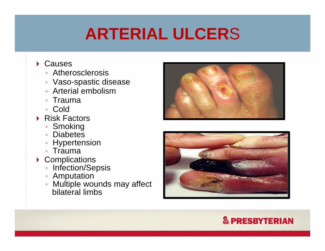
ARTERIAL ULCERSCauses◦ Atherosclerosis◦ Vaso-spastic disease◦ Arterial embolism◦ Trauma◦ ColdRisk Factors◦ Smoking◦ Diabetes◦ Hypertension◦ TraumaComplications◦ Infection/Sepsis◦ Amputation◦ Multiple wounds may affect
bilateral limbs
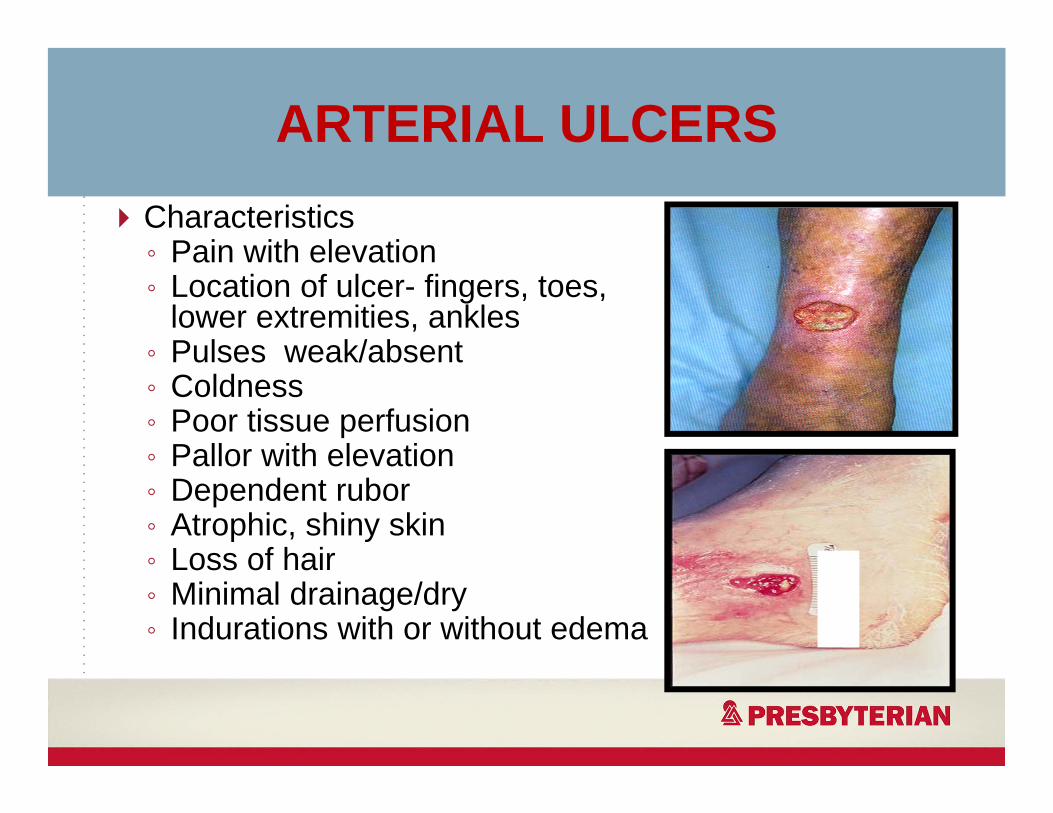
ARTERIAL ULCERSCharacteristics◦ Pain with elevation◦ Location of ulcer- fingers, toes,
lower extremities, ankles◦ Pulses weak/absent◦ Coldness◦ Poor tissue perfusion◦ Pallor with elevation◦ Dependent rubor◦ Atrophic, shiny skin◦ Loss of hair◦ Minimal drainage/dry◦ Indurations with or without edema
VENOUS ULCERS
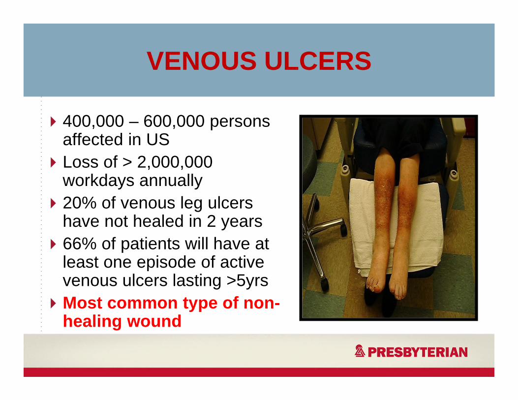
400,000 – 600,000 persons affected in USLoss of > 2,000,000 workdays annually20% of venous leg ulcers have not healed in 2 years66% of patients will have at least one episode of active venous ulcers lasting >5yrsMost common type of non-healing wound
CAUSES OF VENOUS HYPERTENSION
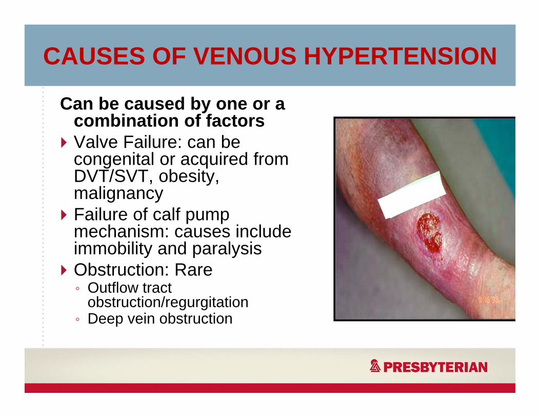
Can be caused by one or a combination of factorsValve Failure: can be congenital or acquired from DVT/SVT, obesity, malignancyFailure of calf pump mechanism: causes include immobility and paralysisObstruction: Rare◦ Outflow tract
obstruction/regurgitation◦ Deep vein obstruction
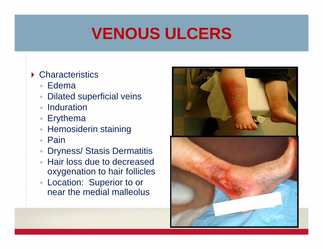
VENOUS ULCERS
Characteristics◦ Edema◦ Dilated superficial veins◦ Induration ◦ Erythema◦ Hemosiderin staining◦ Pain◦ Dryness/ Stasis Dermatitis◦ Hair loss due to decreased
oxygenation to hair follicles◦ Location: Superior to or
near the medial malleolus
CODING A VENOUS ULCER
• SENERIO:• 65M patient who hit lateral left ankle on a coffee
table two weeks ago. There is pitting edema, the LLE is reddened with a wound noted on the left lateral ankle. Stasis dermatitis noted LLE
• Patient has a history of venous insufficiency and ulcerations in the past.
• Trauma ?? Venous Ulcer?? Varicose Veins??
9
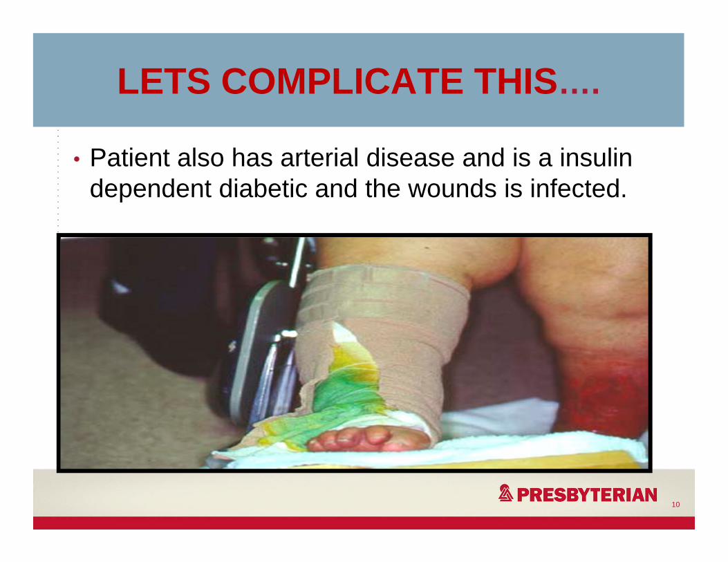
LETS COMPLICATE THIS….
• Patient also has arterial disease and is a insulin dependent diabetic and the wounds is infected.
10
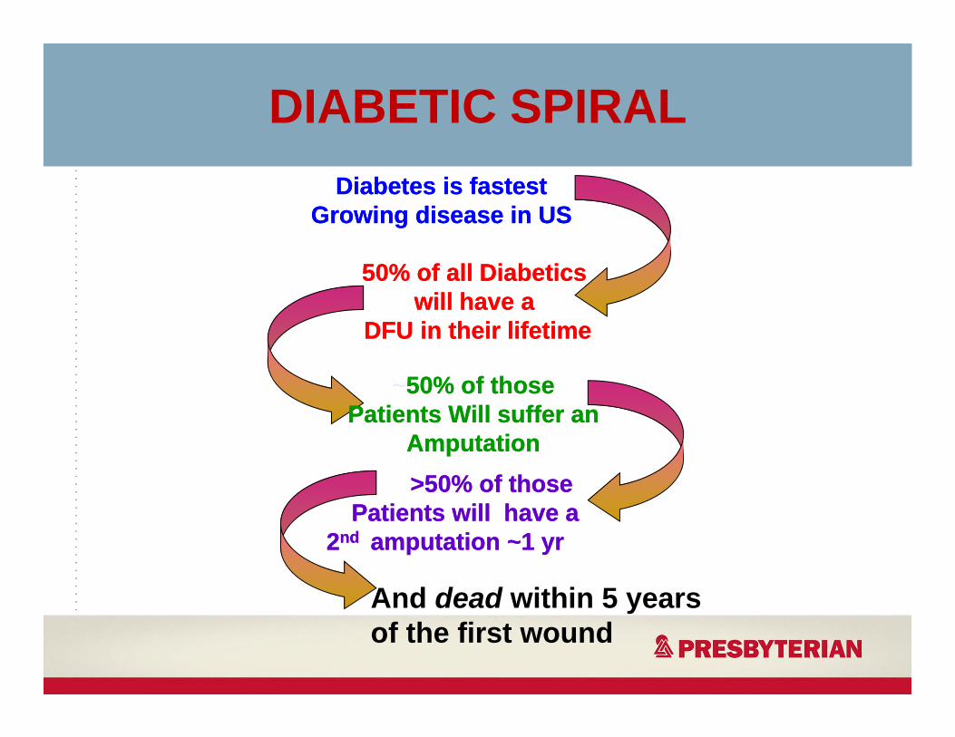
DIABETIC SPIRALDiabetes is fastest
Growing disease in USDiabetes is fastest
Growing disease in US
50% of all Diabetics will have a
DFU in their lifetime
50% of all Diabetics will have a
DFU in their lifetime
~50% of those Patients Will suffer an
Amputation
~50% of those Patients Will suffer an
Amputation >50% of those
Patients will have a 2nd amputation ~1 yr
>50% of those Patients will have a
2nd amputation ~1 yr
And dead within 5 years of the first wound
DIABETIC FOOT ULCERS
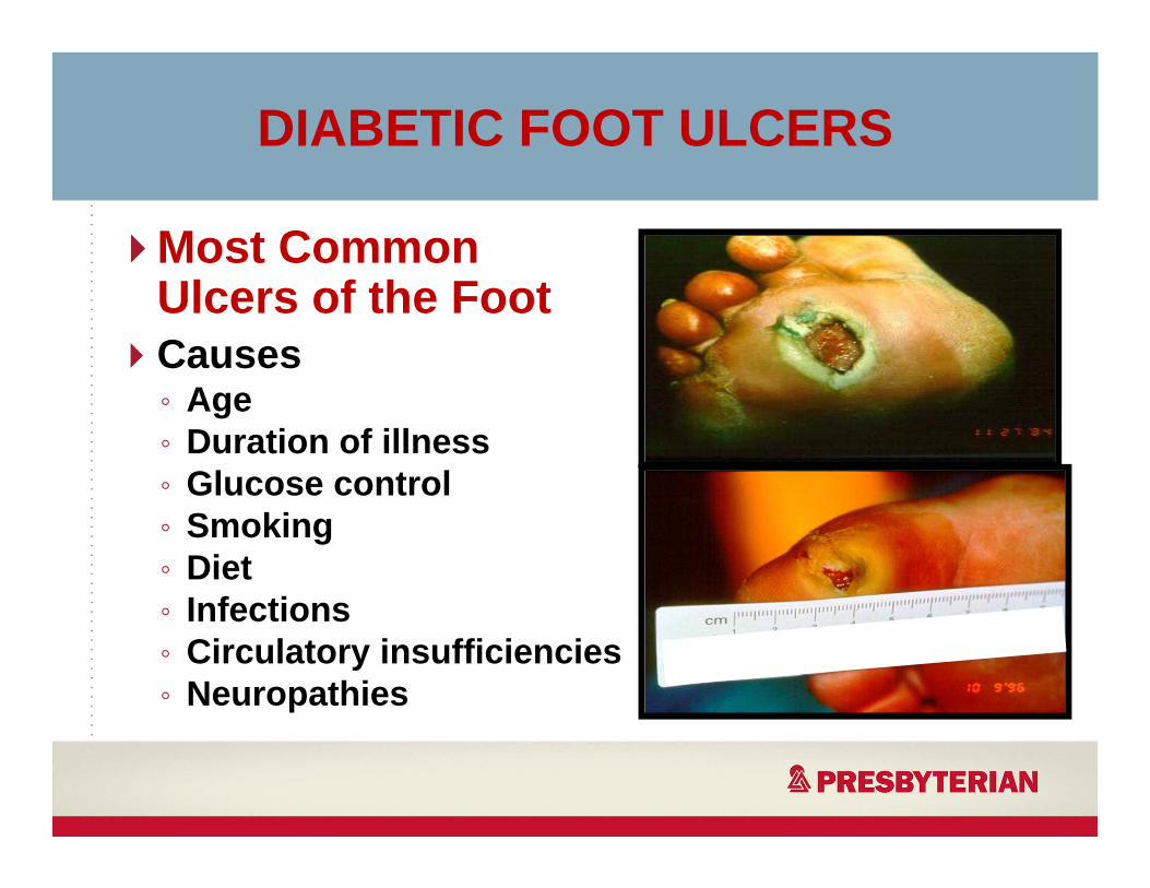
Most Common Ulcers of the Foot Causes◦ Age◦ Duration of illness◦ Glucose control◦ Smoking◦ Diet◦ Infections ◦ Circulatory insufficiencies◦ Neuropathies
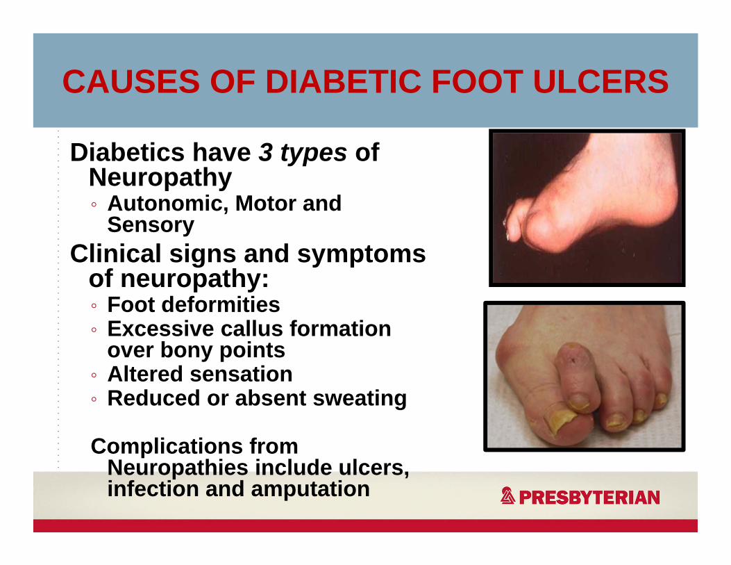
CAUSES OF DIABETIC FOOT ULCERS
Diabetics have 3 types of Neuropathy◦ Autonomic, Motor and
SensoryClinical signs and symptoms
of neuropathy:◦ Foot deformities◦ Excessive callus formation
over bony points◦ Altered sensation◦ Reduced or absent sweating
Complications from Neuropathies include ulcers, infection and amputation
DIABETIC FOOT ULCERS
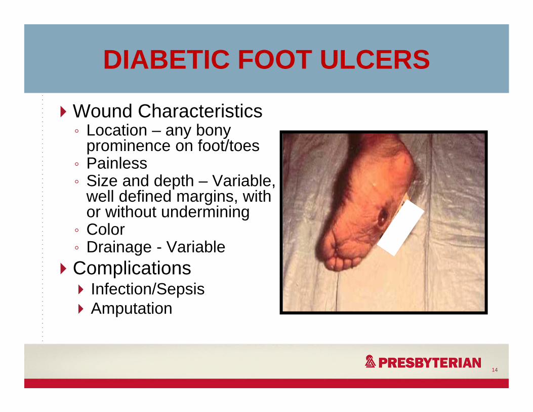
Wound Characteristics◦ Location – any bony
prominence on foot/toes◦ Painless◦ Size and depth – Variable,
well defined margins, with or without undermining
◦ Color ◦ Drainage - VariableComplications
Infection/SepsisAmputation
14
DIABETIC FOOT ULCERS VS. PRESSURE ULCERS
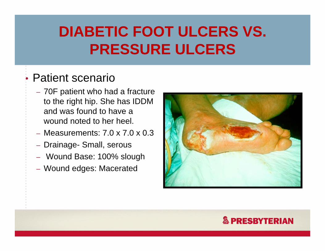
• Patient scenario– 70F patient who had a fracture
to the right hip. She has IDDM and was found to have a wound noted to her heel.
– Measurements: 7.0 x 7.0 x 0.3– Drainage- Small, serous– Wound Base: 100% slough– Wound edges: Macerated
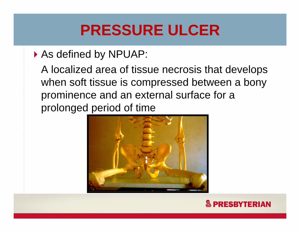
PRESSURE ULCERAs defined by NPUAP: A localized area of tissue necrosis that develops when soft tissue is compressed between a bony prominence and an external surface for a prolonged period of time
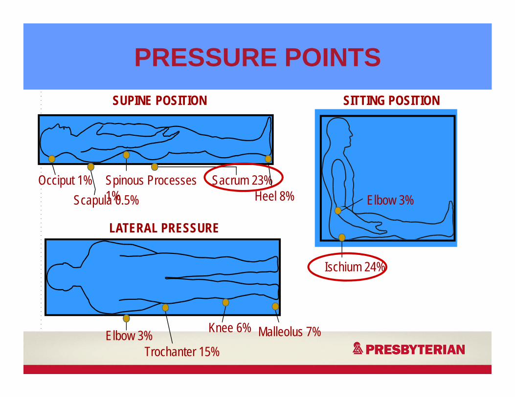
PRESSURE POINTS
Occiput 1%Scapula 0.5%
Spinous Processes 1%
Sacrum 23%Heel 8%
Elbow 3%Trochanter 15%
Knee 6% Malleolus 7%
SUPINE POSITION
LATERAL PRESSURE
SITTING POSITION
Ischium 24%
Elbow 3%
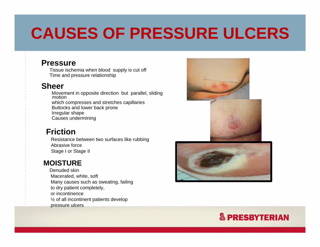
CAUSES OF PRESSURE ULCERSPressure
Tissue ischemia when blood supply is cut offTime and pressure relationship
SheerMovement in opposite direction but parallel, sliding motion which compresses and stretches capillariesButtocks and lower back proneIrregular shapeCauses undermining
FrictionResistance between two surfaces like rubbingAbrasive forceStage I or Stage II
MOISTURE Denuded skinMacerated, white, softMany causes such as sweating, failing to dry patient completely,or incontinence½ of all incontinent patients develop pressure ulcers
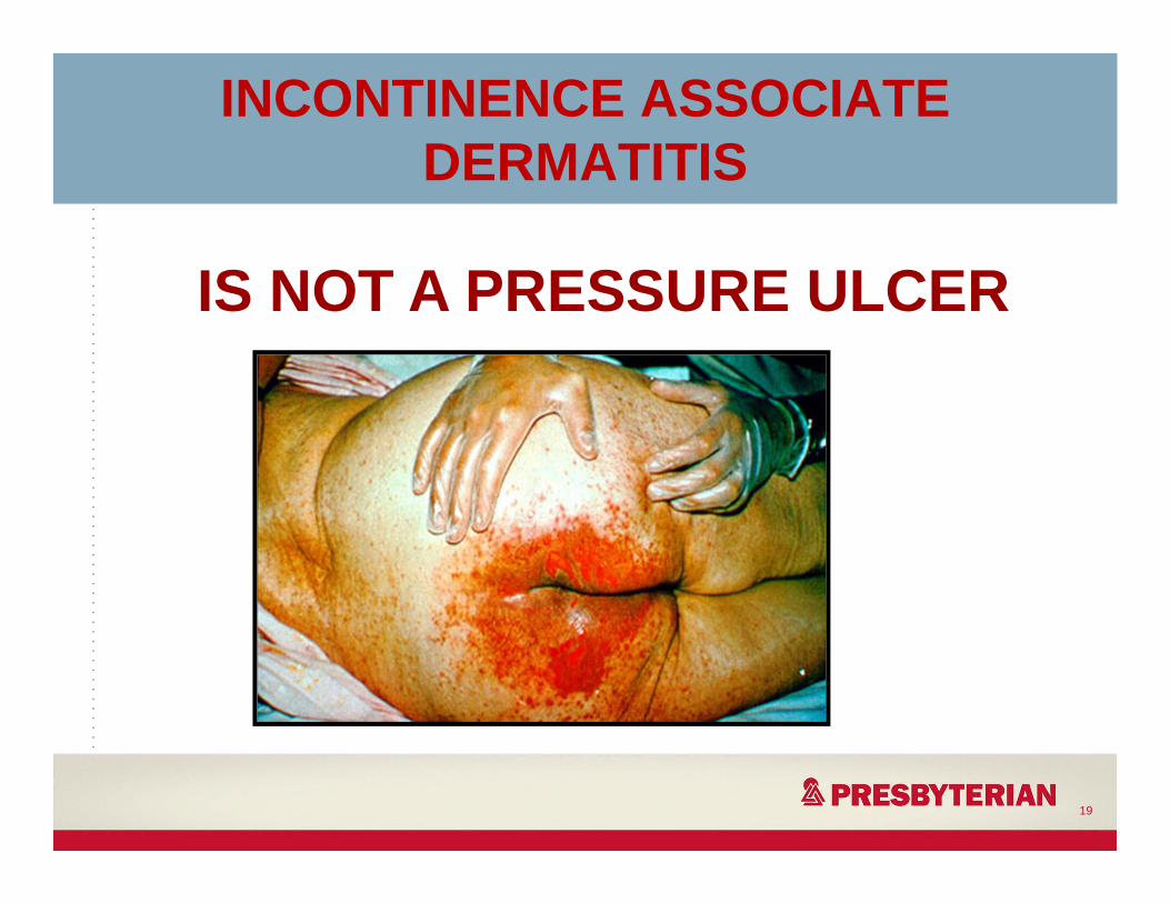
INCONTINENCE ASSOCIATE DERMATITIS
19
IS NOT A PRESSURE ULCER
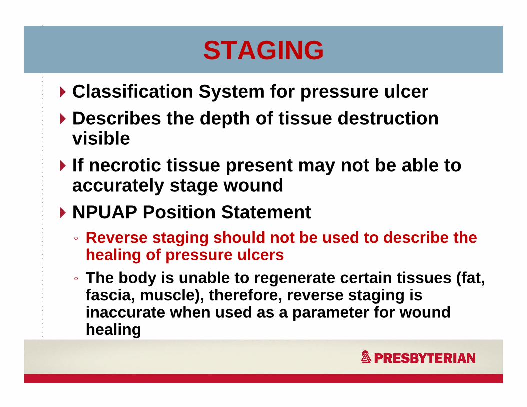
STAGINGClassification System for pressure ulcer Describes the depth of tissue destruction visibleIf necrotic tissue present may not be able to accurately stage woundNPUAP Position Statement◦ Reverse staging should not be used to describe the
healing of pressure ulcers◦ The body is unable to regenerate certain tissues (fat,
fascia, muscle), therefore, reverse staging is inaccurate when used as a parameter for wound healing
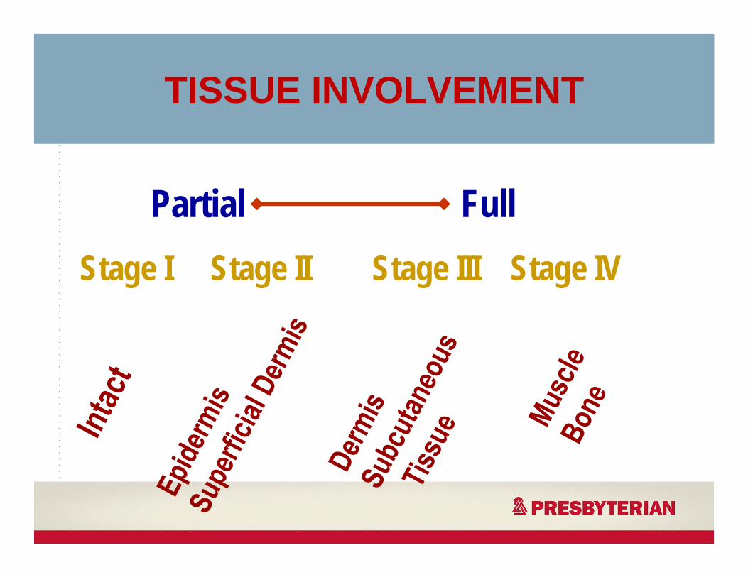
TISSUE INVOLVEMENT
Partial FullStage I Stage II Stage III Stage IV
Associated Factors◦ Mobility status◦ Activity status◦ Mental status◦ Nutritional status◦ Incontinence◦ Chronic diseases◦ Medications
Wound Characteristics◦ Stage◦ Location◦ Pain◦ Size and depth◦ Color◦ Drainage
PRESSURE ULCERS
22
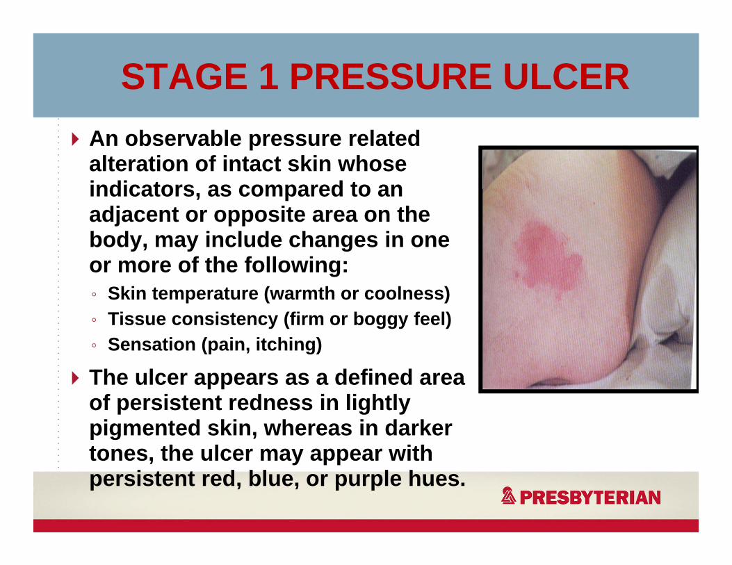
STAGE 1 PRESSURE ULCERAn observable pressure related alteration of intact skin whose indicators, as compared to an adjacent or opposite area on the body, may include changes in one or more of the following: ◦ Skin temperature (warmth or coolness)◦ Tissue consistency (firm or boggy feel)◦ Sensation (pain, itching)
The ulcer appears as a defined area of persistent redness in lightly pigmented skin, whereas in darker tones, the ulcer may appear with persistent red, blue, or purple hues.
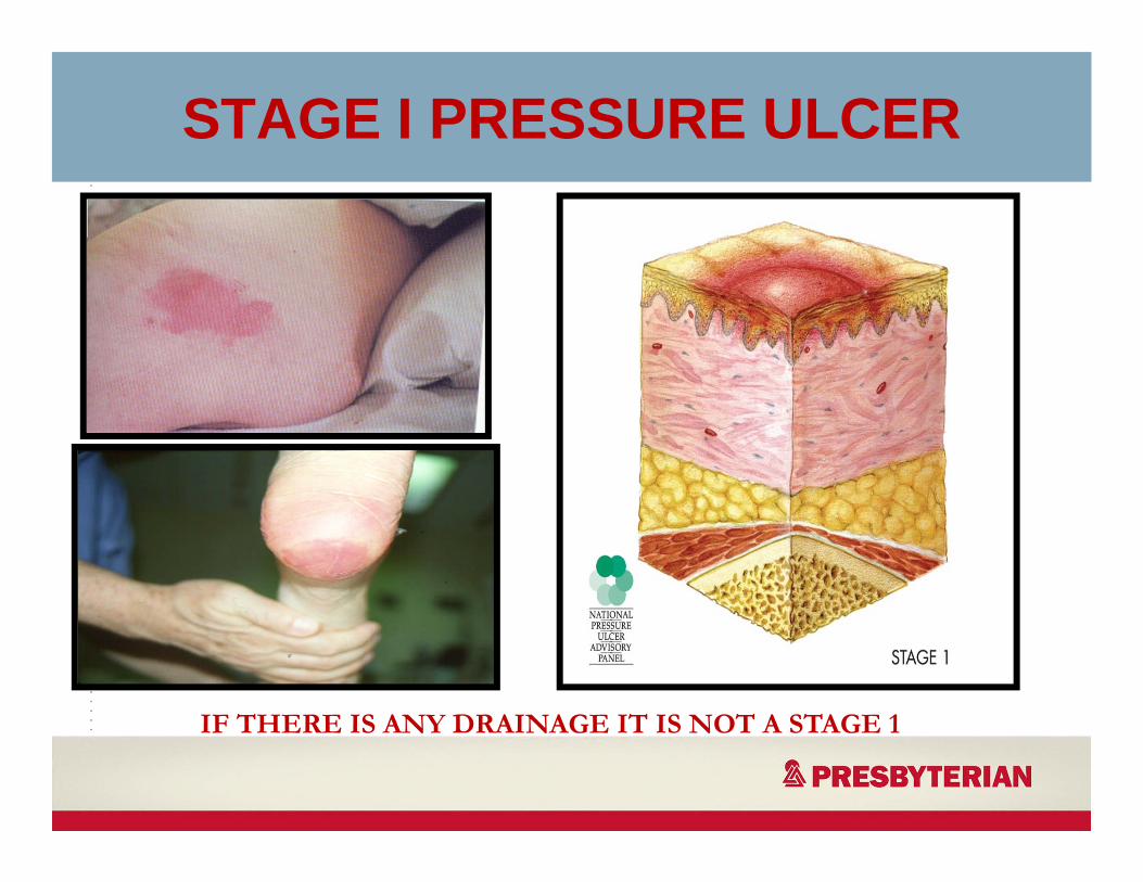
STAGE I PRESSURE ULCER
IF THERE IS ANY DRAINAGE IT IS NOT A STAGE 1
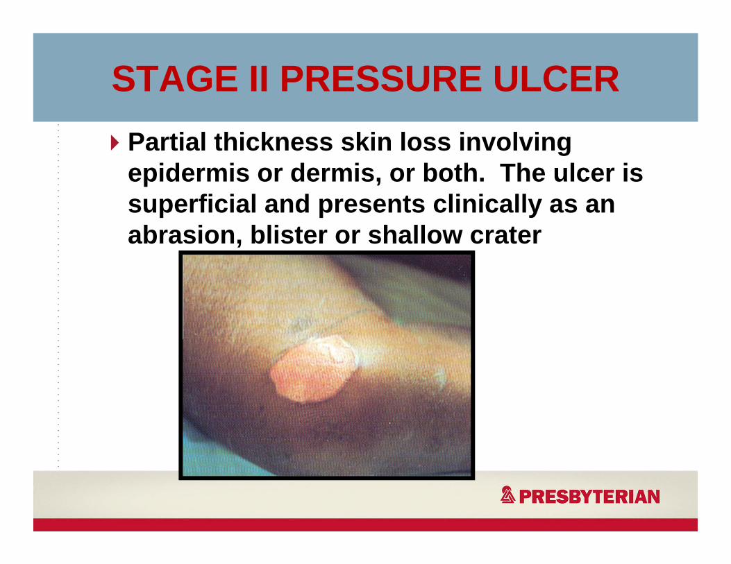
STAGE II PRESSURE ULCERPartial thickness skin loss involving epidermis or dermis, or both. The ulcer is superficial and presents clinically as an abrasion, blister or shallow crater
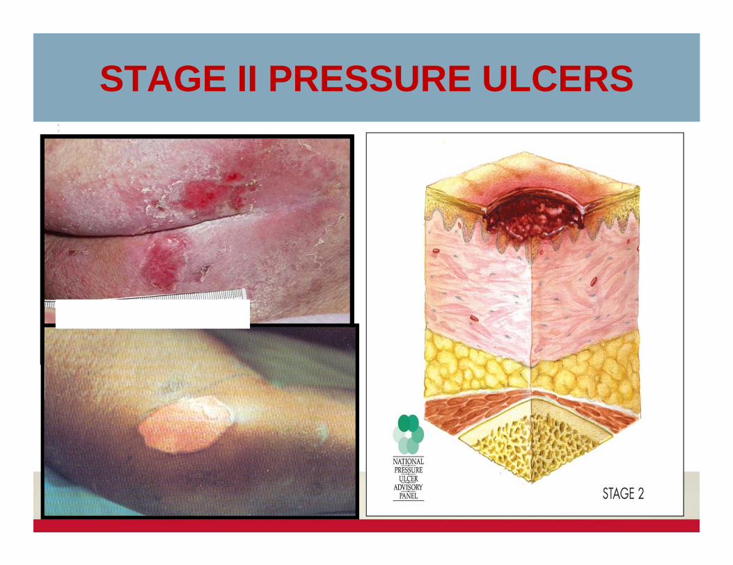
STAGE II PRESSURE ULCERS
STAGE III PRESSURE ULCER
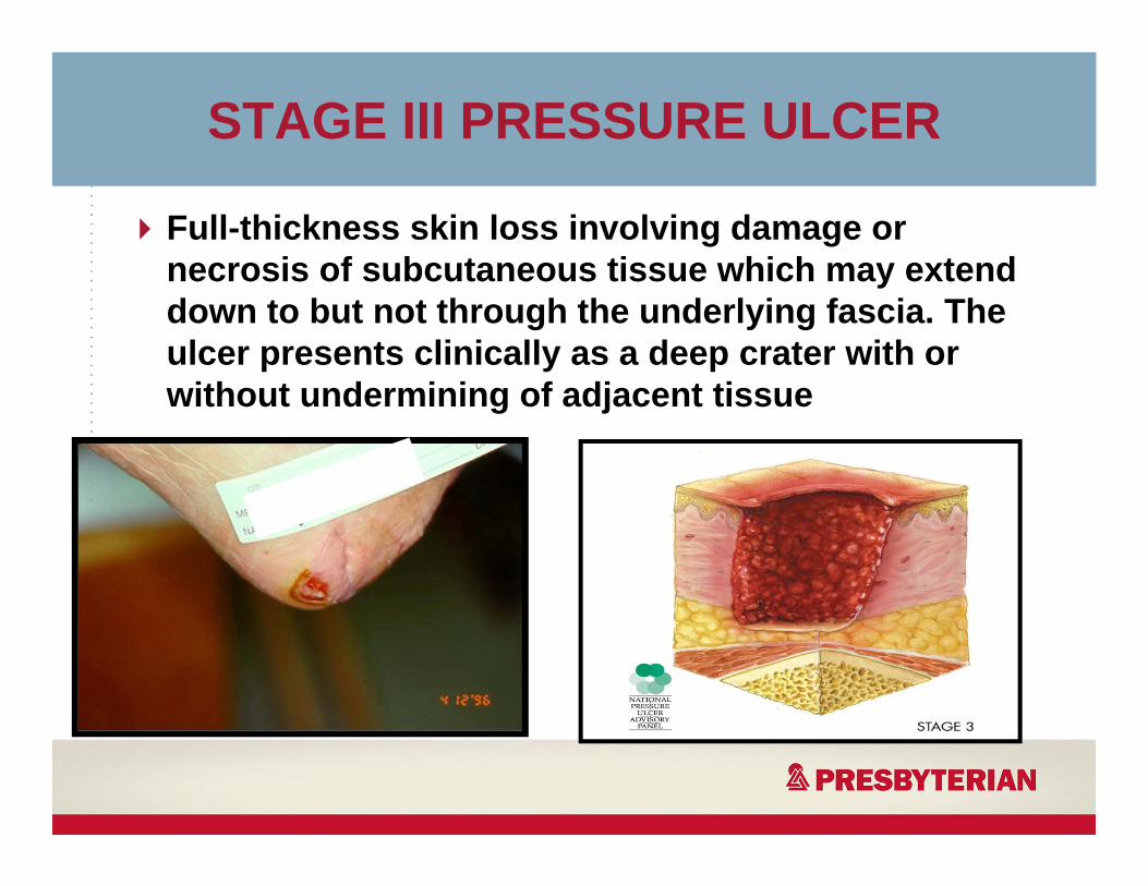
Full-thickness skin loss involving damage or necrosis of subcutaneous tissue which may extend down to but not through the underlying fascia. The ulcer presents clinically as a deep crater with or without undermining of adjacent tissue
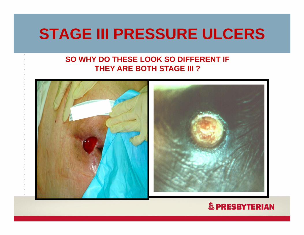
STAGE III PRESSURE ULCERSSO WHY DO THESE LOOK SO DIFFERENT IF
THEY ARE BOTH STAGE III ?
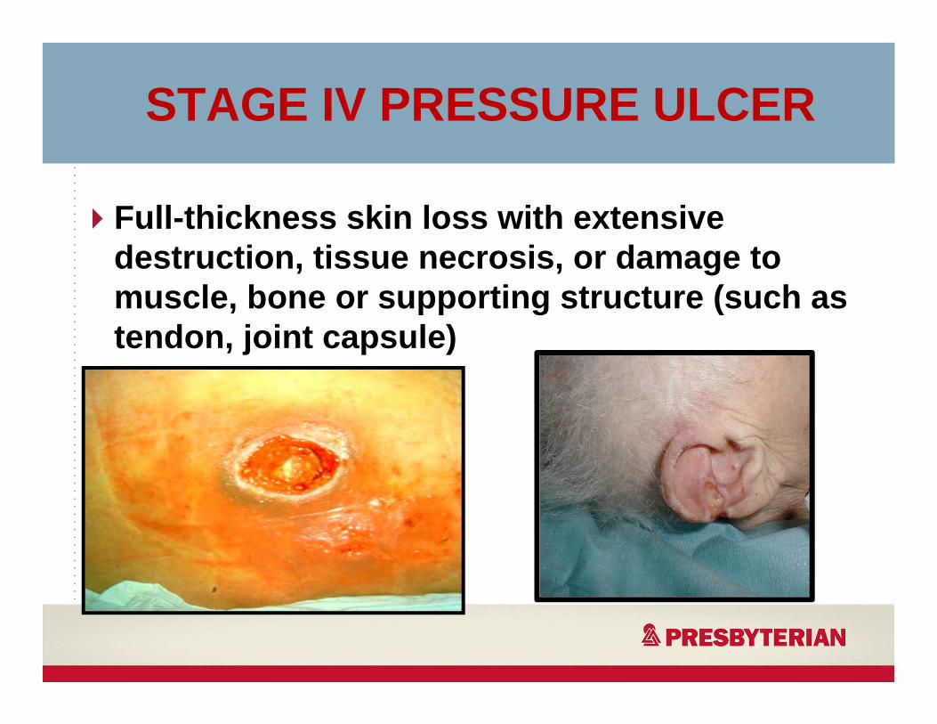
STAGE IV PRESSURE ULCER
Full-thickness skin loss with extensive destruction, tissue necrosis, or damage to muscle, bone or supporting structure (such as tendon, joint capsule)
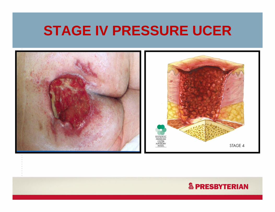
STAGE IV PRESSURE UCER
SUSPECTED DEEP TISSUE INJURY
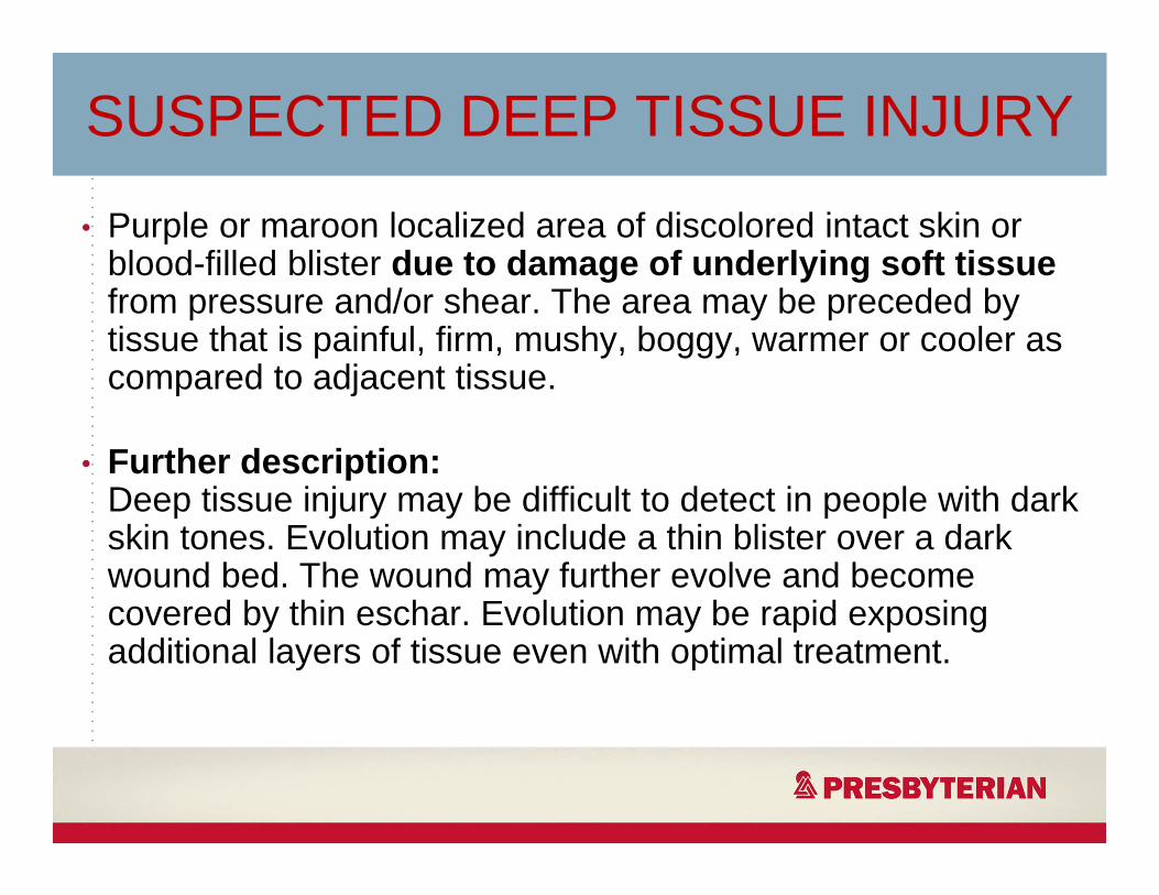
• Purple or maroon localized area of discolored intact skin or blood-filled blister due to damage of underlying soft tissuefrom pressure and/or shear. The area may be preceded by tissue that is painful, firm, mushy, boggy, warmer or cooler as compared to adjacent tissue.
• Further description:Deep tissue injury may be difficult to detect in people with dark skin tones. Evolution may include a thin blister over a dark wound bed. The wound may further evolve and become covered by thin eschar. Evolution may be rapid exposing additional layers of tissue even with optimal treatment.
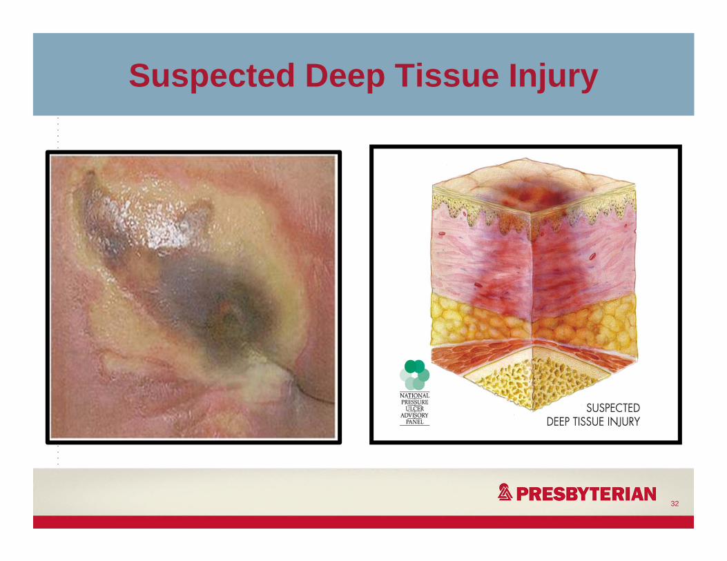
Suspected Deep Tissue Injury
32
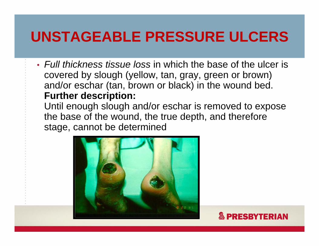
UNSTAGEABLE PRESSURE ULCERS
• Full thickness tissue loss in which the base of the ulcer is covered by slough (yellow, tan, gray, green or brown) and/or eschar (tan, brown or black) in the wound bed.Further description:Until enough slough and/or eschar is removed to expose the base of the wound, the true depth, and therefore stage, cannot be determined
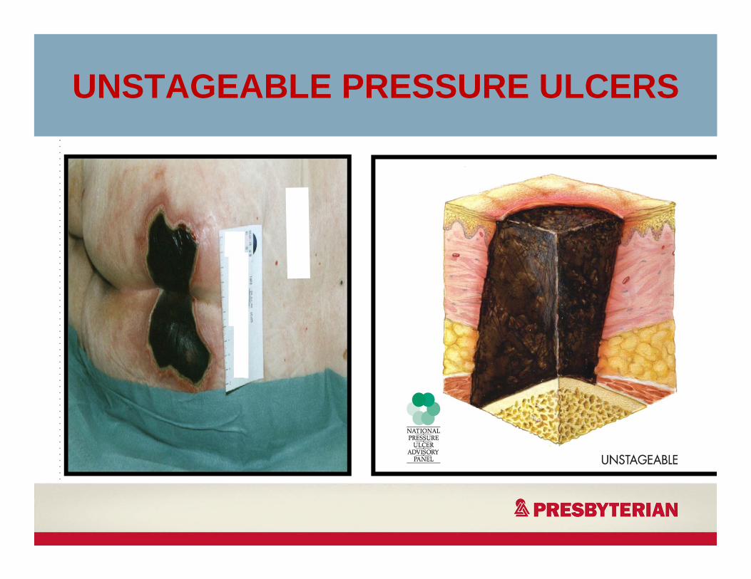
UNSTAGEABLE PRESSURE ULCERS
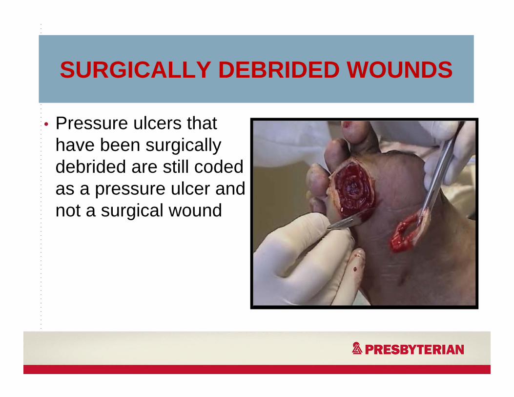
SURGICALLY DEBRIDED WOUNDS
• Pressure ulcers that have been surgically debrided are still coded as a pressure ulcer and not a surgical wound
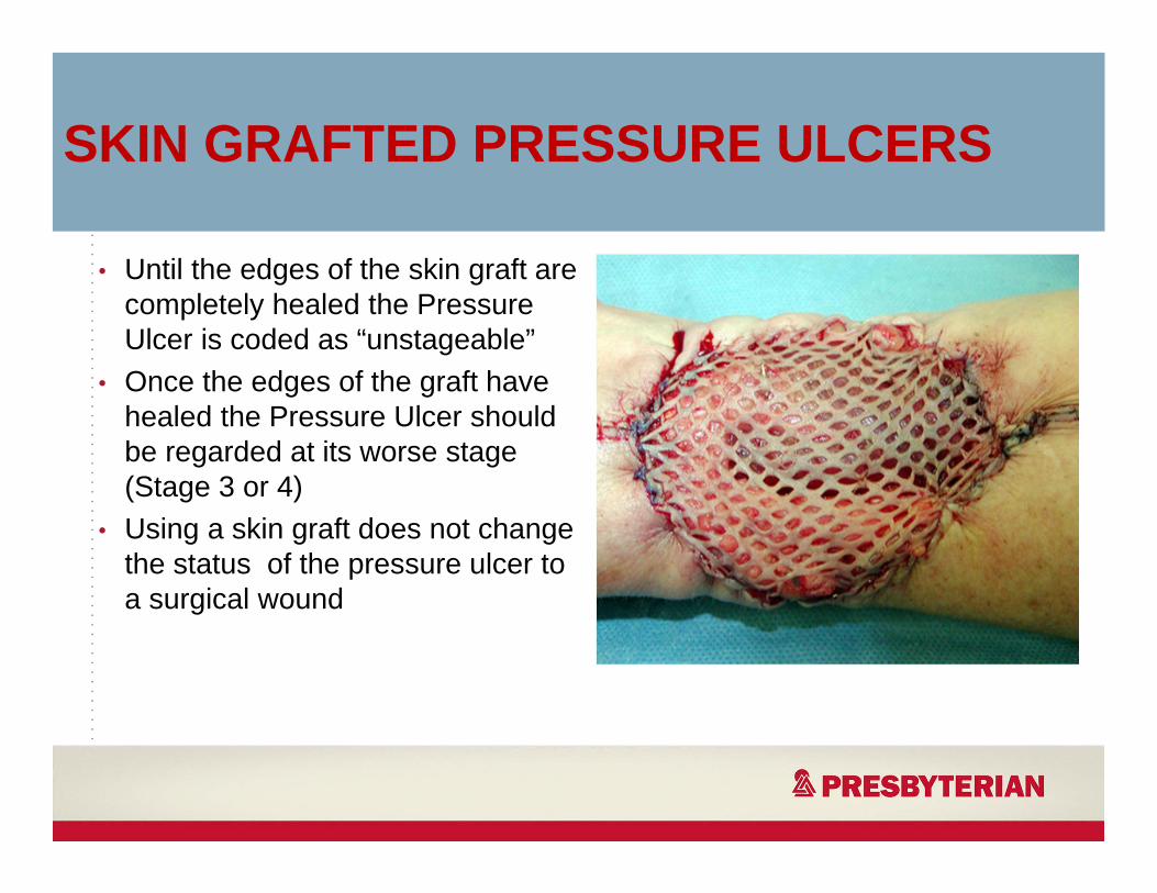
SKIN GRAFTED PRESSURE ULCERS
• Until the edges of the skin graft are completely healed the Pressure Ulcer is coded as “unstageable”
• Once the edges of the graft have healed the Pressure Ulcer should be regarded at its worse stage (Stage 3 or 4)
• Using a skin graft does not change the status of the pressure ulcer to a surgical wound
SURGICAL CLOSURE WITH FLAP
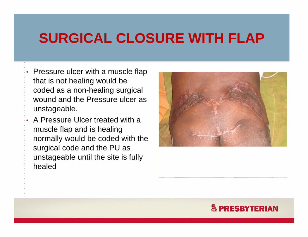
• Pressure ulcer with a muscle flap that is not healing would be coded as a non-healing surgical wound and the Pressure ulcer as unstageable.
• A Pressure Ulcer treated with a muscle flap and is healing normally would be coded with the surgical code and the PU as unstageable until the site is fully healed
00000000000000000000000000000000000000000000000000000000000000000000000000000000000000000000000000
BURN INJURIES
RULE OF NINES
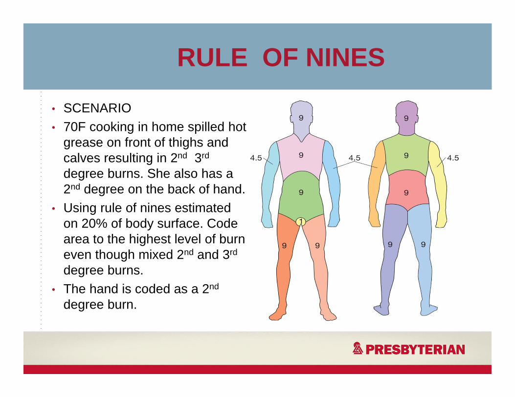
• SCENARIO• 70F cooking in home spilled hot
grease on front of thighs and calves resulting in 2nd 3rd
degree burns. She also has a 2nd degree on the back of hand.
• Using rule of nines estimated on 20% of body surface. Code area to the highest level of burn even though mixed 2nd and 3rd
degree burns.• The hand is coded as a 2nd
degree burn.
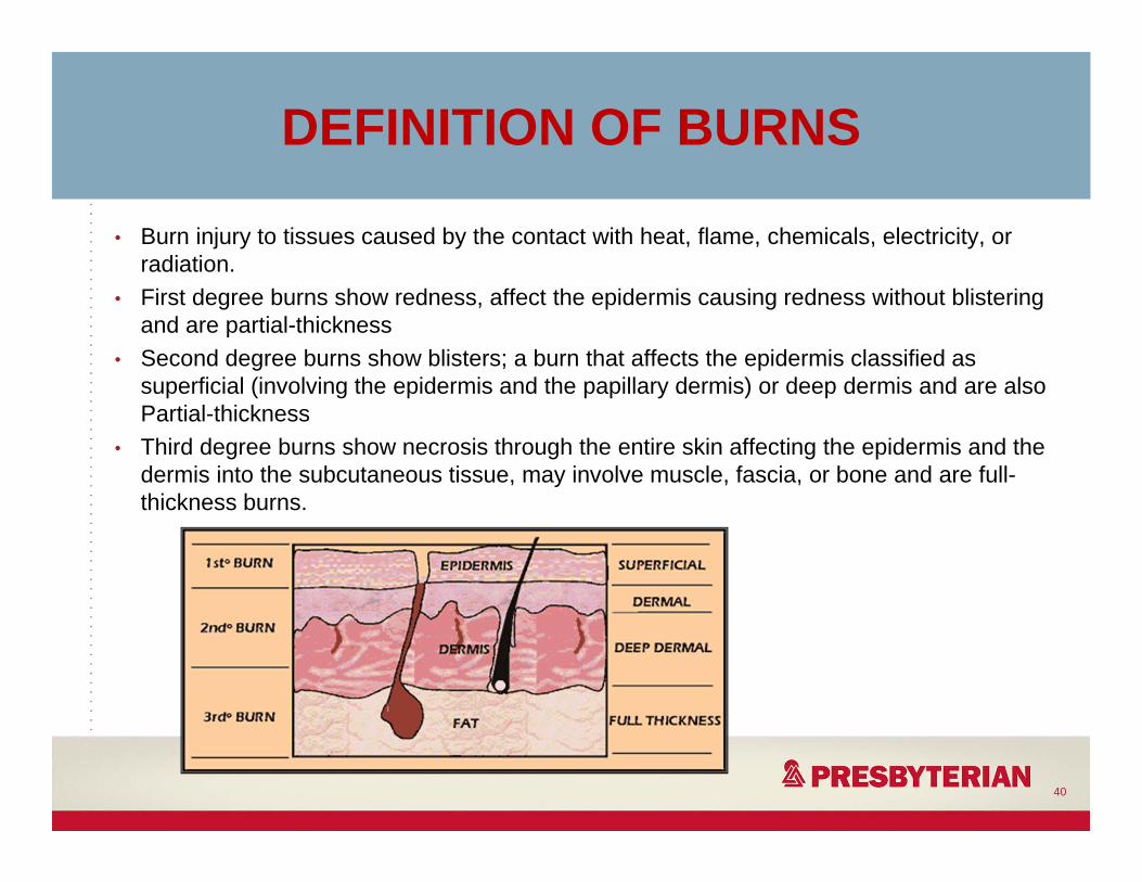
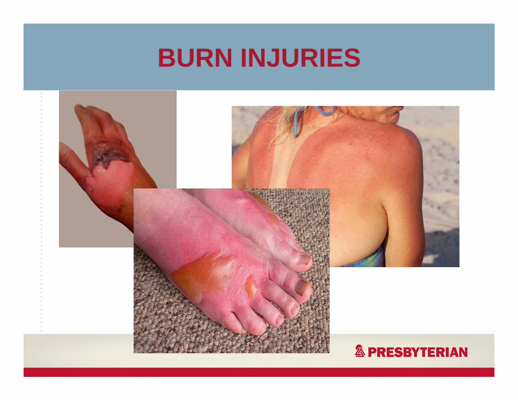
DEFINITION OF BURNS
• Burn injury to tissues caused by the contact with heat, flame, chemicals, electricity, or radiation.
• First degree burns show redness, affect the epidermis causing redness without blistering and are partial-thickness
• Second degree burns show blisters; a burn that affects the epidermis classified as superficial (involving the epidermis and the papillary dermis) or deep dermis and are also Partial-thickness
• Third degree burns show necrosis through the entire skin affecting the epidermis and the dermis into the subcutaneous tissue, may involve muscle, fascia, or bone and are full-thickness burns.
40
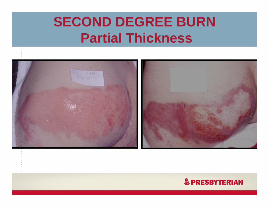
SECOND DEGREE BURN Partial Thickness
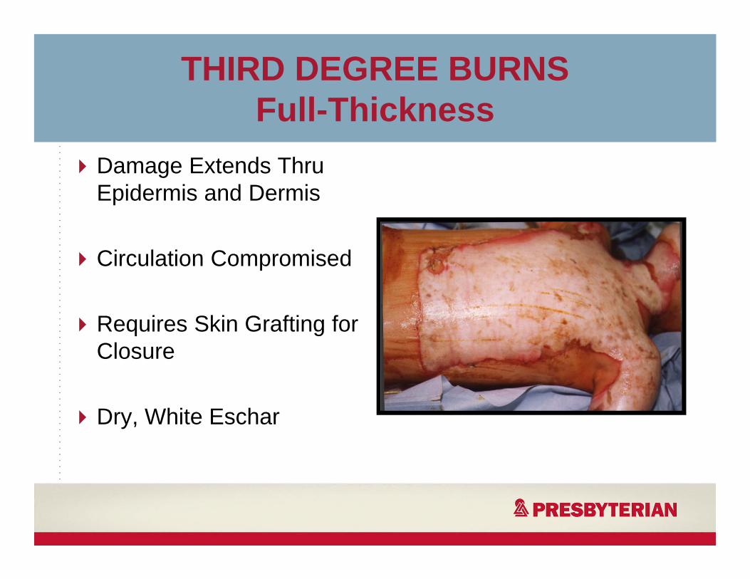
Damage Extends Thru Epidermis and Dermis
Circulation Compromised
Requires Skin Grafting for Closure
Dry, White Eschar
THIRD DEGREE BURNSFull-Thickness
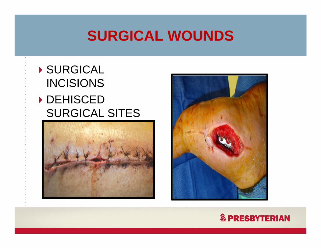
SURGICAL WOUNDS
SURGICAL INCISIONSDEHISCED SURGICAL SITES
SKIN TEARS
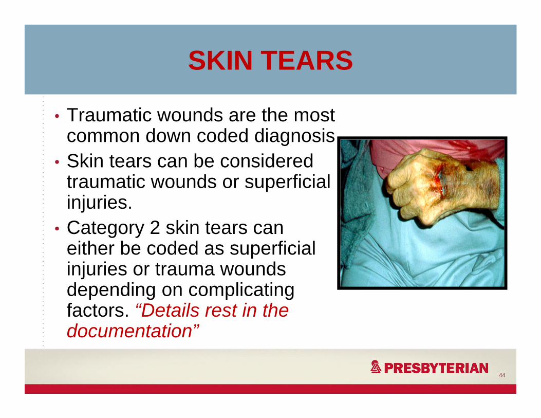
• Traumatic wounds are the most common down coded diagnosis
• Skin tears can be considered traumatic wounds or superficial injuries.
• Category 2 skin tears can either be coded as superficial injuries or trauma wounds depending on complicating factors. “Details rest in the documentation”
44
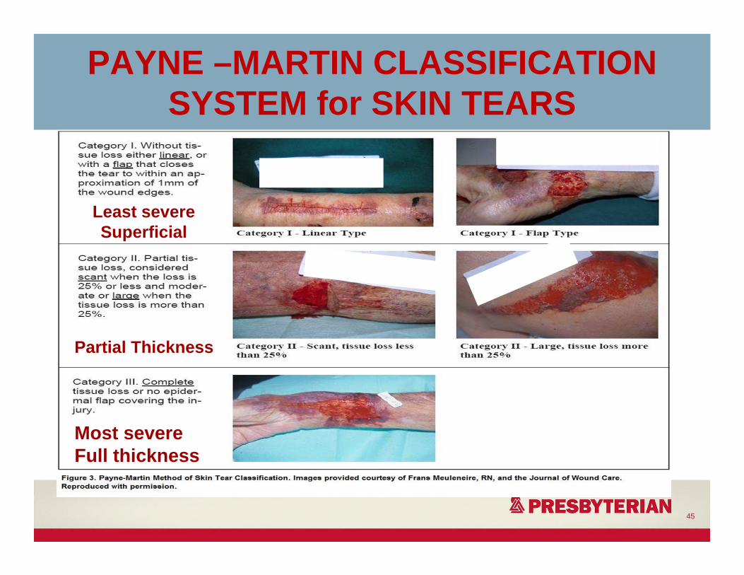
PAYNE –MARTIN CLASSIFICATION SYSTEM for SKIN TEARS
45
Least severeSuperficial
Most severeFull thickness
Partial Thickness
SKIN TEAR: MY DOCUMENTATION
• Location- Rt lower arm• Measurements: 5.0cm x 5.0cm x 0.3cm• Wound bed- 50% granulation tissue, 50% slough • Drainage: Large, serous sanguineous• Peri-wound: Red, warm to touch, painful• Wound edges: Jagged, rolled
46


Questions?
48