Embed Size (px)
DESCRIPTION
Major Electrolytes. Iron. Serum Iron Total Iron Binding Capacity (TIBC) Unsaturated Iron Binding Capacity (UIBC) Serum ferritin transferrin. Laboratory tests for investigation of iron disorder. Three stages of Iron deficiency anemia. - PowerPoint PPT Presentation
Citation preview
Major Electrolytes

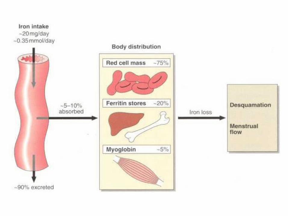
Iron
Laboratory tests for investigation of iron disorder
Serum IronTotal Iron Binding Capacity (TIBC)Unsaturated Iron Binding Capacity (UIBC)Serum ferritintransferrin
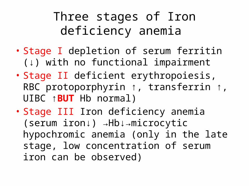
Three stages of Iron deficiency anemia
• Stage I depletion of serum ferritin (↓) with no functional impairment
• Stage II deficient erythropoiesis, RBC protoporphyrin ↑, transferrin ↑, UIBC ↑BUT Hb normal)
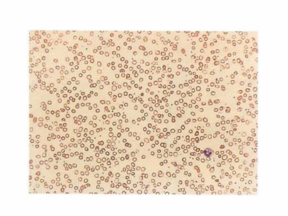
• Stage III Iron deficiency anemia (serum iron↓) →Hb↓→microcytic hypochromic anemia (only in the late stage, low concentration of serum iron can be observed)
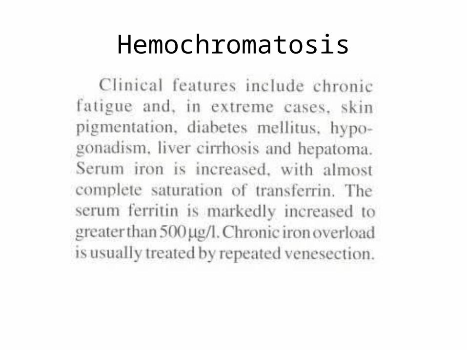
Hemochromatosis
Calcium
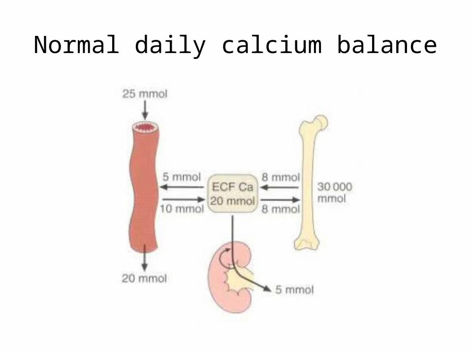
Normal daily calcium balance
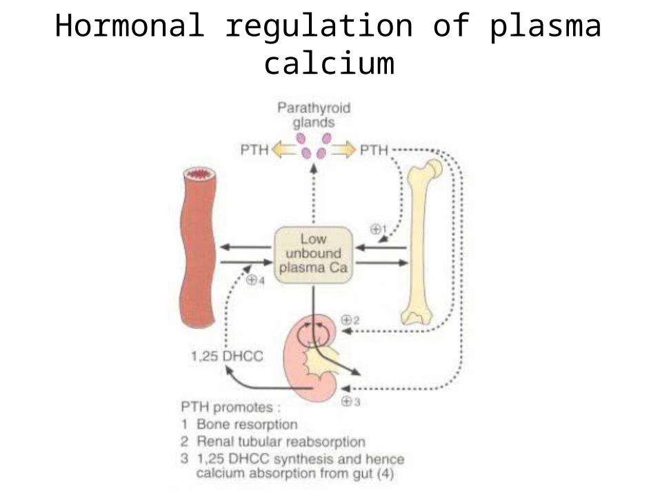
Hormonal regulation of plasma calcium
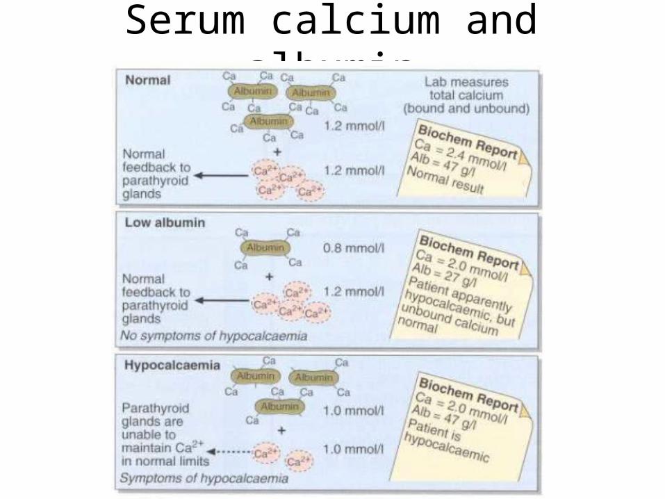
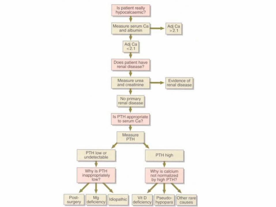
Serum calcium and albumin
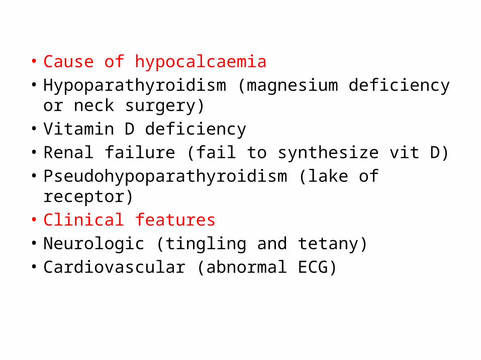
• Cause of hypocalcaemia• Hypoparathyroidism (magnesium deficiency or
neck surgery)• Vitamin D deficiency • Renal failure (fail to synthesize vit D)• Pseudohypoparathyroidism (lake of receptor)• Clinical features• Neurologic (tingling and tetany)• Cardiovascular (abnormal ECG)
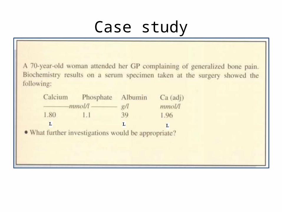
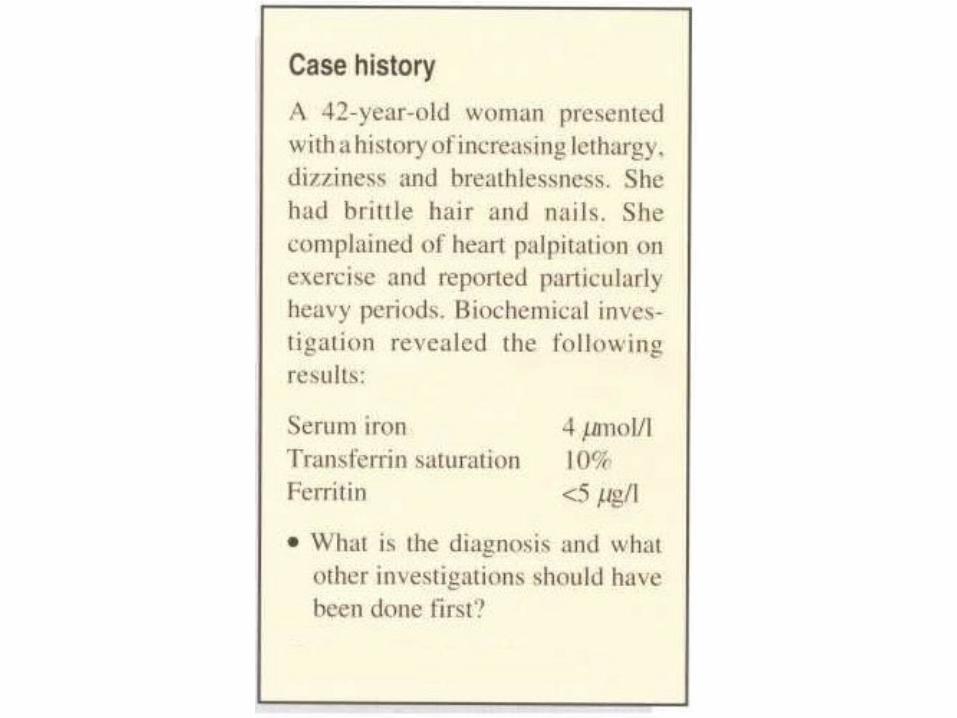
Case study
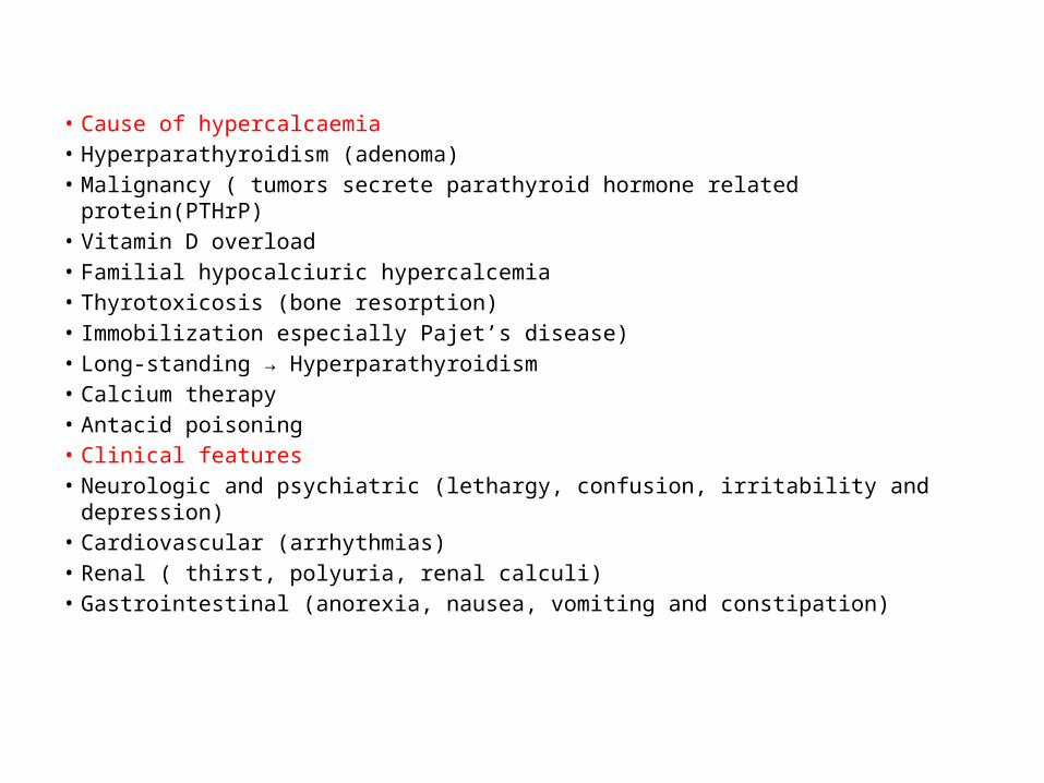
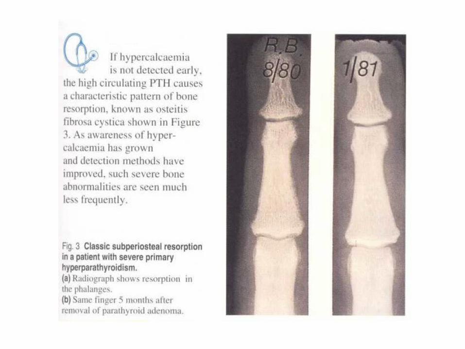
• Cause of hypercalcaemia• Hyperparathyroidism (adenoma)• Malignancy ( tumors secrete parathyroid hormone related protein(PTHrP)• Vitamin D overload• Familial hypocalciuric hypercalcemia• Thyrotoxicosis (bone resorption)• Immobilization especially Pajet’s disease)• Long-standing → Hyperparathyroidism • Calcium therapy• Antacid poisoning• Clinical features• Neurologic and psychiatric (lethargy, confusion, irritability and depression)• Cardiovascular (arrhythmias)• Renal ( thirst, polyuria, renal calculi)• Gastrointestinal (anorexia, nausea, vomiting and constipation)
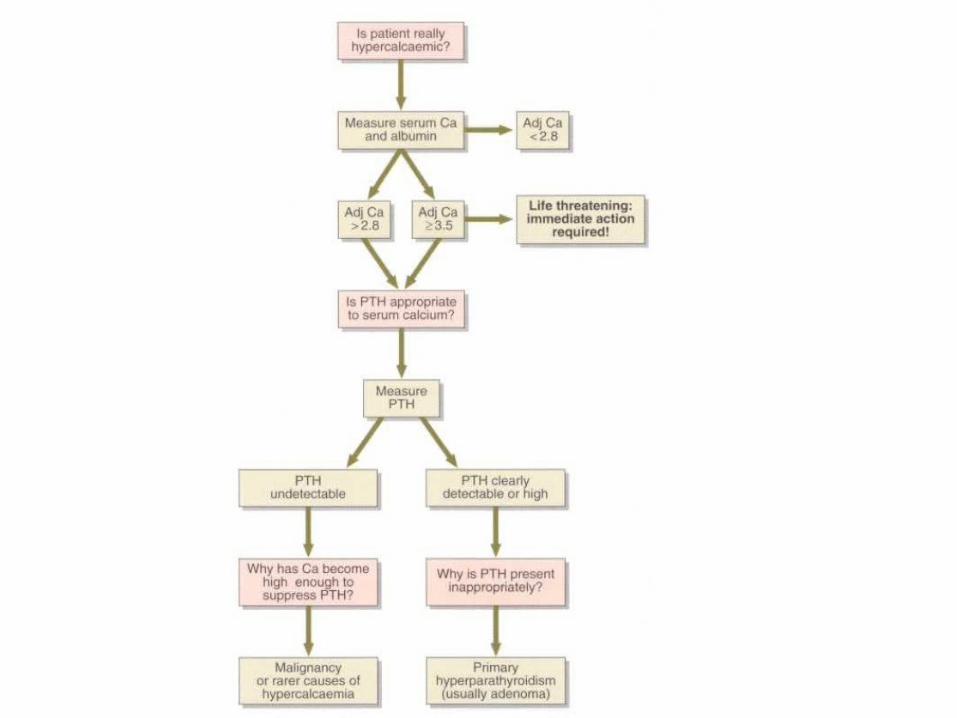
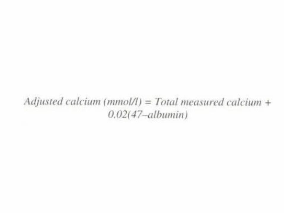
Calcium measurement
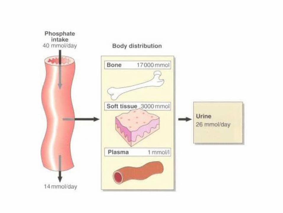
Phosphate
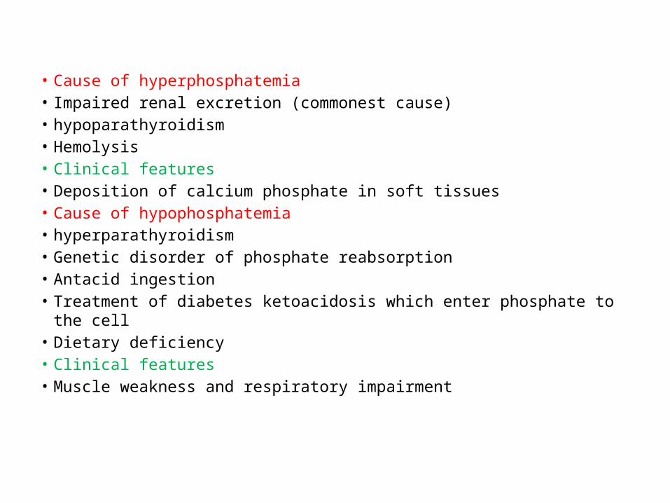
• Cause of hyperphosphatemia• Impaired renal excretion (commonest cause)• hypoparathyroidism • Hemolysis• Clinical features• Deposition of calcium phosphate in soft tissues• Cause of hypophosphatemia• hyperparathyroidism • Genetic disorder of phosphate reabsorption• Antacid ingestion• Treatment of diabetes ketoacidosis which enter phosphate to the cell• Dietary deficiency• Clinical features• Muscle weakness and respiratory impairment
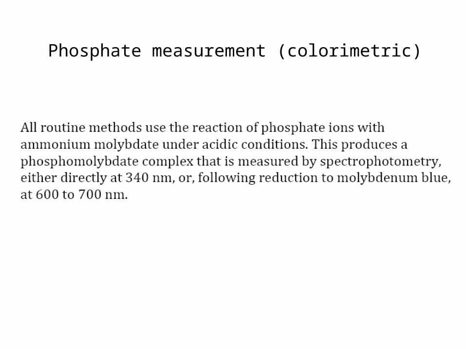
Phosphate measurement (colorimetric)
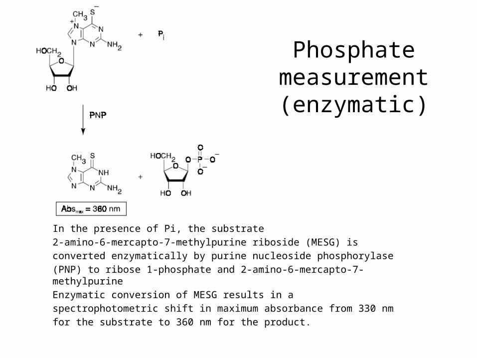
In the presence of Pi, the substrate2-amino-6-mercapto-7-methylpurine riboside (MESG) isconverted enzymatically by purine nucleoside phosphorylase(PNP) to ribose 1-phosphate and 2-amino-6-mercapto-7-methylpurineEnzymatic conversion of MESG results in aspectrophotometric shift in maximum absorbance from 330 nmfor the substrate to 360 nm for the product.
Phosphate measurement
(enzymatic)

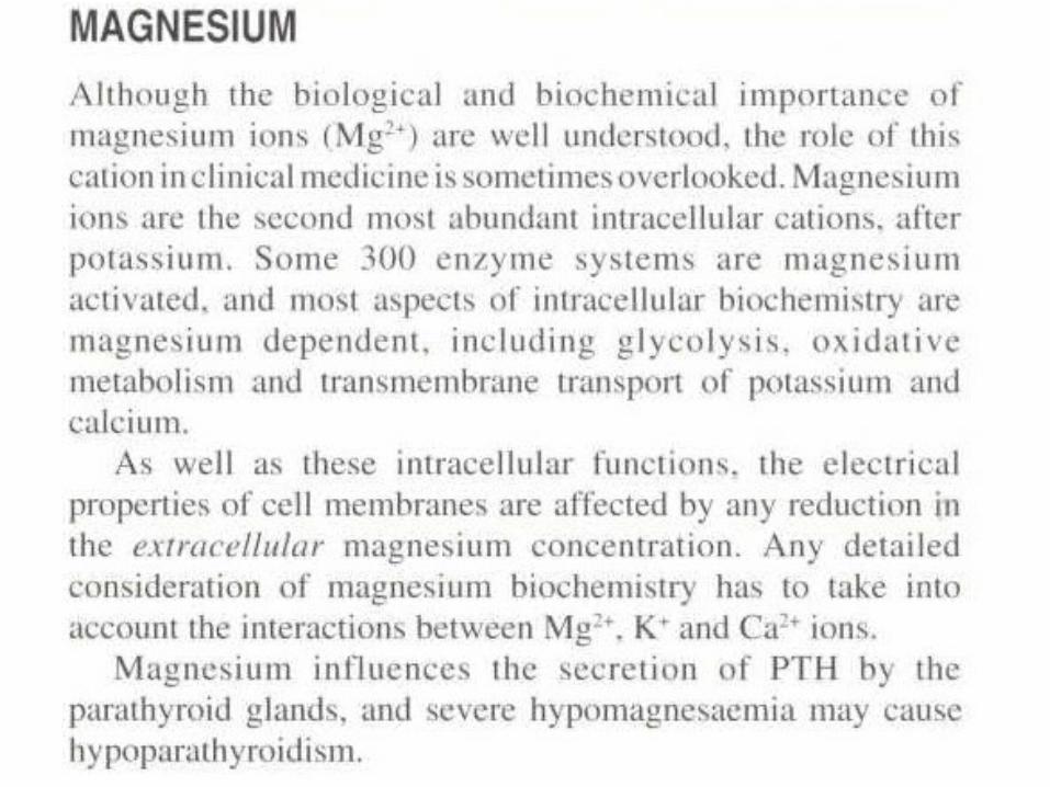
Magnesium
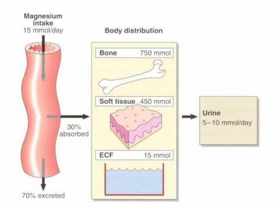
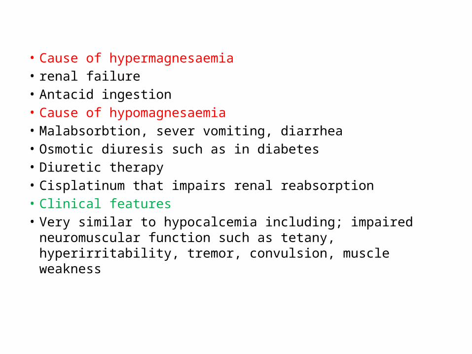
• Cause of hypermagnesaemia• renal failure• Antacid ingestion• Cause of hypomagnesaemia• Malabsorbtion, sever vomiting, diarrhea• Osmotic diuresis such as in diabetes• Diuretic therapy• Cisplatinum that impairs renal reabsorption• Clinical features• Very similar to hypocalcemia including; impaired neuromuscular
function such as tetany, hyperirritability, tremor, convulsion, muscle weakness
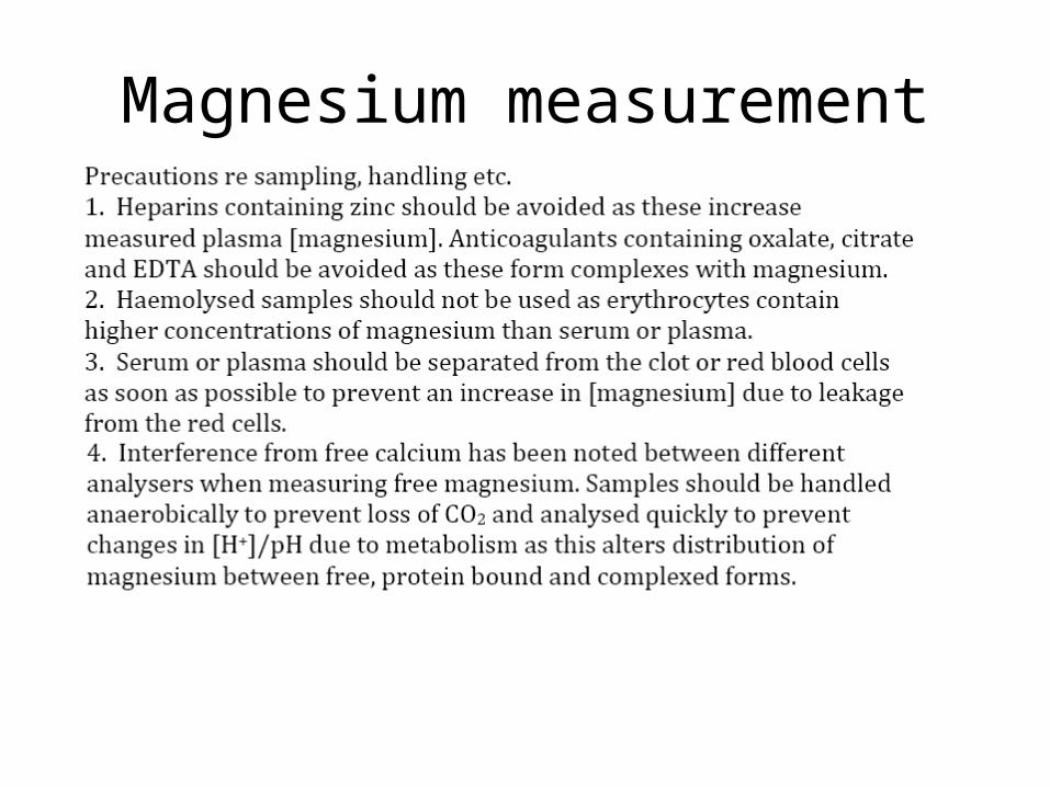
Magnesium measurement
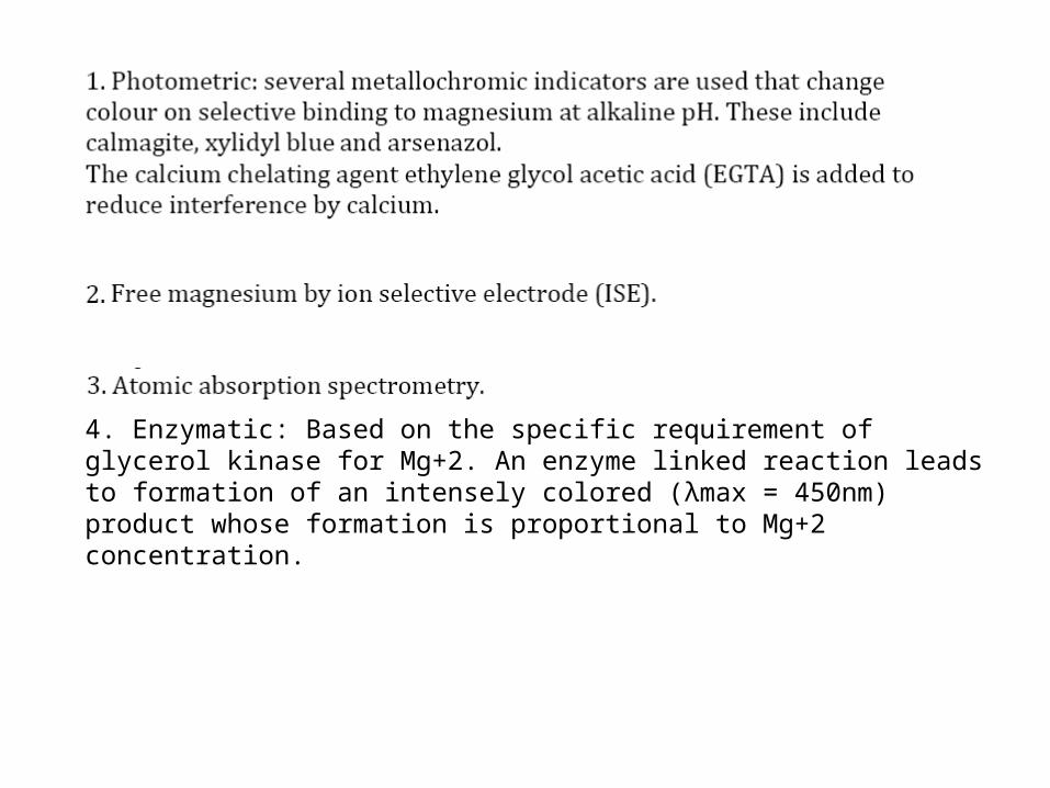
4. Enzymatic: Based on the specific requirement of glycerol kinase for Mg+2. An enzyme linked reaction leads to formation of an intensely colored (λmax = 450nm) product whose formation is proportional to Mg+2 concentration.

Copper
Copper
• Biochemical role (as a cofactor for metaloenzymes, ceruloplasmin, cytochrome c oxidase, dopamine β-hydroxylase, superoxide dismutase and tyrosinase
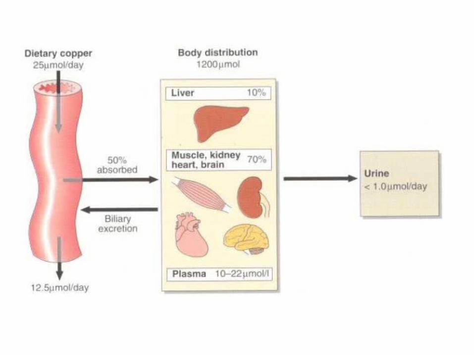
• Absorption: stomach and intestine• Transport: to the liver by Alb and then
ceruloplasmin • Distribution: liver, kidney, muscle and bone• Excretion: bile and urine
Laboratory tests
• Serum copper (Under acid environment, copper present in the sample reacts, with the chromogen Di-Br-PAESA forming a colored blue complex. The intensity of the colored complex is
proportional to the copper concentration in the sample.)• Ceruloplasmin (ELISA and emzymatic based on
oxidase activity of ceruloplasmin on synthetic substrate .
• Urinary copper
• Copper deficiency (in infants and intestinal bypass surgery or parenteral nutrition) signs:
• mental retardation, depigmentation, anemia, hypotonia and scorbutic changes in bone, Iron-resistant microcytic hypochromic anemia.
• Copper toxicity• By administration of copper sulfate solutions, • Renal tubular damage• Damage to tissues• Treatment• Penicillamine
Wilson’s disease
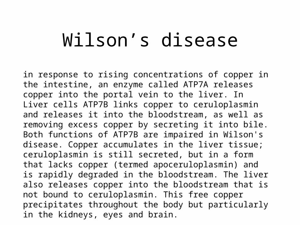
in response to rising concentrations of copper in the intestine, an enzyme called ATP7A releases copper into the portal vein to the liver. In Liver cells ATP7B links copper to ceruloplasmin and releases it into the bloodstream, as well as removing excess copper by secreting it into bile. Both functions of ATP7B are impaired in Wilson's disease. Copper accumulates in the liver tissue; ceruloplasmin is still secreted, but in a form that lacks copper (termed apoceruloplasmin) and is rapidly degraded in the bloodstream. The liver also releases copper into the bloodstream that is not bound to ceruloplasmin. This free copper precipitates throughout the body but particularly in the kidneys, eyes and brain.
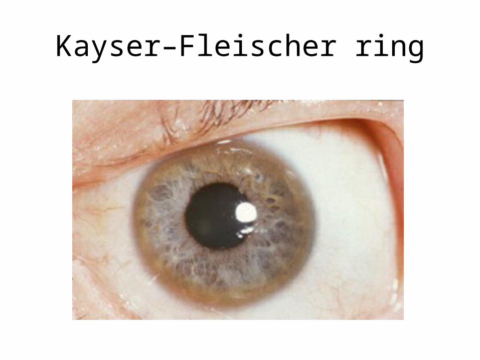
Kayser–Fleischer ring
Menkes syndrome
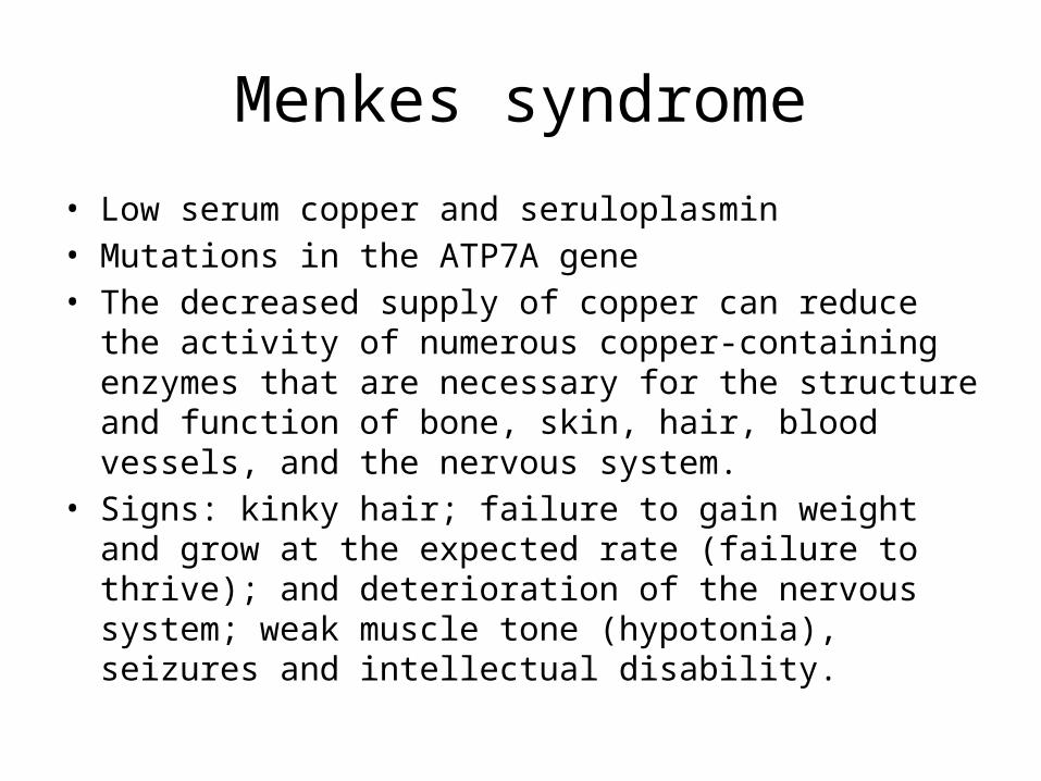
• Low serum copper and seruloplasmin• Mutations in the ATP7A gene• The decreased supply of copper can reduce the activity of
numerous copper-containing enzymes that are necessary for the structure and function of bone, skin, hair, blood vessels, and the nervous system.
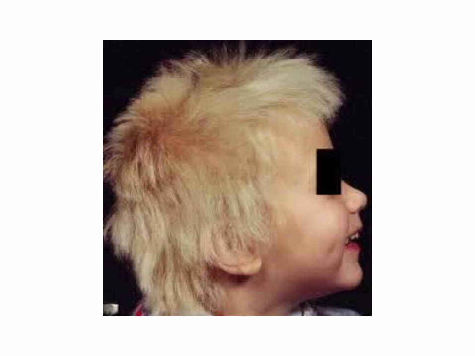
• Signs: kinky hair; failure to gain weight and grow at the expected rate (failure to thrive); and deterioration of the nervous system; weak muscle tone (hypotonia), seizures and intellectual disability.

Zinc

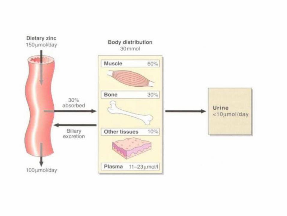
• Zinc is a co-factor in DNA and protein synthesis and cell division
• In plasma it is bound to albumin (90%) and α2-macroglobulin
• Located mainly in muscle and bone• Excretion: urine, bile, pancreatic fluid and milk in
lactating mothers• Deficiency a result of low intake or cadmium
poisoning: hair loss, skin rash, wound breakdown and delayed healing; Anorexia
Sodium
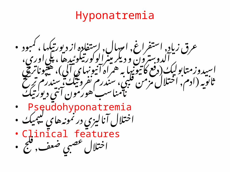
Hyponatremia
• عرق زياد, استفراغ, اسهال, استفاده از ديورتيكها ، كمبود آلدوسترون و ديگر مينرالوكورتيكوئيدها ، پلي اوري، اسيدوزمتابوليك(دفع كاتيونها به همراه آنيونهاي آلي)، هيپوناترمي ثانويه (ادم, اختالل مزمن قلبي، سندرم نفروتيك، سندرم ترشح نامناسب هورمون آنتي ديورتيك
• Pseudohyponatremia• اختالل آناليزي در نمونه هاي ليپميك• Clinical features• اختالل عصبي ضعف, فلج
Hypernatremia
• تزریق محلول های سالین• ،انسولین درمانی• هیپرآلدوسترونیسم• Hypernatriuria• هيپرناتری اورياي فيزيولوژيك در مواقع افزايش جذب و• .پس از دیورز قاعدگي ديده مي شود• Hyponatriuria• در نتيجه كاهش دريافت سدیم و يا احتباس آب و سدیم
قاعدگي ايجاد مي گردد, هيپرآلدوسترونيسم GFR قبل ازوكاهش
Potassium

• سلولي داخل عمده كاتيون• و گردد می پروکسیمال توبولهای در بازجذب
دیستال در ترشح
Hypokalemia
• ، ورود پتاسيم خارج سلولي به كاهش دريافت داخل سلولي
• افزايش دفع مايعات، انسولين درماني• آلكالوز (توقف آنتی پورت پتاسیم- پروتون)
• استفراغ, اسهال• اسيدوز توبولهاي كليه• آلدوسترونيسم
• دفع پتاسیم از کلیه←خروج پتاسیم از سلول ← ورود پروتون به سلول ← آلکالوز
Hyperkalemia
• افزايش خروج از سلول در حالتهاي دهيدراتاسيون, هيپوكسي بافتي, هموليز,
� سوختگي هاي شديد, فعاليت شديد عضالني(كالصدمات بافتي), كتواسيدوز ديابتي
• اختالل در دفع كليوي• كمبود آلدوسترون
Sampling
• رقتي اثر شديد← هموليز
• K• Hemolysis• Serum > plasma
اندازه گيري سديم
• ISE• Flame spectrophotometry• Internal standard• Calibration with low and high concentration of analyte• Enzymatic• Using galactosidase and (ONPG) O-nitrophenyl-β-D-
galactopyranoside as enzyme and substrate• Chromogenic ionophore, chromolyte, cryptand
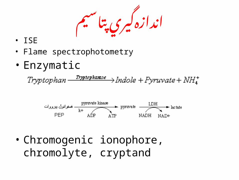
اندازه گيري پتاسيم• ISE• Flame spectrophotometry
• Enzymatic
• Chromogenic ionophore, chromolyte, cryptand
Chloride
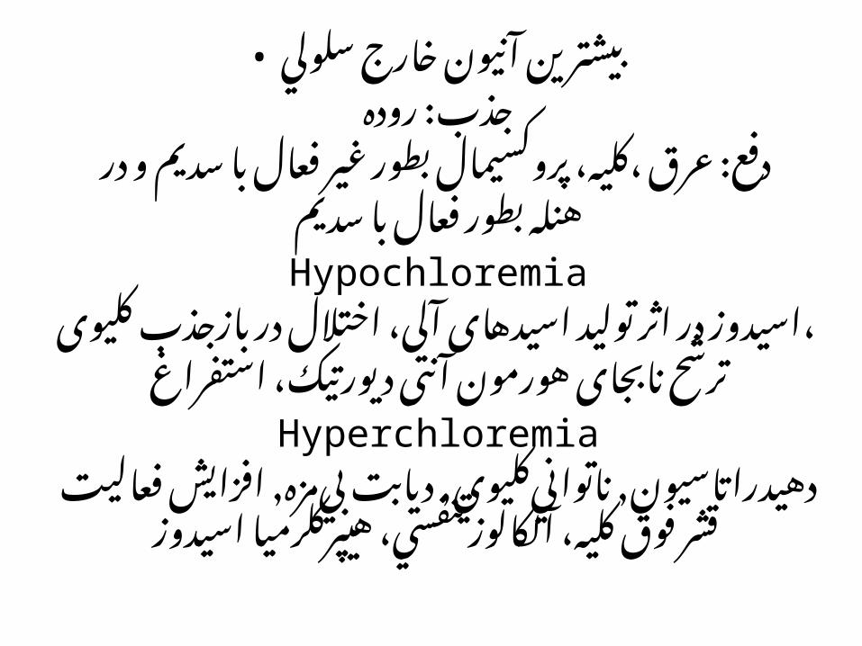
• بيشترين آنيون خارج سلوليجذب: روده
دفع: عرق ،کلیه، پروکسیمال بطور غیر فعال با سدیم و در
هنله بطور فعال با سدیمHypochloremia
اسیدوز در اثر تولید اسیدهای آلی، اختالل در بازجذب ،کلیوی
ترشح نابجای هورمون آنتی دیورتیک، استفراغHyperchloremia
دهيدراتاسيون, ناتواني كليوي, ديابت بي مزه, افزايش هيپركلرميا فعاليت قشر فوق كليه، آلكالوز تنفسي،
اسيدوز
اندازه گيري كلرايد
• در مدفوع: در بیماری آلكالوز هيپوكلريك hyperchlororrhea مادرزادي با
• پس از خون گيري در سرم, پالسما, ادرار و عرق . سلولها بايستي بسرعت از پالسما جدا شوند چرا كه وقتي خون در معرض هوا قرار گيرد بعلت از دست رفتن دی اکسید کربن توزيع کلر بين سلولهاي
.خوني و پالسما تغيير مي كند
متد های اندازه گيري كلرايد
• Coulometric – Amperometric titration• with silver ions. In the chloride titrator, a
constant• direct current is passed between a pair of silver
electrodes, causing release of silver ions• into the titration solution at a constant rate. The
silver ions react with chloride to• precipitate (Ag+ + Cl - ===> AgCl).• ISE