Embed Size (px)
Citation preview

Biochemical Characterization and Antibiotic Sensitivity Profile of Streptococcus suis Isolates from Swine Slaughtered in Selected Cavite Abattoirs Chester Joshua V. Saldaña, DVM and Marie Bernadette Bono, DVM
Physical Properties of the Snake Plant (Sanseviera hyacinthoides) Leaf-Derived Suture Chester Joshua V. Saldaña, DVM and Rona Karen C. Ferre, DVM
Managing Pawikan Conservation and Protection Project in Naic, Cavite Through Partnership: The CvSU Experience John Xavier B. Nepomuceno and Alexander F. Ferre, PhD
Response of Dragon Fruit (Hylocereus undatus Haw. Britton and Rose) Clonal Cuttings to Healing Duration and Fungicide Application Teddy F. Tepora, PhD and Regina M. Nosa
Maiden Issue | January - June 2014 ISSN: 2244-064X
1
9
17
27

CvSU Research Journal is the official peer-reviewed academic journal of the Cavite State University, Indang, Cavite, published biannually by the Office of the Vice President for Research, Extension, Continuing Education and Training Services (OVPRECETS). The publication is a compendium of interdisciplinary academic and research works of the different fields of specialization of the University’s colleges, units and campuses. Contents of this journal should not be reprinted, reproduced or transmitted without the written permission from the publisher.
EDITORIAL BOARD
Editors-in-Chief Adelfa M. Basaen, PhD Almira G. Magcawas
Associate Editors
Hosea DL. Matel, PhD Miriam D. Baltazar, PhD Rhodora S. Crizaldo, PhD Melbourne R. Talactac, DVM Evelyn O. Singson, PhD Rhodora V. Nuestro, PhD
Editorial Assistants Leyma L. Cero, PhD Gary A. Pareja Circulation Managers Teddy F. Tepora, PhD Lorna C. Matel Lay-out Artist Tirso P. Arreza Bernadette A. Sisante CONSULTANTS Divinia C. Chavez, PhD Alejandro DC. Mojica, Sr., PhD
ON THE COVER The Laya at Diwa is a brass artwork created by Jonnel P. Castillo and it is the University’s ideals of TRUTH, EXCELLENCE and SERVICE. It has five basic elements: a female figure holding a book, represents today’s modern woman - a mother, a nurturer, a mentor and a servant to her fellow citizens; the male figure with a pen and torch, represents the distinguished Filipino father; the child figure with a dove, represents the youth in general and recognized as the proverbial hope of the future that needs molding and nurturing; the dove being reached out by the child, symbolizes peace and freedom; and the central pillar, symbolizes growth and development which represents the humanity’s common ambition for an improved quality of life.
For correspondence, write to: The Editor-in-Chief CvSU Research Journal Cavite State University Indang, 4122 Cavite Philippines Tel. No. : (046) 443-9104 Email : [email protected] Website: cvsuresearchjournal.webs.com

Biochemical Characterization and Antibiotic Sensitivity Profile of Streptococcus suis Isolates from Swine Slaughtered in Selected Cavite Abattoirs Chester Joshua V. Saldaña, DVM and Marie Bernadette Bono, DVM
Physical Properties of the Snake Plant (Sanseviera hyacinthoides) Leaf-Derived Suture Chester Joshua V. Saldaña, DVM and Rona Karen C. Ferre, DVM
Managing Pawikan Conservation and Protection Project in Naic, Cavite Through Partnership: The CvSU Experience John Xavier B. Nepomuceno and Alexander F. Ferre, PhD
Response of Dragon Fruit (Hylocereus undatus Haw. Britton and Rose) Clonal Cuttings to Healing Duration and Fungicide Application Teddy F. Tepora, PhD and Regina M. Nosa
Maiden Issue | January - June 2014 ISSN: 2244-064X
1
9
17
27


FOREWORD
The Cavite State University (CvSU) is committed to generate new and useful innovations and technologies that would benefit its clientele. It is also steadfast to disseminate these outputs of researches to its target users. While doing these, CvSU encourages intellectual exchange as well as promotes and strengthens research culture in the University. This maiden issue of the CvSU Research Journal is a testament to the commitment of the University to contribute in addressing the needs of the industry and the community. This publication is a peer-reviewed academic journal and a compendium of interdisciplinary academic and research works of the different fields of specialization of the University‘s colleges, units and campuses. This issue is just the beginning of the University‘s journey to ensure that products of Research and Development would reach its intended beneficiaries. In the next issues, CvSU would continue to publish relevant and responsive researches. We hope that readers would find this publication useful and would inspire others to disseminate their research outputs.
DIVINIA C. CHAVEZ President


INTRODUCTION Streptococcus suis (S. suis) is a bacterium of increasing importance in the swine industry. S. suis is found in the upper respiratory tract, particularly in the tonsils and nasal cavities and in the genital and alimentary tracts of pigs with infections associated with Streptococcosis. Transmission of infection in pigs is mainly through nose-to-nose contact, fomites and flies or by aerosol over short distances (Gottschalk, 2004). There are currently 35 known capsular serotypes of S. suis and many of these serotypes cause outbreaks of disease in pigs, especially in recently weaned pigs (Higgins et al., 1995). Infection of many organs and rapid death (septicemia) and/or infection of the brain resulting to dizziness, paddling, convulsions and death (meningitis) are the most common forms (Sanford and Tilker, 1982). Outbreaks have been recognized worldwide in countries where pig production is well developed (Staats et al., 1997). Farmers and veterinarians classified the impact of a S. suis serotype 2 meningitis on the well-being of affected pigs as very severe in
comparison with swine influenza or enteric diseases (Bennett & Ijpelaar, 2003). The control of diseases caused by S. suis demands great efforts at swine farms. The costs of drugs, extra labor and high mortality rates have significant impact on their economics. Concomitant with the high use of antibiotics to control streptococcal disease at swine farms, the hazard of antibiotic resistance increases, which is a threat for animal as well as public health (Hendriksen et al., 2008). S. suis is also a zoonotic agent which affects predominantly people who handle pigs routinely like farmers and butchers (Arends and Zanen, 1988). The bacterium is thought to enter primarily through skin cuts and abrasions. Sometimes, the bacterium provokes larger outbreaks in man like in the Sichuan province in China (WHO, 2005; Yu et al., 2006; Yu et al., 2005). Experts in communicable diseases classify S. suis meningitis in man as an extremely violent disease (Bennett and Ijpe-laar, 2003).
Biochemical Characterization and Antibiotic Sensitivity Profile of Streptococcus suis Isolates from Swine Slaughtered
in Selected Cavite Abattoirs
Chester Joshua V. Saldaña, DVM1 and Marie Bernadette Bono, DVM
2
1 Assistant Professor, College of Veterinary Medicine and Biomedical Sciences, Cavite State University, Indang, Cavite
2 Former undergraduate student, Cavite State University, Indang, Cavite
ABSTRACT
The study was conducted to determine the biochemical characters and antibiotic sensitivity profile of Streptococcus suis (S. suis) from the palatine tonsil of 267 swine slaughtered in three abattoirs in Cavite. After cultural, morphological and biochemical characterization, S. suis was detected in 2.24% tonsils of the total number of swine, with the highest percentage (4.92%) from swine in Indang abattoir, followed by Tagaytay City (1.61%) and last in Alfonso, Cavite (1.22%). The susceptibility of S. suis isolates to eight antibiotics was studied by disk diffusion method using Mueller Hinton agar supplemented with defibrinated sheep blood. Results revealed that more of the isolates were found to be susceptible to norfloxacin (83.3%) than to gentamicin (66.7%) and fosfomycin (66.7%). On the other hand, isolates were resistant to tetracycline (83.3%), ampicillin (66.7%), clindamycin (66.7%), erythromycin (50.0%) and trimethoprim- sulfamethoxazole (50.0%).
Keywords: Streptococcosis, antibiotics
CvSU Research Journal 1
2 Maiden Issue (January - June 2014)
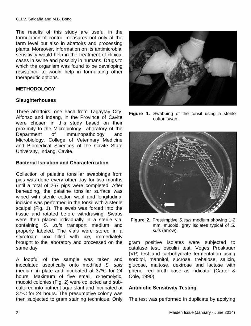
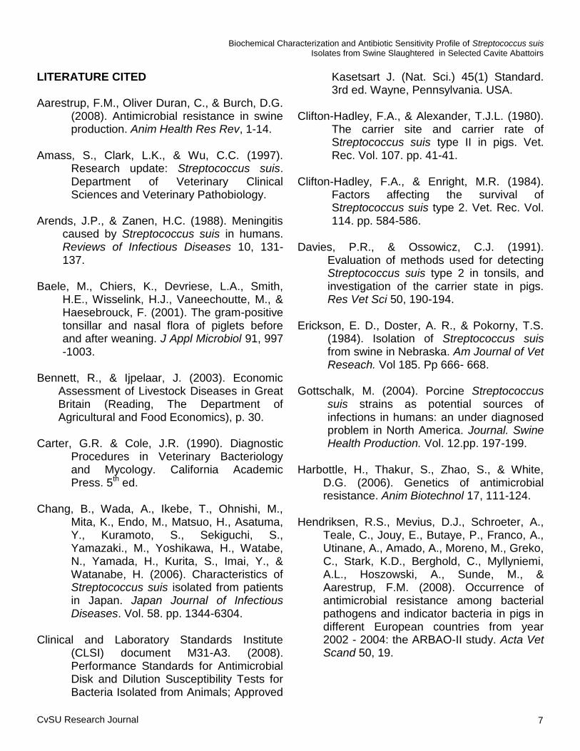
The results of this study are useful in the formulation of control measures not only at the farm level but also in abattoirs and processing plants. Moreover, information on its antimicrobial sensitivity would help in the treatment of clinical cases in swine and possibly in humans. Drugs to which the organism was found to be developing resistance to would help in formulating other therapeutic options. METHODOLOGY Slaughterhouses Three abattoirs, one each from Tagaytay City, Alfonso and Indang, in the Province of Cavite were chosen in this study based on their proximity to the Microbiology Laboratory of the Department of Immunopathology and Microbiology, College of Veterinary Medicine and Biomedical Sciences of the Cavite State University, Indang, Cavite. Bacterial Isolation and Characterization Collection of palatine tonsillar swabbings from pigs was done every other day for two months until a total of 267 pigs were completed. After beheading, the palatine tonsillar surface was wiped with sterile cotton wool and longitudinal incision was performed in the tonsil with a sterile scalpel (Fig. 1). The swab was forced into the tissue and rotated before withdrawing. Swabs were then placed individually in a sterile vial containing S. suis transport medium and properly labeled. The vials were stored in a styrofoam box filled with ice, immediately brought to the laboratory and processed on the same day. A loopful of the sample was taken and inoculated aseptically onto modified S. suis medium in plate and incubated at 37ºC for 24 hours. Maximum of five small, α-hemolytic, mucoid colonies (Fig. 2) were collected and sub-cultured into nutrient agar slant and incubated at 37ºC for 24 hours. The presumptive colony was then subjected to gram staining technique. Only
gram positive isolates were subjected to catalase test, esculin test, Voges Proskauer (VP) test and carbohydrate fermentation using sorbitol, mannitol, sucrose, trehalose, salicin, glucose, maltose, dextrose and lactose with phenol red broth base as indicator (Carter & Cole, 1990). Antibiotic Sensitivity Testing The test was performed in duplicate by applying
Figure 1. Swabbing of the tonsil using a sterile cotton swab.
Figure 2. Presumptive S.suis medium showing 1-2 mm, mucoid, gray isolates typical of S. suis (arrow).
C.J.V. Saldaña and M.B. Bono

CvSU Research Journal 3
a commercially available filter paper disc impregnated with antibiotics and diffused through the agar plate on which it is applied. The antibiotics used in the test were: ampicillin (10µg), clindamycin (2µg), erythromycin (15µg), fosfomycin (5µg), gentamicin (10µg), norfloxacin (10µg), tetracycline (30µg), and trimethorprim sulfamethoxazole (TMPS) (25µg). The method followed the recommended guidelines of the Clinical and Laboratory Standards Institute (CLSI, 2008). The organism tested was adjusted to a viable count of approximately 1.5 x 10
8 cells/ml. The
inoculum was spread over the entire surface of the Mueller Hinton agar supplemented with 5% defibrinated sheep blood and then impregnated discs were applied. After 24 hours of incubation at 37˚C, the diameter of zones of inhibition around the disc was measured using a vernier caliper and was compared to the interpretative table set by the manufacturer (Difco®) to
determine whether the organism tested is susceptible, intermediate or resistant (Table 1). Data Analysis Descriptive analysis using frequency counts, percentages and means were used in the interpretation and presentation of data.
RESULTS AND DISCUSSION Isolation and Biochemical Characterization of Streptococcus suis The distribution of slaughtered swine positive for S.suis by abattoir is shown in Table 2. The overall prevalence of S. suis in the palatine tonsils of 267 swine was 2.24% (6/267), with the highest occurrence in Indang 4.92% (3/61), followed by Tagaytay City 1.61% (2/124) and lastly, Alfonso 1.22% (1/82). The study documented the presence of Streptococcus suis among slaughtered pigs in Cavite. S. suis can be found in many organs of diseased animals, such as the nose, tonsils, lungs, and the intestinal and genital tracts (Baele et al., 2001). The presence of S. suis in organs of clinically healthy pigs is mostly transient. However, the tonsils can harbor S. suis colonies deep in the crypts for prolonged periods, without
any clinical sign of disease (Davies & Ossowicz, 1991). Clinically infected herds in the United States have yielded tonsillar carrier rates from 0 to 80% (Erickson et al., 1984). The carrier rate in young weaned pigs in intensive housing may be in excess of 50% when there is a high incidence of clinical cases, and could be as low as 3% when no clinical cases occur (Clifton-Hadley et al., 1980). This suggests that some herds are
Biochemical Characterization and Antibiotic Sensitivity Profile of Streptococcus suis Isolates from Swine Slaughtered in Selected Cavite Abattoirs
Table 1. Reference values for susceptibility and resistance based on diameter of zone of inhibition in response to different antibiotics (Difco®).
ANTIBIOTIC RESISTANT (mm) INTERMEDIATE (mm)
SUSCEPTIBLE (mm)
Ampicillin < 11 12-13 >14
Clindamycin < 14 15-16 >17
Erythromycin < 13 14-17 >18
Fosfomycin < 10 11-14 >15
Gentamicin < 12 - >13
Norfloxacin < 12 13-16 >17
Tetracycline < 14 15-18 >19
TMPS < 10 11-15 >16

4
infected and others are not, or the organism may persist at undetectable level probably deep in the tonsillar tissue where it is periodically multiplying. Despite the low occurrence rate of S. suis, this represents a potential source of infection for the slaughterhouse workers. S. suis is a public health hazard in workers in the swine industry. Humans can be infected with S. suis when they handle infected pig carcasses or meat, especially if the handlers have cuts and abrasions. There is a possible transmission via ingestion or through mucous membranes such as conjunctiva. Human infection can be severe, with meningitis, septicemia, endocarditis, and deafness as possible outcomes of infection (Yu et al., 2006).
Since severity of infection in human has been
established (WHO, 2005), preventive measures have to be taken. These measures are limited to hygienic procedures such as protecting cuts and abrasions and washing with soap and water after handling potential sources of infection. Exposed persons should therefore be informed of the hazard. The S. suis isolates identified showed short chains of gram positive cocci (Fig. 3). Biochemical reactions of S. suis isolates obtained were production of acetoin or Voges Proskauer negative (Fig. 4A), catalase negative (Fig. 4B) and esculin hydrolysis (Fig. 4C).
Table 2. Percentage distribution of slaughtered swine positive for S. suis by abattoir.
ABBATOIR NUMBER OF SWINE EXAMINED
PERCENT (%) SWINE POSITIVE
NUMBER OF PRESUMP-TIVE ISOLATES
Alfonso 82 1(1. 22 %) 87
Tagaytay 124 2 (1. 61 %) 214
Indang 61 3 (4. 92 %) 500
Total 267 6 (2.24 %) 214
Figure 3. A gram positive S. suis isolate from swine tonsil from Tagaytay City abattoir (1000x).
C
A
B
D
1 2 3 4 5 6 7
Figure 4. Biochemical reactions of S. suis isolate. A: Voges Proskauer negative, B. Catalase nagative, C. Esculin hydrolysis, D. Sugar Fermentation: 1. Sorbitol -, 2. Maltose -, 3. Lactose +, 4 . Glucose +, 5. Dextrose +, 6. Mannitol -, and 7. Trehalose +.
C.J.V. Saldaña and M.B. Bono
Maiden Issue (January - June 2014)

CvSU Research Journal 5
Further, acid reaction was shown from carbohydrate fermentation test on lactose (+), glucose (+), dextrose (+) and trehalose (+) while negative reaction was observed for sorbitol (-), maltose (-) and mannitol (-) (Fig. 4D). Antibiotic Sensitivity Profile of Streptococcus suis Isolates The confirmed S. suis isolates were subjected to the antibiotic sensitivity test and results of the reaction based on the diameter of zones of inhibition are presented in Table 3. The zones of inhibition obtained were compared to the reference values of measurement in Table 1. Majority of the samples were resistant to ampicillin, tetracycline and clindamycin but were susceptible to gentamicin, norfloxacin and fosfomycin (Fig. 5). Moreover, some isolates were resistant to erythromycin and trimethorprim sulfamethoxazole (TMPS) but some were also susceptible to the two drugs. A study by Vela et al. (2005) showed that more than 87% of the S. suis isolates were resistant to tetracyclines and clindamycin which are similar to the results of this study. Moreover, the study found out that the isolates were also resistant to sulfonamides and macrolides. In addition, Chang et al. (2006) showed that S. suis is resistant to erythromycin and clindamycin but susceptible to ampicillin, penicillin G, cefotaxime and
ciprofloxacin. In this study, five of the isolates were resistant to tetracycline and four isolates to clindamycin. Isolates from Tagaytay City were found to be susceptible to the said antibiotics. Similar result was obtained by Tian et al. (2004), wherein a high resistance to tetracycline was observed. However, two isolates from Indang were susceptible to ampicillin as compared to the study by Chang et al., wherein all S. suis isolates were susceptible to ampicillin. The degree of resistance of the isolates was
Table 3. Percentage of S. suis isolates that are resistant, intermediate and susceptible to the antibiotics tested.
ANTIBIOTIC RESISTANT INTERMEDIATE SUSCEPTIBLE
Ampicillin (10µg) 66.7% (4/6) - 33.3% (2/6)
Clindamycin (2µg) 66.7% (4/6) - 33.3% (2/6)
Erythromycin (15µg) 50.0% (3/6) - 50.0% (3/6)
Fosfomycin (5µg) 33.3% (2/6) - 66.7% (4/6)
Gentamicin (10µg) 33.3% (2/6) - 66.7% (4/6)
Norfloxacin (10µg) 16.7% (1/6) - 83.3% (5/6)
Tetracycline (30µg) 83.3% (5/6) - 16.7% (1/6)
TMPS (25µg) 50.0% (3/6) 16.7% (1/6) 33.3% (2/6)
Figure 5. S. suis isolate from Alfonso, Cavite showing resistance to erythromycin (E), clindamycin (DA), TMPS (SXT) and tetracycline (TE), and susceptible to gentamicin (CN), norfloxacin (NOR), fosfomycin (FOS).
Biochemical Characterization and Antibiotic Sensitivity Profile of Streptococcus suis Isolates from Swine Slaughtered in Selected Cavite Abattoirs

6
higher in tetracycline with 83.3% (5/6) as compared to that in ampicillin and clindamycin with both 66.7% (4/6). On the other hand, these isolates showed greater susceptibility to norfloxacin with 83.3% (5/6) as against to that in gentamicin and fosfomycin which were 66.7% (4/6). In the study conducted by Amass et al. (1997), 91% of the presumptive S. suis isolated were susceptible to ampicillin and less than 50% of the isolates were susceptible to tetracycline. In addition, Vela et al. (2005) showed that 87% of the isolated S. suis organisms were resistant to tetracycline and clindamycin. Whereas, in the study conducted by Tian et al. (2004) 41% of the isolated S. suis were resistant to erythromycin, 24% to tetracycline and most isolates were resistant to trimethophrim + sulphonamides. The results of antibiotic sensitivity test of S. suis revealed that the resistance and susceptibility between samples varied. Amass et al. (1997) stated that the use of antimicrobial susceptibility testing on herd isolates of S. suis is recommended because of the variability in sensitivity between isolates. Moreover, Amass et al. (1997) stated that early weaning management protocols should not be used to prevent S. suis from infecting pigs. Blanket medication of pigs with the goal of eliminating S. suis from the herd was not recommended because it was not effective and it may also lead to antibiotic resistance among streptococci. Moreover, the optimization of management and environment of pigs, coupled with strategic medication of ill animals, was highly recommended for the control and prevention of mortality caused by streptococcosis. The use of antimicrobial drugs has several benefits in the treatment of streptococcal related diseases. However, antimicrobial resistance is an increasing threat for animal health as well as public health (Harbottle et al., 2006; Hendriksen et al., 2008). In general, the large amounts of
drugs used especially in prevention of diseases facilitate the emergence and development of bacterial resistance (Aarestrup et al., 2008). As a consequence, it already has been shown that the use of antibiotics in livestock contributes to the increased prevalence of antibiotic-resistant bacteria that are of significance, not only in swine production but in public health as well (Mathew et al., 2007). CONCLUSION AND RECOMMENDATION Streptococcus suis is present in the palatine tonsils of slaughtered swine in Cavite with an overall prevalence of 2.24%. Although at a low occurrence rate, this represents a potential source of infection for the slaughterhouse workers. Thus, exposed persons should be informed of the hazard. Majority of the isolates was found to be susceptible to norfloxacin, gentamicin, and fosfomycin, and were resistant to tetracycline, ampicillin, clindamycin, erythromycin and trimethoprim-sulfamethoxazole. The results presented in this study should therefore not be blindly used in the choice of drug against S. suis; rather, it gives an indication of the present in vitro pattern of susceptibility to antibiotics of the bacteria. Regular follow-up studies could establish changing patterns in drug susceptibility and resistance. Additional biochemical tests such as inulin, hippurate and lancefield grouping are recommended to identify Streptococcus suis serotype. More sensitive means of isolation such as the use of immunomagnetic separation (IMS) techniques coupled with direct culture as well as immunohistochemistry procedures to demonstrate the organisms in tissues and Polymerase Chain Reaction (PCR) are likewise recommended. Moreover, epidemiological studies could help establish the presence of S. suis organisms at the farm level.
C.J.V. Saldaña and M.B. Bono
Maiden Issue (January - June 2014)
CvSU Research Journal 7
LITERATURE CITED Aarestrup, F.M., Oliver Duran, C., & Burch, D.G.
(2008). Antimicrobial resistance in swine production. Anim Health Res Rev, 1-14.
Amass, S., Clark, L.K., & Wu, C.C. (1997).
Research update: Streptococcus suis. Department of Veterinary Clinical Sciences and Veterinary Pathobiology.
Arends, J.P., & Zanen, H.C. (1988). Meningitis
caused by Streptococcus suis in humans. Reviews of Infectious Diseases 10, 131-137.
Baele, M., Chiers, K., Devriese, L.A., Smith,
H.E., Wisselink, H.J., Vaneechoutte, M., & Haesebrouck, F. (2001). The gram-positive tonsillar and nasal flora of piglets before and after weaning. J Appl Microbiol 91, 997-1003.
Bennett, R., & Ijpelaar, J. (2003). Economic
Assessment of Livestock Diseases in Great Britain (Reading, The Department of Agricultural and Food Economics), p. 30.
Carter, G.R. & Cole, J.R. (1990). Diagnostic
Procedures in Veterinary Bacteriology and Mycology. California Academic Press. 5
th ed.
Chang, B., Wada, A., Ikebe, T., Ohnishi, M.,
Mita, K., Endo, M., Matsuo, H., Asatuma, Y., Kuramoto, S., Sekiguchi, S., Yamazaki., M., Yoshikawa, H., Watabe, N., Yamada, H., Kurita, S., Imai, Y., & Watanabe, H. (2006). Characteristics of Streptococcus suis isolated from patients in Japan. Japan Journal of Infectious Diseases. Vol. 58. pp. 1344-6304.
Clinical and Laboratory Standards Institute
(CLSI) document M31-A3. (2008). Performance Standards for Antimicrobial Disk and Dilution Susceptibility Tests for Bacteria Isolated from Animals; Approved
Kasetsart J. (Nat. Sci.) 45(1) Standard. 3rd ed. Wayne, Pennsylvania. USA.
Clifton-Hadley, F.A., & Alexander, T.J.L. (1980).
The carrier site and carrier rate of Streptococcus suis type II in pigs. Vet. Rec. Vol. 107. pp. 41-41.
Clifton-Hadley, F.A., & Enright, M.R. (1984).
Factors affecting the survival of Streptococcus suis type 2. Vet. Rec. Vol. 114. pp. 584-586.
Davies, P.R., & Ossowicz, C.J. (1991).
Evaluation of methods used for detecting Streptococcus suis type 2 in tonsils, and investigation of the carrier state in pigs. Res Vet Sci 50, 190-194.
Erickson, E. D., Doster, A. R., & Pokorny, T.S.
(1984). Isolation of Streptococcus suis from swine in Nebraska. Am Journal of Vet Reseach. Vol 185. Pp 666- 668.
Gottschalk, M. (2004). Porcine Streptococcus
suis strains as potential sources of infections in humans: an under diagnosed problem in North America. Journal. Swine Health Production. Vol. 12.pp. 197-199.
Harbottle, H., Thakur, S., Zhao, S., & White,
D.G. (2006). Genetics of antimicrobial resistance. Anim Biotechnol 17, 111-124.
Hendriksen, R.S., Mevius, D.J., Schroeter, A.,
Teale, C., Jouy, E., Butaye, P., Franco, A., Utinane, A., Amado, A., Moreno, M., Greko, C., Stark, K.D., Berghold, C., Myllyniemi, A.L., Hoszowski, A., Sunde, M., & Aarestrup, F.M. (2008). Occurrence of antimicrobial resistance among bacterial pathogens and indicator bacteria in pigs in different European countries from year 2002 - 2004: the ARBAO-II study. Acta Vet Scand 50, 19.
Biochemical Characterization and Antibiotic Sensitivity Profile of Streptococcus suis Isolates from Swine Slaughtered in Selected Cavite Abattoirs

8
Higgins, R., Gottchalk, M., Boudreau, M., Lebrun, A., & Henrichsen, J. (1995). Description of six new Streptococcus suis capsular types. Journal Vet Diagnostic Invention. Vol. 7. pp 405- 406.
Mathew, A.G., Cissell, R., & Liamthong, S.
(2007). Antibiotic resistance in bacteria associated with food animals: a United States perspective of livestock production. Foodborne Pathog Dis 4, 115-133.
Staats, J.J., Feder, I., Okwumabua, O., &
Chengappa, M.M. (1997). Streptococcus suis: past and present. Veterinary-Research-Communications 21, 381-407.
Sanford, S.E., & Tilker, A.M.E. (1982).
Streptococcus suis type-II associated diseases in swine: Observations of a one-year study. Journal of American Veterinary Medical Association. Vol. 181. pp 673-676.
Tian, Y, Aarestrup, F.M., & Lu, C.P. (2004).
Characterization of Streptococcus suis serotype 7 isolates from pigs in Denmark. Veterinary Microbiology. Vol. 103. pp. 55-62.
Vela, A.I., Moreno, M.A., Cebolla, J.A., Gonzalez
S., Latre, M.V., Dominguez, L., & Fernandez-Garayzabal, J.F. (2005). Antimicrobial susceptibility of clinical strains of Streptococcus suis isolated from pigs in Spain. Veterinary Microbiology. Vol. 105. pp 143-147.
WHO. (2005). Outbreak associated with
Streptococcus suis in pigs in China: Update (World Health Organization; The Western Pacific Regional Office). Rec. Vol. 80. pp.269–76.
Yu, H., Jing, H., Chen, Z., Zheng, H., Zhu, X.,
Wang, H., Wang, S., Liu, L., Zu, R., Luo, L., Xiang, N., Liu, H., Liu, X., Shu, Y., Lee, S.S., Chuang, S.K., Wang, Y., Xu, J., &
Yang, W. (2006). Human Streptococcus suis outbreak, Sichuan, China. Emerg Infect Dis 12, 914-920.
Yu, H.J., Liu, X.C., Wang, S.W., Liu, L.G., ZU,
R.Q., Zhong, W.J., Zhu, X.P., Xiang, N.J., Yuan, H., Meng, L., OU, Y.B., Gao, Y.J., LV, Q., Huang, Y., AN, X.D., Huang, T., Zhou, X.Y., Feng, L., Pang, Q.D., & Yang, W.Z. (2005). Matched case-control study for risk factors of human Streptococcus suis infection in Sichuan Province, China. Zhonghua Liu Xing Bing Xue Za Zhi 26, 636-639.
C.J.V. Saldaña and M.B. Bono
Maiden Issue (January - June 2014)
CvSU Research Journal 9
Physical Properties of the Snake Plant (Sanseviera hyacinthoides) Leaf-Derived Suture
Chester Joshua V. Saldaña, DVM
1 and Rona Karen C. Ferre, DVM
1
1 Assistant Professors, College of Veterinary Medicine and Biomedical Sciences, Cavite State University, Indang, Cavite
ABSTRACT
The physical properties of snake plant (Sanseviera hyacinthoides) leaf-derived suture were assessed by the straight pull, knot pull test, capillarity test, and effects of sterilization. Result of the study revealed that the snake plant leaf-derived suture mean tensile strength (1.48 kgf) is dependent on its diameter (0.43 mm). The tensile strength of the suture decreased after soaking and knotting (57%) in square knot, surgeon‘s knot and reinforced surgeon‘s knot (59%). Prolonged soaking time (60 min) in 5% saline bath altered the tensile strength of the tested suture. The snake plant leaf-derived suture possesses capillarity attraction absorbing about an average of 63% water relative to its dry weight. Moreover, autoclave sterilization reduced the tensile strength of the tested suture in knot pull test but increased under straight pull test.
Keywords: suture, capillarity, knot, tensile strength
INTRODUCTION
The primary aim in the field of surgery is imparting wound care by preserving the intact tissues and supporting the damaged parts. Surgical suture is the most common medical device which is used to maintain apposition of tissues that have been separated by surgical incision or external violence, providing the best situation for apposed wound edges to heal normally (Blood, 1988). In any surgical technique, sutures serve an important approximation of tissues as the wound heals (Turner, 1982). Sutures are classified as natural versus synthetic, absorbable versus non-absorbable and multifilament versus monofilament in different sizes (Yang, 2010). Non-absorbable sutures can be made from nylon, polypropylene, stainless steel, cotton fibres, teflon, polyester, dacron and a variety of less commonly used synthetic materials (Greenwald et al., 1994). Even though synthetic sutures are highly efficient, their cost of production is higher and not affordable by common people and also, their method of production is not eco-friendly unlike the natural non-absorbable suture like silk, linen and cotton.
In the United States Pharmacopoeia (USP), the tensile strength and cross-diameter are the only physical data that are used to specify surgical suture materials (Holmlund, 1999). Because the degree of the force varies and the healing time needed for different wounds in different tissues varies, the sutures also vary in their strength profiles (Greenberg & Clark, 2009). Many other properties such as capillarity and knot properties are of great interest to the surgeons. Characteristics of ideal suture material suitable to any surgical procedure are (1) ease or comfortable in handling, (2) elasticity to lessen shrinkage and strangulation of the tissue, (3) must elicit minimal reaction without inducing an environment favorable to bacterial growth, (4) high tensile strength with a small diameter of 2-0 or 0.3 mm, (5) as strong as, but not greatly stronger than the tissue being sutured, (6) maintain a knot without slippage, and (7) absorbed in a predictable manner in 30 to 60 days or until fragments are dissolved without harmful end products. In addition, it should be non-electrolytic, non-capillary, non-allergenic, and non-carcinogenic to prevent the harmful effects it might induce to the skin (Bojrab, 1990;

Maiden Issue (January - June 2014) 10
Greenberg & Clark, 2009). Skin wounds are usually closed using variety of materials such as silk, nylon, synthetic polymers, metal clips and staples. Silk which is the most commonly used suture material demonstrates high tissue reactivity (Crane, 1983). Despite millennia of experience with wound closure biomaterials, no study or surgeon has yet identified the perfect suture for all situations (Greenberg & Clark, 2009). At present, there is no ideal suture material although many commercially available sutures possess majority of the above characteristics. This study investigated the physical properties of snake plant leaf fiber as possible suture material. Snake plant (Sanseviera hyacinthoides) is a herbaceous perennial plant belonging to family Agavaceae, with most species native to tropical Africa and India (Klingaman, 1999). Sansevierias are popular garden or indoor plants and its uses include manufacture of rope, fishing lines, cordage, fine matting, bowstring and clothing in which fibers of leaves are most often used (Everett, 1982; Gangstad et al., 1951; Mbugua & Moore, 1996). Watt and Breyer-Brandwijk (1962) reported on the use of Sansevieria fiber for binding fractures and for making ceremonial garb. It also exhibits antimicrobial activity against gram positive and gram negative bacteria (Aliero et al., 2008). Natural fibers are eco-friendly, abundantly available, renewable, and cheap and have low density.
The biodegradability of plant fibers can
contribute to a healthy ecosystem while their low cost and high performance fulfill the economic interest too (Kispotta, 2008).
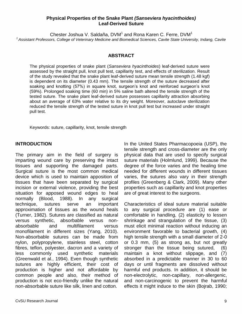
METHODOLOGY Extraction of Snake Plant Leaf Fibers Mature snake plant leaves (Fig. 1) at least 10 in long, were collected, cleaned and dried with a towel. Both ends of each leaf were cut, 1.5 in from the tip and the base, then pounded with the use of hammer on a clean flat surface. The fibers
Figure 1. Mature leaves of snake plant (Sanseviera hyacinthoides).
Figure 2. Dried plant fiber from snake plant (Sanseviera hyacinthoides).
Figure 3. Snake plant leaf-derived suture.
C.J.V. Saldaña and R.K.C. Ferre

CvSU Research Journal 11
were manually extracted, air-dried (Fig. 2) and bundled into 10 fibers to create a suture (Fig. 3). A bundle of 10 fibers was tied at one end by overhand knot and was divided into two groups: five strands were held by one hand, the other five by the other hand while holding the tied end with a clip. The fibers were twisted by alternately placing each bundle over the other, twisting in a clockwise direction until twisting of the strands were completed and tied at the other end of the bundle. It was then pressed with the use of minimum heat (>1 iron setting) with a hot plate iron (Standard
®) and were stored in a plastic
container with a dessicant for three days. The dried fibers were brought to the Philippine Textile Research Institute (PTRI) - Department of Science and Technology (DOST) for physical property testing. A total of 360 sutures were used in the study, each treatment having three replicates with ten samples per replicate. The sutures were randomly selected and cut evenly into 30 cm long. The treatments were the varying lengths of soaking time in 5% saline bath which were 15, 30 and 60 min heated at the same temperature of 37
oC. Dry or suture not soaked in saline bath
was used as control for the study. Tests for Physical Properties Tensile Strength and Knot Security The failure or break load of a suture material for a given suture size describes its tensile strength, which is the amount of weight in pounds or kilograms that is necessary to cause the suture to rupture. Minimum baseline suture tensile strengths are standardized by suture size and readily available from the USP (Greenberg, & Clark, 2009). The diameter of the sutures prior to testing was measured in millimeters using a vernier caliper. For knot pull test, the ten sutures per replicate were tied into different knots: square knot, surgeon‘s knot and reinforced surgeon‘s knot.
Likewise, the same number of sample sutures subjected for straight-pull test. To measure the breaking strength in terms of kilogram force (kgf), the sutures were then placed in Instron Universal Tensile Strength tester, allowing it to pull the suture until it breaks. Capillarity Ten pieces of sutures per replicate for each treatment were weighed on analytical balance prior to soaking in 5% saline bath to get the initial weight in grams. Afterwards, these were soaked in 5% saline bath for 15, 30 and 60 min, except for the control represented by the dry suture. When length of soaking time was reached, the soaked sutures were allowed to drip until no water dropped and again weighed to get the final weight. The difference between the final weight and the initial weight was computed to approximate the amount of water absorbed by the suture which represents the capillarity. Effects of Sterilization on Physical Properties Ten sample sutures per replicate for each treatment were sterilized in an autoclave for 15 min at 121°C and 15 psi or 2 atm (Nelson & Molintoris, 1992). The sterilized sutures from dry (10 samples) and treated sutures (10 samples) were subjected to straight pull test and knot pull test to determine their tensile strength and knot security. Data Analysis Data on tensile strength under straight pull test, knot security and knot pull test, capillarity and effect of sterilization on the suture were gathered and analyzed using Analysis of Variance. RESULTS AND DISCUSSION Leaf Tensile Strength and Diameter of Snake Plant Derived Suture The mean tensile strength of the dry suture (control) is 1.48 kgf or 3.26 lb and with an
Physical Properties of the Snake Plant (Sanseviera hyacinthoides) Leaf-Derived Suture

12
average diameter of 0.43 mm or 1 (USP) (Table 1). The diameter and tensile strength of the soaked sutures were positively correlated and indicate that, as the diameter increases, the tensile strength also increases. According to Bojrab (1990), an ideal suture material has a high tensile strength in small diameter (8 lb at 4-0 or 0.15 mm) in vivo and in vitro. This is to avoid tissue trauma by the implanted part. Oversized suture causes tissue reactions and knots poorly, while small sutures cause less tissue reaction and form a smaller knot. However, very thin sutures easily cut friable tissues or skin if applied under tension (Allen, 1987). Commercially available non-absorbable sutures of veterinary use include silk, cotton, polypropylene, polyester and nylon. Silk, cotton and polyester are available in sizes 3-0 (0.2 mm) and 2-0 (0.3 mm), while polypropylene and nylon are available in 2-0. The tensile strength of 3-0 silk is 5.3 lbs and 2-0 for 8.2 lbs. Cotton suture at 3-0 and 2-0 has a tensile strength of 4 lbs and 5.5 lbs, respectively. Likewise, the tensile strengths of polyester are 8.8 lbs and 14.5 lbs for 3-0 and 2-0, respectively. On the other hand, polypropylene has a tensile strength of 7.6 lbs and nylon has 9 lbs. Comparing the above values to the dry snake plant leaf-derived suture, the tensile strength is relatively low at 1 or 0.43 mm diameter. Aside from the tensile strength of the suture, tissue strength should also be considered in choosing suture material. According to Allen (1987), suture strength should not exceed tissue
strength. The strength of a particular tissue depends on its anatomic site and histological condition. Lai (2007) noted that the strength of fat, muscle, skin and fascia are 0.2, 1.27, 1.82 and 3.77 kgf, respectively. Based on the tensile strength of the tissues and mean tensile strength of the tested suture (1.480 kgf), the snake plant leaf-derived suture might be used on fat and skin. Effect of Knot on Tensile Strength Each suture material has recognizable tensile strength, which is the amount of weight in pounds or kilograms, necessary to cause suture to rupture. Typically, this measurement is presented in two forms, straight-pull and knot-pull. Straight-pull and knot-pull reflect the reduction in any given suture strength when knotted (Greenberg & Clark, 2009). The tensile strength of the sutures under straight pull test was significantly different from the tensile strength of the knotted sutures, particularly on dry, at 15 and 30 min soaking wherein the average decreases in tensile strength from straight pull test are 57.3%, 51.3% and 30.0%, respectively. Meanwhile, there is no statistical difference between the tensile strength of straight and the knotted sutures soaked for 60 min. The tensile strength of the sutures decreases by 30-55% when knotted compared to its original state (Henderson, 2007). According to Allen (1987), knotting method influences the structural properties of the suture and should be considered in tying knot under
Table 1. Mean diameter (mm) and tensile strength (kgf) of snake plant leaf-derived suture from different treatments subjected to straight- pull test.
MEAN DIAMETER (mm)
TENSILE STRENGTH (kgf)
R VALUES
Dry 0.43 1.48 0.44
15 min 0.41 2.08 0.59
30 min 0.43 1.62 0.23
60 min 0.49 0.89 0.02
C.J.V. Saldaña and R.K.C. Ferre
Maiden Issue (January - June 2014)

CvSU Research Journal 13
tension. In addition, the pliability (ability of suture to change its shape), memory (ability of suture to return to its shape), surface friction (roughness or smoothness of the suture) and size affect the security of knotting.
Effect of Soaking on Tensile Strength In the straight pull test, the tensile strength of dry suture is not significantly different with the sutures soaked for 15 and 30 min. However, a significant decrease of 40% was obtained for the suture soaked for 60 min (Table 2). In the knot pull test, differences were noted in the dry and sutures soaked for 15 and 30 min. The mean values increased by 30-50% after the sutures were soaked. On the other hand, the mean tensile strength of suture soaked for 60 min decreased by 1-30%, which is significantly different to that of the sutures soaked for 15 and 30 min in the surgeon‘s and reinforced surgeon‘s knot, but not significantly different to that of the square knotted suture.
Results suggest that prolonged soaking time (60 min) together with warm temperature (37°C) weaken the suture. Anand (1996) cited that sutures with temperature dependent hydrolysis
brought about by boiling water or steam, such as silk and other natural sutures cause gradual loss of strength. Use of accelerated test conditions, such as exposure to temperature higher than 37°C and pH greater than 11 or pH less than 3 speed up degradation process. Apart from this, the fluid from which the sutures were soaked
requires recognition for the presence of cations and anions at a specific pH, since cations accelerate degradation at high pH whereas anions accelerate degradation at low pH. According to Henderson (2007), knot security usually decreases with wetting, however, it can be stabilized by increasing the number of throws, accuracy of knot and tightness of knot tied. The increased number of throws will make knot security more stable because of the distribution of friction and tensile forces (Turner, 1982). Capillarity of Snake Plant Leaf-Derived Suture The snake plant leaf-derived suture absorbs water (Table 3). Capillarity is attributed to the twisted conformation of the fibers since having interstices allow the contact of liquid to solid in between its small spaces. Aside from this, capillary attraction of the suture can be related to the nature of the fibers within the plant.
Table 2. Tensile strength of snake plant leaf-derived sutures in different treatments under straight pull and knot pull test (kgf).
SOAKING TIME (min)
STRAIGHT PULL TEST
KNOT PULL TEST AVERAGE % DECREASE
FROM STRAIGHT-PULL TEST
Square knot
Surgeon’s knot
Reinforced surgeon’s knot
Dry 1.480a 0.630
b 0.630
b 0.605
b 57.3
15 2.080a 0.915
b 1.195
b 0.905
b 51.3
30 1.620a 1.115
b 1.220
b 1.045
b 30.0
60 0.885a 0.905
a 0.830
a 0.655
a 10.0
*Values followed by common letters are not significant using P <0.05.
Physical Properties of the Snake Plant (Sanseviera hyacinthoides) Leaf-Derived Suture

14
According to Richardson (2006), snake plant leaf fiber cells have a small lumen inside allowing the absorption of water. When the sutures are soaked for 15 min, the weight increased from 0.0165 g to 0.0677 g indicating that water comprised 75% of its weight. After 30 min of soaking, the weight of the suture increased by 60% from its initial weight of 0.0194 g. At 60 min soaking time, the weight of the sutures increased by 54% from its initial weight of 0.0206 g. There is a significant increase from the initial weight of the suture after soaking. However, the difference in final weight and amount of water absorbed by the suture at
different lengths of soaking time was not significant. The duration of soaking has no effect on the amount of water absorbed by the snake plant leaf-derived suture. The tested sutures are comparable with other available natural sutures such as silk and cat gut that also have the capability of capillary attraction. Effect of Sterilization on Tensile Strength The tensile strength of sutures under straight pull test variably changed after sterilization in autoclave. An increase in tensile strength after sterilization was observed for the dry, 30 and 60 min soaked sutures. Whereas, a decrease in
Table 3. Mean capillarity of snake plant leaf- derived suture in different soaking times.
Table 4. Mean tensile strength of sterilized and not sterilized snake plant leaf-derived suture.
a,bmeans within the same columns with different superscripts are significantly different (P < 0.05)
o the Group did not undergo soaking
c,d means within two adjacent rows (initial weight and final weight) with different superscripts are significantly different
(P< 0.05)
SOAKING TIME (min)
DIAMETER (mm)
INITIAL WEIGHT (g)
FINAL WEIGHT (g)
DIFFERENCE WEIGHT
(g)
Dry 0.425a,o
0.0188a,o
0.0188b,o
0.000b,o
15 0.396a 0.0165
a,d 0.0677
a,c 0.024
a
30 0.432a 0.0194
a.d 0.0484
a,c 0.029
a
60 0.447a 0.0206
a,d 0.0453
a,c 0.025
a
SOAKING TIME (min)
STRAIGHT-PULL TEST
(kgf)
KNOT-PULL TEST
Square Knot (kgf) Surgeon’s Knot
(kgf)
Reinforced Surgeon’s Knot
(kgf)
NS S NS S NS S NS S
Dry 1.480a 1.775
a 0.630
a 0.550
a 0.630
a 0.525
a 0.605
a 0.595
a
15 2.080a 1.645
a 0.915
a 0.670
b 1.195
a 0.920
b 0.905
a 0.695
b
30 1.620a 2.035
a 1.115
a 0.880
b 1.220
a 0.860
b 1.045
a 0.865
b
60 0.885b 1.620
a 0.905
a 0.910
a 0.830
a 0.880
a 0.655
a 0.925
a
NS not sterilized
S sterilized
a,b means within two adjacent rows (between sterilized and not sterilized) sutures with different superscripts are
significantly different (P< 0.05)
C.J.V. Saldaña and R.K.C. Ferre
Maiden Issue (January - June 2014)

CvSU Research Journal 15
tensile strength was observed for sterilized suture soaked for 15 min (Table 4). On the other hand, in the knot pull test, the tensile strength decreased in the dry, 15 and 30 min soaking but increased in 60 min soaking for the sterilized knotted sutures (Table 4). Significant differences in tensile strength between sterilized and non-sterilized sutures were noted in the 15 and 30 min soaking time in knot pull test for the square, surgeon and reinforced surgeon‘s knots. However, no significant difference was noted in the dry and 60 min soaked suture in the knot pull test for all knots (Table 4). This indicates that sterilization decreases the tensile strength in knotted sutures soaked in 15 and 30 min. Aside from the decreasing effect of knotting to the tensile strength, exposure of sutures to increased humidity during steam sterilization may have caused alteration in its structural properties and thus decreasing its tensile strength. Steam sterilization reduces the strength of the available non-absorbable sutures, and is contraindicated for other absorbable sutures due to exposure to increased humidity that causes suture degradation (Bojrab, 1990). However, sterilization of knotted suture under prolonged soaking (60 min) slightly increased tensile strength. CONCLUSION Based on the results of this study, the snake plant leaf-derived suture mean tensile strength is dependent on its diameter. The tensile strength of the suture decreased after prolonged soaking and knotting. Compared to commercially available suture, the tensile strength of the snake plant leaf-derived suture is relatively low at 1 or 0.43 mm diameter. The snake plant leaf-derived suture possesses capillarity attraction absorbing about an average of 63% water relative to its dry weight. Moreover, sterilization using an autoclave reduces the tensile strength of the tested plant fiber suture.
LITERATURE CITED Aliero, A.A., Jimoh, F.O & Afolayan, A.J. (2008).
Antioxidant and antibacterial properties of Sansevieria hyacinthoides. International Journal of Pure and Applied Sciences, 2(3): 103-110.
Allen, A.R. (1987). Fundamental technique in
veterinary surgery. 3rd
ed. W. B. Saunders Co. London.
Anand, S. (1996). Medical textile 1996
international conference. Woodward Publishing. Bolton Institute. England, U.K.
Blood, D.C. (1988). Bailliere‘s Comprehensive
Veterinary Dictionary. W.B. Saunders Co. Oval Road, London.
Bojrab, M.J. (1981). Pathophysiology in small
animal surgery. Washington Square, Philadelphia.
Bojrab, M.J. (1990). Current techniques in small
animal surgery, 3rd
Ed. Washington Square, Philadelphia.
Crane, S. W. (1983). Suture materials. In Bojrab
M. J. (ed): Current techniques in small animal surgery. 2
nd ed. Philadelphia: Lea
and Febiger. Everett, E.H. (1982). Encyclopedia of
horticulture. Vol. 9. Garland Publishing Inc., New York and London.
Gangstad, E.O., Joyner, J.F. & Seale, C.C.
(1951). Agronomic characteristics of Sansevieria species. Tropical Agriculture Journal. 28:204-214.
Greenberg, J.A. & Clark, R.M. (2009). Advances
in suture materials for obstetrics and gynecologic surgery. Reviews in Obstetrics and Gynecology. Vol. 2 No. 3: 143-158.
Physical Properties of the Snake Plant (Sanseviera hyacinthoides) Leaf-Derived Suture
16
Greenwald, D., Shumway, S., Alber, P., & Gottlieb, L. (1994). Mechanical comparison of 10 suture materials before and after in Vivo Incubation‖, Journal of Surgical Research, Academic Press Inc.; 56, 372-377.
Holmlund, E. W. (1999). Physical properties of
surgical suture materials: stress-strain relationship, stress-relaxation and irreversible elongation‖, Dept. of Surgery, University of Umea, Sweden; Ann. Surg. pp. 189.
Henderson, R.A. (2007). The Veterinarian‘s
suture guide. College of Veterinary Medicine, Auburn University. http://www.vetmed.aurburn.edu./~henderson/guide/guide6.htm.
Kispotta, U.G. (2008). Synthesis and
characterization of bio-composite material. Department of Physics, National Institute of Technology, Rourkela. 1(5) pp. 11.
Klingaman, G. (1999). Ornamentals Extension
News. University of Arkansas, Division of Agriculture. Cooperative Extension Service.
Lai, S.Y. (2007). Sutures and Needles.
eMedicine World Medical Library. http://www.emedicine.com/ent/topic38/htm.
Mbugua, P.K. & Moore, D.M. (1996).
Taxonomic studies of the genus Sansevieria (Dracaenaceae). In The Biodiversity of African Plants. Edited by L.J.G. van der Maesen, X.M., van der Burgt & J.M., van Medenbach de Rooy. Pp. 489-492. Kluwer Academic Publishers, Dordrecht, Netherlands.
Nelson, E.A. & Molintoris, E. (1992). Autoclave
(Steam Sterilizer). Sect. 12. 3. In Isenberg, H. D., editor. Clinical Microbiology Procedures Handbook. American Society for Microbiology, Washington, D.C.
Olivia, C. (2005). An assessment of medicinal hemp plant extracts as natural antibiotic and immune modulation phytotherapies. M.Sc Thesis. Faculty of Natural Sciences, University of the Western Cape.
Richardson, W.N. (2006). Plants, agriculture and
human society. Massachusetts. W.H. Benjamin Inc. pp. 173-179.
Turner, A. (1982). Techniques in large animal
surgery. Bailliere Tindall, London. Watt, J.M. & Breyer-Brandwijk, M.G. (1962). The
medicinal and poisonous plants of Southern and Eastern Africa. E and S Livingstone Ltd., Edinburgh, United Kingdom.
Wunderlin, R.P. (2004). Atlas of Florida vascular
plants. Institute of Systematic Botany. Florida. DK Publishing. pp. 154-155.
Yang, C.S., & Chen, C.Y. (2010). The effect of
suture size on skin wound healing strengths in rats. Journal of Medical and Biological Engineering. 31(5) 339-343.
C.J.V. Saldaña and R.K.C. Ferre
Maiden Issue (January - June 2014)

CvSU Research Journal 17
Managing Pawikan Conservation and Protection Project in Naic, Cavite Through Partnership: The CvSU Experience
John Xavier B. Nepomuceno
1 and Alexander F. Ferre, PhD
2
1Instructor, Cavite State University, Naic, Cavite
2Professor, Cavite State University, Indang, Cavite
ABSTRACT
Reported and proven incidences of nesting, hatching and poaching of eggs of marine turtle in some coastal villages in Naic, Cavite were revealed and validated by the Department of Environment and Natural Resources–Protected and Wildlife Bureau (DENR-PAWB) during informal survey in the community. These documented cases paved the way to the establishment of a ―Pawikan Center‖ by way of the Pawikan Conservation and Protection Project (PCPP). This study documented the initial conservation and protection efforts for marine turtles, specifically Olive Ridleys (Lepidochelys olivacea) in Naic. In the ten months of implementation, seminars, workshops and series of information dissemination were conducted to develop and enhance relevant skills and knowledge on marine turtles, its protection and conservation. The project, being community-based in the coastal village of Labac, utilized participatory approach in the actual conservation through monitoring of nesting marine turtles, hatchery management, releasing of hatchlings and tagging of stranded adult marine turtles. The coastal community residents became more vigilant in reporting nesting incidents. Field conservationists enhanced their leadership and management skills in dealing with people visiting the hatchery. The PCPP has since been instrumental as basis for policy making on issues related to the wildlife by the local council. The established baseline information showed that 2,091 marine turtle eggs were protected in the hatchery with 84% or 1,764 eggs hatched and released to the sea.
Keywords: marine turtle, Olive Ridley, pawikan, nesting, poaching
INTRODUCTION Marine turtles, popularly known in the Philippines as pawikan, belong to the sub-order Cryptodira, families Dermochelyidae and Cheloniidae. There are more than 220 species of turtles in the world, but only seven are considered marine (saltwater). Five of the marine species are present in the Philippines. These are the Green (Chelonia mydas), Hawksbill (Eretmochelys imbricata), Loggerhead (Caretta caretta), Olive Ridley (Lepidochelys olivacea) and the Leatherback turtles (Dermochelys coriacea). A typical Philippine marine turtle weighs between 180 to 210 kg and, unlike land turtles, cannot retract its head and limbs under its streamlined shell. It has large
upper eyelids that protect its eyes, but has no external ear opening. Awkward on land, it is more active and graceful in the water, travelling as fast as 32 km/hr using its long paddle-like fore and hind flippers. Marine turtles vary in color: olive-green, yellow, greenish-brown, or black. The most common species in the Philippines is the Green Sea Turtle, which is also found in all tropical and sub-tropical seas. Its most distinct feature is a more blunt and wider head than that of the Hawksbill Turtle. It grows up to 1.5 m long and weighs up to 185 kg (Carbayas, 2012). Scientists refer to marine turtles as the only living remnants of the dinosaur age, but maybe not for long. Unless sincere efforts are

18
undertaken, marine turtles might follow dinosaurs into extinction (Carbayas, 2012). Marine turtles are essential to the marine ecosystem and their presence serves as indicator of good and healthy marine habitat. Exploitation of marine turtles in the Philippines led to its current status as one of the endangered species. One of the government‘s executive agencies, the Department of Environment and Natural Resources (DENR), is tasked to conserve and save the marine turtles along with other threatened, endangered and near extinct wildlife resources. Through Executive Order No. 542, Task Force Pawikan, presently known as Pawikan Conservation and Protection Project (PCPP), was created on June 26, 1979 with main purpose of conserving the marine turtles. In addition, the Philippine Wildlife Resources Conservation and Protection Act (RA 9147 of July 30, 2001) was also enacted to prohibit the killing, maltreatment, infliction of injury, trading, collecting, hunting, possessing, and transporting of marine turtles, including their eggs and by-products. More so, the Fisheries Code of 1998 (RA 8550) enforces related prohibitions in order to protect the marine turtles. Background of the Project The Olive Ridley, Lepidochelys olivacea, was identified to be nesting in Naic, in the Province of Cavite. This prompted the DENR through its Protected Areas and Wildlife Bureau (PAWB) to introduce PCPP in the area. Naic is one of the coastal towns of Cavite located 47 km from Manila. It is considered coastal with nine out of its 30 villages adjacent to the waters of Manila Bay. Coastlines of Naic are about 1 - 3 km away from the town proper or Poblacion. The local government unit of Naic (LGU-Naic) supports its marine fisheries by providing technical assistance to the fishermen and adopting fish farmers to increase their productivity. Furthermore, the local government
protects, conserves and rehabilitates its marine system by conducting activities like river and coastal cleanup, coral rehabilitation, and mangrove reforestation, in coordination with other sectors. The biggest fishery extension project of the municipality is the Naic Fish Sanctuary (NFS) Project chaired by the Cavite State University – Naic (CvSU-Naic). The local government, specifically the Mayor‘s Office, serves as the adviser. Some private and other public organizations participate in the project. NFS started its operation in 2003 and has listed various fishery issues confronting the municipality such as biological degradation, natural calamities, water pollution, sand mining and illegal fishing.
The initial meetings and invitations for the PCPP-Naic involved the member organizations of the NFS Project Management Team. The experiences of the organizations in managing the sanctuary propelled the preparation for the needed plans and activities of the PCPP.
Operational Framework Government agencies, namely: DENR, LGU-Naic and CvSU-Naic, NGO through Shoreline Kabalikat sa Kaunlaran Incorporated (SKKI), and the private sector, namely: Far East Maritime Foundation Incorporated (FEMFI), International Maritime and Offshore Safety Training Institute (IMOSTI), Tropical Garden Resort (TGR), and Sabrina Fair Philippines Incorporated (SFPI) entered into a Memorandum of Agreement (MOA) for the marine turtle conservation and protection. As key players, each partner agency provided expertise and resources delineated in the MOA to attain the project‘s objectives and sustainability. Operating under the existing laws for government institutional projects, both national and local agencies implemented PCPP-Naic as a community-based project wherein the community leads the actual field work by patrolling, monitoring, managing the hatchery, tagging and releasing marine turtles. An
J.X.B. Nepomuceno and A.F. Ferre
Maiden Issue (January - June 2014)
CvSU Research Journal 19
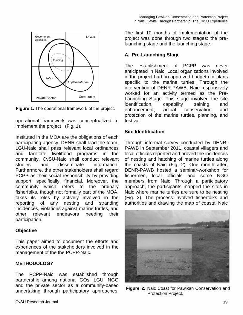
operational framework was conceptualized to implement the project (Fig. 1). Instituted in the MOA are the obligations of each participating agency. DENR shall lead the team. LGU-Naic shall pass relevant local ordinances and facilitate livelihood programs in the community. CvSU-Naic shall conduct relevant studies and disseminate information. Furthermore, the other stakeholders shall regard PCPP as their social responsibility by providing support, specifically, financial. Moreover, the community which refers to the ordinary fisherfolks, though not formally part of the MOA, takes its roles by actively involved in the reporting of any nesting and stranding incidences, violations against marine turtles, and other relevant endeavors needing their participation.
Objective This paper aimed to document the efforts and experiences of the stakeholders involved in the management of the the PCPP-Naic. METHODOLOGY
The PCPP-Naic was established through partnership among national GOs, LGU, NGO and the private sector as a community-based undertaking through participatory approaches.
The first 10 months of implementation of the project was done through two stages: the pre-launching stage and the launching stage.
A. Pre-Launching Stage The establishment of PCPP was never anticipated in Naic. Local organizations involved in the project had no approved budget nor plans specific to the marine turtles. Through the intervention of DENR-PAWB, Naic responsively worked for an activity termed as the Pre-Launching Stage. This stage involved the site identification, capability training and enhancement, actual conservation and protection of the marine turtles, planning, and festival.
Site Identification Through informal survey conducted by DENR-PAWB in September 2011, coastal villagers and local officials reported and proved the incidences of nesting and hatching of marine turtles along the coasts of Naic (Fig. 2). One month after, DENR-PAWB hosted a seminar-workshop for fishermen, local officials and some NGO members from Naic. Through a participatory approach, the participants mapped the sites in Naic where marine turtles are sure to be nesting (Fig. 3). The process involved fisherfolks and authorities and drawing the map of coastal Naic
Government Agencies
NGOs
Community
Policy Implementation
Private Sector
Funding
Figure 1. The operational framework of the project.
Managing Pawikan Conservation and Protection Project in Naic, Cavite Through Partnership: The CvSU Experience
Figure 2. Naic Coast for Pawikan Conservation and Protection Project.

20
and putting marks on areas which historically served as nesting sites for marine turtles. Out of the nine coastal villages or barangays, four barangays were identified as nesting sites, namely: Labac, Mabulo, Bagong Kalsada and Bucana Malaki. Capability Training and Enhancement The above mentioned three-day seminar-workshop included lectures on species identification, biology, behavior and laws about marine turtles. Hands–on experience specifically focused on transplanting newly-laid eggs for an ex situ nest (Fig. 4) and hatchery management (Fig. 5). Transplanting of eggs from the original nest to a nest in a hatchery is crucial in conserving and protecting the marine turtles. Eggs laid in unsafe areas would lead to its spoilage or breakage. Threats to eggs include high tide, predator animals and poaching.
Participants were given an exposure trip to the PCPP in Morong, Bataan. Bataan has one of the
most well-known marine turtle conservation efforts in the country. Participants had time to consult with the conservationists in Bataan and witness their activities. Conservation and Protection of the Marine Turtles As an outcome of the seminar-workshop, Wildlife Enforcement Officers (WEOs) were officially assigned by PAWB from among the participants. Incidentally, one week after the seminar-workshop, nesting marine turtles were sighted in Labac, thus the gained capability to conserve and protect wildlife, specifically for marine turtles was effected through proper procedures. The WEOs, who used to be ordinary citizens, engaged fellow fisherfolks in regularly monitoring their coastal areas. This is also in consideration with the information from PAWB that the nesting season for marine turtles usually occurs during the last quarter of the year.
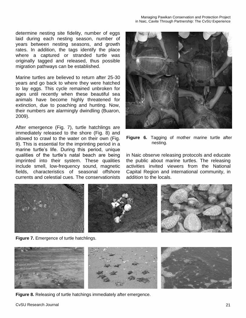
The local conservationists record necessary information on nesting marine turtles. Data include length and width of its carapace, and total number of eggs laid and transplanted to the hatchery. Prior to the release into the sea, mother marine turtles are tagged (Fig. 6) for future monitoring. Tagging is a method to
Figure 4. Transplanting of marine turtle eggs.
Figure 5. Hatchery of marine turtle eggs.
J.X.B. Nepomuceno and A.F. Ferre
Maiden Issue (January - June 2014)
Figure 3. Nesting Olive ridley turtle in Naic.
CvSU Research Journal 21
determine nesting site fidelity, number of eggs laid during each nesting season, number of years between nesting seasons, and growth rates. In addition, the tags identify the place where a captured or stranded turtle was originally tagged and released, thus possible migration pathways can be established. Marine turtles are believed to return after 25-30 years and go back to where they were hatched to lay eggs. This cycle remained unbroken for ages until recently when these beautiful sea animals have become highly threatened for extinction, due to poaching and hunting. Now, their numbers are alarmingly dwindling (Buaron, 2009).
After emergence (Fig. 7), turtle hatchlings are immediately released to the shore (Fig. 8) and allowed to crawl to the water on their own (Fig. 9). This is essential for the imprinting period in a marine turtle‘s life. During this period, unique qualities of the turtle‘s natal beach are being imprinted into their system. These qualities include smell, low-frequency sound, magnetic fields, characteristics of seasonal offshore currents and celestial cues. The conservationists
in Naic observe releasing protocols and educate the public about marine turtles. The releasing activities invited viewers from the National Capital Region and international community, in addition to the locals.
Figure 6. Tagging of mother marine turtle after nesting.
Figure 7. Emergence of turtle hatchlings.
Figure 8. Releasing of turtle hatchings immediately after emergence.
Managing Pawikan Conservation and Protection Project in Naic, Cavite Through Partnership: The CvSU Experience

22
Planning In between the actual conservation and protection activities, LGU-Naic initiated meetings with partner agencies and planned for the project‘s grand activity, culminating the pre-launching stage. In said meetings, obligations of each partner agency were defined and delineated.
Pawikan Festival On January 31, 2012, the Pawikan Festival was held to highlight and culminate the pre-launching stage for PCPP-Naic with the theme, “Pawikan: Yaman ng Karagatan, Kaya Sagipin, Arugain, Padamihin”. A MOA forged among the partner organizations was signed during the day-long festival. Other events held were ceremonial releasing of hatchlings, marine turtle exhibit, photo shoot contest, beach volleyball, mural painting contest, film showing, and entertainment. B. Launching Stage Signing of the PCPP-Naic MOA marked the beginning of the Launching Stage. Such MOA strengthens the partnership and defines the role of each organizations involved in the project.
MOA Signing The MOA strengthens the partnership and defines the role of each organization involved in the project. The MOA signed among the partner-agencies identified the project as “DENR, LGU-Naic, CvSU-Naic, SKKI, FEMFI, IMOSTI, TGR and SFPI Project for Naic Pawikan Conservation and Protection”.
Expansion of the Conservation and Protection of the Marine Turtles Part of the collective obligations of all the partners is the preparation of a Marine Turtle Conservation Action Plan (MTCAP). Initial discussion among partners favored the expansion of the project by including other coastal villages of Naic in conservation and protection efforts. Meanwhile, the plan is still being drafted and shall outline the specific activities and implementation strategies to be carried out by the management team for five years. This shall also lead the team in realizing the project‘s objectives.
In celebration of ―Month of the Ocean‖ with the theme, “Buhay Dagat, Buhay Natin,” and of the ―International Day for Biological Diversity (Marine Biodiversity),‖ partners gathered in CvSU-Naic. The event was initiated by DENR-Cavite, in collaboration with the Bureau of Fisheries and Aquatic Resources (BFAR) Region IVA, Philippine Coast Guard (PCG) and LGU-Naic. During the event, locals from other Naic coastal villages confirmed that their areas are also nesting sites of marine turtles. Such confirmation validated the previous assumption that the entire coastline of Naic is a marine turtle nesting site.
In response to the need for expanding the conservation and protection activities in Naic, the first seminar in Labac was replicated and echoed to new participants from other coastal villages of Naic. Partners calendared further capability training,
Figure 9. Turtle hatchling finding its own way to the sea.
J.X.B. Nepomuceno and A.F. Ferre
Maiden Issue (January - June 2014)

CvSU Research Journal 23
workshop and consultation in order to equip each coastal villages of Naic prior to the succeeding nesting seasons of marine turtles. Monitoring Success of the project was gleaned on the increase of incidences of female marine turtles nesting on the coastline, as well as increase in the total number of laid eggs and high and increasing number of hatchlings. PCPP-Naic understands that reliable analysis on
the project success will require years of constant monitoring. Meanwhile, based from the initial conservation efforts, 2,091 eggs were transplanted in the hatchery. Most of the eggs were laid in Labac, aside from 99 eggs which were taken from the nearby village of Bancaan. It takes 45-65 days of incubation before the eggs hatch. Monitoring showed that 1,764 or 84% of the transplanted eggs hatched. All of the hatchlings were released to the sea (Table 1). While the eggs were kept safe in the hatchery, the unhatched were believed to be due to mishandling during the transplantation process.
Table 1. Hatchery data of marine turtles conserve at Labac, Naic, Cavite from October 2011 – February 2012.
DATE NO. OF EGGS
TRANSPLANTED NO. OF HATCHLINGS
RELEASED DATE OF
EMERGENCE
October 15, 2011 100 81 December 1, 2011
October 15, 2011 107 77 December 1, 2011
October 15, 2011 81 67 December 1, 2011
November 10, 2011 134 94 January 1, 2012
November 11, 2011 115 102 January 3, 2012
November 15, 2011 116 65 January 9, 2012
November 21, 2011 115 112 January 19, 2012
November 24, 2011 93 89 January 19, 2012
December 1, 2011 121 112 January 30, 2012
December 2, 2011 93 89 January 29, 2012
December 4, 2011 86 82 January 29, 2012
December 11, 2011 20 14 February 3, 2012
December 14, 2011 83 87 February 6, 2012
December 14, 2011 106 102 February 6, 2012
December 15, 2011 86 63 February 8, 2012
December 18, 2011 104 101 February 12, 2012
December 19, 2011 112 107 February 12, 2012
December 23, 2011 113 110 February 16, 2012
December 24, 2011 102 97 February 17, 2012
January 19, 2012 99 98 March 9, 2012
February 26, 2012 105 15 April 15, 2012
Total 2,091 1,764
Managing Pawikan Conservation and Protection Project in Naic, Cavite Through Partnership: The CvSU Experience
24
Local conservationists also monitored stranding and rescued marine turtles. Stranded marine turtles were tagged and were then released back to the sea. On the other hand, within ten months, three dead adult marine turtles were recorded. To preserve the dead turtles, these were stuffed, skeletonized and kept at CvSU-Naic to serve as an instruction material. Moreover, a rescued marine turtle from Zambales, a green sea turtle (Chelonia mydas), was turned over to CvSU-Naic.
Information Dissemination Aside from the usual consultations, meetings and seminars, PCPP distributes a booklet containing important information about marine turtle conservation and protection. The booklet contains necessary information like species identification, biology, behavior and laws about marine turtles. The material is written in Tagalog for the clients to easily understand its content. Presently, CvSU-Naic considers marine turtles as part of its extension priorities. On-going research activities in the Campus Information and Technology Department include marine turtle software development. When the software is ready, it is expected to provide a new material in information dissemination. Meanwhile, LGU-Naic is using its social networking sites to disseminate information about marine turtles. The campus-based student organization of the Philippine Rural Reconstruction Youth Association of CvSU-Naic has included marine turtle conservation and protection in its program. The student organization is expected to assist partner agencies in conducting marine turtle awareness activities.
DISCUSSION Documenting the management experiences of the team conserving and protecting the marine turtles in Naic can be gleaned in various perspectives. The following discussions present the reasons for management decisions for
actions, constraints and challenges met, and consequences and implications of management decisions. Reasons for Management Decisions or Actions The validated information on marine turtle nesting in some coastal villages of Naic, Cavite paved the way to the forged partnership among government, non-government and private organizations adhering to existing Philippine laws on conservation of wildlife and natural resources. After series of consultations, meetings, seminars and workshops, the partner-agencies have gained deeper insights and skills on marine turtle management and conservation. The success of the pre-launching and launching stages, propelled the partner-agencies to outline a five-year Marine Turtle Conservation Action Plan that will further expand the conservation and protection efforts of the marine turtles in Naic.
Constraints and Challenges The marine turtle conservation and protection project in Naic, being newly-established, encountered problems in the community that are seriously challenging the management team in attaining its set objectives. Among these problems are: Egg poaching. In a study conducted by CvSU-Naic, local fishermen admitted that poaching of marine turtle eggs is a common practice. Locals take the laid eggs for food consumption. In addition, there were some locals who admitted eating marine turtle meat in the belief that it is an aphrodisiac. Misconception about marine turtles. In constant consultation with village chiefs of other coastal communities in Naic, it was revealed that many locals do not believe that marine turtles do have economic nor ecological value. In the previously mentioned study, such misconception was validated by fishermen themselves. It was also
J.X.B. Nepomuceno and A.F. Ferre
Maiden Issue (January - June 2014)
CvSU Research Journal 25
revealed that locals associate marine turtles to bad luck. Appearance of such wildlife in their fishing activities is believed to bring less fish catch. In addition, marine turtles are blamed to tear their fish spread in the sea. These bad beliefs about marine turtles are resulting to apathy of the locals towards marine turtles. Little knowledge on proper conservation and protection. The project being community-based in Labac, gained volunteers and supporters not only from the coastal community but also from other places. In some instances, people would gather to witness marine turtles nesting or being released. The crowd, while claiming to be concerned with the marine turtles, turned as threats by having little knowledge about the conservation. The amazed observers tend to break some protocols while others intrude the pathways trailed by the marine turtles being released. Most notably, many handle the marine turtles without care. Insufficient conservation and protection capability and skills. The management team, particularly those in the field, was equipped through seminars, trainings and workshops in marine turtle identification, tagging, nesting and releasing. However, actual conservation and protection showed that further enhancement in the capability and skills training are needed. This includes crowd management, rescuing wounded marine turtles and law enforcement. Illegal fishing. The use of illegal fishing methods is detrimental not only to fishes but also to the entire marine ecosystem including the marine turtles. Records from the Philippine National Police – Cavite Maritime Group showed that illegal fishing still occurs in Naic, as in other parts of the Manila Bay. Dynamite fishing does not only destroy the marine habitat, but also directly harms the lives of the marine turtles. In addition, entanglement of the marine turtles to fishing nets can cause drowning. Marine turtles need to surface to take in air. Hooks and propellers inflict injury to the marine turtles.
Insufficient budget. Project partners provide counterpart and share resources in various activities related to PCPP. However, meeting the objectives of the project demands more financial support in order to undertake activities such as research, instructional material preparation, information dissemination, and monitoring. Consequences of Management Decisions The implementation of the project, being community-based, gained further support from the locals. Some fisherfolks were identified as active patrollers, getting no compensation aside from the ―fulfillment‖ which they claim to experience everytime they work for PCPP. The coastal community also became vigilant in reporting nesting incidents. During hatching times, information would spread through word of mouths, social networking sites and text messages. Locals, as well as visitors from Manila and foreign countries would come to witness the event. In addition, field conservationists were able to enhance their leadership skills in dealing with different people visiting the hatchery. They also served as inspirations to other local village leaders in promoting, conserving and protecting the marine turtles. On the other hand, the MOA fostered partnership among various sectors. Through such, the implementation of the project gains ensured sustainability. The partnership promotes sharing of expertise and resources between and among partner agencies. The stakeholders are also drafting a five-year plan that will give direction for attaining the project‘s objectives.
Implications of the Management Decisions or Actions Through the institutionalization of the project, conservation and protection of the marine turtles in Naic is ensured. In addition, the MOA puts the project as expandable and extendable to other coastal villages of Naic. Furthermore, the MOA
Managing Pawikan Conservation and Protection Project in Naic, Cavite Through Partnership: The CvSU Experience

26
also implies budget allocation from various partners for sustaining PCPP.
This project also supports other coastal management projects of Naic, particularly the fish sanctuary project. PCPP also contributes to the widening perspective of the community in terms of marine resources. Marine turtle conservation is now included in Naic‘s regular activities like mangrove reforestation, coastal and river cleanup, and coral rehabilitation.
PCPP is also instrumental as basis for local council‘s policy making. A relevant resolution in support of the project is presently being drafted, which will then be reviewed by the Sangguniang Bayan of Naic. DENR-PAWB also proposes that Naic be declared as a protected marine turtle site. Strong ordinances and strict implementation of laws are major ways in addressing the challenges faced by the conservation team.
In a short period of time, Naic was able to provide conservation and protection efforts for the marine turtles. This placed Naic as a trending topic in various national and local discussions with respect to coastal and environmental protection. The efforts were even recognize by the national government enlisting Naic from where PCPP is active.
CONCLUSION
Validated nesting incidences of marine turtles paved the way towards an institutionalized Pawikan Conservation and Protecion Project in Naic. This became possible through the partnership fostered among national and local government agencies, non-government organization, and private sectors.
In 10 months of the projects' implementation, the management encountered contraints and challenges such as egg poaching, misconception of the community about marine turtles, little knowledge on proper conservation and protection, insufficient conservation and protection capability and skills, illegal fishing and insufficient budget.
Strong ordinances and strict implementation of laws are major ways in addressing the challenges faced by the conservation team. Finalization and approval of the five-year plan will also help and give direction for attaining the project‘s objectives. Moreover, strengthening the participation of the community, particularly the fisherfolks, will result to the project‘s success.
As expected, continuing the conservation and protection methods will lead to the increase of number of marine turtles nesting, hatching and being released in Naic.
RECOMMENDATIONS
The following are recommended to ensure efficient and effective implementation of the PCPP-Naic:
Intensify marine turtle awareness campaign
in the entire coastal community of Naic by using relevant media.
Partner with other business and private sectors located in other villages of Naic in expanding the project area.
Conduct trainings and workshops for present and future conservationists of Naic in order to develop and enhance their skills in various aspects of the project.
Conduct research activities that will improve project management.
Consider external funding to further sustain the project.
LITERATURE CITED
Buaron, D.R.A. (2009). Pawikan conservation in
Philippines. Retrieved from
www.oceanheritage.com.ph/pawikan-
conservation-project, Nov 2009.
Carbayas, R.V. (2012). Philippine navy rescue
117 endangered pawikan in Palawan. Retrieved from Philippine Information Agency, news.pia.gov.ph/index.php?article=1421355199010.
J.X.B. Nepomuceno and A.F. Ferre
Maiden Issue (January - June 2014)
Response of Dragon Fruit (Hylocereus undatus Haw. Britton and Rose) Clonal Cuttings to Healing Duration and Fungicide Application
Teddy F. Tepora, PhD
1 and Regina M. Nosa
2
1Assistant Professor, Cavite State University, Indang, Cavite
2Professor, Cavite State University, Indang, Cavite
ABSTRACT
The study was conducted to determine the effect of different healing durations and fungicide application on the growth and rooting performance of dragon fruit clonal cuttings. The interaction of healing duration and fungicide treatment significantly affected the stem diameter, percent disease infection and number of roots. Fungicide application significantly affected length and number of roots, length and diameter of stems and percent disease infection in dragon fruit clonal cuttings. Healing duration also showed positive effect on stem width, number and length of roots, and percent disease infection. The clonal cuttings subjected to fungicide treatment at eight days healing period gave optimal results in terms of growth and rooting performance.
Keywords: clonal cuttings, fungicide, healing period, dragon fruit
INTRODUCTION Dragon fruit (Hylocereus undatus Haw. Britton and Rose) is a vining, terrestrial or epiphytic plant belonging to the cactus family. It has three-sided, evergreen, winged and fleshy stem that climbs on any type of support, reaching from a few inches up to 20 ft long in mature plants. At each node of the trunk or primary stem, a group of four to five spines are attached, on which secondary stems come out. This plant has both ground and aerial roots. Flowers are ornate and blooms, only for a night. Fruits are egg-shaped about 7 to 10 cm in diameter, 10 to 15 cm in length and 300 to 700 g in weight. The skin of the fruit is leathery, thin, brilliant reddish, with several greenish, soft flat scales projecting towards the end of the fruit. This night blooming cactus can have four to six fruiting cycles per year and may be propagated by either seed or stem (Trade Winds Fruit, 2004). The dragon fruit was introduced in the Philippines in the 1900‘s through trading and exchange of goods between the Spaniards and Filipinos (Aquino, 2005). The crop is also grown in Vietnam, Thailand, Malaysia and Taiwan. The Philippines is now gaining a competitive edge through mass production of dragon fruit, particularly in Cavite, Davao, Bukidnon and
Ilocos Region due to its nutritional value and high potential as a crop. The primary problem in dragon fruit farming is the widespread development of disease in the plantation which causes production failure and low quality of planting materials. The same problems are encountered by the propagators of the said fruit in South Florida (Florida Agroindustry, 2005). Seedlings are prone to diseases such as stem rot (Xanthomonas campestris), collar rot (Phytopthora sp.) and root rot (Fusarium sp., Alternara sp.) which are usually caused by too much water and moist condition of the soil, particularly in countries having humid climate (Bijlmakers, 2008). To date, information on the appropriate nursery management protocols to prevent and control disease infection on dragon fruit is scarce. Hence, result of this study serves as baseline information for better propagation techniques to produce high quality planting materials. The study aimed to develop a nursery management protocol that will minimize disease in dragon fruit clonal seedlings and produce high quality planting materials. It determined the response of dragon fruit clonal cuttings to healing duration and fungicide application.
CvSU Research Journal 27

METHODOLOGY
This study used the red skin-white flesh variety of dragon fruit. It focused on the growth and rooting performance of clonal seedlings as affected by healing duration and fungicide treatment. A 4 x 2 factorial in Randomized Complete Block Design (RCBD) with three replications was used in the study. Factor A was healing duration while Factor B was fungicide application. Two days, four days, six days or eight days healing periods with or without fungicide application were used as treatments. Selected 12-in long healthy stem cuttings were taken from green, disease-free, incision-free and moderately succulent good parent material. Stem cuttings were soaked in mancozeb solution (one tablespoon of fungicide powder dissolved in one liter of water) for 24 hours. After soaking, cuttings were applied with fungicide paste, prepared by adding a small amount of water to fungicide powder. The dragon fruit cuttings applied with fungicide were allowed to heal in a clean dry place subscribing to the healing duration treatments. A mixture of sand, garden soil, coconut husk and compost at 1:1:1:1 ratio was used as growing medium. The planting area was thoroughly cleaned and sanitized to ensure that it is free from pests before planting. A net house was also prepared
to allow the right amount of light to penetrate the dragon fruit seedlings. After the healing period, the cuttings were planted directly into the polyethylene bag. The clonal cuttings were inserted into seedling bags at half the level of the media to allow suitable space for root expansion. The newly planted cuttings were watered using a water sprinkler every other day. Data Gathering The data gathered include length and diameter of stem, number and length of roots, and percent disease infection which was determined using the formula: Data Analysis The Analysis of Variance for factorial experiment in RCBD was used to analyze the data gathered. Significant results were further tested using Duncan‘s Multiple Range Test (DMRT) for mean comparison. RESULTS AND DISCUSSION Length of Stem The analysis of variance showed insignificant
= —————————————————— x 100 Percent disease infection
Number of diseased clonal seedlings
Total number of clonal seedling per replication
TREATMENT
STEM LENGTH (cm)
One Month
Two Months
Three Months
Four Months
No fungicide + 2 days healing 27.38 28.68 29.76 30.75
No fungicide + 4 days healing 28.30 28.68 30.51 31.40
No fungicide + 6 days healing 29.33 29.85 31.01 31.23 No fungicide + 8 days healing 27.38 31.20 29.46 31.53
With fungicide + 2 days healing 29.46 29.85 31.40 31.63
With fungicide + 4 days healing 29.33 30.51 31.60 31.63
With fungicide + 6 days healing 31.6 31.63 31.85 32.63
With fungicide + 8 days healing 30.86 31.35 31.85 32.43
Table 1. Mean stem length (cm) of dragon fruit clonal cuttings as affected by healing period and fungicide application at one to four months after planting.
28 Maiden Issue (January - June 2014)
T.F. Tepora and R.M. Nosa

differences on the mean stem length of dragon fruit clonal cuttings across treatments as affected by the interaction of healing duration and fungicide application from the first to fourth month after planting (Table 1). However, there were significant differences among treatment means with regard to stem length as affected by fungicide application at three and four months after planting (Fig. 1). At these periods after planting, clonal cuttings applied with fungicide had significantly longer stem than untreated cuttings. The data indicated that fungicide application helped in the promotion of stem length of dragon fruit clonal cuttings. The finding corroborates the results of the study undertaken by the Institute of Food and Agricultural Science of the University of Florida (2005) conducted in Costa Rica and El Salvador. Farmers apply fungicide to the dragon fruit clonal cuttings to have supplementary resistance against certain fungi that enter the stem cuts. Due to fungicide application, pathogens causing disease could not penetrate the clonal cuttings; thus, providing better stem performance of dragon fruit in terms of height and width.
Diameter of Stem On the interaction effect of healing duration and fungicide application on stem diameter of dragon fruit clonal cuttings at one to four months after planting (Table 2), statistical analysis revealed that there were no significant differences among means of all treatment combinations at one to three months after planting. However, significant differences were observed at four months after planting. The results indicate that clonal cuttings applied with fungicide and subjected to longer healing duration resulted to thicker stem. Significant differences were also noted among the mean stem width of plants as affected by healing duration at two and three months after planting (Fig. 2). The data indicate that clonal cuttings subjected to longer healing duration resulted to thicker stem. The results can be attributed to the callusing provided by the period of healing. This confirms the results of Schultz (2009) that placing the succulent cuttings in a cool, dry area for at least a week out of direct sunlight will help to heal dry or callus the wound or cuts that promotes the growth of plants.
= —————————————————— x 100
A B
28
29
30
31
32
33
34
35
Mea
n S
tem
Len
gth
(cm
)
Without Fungicide With Fungicide
Figure 1. Mean stem length (cm) of dragon fruit clonal cuttings as affected by fungicide application at three months (A) and four months (B) after planting.
A B
0
1
2
3
4
5
6
7
2 Days Healing 4 Days Healing 6 Days Healing 8 Days Healing
Mean S
tem
Wid
th (
cm
)
b
a
b a
a
b
a
b
aa
Figure 2. Mean stem diameter (cm) of dragon fruit clonal cuttings as affected by healing duration at two months (A) and three months (B) after planting.
Response of Dragon Fruit (Hylocereus undatus Haw. Britton and Rose) Clonal Cuttings to Healing Duration and Fungicide Application
CvSU Research Journal 29

The analysis of variance also revealed significant differences among treatment means with regard to the mean stem width of dragon fruit clonal cuttings as affected by fungicide application at three and four months after planting (Fig. 3). The findings confirm the results of the study by the Institute of Food and Agricultural Science of the University of Florida (2005) conducted in Costa Rica and El Salvador. Farmers apply fungicide to the dragon fruit clonal cuttings to have supplementary resistance against certain fungi that enter the stem cuts. The application of fungicide hinders the penetration of pathogens causing disease on stem cuts. This provides better stem performance of dragon fruit in terms of height and width. Number of Pseudostems The analysis of variance revealed insignificant differences on mean number of pseudostems across treatments as affected by the interaction of fungicide application and healing duration at one to four months after planting (Table 3).
*Means in a column with different letters are significantly different at 5% level by DMRT.
A B
0
1
2
3
4
5
6
7
8
Mean S
tem
Wid
th (
cm
)
Without Fungicide With Fungicide
Figure 3. Mean stem width (cm) of dragon fruit clonal cuttings as affected by fungicide application at three (A) and four (B) months after planting.
Table 2. Mean stem diameter (cm) of dragon fruit clonal cuttings as affected by healing duration and fungicide application at one to four months after planting.
TREATMENT
STEM DIAMETER (cm)
One
Month
Two
Months
Three
Months
Four
Months
No fungicide + 2 days healing 4.40 4.78 4.9 5.37c
No fungicide + 4 days healing 3.98 4.83 5.4 5.88bc
No fungicide + 6 days healing 4.55 5.25 5.78 6.02 bc
No fungicide + 8 days healing 5.26 5.63 5.83 6.22b
With fungicide + 2 days healing 4.73 5.32 5.83 6.22b
With fungicide + 4 days healing 4.36 5.33 6.07 6.27b
With fungicide + 6 days healing 5.35 5.55 6.25 7.30b
With fungicide + 8 days healing 4.82 6.22 7.12 7.65a
T.F. Tepora and R.M. Nosa
30 Maiden Issue (January - June 2014)

Length of Pseudostems The analysis of variance on the interaction effect of healing duration and fungicide application on the length of pseudostems of dragon fruit clonal seedlings at one to four months after planting revealed insignificant differences (Table 4).
Number of Roots Statistical analysis revealed that there were insignificant differences among the mean number of roots across treatments as affected by the interaction of healing duration and fungicide application at one to three months after
TREATMENT
NUMBER OF ARMS
One Month Two
Months Three
Months Four
Months
No fungicide + 2 days healing 3.00 3.43 3.96 4.33
No fungicide + 4 days healing 2.50 3.17 3.83 5.00
No fungicide + 6 days healing 2.67 2.97 3.83 4.33
No fungicide + 8 days healing 2.00 3.67 4.00 4.33
With fungicide + 2 days healing 2.83 3.50 4.83 5.83
With fungicide + 4 days healing 3.83 4.16 5.00 6.17
With fungicide + 6 days healing 3.17 3.50 5.33 5.83
With fungicide + 8 days healing 1.83 3.83 5.33 6.00
Table 4. Mean length of pseudostems (cm) of dragon fruit clonal cuttings as affected by healing duration and fungicide application at one to four months after planting.
Table 3. Mean number of pseudostems of dragon fruit clonal cuttings as affected by healing duration and fungicide application at one to four months after planting.
TREATMENT
LENGTH OF ARMS (cm)
One Month
Two Months
Three Months
Four Months
No fungicide + 2 days healing 9.93 16.63
16.72
19.59
No fungicide + 4 days healing 11.57 15.77 16.96
17.67
No fungicide + 6 days healing 9.33 15.88
17.26
17.45
No fungicide + 8 days healing 10.00 16.48 16.70
18.64
With fungicide + 2 days healing 11.91 17.95 20.62 22.00
With fungicide + 4 days healing 9.69 14.63 15.96 17.09
With fungicide + 6 days healing 9.20 12.37 15.37 15.57
With fungicide + 8 days healing 9.51 16.20 15.77 16.76
Response of Dragon Fruit (Hylocereus undatus Haw. Britton and Rose) Clonal Cuttings to Healing Duration and Fungicide Application
CvSU Research Journal 31
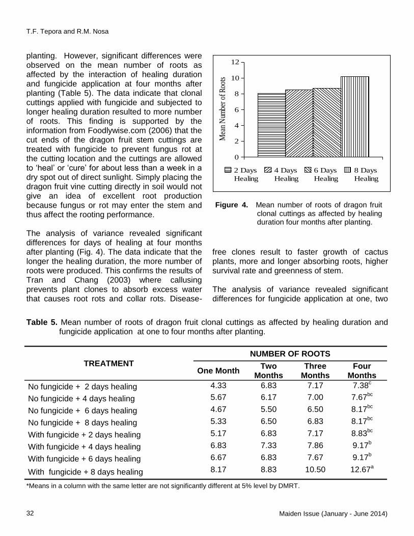
planting. However, significant differences were observed on the mean number of roots as affected by the interaction of healing duration and fungicide application at four months after planting (Table 5). The data indicate that clonal cuttings applied with fungicide and subjected to longer healing duration resulted to more number of roots. This finding is supported by the information from Foodlywise.com (2006) that the cut ends of the dragon fruit stem cuttings are treated with fungicide to prevent fungus rot at the cutting location and the cuttings are allowed to ‗heal‘ or ‗cure‘ for about less than a week in a dry spot out of direct sunlight. Simply placing the dragon fruit vine cutting directly in soil would not give an idea of excellent root production because fungus or rot may enter the stem and thus affect the rooting performance. The analysis of variance revealed significant differences for days of healing at four months after planting (Fig. 4). The data indicate that the longer the healing duration, the more number of roots were produced. This confirms the results of Tran and Chang (2003) where callusing prevents plant clones to absorb excess water that causes root rots and collar rots. Disease-
free clones result to faster growth of cactus plants, more and longer absorbing roots, higher survival rate and greenness of stem. The analysis of variance revealed significant differences for fungicide application at one, two
TREATMENT
NUMBER OF ROOTS
One Month Two
Months
Three Months
Four Months
No fungicide + 2 days healing 4.33 6.83 7.17 7.38c
No fungicide + 4 days healing 5.67 6.17 7.00 7.67bc
No fungicide + 6 days healing 4.67 5.50 6.50 8.17bc
No fungicide + 8 days healing 5.33 6.50 6.83 8.17bc
With fungicide + 2 days healing 5.17 6.83 7.17 8.83bc
With fungicide + 4 days healing 6.83 7.33 7.86 9.17b
With fungicide + 6 days healing 6.67 6.83 7.67 9.17b
With fungicide + 8 days healing 8.17 8.83 10.50 12.67a
*Means in a column with the same letter are not significantly different at 5% level by DMRT.
Table 5. Mean number of roots of dragon fruit clonal cuttings as affected by healing duration and fungicide application at one to four months after planting.
0
2
4
6
8
10
12
2 Days
Healing
4 Days
Healing
6 Days
Healing
8 Days
Healing
Mea
n N
umbe
r of
Roo
ts
Figure 4. Mean number of roots of dragon fruit clonal cuttings as affected by healing duration four months after planting.
T.F. Tepora and R.M. Nosa
32 Maiden Issue (January - June 2014)

and four months after planting (Fig. 5). The data indicate that the number of roots is promoted by the application of fungicide. The results of Silvan (2000) support the findings of the study. In planting succulents, fungicide application stimulates the natural tendency of the roots to have resistance to pathogens, so the rooting
takes place in a much shorter time and development of more and longer roots is promoted. Length of Roots The analysis of variance revealed insignificant differences on the mean length of roots of plant clones across treatments as affected by the interaction of healing duration and fungicide application at one to four months after planting (Table 6). On the other hand, significant differences were found on treatments subjected to different healing duration at four months after planting (Fig. 6). The data indicate that healing duration promotes root elongation of dragon fruit clonal cuttings. Significant differences were also noted on the mean length of roots at three and four months after planting as affected by fungicide application (Fig. 6). The results are in agreement with Silvan (2000) that the length of roots is promoted by fungicide application through the stimulation of
A B C
0
2
4
6
8
10
12
Mean
Nu
mb
er o
f R
oots
Without Fungicide With Fungicide
Figure 5. Mean number of roots of dragon fruit clonal cuttings as affected by fungicide application at one month (A), two months (B) and four months (C) after planting.
TREATMENT
LENGTH OF ROOTS (cm)
One Month
Two Months
Three Months
Four Months
No fungicide + 2 days healing 18.06 23.11 23.71 32.50
No fungicide + 4 days healing 16.17 19.85 22.13 33.58
No fungicide + 6 days healing 19.82 22.72 24.27 37.31
No fungicide + 8 days healing 16.88 26.00 26.00 31.90
With fungicide + 2 days healing 15.90 26.33 31.90 22.33
With fungicide + 4 days healing 15.65 21.70 22.02 25.10
With fungicide + 6 days healing 25.63 25.75 34.52 35.47
With fungicide + 8 days healing 24.17 29.12 34.50 38.55
Table 6. Mean length (cm) of roots of dragon fruit clonal cuttings as affected by healing duration and fungicide application at one to four months after planting.
Response of Dragon Fruit (Hylocereus undatus Haw. Britton and Rose) Clonal Cuttings to Healing Duration and Fungicide Application
CvSU Research Journal 33

Percent Disease Infection Significant differences were observed at three and four months after planting on percent disease infection as affected by the interaction of fungicide application and healing duration (Table 7).
CONCLUSIONS Based on the results of the study, the following conclusions were drawn: 1. The interaction of healing duration and
fungicide application promoted the growth of dragon fruit clonal cuttings through thicker stems of the plant and greater resistance against diseases.
2. Fungicide application encouraged the growth of the dragon fruit clonal cuttings through better development of stem in terms of length and diameter, and promotion of resistance against disease. Rooting performance was also affected by fungicide application resulting to more and longer roots. Hence, treatments with fungicide enhanced growth and rooting performance of dragon fruit clonal seedlings.
3. Better plant growth, in terms of more, longer and thicker stems and promotion of resistance against diseases, was achieved with the eight days healing duration. Hence, the longer the healing duration, the better the
0
5
10
15
20
25
30
35
40
2 Days
Healing
4 Days
Healing
6 Days
Healing
8 Days
Healing
Mea
n L
engt
h of R
oots
(cm
)
c c
b
a
Figure 6. Mean length of roots (cm) of dragon fruit clonal cuttings as affected by healing duration at four months after planting.
TREATMENTS
DISEASE INFECTION (%)
One Month
Two Months
Three Months
Four Months
No fungicide + 2 days healing 0.00 14.43 25.80a 36.36
a
No fungicide + 4 days healing 0.00 7.78 22.44ab
31.20b
No fungicide + 6 days healing 0.00 2.22 11.16b 26.11
b
No fungicide + 8 days healing 0.00 0.00 0.00c 10.80
c
With fungicide + 2 days healing 0.00 0.00 0.00c 10.80
c
With fungicide + 4 days healing 0.00 0.00 0.00c 3.60
c
With fungicide + 6 days healing 0.00 0.00 0.00c 0.00
c
With fungicide + 8 days healing 0.00 0.00 0.00c 0.00
c
*Means in a column with different letters are significantly different at 5% level by DMRT.
Table 7. Percent disease infection on dragon fruit clonal cuttings as affected by healing duration and fungicide application at one to four months after planting.
T.F. Tepora and R.M. Nosa
34 Maiden Issue (January - June 2014)

growth and rooting performance of dragon fruit clonal cuttings.
4. Among all treatments, the clonal cuttings applied with fungicide and subjected to the longest healing duration gave optimal results in terms of growth and rooting performance.
RECOMMENDATIONS 1. The application of fungicide and exposure of
clonal cuttings to eight days of healing is highly recommended to achieve optimum growth of dragon fruit clonal cuttings.
2. The uses and benefits of healing duration and fungicide application in other clonal cuttings of other dragon fruit varieties should be examined.
3. Further investigations are needed to explore the long-term effect of fungicide application and healing duration in the yield performance of dragon fruit.
LITERATURE CITED Aquino, M.U. (2005). The potential of the dragon
fruit. BAR Digest: Research and Development. Vol 7. No.3.
Bijmakers, H. (2008). The dragon fruit. Retrieved
December 22, 2008, from http://www.bijlmakers.com/fruits/dragonfruit.html.
Florida Agrindustry. (2005). A different fruit.
Retrieved March 26, 2009, from www.florida.agriculture.com/agindustry.html.
Foodlywise.com. (2006). Dragon fruit | stem
cutting method. Retrieved December 22, 2008, from http://dragonfruit.foodlywise.com/how to grow dragonfruit/growing dragonfruit commercially/growing dragonfruit from cuttings/dragonfruit stem cuttings/dragon fruit cuttings stem.html.
Institute of Food and Agricultural Sciences (IFAS) (2005). University of Florida.
Schultz, E.P. (2009). Simple way to propagate succulents Callusing. p. 11-12.
Silvan, K.R. (2000). Plant diseases, prevention and cure. Fungicide Application and other Engineered Method to Avoid Basal Root Disease. p.124.
Trade Winds Fruit. (2004). Dragonfruit. Retrieved December 22, 2008, from www.tradewindsfruit.com/dragonfruit.html.
Tran, D.I. & Chang, M.C. (2003). Vietnamese crops. A step-by-step pamphlet for propagating vietnamese crops.
Response of Dragon Fruit (Hylocereus undatus Haw. Britton and Rose) Clonal Cuttings to Healing Duration and Fungicide Application
CvSU Research Journal 35

GUIDELINES FOR AUTHORS
The Cavite State University Research Journal is the official peer-reviewed academic journal of the Cavite State University published biannually. The publication is a compendium of interdisciplinary academic and research works of the different fields of specialization of the University‘s colleges, units and campuses. Articles and manuscripts are subject to scholarly review by the editorial staff and at least three external reviewers who are experts on the specified disciplines. The different types of articles or manuscripts considered are; original Full Academic and Original Research Papers, Research Notes, Research Reviews and Case Reports. Research papers with funding from international, national, regional and local institutions are given priority for publication. Types of Papers Original research paper is a full length article that
consists of documentation, discussions of results and findings arising from researches, completed studies and technical achievement in basic or applied research that have not been published elsewhere. A research paper usually consists of 10-30 double-spaced typewritten pages of the complete text, figures, illustrations, tables and references. Research articles may also be empirical or conceptual/theoretical as in the fields of education, humanities, social science, psychology, literature and languages.
Research note/Short communication is a short description of important current research findings or observations made in a limited area of investigation (not a full length article) that warrants immediate publication and are considered urgent. The article contains abstract, body, conclusions/recommendations and literature cited and must not exceed four double spaced typewritten pages. It is important that the author specifies that the article submitted is a research note.
Case report is a detailed report usually in the field of medicine and health science describing an unusual or novel occurrence of an individual/patient citing the symptoms, signs, diagnosis, treatment and follow-up treatment including demographic profile. This type of report also applies to the fields of education, social science and criminal justice.
Review article is a narrative paper providing accumulative results of many different articles on a particular topic coherent to the state of the art in its own field. It does not cover original research but provide information about the topic and journal references to the original research. It must have an abstract, introduction and literature cited.
Distribution of Topics per Issue If possible, no more than two articles per field of specialization shall be included for publication per issue of the CvSU Research Journal, depending on how varied and on the number of articles submitted for publication for any one issue. A list of fields of specialization of various colleges of the University is enumerated below: Animal and Veterinary Science Fisheries Crop Science Crop Protection Horticulture Farming Systems Nursing Medical Technology Plant Pathology Social Sciences Languages Biological Sciences Biotechnology Communication Economics/Agricultural Economics Business Management/Agribusiness
Management Development Studies Education Hotel and Restaurant Management Human Resources Management Computer Engineering Agricultural Engineering Computer Science Information Technology Civil Engineering Criminology/Criminal Justice Sports/Recreational Management/ Physical Education Other Fields
GENERAL INSTRUCTIONS Submission of Manuscripts
Authors must submit the following: Manuscript (three clear printed hard copies and a CD copy) Accomplished Cover Sheet All document requirements should be sent to: The Editor-in-Chief Cavite State University Research Journal Office of the Vice President for RECETS Cavite State University Indang, 4122 Cavite Philippines
Electronic submission and communication are encouraged. Manuscript submitted through online must be in Microsoft Word file. Only complete manuscripts and those that conform to the required format will be reviewed. The editorial staff will issue an acknowledgement receipt once the manuscript is received. To follow-up manuscripts, correspondence should be addressed to the Editor-in-Chief at Telefax No. (63) 46 443-9104 or email [email protected].
Eligibility Faculty-researchers of the University may submit their original research paper, research notes, research reviews or case reports. Research papers with funding from international, national, regional and local institutions are given priority for publication. Copyright Notice Papers submitted should not have been published nor submitted elsewhere. For paper with more than one author, its publication must be with the consent of all co-authors. A paper cannot be withdrawn after it has been accepted for publication. The article cannot also be published in any form of language without the consent of the copyright holders. The authors shall agree to the automatic transfer of the copyright to the Cavite State University and will be required to sign a Copyright Agreement when the paper is accepted for publication. In the event that the author wishes to reproduce or modify parts of the
paper, permission from the Cavite State University through the Editor-in-Chief must be obtained. Authorship The primary author/s shall be the person/s with substantial contribution to the paper. Participation includes conceptualization, source of novelty, design, analysis and interpretation of data, drafting and writing of the paper. Manuscript Format All parts of manuscript should be typed on one side of page, double-spaced on Letter (8 ½‖ x 11‖) typewriting paper with one-inch margin on all sides and consecutively paginated on the upper right hand corner of the page. Arial with 12 point font size is preferred, using Microsoft Word and limited to 30 pages (10 printed pages) including tables, figures and references. Footnotes are not to be used in the text. The format found in the style guide of the American Psychological Association (APA) shall be used as reference for the journal. The International System of Units (SI) must be used. Write out numbers one to nine, Arabic numeral is preferred. All pages of text should have line numbers. Only the first word, proper nouns and acronyms are capitalized. Full length paper must be organized in the following order: Title page (page 1) should contain the full article title, the complete name/s and address of each author, followed by the name and address of the institution where the work was carried out. Limit the title to a maximum of 15 words. Avoid active verbs, abbreviations, acronyms and punctuation marks in the title.
Abstract (page 2) must be written in one paragraph, concise and understandable when printed separately. It should provide a brief summary of the research, its objectives, methods, significant findings and implications and major conclusion. A statement of rationale may also be included. The abstract must be limited to 250 words. Keywords should be listed immediately after the Abstract to facilitate retrieval of references and indexing journal articles. Six key words from the title may be included.

Text or Body starts on page 3 and presented uninterrupted on consecutive pages and line numbered. Organize text into sections labeled as Introduction, Methodology, Results and Discussion, Conclusion and Recommendation (if any). These are written as headings of separate sections, center-aligned, bold face, and worded exactly as mentioned. Subheadings should be aligned to the left of the page and set in bold face. In case of third-order subheadings, these can take the form paragraph lead-ins, written in bold face, indented and run with the text, separated by a period. The sequence of presentation of the text should be as follows:
Introduction provides sufficient but brief background information and rationale of the paper. This section includes relevant review of literature and objectives of the study. Methodology should give sufficient information so that other investigators can repeat the study. This section must be arranged chronologically, by succession of procedures or in any other logical manner, tables and figures can be included. For well-established methods, a reference to the technique should be given. Generic names of products and equipment should be used unless the author considers the trade names or manufacturers‘ names essential or very specific to the purpose and methodology of the study. The manufacturer‘s name and location (town, country) should be given in
parenthesis. Details of procedures and anesthetics used and doses given should be provided. The composition of buffers, solutions and culture media should be specified. For solutions, the term % must be defined as w/v of v/v. Supply complete citations, including author (editor), title, year, publisher and version number for computer software mentioned in the manuscript. Sufficient details of the experimental design, sampling procedures and statistical analysis should be given so that readers can assess the adequacy and validity of testing the hypotheses of interest. The numbers of experimental units used and the way in which they are allocated to treatments should be described. Omitted observations must be justified. The full binomial Latin names should be given for all experimental plants and animals. The breed or strain of animals should be stated and details of age, weight and sex. Authors should indicate that investigations have been approved by the local ethical committee (IACUC) and that consent has been obtained from patients/owners/respondents. Results depend on the type of paper and may be presented by means of text, tables and figures. This section maybe divided into subheadings. Clarity of argument, cohesion and brevity must be observed. Repetitive descriptions that are already clear in tables, graphs and illustrations must be avoided. Tables, figures, graphs or illustrations must be numbered in the order in which they are mentioned
ORIGINAL OR FULL LENGTH RESEARCH
RESEARCH NOTE/SHORT COMMUNICATION
REVIEW
Title Page Title Page Title Page
Abstract (limited to 250 words) Abstract (limited to150 words) Abstract
Text (Body) Introduction
Methodology
Results and Discussion
Conclusion
Recommendation/s
Body must have Conclusion and/or Recommendation/s
Body must have Introduction
Literature Cited Literature Cited Literature Cited
Acknowledgment
Tables
Figures
Case Report may follow above format or with modifications

in the text and must all be cited in the text. In Discussion, results are extensively interpreted in relation to the objectives of the study. It may be compared or contrasted to previously published works. It should explain the significance of the results. Avoid excessive review. Results and discussion are usually combined. Conclusion and Recommendation (if any), may be included in the results and discussion or presented as separate for emphasis of significant findings. Abstract, methodology and results will be stated in past tense, whereas most of introduction, some discussion and all conclusions will be in the present tense. Acknowledgments should be limited to persons or institutions (other than your institution or any potential conflict of interest) which have substantial contribution to the work, personally or financially. This section should be briefly stated under separate heading placed immediately before References. Literature cited include only published papers mentioned in the text and should be arranged alphabetically by the last names of the authors between and within each reference. Where there are two authors, both should be named and only the first author‘s name and initials are inverted. With three or more authors, only the first author‘s name is given and ―et al.‖ for the rest. . In the text, the name of the author and the year of publication should be in parenthesis, e.g. (Cruz, 2008). For more than two authors, et al. should be used after the name of the senior author, e.g. (Yap et al.,1990) should be used. The American Psychological Association (APA) citation style shall be used as reference for the journal. For electronic references that are retrieved from a website, type the date of retrieval after the website. Tables must be typed double spaced on a separate sheet consist of title, body and footnotes. The title should be brief and descriptive of the information in the table. Only essential data must be incorporated in tables and graphs. Columns must not be separated by vertical lines and compared data read down, not across. Internal horizontal line is allowed only in the heading. First word of the heading is capitalized and the rest in lowercase except proper nouns and no period at end of title. Font size must not be reduced to accommodate data. Symbols and abbreviations used are defined below the table. Avoid crowded tables. Data should be presented either in tabular or graphical form and never both. Tables should be understandable, self-explanatory
without extensive reference to the text. Tables should supplement and not duplicate the text. They should be numbered in the order of their citation in the text and in Arabic numerals. All abbreviations used should be explained in the footnotes.
Figures should be in lowercase type except for the first letter. It includes line drawings, photographs, diagrams, computer plots or graphs. These should be typed double spaced on a separate sheet and should carry a brief descriptive heading and numbered and listed the same manner as they are mentioned in the text. Accompanying glossy print photographs must be mounted (use double sided tape) on copy paper with proper label below the photo indicating figure number and descriptive title. Additional copies of figures shall be required when paper is accepted. Color figures and illustrations are printed without charge to the author at the discretion of the editorial board. If Photoshop (TIFF or JPEG) files were supplied, images should be at least 300 dpi for color or 600 dpi for black and gray scale at the final published width. Figures should not be enclosed in boxes and Arial font is preferred. Reference lists, figures, tables, and figure/list captions should all be on separate sheets, all of which should be double-spaced and numbered. Standard nomenclature should be used. Unfamiliar terms, abbreviations, acronyms and symbols must be defined at first mention. Research note/Short communication should not exceed four printed pages excluding tables, figures and references. The article should have an abstract of not more than 150 words, body, conclusion and recommendation and literature cited.
Research review must have an abstract, introduction, body and literature cited. Privacy Statement The names and email addresses entered in this journal will be used exclusively for the stated purposes of this journal and will not be made available for any other purpose or to any other party.