Embed Size (px)
Citation preview
© 2013 WILEY-VCH Verlag GmbH & Co. KGaA, Weinheim 1
www.advmat.dewww.MaterialsViews.com
wileyonlinelibrary.com
CO
MM
UN
ICATIO
N
Magnetically Engineered Microcapsules as Intracellular Anchors for Remote Control Over Cellular Mobility
Anton M. Pavlov , Bruno G. De Geest ,* Benoit Louage , Lien Lybaert , Stefaan De Koker , Zdenek Koudelka , Andrei V. Sapelkin , and Gleb B. Sukhorukov *
In vivo delivery of ex vivo cultured cells is of interest for cell therapy, including stem cells and immune cells, for regenerative medicine and the treatment of genetic defects and cancer. [ 1–4 ] A particular challenge is to target these cells to a desired site of action in the body as nature does not have a homing mecha-nism to guide externally delivered cells to a designated tissue or organ. To address this issue, the use of an external force, such as a magnetic fi eld, could be an attractive option. Therefore, cells have to be impregnated with magnetic material, such as iron oxide particles, which are inert and biocompatible. Bun-dles of magnetic nanoparticles, when internalized by cells or linked to the cell surface as inert backpacks, could render the whole cell responsive to a magnetic fi eld. Currently, only a few approaches are available to magnetically label living cells. [ 5 ] Typically, magnetic nanoparticles are modifi ed on their surface with a ligand, promoting binding and internalization by cells. Such approach has for example shown to be well suited for MRI imaging. [ 6–8 ] On the other hand magnetically guided drug delivery has also been reported by conjugating drug molecules to magnetic particles followed by magnetically guided site-spe-cifi c delivery. [ 9,10 ]
A highly interesting strategy would be to combine these prin-ciples and to design vesicle-like constructs that allow magnetic guidance of living cells while simultaneously offering the pos-sibility to deliver bioactive molecules, either through sustained
release or triggered release. The layer-by-Layer technique [ 11 ] pro-vides a unique tool for the fabrication of multifunctional nano- and microcapsules where bioactive molecules and magnetic materials can easily be engineered into one entity. [ 12 ] Intensive research in the past few years has demonstrated that LbL cap-sules can be internalized by different types of living cells, can be degraded intracellularly in vitro and in vivo and can deliver a low and high molecular weight drug payloads. [ 13–16 ] Release of an encapsulated payload can be stimulated by various external physico-chemical triggers. [ 13,17 ] Due to the presence of mag-netite nanoparticles (MNP) which enable contrast for MRI imaging, the carriers can potentially be visualized in vivo. At the same time, MNP could be used for triggered release of pay-load once delivered to the site by an alternating magnetic fi eld or ultrasound. [ 10 ] These properties make LbL capsules poten-tially attractive carriers for drug delivery applications.
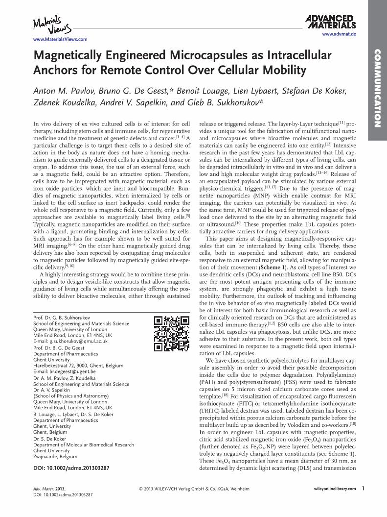
This paper aims at designing magnetically-responsive cap-sules that can be internalized by living cells. Thereby, these cells, both in suspended and adherent state, are rendered responsive to an external magnetic fi eld, allowing for manipula-tion of their movement ( Scheme 1 ). As cell types of interest we use dendritic cells (DCs) and neuroblastoma cell line B50. DCs are the most potent antigen presenting cells of the immune system, are strongly phagocytic and exhibit a high tissue mobility. Furthermore, the outlook of tracking and infl uencing the in vivo behavior of ex vivo magnetically labeled DCs would be of interest for both basic immunological research as well as for clinically oriented research on DCs that are administered as cell-based immune-therapy. [ 1,2 ] B50 cells are also able to inter-nalize LbL capsules via phagocytosis, but unlike DCs, are more adhesive to their substrate. In the present work, both cell types were examined in response to a magnetic fi eld upon internali-zation of LbL capsules.
We have chosen synthetic polyelectrolytes for multilayer cap-sule assembly in order to avoid their possible decomposition inside the cells due to polymer degradation. Poly(allylamine) (PAH) and poly(styrensulfonate) (PSS) were used to fabricate capsules on 5 micron sized calcium carbonate cores used as template. [ 18 ] For visualization of encapsulated cargo fl uorescein isothiocyanate (FITC)-or tetramethylrhodamine isothiocyanate (TRITC) labeled dextran was used. Labeled dextran has been co-precipitated within porous calcium carbonate particle before the multilayer build up as described by Volodkin and co-workers. [ 18 ] In order to engineer LbL capsules with magnetic properties, citric acid stabilized magnetic iron oxide (Fe 3 O 4 ) nanoparticles (further denoted as Fe 3 O 4 -NP) were layered between polyelec-trolyte as negatively charged layer constituents (see Scheme 1 ). These Fe 3 O 4 nanoparticles have a mean diameter of 30 nm, as determined by dynamic light scattering (DLS) and transmission DOI: 10.1002/adma.201303287
Prof. Dr. G. B. SukhorukovSchool of Engineering and Materials Science Queen Mary, University of London Mile End Road , London , E1 4NS , UK E-mail: [email protected] Prof. Dr. B. G. De GeestDepartment of PharmaceuticsGhent University Harelbekestraat 72 , 9000 , Ghent , Belgium E-mail: [email protected] Dr. A. M. Pavlov, Z. KoudelkaSchool of Engineering and Materials ScienceDr. A. V. Sapelkin(School of Physics and Astronomy) Queen Mary, University of London Mile End Road , London , E1 4NS , UK B. Louage, L. Lybaert, Dr. S. De Koker Department of Pharmaceutics Ghent, UniversityGhent , Belgium Dr. S. De KokerDepartment of Molecular Biomedical Research Ghent University Zwijnaarde , Belgium
Adv. Mater. 2013, DOI: 10.1002/adma.201303287
2
www.advmat.dewww.MaterialsViews.com
wileyonlinelibrary.com © 2013 WILEY-VCH Verlag GmbH & Co. KGaA, Weinheim
CO
MM
UN
ICATI
ON
electron microscopy (TEM), and a zeta-potential of -25 mV (as determined via electrophoretic mobility measurements) and were synthesized according to the Massart method. [ 13 ] After bilayers of PSS/PAH have been deposited with magnetic nanoparticles in middle, the calcium carbonate cores were dis-solved in EDTA. As low molecular weight species such as ions can freely diffuse through the multilayer membrane and larger
species cannot, hollow capsules are obtained with fl uorescent dextran entrapped within the capsules’ hollow void.
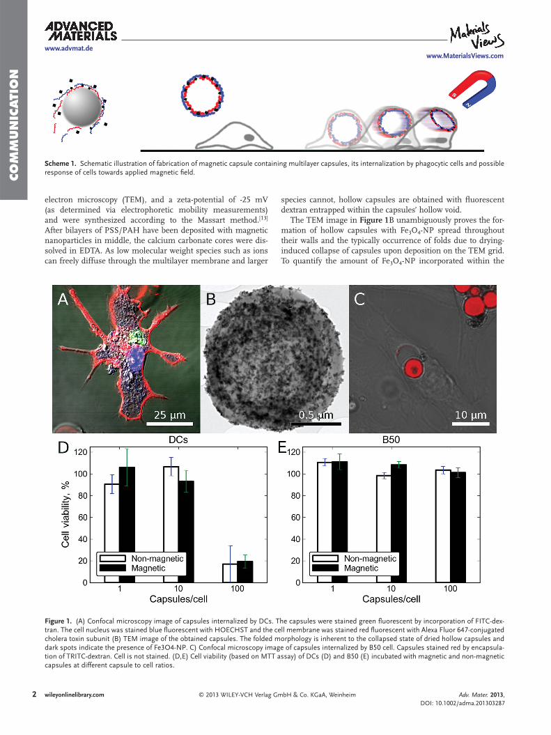
The TEM image in Figure 1 B unambiguously proves the for-mation of hollow capsules with Fe 3 O 4 -NP spread throughout their walls and the typically occurrence of folds due to drying-induced collapse of capsules upon deposition on the TEM grid. To quantify the amount of Fe 3 O 4 -NP incorporated within the
Scheme 1. Schematic illustration of fabrication of magnetic capsule containing multilayer capsules, its internalization by phagocytic cells and possible response of cells towards applied magnetic fi eld.
Figure 1. (A) Confocal microscopy image of capsules internalized by DCs. The capsules were stained green fl uorescent by incorporation of FITC-dex-tran. The cell nucleus was stained blue fl uorescent with HOECHST and the cell membrane was stained red fl uorescent with Alexa Fluor 647-conjugated cholera toxin subunit (B) TEM image of the obtained capsules. The folded morphology is inherent to the collapsed state of dried hollow capsules and dark spots indicate the presence of Fe3O4-NP. C) Confocal microscopy image of capsules internalized by B50 cell. Capsules stained red by encapsula-tion of TRITC-dextran. Cell is not stained. (D,E) Cell viability (based on MTT assay) of DCs (D) and B50 (E) incubated with magnetic and non-magnetic capsules at different capsule to cell ratios.
Adv. Mater. 2013, DOI: 10.1002/adma.201303287
3
www.advmat.dewww.MaterialsViews.com
wileyonlinelibrary.com© 2013 WILEY-VCH Verlag GmbH & Co. KGaA, Weinheim
CO
MM
UN
ICATIO
N
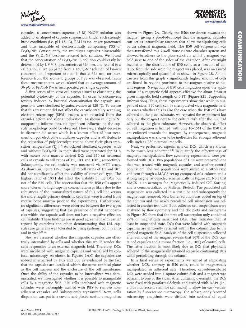
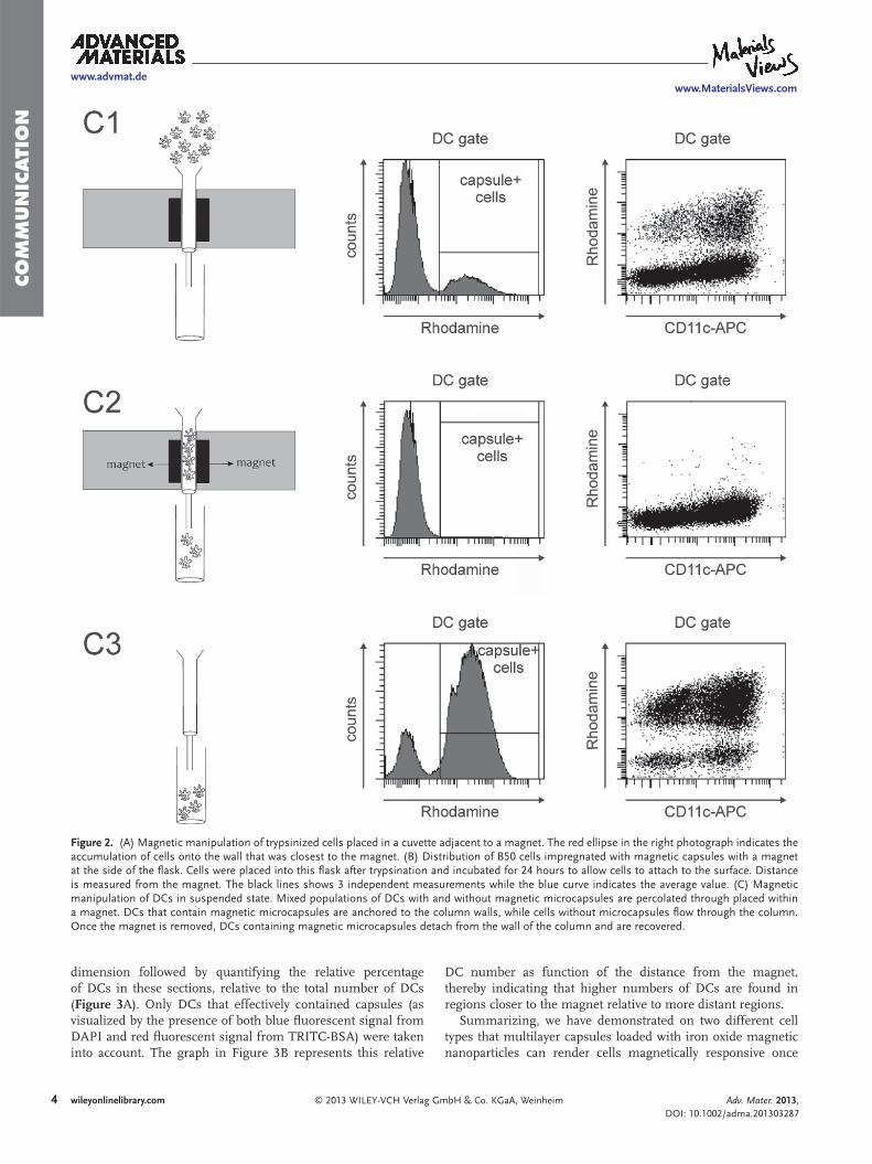
shown in Figure 2 A. Clearly, the B50s are drawn towards the magnet, giving a proof-of-concept that the magnetic capsules can act as intracellular anchors that allow manipulating cells by an external magnetic fi eld. The B50 cell suspension was then transferred to a 2-well Nunc culture chamber system and allowed to adhere to the glass substrate whilst a magnet was held next to one of the sides of the chamber. After overnight incubation, the distribution of B50 cells, as a function of dis-tance from the side were the magnet was placed, was measured microscopically and quantifi ed as shown in Figure 2 B. As one can see from this graph a signifi cantly higher amount of cells are found in regions proximate to the magnet relative to dis-tant regions. Navigation of B50 cells migration upon the appli-cation of a magnetic fi eld appears effective for about 5mm at given magnetic fi eld strength of 0.2Tl (Figure S2B, Supporting Information). Thus, these experiments show that while in sus-pended state, B50 cells can be manipulated via a magnetic fi eld. To assess whether this is also the case when the B50 cells have adhered to the glass substrate, we repeated the experiment but only put the magnet next to the culture dish after the B50 had adhered to the glass substrate. However, the observed. effect on cell migration is limited, with only 10–15M of the B50 that are enforced towards the magnet. By consequence, magnetic manipulation was shown to be ineffective for strongly adherent cells such as B50 neuronal rat cells.
Next, we performed experiments on DCs, which are known to be much less adherent. [ 23 ] To quantify the effectiveness of magnetic manipulation, fl ow cytometry experiments were per-formed with DCs. Two populations of DCs were prepared: one that was treated with magnetic capsules and another control population. The two populations were mixed in a 50:50 ratio and sent through a MACS set-up composed of a column and a strong magnet as depicted schematically in Figure 2 C. Note that MACS is an acronym for ‘Magnetically Induced Cell Sorting’ and is commercialized by Miltenyi Biotech. The percolated cell suspension was collected in a test tube and subsequently the magnet was removed. New buffer solution was added on top of the column and the newly percolated cell suspension was col-lected in another test tube. Both collected cell suspensions were analyzed by fl ow cytometry and the dot plots and histograms in Figure 2 C show that the fi rst cell suspension only contained 28% of magnetically sensitized DCs. This indicates that, at least in suspended state, DCs that were labeled with magnetic capsules are effi ciently retained within the column due to the applied magnetic fi eld. Analysis of the cell suspension collected after removal of the magnet reveals that 90% of the DCs con-tained capsules and a minor fraction (i.e., 10%) of control cells. The latter fraction is most likely due to DCs that physically adhered to the magnetically retained (capsule containing) DCs while percolating through the column.
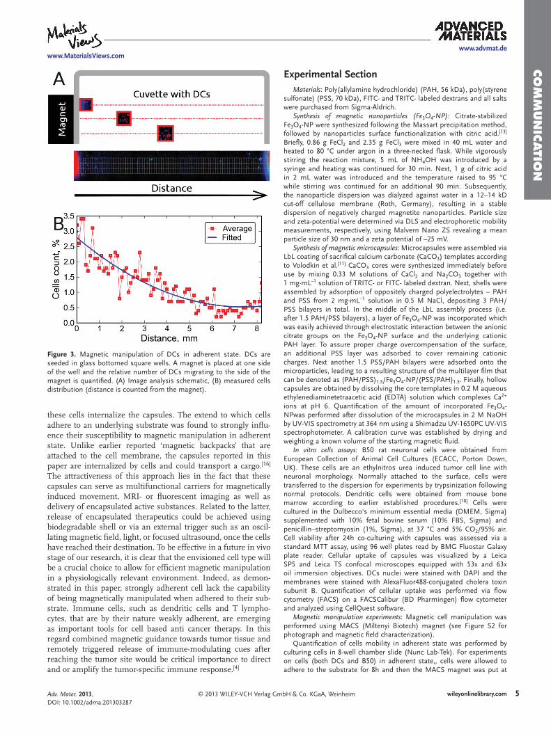
In a fi nal series of experiments we aimed at elucidating whether DCS, contrary to B50 cells, could be magnetically manipulated in adherent sate. Therefore, capsule-incubated DCs were seeded into a square culture dish and a magnet was adjacent to one of the sides. After culturing overnight, the DCs were fi xed with paraformaldehyde and stained with DAPI (i.e., a blue fl uorescent stain for cell nuclei) to allow for easy visuali-zation by fl uorescence microscopy. The subsequently recorded microscopy snapshots were divided into sections of equal
capsules, a concentrated aqueous (2 M) NaOH solution was added to an aliquot of capsule suspension. Under such strongly basic conditions (i.e. pH 12–13), PAH is no longer protonated and thus incapable of electrostatically complexing PSS or Fe 3 O 4 -NP. Consequently, the multilayer capsules disassemble and the Fe 3 O 4 -NP become released into solution. We found that the concentration of Fe 3 O 4 -NP in solution could easily be determined by UV-VIS spectrometry at 364 nm, and related to a calibration curve prepared from a Fe 3 O 4 -NP solution of known concentration. Important to note is that at 364 nm, no inter-ference from the aromatic groups of PSS was observed. From these measurements we calculated that an average amount of 36 pG of Fe 3 O 4 -NP was incorporated per single capsule.
A fi rst series of in vitro cell assays aimed at elucidating the potential cytotoxicity of the capsules. In order to circumvent toxicity induced by bacterial contamination the capsule sus-pensions were sterilized by autoclavation at 120 °C. To assure that this treatment did not affect the capsule stability scanning electron microscopy (SEM) images were recorded from the capsules before and after autoclavation. As shown in Figure S1 (supporting information), no signifi cant infl uence on the cap-sule morphology could be observed. However, a slight decrease in diameter did occur, which is a known effect of heat treat-ment on polyelectrolyte multilayer capsules and is attributed to the relaxation of polyelectrolyte chains above their glass tran-sition temperature (T g ). [ 19 ] Autoclaved sterilized capsules, with and without Fe 3 O 4 -NP in their shell were incubated for 24 h with mouse bone marrow derived DCs and B50 rat neuronal cells at capsule to cell ratios of 1:1, 10:1 and 100:1, respectively. Subsequently, the cell toxicity was measured via MTT assay. As shown in Figure 1 D-E, capsule to cell ratios of 1:1 and 10:1 did not signifi cantly affect the viability of either cell type. The highest ratio of 100:1 did affect the viability of the DCs but not of the B50 cells. The observation that the B50 cells appear more tolerant to high capsule concentrations is likely due to the robustness of the immortalized nature of this cell line versus the more fragile primary dendritic cells that were isolated from mouse bone marrow prior to the experiments. Furthermore, no signifi cant differences were observed between the two types of capsules, suggesting that the presence of metal nanoparti-cles within the capsule wall does not have a negative effect on cell viability. These fi ndings are in good agreement with earlier reports by ourselves and other, demonstrating that LbL cap-sules are generally well tolerated by living systems, both in vitro and in vivo. [ 20–22 ]
Next we assessed whether the magnetic capsules are effec-tively internalized by cells and whether this would render the cells responsive to an external magnetic fi eld. Therefore, DCs were incubated with magnetic capsules and visualized by con-focal microscopy. As shown in Figures 1 A,C, the capsules are indeed internalized by DCs and B50 as evidenced by the fact that the capsules are localized within the same confocal plane as the cell nucleus and the enclosure of the cell membrane. Once the ability of the capsules to be internalized was dem-onstrated, we investigated whether it is possible to manipulate cells by a magnetic fi eld. B50 cells incubated with magnetic capsules were thoroughly washed with PBS to remove non-adherent capsules and subsequently trypsinized. Next, cells dispersion was put in a cuvette and placed next to a magnet as
Adv. Mater. 2013, DOI: 10.1002/adma.201303287
4
www.advmat.dewww.MaterialsViews.com
wileyonlinelibrary.com © 2013 WILEY-VCH Verlag GmbH & Co. KGaA, Weinheim
CO
MM
UN
ICATI
ON
DC number as function of the distance from the magnet, thereby indicating that higher numbers of DCs are found in regions closer to the magnet relative to more distant regions.
Summarizing, we have demonstrated on two different cell types that multilayer capsules loaded with iron oxide magnetic nanoparticles can render cells magnetically responsive once
dimension followed by quantifying the relative percentage of DCs in these sections, relative to the total number of DCs ( Figure 3 A). Only DCs that effectively contained capsules (as visualized by the presence of both blue fl uorescent signal from DAPI and red fl uorescent signal from TRITC-BSA) were taken into account. The graph in Figure 3 B represents this relative
Figure 2. (A) Magnetic manipulation of trypsinized cells placed in a cuvette adjacent to a magnet. The red ellipse in the right photograph indicates the accumulation of cells onto the wall that was closest to the magnet. (B) Distribution of B50 cells impregnated with magnetic capsules with a magnet at the side of the fl ask. Cells were placed into this fl ask after trypsination and incubated for 24 hours to allow cells to attach to the surface. Distance is measured from the magnet. The black lines shows 3 independent measurements while the blue curve indicates the average value. (C) Magnetic manipulation of DCs in suspended state. Mixed populations of DCs with and without magnetic microcapsules are percolated through placed within a magnet. DCs that contain magnetic microcapsules are anchored to the column walls, while cells without microcapsules fl ow through the column. Once the magnet is removed, DCs containing magnetic microcapsules detach from the wall of the column and are recovered.
Adv. Mater. 2013, DOI: 10.1002/adma.201303287
5
www.advmat.dewww.MaterialsViews.com
wileyonlinelibrary.com© 2013 WILEY-VCH Verlag GmbH & Co. KGaA, Weinheim
CO
MM
UN
ICATIO
N
Experimental Section Materials : Poly(allylamine hydrochloride) (PAH, 56 kDa), poly(styrene
sulfonate) (PSS, 70 kDa), FITC- and TRITC- labeled dextrans and all salts were purchased from Sigma-Aldrich.
Synthesis of magnetic nanoparticles (Fe 3 O 4 -NP) : Citrate-stabilized Fe 3 O 4 -NP were synthesized following the Massart precipitation method, followed by nanoparticles surface functionalization with citric acid. [ 13 ] Briefl y, 0.86 g FeCl 2 and 2.35 g FeCl 3 were mixed in 40 mL water and heated to 80 °C under argon in a three-necked fl ask. While vigorously stirring the reaction mixture, 5 mL of NH 4 OH was introduced by a syringe and heating was continued for 30 min. Next, 1 g of citric acid in 2 mL water was introduced and the temperature raised to 95 °C while stirring was continued for an additional 90 min. Subsequently, the nanoparticle dispersion was dialyzed against water in a 12–14 kD cut-off cellulose membrane (Roth, Germany), resulting in a stable dispersion of negatively charged magnetite nanoparticles. Particle size and zeta-potential were determined via DLS and electrophoretic mobility measurements, respectively, using Malvern Nano ZS revealing a mean particle size of 30 nm and a zeta potential of –25 mV.
Synthesis of magnetic microcapsules : Microcapsules were assembled via LbL coating of sacrifi cal calcium carbonate (CaCO 3 ) templates according to Volodkin et al . [ 11 ] CaCO 3 cores were synthesized immediately before use by mixing 0.33 M solutions of CaCl 2 and Na 2 CO 3 together with 1 mg·mL −1 solution of TRITC- or FITC- labeled dextran. Next, shells were assembled by adsorption of oppositely charged polyelectrolytes – PAH and PSS from 2 mg·mL −1 solution in 0.5 M NaCl, depositing 3 PAH/PSS bilayers in total. In the middle of the LbL assembly process (i.e. after 1.5 PAH/PSS bilayers), a layer of Fe 3 O 4 -NP was incorporated which was easily achieved through electrostatic interaction between the anionic citrate groups on the Fe 3 O 4 -NP surface and the underlying cationic PAH layer. To assure proper charge overcompensation of the surface, an additional PSS layer was adsorbed to cover remaining cationic charges. Next another 1.5 PSS/PAH bilayers were adsorbed onto the microparticles, leading to a resulting structure of the multilayer fi lm that can be denoted as (PAH/PSS) 1.5 /Fe 3 O 4 -NP/(PSS/PAH) 1.5 . Finally, hollow capsules are obtained by dissolving the core templates in 0.2 M aqueous ethylenediaminetetraacetic acid (EDTA) solution which complexes Ca 2+ ions at pH 6. Quantifi cation of the amount of incorporated Fe 3 O 4 -NPwas performed after dissolution of the microcapsules in 2 M NaOH by UV-VIS spectrometry at 364 nm using a Shimadzu UV-1650PC UV-VIS spectrophotometer. A calibration curve was established by drying and weighting a known volume of the starting magnetic fl uid.
In vitro cells assays : B50 rat neuronal cells were obtained from European Collection of Animal Cell Cultures (ECACC, Porton Down, UK). These cells are an ethylnitros urea induced tumor cell line with neuronal morphology. Normally attached to the surface, cells were transferred to the dispersion for experiments by trypsinization following normal protocols. Dendritic cells were obtained from mouse bone marrow according to earlier established procedures. [ 18 ] Cells were cultured in the Dulbecco's minimum essential media (DMEM, Sigma) supplemented with 10% fetal bovine serum (10% FBS, Sigma) and penicillin–streptomyosin (1%, Sigma), at 37 °C and 5% CO 2 /95% air. Cell viability after 24h co-culturing with capsules was assessed via a standard MTT assay, using 96 well plates read by BMG Fluostar Galaxy plate reader. Cellular uptake of capsules was visualized by a Leica SP5 and Leica TS confocal microscopes equipped with 53x and 63x oil immersion objectives. DCs nuclei were stained with DAPI and the membranes were stained with AlexaFluor488-conjugated cholera toxin subunit B. Quantifi cation of cellular uptake was performed via fl ow cytometry (FACS) on a FACSCalibur (BD Pharmingen) fl ow cytometer and analyzed using CellQuest software.
Magnetic manipulation experiments : Magnetic cell manipulation was performed using MACS (Miltenyi Biotech) magnet (see Figure S2 for photograph and magnetic fi eld characterization).
Quantifi cation of cells mobility in adherent state was performed by culturing cells in 8-well chamber slide (Nunc Lab-Tek). For experiments on cells (both DCs and B50) in adherent state,, cells were allowed to adhere to the substrate for 8h and then the MACS magnet was put at
these cells internalize the capsules. The extend to which cells adhere to an underlying substrate was found to strongly infl u-ence their susceptibility to magnetic manipulation in adherent state. Unlike earlier reported ‘magnetic backpacks’ that are attached to the cell membrane, the capsules reported in this paper are internalized by cells and could transport a cargo. [ 16 ] The attractiveness of this approach lies in the fact that these capsules can serve as multifunctional carriers for magnetically induced movement, MRI- or fl uorescent imaging as well as delivery of encapsulated active substances. Related to the latter, release of encapsulated therapeutics could be achieved using biodegradable shell or via an external trigger such as an oscil-lating magnetic fi eld, light, or focused ultrasound, once the cells have reached their destination. To be effective in a future in vivo stage of our research, it is clear that the envisioned cell type will be a crucial choice to allow for effi cient magnetic manipulation in a physiologically relevant environment. Indeed, as demon-strated in this paper, strongly adherent cell lack the capability of being magnetically manipulated when adhered to their sub-strate. Immune cells, such as dendritic cells and T lympho-cytes, that are by their nature weakly adherent, are emerging as important tools for cell based anti cancer therapy. In this regard combined magnetic guidance towards tumor tissue and remotely triggered release of immune-modulating cues after reaching the tumor site would be critical importance to direct and or amplify the tumor-specifi c immune response. [ 4 ]
Figure 3. Magnetic manipulation of DCs in adherent state. DCs are seeded in glass bottomed square wells. A magnet is placed at one side of the well and the relative number of DCs migrating to the side of the magnet is quantifi ed. (A) Image analysis schematic, (B) measured cells distribution (distance is counted from the magnet).
Adv. Mater. 2013, DOI: 10.1002/adma.201303287
6
www.advmat.dewww.MaterialsViews.com
wileyonlinelibrary.com © 2013 WILEY-VCH Verlag GmbH & Co. KGaA, Weinheim
CO
MM
UN
ICATI
ON [1] Y. Hori , A. M. Winans , C. C. Huang , E. M. Horrigan , D. J. Irvine ,
Biomaterials 2008 , 29 , 3671 – 3682 . [2] P. J. Tacken , I. J. M. de Vries , R. Torensma , C. G. Figdor , Nat. Rev.
Immunol. 2007 , 7 , 790 – 802 . [3] C. Yee , J. A. Thompson , D. Byrd , S. R. Riddell , P. Roche , E. Celis ,
P. D. Greenberg , Proc. Natl. Acad. Sci. USA 2002 , 99 , 16168 – 16173 . [4] M. A. Morse , R. E. Coleman , G. Akabani , N. Niehaus , D. Coleman ,
H. K. Lyerly , Cancer Res. 1999 , 59 , 56 – 58 . [5] J. Jiang , Y. Q. Chen , Y. K. Zhu , X. H. Yao , J. Qi , Cancer Biother. Radi-
opharmaceut. 2011 , 26 , 461 – 467 . [6] K. Andreas , R. Georgieva , M. Ladwig , S. Mueller , M. Notter ,
M. Sittinger , J. Ringe , Biomaterials 2012 , 33 , 4515 – 4525 . [7] A. S. Arbab , G. T. Yocum , H. Kalish , E. K. Jordan , S. A. Anderson ,
A. Y. Khakoo , E. J. Read , J. A. Frank , Blood 2004 , 104 , 1217 – 1223 . [8] C. Wang , X. X. Ma , S. Q. Ye , L. Cheng , K. Yang , L. Guo , C. H. Li ,
Y. G. Li , Z. Liu , Adv. Funct. Mater. 22 , 2363 – 2375 . [9] A. Baeza , E. Guisasola , E. Ruiz-Hernandez , M. Vallet-Regi , Chem.
Mater. 24 , 517 – 524 . [10] T. Hoare , J. Santamaria , G. F. Goya , S. Irusta , D. Lin , S. Lau ,
R. Padera , R. Langer , D. S. Kohane , Nano Lett. 2009 , 9 , 3651 – 3657 . [11] G. Decher , Science 1997 , 277 , 1232 – 1237 . [12] Z. H. Lu , M. D. Prouty , Z. H. Guo , V. O. Golub , C. Kumar , Y. M. Lvov ,
Langmuir 2005 , 21 , 2042 – 2050 . [13] M. N. Antipina , G. B. Sukhorukov , Adv. Drug Deliv. Rev. 63 , 716 – 729 . [14] S. De Koker , R. Hoogenboom , B. G. De Geest , Chem. Soc. Rev. 41 ,
2867 – 2884 . [15] G. K. Such , A. P. R. Johnston , F. Caruso , Chem. Soc. Rev. 40 , 19 – 29 . [16] W. J. Tong , X. X. Song , C. Y. Gao , Chem. Soc. Rev. 41 , 6103 – 6124 . [17] B. G. De Geest , N. N. Sanders , G. B. Sukhorukov , J. Demeester ,
S. C. De Smedt , Chem. Soc. Rev. 2007 , 36 , 636 – 649 . [18] D. V. Volodkin , N. I. Larionova , G. B. Sukhorukov , Biomacromole-
cules 2004 , 5 , 1962 – 1972 . [19] K. Kohler , D. G. Shchukin , G. B. Sukhorukov , H. Mohwald , Macro-
molecules 2004 , 37 , 9546 – 9550 . [20] B. G. De Geest , R. E. Vandenbroucke , A. M. Guenther ,
G. B. Sukhorukov , W. E. Hennink , N. N. Sanders , J. Demeester , S. C. De Smedt , Adv. Mater. 2006 , 18 , 1005 .
[21] S. De Koker , B. G. De Geest , C. Cuvelier , L. Ferdinande , W. Deckers , W. E. Hennink , S. De Smedt , N. Mertens , Adv. Funct. Mater. 2007 , 17 , 3754 – 3763 .
[22] A. M. Javier , O. Kreft , M. Semmling , S. Kempter , A. G. Skirtach , O. T. Bruns , P. del Pino , M. F. Bedard , J. Raedler , J. Kaes , C. Plank , G. B. Sukhorukov , W. J. Parak , Adv. Mater. 2008 , 20 , 4281 – 4287 .
[23] K. A. Brown , P. Bedford , M. Macey , D. A. McCarthy , F. Leroy , A. J. Vora , A. J. Stagg , D. C. Dumonde , S. C. Knight , Clin. Exp. Immunol. 1997 , 107 , 601 – 607 .
one side of the 8-well chamber slide and the cells were cultured further overnight. For experiments on B50 cells is suspended state, trypsinized cell suspensions were introduced to the chamber and magnet was immediately put to the side of chamber to allow cells to adhere while being infl uenced by magnetic fi eld. Next, the cells were fi xed with 4% paraformaldehyde and stained with DAPI. Series of fl uorescent microscopy images in three channels (transmission, DAPI for cells nuclei and TRITC for microcapsules) were taken to form a large panoramic image of the chamber from one side to side ( ∼ 8 mm), along the axis of magnetic fi eld (X-axis). This large image was then analyzed for the distribution of cells. The image was divided into frames with sides of 100 μ m along X axis, and each frame was presenting a single point on a graph. The other side of a frame was 200 μ m long. Each frame was then divided into 3 smaller sub-frames, perpendicularly to X axis, so that the sub-frames were at similar distance from the magnet (Figure 3 A). Number of cells that have internalized microcapsules (cells without capsules, as judged by fl uorescent images, were not taken into account) were counted in 3 sub-frames for each frame of the image. Then, the numbers obtained for each sub-frame line were summed to get the total number of cells in sub-frames line. Number of cells in each sub-frame was then referred to corresponding total to get the percentage of impregnated cells found in each subf-rame. For each frame the numbers for individual sub-frames were then averaged resulting in a single averaged percentage number for a whole frame. By doing that, a graph of cells dispersion was plotted, showing the dispersion of cells across the chamber (Figures 2 B and 3 ).
Supporting Information Supporting Information is available from the Wiley Online Library or from the author.
Acknowledgements The work was funded in part by BBSRC research grant BB/J001473/1 “Targeted drug delivery to neurons and glia using light-and fi eld-sensitive microcapsules”. BGDG thanks FWO-Flanders and UGhent (BOF-ZAP) for funding. BGDG, AMP and GBS thank UGhent (BOF-bilateral) for funding. BL and LL thanks the IWT-Flanders for a PhD scholarship. AMP thanks SEMS DTA PhD studentship program. ZK thanks Barts and the London School of Medicine and Dentistry for the Rodd Flower Scholarship.
Received: July 16, 2013 Published online:
Adv. Mater. 2013, DOI: 10.1002/adma.201303287