Embed Size (px)
Citation preview

J Neurosurg / March 16, 2012
DOI: 10.3171/2012.2.JNS102032
1
OligOdendrOglial tumors are predominantly su-pratentorial, diffuse primary brain tumors and represent the second most common glioma in
adults after glioblastoma multiforme.11 Closely related to diffuse fibrillary astrocytomas both clinically and biologi-
cally, they are classified as pure oligodendrogliomas or mixed oligoastrocytomas and are graded histologically as low grade (WHO Grade II) or anaplastic (WHO Grade III).20 Similar to fibrillary astrocytomas, oligodendroglial tumors that show enhancement on conventional MRI af-ter administration of gadolinium contrast medium have long been thought to portend a poorer prognosis.18,22 In fact, the presence of contrast enhancement has been con-sidered a central criterion in some grading systems for oligodendroglial tumors.10 Furthermore, the presence of
Magnetic resonance imaging volumetric assessment of the extent of contrast enhancement and resection in oligodendroglial tumors
Clinical articleTejas sankar, M.D.C.M., F.r.C.s.C.,1,2 nina Z. Moore, M.s.e.,1 joshua johnson, B.sC.,1 Lynn s. ashBy, M.D.,3 aDrienne C. sCheCk, Ph.D.,1,3 WiLLiaM r. shaPiro, M.D.,4 kris a. sMiTh, M.D.,1 roBerT F. sPeTZLer, M.D.,1 anD Mark C. PreuL, M.D.1
1Division of Neurological Surgery, 4Division of Neurology, and 3Department of Neuro-Oncology Research, Barrow Neurological Institute, St. Joseph’s Hospital and Medical Center, Phoenix, Arizona; and 2Division of Neurosurgery, University of Alberta, Edmonton, Alberta, Canada
Object. Oligodendrogliomas that enhance on MR images are associated with poor prognosis. However, the im-portance of the volume of enhancing tumor tissue, and the extent of its resection, is uncertain. The authors examined the prognostic significance of preoperative and residual postoperative enhancing tissue volumes in a large single-center series of patients with oligodendroglioma. They also examined the relationship between enhancement and characteristic genetic signatures in oligodendroglial tumors, specifically deletion of 1p and 19q (del 1p/19q).
Methods. The authors retrospectively analyzed 100 consecutive cases of oligodendroglioma involving patients who had undergone T1-weighted gadolinium-enhanced MRI at diagnosis and immediately after initial surgical inter-vention. The presence of preoperative enhancement was determined by consensus. Preoperative and residual postop-erative volumes were measured using a quantitative, semiautomated method by a single blinded observer. Intrarater reliability for preoperative volumes was confirmed by remeasurement in a subset of patients 3 months later. Intrarater and interrater reliability for residual postoperative volumes was confirmed by remeasurement of these volumes by both the original and a second blinded observer. Multivariate analysis was used to assess the influence of contrast enhancement at diagnosis and the volume of pre- and postoperative contrast-enhancing tumor tissue on time to re-lapse (TTR) and overall survival (OS), while controlling for confounding clinical, pathological, and genetic factors.
Results. Sixty-three of 100 patients had enhancing tumors at initial presentation. Presence of contrast enhance-ment at diagnosis was related to reduced TTR and OS on univariate analysis but was not significantly related on mul-tivariate analysis. In enhancing tumors, however, greater initial volume of enhancing tissue correlated with shortened TTR (p = 0.00070). Reduced postoperative residual enhancing volume and a relatively greater resection of enhancing tissue correlated with longer OS (p = 0.0012 and 0.0041, respectively). Interestingly, patients in whom 100% of en-hancing tumor was resected had significantly longer TTR (174 vs 64 weeks) and OS (392 vs 135 weeks) than those with any residual enhancing tumor postoperatively. This prognostic benefit was not consistently maintained with greater than 90% or even greater than 95% resection of enhancing tissue. There was no relationship between presence or volume of enhancement and del 1p/19q.
Conclusions. In enhancing oligodendrogliomas, completely resecting enhancing tissue independently improves outcome, irrespective of histological grade or genetic status. This finding supports aggressive resection and may impact treatment planning for patients with these tumors.(http://thejns.org/doi/abs/10.3171/2012.2.JNS102032)
key WorDs • oligodendroglioma • prognosis • contrast enhancement • resection • volumetric analysis • oncology • survival
1
Abbreviations used in this paper: DICOM = Digital Imaging and Communications in Medicine; ICC = intraclass correlation; MIPAV = Medical Image Processing and Visualization; OS = overall sur-vival; TTR = time to relapse; VOI = volume of interest.
See the corresponding editorial, DOI: 10.3171/2011.10.JNS111437.

T. Sankar et al.
2 J Neurosurg / March 16, 2012
new foci of enhancement in a previously nonenhancing tumor is usually considered highly suggestive of malig-nant degeneration.7,8 Recent work, however, suggests a weaker correlation between tumor enhancement and his-tological grade than originally thought: Many anaplastic oligodendroglial tumors do not enhance, while a signifi-cant minority of low-grade ones do.12,37
Current treatment strategies for oligodendroglial tumors include—either individually or in combination—observation with serial imaging, biopsy alone, resective surgery, radiotherapy, and chemotherapy. However, the optimal management of these tumors, especially those graded as WHO Grade II, remains uncertain.21 In par-ticular, the timing and extent of resection and its impact on survival are controversial. In most centers, resective surgery is offered for biopsy-proven or suspected oligo-dendroglial tumors that are 1) causing symptomatic mass effect, 2) causing epilepsy, or 3) demonstrating unequivo-cal growth on serial imaging studies. Surgery is also commonly offered when tumors demonstrate contrast en-hancement at diagnosis or enhance on follow-up imaging. It has not yet been shown conclusively that more extensive resection improves survival in patients with these tumors. Furthermore, no published studies have used quantitative volumetric methods to assess extent of resection, nor has the prognostic significance of residual contrast-enhanc-ing tumor after surgery been examined specifically. Ad-dressing the question of resection is particularly relevant given that oligodendroglial tumors exhibiting deletion of chromosome regions 1p and 19q (del 1p/19q) are highly sensitive to chemotherapy.4,5 These tumors also have a more favorable natural history,14,33 which suggests that a less aggressive approach to surgery altogether may be a reasonable approach in patients in this subgroup. Unfor-tunately, the relationship between contrast enhancement and del 1p/19q—among other molecular signatures—is also unclear.
We therefore were interested in the significance of contrast enhancement as it relates to the prognosis and surgical management of oligodendroglial tumors. Spe-cifically, our objectives were: 1) to determine the inde-pendent prognostic significance of contrast enhancement on MRI at diagnosis in adults with a supratentorial oligo-dendroglial tumor; 2) to determine the prognostic signifi-cance of the initial volume of contrast-enhancing tissue and the presence and volume of residual enhancing tissue after resection in patients with enhancing tumors; and 3) to determine if preoperative contrast enhancement and common genetic alterations in oligodendroglial tumors, most notably del 1p/19q, were correlated.
MethodsPatient Selection
The comprehensive, prospectively updated brain tumor database at the Barrow Neurological Institute of St. Joseph’s Hospital and Medical Center was queried to identify all patients with a histological diagnosis of an oligodendroglial tumor whose initial surgical procedure (biopsy or resection) was between January 1992 and January 2001. Two hundred thirteen patients met these
criteria. Of these, patients included in the study had to meet 3 additional criteria: 1) they had undergone preop-erative axial T1-weighted MRI with gadolinium contrast medium; 2) they had also undergone an immediate post-operative (within 72 hours) axial T1-weighted MRI with gadolinium contrast medium; and 3) their archived pre- and postoperative MRI studies were available in DICOM format for image processing. Altogether, 100 patients (55 male, 45 female) met these criteria for inclusion.
Patient TreatmentAll cases involving patients in the study were re-
viewed by an institutional tumor board. Treatment plans for each patient were reached by consensus between neu-rosurgery, radiation oncology, and neurooncology spe-cialists. In particular, the decision to treat up front with radiation or chemotherapy was made on a case-by-case basis.
Image Processing and MRI Volumetric AnalysisDigital MR images in DICOM format for each patient
were stripped of overt identifiers, numerically coded, and stored on a personal computer workstation. Image pro-cessing was completed with the freely available, public domain MIPAV version 4.0 software package (Medical Image Processing and Visualization, National Institutes of Health). The MR images were first transformed into normalized Talairach image space to account for varia-tions in image acquisition and head shape.35 They also underwent standard automated correction for intensity nonuniformity due to radiofrequency inhomogeneity of the MRI scanner and intensity standardization using the inhomogeneity N3 algorithm included in MIPAV, based on the method published by Sled et al.32 Two trained ob-servers (authors T.S. and N.Z.M.) determined the pres-ence or absence of contrast enhancement on each scan by consensus. Volumetric segmentation of enhancing tumor tissue was then completed by a single trained observer blinded to patient identity (N.Z.M.). We assessed intrarat-er reliability for preoperative enhancing tumor volume by having the same observer (N.Z.M.) resegment the scans in 20 randomly selected cases 3 months later. For post-operative residual enhancing volume, we assessed intra-rater reliability by having the original observer (N.Z.M.) resegment enhancing tissue on all postoperative scans 1 year after initial segmentation. Given the importance of the volume of residual postoperative enhancing tumor tis-sue in this study, we further assessed interrater reliability by having a second blinded observer (J.J.) also segment residual enhancing tissue on all postoperative scans.
We used a semiautomated method to segment en-hancing tumor tissue in each patient. The process involved first selecting a seed voxel in an area of obviously enhanc-ing tumor tissue on an axial T1-weighted MRI slice. Next, we applied the “Paint Grow” algorithm within MIPAV to include all adjacent enhancing voxels on the same slice whose intensity was between 20% below and 20% above the intensity of the enhancing index voxel (Fig. 1). The process was repeated for every tumor-containing slice in the MRI study for each patient. The trained observer then

J Neurosurg / March 16, 2012
Contrast enhancement in oligodendroglial tumors
3
performed manual correction in the axial, coronal, and sagittal planes. Areas of obvious cyst or necrosis were ex-cluded. Special care was taken on postoperative scans to avoid including adjacent vessels that might otherwise be considered erroneously to represent residual enhancing tumor tissue. Painted areas in each slice were summed to create a VOI. The volume of the VOI (representing the to-tal volume of contrast-enhancing tissue in a given patient) was then computed by MIPAV, taking into account slice thickness and interslice spacing.
Deletion Analysis of 1p/19qDeletion analysis was done using fluorescence in situ
hybridization. Unstained sections of 5-mm thickness were deparaffinized in xylene, placed in Lugol iodine solution for 5 minutes, washed in 2.5% sodium thiocyanate un-til clear, and then dehydrated in ethanol. The slides were placed in 10 mM citric acid (pH 6) and microwaved on high for 5 minutes. The sections were then digested in a pepsin/0.9% NaCl solution, pH 1.5, for 60 minutes at 37°C, dehydrated in graded ethanols, and air dried. Del 1p/19q was determined using the Vysis LSI 1p36/LSI 1q25 and LSI 19q13/19p13 dual-color probes (Abbott Mo-lecular). Probes for del 1p/19q were placed on the slides, sealed under a coverslip, denatured at 80°C for 3 minutes, and hybridized for 24 hours at 37°C. Slides were then
washed in 1.5 M urea/0.1× saline–sodium citrate for 15 minutes at 45°C, rinsed briefly twice with saline–sodium citrate and then dried in darkness. Counterstaining was done using Vectashield counterstain with 4 ,́6-diamidino-2-phenylindole (DAPI, Vector Laboratories).
Fluorescence in situ hybridization results were viewed on a Zeiss Pascal 5 laser scanning confocal mi-croscope (Carl Zeiss, Inc.). For each hybridization, sig-nals were counted in at least 80 cells in the region of the tumor specified by the neuropathologist. Deletion of chromosome region 1p was determined using a probe for 1p36 with a control probe for 1q36. Deletion of chromo-some region 19q was determined using a probe for 19q13 with a control probe for 19p13. Deletion was defined as probe signal loss in the presence of a control signal for each chromosome copy. A normal ratio was approximate-ly 1.0. Any ratio less than 0.8 and/or the presence of more than 20% individual nuclei with deletion was scored as deleted.
Study End Points and Statistical AnalysisClinical follow-up for all 100 patients was available
through 2007. Primary outcome measures were 1) time to relapse (TTR) and 2) overall survival (OS). Relapse was defined by the occurrence of one of the following: reoperation, tumor progression on imaging, or a change
Fig. 1. Semiautomated method for segmentation of enhancing tumor tissue using MIPAV version 4.0 soft-ware. A: The observer selects a single seed voxel (ar-row) in an area of frankly enhancing tumor. B: The “Paint Grow” algorithm is then initiated. In every patient, the same threshold above and below the seed voxel intensity—the delta range (red arrows)—is selected. All voxels within the delta range are automatically painted on each slice of the MRI study. C: Painted voxels are converted to a VOI, and the VOI is verified in all 3 dimensions by the observer. MIPAV then computes the volume of the VOI, taking into account slice thickness and spacing. The computed value represents the total volume of enhancing tissue.

T. Sankar et al.
4 J Neurosurg / March 16, 2012
in therapy. Patients who had not relapsed or who were still alive at the end of the follow-up period were right-censored in any statistical analyses.
The entire patient cohort (n = 100) was used to assess the impact of contrast enhancement at diagnosis on TTR and OS. Only cases in which patients had preoperative contrast enhancement (n = 63) were used to assess the im-pact of pre- and postoperative enhancing tumor volume on outcome. Volumetric data were analyzed both using continuous variables (preoperative volume, postopera-tive volume, % resection of enhancing tissue) and using nominal variables (100% resection, > 95% resection, and > 90% resection of enhancing tissue).
Univariate analysis was achieved by comparing Ka-plan-Meier survival curves for patients in distinct groups (for example, enhancing vs nonenhancing) using the log-rank test. A threshold of p < 0.05 was used for statistical significance. Survival curves were plotted and analyzed using GraphPad Prism software version 4 (GraphPad Software, Inc.). Multivariate analysis was performed using Cox proportional hazards regression modeling controlled for confounding clinical, pathological, and molecular variables, including sex, age, presence of an astrocytic component, WHO grade, presence of a cystic component, MIB-1, Ki 67 labeling index, early and late radiotherapy, early and late chemotherapy, del 1p/19q, 1p ratio, 19q ratio, % 1p loss, % 19q loss, 1p deletion, 19q deletion, epidermal growth factor receptor amplification, and chromosome 7 polyploidy. The threshold of statisti-cal significance was set at p < 0.05. Three different multi-variate analyses were performed: 1) for the entire patient cohort (Table 1), with enhancement as a categorical vari-able (present vs absent); 2) for the enhancing subgroup, with volume of enhancement as a continuous variable; and 3) for the enhancing subgroup (Table 2), with volume of enhancement as a nominal variable. All multivariate analyses were performed using SAS/STAT software ver-sion 8.2 (SAS Institute, Inc.).
Both intra- and interrater reliability were expressed as percentages computed by the following formula: % difference = (100 × |a—b|/a), where a represents the first measured volume and b represents the volume measured at a later time either by the same observer or by a differ-ent observer. Both intra- and interrater agreement were also assessed by computing an intraclass correlation (ICC) statistic. An ICC coefficient greater than 0.8 signi-fies excellent concordance between 2 raters.31 The ICC coefficients were calculated using IBM SPSS Statistics software for Mac version 19, release 19.0.0 (IBM, Inc.).
ResultsPresence and Distribution of Preoperative Contrast Enhancement
On initial imaging 63% of tumors showed contrast enhancement. Of WHO Grade II tumors, 41.94% en-hanced (Table 3), whereas 97.37% of WHO Grade III tu-mors enhanced (that is, all but 1 WHO Grade III tumor). This difference was statistically significant (p = 0.02).Volumetric Data
On average, 86.26% of enhancing tumor tissue was
resected (Table 3). In only 16 (25.40%) of 63 patients was complete resection of all enhancing tissue achieved.
Intrarater and Interrater Reliability For preoperative enhancing tumor volume, intrarater
reliability calculated from a resegmentation by original observer N.Z.M. of 20 randomly selected patient scans was 11.89%, which corresponded to a robust ICC coef-ficient of 0.85 (95% CI 0.77–0.93, p < 0.01), indicating excellent concordance (Fig. 2).
For postoperative residual enhancing tumor volume, intrarater reliability calculated after resegmentation of all patient scans by original observer N.Z.M. was 5.92%, with a corresponding ICC coefficient of 0.83 (95% CI 0.72–0.90, p < 0.0001) signifying excellent concordance. Interrater reliability (that is, original volumes segmented by observer N.Z.M. compared with volumes segmented by observer J.J.) was 7.29%, with a corresponding ICC coefficient of 0.95 (95% CI 0.91–0.97, p < 0.0001) sig-nifying even stronger concordance. Phrased differently, mean residual enhancing tumor volume based on the original observations of observer N.Z.M. (Table 3) was 2.36 ± 3.85 cm3 (mean percentage resection of 86.26% ± 19.49%). Repeat segmentation by observer N.Z.M. re-vealed a mean residual enhancing tumor volume of 2.47
TABLE 1: Clinical, pathological, and biological characteristics across all patients*
Clinical Variable Value
total no. of pts 100mean age (yrs) 42.6 ± 12.7sex (no. of male pts) 55median time to relapse (wks) 113median survival (wks) 257cystic tumor 29astrocytic component 51malignant (WHO Grade III) 38MIB-1 >10% 43mean Ki 67 labeling index 6.68 ± 8.35early radiotherapy (at time of first diagnosis) 48late radiotherapy (at time of relapse) 36early chemotherapy 33late chemotherapy 48LOH 1p/19q 131p deletion 2819q deletion 20mean 1p ratio 0.88 ± 0.17mean 19q ratio 0.92 ± 0.14mean % 1p loss 23.12 ± 31.46mean % 19q loss 16.70 ± 27.76EGFR amplification 7chromosome 7 polyploidy 25
* EGFR = epidermal growth factor receptor; LOH = loss of heterozy-gosity; pts = patients.

J Neurosurg / March 16, 2012
Contrast enhancement in oligodendroglial tumors
5
± 4.50 cm3 (mean % resection of 86.34% ± 20.12%). Seg-mentation by new observer J.J. produced a mean residual enhancing tumor volume of 2.21 ± 3.73 cm3 (mean % re-section of 85.04% ± 23.53%). There were no significant differences in TTR or OS within or between observers (Tables 4 and 5). In summary, absolute residual tumor volumes and overall percentage resections were highly concordant across all observers and time points, further confirming robust intra- and interrater reliability.
Impact of Contrast Enhancement and Resection of Enhancing Tissue on Survival
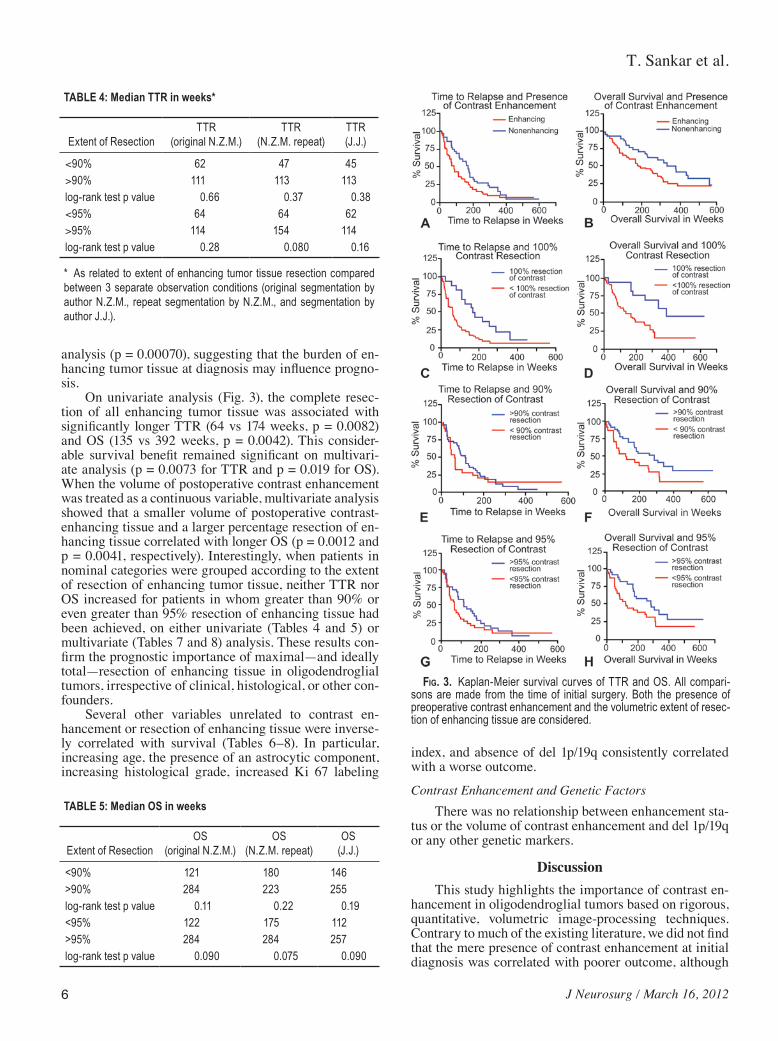
Figure 3 depicts Kaplan-Meier survival curves show-ing TTR and OS from the time of initial surgery as they relate to the presence of preoperative contrast enhance-ment and the volumetric extent of resection of enhancing tissue. Tables 6–8 summarize the results of multivariate analyses examining the survival impact of several clini-cal or biological variables in addition to contrast enhance-ment and resection.
On univariate analysis, the presence of contrast en-hancement at diagnosis correlated with shorter median TTR (83 vs 169 weeks, p = 0.040) and showed a strong trend toward correlation with shorter OS (196 vs 382 weeks, p = 0.063) (Fig. 3). However, these associations did not hold true on multivariate analysis (Table 6). In enhancing tumors, greater preoperative enhancing tumor volume correlated with shortened TTR on multivariate
TABLE 2: Clinical, pathological, and biological characteristics across patients with enhancing tumors only
Clinical Variable Value
total no. of pts 63mean age (yrs) 43.6 ± 12.9sex (no. of male pts) 37median TTR (wks) 83median OS (wks) 196cystic tumor 26astrocytic component 34malignant (WHO Grade III) 37MIB-1 >10% 34mean Ki 67 labeling index 8.76 ± 9.53early radiotherapy 38late radiotherapy 15early chemotherapy 26late chemotherapy 29LOH 1p/19q 101p deletion 1719q deletion 15mean 1p ratio 0.88 ± 0.16mean 19q ratio 0.91 ± 0.15mean % 1p loss 21.27 ± 30.75mean % 19q loss 17.92 ± 28.52EGFR amplification 6chromosome 7 polyploidy 21
TABLE 3: Preoperative and postoperative volumetric data for patients with enhancing tumors
Variable Value
enhancing WHO Grade II tumors 26/62 (41.94%)enhancing WHO Grade III tumors 37/38 (97.37%)tumors w/ astrocytic component & enhancement 34/51 (66.67%)mean preop contrast-enhancing vol (cm3) 25.02 ± 27.15mean postop contrast-enhancing vol (cm3) 2.36 ± 3.85mean % resection of enhancing vol 86.26 ± 19.49no. (%) of pts w/ 100% resection of enhancing vol 16/63 (25.40)no. (%) of pts w/ >95% resection of enhancing vol 29/63 (46.03)no. (%) of pts w/ >90% resection of enhancing vol 38/63 (60.32)
Fig. 2. Kaplan-Meier survival curves of TTR and OS demonstrat-ing repeated measurements to ensure intra- and interrater reliability. All comparisons are made from the time of initial surgery. Both the pres-ence of preoperative contrast enhancement and the volumetric extent of resection of enhancing tissue are considered.
T. Sankar et al.
6 J Neurosurg / March 16, 2012
analysis (p = 0.00070), suggesting that the burden of en-hancing tumor tissue at diagnosis may influence progno-sis.
On univariate analysis (Fig. 3), the complete resec-tion of all enhancing tumor tissue was associated with significantly longer TTR (64 vs 174 weeks, p = 0.0082) and OS (135 vs 392 weeks, p = 0.0042). This consider-able survival benefit remained significant on multivari-ate analysis (p = 0.0073 for TTR and p = 0.019 for OS). When the volume of postoperative contrast enhancement was treated as a continuous variable, multivariate analysis showed that a smaller volume of postoperative contrast-enhancing tissue and a larger percentage resection of en-hancing tissue correlated with longer OS (p = 0.0012 and p = 0.0041, respectively). Interestingly, when patients in nominal categories were grouped according to the extent of resection of enhancing tumor tissue, neither TTR nor OS increased for patients in whom greater than 90% or even greater than 95% resection of enhancing tissue had been achieved, on either univariate (Tables 4 and 5) or multivariate (Tables 7 and 8) analysis. These results con-firm the prognostic importance of maximal—and ideally total—resection of enhancing tissue in oligodendroglial tumors, irrespective of clinical, histological, or other con-founders.
Several other variables unrelated to contrast en-hancement or resection of enhancing tissue were inverse-ly correlated with survival (Tables 6–8). In particular, increasing age, the presence of an astrocytic component, increasing histological grade, increased Ki 67 labeling
index, and absence of del 1p/19q consistently correlated with a worse outcome.
Contrast Enhancement and Genetic FactorsThere was no relationship between enhancement sta-
tus or the volume of contrast enhancement and del 1p/19q or any other genetic markers.
DiscussionThis study highlights the importance of contrast en-
hancement in oligodendroglial tumors based on rigorous, quantitative, volumetric image-processing techniques. Contrary to much of the existing literature, we did not find that the mere presence of contrast enhancement at initial diagnosis was correlated with poorer outcome, although
TABLE 4: Median TTR in weeks*
Extent of ResectionTTR
(original N.Z.M.)TTR
(N.Z.M. repeat)TTR(J.J.)
<90% 62 47 45>90% 111 113 113log-rank test p value 0.66 0.37 0.38<95% 64 64 62>95% 114 154 114log-rank test p value 0.28 0.080 0.16
* As related to extent of enhancing tumor tissue resection compared between 3 separate observation conditions (original segmentation by author N.Z.M., repeat segmentation by N.Z.M., and segmentation by author J.J.).
TABLE 5: Median OS in weeks
Extent of ResectionOS
(original N.Z.M.)OS
(N.Z.M. repeat)OS
(J.J.)
<90% 121 180 146>90% 284 223 255log-rank test p value 0.11 0.22 0.19<95% 122 175 112>95% 284 284 257log-rank test p value 0.090 0.075 0.090
Fig. 3. Kaplan-Meier survival curves of TTR and OS. All compari-sons are made from the time of initial surgery. Both the presence of preoperative contrast enhancement and the volumetric extent of resec-tion of enhancing tissue are considered.

J Neurosurg / March 16, 2012
Contrast enhancement in oligodendroglial tumors
7
a larger initial burden of enhancing tissue was a negative prognostic factor. Our more robust findings relate to re-section of contrast-enhancing tumor tissue: In tumors that enhanced, increasing resection of the enhancing portion was independently associated with longer TTR and OS. Importantly, these improved outcomes appeared to be accounted for predominantly by cases in which enhanc-ing tumor tissue was resected completely. The presence of any residual enhancement postoperatively—even less than 5% of the original enhancing volume—left progno-sis unimproved. Furthermore, we found no relationship between the presence of del 1p/19q and the presence or volume of contrast enhancement.
The Meaning of Contrast EnhancementContrast enhancement of brain tumors on MRI is
thought to occur when the blood-brain barrier is disturbed by invasive tumor cells. The breach in the barrier allows the extravasation of gadolinium from within tumor ves-sels and its subsequent accumulation, which is detectable on T1-weighted sequences.3,12,37 In a limited number of studies in which biopsies have been obtained from frank-ly enhancing tumor regions, histological analysis has sug-gested that enhancing tissue may also represent regions of neovascularity38 or, perhaps, nodules of increased neo-plastic cell density.37 There is, therefore, some evidence that enhancing tissue represents intratumoral foci with a greater malignant potential, even in the absence of fea-tures of frank histological malignancy. It follows that a larger volume of such tissue may increase the risk of tu-mor recurrence, produce greater resistance to therapy, and eventually lead to a poor outcome. Our findings support this prediction. Although we did not specifically obtain and examine biopsies from contrast-enhancing tumor re-gions, our results suggest that the presence of enhancing tissue in an oligodendroglial tumor predicts a worse prog-nosis and that this effect is independent of—instead of rigidly tied to—histological grade. Predictably, we found
TABLE 6: Results of multivariate analysis examining the impact of the presence of contrast enhancement on TTR and OS across the entire patient group (n = 100)*
End Point & Variable p Value HR
TTR preop enhancement 0.49 — astrocytic component 0.010 1.802 WHO Grade III 0.023 1.952 cystic 0.0013 2.399OS preop enhancement 0.25 — age 0.0090 1.036 WHO Grade III <0.00010 4.626 del 1p/19q 0.00060 0.977
* Volumetric variables are shown in bold type. Variables unrelated to enhancement but significantly correlated with TTR or OS are also listed. Italic type indicates statistically significant p values.
TABLE 7: Results of the first multivariate analysis examining the impact of the volume of contrast enhancement on TTR and OS in patients with enhancing tumors (n = 63)*
End Point & Variable p Value HR
TTR preop enhancing volume 0.00070 >1.000
postop enhancing volume 0.17 —
% contrast resection 0.65 — Ki 67 labeling index 0.0019 1.052 del 1p/19q 0.00030 0.163OS preop enhancing volume 0.090 —
postop enhancing volume 0.0012 0.833
% contrast resection 0.0041 0.965 astrocytic component 0.029 2.463 MIB-1 >10% 0.0020 3.240 Ki 67 labeling index 0.0016 1.088 del 1p/19q <0.00010 0.955 early chemotherapy 0.023 0.371 late radiotherapy 0.0041 0.189
* Volumetric variables are identified in bold type, and were considered as continuous variables in this analysis. Italic text indicates statistically significant p values.
TABLE 8: Results of the second multivariate analysis examining the impact of the volume of contrast enhancement on TTR and OS in patients with enhancing tumors (n = 63)*
End Point & Variable p Value HR
TTR 100% resection of contrast 0.0073 0.239
95–99% resection of contrast 0.54 —
90–99% resection of contrast 0.31 — WHO Grade III 0.00010 4.458 del 1p/19q 0.00070 0.172 early chemotherapy 0.028 0.434OS 100% resection of contrast 0.019 0.487
95–99% resection of contrast 0.34 —
90–99% resection of contrast 0.23 — astrocytic component 0.0087 2.522 MIB-1 >10% 0.0064 2.794 del 1p/19q 0.00090 0.972
* Volumetric variables are identified in bold type, and were considered as nominal, categorical variables in this analysis. Variables unrelated to volume of enhancement but significantly correlated with TTR or OS are also listed. Italic type indicates statistically significant p values.

T. Sankar et al.
8 J Neurosurg / March 16, 2012
that the overall burden of enhancing tissue on initial diag-nosis was directly related to outcome.
Oligodendrogliomas and ResectionRelatively few studies have addressed—either in iso-
lation or as part of a larger study—the prognostic effect of extent of resection of oligodendroglial tumors.6,8,10,16,17,19,
25,28–30,34 The existing studies are all retrospective, and their conclusions are conflicting. Some have reported no statistically significant difference in OS between resec-tion and biopsy groups,10,17,34 while others have found that the extent of surgery correlated positively with increased survival either independently6,25,29,30 or when surgery was followed by radiotherapy.19 As with most studies exam-ining prognostic factors in brain tumors, these studies are weakened by methodological concerns such as small sample size, the combination of pediatric and adult popu-lations despite clearly better prognosis in the pediatric group,6,19,25,30,34 and a failure to distinguish between histo-logical variants of oligodendroglial tumors (that is, oligo-dendroglioma vs oligoastrocytoma).25 Furthermore, none of these studies used reproducible, quantitative volumetric methods. Instead, they relied on highly subjective—and inaccurate—estimates of the extent of tumor resection by individual neurosurgeons (for example, “gross-total re-section,” “subtotal resection,” or “biopsy”).
Although our study is also retrospective, it does over-come some of the above methodological shortcomings. First, our study population was a sizable, single-institu-tion cohort of patients who all underwent similar stan-dard imaging and therapeutic regimens and who were fol-lowed up for at least 5 years. No pediatric patients were included. Second, we used a standardized, semiautomat-ed method to generate quantitative pre- and postoperative volumetric data from MRI studies. We rigorously ensured objectivity by blinding observers to patient identity and demonstrated good intrarater reliability with our volume measurement methodology. Third, we limited volumetric analysis to enhancing tumors only. Consequently, there were fewer problems dealing with irregular tumor bound-aries or peritumoral edema, which can produce inaccu-racies in measurements of tumor volumes. Our focused results apply only to a subset of oligodendroglial tumors and hence are more reliable than previously reported find-ings. To our knowledge, no quantitative volumetric study of resection limited to oligodendrogliomas encompasses both enhancing and nonenhancing tumors. Undertaking such a study would require a consistent and automated method of reliably detecting infiltrative tumor boundar-ies, possibly by using metabolic imaging (for example, MR spectroscopy)9,27 or MRI texture analysis techniques.1
Contrast Enhancement, del 1p/19q, and Implications for Resection
We found no relationship between the presence or volume of contrast enhancement and characteristic ge-netic markers of oligodendroglial tumors, particularly del 1p/19q. Some previous studies have looked at imag-ing correlates of molecular or genetic features in oligo-dendrogliomas.2,15,23 Megyesi et al.23 found that del 1p/19q was associated with an indistinct tumor boundary on T1-
weighted MR images, the presence of intratumoral cal-cification, and paramagnetic susceptibility effect. Sim-i larly, Jenkinson et al.15 found that an indistinct tumor border and calcification correlated with tumors showing del 1p/19q, but they found no association with paramag-netic susceptibility effect. In both studies, the presence of contrast enhancement was more likely to be associ-ated with a higher tumor grade, but neither study found that contrast enhancement was correlated with del 1p/19q. These results are consistent with our data. Admittedly, our study focused only on contrast enhancement, and we did not examine MR images for other imaging features that might suggest the presence of specific genetic sig-natures, in large part because we consciously wanted to avoid subjective criteria in our analyses. Nevertheless, recently developed quantitative imaging techniques may allow completely noninvasive detection of del 1p/19q on routine MRI studies with good sensitivity and specificity, potentially obviating the need for molecular diagnostic analysis of actual tumor tissue.2
Whether detected in tumor tissue specimens or by noninvasive techniques, the presence of del 1p/19q raises an important clinical dilemma in the context of our own findings. It is known that del 1p/19q is a marker of a rela-tively benign natural history14,33 as well as an improved response to chemotherapy.4,5 Consequently, avoiding or delaying invasive treatments such as aggressive resective surgery, which have the potential to cause neurological morbidity, appears to be an attractive option in patients with del 1p/19q. This is particularly true in young patients with low-grade oligodendrogliomas harboring the genetic signature. Even in older patients or in those with high-grade tumors, it has been argued that del 1p/19q should prompt the early initiation of chemotherapy as the sole treatment modality. In a nonrandomized study of patients with newly diagnosed anaplastic oligodendroglioma, Mik kel sen et al.24 concluded that patients harboring del 1p/19q can be treated safely and efficaciously with temo-zolomide alone.
Given this favorable response to up-front chemother-apy, is there a role for resective surgery? Should surgical goals change for enhancing tumors? To date, data have been insufficient to answer these questions. In the study by Mikkelsen et al.,24 only 11 (22.9%) of 48 patients un-derwent gross-total resection, and the presence or extent of contrast enhancement was not reported. We believe that 3 key points emerge from our data. First, contrast en-hancement is unrelated to del 1p/19q, suggesting that this genetic aberration does not affect tumor vasculature but is instead an intrinsic feature of the tumor cells affecting their response to therapy and/or growth. Second, the pres-ence and volume of contrast enhancement are indepen-dent negative prognostic factors. Third, maximal and, if possible, total resection of contrast-enhancing tissue sig-nificantly improves prognosis. Clearly, our data support a more aggressive surgical approach to oligodendroglial tumors. Specifically, an attempt should be made to resect all enhancing tissue if safely possible, because doing so is likely to be beneficial regardless of the genetic features of the tumor. To that end, intraoperative or immediate post-operative gadolinium-enhanced MRI, with a clear intent to return to the operative site for additional debulking of

J Neurosurg / March 16, 2012
Contrast enhancement in oligodendroglial tumors
9
residual enhancing tissue if necessary, may be useful ad-juncts. Neuronavigation and awake craniotomy or alter-native mapping strategies also may be of use in maximiz-ing removal of enhancing tissue in or near eloquent areas.
Study LimitationsOur study is not without some weaknesses. As men-
tioned, it is retrospective, and cannot provide Class I evi-dence to support our conclusions, despite our rigorous volumetric and statistical methods. In addition, no single standardized therapeutic protocol was used across all patients. Consequently, it is possible that over the rela-tively long follow-up period there were changes in over-all treatment patterns (for example, the widespread use of temozolomide in patients treated since 2002). This issue is partially offset by our single-institution patient population managed by a relatively unchanged treatment team over the course of the study. There could also be some concern about the representativeness of the popu-lation given the large number of patients that had to be excluded due to inadequate imaging, although there is no reason to think that such patients would be qualitative- ly different from their counterparts with complete im-aging sets. Furthermore, our genetic/molecular analyses did not include an assessment of promoter methylation of the methyl-guanine methyl transferase (MGMT) gene, which, like del 1p/19q, may be associated with improved prognosis and responsiveness to chemotherapy with al-kylating agents.26,36
Any study that uses human observers to generate volumetric data related to tumor size and extent of re-section is open to criticisms of bias related to subjective judgments made during the segmentation process. We attempted to validate our volumetric technique by as-sessing intra- and interrater reliability, which were both found to be satisfactory. Ideally, studies such as ours would employ fully automated tumor segmentation tech-niques free of the possibility of human bias. While some “fuzzy” algorithms to accomplish this goal have recently been developed, most fully automated tumor classifiers are plagued by systematic errors and significant problems with computational economy.13 Until these issues are re-solved, semiautomated techniques such as we have em-ployed must suffice as an acceptable alternative.
ConclusionsQuantitative MRI volumetric analysis strongly sug-
gests that among enhancing oligodendroglial tumors, the burden of initial contrast-enhancing tumor tissue and the degree of resection of this tissue are related to outcome. These relationships remain significant irrespective of histological grade or molecular genetic classification. What ever the biological significance of contrast-enhanc-ing tissue is, its complete resection independently and dramatically improves survival. Consequently, resective surgery remains vitally important in the management of patients with these tumors. If possible, surgical planning and intraoperative adjuncts should further the goal of complete resection before adjuvant therapies are initi-ated.
Disclosure
The authors report no conflict of interest concerning the mate-rials or methods used in this study or the findings specified in this paper.
Author contributions to the study and manuscript prepara-tion include the following. Conception and design: Preul, Sankar. Ac quisition of data: Sankar, Moore, Johnson. Analysis and inter-pretation of data: Sankar, Moore. Drafting the article: Preul, Sankar, Moore, Scheck, Smith. Critically revising the article: Preul, Sankar. Sta tistical analysis: Sankar, Moore. Administrative/technical/mate-rial support: Preul, Ashby, Shapiro, Smith, Spetzler. Study supervi-sion: Preul, Sankar.
References
1. Assefa D, Keller H, Ménard C, Laperriere N, Ferrari RJ, Yeung I: Robust texture features for response monitoring of glioblastoma multiforme on T1-weighted and T2-FLAIR MR images: a preliminary investigation in terms of identification and segmentation. Med Phys 37:1722–1736, 2010
2. Brown R, Zlatescu M, Sijben A, Roldan G, Easaw J, Forsyth P, et al: The use of magnetic resonance imaging to noninvasively detect genetic signatures in oligodendroglioma. Clin Cancer Res 14:2357–2362, 2008
3. Byrne TN: Imaging of gliomas. Semin Oncol 21:162–171, 1994
4. Cairncross G, Berkey B, Shaw E, Jenkins R, Scheithauer B, Brachman D, et al: Phase III trial of chemotherapy plus radio-therapy compared with radiotherapy alone for pure and mixed anaplastic oligodendroglioma: Intergroup Radiation Therapy Oncology Group Trial 9402. J Clin Oncol 24:2707–2714, 2006
5. Cairncross JG, Ueki K, Zlatescu MC, Lisle DK, Finkelstein DM, Hammond RR, et al: Specific genetic predictors of che-motherapeutic response and survival in patients with anaplas-tic oligodendrogliomas. J Natl Cancer Inst 90:1473–1479, 1998
6. Celli P, Nofrone I, Palma L, Cantore G, Fortuna A: Cerebral oligodendroglioma: prognostic factors and life history. Neu-rosurgery 35:1018–1035, 1994
7. Chaichana KL, McGirt MJ, Laterra J, Olivi A, Quiñones-Hi-nojosa A: Recurrence and malignant degeneration after resec-tion of adult hemispheric low-grade gliomas. Clinical article. J Neurosurg 112:10–17, 2010
8. Chaichana KL, McGirt MJ, Niranjan A, Olivi A, Burger PC, Quinones-Hinojosa A: Prognostic significance of contrast-enhancing low-grade gliomas in adults and a review of the lit erature. Neurol Res 31:931–939, 2009
9. Cohen BA, Knopp EA, Rusinek H, Babb JS, Zagzag D, Gonen O: Assessing global invasion of newly diagnosed glial tumors with whole-brain proton MR spectroscopy. AJNR Am J Neu roradiol 26:2170–2177, 2005
10. Daumas-Duport C, Tucker ML, Kolles H, Cervera P, Beuvon F, Varlet P, et al: Oligodendrogliomas. Part II: A new grading system based on morphological and imaging criteria. J Neu-rooncol 34:61–78, 1997
11. Giannini C, Scheithauer BW, Weaver AL, Burger PC, Kros JM, Mork S, et al: Oligodendrogliomas: reproducibility and prognostic value of histologic diagnosis and grading. J Neu-ropathol Exp Neurol 60:248–262, 2001
12. Ginsberg LE, Fuller GN, Hashmi M, Leeds NE, Schomer DF: The significance of lack of MR contrast enhancement of su-pratentorial brain tumors in adults: histopathological evalua-tion of a series. Surg Neurol 49:436–440, 1998
13. Harati V, Khayati R, Farzan A: Fully automated tumor seg-mentation based on improved fuzzy connectedness algorithm in brain MR images. Comput Biol Med 41:483–492, 2011
14. Jenkins RB, Blair H, Ballman KV, Giannini C, Arusell RM, Law M, et al: A t(1;19)(q10;p10) mediates the combined dele-

T. Sankar et al.
10 J Neurosurg / March 16, 2012
tions of 1p and 19q and predicts a better prognosis of patients with oligodendroglioma. Cancer Res 66:9852–9861, 2006
15. Jenkinson MD, du Plessis DG, Smith TS, Joyce KA, Warnke PC, Walker C: Histological growth patterns and genotype in oligodendroglial tumours: correlation with MRI features. Brain 129:1884–1891, 2006
16. Keles GE, Lamborn KR, Berger MS: Low-grade hemispheric gliomas in adults: a critical review of extent of resection as a factor influencing outcome. J Neurosurg 95:735–745, 2001
17. Kros JM, Pieterman H, van Eden CG, Avezaat CJ: Oligoden-droglioma: the Rotterdam-Dijkzigt experience. Neurosur-gery 34:959–966, 1994
18. Lee YY, Van Tassel P: Intracranial oligodendrogliomas: im-aging findings in 35 untreated cases. AJR Am J Roentgenol 152:361–369, 1989
19. Lindegaard KF, Mørk SJ, Eide GE, Halvorsen TB, Hatlevoll R, Solgaard T, et al: Statistical analysis of clinicopathological features, radiotherapy, and survival in 170 cases of oligoden-droglioma. J Neurosurg 67:224–230, 1987
20. Louis DN, Ohgaki H, Wiestler OD, Cavenee WK (eds): World Health Organization Classification of Tumours of the Central Nervous System, ed 4. Lyon: IARC, 2007
21. Lwin Z, Gan HK, Mason WP: Low-grade oligodendroglioma: current treatments and future hopes. Expert Rev Anticancer Ther 9:1651–1661, 2009
22. Margain D, Peretti-Viton P, Perez-Castillo AM, Martini P, Salamon G: Oligodendrogliomas. J Neuroradiol 18:153–160, 1991
23. Megyesi JF, Kachur E, Lee DH, Zlatescu MC, Betensky RA, Forsyth PA, et al: Imaging correlates of molecular signatures in oligodendrogliomas. Clin Cancer Res 10:4303–4306, 2004
24. Mikkelsen T, Doyle T, Anderson J, Margolis J, Paleologos N, Gutierrez J, et al: Temozolomide single-agent chemotherapy for newly diagnosed anaplastic oligodendroglioma. J Neurooncol 92:57–63, 2009
25. Mørk SJ, Lindegaard KF, Halvorsen TB, Lehmann EH, Sol-gaard T, Hatlevoll R, et al: Oligodendroglioma: incidence and biological behavior in a defined population. J Neurosurg 63: 881–889, 1985
26. Ney DE, Lassman AB: Molecular profiling of oligodendrogli-omas: impact on prognosis, treatment, and future directions. Curr Oncol Rep 11:62–67, 2009
27. Pirzkall A, Li X, Oh J, Chang S, Berger MS, Larson DA, et al: 3D MRSI for resected high-grade gliomas before RT: tumor extent according to metabolic activity in relation to MRI. Int J Radiat Oncol Biol Phys 59:126–137, 2004
28. Scerrati M, Roselli R, Iacoangeli M, Pompucci A, Rossi GF:
Prognostic factors in low grade (WHO grade II) gliomas of the cerebral hemispheres: the role of surgery. J Neurol Neu-rosurg Psychiatry 61:291–296, 1996
29. Schiffer D, Dutto A, Cavalla P, Bosone I, Chiò A, Villani R, et al: Prognostic factors in oligodendroglioma. Can J Neurol Sci 24:313–319, 1997
30. Shaw EG, Scheithauer BW, O’Fallon JR, Tazelaar HD, Da-vis DH: Oligodendrogliomas: the Mayo Clinic experience. J Neurosurg 76:428–434, 1992
31. Shrout PE, Fleiss JL: Intraclass correlations: uses in assessing rater reliability. Psychol Bull 86:420–428, 1979
32. Sled JG, Zijdenbos AP, Evans AC: A nonparametric method for automatic correction of intensity nonuniformity in MRI data. IEEE Trans Med Imaging 17:87–97, 1998
33. Smith JS, Perry A, Borell TJ, Lee HK, O’Fallon J, Hosek SM, et al: Alterations of chromosome arms 1p and 19q as predic-tors of survival in oligodendrogliomas, astrocytomas, and mixed oligoastrocytomas. J Clin Oncol 18:636–645, 2000
34. Sun ZM, Genka S, Shitara N, Akanuma A, Takakura K: Fac-tors possibly influencing the prognosis of oligodendroglioma. Neurosurgery 22:886–891, 1988
35. Talairach J, Tournoux P: Coplanar Stereotaxic Atlas of the Human Brain. Stuttgart: Thieme, 1988
36. van den Bent MJ, Dubbink HJ, Sanson M, van der Lee-Haar-loo CR, Hegi M, Jeuken JW, et al: MGMT promoter methyla-tion is prognostic but not predictive for outcome to adjuvant PCV chemotherapy in anaplastic oligodendroglial tumors: a report from EORTC Brain Tumor Group Study 26951. J Clin Oncol 27:5881–5886, 2009
37. White ML, Zhang Y, Kirby P, Ryken TC: Can tumor contrast enhancement be used as a criterion for differentiating tumor grades of oligodendrogliomas? AJNR Am J Neuroradiol 26: 784–790, 2005
38. Zagzag D, Goldenberg M, Brem S: Angiogenesis and blood-brain barrier breakdown modulate CT contrast enhancement: an experimental study in a rabbit brain-tumor model. AJR Am J Roentgenol 153:141–146, 1989
Manuscript submitted December 2, 2010.Accepted February 8, 2012.Please include this information when citing this paper: pub-
lished online March 16, 2012; DOI: 10.3171/2012.2.JNS102032.Address correspondence to: Mark C. Preul, M.D., c/o Neurosci-
ence Publications, Barrow Neurological Institute, St. Joseph’s Hos-pital and Medical Center, 350 West Thomas Road, Phoenix, Arizona 85013. email: [email protected].