Embed Size (px)
Citation preview
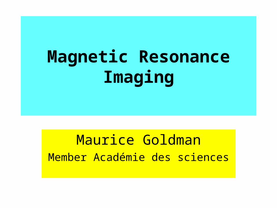
Magnetic Resonance Imaging
Maurice GoldmanMember Académie des sciences
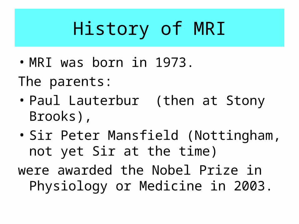
History of MRI
• MRI was born in 1973.
The parents:
• Paul Lauterbur (then at Stony Brooks),
• Sir Peter Mansfield (Nottingham, not yet Sir at the time)
were awarded the Nobel Prize in Physiology or Medicine in 2003.
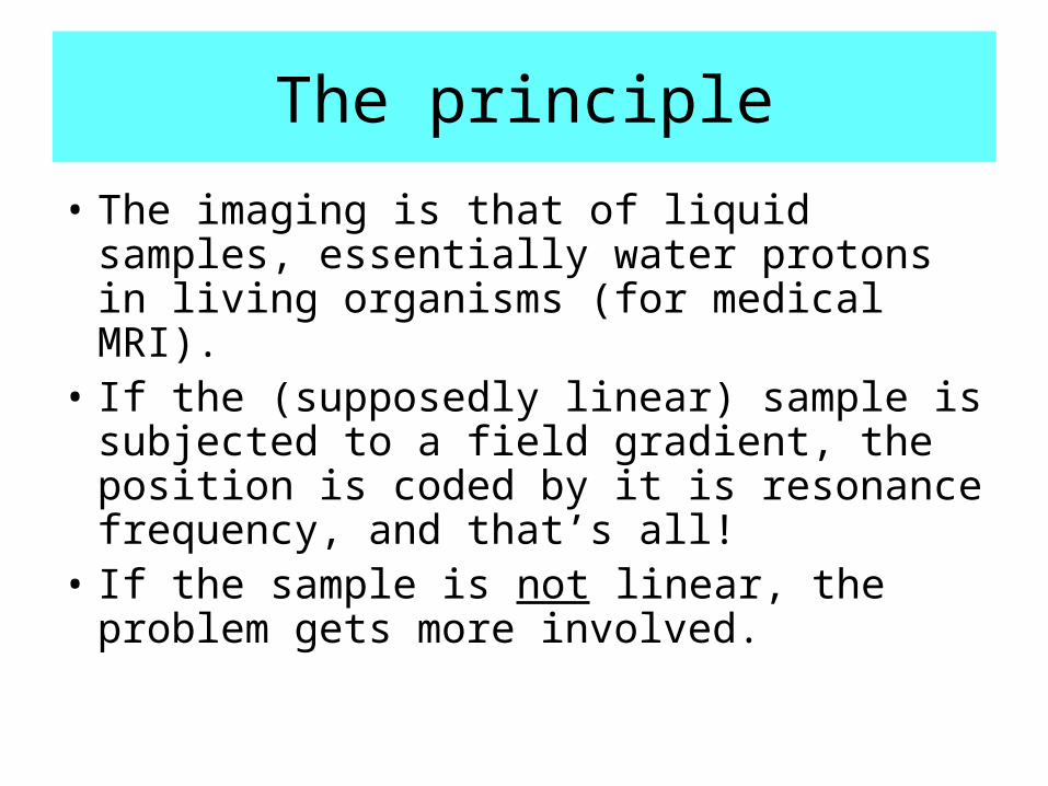
The principle
• The imaging is that of liquid samples, essentially water protons in living organisms (for medical MRI).
• If the (supposedly linear) sample is subjected to a field gradient, the position is coded by it is resonance frequency, and that’s all!
• If the sample is not linear, the problem gets more involved.
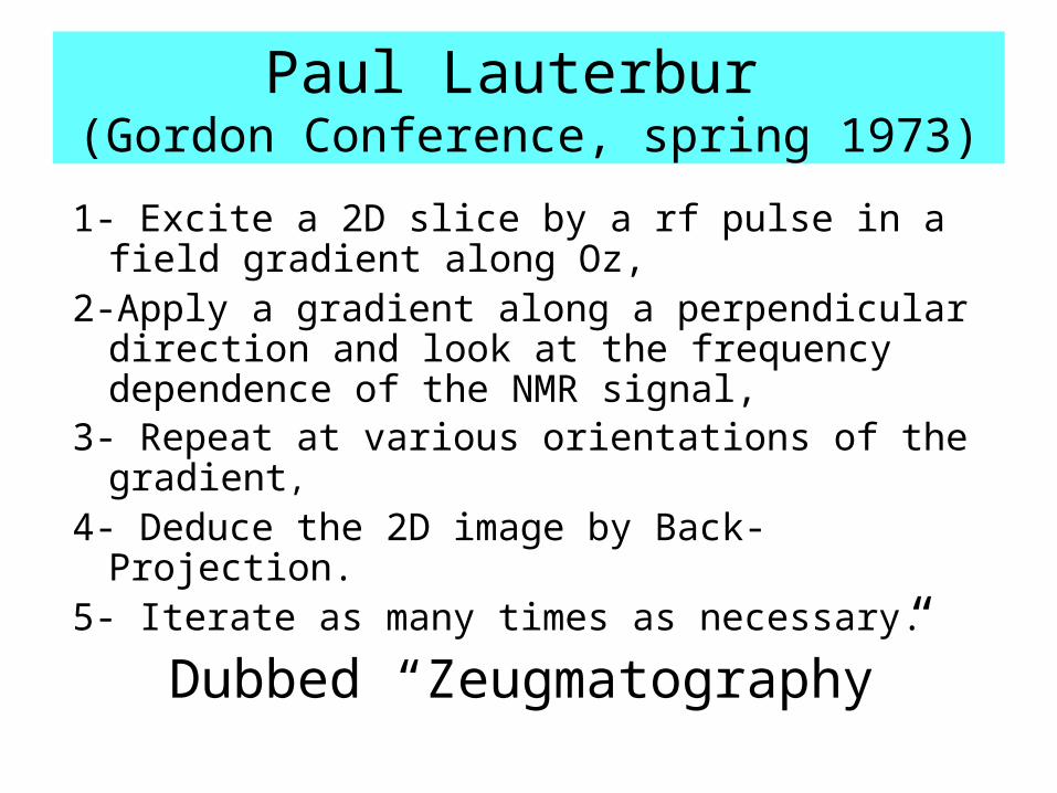
Paul Lauterbur (Gordon Conference, spring 1973)
1- Excite a 2D slice by a rf pulse in a field gradient along Oz,
2-Apply a gradient along a perpendicular direction and look at the frequency dependence of the NMR signal,
3- Repeat at various orientations of the gradient,4- Deduce the 2D image by Back-Projection.5- Iterate as many times as necessary.
Dubbed “Zeugmatography”
G
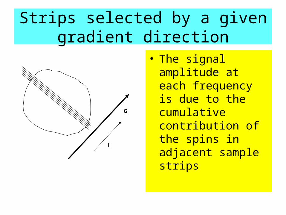
Strips selected by a given gradient direction
• The signal amplitude at each frequency is due to the cumulative contribution of the spins in adjacent sample strips
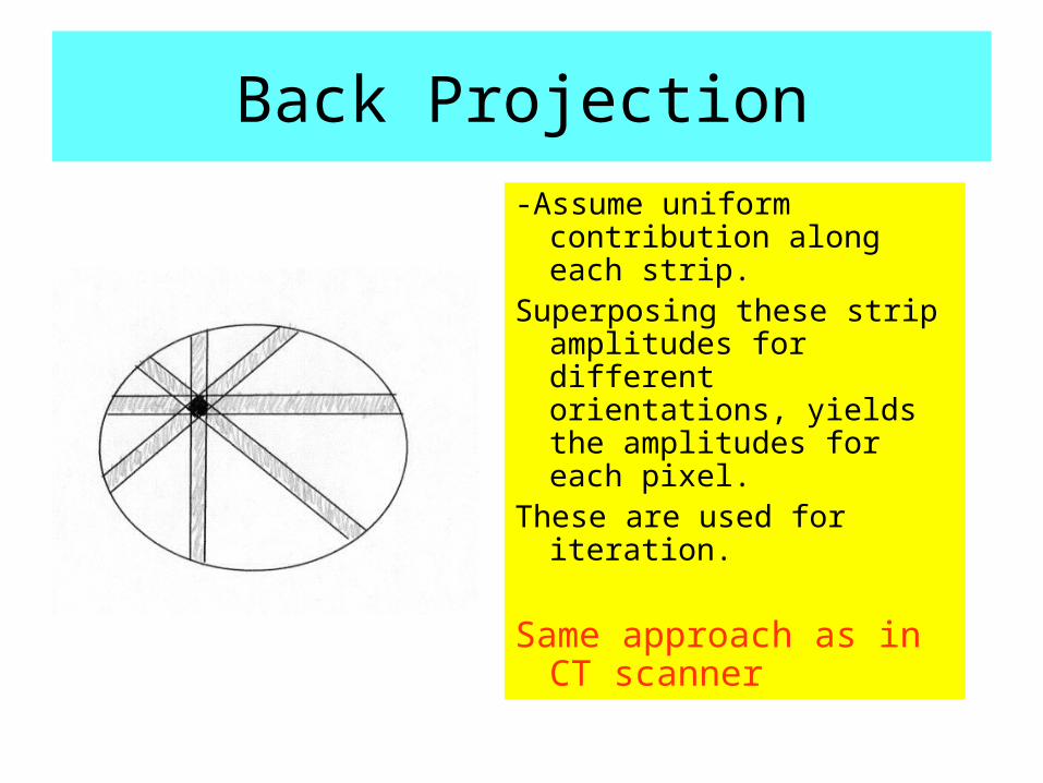
Back Projection
-Assume uniform contribution along each strip.
Superposing these strip amplitudes for different orientations, yields the amplitudes for each pixel.
These are used for iteration.
Same approach as in CT scanner
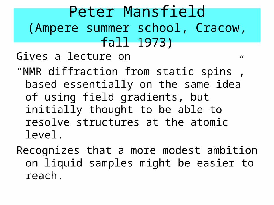
Peter Mansfield(Ampere summer school, Cracow, fall 1973)
Gives a lecture on
“NMR diffraction from static spins”, based essentially on the same idea of using field gradients, but initially thought to be able to resolve structures at the atomic level.
Recognizes that a more modest ambition on liquid samples might be easier to reach.
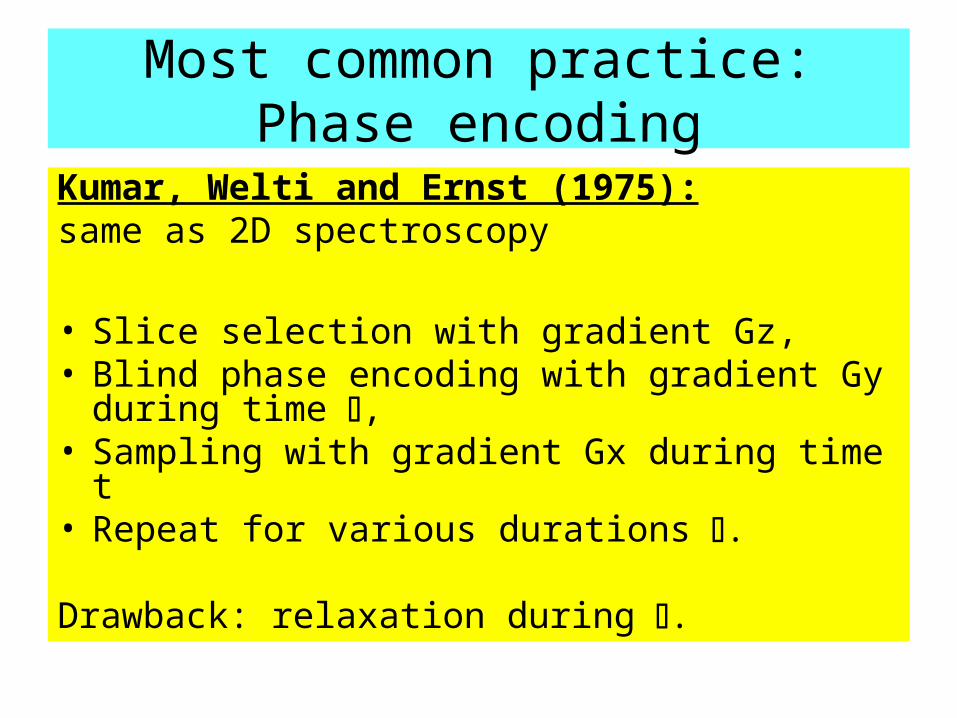
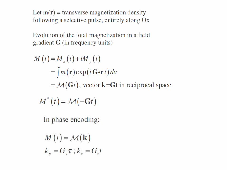
Most common practice:Phase encoding
Kumar, Welti and Ernst (1975):same as 2D spectroscopy
• Slice selection with gradient Gz,• Blind phase encoding with gradient Gy during
time ,• Sampling with gradient Gx during time t• Repeat for various durations .
Drawback: relaxation during .
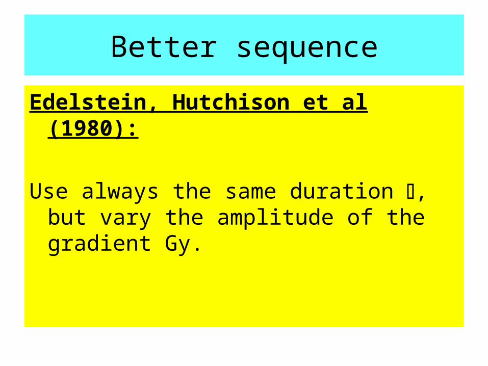
Better sequence
Edelstein, Hutchison et al (1980):
Use always the same duration , but vary the amplitude of the gradient Gy.
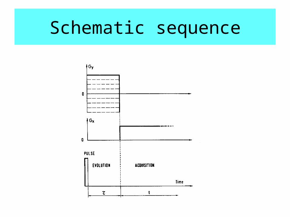
Schematic sequence
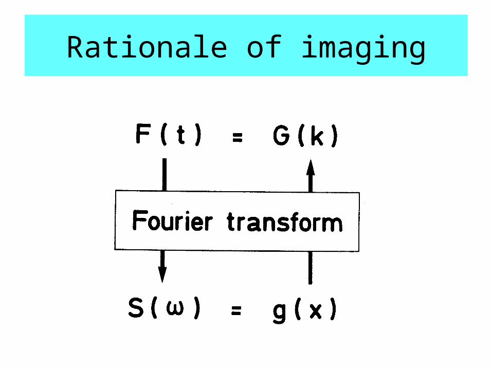
Rationale of imaging
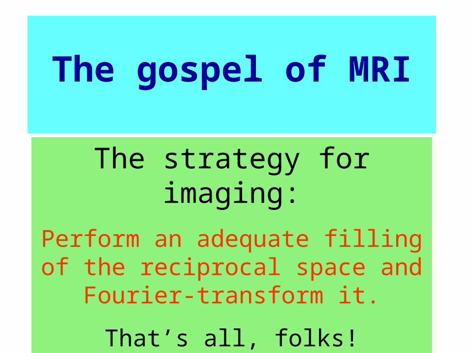
The strategy for imaging:
Perform an adequate filling of the reciprocal space and Fourier-
transform it.
That’s all, folks!
The gospel of MRI
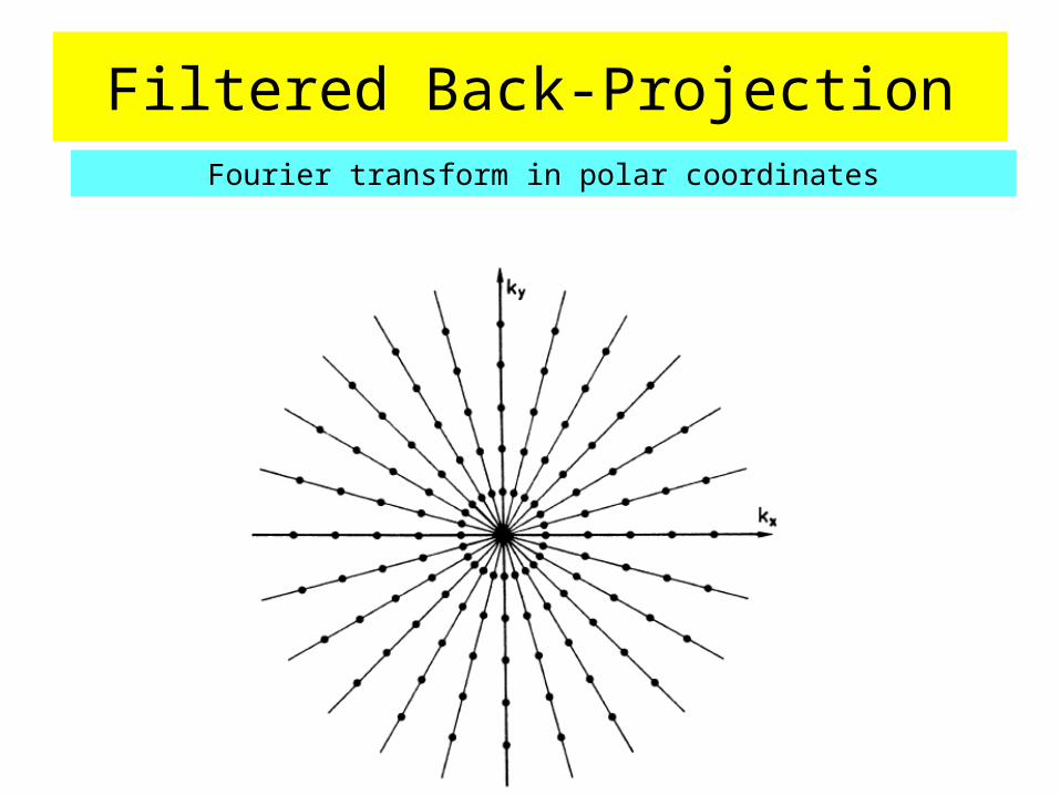
Filtered Back-ProjectionFourier transform in polar coordinates
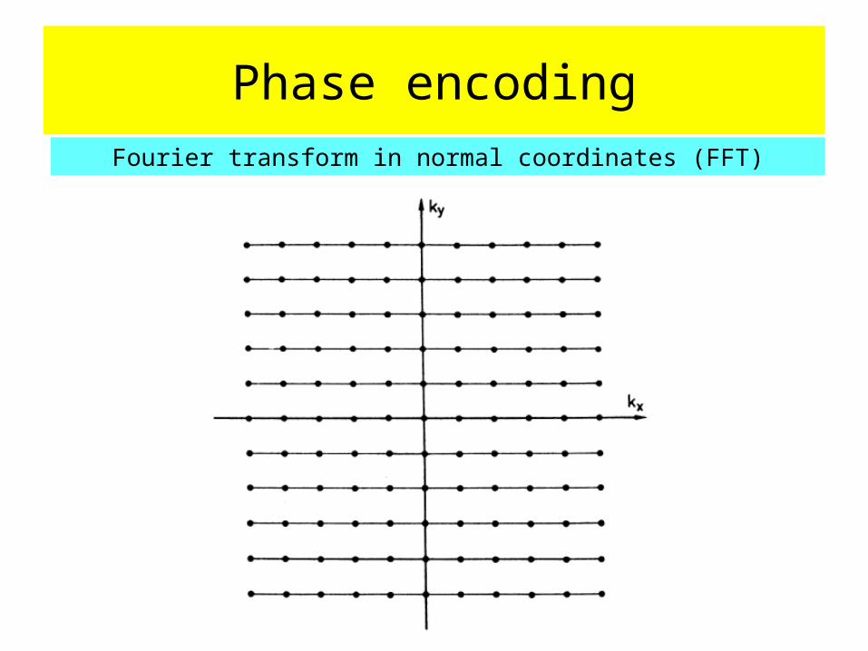
Phase encodingFourier transform in normal coordinates (FFT)
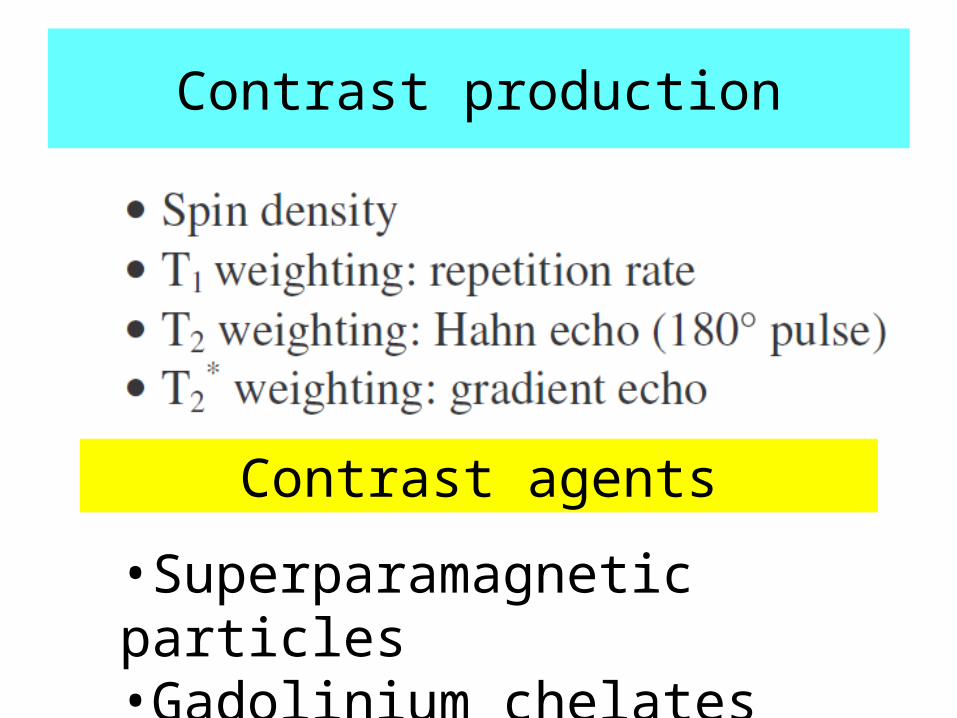
Contrast production
Contrast agents
•Superparamagnetic particles•Gadolinium chelates
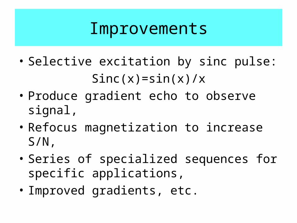
Improvements
• Selective excitation by sinc pulse:
Sinc(x)=sin(x)/x
• Produce gradient echo to observe signal,
• Refocus magnetization to increase S/N,
• Series of specialized sequences for specific applications,
• Improved gradients, etc.
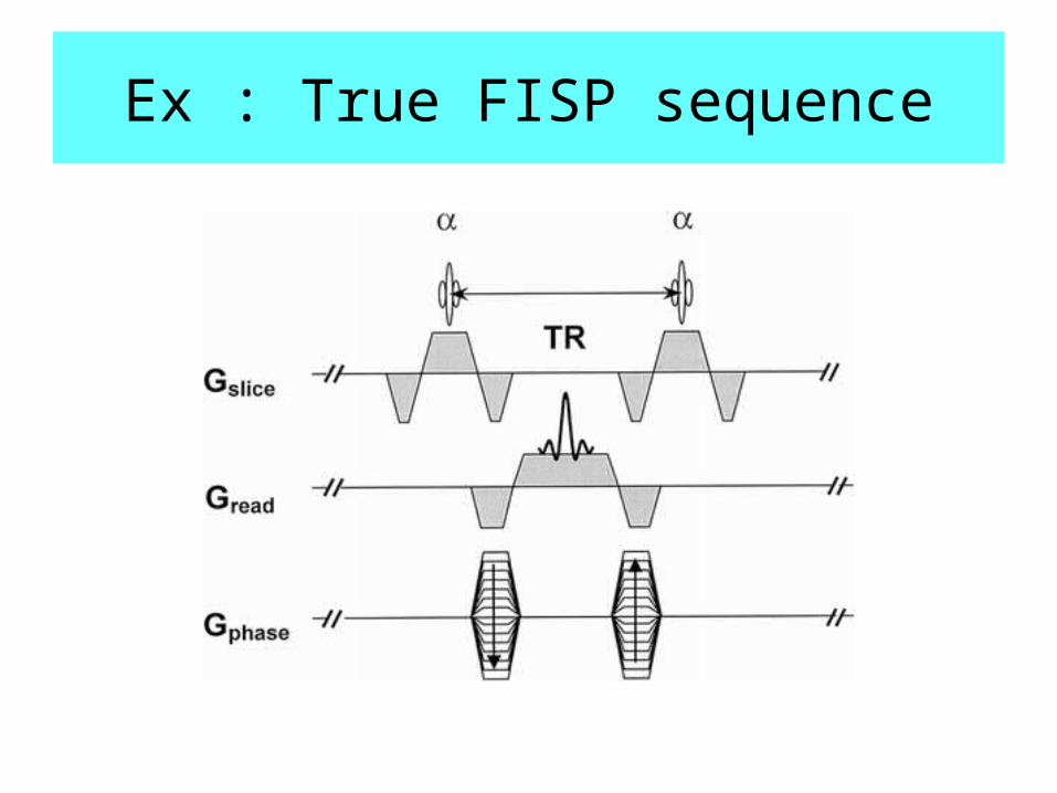
Ex : True FISP sequence
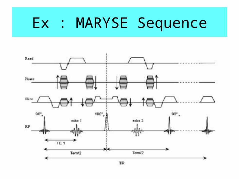
Ex : MARYSE Sequence
Other measurements
• Flow :
phase imaging or presaturation of fov.
• Diffusion tensor:
bouncing on cell walls modifies apparent diffusion coefficient
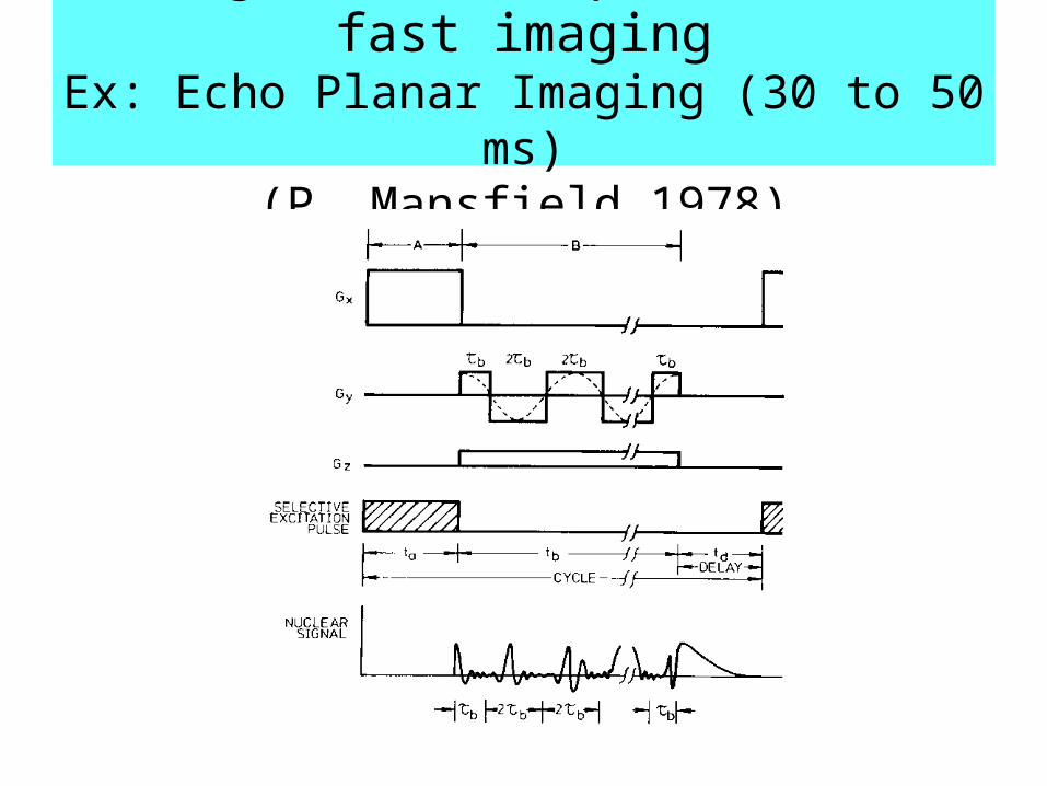
Single-shot sequences for fast imagingEx: Echo Planar Imaging (30 to 50 ms)
(P. Mansfield 1978)
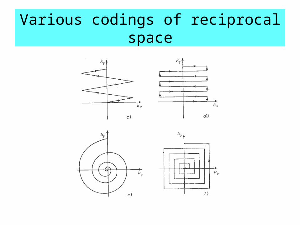
Various codings of reciprocal space
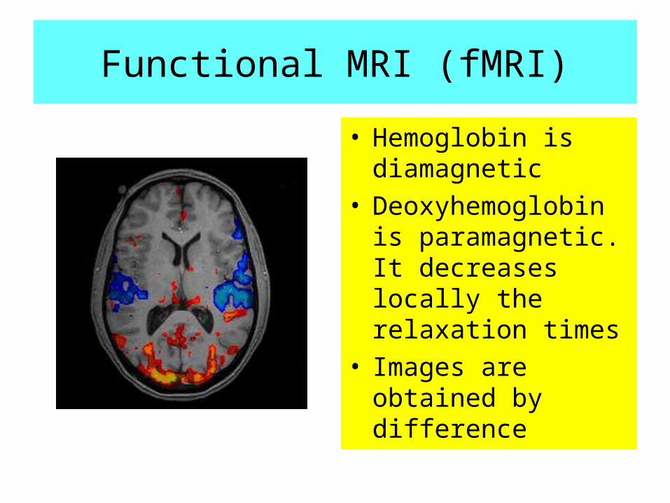
Functional MRI (fMRI)
• Hemoglobin is diamagnetic
• Deoxyhemoglobin is paramagnetic. It decreases locally the relaxation times
• Images are obtained by difference
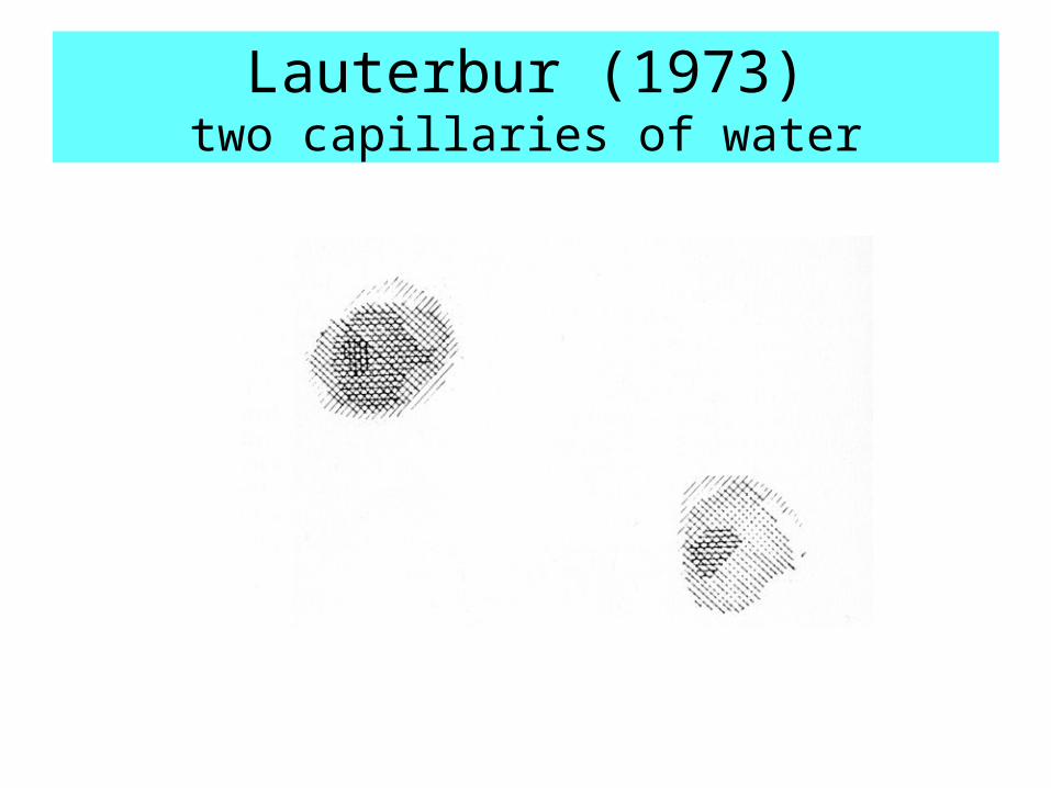
Lauterbur (1973)two capillaries of water
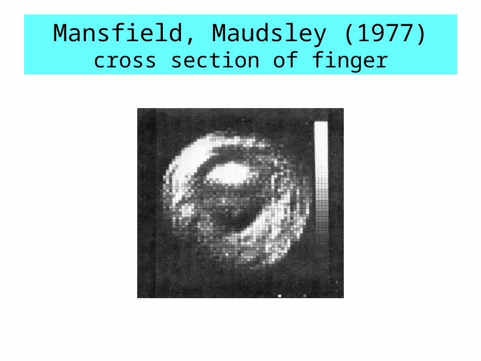
Mansfield, Maudsley (1977)cross section of finger
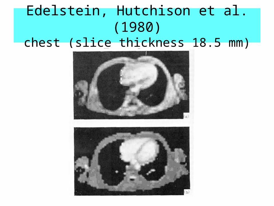
Edelstein, Hutchison et al. (1980)chest (slice thickness 18.5 mm)
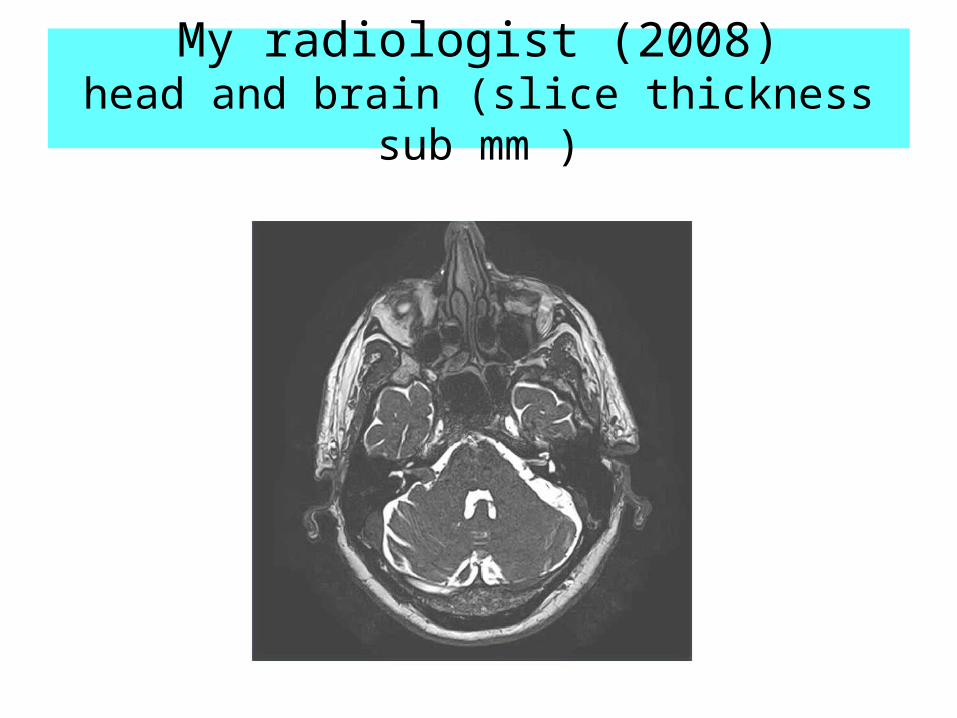
My radiologist (2008)head and brain (slice thickness sub mm )


![Académie Goncourt · Oscar Wilde. [1] (1986) avec Académie Goncourt comme Éditeur scientifique Cervantès [1] (1986) avec Académie Goncourt comme Éditeur scientifique Pascal](https://img.pdfslide.us/doc/110x75/60b352b3a9d4ba28e70e44c8/acadmie-goncourt-oscar-wilde-1-1986-avec-acadmie-goncourt-comme-diteur.jpg)