Embed Size (px)
Citation preview
Research ArticleMagnetic-Guided Capsule Endoscopy in the Diagnosis ofGastrointestinal Diseases in Minors
Yuting Qian , Tingting Bai, Juanjuan Li, Yi Zang, Tong Li, Mingping Xie, Qi Wang,Lifu Wang , and Ruizhe Shen
Department of Gastroenterology, Ruijin Hospital Affiliated to Shanghai Jiaotong University School of Medicine, 197 SecondRuijin Road, Shanghai 200025, China
Correspondence should be addressed to Lifu Wang; [email protected] and Ruizhe Shen; [email protected]
Received 8 April 2018; Revised 3 August 2018; Accepted 23 August 2018; Published 18 September 2018
Academic Editor: Hauke S. Heinzow
Copyright © 2018 Yuting Qian et al. This is an open access article distributed under the Creative Commons Attribution License,which permits unrestricted use, distribution, and reproduction in any medium, provided the original work is properly cited.
Objective. This study aimed at investigating the clinical value of magnetic-guided capsule endoscopy (MGCE) in the diagnosis ofgastrointestinal diseases in minors. Methods. Eighty-four minor patients hospitalized in the pediatric department at RuijinHospital between June 2015 and January 2018 were enrolled for this study. Following bowel preparation, all patients underwentMGCE. The feasibility, safety, diagnostic yield, and sensitivity of MGCE were analyzed. Patients were followed up for more than2 weeks. Results. The main indications for MGCE in minors were Crohn’s disease, gastrointestinal bleeding, and abdominalpain. The main causes of gastric disease were gastric inflammatory hyperplasia, exudative gastritis, and polyps. The mostcommon small bowel diseases in minors were Crohn’s disease, Henoch-Schonlein purpura, and polyps. The diagnostic yield inthe stomach and small intestine was 13.1% and 28.6%, respectively, and the sensitivity was 100% and 96.0%, respectively. Noadverse events occurred. Conclusion. MGCE is a safe, effective, and well-tolerated procedure with good sensitivity and has apotential clinic value for the diagnosis of gastrointestinal diseases in minors.
1. Introduction
Minor is a special group of patients, whose symptoms anddiseases are often different from those of adults, especiallyfor gastrointestinal diseases. In the past 30 years, esophago-gastroduodenoscopy (EGD) has been considered as the goldstandard for gastric disease investigation but the discomfortlimits its use in minors. The appearance of capsule endos-copy (CE) has become a milestone in small bowel examina-tion [1–3]. In 2009, indications of CE have been broadenedto patients more than 2 years old by the United States Foodand Drug Administration (FDA) and CE is proven safe topediatrics [4–8]. With the continuous innovation of CE tech-nology, magnetic-guided capsule endoscopy (MGCE) showsits advantages in gastric disease investigation [9–12]. How-ever, there is a lack of the clinical value of magnetic-guidedcapsule endoscopy in pediatric gastrointestinal diseases.Until now, the decision of the clinical value of MGCE is basedon adult research and empirical data based on CE [5, 13, 14].
Therefore, this prospective study aims at investigating thediagnostic value of magnetic-guided capsule endoscopy forminors’ gastrointestinal diseases.
2. Materials and Methods
2.1. Study Cohorts. This study recruited patients from 6 to 18years old hospitalized in the pediatric department from June2015 to January 2018 at Ruijin Hospital Affiliated toShanghai Jiaotong University, and the study was approvedby the Ruijin Hospital Ethics Committee. Inclusion criteriawere IBDU (inflammatory dowel disease unclassified), sus-pected Crohn’s disease, hematemesis, hematochezia, melena,anemia, abdominal pain, diarrhea, abdominal distension, ele-vated tumor markers, constipation, and dyspepsia. Patientswere divided into five groups by complains. The mainexclusion criteria were metal implants (cardiac pacemaker,metal valves, metal prosthesis, etc.), impairment of gastroin-testinal movement, suspected or diagnosed gastrointestinal
HindawiGastroenterology Research and PracticeVolume 2018, Article ID 4248792, 8 pageshttps://doi.org/10.1155/2018/4248792
obstruction, and history of abdominal surgery. All patientsand their guardians were informed of the related processand potential risks duringMGCE examination, and all signedconsents.
2.2. Magnetic-Guided Capsule Endoscopy System. TheNaviCam™ magnetic-guided capsule endoscopy system(Shanghai ANKON Medical Technology Co. Ltd.) wasapplied in this study. The components included capsulerobot, magnetic-guided capsule endoscopy examinationbed, translation and rotary table, magnet, console, portablerecorder, capsule locator, and ESNavi software. The cap-sule robot was a capsule-like equipment with the size of12mm× 28mm and took 2 frames per second. The obser-vation view was 140± 10°, the working temperature was20–40°C, and the working time was >8h. The captureddata of the capsule can be instantly transmitted by thedata line to the operating table for real-time observation.The activity of the capsule was controlled by the C-armmagnetic field system.
2.3. Preparations. CTE or MRE examination was performedbefore MGCE examination to exclude intestinal obstruction.Subjects were required to carry out low-residue diet 3 daysbefore the examination. At 8 pm the day before MGCEexamination, patients drank polyethylene glycol electrolytepowder (Hengkangzhengqing, Jiangxi Hengkang Pharma-ceutical Co. Ltd.) for intestinal cleaning. Patients youngerthan 10 years old or less than 40 kg took 25ml/kg laxative,and patients older than 10 years and more than 40 kg took2000ml of laxative. On the day of examination, the subjectstook 200–300ml of water at 6 am. 10ml of simethicone(Bo Xi, Berlin-Chemie AG) was taken 60min before theexamination. All metal belongings were removed (keys,metal dentures, mobile phones, watches, magnetic cards,etc.). Patients were demanded to take in 100ml to 200mlbefore MGCE examination. During the inspection process,if the vision was not clear, patients were asked to continuedrinking water until the field of view was satisfied.
2.4. Procedures. The patients put on the portable recorderand lied on the operating bed, then swallowed the capsulerobot with 5ml water through a suction tube. The physicianblinded to CTE/MRE result carried out real-time monitoring.During the gastric examination, patients changed positionsas ordered for better gastric observation. After finishing gas-tric investigation, the patients kept walking around and thecapsules’ location was evaluated 2 h later. Capsule retentionin the stomach over 3 hr was applied to the duodenum by atrap under gastroscopy. After 8–12 h, the portable recorderand the suit were recovered and the data of the recorderwas exported. Two experienced physicians blinded to eachother’s results were selected to read the imaging. Differentresults were finally discussed for agreements. All patientsunderwent gastroscopy and colonoscopy before or afterMGCE examination. Patients with positive findings in thesmall bowel underwent DBE (Figure 1). Patients were hospi-talized for 2–3 days and followed up for more than 2 weeks inorder to estimate adverse events.
2.5. Main Outcome Measurements. The main outcome mea-surements are as follows: (1) gastric examination time;(2) small bowel transit time; (3) the completion rate ofstomach examination; (4) the completion rate of smallbowel examination (the capsule across the ileocecal valveduring examination time); (5) the diagnostic yield (the rateof positive findings) and sensitivity of MGCE in both thestomach and small bowel examinations; and (6) the occur-rence of adverse events.
2.6. Statistical Analysis. All the data were represented bymean and standard deviation. Pearson’s chi-squared testwas used for comparisons of subgroups. Fisher’s test wasaccurately used for a value less than 5. In this study, the pvalue of a double-tailed case less than 0.05 presented statisti-cally significant differences. All statistical analyses wereachieved by IBM SPSS version 2.0.
3. Results
3.1. Study Populations. 84 patients were finally enrolled inthis study, with an average age of 12± 3.2 years, of which53 (63.1%) were male. Subgroup I included 35 cases withabdominal pain (41.7%), and the levels of C-reactive pro-tein (CRP) and erythrocyte sedimentation rate (ESR) were18.6± 14.3mg/l and 20.9± 12.7mm/H, respectively. Sub-group II involved 22 cases with gastrointestinal bleeding:melena (6 patients, 7.1%), hematochezia (6 patients, 7.1%),hematemesis (3 patients, 3.6%), and anemia (7 patients,8.3%). Patients with anemia had the average hemoglobin of52.5± 14.0 g/l and underwent blood routine, blood smear,hemolytic anemia complete set, and bone wear, but therewere no evidences of hematological diseases. They allreceived blood transfusion. Subgroup III enrolled 8 IBDcases: 6 cases with IBDU (6 patients, 7.1%) and 2 cases withsuspected Crohn’s disease (2 patients, 2.4%). Subgroup IVinvolved 3 cases with diarrhea (3.6%); subgroup V included16 cases with other gastrointestinal discomforts: dyspepsia(12 patients, 14.3%), abdominal distension (2 patients,2.4%), constipation (1 patient, 1.2%), and high level ofCA199 (1 patient, 1.2%). All the patients successfully swal-lowed the capsule robot without mistaken aspiration. Thegastric examination time was 12.1± 6.2min. The mean smallintestinal transit time was 248.5± 97.7min. Two patients gotmetoclopramide to promote gastrointestinal dynamics. Apatient’s capsule was sent to the duodenum by gastroscopy.All patients completed gastric examination. A case (1.2%)did not complete the small bowel examination and thecapsule reached the terminal ileum. During the examination,no patient felt discomfort. All capsules were dischargedwithin two weeks, without capsule retention, intestinalobstruction, perforation, or mistaken aspiration.
3.2. Positive Findings
3.2.1. Positive Findings in the Stomach. Eleven patients hadlesions in their stomach (13.1%), including 2 patients(3.6%) with gastric inflammatory hyperplasia, 2 (2.4%) withexudative gastritis, 2 (2.4%) with polyps, 1 (1.2%) with ero-sive lesions (1, 1.2%), 1 (1.2%) with an ulcer, 1 (1.2%) with
2 Gastroenterology Research and Practice
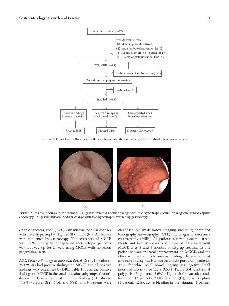
ectopic pancreas, and 1 (1.2%) with mucosal nodular changeswith plica hypertrophy (Figures 2(a) and 2(b)). All lesionswere confirmed by gastroscopy. The sensitivity of MGCEwas 100%. The patient diagnosed with ectopic pancreaswas followed up for 2 years using MGCE with no lesionprogression seen.
3.2.2. Positive Findings in the Small Bowel.Of the 84 patients,25 (29.8%) had positive findings on MGCE and all positivefindings were confirmed by DBE. Table 1 shows the positivefindings on MGCE in the small intestine subgroups. Crohn’sdisease (CD) was the most common finding (10 patients,11.9%) (Figures 3(a), 3(b), and 3(c)), and 9 patients were
diagnosed by small bowel imaging including computedtomography enterography (CTE) and magnetic resonanceenterography (MRE). All patients received systemic treat-ments and had symptom relief. Two patients underwentMGCE after 3 and 6 months of step-up treatments; onepatient showed mucosal improvement on MGCE, and theother achieved complete mucosal healing. The second mostcommon finding was Henoch-Schonlein purpura (4 patients,4.8%) for which small bowel imaging was negative. Smallintestinal ulcers (3 patients, 3.6%) (Figure 3(d)), intestinalpolyposis (3 patients, 3.6%) (Figure 3(e)), vascular mal-formation (2 patients, 2.4%) (Figure 3(f)), intussusception(1 patient, 1.2%), active bleeding in the jejunum (1 patient,
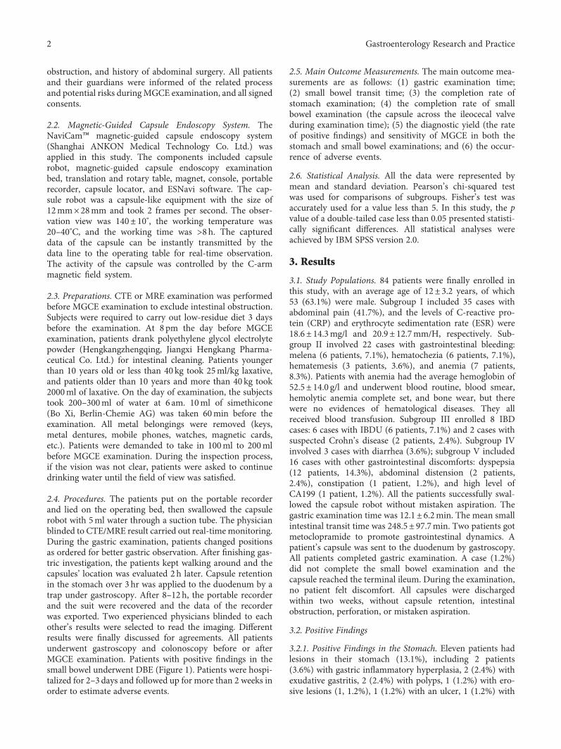
Subjects recruited (n=87)
Exclude criteria (n=2)(i) Metal implantations(n=0)
(ii) Impaired bowel movement (n=0) (iii) Suspected or known obstruction(n=1)(iv) History of gastrointestinal tract(n=1)
Gastrointestinal preparation (n=84)
Exclude (n=0)
Enrolled (n=84)
Positive findingsin stomach (n=11)
Positive findings insmall bowel (n = 24)
Uncompleted smallbowel examination
Proceed EGD
CTE/MRE (n=85)
Exclude: suspected obstruction(n=1)
Proceed DBE Proceed colonoscopy
Figure 1: Flow chart of the study. EGD: esophagogastroduodenoscopy; DBE: double-balloon enteroscopy.
(a) (b)
Figure 2: Positive findings in the stomach: (a) gastric mucosal nodular change with fold hypertrophy found by magnetic-guided capsuleendoscopy; (b) gastric mucosal nodular change with fold hypertrophy verified by gastroscopy.
3Gastroenterology Research and Practice
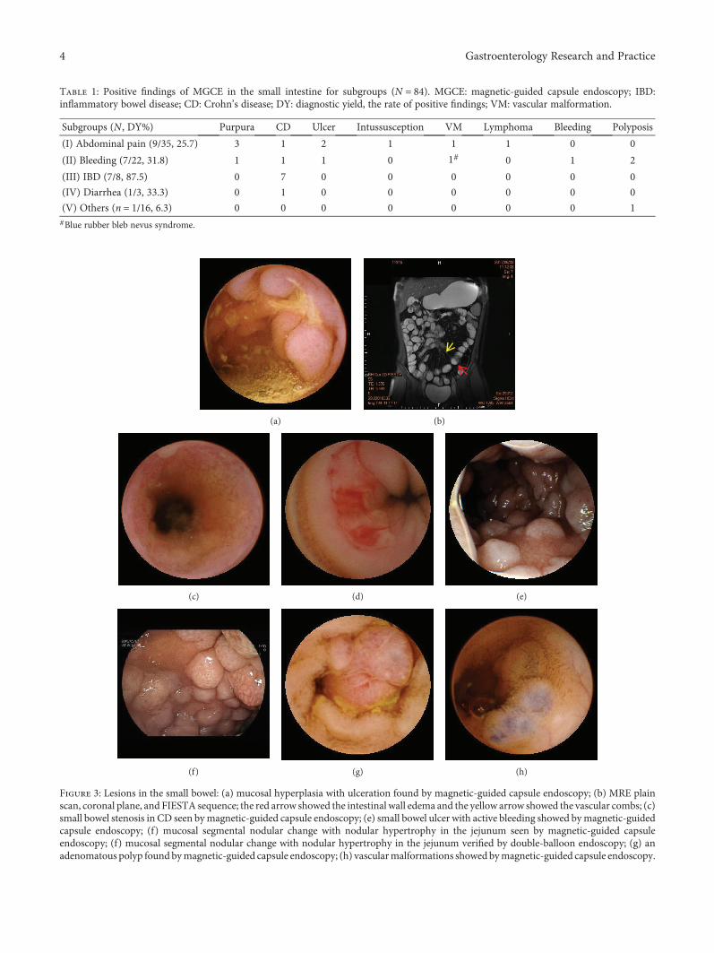
Table 1: Positive findings of MGCE in the small intestine for subgroups (N = 84). MGCE: magnetic-guided capsule endoscopy; IBD:inflammatory bowel disease; CD: Crohn’s disease; DY: diagnostic yield, the rate of positive findings; VM: vascular malformation.
Subgroups (N , DY%) Purpura CD Ulcer Intussusception VM Lymphoma Bleeding Polyposis
(I) Abdominal pain (9/35, 25.7) 3 1 2 1 1 1 0 0
(II) Bleeding (7/22, 31.8) 1 1 1 0 1# 0 1 2
(III) IBD (7/8, 87.5) 0 7 0 0 0 0 0 0
(IV) Diarrhea (1/3, 33.3) 0 1 0 0 0 0 0 0
(V) Others (n = 1/16, 6.3) 0 0 0 0 0 0 0 1#Blue rubber bleb nevus syndrome.
(a) (b)
(c) (d) (e)
(f) (g) (h)
Figure 3: Lesions in the small bowel: (a) mucosal hyperplasia with ulceration found by magnetic-guided capsule endoscopy; (b) MRE plainscan, coronal plane, and FIESTA sequence; the red arrow showed the intestinal wall edema and the yellow arrow showed the vascular combs; (c)small bowel stenosis in CD seen bymagnetic-guided capsule endoscopy; (e) small bowel ulcer with active bleeding showed bymagnetic-guidedcapsule endoscopy; (f) mucosal segmental nodular change with nodular hypertrophy in the jejunum seen by magnetic-guided capsuleendoscopy; (f) mucosal segmental nodular change with nodular hypertrophy in the jejunum verified by double-balloon endoscopy; (g) anadenomatous polyp found bymagnetic-guided capsule endoscopy; (h) vascularmalformations showed bymagnetic-guided capsule endoscopy.
4 Gastroenterology Research and Practice
1.2%), and non-Hodgkin’s lymphoma confirmed by biopsy(Figures 3(g) and 3(h)) were also found on MGCE. In apatient in subgroup I, the capsule did not pass the ileoce-cal valve during the examining time, but there was noobvious abnormality found in the small intestine. Thispatient underwent colposcopy which showed no lesion inthe terminal ileum. A patient with a negative MGCE wasdiagnosed with nonspecific enteritis by MRE. Other patientswith a negative MGCE underwent other supplemental smallintestinal imaging examinations including computedtomography (CT), CTE, MRE, and digital subtractionangiography (DSA). There were no missed diagnoses andno patient was misdiagnosed.
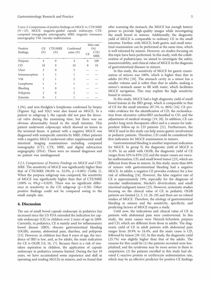
3.2.3. Comparisons of Positive Findings on MGCE and CTE/MRE. The sensitivity of MGCE was significantly higher thanthat of CTE/MRE (96.0% vs. 52.0%, p = 0 001) (Table 2).When the purpura subgroup was compared, the sensitivityof MGCE was significantly higher than that of CTE/MRE(100% vs. 0%,p = 0 029). There was no significant differ-ence in sensitivity in the CD subgroup (p = 0 50). Otherpositive findings could not be compared owing to thesmall sample size.
4. Discussion
The use of small bowel capsule endoscopy in pediatrics hasincreased since the US FDA extended the indication for cap-sule endoscopy (CE) to children over 2 years of age in 2009.Currently, in pediatrics, CE is mainly used for inflammatorybowel disease (IBD), obscure gastrointestinal bleeding(OGIB), anemia, abdominal pain, diarrhea, and polyposis[15]. However, in children less than 8 years of age, the inci-dence of IBD is low, and, as for adults, the main indicationfor CE is OGIB [14, 16, 17]. Because there is a risk of mis-taken aspiration in children, the application of capsuleendoscopy in pediatrics remains limited. During the last 10years, we have accumulated some experience and skill inoperating and reading MGCEs in minors, and we found that
after scanning the stomach, the MGCE has enough batterypower to provide high-quality images while investigatingthe small bowel in minors. Additionally, the diagnosticyield of MGCE is comparable to ordinary CE in the smallintestine. Further, with MGCE, both gastric and small intes-tinal examination can be performed at the same time, whichis well tolerated by minors. However, no studies focusing onthis topic have been performed. In this study, with the collab-oration of pediatricians, we aimed to investigate the safety,maneuverability, and clinical value of MGCE in the diagnosisof gastrointestinal diseases in minors.
In this study, the sensitivity of MGCE for gastric exami-nation of minors was 100%, which is higher than that inadults (61.9%) [18]. The stomach cavity in a minor has asmaller volume and is softer than that in adults, making aminor’s stomach easier to fill with water, which facilitatesMGCE navigation. This may explain the high sensitivityfound in minors.
In this study, MGCE had a high diagnostic yield of smallbowel lesions in the IBD group, which is comparable to thatof CE for the small intestine (87.5% vs. 86%) [16]. CE pro-vides evidence for the identification of CD, the reclassifica-tion from ulcerative colitis/IBD unclassified to CD, and theadjustment of medical strategy [19, 20]. In addition, CE canpredict long-term therapeutic effect and is a means for inde-pendent follow-up in CD patients [21]. Furthermore, theMGCE used in this study can help assess gastric involvementin pediatric patients. Therefore, CD could be considered thefirst indication for MGCE examination in minors.
Gastrointestinal bleeding is another important indicationfor MGCE. In group II, the diagnostic yield of MGCE is31.8%. In an adult with OGIB, the diagnostic yield of CEranges from 32% to 83% [22] and the major causes are vascu-lar malformation, CD, and small bowel tumor [23], which aredifferent from those in minors. In this study, more than 60%of minors with gastrointestinal bleeding had a negativeMGCE. In adults, a negative CE provides evidence for a lowrisk of rebleeding [24]. However, the false negative rate ofCE is approximately 19%, especially for the diagnosis ofvascular malformation, Meckel’s diverticulum, and smallintestinal malignant tumor [25]. However, systematic studiesfocusing on the clinical value of CE in pediatric OGIBpatients are limited [2, 5, 13, 26–29] and there are no relatedstudies of MGCE. Therefore, the etiology of gastrointestinalbleeding in minors and the sensitivity, specificity, andpredicting factors of MGCE require a study.
Until now, the indications and clinical value of CE inpatients with abdominal pain were controversial. In thisstudy, the main causes were Henoch-Schonlein purpuraand CD, which are different from those in adults. The diag-nostic yield of CE in adult patients with abdominal painranges from 20.9% to 24.4%, and the main cause is CD,followed by tumor [30–32]. In this study, the diagnostic yield(25.7%) was slightly higher than that of the adults. Thereasons for this could be (1) the patients recruited were hos-pitalized, and the syndrome may be more severe in them inoutpatients; (2) the patients enrolled in this study had ele-vated C-reactive protein or erythrocyte sedimentation rate,which may be an effective predictor for positive CE findings
Table 2: Comparisons of positive findings on MGCE vs. CTE/MRE(N = 25). MGCE: magnetic-guided capsule endoscopy; CTE:computed tomography enterography; MRE: magnetic resonanceenterography; VM: vascular malformation.
Positivefindings
CE(N)
CTE/MRE(N)
Confirmedcases (N)
Miss rate(%)
CECTE/MRE
Purpura 4 0 4 0 100
CD 10 9 10 0 10
Ulcer 3 0 3 — —
Intussusception 1 1 1 — —
VM 2 1 2 — —
Lymphoma 1 1 1 — —
Bleeding 1 0 1 — —
Polyposis 2 0 2 — —
Enteritis 0 1 1 — —
5Gastroenterology Research and Practice
in patients with abdominal pain [33, 34]; and (3) minorsinvolved in this study had a high incidence of Henoch-Schonlein purpura, which was not frequently found in adultpatients. Therefore, large-scale clinical trials confirming theclinical value of MGCE in minors with abdominal pain arestill needed.
In the diarrhea subgroup, one patient was diagnosedwith CD, and the diagnostic yield of MGCE was higherthan that of the adults using CE (33.3% vs. 14%) [35].Although CE is not recommended as a first-line examina-tion for diarrhea, it facilitates making the diagnosis in 9%of patients with negative conventional endoscopy andimaging examination results [35]. The most commoncause is IBD, and hematochezia and hypoalbuminemiaon CE are positive predictors for it [36]. There is a lackof clinical studies on CE for patients with dyspepsia, con-stipation, high level CA 19-9, and abdominal distention,especially in the pediatric population. MGCE may not besuitable for such patients as a first-line procedure, but itremains valuable for suspected IBD in minors.
Currently, CTE and MRE are widely used for diseases ofthe small intestine, and the diagnostic yield is comparable tothat of CE [37, 38]. In this study, MGCE had a better sensitiv-ity than that of CTE and MRE in minors. The possible rea-sons for this advantage maybe as follows: (1) MGCE isbetter for scanning the lesions on the mucosal surface, whichis where lesions in minors primarily occur; and (2) minorshave difficulty cooperating with the CTE and MRE examina-tion processes. However, CTE and MRE can provide supple-mentary information and, for safety, are still recommendedbefore MGCE in minors to avoid capsule retention.
Swallowing problems in minors during MGCE remainsa challenge, especially for patients younger than 8 years ofage. We thoroughly explained the procedure process andtechniques to the patients and their guardians to comfortthem and reduce fear. Patient guidance and encourage-ment from doctors can help children swallow the capsulesmoothly, even for those who are 6 years of age [39].The overall incidence rate of adverse events is 2% [40],and capsule retention is one of the most common compli-cations, especially for CD patients and patients less than10 years of age [41]. In children, the high risk of capsuleretention may be due to the narrow diameter of theintestinal cavity and the high incidence of CD-relatedintestinal stenosis. However, during our follow-up period,no adverse events occurred. We consider MGCE to be asafe procedure for minors.
There are some limitations to this study. First, the effi-ciency of MGCEs in the subgroups could not be comparedowing to the small sample size. Second, longer follow-up timeis needed to confirm the cure rate, recurrence rate, andaccuracy of the original diagnosis. Finally, due to potentialswallowing problems, patients less than 6 years of age werenot recruited for this study, which may have led to some bias.
5. Conclusion
MGCE is a safe, well-tolerated, and feasible procedure with ahigh diagnostic yield and sensitivity in minor patients. It has
important clinical value and great prospects in the diagnosisof gastrointestinal diseases in minors.
Data Availability
All data can be acquired by connecting the correspondingauthors.
Disclosure
Yuting Qian and Tingting Bai contributed equally to thispaper.
Conflicts of Interest
The authors declare no competing interests.
Acknowledgments
This work was supported by the National Natural ScienceFoundation of China (81672719, 81702740).
References
[1] F. Argüelles Arias, E. Donat, I. Fernández-Urien et al., “Guide-line for wireless capsule endoscopy in children and adoles-cents: a consensus document by the SEGHNP (SpanishSociety for Pediatric Gastroenterology, Hepatology, andNutrition) and the SEPD (Spanish Spanish Society for Diges-tive Diseases),” Revista Española de Enfermedades Digestivas,vol. 107, 2015.
[2] S. Oliva, M. Pennazio, S. A. Cohen et al., “Capsule endoscopyfollowed by single balloon enteroscopy in children withobscure gastrointestinal bleeding: a combined approach,”Digestive and Liver Disease, vol. 47, no. 2, pp. 125–130, 2015.
[3] K. N. Shim, J. S. Moon, D. K. Chang et al., “Guideline for cap-sule endoscopy: obscure gastrointestinal bleeding,” ClinicalEndoscopy, vol. 46, no. 1, pp. 45–53, 2013.
[4] C. Dupont-Lucas, M. Bellaïche, O. Mouterde et al.,“Capsule endoscopy in children: which are the best indica-tions?,” Archives de Pédiatrie, vol. 17, no. 9, pp. 1264–1272, 2010.
[5] J. Viala, L. Michaud, M. Bellaiche, and A. Lachaux, “When andhow should small-bowel capsule endoscopy be used in chil-dren?,” Archives de Pédiatrie, vol. 24, no. 4, pp. 391–398, 2017.
[6] B. Vadamalayan, M. Hii, J. Kark, and I. Bjarnason, “Feasibilityof small bowel capsule endoscopy in children under the age of4 years: a single centre experience,” Frontline Gastroenterology,vol. 3, no. 4, pp. 267–271, 2012.
[7] A. Fritscher-Ravens, P. Scherbakov, P. Bufler et al., “Thefeasibility of wireless capsule endoscopy in detecting smallintestinal pathology in children under the age of 8 years: amulticentre European study,” Gut, vol. 58, no. 11,pp. 1467–1472, 2009.
[8] M. Oikawa-Kawamoto, T. Sogo, T. Yamaguchi et al., “Safetyand utility of capsule endoscopy for infants and young chil-dren,” World Journal of Gastroenterology, vol. 19, no. 45,pp. 8342–8348, 2013.
[9] J. F. Rey, H. Ogata, N. Hosoe et al., “Feasibility of stomachexploration with a guided capsule endoscope,” Endoscopy,vol. 42, no. 07, pp. 541–545, 2010.
6 Gastroenterology Research and Practice
[10] P. Swain, A. Toor, F. Volke et al., “Remote magnetic manipu-lation of a wireless capsule endoscope in the esophagus andstomach of humans (with videos),” Gastrointestinal Endos-copy, vol. 71, no. 7, pp. 1290–1293, 2010.
[11] J. Keller, C. Fibbe, F. Volke et al., “Inspection of the humanstomach using remote-controlled capsule endoscopy: a feasi-bility study in healthy volunteers (with videos),” Gastrointesti-nal Endoscopy, vol. 73, no. 1, pp. 22–28, 2011.
[12] Z. Liao, X. D. Duan, L. Xin et al., “Feasibility and safety ofmagnetic-controlled capsule endoscopy system in examina-tion of human stomach: a pilot study in healthy volunteers,”Journal of Interventional Gastroenterology, vol. 2, no. 4,pp. 155–160, 2012.
[13] H. Nuutinen, K. L. Kolho, P. Salminen et al., “Capsule endos-copy in pediatric patients: technique and results in our first 100consecutive children,” Scandinavian Journal of Gastroenterol-ogy, vol. 46, no. 9, pp. 1138–1143, 2011.
[14] N. Zevit and R. Shamir, “Wireless capsule endoscopy of thesmall intestine in children,” Journal of Pediatric Gastroenterol-ogy and Nutrition, vol. 60, no. 6, pp. 696–701, 2015.
[15] S. A. Cohen and A. I. Klevens, “Use of capsule endoscopy indiagnosis and management of pediatric patients, based onmeta-analysis,” Clinical Gastroenterology and Hepatology,vol. 9, no. 6, pp. 490–496, 2011.
[16] R. A. Enns, L. Hookey, D. Armstrong et al., “Clinical practiceguidelines for the use of video capsule endoscopy,” Gastroen-terology, vol. 152, no. 3, pp. 497–514, 2017.
[17] H. Yamamoto, H. Ogata, T. Matsumoto et al., “Clinicalpractice guideline for enteroscopy,” Digestive Endoscopy,vol. 29, no. 5, pp. 519–546, 2017.
[18] U. W. Denzer, T. Rösch, B. Hoytat et al., “Magnetically guidedcapsule versus conventional gastroscopy for upper abdominalcomplaints: a prospective blinded study,” Journal of ClinicalGastroenterology, vol. 49, no. 2, pp. 101–107, 2015.
[19] S. B. Min, M. Le-Carlson, N. Singh et al., “Video capsuleendoscopy impacts decision making in pediatric IBD: a singletertiary care center experience,” Inflammatory Bowel Diseases,vol. 19, no. 10, pp. 2139–2145, 2013.
[20] I. M. Gralnek, S. A. Cohen, H. Ephrath et al., “Small bowelcapsule endoscopy impacts diagnosis and management ofpediatric inflammatory bowel disease: a prospective study,”Digestive Diseases and Sciences, vol. 57, no. 2, pp. 465–471, 2012.
[21] Y. Niv, “Small-bowel mucosal healing assessment by capsuleendoscopy as a predictor of long-term clinical remission inpatients with Crohn’s disease: a systematic review and meta-analysis,” European Journal of Gastroenterology & Hepatol-ogy, vol. 29, no. 7, pp. 844–848, 2017.
[22] Y. W. Min and D. K. Chang, “The role of capsule endoscopy inpatients with obscure gastrointestinal bleeding,” ClinicalEndoscopy, vol. 49, no. 1, pp. 16–20, 2016.
[23] C. S. E. Wang, J. N. Li, Y. N. Wang, H. Yang, and J. M.Qian, “Diagnostic value analysis of capsule endoscopy inobscure gastrointestinal bleeding patients of different ages,”Zhonghua Yi Xue Za Zhi, vol. 97, no. 36, pp. 2848–2851,2017.
[24] P. Katsinelos, J. Kountouras, G. Chatzimavroudis et al.,“Factors predicting a positive capsule endoscopy in pastovert obscure gastrointestinal bleeding: a multicenter retro-spective study,” Hippokratia, vol. 20, no. 2, pp. 127–132,2016.
[25] C. Van de Bruaene, P. Hindryckx, C. Snauwaert et al., “Thepredictive value of negative capsule endoscopy for the indica-tion of obscure gastrointestinal bleeding: no reassurance inthe long term,” Acta Gastro-Enterologica Belgica, vol. 79,no. 4, pp. 405–413, 2016.
[26] B. Sahn and S. Bitton, “Lower gastrointestinal bleeding inchildren,” Gastrointestinal Endoscopy Clinics of NorthAmerica, vol. 26, no. 1, pp. 75–98, 2016.
[27] M. Thomson, “Paediatrics: diagnostic yield of paediatric lowergastrointestinal endoscopy,” Nature Reviews Gastroenterology& Hepatology, vol. 13, no. 7, pp. 382–384, 2016.
[28] P. Katsinelos, G. Lazaraki, A. Gkagkalis et al., “The role ofcapsule endoscopy in the evaluation and treatment ofobscure-overt gastrointestinal bleeding during daily clinicalpractice: a prospective multicenter study,” Scandinavian Jour-nal of Gastroenterology, vol. 49, no. 7, pp. 862–870, 2014.
[29] Z. Liao, R. Gao, F. Li et al., “Fields of applications, diagnosticyields and findings of OMOM capsule endoscopy in 2400Chinese patients,” World Journal of Gastroenterology, vol. 16,no. 21, pp. 2669–2676, 2010.
[30] M. Xue, X. Chen, L. Shi, J. Si, L. Wang, and S. Chen,“Small-bowel capsule endoscopy in patients with unex-plained chronic abdominal pain: a systematic review,”Gastrointestinal Endoscopy, vol. 81, no. 1, pp. 186–193,2015.
[31] J. Egnatios, K. Kaushal, D. Kalmaz, and A. Zarrinpar,“Video capsule endoscopy in patients with chronic abdom-inal pain with or without associated symptoms: a retro-spective study,” PLoS One, vol. 10, no. 4, articlee0126509, 2015.
[32] L. Yang, Y. Chen, B. Zhang et al., “Increased diagnostic yield ofcapsule endoscopy in patients with chronic abdominal pain,”PLoS One, vol. 9, no. 1, article e87396, 2014.
[33] M. Nakano, S. Oka, S. Tanaka et al., “Indications forsmall-bowel capsule endoscopy in patients with chronicabdominal pain,” Internal Medicine, vol. 56, no. 12,pp. 1453–1457, 2017.
[34] P. Katsinelos, K. Fasoulas, A. Beltsis et al., “Diagnostic yieldand clinical impact of wireless capsule endoscopy in patientswith chronic abdominal pain with or without diarrhea: a Greekmulticenter study,” European Journal of Internal Medicine,vol. 22, no. 5, pp. e63–e66, 2011.
[35] L. C. Fry, E. J. Carey, A. D. Shiff et al., “The yield of capsuleendoscopy in patients with abdominal pain or diarrhea,”Endoscopy, vol. 38, no. 5, pp. 498–502, 2006.
[36] H. J. Song, J. S. Moon, S. R. Jeon et al., “Diagnostic yield andclinical impact of video capsule endoscopy in patients withchronic diarrhea: a Korean multicenter CAPENTRY study,”Gut and Liver, vol. 11, no. 2, pp. 253–260, 2017.
[37] M. Choi, S. Lim, M. G. Choi, K. N. Shim, and S. H. Lee,“Effectiveness of capsule endoscopy compared with otherdiagnostic modalities in patients with small bowel Crohn’sdisease: a meta-analysis,” Gut and Liver, vol. 11, no. 1,pp. 62–72, 2017.
[38] E. Casciani, G. D. Nardo, S. Chin et al., “MR Enterographyin paediatric patients with obscure gastrointestinal bleed-ing,” European Journal of Radiology, vol. 93, pp. 209–216,2017.
[39] B. A. Barth, S. Banerjee, Y. M. Bhat et al., “Equipment forpediatric endoscopy,” Gastrointestinal Endoscopy, vol. 76,no. 1, pp. 8–17, 2012.
7Gastroenterology Research and Practice

[40] M. Rezapour, C. Amadi, and L. B. Gerson, “Retention associ-ated with video capsule endoscopy: systematic review andmeta-analysis,” Gastrointestinal Endoscopy, vol. 85, no. 6,pp. 1157–1168.e2, 2017, e1152.
[41] Y. J. Lim, O. Y. Lee, Y. T. Jeen et al., “Indications for detection,completion, and retention rates of small bowel capsule endos-copy based on the 10-year data from the Korean capsuleendoscopy registry,” Clinical Endoscopy, vol. 48, no. 5,pp. 399–404, 2015.
8 Gastroenterology Research and Practice

Stem Cells International
Hindawiwww.hindawi.com Volume 2018
Hindawiwww.hindawi.com Volume 2018
MEDIATORSINFLAMMATION
of
EndocrinologyInternational Journal of
Hindawiwww.hindawi.com Volume 2018
Hindawiwww.hindawi.com Volume 2018
Disease Markers
Hindawiwww.hindawi.com Volume 2018
BioMed Research International
OncologyJournal of
Hindawiwww.hindawi.com Volume 2013
Hindawiwww.hindawi.com Volume 2018
Oxidative Medicine and Cellular Longevity
Hindawiwww.hindawi.com Volume 2018
PPAR Research
Hindawi Publishing Corporation http://www.hindawi.com Volume 2013Hindawiwww.hindawi.com
The Scientific World Journal
Volume 2018
Immunology ResearchHindawiwww.hindawi.com Volume 2018
Journal of
ObesityJournal of
Hindawiwww.hindawi.com Volume 2018
Hindawiwww.hindawi.com Volume 2018
Computational and Mathematical Methods in Medicine
Hindawiwww.hindawi.com Volume 2018
Behavioural Neurology
OphthalmologyJournal of
Hindawiwww.hindawi.com Volume 2018
Diabetes ResearchJournal of
Hindawiwww.hindawi.com Volume 2018
Hindawiwww.hindawi.com Volume 2018
Research and TreatmentAIDS
Hindawiwww.hindawi.com Volume 2018
Gastroenterology Research and Practice
Hindawiwww.hindawi.com Volume 2018
Parkinson’s Disease
Evidence-Based Complementary andAlternative Medicine
Volume 2018Hindawiwww.hindawi.com
Submit your manuscripts atwww.hindawi.com