Embed Size (px)
Citation preview
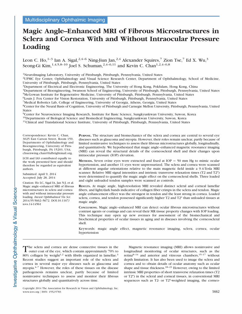
Multidisciplinary Ophthalmic Imaging
Magic Angle–Enhanced MRI of Fibrous Microstructures inSclera and Cornea With and Without Intraocular PressureLoading
Leon C. Ho,1–3 Ian A. Sigal,2,4–6 Ning-Jiun Jan,2,4 Alexander Squires,7 Zion Tse,7 Ed X. Wu,3
Seong-Gi Kim,1,4,5,8–10 Joel S. Schuman,2,4–6,11 and Kevin C. Chan1,2,4–6,8
1NeuroImaging Laboratory, University of Pittsburgh, Pittsburgh, Pennsylvania, United States2UPMC Eye Center, Ophthalmology and Visual Science Research Center, Department of Ophthalmology, School of Medicine,University of Pittsburgh, Pittsburgh, Pennsylvania, United States3Department of Electrical and Electronic Engineering, The University of Hong Kong, Pokfulam, Hong Kong, China4Department of Bioengineering, Swanson School of Engineering, University of Pittsburgh, Pittsburgh, Pennsylvania, United States5McGowan Institute for Regenerative Medicine, University of Pittsburgh, Pittsburgh, Pennsylvania, United States6Louis J. Fox Center for Vision Restoration, University of Pittsburgh, Pittsburgh, Pennsylvania, United States7Medical Robotics Lab, College of Engineering, University of Georgia, Athens, Georgia, United States8Center for the Neural Basis of Cognition, University of Pittsburgh and Carnegie Mellon University, Pittsburgh, Pennsylvania, UnitedStates9Center for Neuroscience Imaging Research, Institute for Basic Science, Sungkyunkwan University, Suwon, Korea10Departments of Biological Science and Biomedical Engineering, Sungkyunkwan University, Suwon, Korea11Clinical and Translational Science Institute, University of Pittsburgh, Pittsburgh, Pennsylvania, United States
Correspondence: Kevin C. Chan,3025 East Carson Street, Room 159,Departments of Ophthalmology andBioengineering, University of Pitts-burgh, Pittsburgh, PA 15203, USA;[email protected].
LCH and IAS contributed equally tothe work presented here and shouldtherefore be regarded as equivalentauthors.
Submitted: April 9, 2014Accepted: July 28, 2014
Citation: Ho LC, Sigal IA, Jan N-J, et al.Magic angle–enhanced MRI of fibrousmicrostructures in sclera and corneawith and without intraocular pressureloading. Invest Ophthalmol Vis Sci.2014;55:5662–5672. DOI:10.1167/iovs.14-14561
PURPOSE. The structure and biomechanics of the sclera and cornea are central to several eyediseases such as glaucoma and myopia. However, their roles remain unclear, partly because oflimited noninvasive techniques to assess their fibrous microstructures globally, longitudinally,and quantitatively. We hypothesized that magic angle–enhanced magnetic resonance imaging(MRI) can reveal the structural details of the corneoscleral shell and their changes uponintraocular pressure (IOP) elevation.
METHODS. Seven ovine eyes were extracted and fixed at IOP ¼ 50 mm Hg to mimic ocularhypertension, and another 11 eyes were unpressurized. The sclera and cornea were scannedat different angular orientations relative to the main magnetic field inside a 9.4-Tesla MRIscanner. Relative MRI signal intensities and intrinsic transverse relaxation times (T2 and T2*)were determined to quantify the magic angle effect on the corneoscleral shells. Three loadedand eight unloaded tendon samples were scanned as controls.
RESULTS. At magic angle, high-resolution MRI revealed distinct scleral and corneal lamellarfibers, and light/dark bands indicative of collagen fiber crimps in the sclera and tendon. Magicangle enhancement effect was the strongest in tendon and the least strong in cornea. Loadedsclera, cornea, and tendon possessed significantly higher T2 and T2* than unloaded tissues atmagic angle.
CONCLUSIONS. Magic angle–enhanced MRI can detect ocular fibrous microstructures withoutcontrast agents or coatings and can reveal their MR tissue property changes with IOP loading.This technique may open up new avenues for assessment of the biomechanical andbiochemical properties of ocular tissues in aging and in diseases involving the corneoscleralshell.
Keywords: magic angle effect, magnetic resonance imaging, sclera, cornea, ocularhypertension
The sclera and cornea are dense connective tissues in theouter coat of the eye, which contain approximately 70% to
80% collagen by weight1,2 with fibrils organized in lamellae.3
Recent studies suggest an important role of the sclera andcornea in several major eye diseases such as glaucoma andmyopia.4–7 However, the roles of these tissues on the diseasepathogenesis remains unclear, partly because of limitednoninvasive techniques to assess and monitor their fibrousstructures globally and quantitatively across time.
Magnetic resonance imaging (MRI) allows noninvasive andlongitudinal monitoring of ocular structures, such as theretina8–14 and anterior and vitreous chambers,15–17 withoutdepth limitation. It has also been used to image the sclera andcornea and to obtain details of ocular anatomy such as ocularshape and tissue thickness.18–23 However, owing to the limitedintrinsic MRI properties of short transverse relaxation times (T2or T2*) in the scleral and corneal tissues, in conventional MRIsequences such as T2- or T2*-weighted imaging, the corneo-
Copyright 2014 The Association for Research in Vision and Ophthalmology, Inc.
www.iovs.org j ISSN: 1552-5783 5662
scleral shell possesses low signal intensity and appears dark inthe images.23,24 To date, the highest-resolution MRI study ofthe globe19 has required exogenous contrast agents andcoatings to delineate the corneoscleral shell from the darksurroundings. To the best of our knowledge, none of thetechniques aforementioned has been able to visualize theinternal structures of the sclera or cornea, or the effects ofintraocular pressure (IOP) alterations on these tissues.Understanding the microstructures of the corneoscleral shelland their changes upon IOP loading is particularly importantfor resolving the recent debates on how the biomechanics andbiochemistry of the fibrous corneoscleral shell may impair thenearby retina and optic nerve under ocular hypertension inglaucoma.25,26 This may guide more targeted strategies forpreserving vision from this irreversible disease, which in turnmay help reduce the prevalence of this second leading cause ofblindness worldwide.
The magic angle effect describes an orientation-dependentMR signal change arising when imaging highly ordered fibrousstructures.27 In highly ordered, collagen-rich fibrous tissues,such as the sclera and cornea, bounded protons are subject todipolar interactions within a static magnetic field, which resultin low T2 and T2*. The strength of these interactions dependson the angle h between a fiber and the main magnetic field (Bo)of the MRI scanner by the term 3 cos2 h � 1. As the dipolarinteractions are minimized at 3 cos2 h�1¼0, that is, when thetissues are oriented at h » 54.78 or the ‘‘magic angle,’’ thetissue MR signal intensities can be enhanced to the greatestdegree and the MR sensitivity can be maximized.27 This magicangle effect has been demonstrated extensively in MRI studiesof fibrous structures in normal and diseased tendons,28,29
menisci,30 ligaments,31 and cartilages32–35 but has not yet beeninvestigated in ophthalmic research. In this study, wehypothesized that high-field, magic angle–enhanced magneticresonance microscopy can be used to reveal details of thefibrous structures in the corneoscleral shell, and specifically,that it can reveal the lamellar fibers and the waviness, or crimp,of the collagen fibers that form the corneoscleral shell. Further,we hypothesized that the high sensitivity of magic angle effectto the structural alignment of the collagen fibers wouldimprove the detection of the MRI signal differences in oculartissues between no-load and elevated IOP conditions. Inaddition, we evaluated the intrinsic MR tissue properties interms of the absolute transverse relaxation time constants, T2and T2*, of the sclera and cornea by using T2 and T2* mappingto characterize quantitatively the extents of magic angle effect
on different types of MR images in both loaded and unloadedtissues.
MATERIALS AND METHODS
Tissue Preparation
Nine pairs of ovine eyes were obtained from the local abattoirand processed within 12 hours of death. The eyes were dividedinto two groups. In group 1, four eyes from two ovines wereimmersion fixed with 10% formalin without cannulation andthen washed in phosphate-buffered saline (PBS). The eyes thenunderwent whole-globe MRI followed by sectioning of thesclera and cornea for high-resolution magic angle–enhancedMRI. These sections were 2- to 5-mm thick, made by manuallycutting in the superior–inferior direction by using a razor.Section location varied, depending on the region to be imaged,namely, cornea and anterior or posterior sclera. In group 2, oneeye from each of the remaining seven ovines was loaded at anIOP of 50 mm Hg by using a gravity perfusion fixation systemand a cannula inserted into the anterior chamber (Jan NJ, et al.IOVS 2013;54:ARVO E-Abstract 65; Sigal et al. IOVS 2013;54:AR-VO E-Abstract 3158). The contralateral eyes were cannulatedbut not pressurized. All eyes were then immersion fixed with10% formalin, washed in PBS, and dissected with razors andsurgical scissors to isolate the sclera and cornea. Similarly, 13ovine Achilles tendons from the same set of animals were alsowashed in PBS and dissected to isolate thin strips and act aspositive controls of the ocular tissues, given the well-knownmagic angle effect in tendon and the similar structuralcompositions between the tendon and corneoscleral shell.Four strips were randomly selected and were loaded by usingalligator clips attached to a rod to keep stretched, followed byimmersion fixation with 10% formalin. The remaining ninestrips were fixed but unloaded. Three loaded tendons andseven unloaded tendons underwent the same MRI scans as theocular tissues in groups 1 and 2. In addition, one of theunloaded tendons was placed in a glass test tube, such that thetendon strip made a ‘‘U-shape’’ at the bottom of the test tube toillustrate how the MR signal enhancement affected thevisualization of collagen fiber crimp structures at differentangles continuously from 08 to 908 to Bo. The test tube wasfilled with 10% formalin and fixed overnight. Afterwards, theformalin was replaced with PBS, and high-resolution MRI wasperformed to the U-shaped tendon. The remaining one loaded
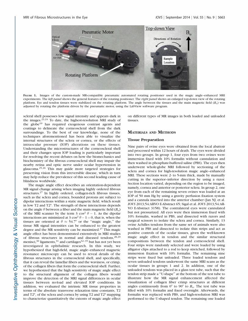
FIGURE 1. Images of the custom-made MR-compatible pneumatic automated rotating positioner used in the magic angle–enhanced MRIexperiments. The left panel shows the general features of the rotating positioner. The right panel shows an enlarged top-down view of the rotatingplatform. Eye and tendon tissues were stabilized on the rotating platform. The angle between the tissues and the main magnetic field (Bo) wasadjusted by rotating the platform driven by the pneumatic motor, using the LabView software program.
MRI of Fibrous Microstructures in the Eye IOVS j September 2014 j Vol. 55 j No. 9 j 5663
and one unloaded tendons were taken for histologic evalua-tions of the collagen fiber crimps. They were cut into ~3-mm-thick and ~1-cm-long strips and processed in 30% sucrose. Thestrips were then embedded and frozen in cryogel andcryosectioned into 50-lm sections. These sections werewashed with water, ethanol, and xylene, stained withhematoxylin-eosin (H&E), and mounted by using Permount.
MRI Protocol
Eye and tendon tissue samples were suspended in agarose gelto prevent vibrational movement during imaging and werescanned by using a 9.4-Tesla/31-cm Varian/Agilent horizontalMRI scanner (Varian/Agilent, Santa Clara, CA, USA). Tissuesamples were positioned relative to Bo by using a custom-madeMR-compatible pneumatic automated rotating positioner (Fig.1).36 In group 1, the whole globe of the intact eye waspositioned and scanned at several orientations to the main
magnetic field (Bo) inside the scanner by using T2*-weightedgradient-echo imaging at voxel size of 60 3 60 3 800 lm3 witha 38-mm-diameter transmit–receive volume coil. For high-resolution microstructural evaluation of tissue sections ingroup 1, the tissues were orientated at 558 relative to Bo insidethe scanner, and T2*-weighted gradient-echo pulse sequenceswere applied at 16 3 16 lm2 to 27 3 27 lm2 in-planeresolution, 400-lm to 700-lm slice thickness, 3-s repetitiontime, and multiple echo times by using a custom-made 12-mm-diameter transmit–receive surface coil. The magic angle effectof the same tissues was also evaluated by examining therelative signal intensities at different angular orientations from08 to 908 relative to Bo, using the same gradient-echo MRIscanning protocol at 30 3 30 lm2 in-plane resolution, 500-lmslice thickness, 3-s repetition time, and 5-ms echo time. Forcomparisons of the absolute MR transverse relaxation timesbetween loaded and unloaded tissue samples in group 2, T2and T2* mapping with spin-echo and gradient-echo MRI pulsesequences was respectively performed by using a custom-made25-mm-diameter transmit–receive surface coil. The loaded andunloaded eye and tendon tissue samples were paired up andpositioned at the same angular orientations as in group 1. TheMRI parameters were as follows: (1) spin-echo sequence:repetition time/echo time ¼ 1000/9.7 ms; echo space time ¼9.7 ms; number of echoes ¼ 3; (2) gradient-echo sequence:repetition time/echo time ¼ 1000/2.9 ms; echo space time ¼3.4 ms; number of echoes¼3. For both spin-echo and gradient-echo sequences in group 2, in-plane resolution ¼ 110 3 110lm2 and slice thickness ¼ 1 mm.
Data Analysis
The MR images were reconstructed by using ImageJ softwarev1.47 (http://imagej.nih.gov/ij/; Wayne Rasband, provided inthe public domain by the National Institutes of Health,Bethesda, MD, USA) for qualitative evaluation of morphologicdetails in the sclera, cornea, and tendon. For quantitative dataanalyses of relative T2- and T2*-weighted signal intensities aswell as absolute T2 and T2* MR transverse relaxation times,regions of interest were manually drawn on the eye and tendontissues in the spin-echo and gradient-echo MR images,respectively, by using ImageJ to extract the signal intensitiesat different echo times and angles to Bo. Intrinsic T2 and T2*values of the loaded and unloaded tissues were derived fromcurve fitting of T2- and T2*-weighted signal decays, respectively,with at least two echo times by using the equations SSE(TE) ¼SSE(0)e(�TE/T2) and SGE(TE) ¼ SGE(0)e(�TE/T2*), or T2 ¼ (TE2 �TE1)/{ln[SSE(TE1)] � ln[SSE(TE2)]} and T2* ¼ (TE2 � TE1)/{ln[SGE(TE1)] � ln[SGE(TE2)]}, where SSE and SGE are the signalintensities (S) from spin-echo (SE) and gradient-echo (GE) MRimages at a particular echo time (TE). Results are presented asmean 6 standard deviation. Measurements between loaded andunloaded tissues, and between 08 and 558 to Bo were comparedwith Student’s t-tests. One-tailed tests were used to confirmprior findings37 that loaded tendons would possess longertransverse relaxation times than unloaded tendons at the magicangle under similar MRI settings. Elsewhere, two-tailed testswere used. Results were considered significant when P < 0.05.
RESULTS
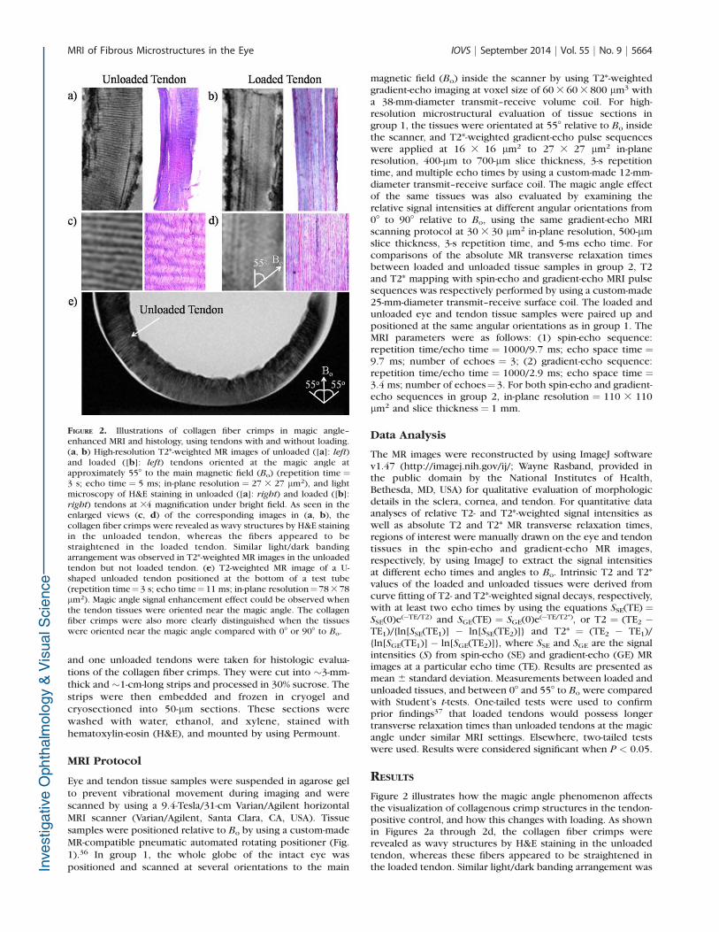
Figure 2 illustrates how the magic angle phenomenon affectsthe visualization of collagenous crimp structures in the tendon-positive control, and how this changes with loading. As shownin Figures 2a through 2d, the collagen fiber crimps wererevealed as wavy structures by H&E staining in the unloadedtendon, whereas these fibers appeared to be straightened inthe loaded tendon. Similar light/dark banding arrangement was
FIGURE 2. Illustrations of collagen fiber crimps in magic angle–enhanced MRI and histology, using tendons with and without loading.(a, b) High-resolution T2*-weighted MR images of unloaded ([a]: left)and loaded ([b]: left) tendons oriented at the magic angle atapproximately 558 to the main magnetic field (Bo) (repetition time ¼3 s; echo time ¼ 5 ms; in-plane resolution ¼ 27 3 27 lm2), and lightmicroscopy of H&E staining in unloaded ([a]: right) and loaded ([b]:right) tendons at 34 magnification under bright field. As seen in theenlarged views (c, d) of the corresponding images in (a, b), thecollagen fiber crimps were revealed as wavy structures by H&E stainingin the unloaded tendon, whereas the fibers appeared to bestraightened in the loaded tendon. Similar light/dark bandingarrangement was observed in T2*-weighted MR images in the unloadedtendon but not loaded tendon. (e) T2-weighted MR image of a U-shaped unloaded tendon positioned at the bottom of a test tube(repetition time¼3 s; echo time¼11 ms; in-plane resolution¼78 3 78lm2). Magic angle signal enhancement effect could be observed whenthe tendon tissues were oriented near the magic angle. The collagenfiber crimps were also more clearly distinguished when the tissueswere oriented near the magic angle compared with 08 or 908 to Bo.
MRI of Fibrous Microstructures in the Eye IOVS j September 2014 j Vol. 55 j No. 9 j 5664
observed in high-resolution T2*-weighted MR images in theunloaded tendon but was not visible in loaded tendon whentissues were oriented at the magic angle at approximately 558to Bo. In the T2-weighted MR image of a U-shaped unloadedtendon (Fig. 2e), the light/dark bands of collagen fiber crimpswere more clearly distinguished when the tissues wereoriented near the magic angle compared with 08 or 908 toBo. Note also the magic angle signal enhancement effect whenthe tendon tissues were oriented near the magic angle.
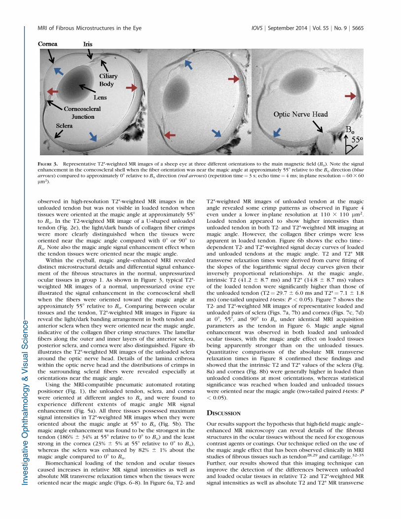
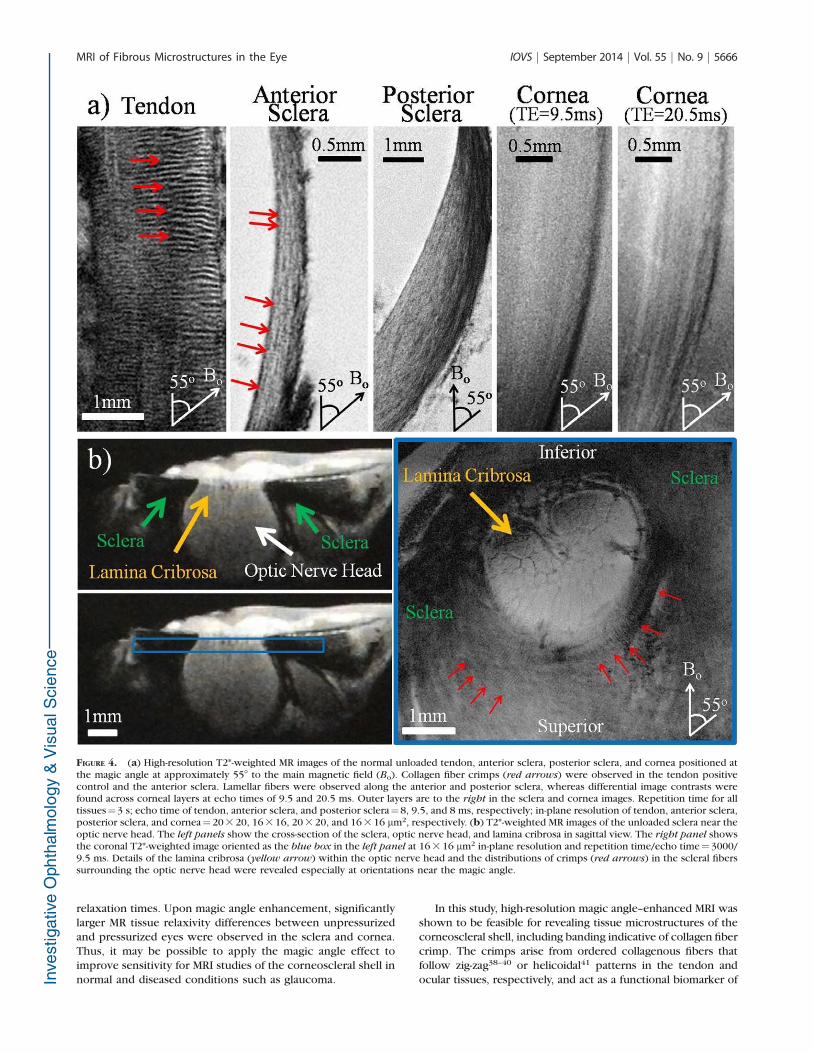
Within the eyeball, magic angle–enhanced MRI revealeddistinct microstructural details and differential signal enhance-ment of the fibrous structures in the normal, unpressurizedocular tissues in group 1. As shown in Figure 3, typical T2*-weighted MR images of a normal, unpressurized ovine eyeillustrated the signal enhancement in the corneoscleral shellwhen the fibers were oriented toward the magic angle atapproximately 558 relative to Bo. Comparing between oculartissues and the tendon, T2*-weighted MR images in Figure 4areveal the light/dark banding arrangement in both tendon andanterior sclera when they were oriented near the magic angle,indicative of the collagen fiber crimp structures. The lamellarfibers along the outer and inner layers of the anterior sclera,posterior sclera, and cornea were also distinguished. Figure 4billustrates the T2*-weighted MR images of the unloaded scleraaround the optic nerve head. Details of the lamina cribrosawithin the optic nerve head and the distributions of crimps inthe surrounding scleral fibers were revealed especially atorientations near the magic angle.
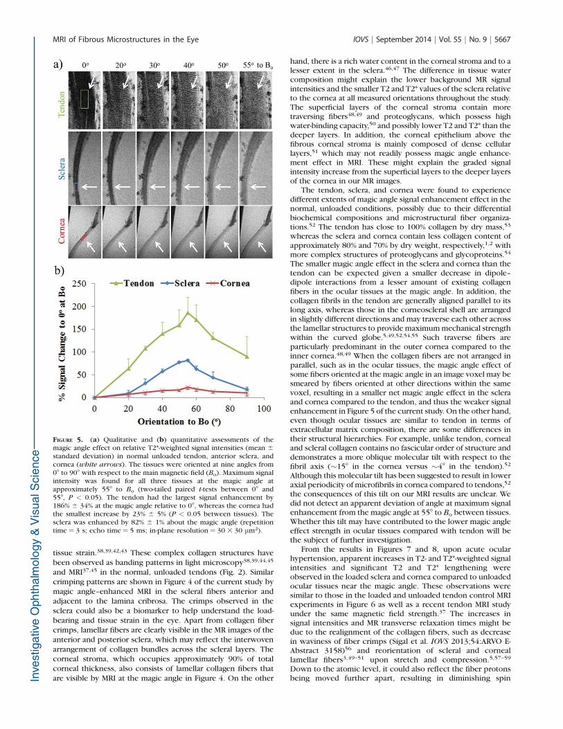
Using the MRI-compatible pneumatic automated rotatingpositioner (Fig. 1), the unloaded tendon, sclera, and corneawere oriented at different angles to Bo and were found toexperience different extents of magic angle MR signalenhancement (Fig. 5a). All three tissues possessed maximumsignal intensities in T2*-weighted MR images when they wereoriented about the magic angle at 558 to Bo (Fig. 5b). Themagic angle enhancement was found to be the strongest in thetendon (186% 6 34% at 558 relative to 08 to Bo) and the leaststrong in the cornea (23% 6 5% at 558 relative to 08 to Bo),whereas the sclera was enhanced by 82% 6 1% about themagic angle compared to 08 to Bo.
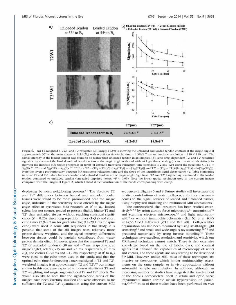
Biomechanical loading of the tendon and ocular tissuescaused increases in relative MR signal intensities as well asabsolute MR transverse relaxation times when the tissues wereoriented near the magic angle (Figs. 6–8). In Figure 6a, T2- and
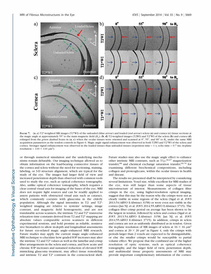
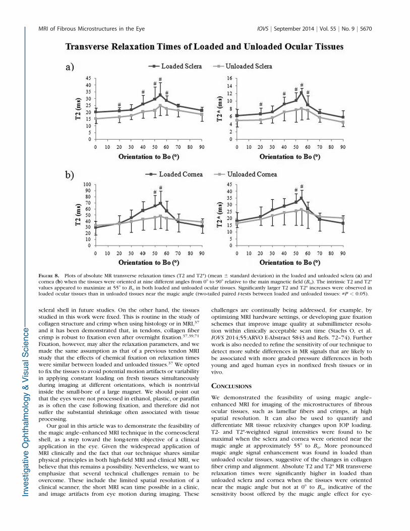
T2*-weighted MR images of unloaded tendon at the magicangle revealed some crimp patterns as observed in Figure 4even under a lower in-plane resolution at 110 3 110 lm2.Loaded tendon appeared to show higher intensities thanunloaded tendon in both T2- and T2*-weighted MR imaging atmagic angle. However, the collagen fiber crimps were lessapparent in loaded tendon. Figure 6b shows the echo time–dependent T2- and T2*-weighted signal decay curves of loadedand unloaded tendons at the magic angle. T2 and T2* MRtransverse relaxation times were derived from curve fitting ofthe slopes of the logarithmic signal decay curves given theirinversely proportional relationships. At the magic angle,intrinsic T2 (41.2 6 8.7 ms) and T2* (14.8 6 8.7 ms) valuesof the loaded tendon were significantly higher than those ofthe unloaded tendon (T2¼ 29.7 6 6.0 ms and T2*¼ 7.1 6 1.8ms) (one-tailed unpaired t-tests: P < 0.05). Figure 7 shows theT2- and T2*-weighted MR images of representative loaded andunloaded pairs of sclera (Figs. 7a, 7b) and cornea (Figs. 7c, 7d)at 08, 558, and 908 to Bo under identical MRI acquisitionparameters as the tendon in Figure 6. Magic angle signalenhancement was observed in both loaded and unloadedocular tissues, with the magic angle effect on loaded tissuesbeing apparently stronger than on the unloaded tissues.Quantitative comparisons of the absolute MR transverserelaxation times in Figure 8 confirmed these findings andshowed that the intrinsic T2 and T2* values of the sclera (Fig.8a) and cornea (Fig. 8b) were generally higher in loaded thanunloaded conditions at most orientations, whereas statisticalsignificance was reached when loaded and unloaded tissueswere oriented near the magic angle (two-tailed paired t-tests: P
< 0.05).
DISCUSSION
Our results support the hypothesis that high-field magic angle–enhanced MR microscopy can reveal details of the fibrousstructures in the ocular tissues without the need for exogenouscontrast agents or coatings. Our technique relied on the use ofthe magic angle effect that has been observed clinically in MRIstudies of fibrous tissues such as tendon28,29 and cartilage.32–35
Further, our results showed that this imaging technique canimprove the detection of the differences between unloadedand loaded ocular tissues in relative T2- and T2*-weighted MRsignal intensities as well as absolute T2 and T2* MR transverse
FIGURE 3. Representative T2*-weighted MR images of a sheep eye at three different orientations to the main magnetic field (Bo). Note the signalenhancement in the corneoscleral shell when the fiber orientation was near the magic angle at approximately 558 relative to the Bo direction (blue
arrows) compared to approximately 08 relative to Bo direction (red arrows) (repetition time¼ 3 s; echo time¼ 4 ms; in-plane resolution¼ 60 3 60lm2).
MRI of Fibrous Microstructures in the Eye IOVS j September 2014 j Vol. 55 j No. 9 j 5665
relaxation times. Upon magic angle enhancement, significantlylarger MR tissue relaxivity differences between unpressurizedand pressurized eyes were observed in the sclera and cornea.Thus, it may be possible to apply the magic angle effect toimprove sensitivity for MRI studies of the corneoscleral shell innormal and diseased conditions such as glaucoma.
In this study, high-resolution magic angle–enhanced MRI wasshown to be feasible for revealing tissue microstructures of thecorneoscleral shell, including banding indicative of collagen fibercrimp. The crimps arise from ordered collagenous fibers thatfollow zig-zag38–40 or helicoidal41 patterns in the tendon andocular tissues, respectively, and act as a functional biomarker of
FIGURE 4. (a) High-resolution T2*-weighted MR images of the normal unloaded tendon, anterior sclera, posterior sclera, and cornea positioned atthe magic angle at approximately 558 to the main magnetic field (Bo). Collagen fiber crimps (red arrows) were observed in the tendon positivecontrol and the anterior sclera. Lamellar fibers were observed along the anterior and posterior sclera, whereas differential image contrasts werefound across corneal layers at echo times of 9.5 and 20.5 ms. Outer layers are to the right in the sclera and cornea images. Repetition time for alltissues¼3 s; echo time of tendon, anterior sclera, and posterior sclera¼8, 9.5, and 8 ms, respectively; in-plane resolution of tendon, anterior sclera,posterior sclera, and cornea¼20 3 20, 16 3 16, 20 3 20, and 16 3 16 lm2, respectively. (b) T2*-weighted MR images of the unloaded sclera near theoptic nerve head. The left panels show the cross-section of the sclera, optic nerve head, and lamina cribrosa in sagittal view. The right panel showsthe coronal T2*-weighted image oriented as the blue box in the left panel at 16 3 16 lm2 in-plane resolution and repetition time/echo time¼ 3000/9.5 ms. Details of the lamina cribrosa (yellow arrow) within the optic nerve head and the distributions of crimps (red arrows) in the scleral fiberssurrounding the optic nerve head were revealed especially at orientations near the magic angle.
MRI of Fibrous Microstructures in the Eye IOVS j September 2014 j Vol. 55 j No. 9 j 5666
tissue strain.38,39,42,43 These complex collagen structures havebeen observed as banding patterns in light microscopy38,39,44,45
and MRI37,45 in the normal, unloaded tendons (Fig. 2). Similarcrimping patterns are shown in Figure 4 of the current study bymagic angle–enhanced MRI in the scleral fibers anterior andadjacent to the lamina cribrosa. The crimps observed in thesclera could also be a biomarker to help understand the load-bearing and tissue strain in the eye. Apart from collagen fibercrimps, lamellar fibers are clearly visible in the MR images of theanterior and posterior sclera, which may reflect the interwovenarrangement of collagen bundles across the scleral layers. Thecorneal stroma, which occupies approximately 90% of totalcorneal thickness, also consists of lamellar collagen fibers thatare visible by MRI at the magic angle in Figure 4. On the other
hand, there is a rich water content in the corneal stroma and to alesser extent in the sclera.46,47 The difference in tissue watercomposition might explain the lower background MR signalintensities and the smaller T2 and T2* values of the sclera relativeto the cornea at all measured orientations throughout the study.The superficial layers of the corneal stroma contain moretraversing fibers48,49 and proteoglycans, which possess highwater-binding capacity,50 and possibly lower T2 and T2* than thedeeper layers. In addition, the corneal epithelium above thefibrous corneal stroma is mainly composed of dense cellularlayers,51 which may not readily possess magic angle enhance-ment effect in MRI. These might explain the graded signalintensity increase from the superficial layers to the deeper layersof the cornea in our MR images.
The tendon, sclera, and cornea were found to experiencedifferent extents of magic angle signal enhancement effect in thenormal, unloaded conditions, possibly due to their differentialbiochemical compositions and microstructural fiber organiza-tions.52 The tendon has close to 100% collagen by dry mass,53
whereas the sclera and cornea contain less collagen content ofapproximately 80% and 70% by dry weight, respectively,1,2 withmore complex structures of proteoglycans and glycoproteins.54
The smaller magic angle effect in the sclera and cornea than thetendon can be expected given a smaller decrease in dipole–dipole interactions from a lesser amount of existing collagenfibers in the ocular tissues at the magic angle. In addition, thecollagen fibrils in the tendon are generally aligned parallel to itslong axis, whereas those in the corneoscleral shell are arrangedin slightly different directions and may traverse each other acrossthe lamellar structures to provide maximum mechanical strengthwithin the curved globe.5,49,52,54,55 Such traverse fibers areparticularly predominant in the outer cornea compared to theinner cornea.48,49 When the collagen fibers are not arranged inparallel, such as in the ocular tissues, the magic angle effect ofsome fibers oriented at the magic angle in an image voxel may besmeared by fibers oriented at other directions within the samevoxel, resulting in a smaller net magic angle effect in the scleraand cornea compared to the tendon, and thus the weaker signalenhancement in Figure 5 of the current study. On the other hand,even though ocular tissues are similar to tendon in terms ofextracellular matrix composition, there are some differences intheir structural hierarchies. For example, unlike tendon, cornealand scleral collagen contains no fascicular order of structure anddemonstrates a more oblique molecular tilt with respect to thefibril axis (~158 in the cornea versus ~48 in the tendon).52
Although this molecular tilt has been suggested to result in loweraxial periodicity of microfibrils in cornea compared to tendons,52
the consequences of this tilt on our MRI results are unclear. Wedid not detect an apparent deviation of angle at maximum signalenhancement from the magic angle at 558 to Bo between tissues.Whether this tilt may have contributed to the lower magic angleeffect strength in ocular tissues compared with tendon will bethe subject of further investigation.
From the results in Figures 7 and 8, upon acute ocularhypertension, apparent increases in T2- and T2*-weighted signalintensities and significant T2 and T2* lengthening wereobserved in the loaded sclera and cornea compared to unloadedocular tissues near the magic angle. These observations weresimilar to those in the loaded and unloaded tendon control MRIexperiments in Figure 6 as well as a recent tendon MRI studyunder the same magnetic field strength.37 The increases insignal intensities and MR transverse relaxation times might bedue to the realignment of the collagen fibers, such as decreasein waviness of fiber crimps (Sigal et al. IOVS 2013;54:ARVO E-Abstract 3158)56 and reorientation of scleral and corneallamellar fibers3,49–51 upon stretch and compression.5,57–59
Down to the atomic level, it could also reflect the fiber protonsbeing moved further apart, resulting in diminishing spin
FIGURE 5. (a) Qualitative and (b) quantitative assessments of themagic angle effect on relative T2*-weighted signal intensities (mean 6standard deviation) in normal unloaded tendon, anterior sclera, andcornea (white arrows). The tissues were oriented at nine angles from08 to 908 with respect to the main magnetic field (Bo). Maximum signalintensity was found for all three tissues at the magic angle atapproximately 558 to Bo (two-tailed paired t-tests between 08 and558, P < 0.05). The tendon had the largest signal enhancement by186% 6 34% at the magic angle relative to 08, whereas the cornea hadthe smallest increase by 23% 6 5% (P < 0.05 between tissues). Thesclera was enhanced by 82% 6 1% about the magic angle (repetitiontime ¼ 3 s; echo time¼ 5 ms; in-plane resolution¼ 30 3 30 lm2).
MRI of Fibrous Microstructures in the Eye IOVS j September 2014 j Vol. 55 j No. 9 j 5667
dephasing between neighboring protons.37 The absolute T2and T2* differences between loaded and unloaded oculartissues were found to be more pronounced near the magicangle, indicative of the sensitivity boost offered by the magicangle effect in eye-related MRI research. At 08 to Bo, loadedsclera, but not cornea, tended to possess slightly higher T2 andT2* than unloaded tissues without reaching statistical signifi-cance (P¼ 0.26). Since long repetition times (1–3 s) and shortecho times (2.9–9.7 ms for gradient-echo; 9.7–29.1 ms for spin-echo) were used in some MRI sequences in this study, it ispossible that some of the MR images were relatively moreproton-density weighted, and the signal intensity differencesbetween tissues could be partially contributed from waterproton density effect. However, given that the measured T2 andT2* of unloaded tendon (~30 ms and ~7 ms, respectively, atmagic angle), sclera (~15 ms and ~5 ms, respectively, at 08 toBo), and cornea (~30 ms and ~17 ms, respectively, at 08 to Bo)were close to the echo times used in this study, and that theoptimal echo time for detecting a maximal signal in T2- and T2*-weighted imaging is at approximately T2 and T2*,60 our imagesshown in this study are expected to possess significant T2 andT2* weighting and magic angle–induced T2 and T2* effects. Wewould also like to note that the signal-to-noise ratios of theimages have been carefully assessed and were observed to besufficient for T2 and T2* quantitation using the current MRI
sequences in Figures 6 and 8. Future studies will investigate therelative contributions of water, collagen, and other macromol-ecules to the signal sources of loaded and unloaded tissues,using biophysical modeling and multimodal MRI assessments.
The corneoscleral shell structure has been studied exten-sively52,55 by using atomic force microscopy,61 transmission62
and scanning electron microscopy,63 and light microscopywith4 or without immunohistochemistry (Jan NJ, et al. IOVS
2014;55:ARVO E-Abstract 3715 and Ref. 48). Collagen fiberorganization has also been measured by using small-angle lightscattering64 and small- and wide-angle x-ray scattering,52,55 andpredicted numerically by using inverse modeling.56 Thesetechniques have excellent resolution and sensitivity, which ourMRI-based technique cannot match. There is also extensiveknowledge based on the use of labels, dyes, and contrastagents that enhance the capabilities of microscopy of scleraand cornea, and these agents are only starting to be developedfor MRI. However, unlike MRI, most of these techniques areinvasive or destructive, which hinder multimodality assess-ments on the same sample, or in vivo applications withoutsubstantial sample manipulation. In addition, although anincreasing number of studies have suggested the involvementof the fibrous corneoscleral shell in retina and optic nerveimpairments under chronic ocular hypertension or glauco-ma,25,26,65 most of these studies have been performed ex vivo
FIGURE 6. (a) T2-weighted (T2WI) and T2*-weighted MR images (T2*WI) showing the unloaded and loaded tendon controls at the magic angle atapproximately 558 to the main magnetic field (Bo) with repetition time/echo time ¼ 1000/9.7 ms and in-plane resolution ¼ 110 3 110 lm2. Thesignal intensity in the loaded tendon was found to be higher than unloaded tendon in all samples. (b) Echo time–dependent T2- and T2*-weightedsignal decay curves of the loaded and unloaded tendons at the magic angle with and without logarithmic scaling (mean 6 standard deviation) forderiving the intrinsic MRI tissue properties in terms of absolute transverse relaxation time constants (T2 and T2*) using the equations SSE(TE) ¼SSE(0)e(�TE/T2) and SGE(TE)¼ SGE(0)e(�TE/T2*), or T2¼ (TE2� TE1)/{ln[SSE(TE1)]� ln[SSE(TE2)]} and T2*¼ (TE2� TE1)/{ln[SGE(TE1)]� ln[SGE(TE2)]}.Note the inverse proportionality between MR transverse relaxation time and the slope of the logarithmic signal decay curve. (c) Table comparingintrinsic T2 and T2* values between loaded and unloaded tendons at the magic angle. Significant T2 and T2* lengthening was found in the loadedtendon compared to unloaded tendon (one-tailed unpaired t-tests: #P < 0.05). Note the lower spatial resolution used in the current imagescompared with the images of Figure 2, which limited direct visualization of the bands corresponding with crimp.
MRI of Fibrous Microstructures in the Eye IOVS j September 2014 j Vol. 55 j No. 9 j 5668
or through numerical simulation and the underlying mecha-nisms remain debatable. Our imaging technique allowed us toobtain information on the load-bearing connective tissues ofthe cornea and sclera without the need for sectioning, staining,labeling, or 3-D structure alignment, which are typical for thestudy of the eye. The images had larger field of view andincreased penetration depth than observed with common toolsused to study the eye, such as optical coherence tomography.Also, unlike optical coherence tomography, which requires aclear central visual axis for imaging of the bases of the eye, MRIdoes not require light sources and can be readily applied toassess patients with obstructed visual axis such as cataract,which commonly coexists with glaucoma in the elderlypopulation. Although the signal intensities in T2- and T2*-weighted imaging are relative to systems’ settings, imagecontrast, and magnetic field homogeneity, and are nottranslatable across scanners, the intrinsic T2 and T2* transverserelaxation time constants derived from T2 and T2* mapping areabsolute values comparable across scanners in differentexperimental sessions, and hold promise as reliable, noninva-sive biomarkers to allow in-depth and longitudinal assessmentsfor future eye-related magic angle–enhanced MRI research.Future studies may apply the current magic angle–enhancedMRI technique to elucidate how graded IOP increases may alterthe intrinsic T2 and T2* values as well as the lamellar and crimpfiber arrangements in the sclera and cornea, and how acute andchronic IOP increases and sustained reduction of elevated IOP,mimicking glaucoma treatment, may affect tissue remodelingand intrinsic T2 and T2* contrasts in the corneoscleral shell.
Future studies may also use the magic angle effect to enhanceother intrinsic MRI contrasts, such as T1q,66,67 magnetizationtransfer,68 and chemical exchange saturation transfer,69,70 forexamining different biochemical compositions, includingcollagen and proteoglycans, within the ocular tissues in healthand disease.
The results we presented shall be interpreted by consideringseveral limitations. Voxel size, while excellent for MRI studies ofthe eye, was still larger than some aspects of tissuemicrostructure of interest. Measurements of collagen fibercrimps in the eye, using higher-resolution optical imaging,suggest that this may be the reason why the crimps were not asclearly visible in some regions of the sclera (Sigal et al. IOVS
2013;54:ARVO E-Abstract 3158) or were even not visible in thecornea (Jan NJ, et al. IOVS 2014;55:ARVO E-Abstract 3715). Thecollagen fiber crimp period on average has been shown to bethe largest in tendon, followed by sclera and cornea (Sigal et al.IOVS 2013;54:ARVO E-Abstract 3158; Jan NJ, et al. IOVS
2014;55:ARVO E-Abstract 3715). In addition, crimps in scleraand cornea are not as uniformly distributed as in tendon. Giventhe in-plane resolution of MR images of sclera at 16 3 16 lm2
and cornea at 20 3 20 lm2 in Figure 4, only the crimps withperiods larger than 2 voxels are expected to be distinguishable,or else the smaller crimps would be susceptible to partialvolume effect. We propose that the combined use of the higherresolution of optic systems, such as optical coherencetomography, and the larger field of view, penetration depth,and multimodal tissue property assessments of MRI mayprovide important complementary information of the corneo-
FIGURE 7. (a, c) T2*-weighted MR images (T2*WI) of the unloaded (blue arrow) and loaded (red arrow) sclera (a) and cornea (c) tissue sections atthe magic angle at approximately 558 to the main magnetic field (Bo); (b, d) T2-weighted images (T2WI) and T2*WI of the sclera (b) and cornea (d)enlarged from the green dashed boxes in (a, c) when the ocular tissues were oriented and scanned at 08, 558, and 908 to Bo under the same MRIacquisition parameters as the tendon controls in Figure 6. Magic angle signal enhancement was observed in both T2WI and T2*WI of the sclera andcornea. Stronger signal enhancement was observed in the loaded tissues than unloaded tissues (repetition time¼ 1 s; echo time¼ 9.7 ms; in-planeresolution¼ 110 3 110 lm2).
MRI of Fibrous Microstructures in the Eye IOVS j September 2014 j Vol. 55 j No. 9 j 5669
scleral shell in future studies. On the other hand, the tissuesstudied in this work were fixed. This is routine in the study ofcollagen structure and crimp when using histology or in MRI,37
and it has been demonstrated that, in tendons, collagen fibercrimp is robust to fixation even after overnight fixation.37,39,71
Fixation, however, may alter the relaxation parameters, and wemade the same assumption as that of a previous tendon MRIstudy that the effects of chemical fixation on relaxation timeswere similar between loaded and unloaded tissues.37 We optedto fix the tissues to avoid potential motion artifacts or variabilityin applying constant loading on fresh tissues simultaneouslyduring imaging at different orientations, which is nontrivialinside the small-bore of a large magnet. We should point outthat the eyes were not processed in ethanol, plastic, or paraffinas is often the case following fixation, and therefore did notsuffer the substantial shrinkage often associated with tissueprocessing.
Our goal in this article was to demonstrate the feasibility ofthe magic angle–enhanced MRI technique in the corneoscleralshell, as a step toward the long-term objective of a clinicalapplication in the eye. Given the widespread application ofMRI clinically and the fact that our technique shares similarphysical principles in both high-field MRI and clinical MRI, webelieve that this remains a possibility. Nevertheless, we want toemphasize that several technical challenges remain to beovercome. These include the limited spatial resolution of aclinical scanner, the short MRI scan time possible in a clinic,and image artifacts from eye motion during imaging. These
challenges are continually being addressed, for example, byoptimizing MRI hardware settings, or developing gaze fixationschemes that improve image quality at submillimeter resolu-tion within clinically acceptable scan time (Stachs O, et al.IOVS 2014;55:ARVO E-Abstract 5843 and Refs. 72–74). Furtherwork is also needed to refine the sensitivity of our technique todetect more subtle differences in MR signals that are likely tobe associated with more graded pressure differences in bothyoung and aged human eyes in nonfixed fresh tissues or invivo.
CONCLUSIONS
We demonstrated the feasibility of using magic angle–enhanced MRI for imaging of the microstructures of fibrousocular tissues, such as lamellar fibers and crimps, at highspatial resolution. It can also be used to quantify anddifferentiate MR tissue relaxivity changes upon IOP loading.T2- and T2*-weighted signal intensities were found to bemaximal when the sclera and cornea were oriented near themagic angle at approximately 558 to Bo. More pronouncedmagic angle signal enhancement was found in loaded thanunloaded ocular tissues, suggestive of the changes in collagenfiber crimp and alignment. Absolute T2 and T2* MR transverserelaxation times were significantly higher in loaded thanunloaded sclera and cornea when the tissues were orientednear the magic angle but not at 08 to Bo, indicative of thesensitivity boost offered by the magic angle effect for eye-
FIGURE 8. Plots of absolute MR transverse relaxation times (T2 and T2*) (mean 6 standard deviation) in the loaded and unloaded sclera (a) andcornea (b) when the tissues were oriented at nine different angles from 08 to 908 relative to the main magnetic field (Bo). The intrinsic T2 and T2*values appeared to maximize at 558 to Bo in both loaded and unloaded ocular tissues. Significantly larger T2 and T2* increases were observed inloaded ocular tissues than in unloaded tissues near the magic angle (two-tailed paired t-tests between loaded and unloaded tissues: #P < 0.05).
MRI of Fibrous Microstructures in the Eye IOVS j September 2014 j Vol. 55 j No. 9 j 5670
related MRI research. We demonstrated a promising techniquethat may open up new avenues of noninvasive assessments ofthe structural, biomechanical, and biochemical characteristicsof the sclera and cornea. Although important technicalchallenges remain to the application in vivo, this techniquehas the potential to enable cross-sectional and longitudinalmonitoring of the functional microstructures of the eye andtheir relationship with aging and diseases involving thecorneoscleral shell, such as acute and chronic ocular hyper-tension, glaucoma, and myopia.
Acknowledgments
The authors thank Chan-Hong Moon, PhD, of the Department ofRadiology at the University of Pittsburgh for his helpful commentson the manuscript.
Supported by the National Institutes of Health Contracts P30-EY008098, R01-EY023966, and UL1-TR000005 (Bethesda, MD,USA); BrightFocus Foundation G2013077 (Clarksburg, MD, USA);Alcon Research Institute Young Investigator Grant (Basel, Switzer-land); Eye and Ear Foundation (Pittsburgh, PA, USA); and Researchto Prevent Blindness (New York, NY, USA).
Disclosure: L.C. Ho, None; I.A. Sigal, None; N.-J. Jan, None; A.Squires, None; Z. Tse, None; E.X. Wu, None; S.-G. Kim, None;J.S. Schuman, None; K.C. Chan, None
References
1. Maurice DM. The Cornea and Sclera. 3rd ed. New York:Academic Press; 1984.
2. Foster CS, de la Maza MS. The Sclera. New York: Springer-Verlag; 1994.
3. Yamabayashi S, Ohno S, Aguilar RN, Furuya T, Hosoda M,Tsukahara S. Ultrastructural studies of collagen fibers of thecornea and sclera by a quick-freezing and deep-etchingmethod. Ophthalmic Res. 1991;23:320–329.
4. Rada JA, Shelton S, Norton TT. The sclera and myopia. Exp Eye
Res. 2006;82:185–200.
5. Pijanka JK, Coudrillier B, Ziegler K, et al. Quantitative mappingof collagen fiber orientation in non-glaucoma and glaucomaposterior human sclerae. Invest Ophthalmol Vis Sci. 2012;53:5258–5270.
6. Sigal IA, Flanagan JG, Ethier CR. Factors influencing opticnerve head biomechanics. Invest Ophthalmol Vis Sci. 2005;46:4189–4199.
7. Quigley HA. Glaucoma: macrocosm to microcosm theFriedenwald lecture. Invest Ophthalmol Vis Sci. 2005;46:2662–2670.
8. Chan KC, Fu QL, Hui ES, So KF, Wu EX. Evaluation of the retinaand optic nerve in a rat model of chronic glaucoma using invivo manganese-enhanced magnetic resonance imaging. Neu-
roimage. 2008;40:1166–1174.
9. Chan KC, Fan SJ, Chan RW, Cheng JS, Zhou IY, Wu EX. In vivovisuotopic brain mapping with manganese-enhanced MRI andresting-state functional connectivity MRI. Neuroimage. 2014;90:235–245.
10. Shih YY, De la Garza BH, Muir ER, et al. Lamina-specificfunctional MRI of retinal and choroidal responses to visualstimuli. Invest Ophthalmol Vis Sci. 2011;52:5303–5310.
11. Berkowitz BA, Sato Y, Wilson CA, de Juan E. Blood-retinalbarrier breakdown investigated by real-time magnetic reso-nance imaging after gadolinium-diethylenetriaminepentaaceticacid injection. Invest Ophthalmol Vis Sci. 1991;32:2854–2860.
12. Chan KC, Cheng JS, Fan S, Zhou IY, Yang J, Wu EX. In vivoevaluation of retinal and callosal projections in early postnataldevelopment and plasticity using manganese-enhanced MRI
and diffusion tensor imaging. Neuroimage. 2012;59:2274–2283.
13. Cheng H, Nair G, Walker TA, et al. Structural and functionalMRI reveals multiple retinal layers. Proc Natl Acad Sci U S A.2006;103:17525–17530.
14. Duong TQ, Ngan SC, Ugurbil K, Kim SG. Functional magneticresonance imaging of the retina. Invest Ophthalmol Vis Sci.2002;43:1176–1181.
15. Chan KC, Fu QL, Guo H, So KF, Wu EX. GD-DTPA enhancedMRI of ocular transport in a rat model of chronic glaucoma.Exp Eye Res. 2008;87:334–341.
16. Berkowitz BA, Roberts R, Luan H, Peysakhov J, Mao X, ThomasKA. Dynamic contrast-enhanced MRI measurements of passivepermeability through blood retinal barrier in diabetic rats.Invest Ophthalmol Vis Sci. 2004;45:2391–2398.
17. Ho LC, Conner IP, Do CW, et al. In vivo assessment of aqueoushumor dynamics upon chronic ocular hypertension andhypotensive drug treatment using gadolinium-enhanced MRI.Invest Ophthalmol Vis Sci. 2014;55:3747–3757.
18. Sadun AA, Carelli V, Bose S, Ross-Cisneros FN, Barboni P,Ahrens ET. First application of extremely high-resolutionmagnetic resonance imaging to study microscopic featuresof normal and LHON human optic nerve. Ophthalmology.2002;109:1085–1091.
19. Norman RE, Flanagan JG, Rausch SM, et al. Dimensions of thehuman sclera: thickness measurement and regional changeswith axial length. Exp Eye Res. 2010;90:277–284.
20. Georgouli T, Chang B, Nelson M, et al. Use of high-resolutionmicroscopy coil MRI for depicting orbital anatomy. Orbit.2008;27:107–114.
21. Goodall N, Kisiswa L, Prashar A, et al. 3-Dimensional modellingof chick embryo eye development and growth using highresolution magnetic resonance imaging. Exp Eye Res. 2009;89:511–521.
22. Singh KD, Logan NS, Gilmartin B. Three-dimensional modelingof the human eye based on magnetic resonance imaging.Invest Ophthalmol Vis Sci. 2006;47:2272–2279.
23. Langner S, Martin H, Terwee T, et al. 7.1 T MRI to assess theanterior segment of the eye. Invest Ophthalmol Vis Sci. 2010;51:6575–6581.
24. Duong TQ, Pardue MT, Thule PM, et al. Layer-specificanatomical, physiological and functional MRI of the retina.NMR Biomed. 2008;21:978–996.
25. Nguyen C, Cone FE, Nguyen TD, et al. Studies of scleralbiomechanical behavior related to susceptibility for retinalganglion cell loss in experimental mouse glaucoma. Invest
Ophthalmol Vis Sci. 2013;54:1767–1780.
26. Sigal IA, Ethier CR. Biomechanics of the optic nerve head. Exp
Eye Res. 2009;88:799–807.
27. Xia Y. Magic-angle effect in magnetic resonance imaging ofarticular cartilage: a review. Invest Radiol. 2000;35:602–621.
28. Fullerton GD, Cameron IL, Ord VA. Orientation of tendons inthe magnetic field and its effect on T2 relaxation times.Radiology. 1985;155:433–435.
29. Erickson SJ, Cox IH, Hyde JS, Carrera GF, Strandt JA, EstkowskiLD. Effect of tendon orientation on MR imaging signalintensity: a manifestation of the ‘‘magic angle’’ phenomenon.Radiology. 1991;181:389–392.
30. Peterfy CG, Janzen DL, Tirman PF, van Dijke CF, Pollack M,Genant HK. ‘‘Magic-angle’’ phenomenon: a cause of increasedsignal in the normal lateral meniscus on short-TE MR images ofthe knee. AJR Am J Roentgenol. 1994;163:149–154.
31. Werpy NM, Ho CP, Kawcak CE. Magic angle effect in normalcollateral ligaments of the distal interphalangeal joint in horsesimaged with a high-field magnetic resonance imaging system.Vet Radiol Ultrasound. 2010;51:2–10.
MRI of Fibrous Microstructures in the Eye IOVS j September 2014 j Vol. 55 j No. 9 j 5671
32. Erickson SJ, Prost RW, Timins ME. The ‘‘magic angle’’ effect:background physics and clinical relevance. Radiology. 1993;188:23–25.
33. Benjamin M, Bydder GM. Magnetic resonance imaging ofentheses using ultrashort TE (UTE) pulse sequences. J Magn
Reson Imaging. 2007;25:381–389.
34. Rubenstein JD, Kim JK, Morova-Protzner I, Stanchev PL,Henkelman RM. Effects of collagen orientation on MR imagingcharacteristics of bovine articular cartilage. Radiology. 1993;188:219–226.
35. Henkelman RM, Stanisz GJ, Kim JK, Bronskill MJ. Anisotropy ofNMR properties of tissues. Magn Reson Med. 1994;32:592–601.
36. Mershon C, Squires A, Gao Y, Chan KC, Tse Z. Magic angleenhanced imaging in high-field MRI using an automated MR-conditional positioner. Proc Intl Soc Magn Reson Med. 2013;21:3474.
37. Mountain KM, Bjarnason TA, Dunn JF, Matyas JR. Thefunctional microstructure of tendon collagen revealed byhigh-field MRI. Magn Reson Med. 2011;66:520–527.
38. Diamant J, Keller A, Baer E, Litt M, Arridge RG. Collagen;ultrastructure and its relation to mechanical properties as afunction of ageing. Proc R Soc Lond B Biol Sci. 1972;180:293–315.
39. Gathercole LJ, Keller A. Crimp morphology in the fibre-forming collagens. Matrix. 1991;11:214–234.
40. Franchi M, Fini M, Quaranta M, et al. Crimp morphology inrelaxed and stretched rat Achilles tendon. J Anat. 2007;210:1–7.
41. Grytz R, Meschke G. Constitutive modeling of crimpedcollagen fibrils in soft tissues. J Mech Behav Biomed Mater.2009;2:522–533.
42. Elliott DH. Structure and function of mammalian tendon. Biol
Rev Camb Philos Soc. 1965;40:392–421.
43. Vidik A, Ekholm R. Light and electron microscopic studies ofcallagen fibers under strain. Z Anat Entwicklungsgesch. 1968;127:154–164.
44. Rigby BJ, Hirai N, Spikes JD, Eyring H. The mechanicalproperties of rat tail tendon. J Gen Physiol. 1959;43:265–283.
45. Pierre-Jerome C, Moncayo V, Terk MR. MRI of the Achillestendon: a comprehensive review of the anatomy, biomechan-ics, and imaging of overuse tendinopathies. Acta Radiol. 2010;51:438–454.
46. Dohlman CH, Bostrom H. Uptake of sulfate by mucopolysac-charides in the rat cornea and sclera. Acta Ophthalmol. 1955;33:455–461.
47. Kaye GI. Stereologic measurement of cell volume fraction ofrabbit corneal stroma. Arch Ophthalmol. 1969;82:792–794.
48. Winkler M, Shoa G, Xie Y, et al. Three-dimensional distributionof transverse collagen fibers in the anterior human cornealstroma. Invest Ophthalmol Vis Sci. 2013;54:7293–7301.
49. Winkler M, Chai D, Kriling S, et al. Nonlinear opticalmacroscopic assessment of 3-D corneal collagen organizationand axial biomechanics. Invest Ophthalmol Vis Sci. 2011;52:8818–8827.
50. William T, Jaeger EA. Duane’s Ophthalmology on DVD-ROM
Edition 2006 [DVD]. Philadelphia, PA: Lippincott Williams &Wilkins; 2006;8.
51. DelMonte DW, Kim T. Anatomy and physiology of the cornea. J
Cataract Refract Surg. 2011;37:588–598.
52. Meek KM, Boote C. The organization of collagen in the cornealstroma. Exp Eye Res. 2004;78:503–512.
53. Fullerton GD, Rahal A. Collagen structure: the molecularsource of the tendon magic angle effect. J Magn Reson
Imaging. 2007;25:345–361.
54. McBrien NA, Jobling AI, Gentle A. Biomechanics of the sclerain myopia: extracellular and cellular factors. Optom Vis Sci.2009;86:E23–E30.
55. Meek KM, Boote C. The use of X-ray scattering techniques toquantify the orientation and distribution of collagen in thecorneal stroma. Prog Retin Eye Res. 2009;28:369–392.
56. Grytz R, Meschke G. A computational remodeling approach topredict the physiological architecture of the collagen fibrilnetwork in corneo-scleral shells. Biomech Model Mechano-
biol. 2010;9:225–235.
57. Fazio MA, Grytz R, Morris JS, et al. Age-related changes inhuman peripapillary scleral strain. Biomech Model Mechano-
biol. 2013;13:551–563.
58. Girard MJ, Dahlmann-Noor A, Rayapureddi S, et al. Quantita-tive mapping of scleral fiber orientation in normal rat eyes.Invest Ophthalmol Vis Sci. 2011;52:9684–9693.
59. Sigal IA, Grimm JL, Jan NJ, Reid K, Minckler DS, Brown DJ. Eye-specific IOP-induced displacements and deformations ofhuman lamina cribrosa. Invest Ophthalmol Vis Sci. 2014;55:1–15.
60. Bandettini PA, Wong EC, Jesmanowicz A, Hinks RS, Hyde JS.Spin-echo and gradient-echo EPI of human brain activationusing BOLD contrast: a comparative study at 1.5 T. NMR
Biomed. 1994;7:12–20.
61. Choi S, Cheong Y, Lee HJ, Lee SJ, Jin KH, Park HK. AFM studyfor morphological and mechanical properties of human scleralsurface. J Nanosci Nanotechnol. 2011;11:6382–6388.
62. Doughty MJ. Observations on the ultrastructure of equatorialscleral collagen fibrils in sheep eyes. Vet Ophthalmol. 2012;15:71–80.
63. Meek KM, Fullwood NJ. Corneal and scleral collagens—amicroscopist’s perspective. Micron. 2001;32:261–272.
64. Danford FL, Yan D, Dreier RA, Cahir TM, Girkin CA, VandeGeest JP. Differences in the region- and depth-dependentmicrostructural organization in normal versus glaucomatoushuman posterior sclerae. Invest Ophthalmol Vis Sci. 2013;54:7922–7932.
65. Burgoyne CF. A biomechanical paradigm for axonal insultwithin the optic nerve head in aging and glaucoma. Exp Eye
Res. 2011;93:120–132.
66. Du J, Statum S, Znamirowski R, Bydder GM, Chung CB,Ultrashort TE. T1rho magic angle imaging. Magn Reson Med.2013;69:682–687.
67. Menezes NM, Gray ML, Hartke JR, Burstein D. T2 and T1rhoMRI in articular cartilage systems. Magn Reson Med. 2004;51:503–509.
68. Kim DK, Ceckler TL, Hascall VC, Calabro A, Balaban RS.Analysis of water-macromolecule proton magnetization trans-fer in articular cartilage. Magn Reson Med. 1993;29:211–215.
69. Saar G, Zhang B, Ling W, Regatte RR, Navon G, Jerschow A.Assessment of glycosaminoglycan concentration changes inthe intervertebral disc via chemical exchange saturationtransfer. NMR Biomed. 2012;25:255–261.
70. Singh A, Haris M, Cai K, et al. Chemical exchange saturationtransfer magnetic resonance imaging of human knee cartilageat 3 T and 7 T. Magn Reson Med. 2012;68:588–594.
71. Matyas J, Edwards P, Miniaci A, et al. Ligament tension affectsnuclear shape in situ: an in vitro study. Connect Tissue Res.1994;31:45–53.
72. Richdale K, Wassenaar P, Teal Bluestein K, et al. 7 Tesla MRimaging of the human eye in vivo. J Magn Reson Imaging.2009;30:924–932.
73. Graessl A, Muhle M, Schwerter M, et al. Ophthalmic magneticresonance imaging at 7 T using a 6-channel transceiverradiofrequency coil array in healthy subjects and patientswith intraocular masses. Invest Radiol. 2014;49:260–270.
74. Zhang Y, Nateras OS, Peng Q, et al. Lamina-specific anatomicmagnetic resonance imaging of the human retina. Invest
Ophthalmol Vis Sci. 2011;52:7232–7237.
MRI of Fibrous Microstructures in the Eye IOVS j September 2014 j Vol. 55 j No. 9 j 5672