Embed Size (px)
Citation preview
11/4/2018
1
Macular Degeneration and Diabetic Retinopathy:Past, Present & Future Daniel K. Bennett, M.D.
Disclosures
Genentech – Speaker and Advisor
11/4/2018
2
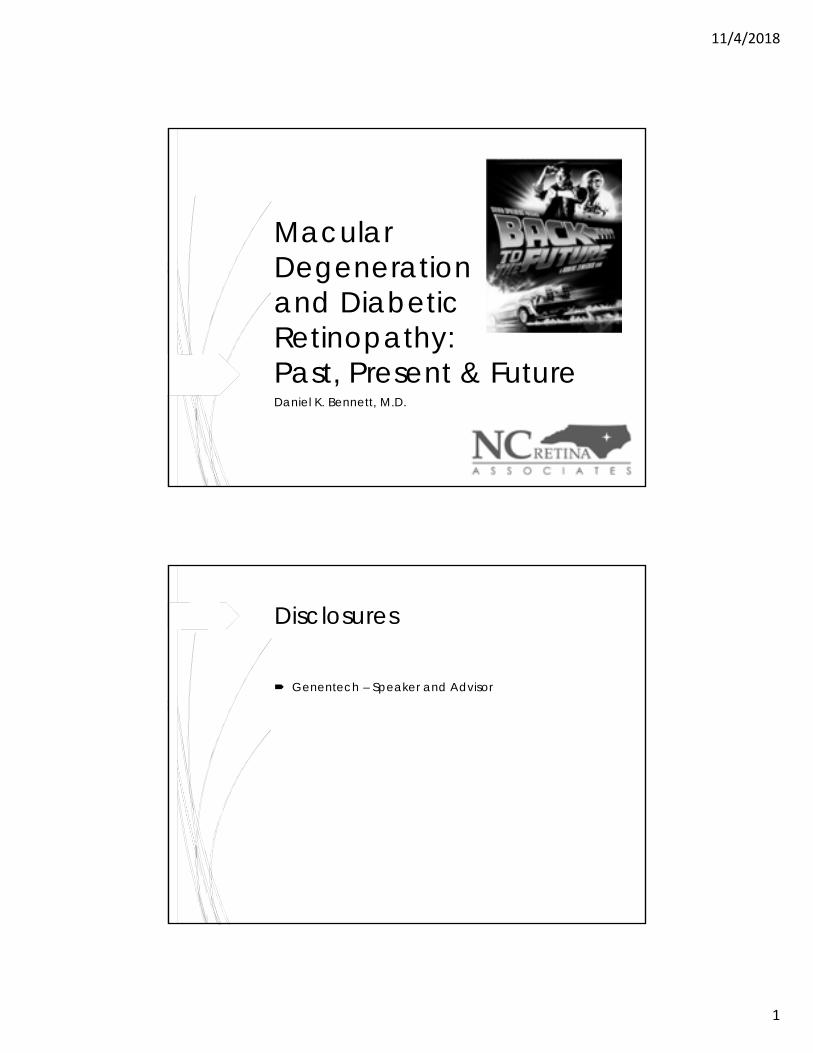
AMD Statistics
Health Care Cost $98 billion in U.S., Canada and Cuba
$343 billion worldwide
Cost of Vision Loss (All Causes) ~$700 billion in U.S.
~$3 trillion worldwide
Substantially higher cost >64yo
Diabetes Statistics
11/4/2018
3
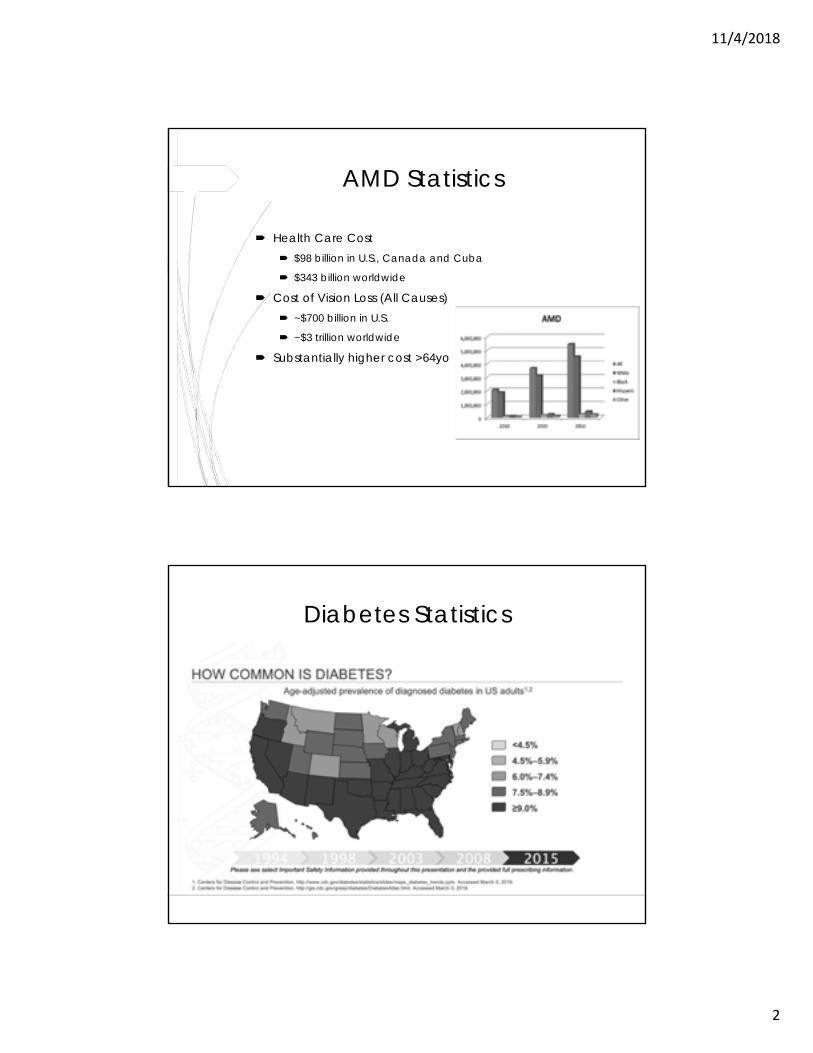
Imaging
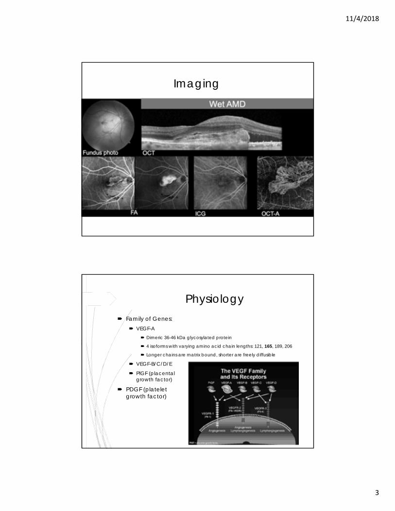
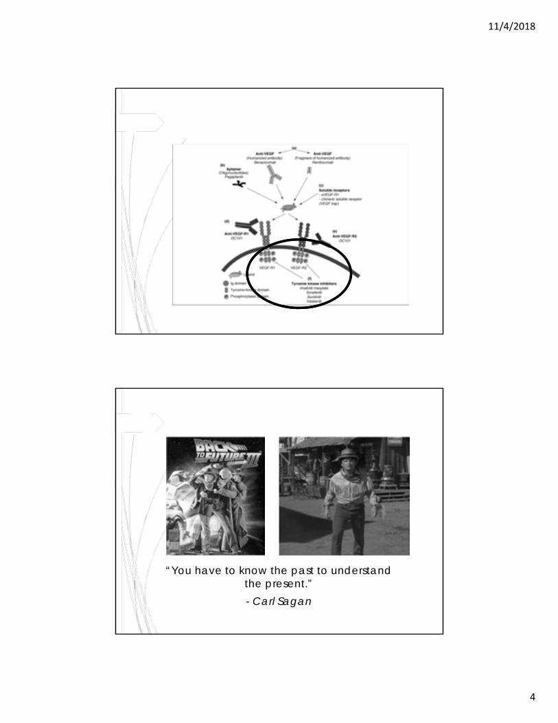
Physiology Family of Genes:
VEGF-A Dimeric 36-46 kDa glycosylated protein
4 isoforms with varying amino acid chain lengths: 121, 165, 189, 206
Longer chains are matrix bound, shorter are freely diffusible
VEGF-B/C/D/E
PlGF (placental growth factor)
PDGF (platelet derived growth factor)
11/4/2018
4
Tyrosine Kinase Pathway
“You have to know the past to understand the present.”- Carl Sagan
11/4/2018
5
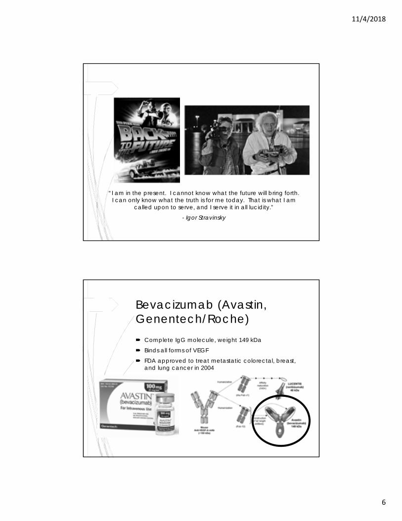
Learning from our mistakes…
Laser Photocoagulation
Macular Photocoagulation Study (MPS) Argon or krypton laser for extra- and juxtafoveal classic
SRNVM versus observation alone
Slowed progression, but eventual loss of foveal vision
Photodynamic Therapy (PDT) Non-thermal, reactive O2 molecules
11/4/2018
6
“I am in the present. I cannot know what the future will bring forth. I can only know what the truth is for me today. That is what I am
called upon to serve, and I serve it in all lucidity.”
- Igor Stravinsky
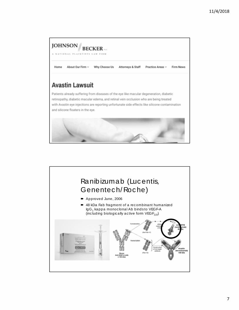
Bevacizumab (Avastin, Genentech/Roche) Complete IgG molecule, weight 149 kDa
Binds all forms of VEGF
FDA approved to treat metastatic colorectal, breast, and lung cancer in 2004
11/4/2018
7
Concerns
“Off-Label”
Compounding pharmacies
Silicone oil bubbles
Efficacy and durability
Ranibizumab (Lucentis, Genentech/Roche) Approved June, 2006
48 kDa Fab fragment of a recombinant humanized IgG1 kappa monoclonal Ab binds to VEGF-A (including biologically active form VEGF110)
11/4/2018
8
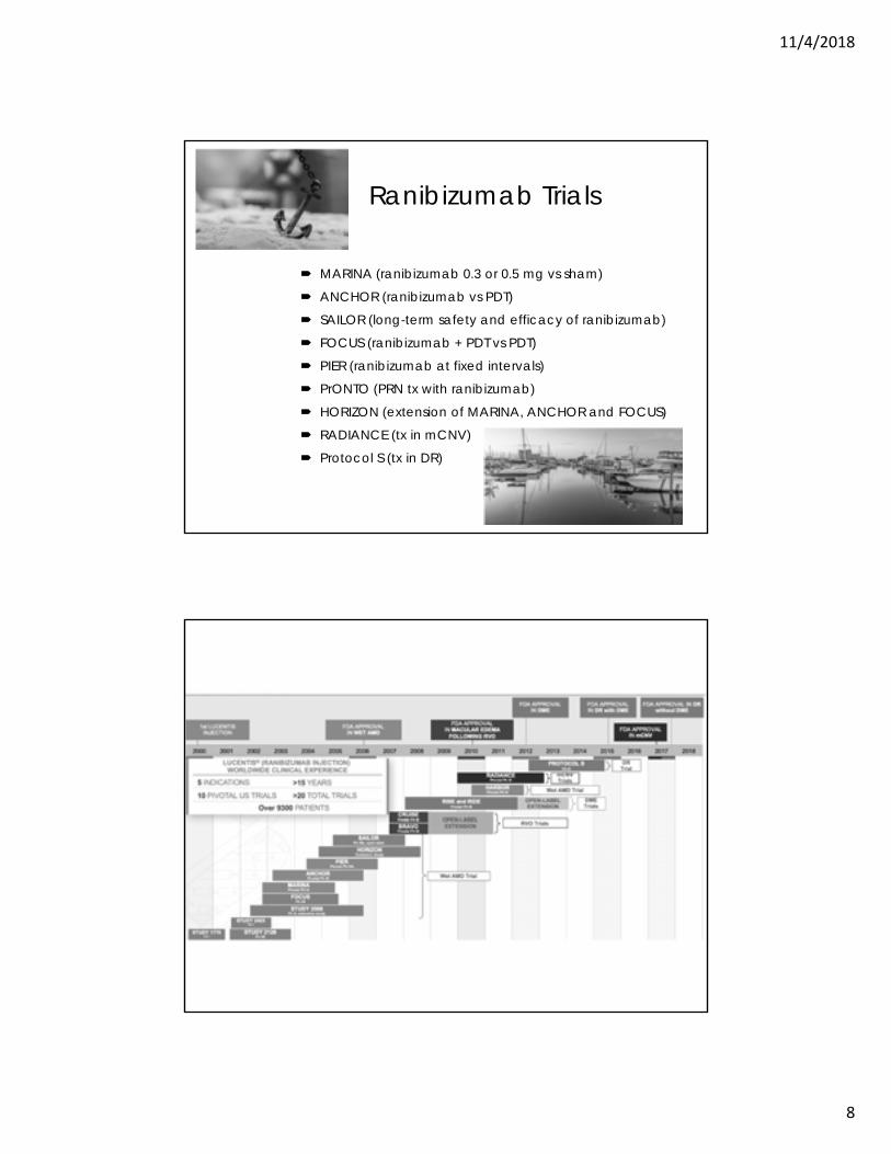
Ranibizumab Trials
MARINA (ranibizumab 0.3 or 0.5 mg vs sham)
ANCHOR (ranibizumab vs PDT)
SAILOR (long-term safety and efficacy of ranibizumab)
FOCUS (ranibizumab + PDT vs PDT)
PIER (ranibizumab at fixed intervals)
PrONTO (PRN tx with ranibizumab)
HORIZON (extension of MARINA, ANCHOR and FOCUS)
RADIANCE (tx in mCNV)
Protocol S (tx in DR)
11/4/2018
9
11/4/2018
10
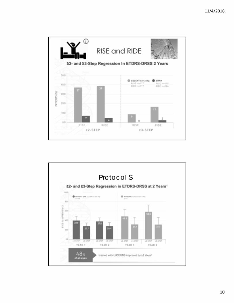
RISE and RIDE
Protocol S
11/4/2018
11
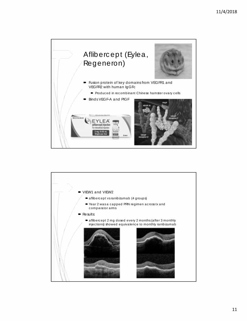
Aflibercept (Eylea, Regeneron)
Fusion protein of key domains from VEGFR1 and VEGFR2 with human IgGFc Produced in recombinant Chinese hamster ovary cells
Binds VEGF-A and PlGF
VIEW1 and VIEW2 aflibercept vs ranibizumab (4 groups)
Year 2 was a capped PRN regimen across tx and comparator arms
Results: aflibercept 2 mg dosed every 2 months (after 3 monthly
injections) showed equivalence to monthly ranibizumab
11/4/2018
12
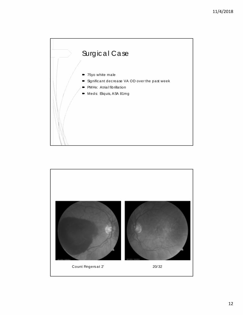
Surgical Case
75yo white male
Significant decrease VA OD over the past week
PMHx: Atrial fibrillation
Meds: Eliquis, ASA 81mg
Count Fingers at 2' 20/32
11/4/2018
13
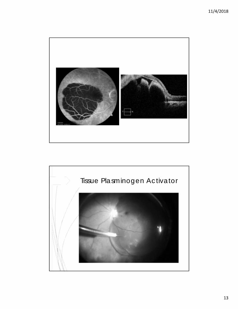
Tissue Plasminogen Activator
11/4/2018
14
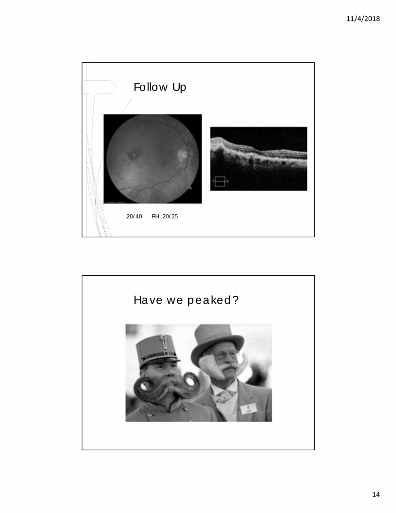
Follow Up
20/40 PH: 20/25
Have we peaked?
11/4/2018
15
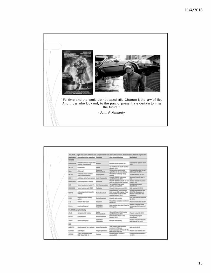
“For time and the world do not stand still. Change is the law of life. And those who look only to the past or present are certain to miss
the future.”
- John F. Kennedy
11/4/2018
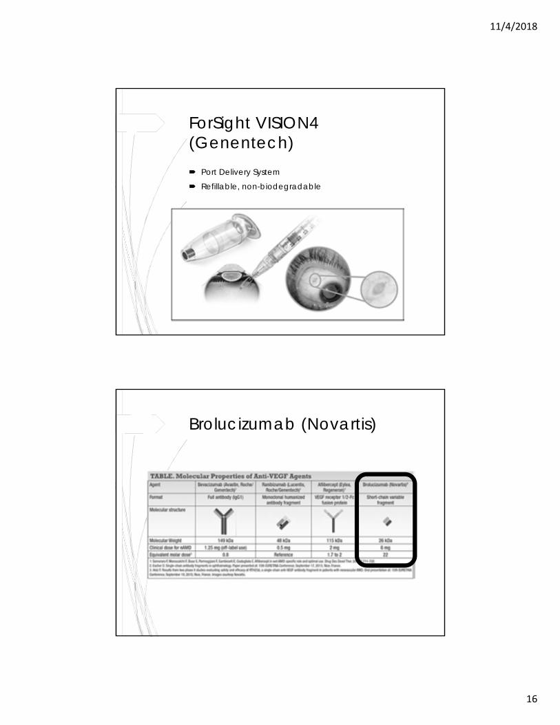
16
ForSight VISION4 (Genentech) Port Delivery System
Refillable, non-biodegradable
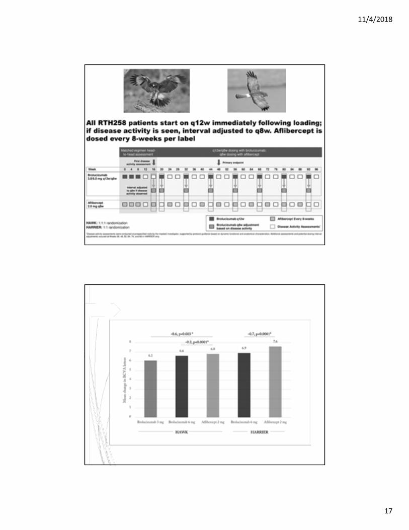
Brolucizumab (Novartis)
11/4/2018
17
11/4/2018
18
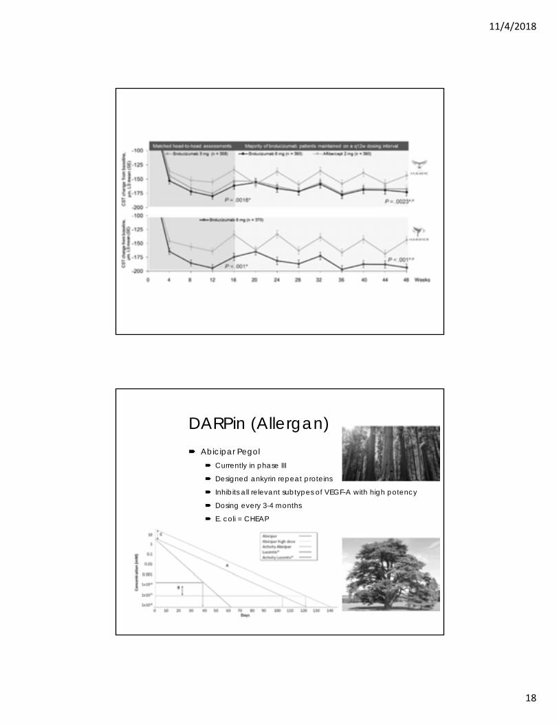
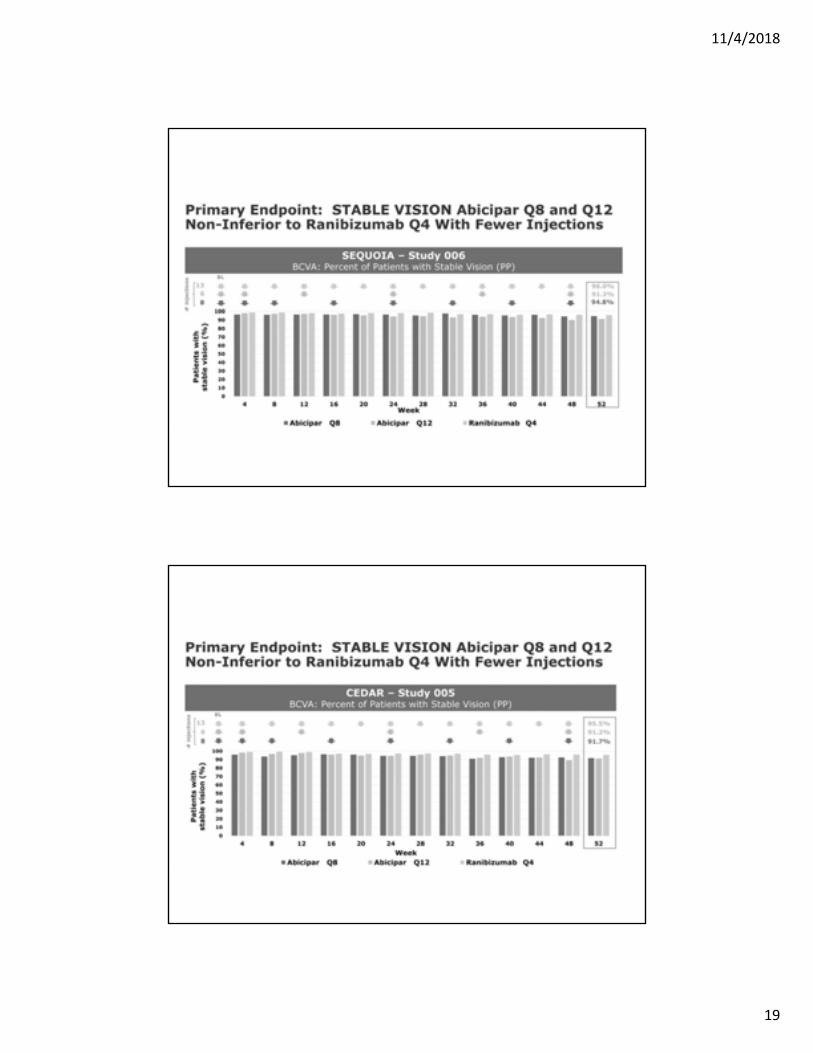
DARPin (Allergan) Abicipar Pegol
Currently in phase III
Designed ankyrin repeat proteins
Inhibits all relevant subtypes of VEGF-A with high potency
Dosing every 3-4 months
E. coli = CHEAP
11/4/2018
19
11/4/2018
20
Fovista (Ophthotech) Anti-PDGF drug
Promising results in combo with Lucentis in phase I/IIa trial, but did not meet phase III endpoint
Patients not responsive to monotherapy
Dual mechanism of action Anti-angiogenic
Anti-fibrotic
11/4/2018
21
Squalamine Lactate (Ohr)
Counteracts VEGF, PDGF and bFGF (basic fibroblast growth factor)
IMPACT study failed in phase III Early results showed improvement in subretinal
hyperreflective material (SHRM)
Anti-angiogenic and anti-fibrotic effects
Bevasiranib (Opko) - Discontinued RNA induced protein silencing complex (siRNA) designed to silence VEGF-A
mRNA
CARE (Cand5 Anti-VEGF RNAi Evaluation) study 1st time siRNA has made a phase III trial
11/4/2018
22
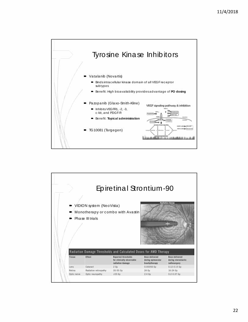
Tyrosine Kinase Inhibitors
Vatalanib (Novartis) Binds intracellular kinase domain of all VEGF receptor
subtypes
Benefit: High bioavailability provides advantage of PO dosing
Pazopanib (Glaxo-Smith-Kline) Inhibits VEGFR1, -2, -3,
c-kit, and PDGF-R
Benefit: Topical administration
TG10081 (Targegen)
Epiretinal Strontium-90
VIDION system (NeoVista)
Monotherapy or combo with Avastin
Phase III trials
11/4/2018
23
The Power of Wine…
CABERNET (CNV Secondary to AMD Treated with Beta Radiation Epiretinal Therapy) Epimacular brachytherapy using a prototype device
Goal to reduce treatment burden
MERLOT (Macular Epiretinal Brachytherapy Versus Lucentis Only) Extension of CABERNET with
identical anti-VEGF regimen in control and treatment arms
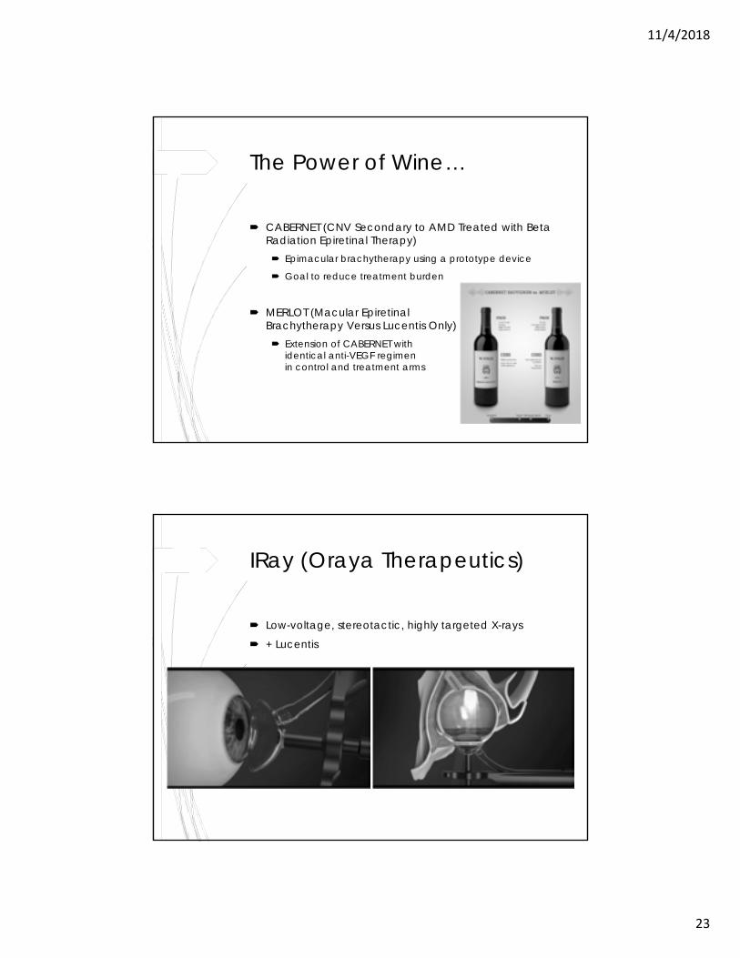
IRay (Oraya Therapeutics)
Low-voltage, stereotactic, highly targeted X-rays
+ Lucentis
11/4/2018
24
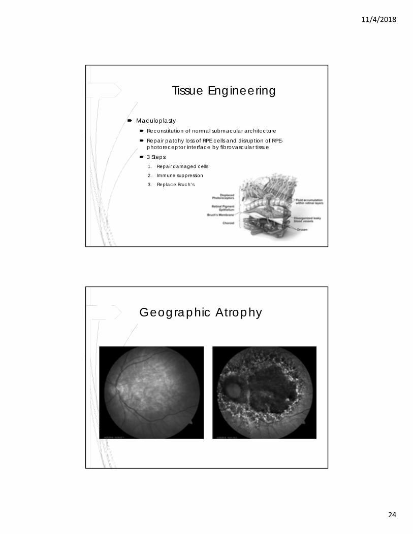
Tissue Engineering
Maculoplasty Reconstitution of normal submacular architecture
Repair patchy loss of RPE cells and disruption of RPE-photoreceptor interface by fibrovascular tissue
3 Steps:1. Repair damaged cells
2. Immune suppression
3. Replace Bruch’s
Geographic Atrophy
11/4/2018
25
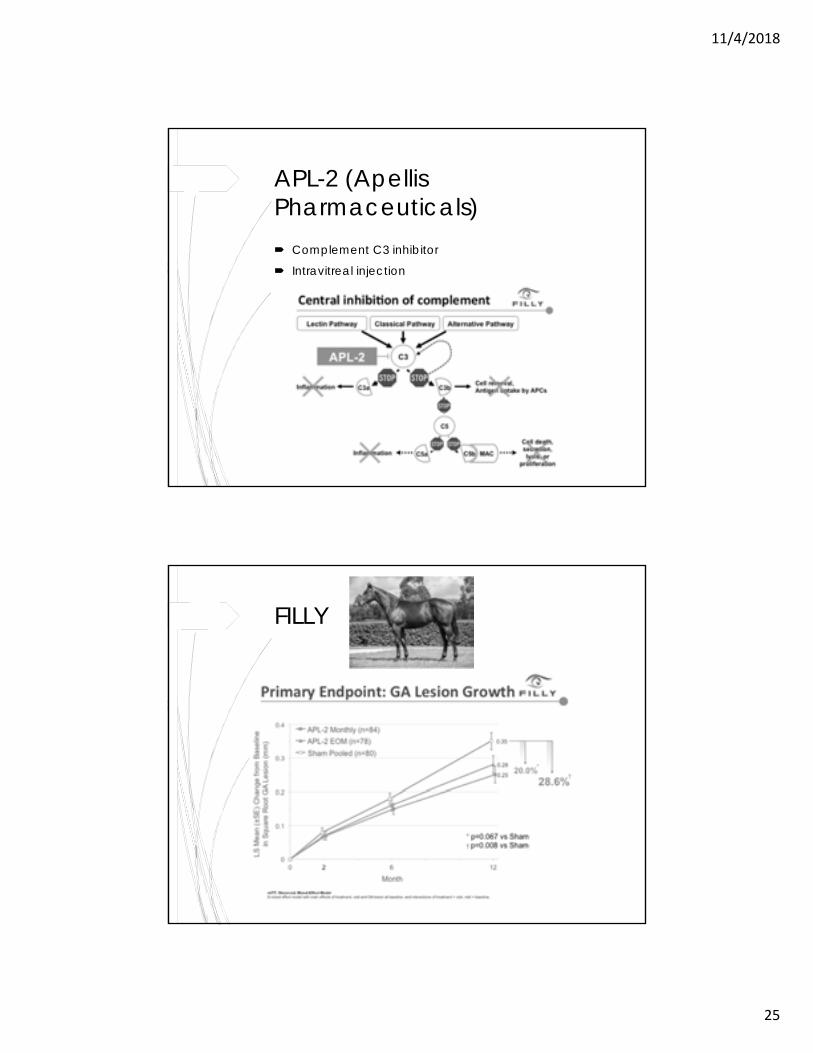
APL-2 (ApellisPharmaceuticals) Complement C3 inhibitor
Intravitreal injection
FILLY
11/4/2018
26
Thank You!Questions?
11/4/2018
1
Sudden Vision LossDaniel K. Bennett, M.D.
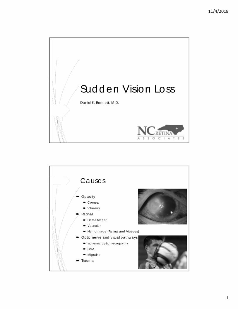
Causes
Opacity Cornea
Vitreous
Retinal Detachment
Vascular
Hemorrhage (Retina and Vitreous)
Optic nerve and visual pathways Ischemic optic neuropathy
CVA
Migraine
Trauma
11/4/2018
2
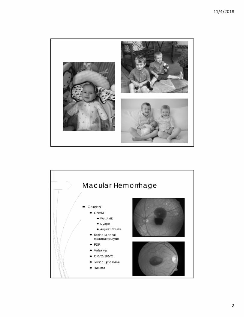
Macular Hemorrhage
Causes: CNVM
Wet AMD
Myopia
Angioid Streaks
Retinal arterial macroaneurysm
PDR
Valsalva
CRVO/BRVO
Terson Syndrome
Trauma
11/4/2018
3
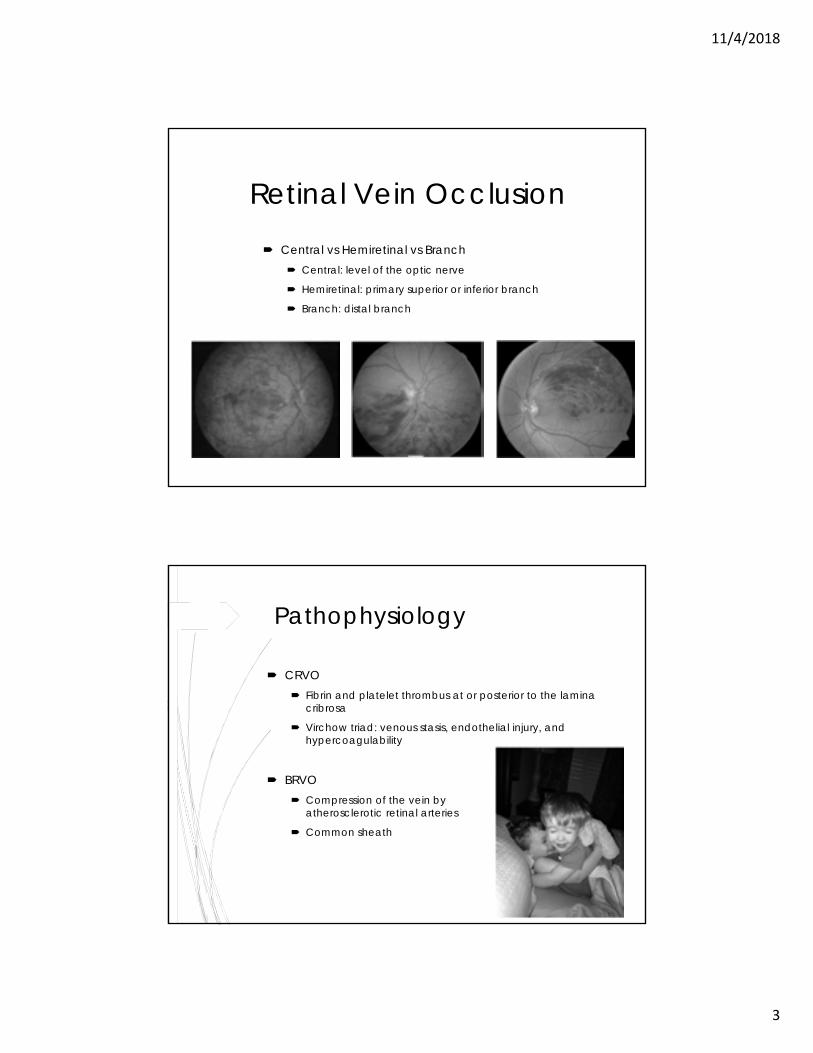
Retinal Vein Occlusion
Central vs Hemiretinal vs Branch Central: level of the optic nerve
Hemiretinal: primary superior or inferior branch
Branch: distal branch
Pathophysiology
CRVO Fibrin and platelet thrombus at or posterior to the lamina
cribrosa
Virchow triad: venous stasis, endothelial injury, and hypercoagulability
BRVO Compression of the vein by
atherosclerotic retinal arteries
Common sheath
11/4/2018
4
Ischemic vsNonischemic Nonischemic CRVO
75-80% of cases, can progress to ischemic
Milder clinical presentation Better overall prognosis
Neovascularization rare
Ischemic CRVO Marked vision decrease (often 20/400 or
worse) Anterior segment neovascularization
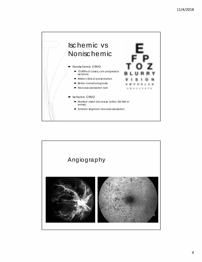
Angiography
11/4/2018
5
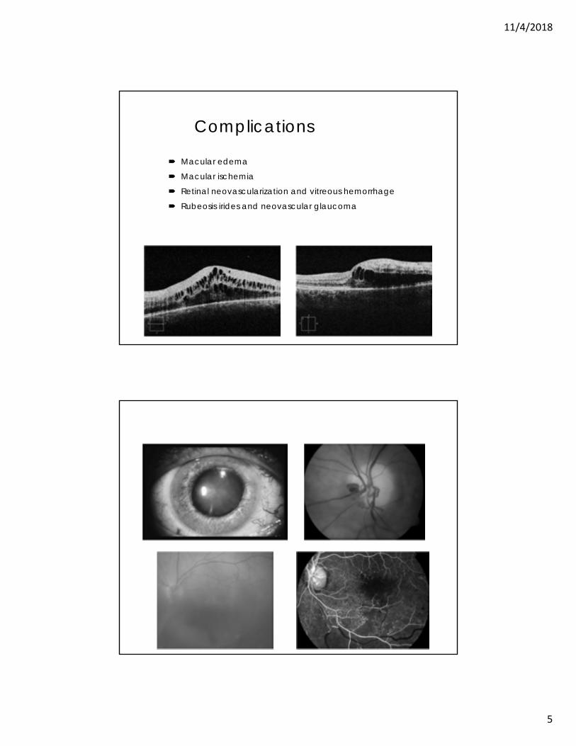
Complications
Macular edema
Macular ischemia
Retinal neovascularization and vitreous hemorrhage
Rubeosis irides and neovascular glaucoma
11/4/2018
6
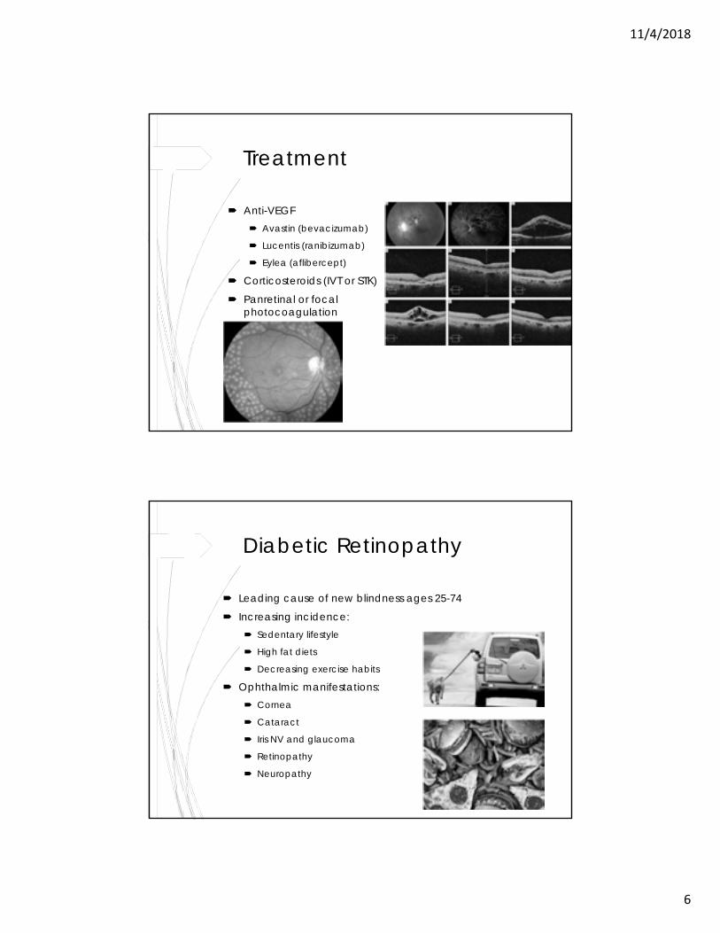
Treatment
Anti-VEGF Avastin (bevacizumab)
Lucentis (ranibizumab)
Eylea (aflibercept)
Corticosteroids (IVT or STK)
Panretinal or focal photocoagulation
Diabetic Retinopathy
Leading cause of new blindness ages 25-74
Increasing incidence: Sedentary lifestyle
High fat diets
Decreasing exercise habits
Ophthalmic manifestations: Cornea
Cataract
Iris NV and glaucoma
Retinopathy
Neuropathy
11/4/2018
7
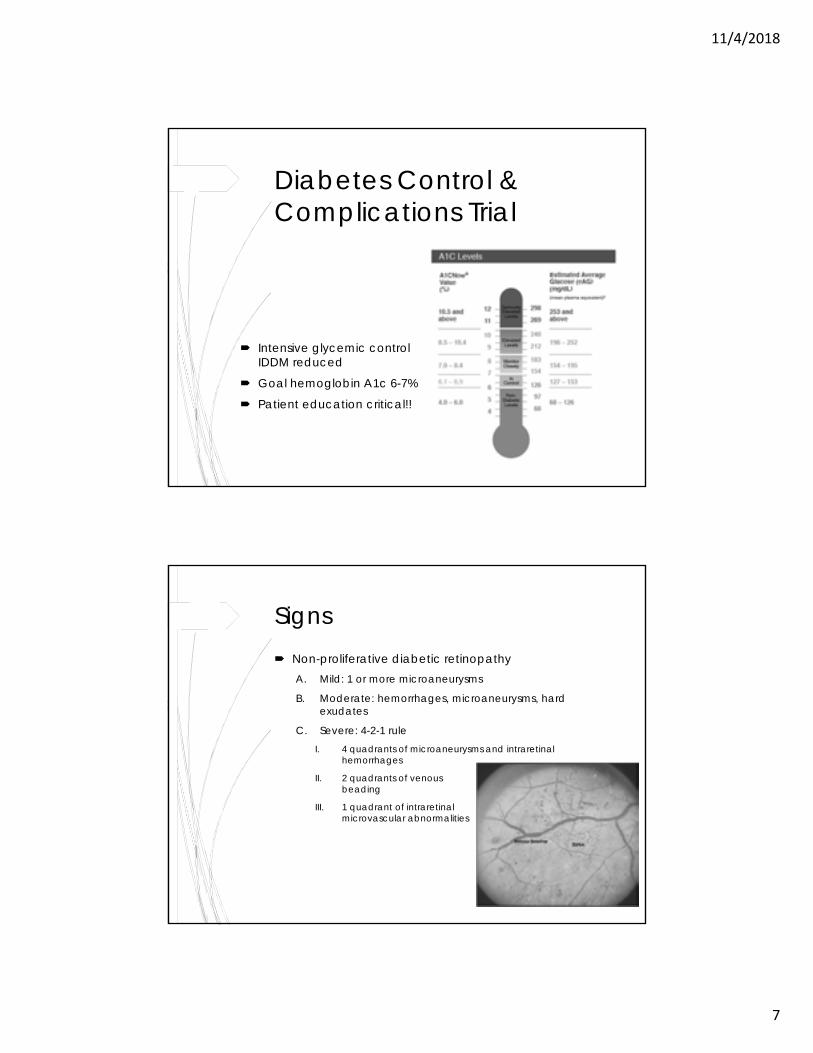
Diabetes Control & Complications Trial
Intensive glycemic control in IDDM reduced complications
Goal hemoglobin A1c 6-7%
Patient education critical!!
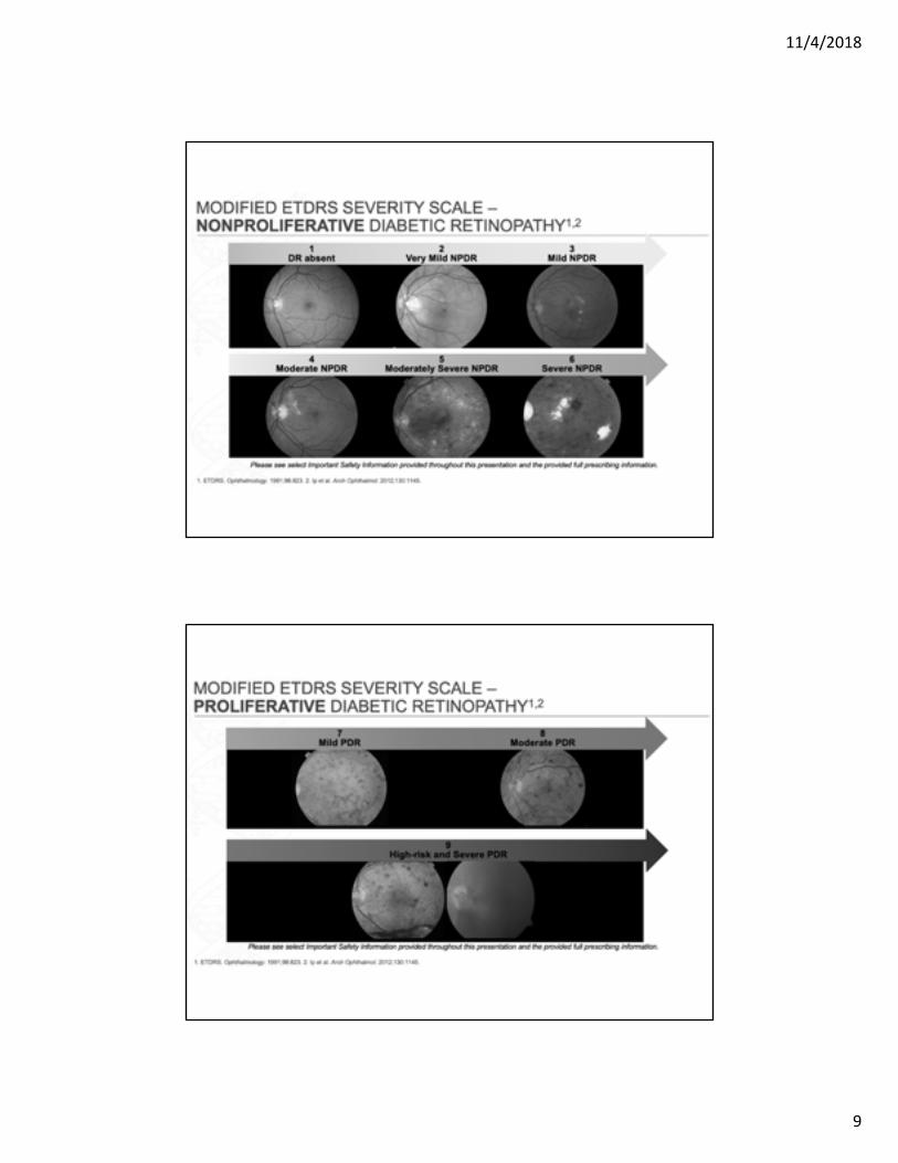
Signs Non-proliferative diabetic retinopathy
A. Mild: 1 or more microaneurysms
B. Moderate: hemorrhages, microaneurysms, hard exudates
C. Severe: 4-2-1 ruleI. 4 quadrants of microaneurysms and intraretinal
hemorrhages
II. 2 quadrants of venous beading
III. 1 quadrant of intraretinalmicrovascular abnormalities
11/4/2018
8
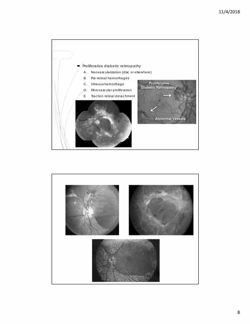
Proliferative diabetic retinopathyA. Neovascularization (disc or elsewhere)
B. Pre-retinal hemorrhages
C. Vitreous hemorrhage
D. Fibrovascular proliferation
E. Traction retinal detachment
11/4/2018
9
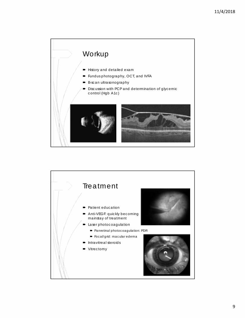
Workup
History and detailed exam
Fundus photography, OCT, and IVFA
B-scan ultrasonography
Discussion with PCP and determination of glycemic control (Hgb A1c)
Treatment
Patient education
Anti-VEGF: quickly becoming mainstay of treatment
Laser photocoagulation Panretinal photocoagulation: PDR
Focal/grid: macular edema
Intravitreal steroids
Vitrectomy
11/4/2018
10
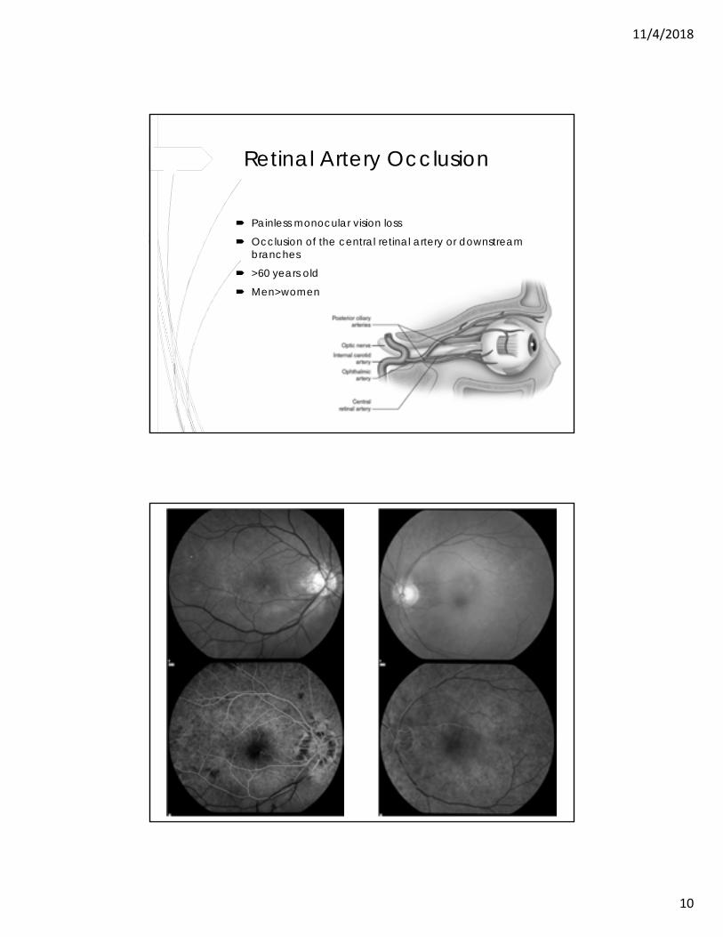
Retinal Artery Occlusion
Painless monocular vision loss
Occlusion of the central retinal artery or downstream branches
>60 years old
Men>women
11/4/2018
11
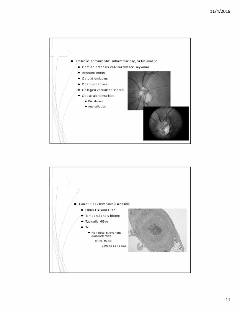
Embolic, thrombotic, inflammatory, or traumatic Cardiac embolus, valvular disease, myxoma
Atherosclerosis
Carotid embolus
Coagulopathies
Collagen vascular diseases
Ocular abnormalities Disc drusen
Arterial loops
Giant Cell (Temporal) Arteritis Order ESR and CRP
Temporal artery biopsy
Typically >55yo
Tx: High dose intravenous
corticosteroids
Solu-Medrol
1,000 mg qd x 3 days
11/4/2018
12
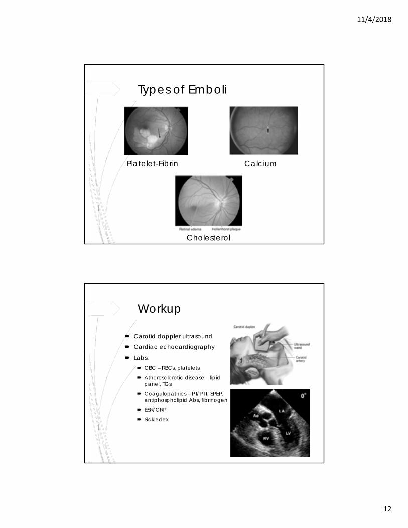
Types of Emboli
Platelet-Fibrin Calcium
Cholesterol
Workup
Carotid doppler ultrasound
Cardiac echocardiography
Labs: CBC – RBCs, platelets
Atherosclerotic disease – lipid panel, TGs
Coagulopathies – PT/PTT, SPEP, antiphospholipid Abs, fibrinogen
ESR/CRP
Sickledex
11/4/2018
13
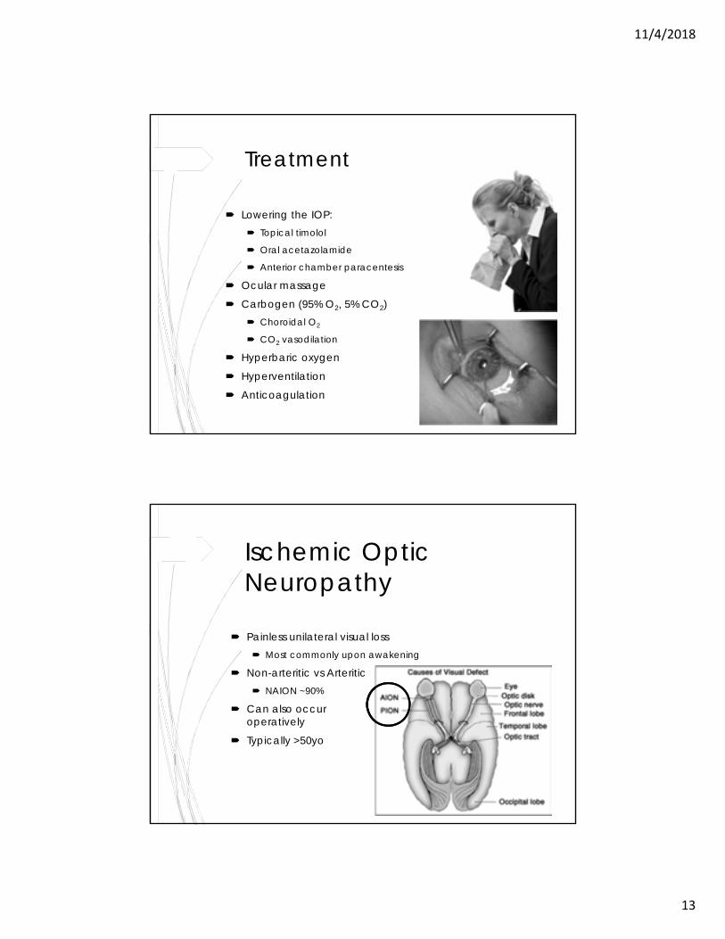
Treatment
Lowering the IOP: Topical timolol
Oral acetazolamide
Anterior chamber paracentesis
Ocular massage
Carbogen (95% O2, 5% CO2) Choroidal O2
CO2 vasodilation
Hyperbaric oxygen
Hyperventilation
Anticoagulation
Ischemic Optic Neuropathy
Painless unilateral visual loss Most commonly upon awakening
Non-arteritic vs Arteritic NAION ~90%
Can also occur post-operatively
Typically >50yo
11/4/2018
14
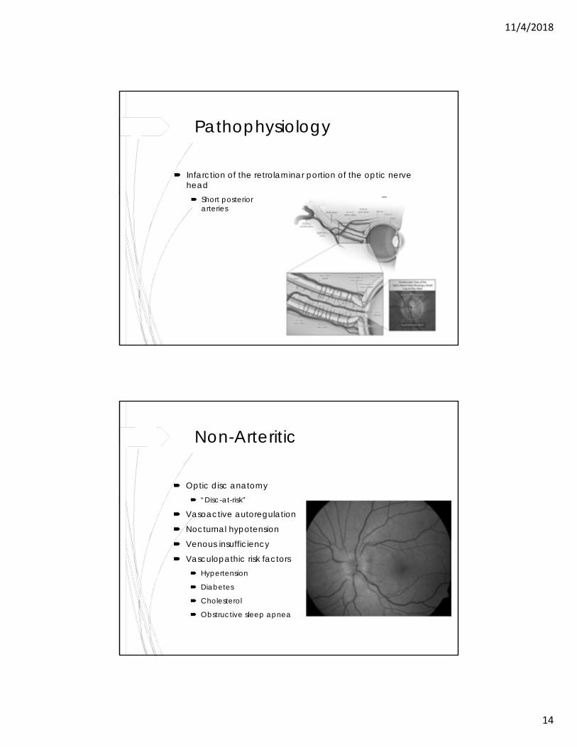
Pathophysiology
Infarction of the retrolaminar portion of the optic nerve head Short posterior ciliary
arteries
Non-Arteritic
Optic disc anatomy “Disc-at-risk”
Vasoactive autoregulation
Nocturnal hypotension
Venous insufficiency
Vasculopathic risk factors Hypertension
Diabetes
Cholesterol
Obstructive sleep apnea
11/4/2018
15
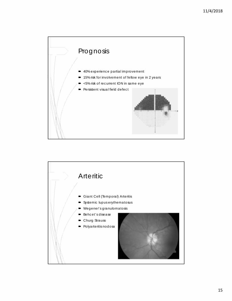
Prognosis
40% experience partial improvement
15% risk for involvement of fellow eye in 2 years
<5% risk of recurrent ION in same eye
Persistent visual field defect
Arteritic
Giant Cell (Temporal) Arteritis
Systemic lupus erythematosus
Wegener’s granulomatosis
Behcet’s disease
Churg Strauss
Polyarteritis nodosa
11/4/2018
16
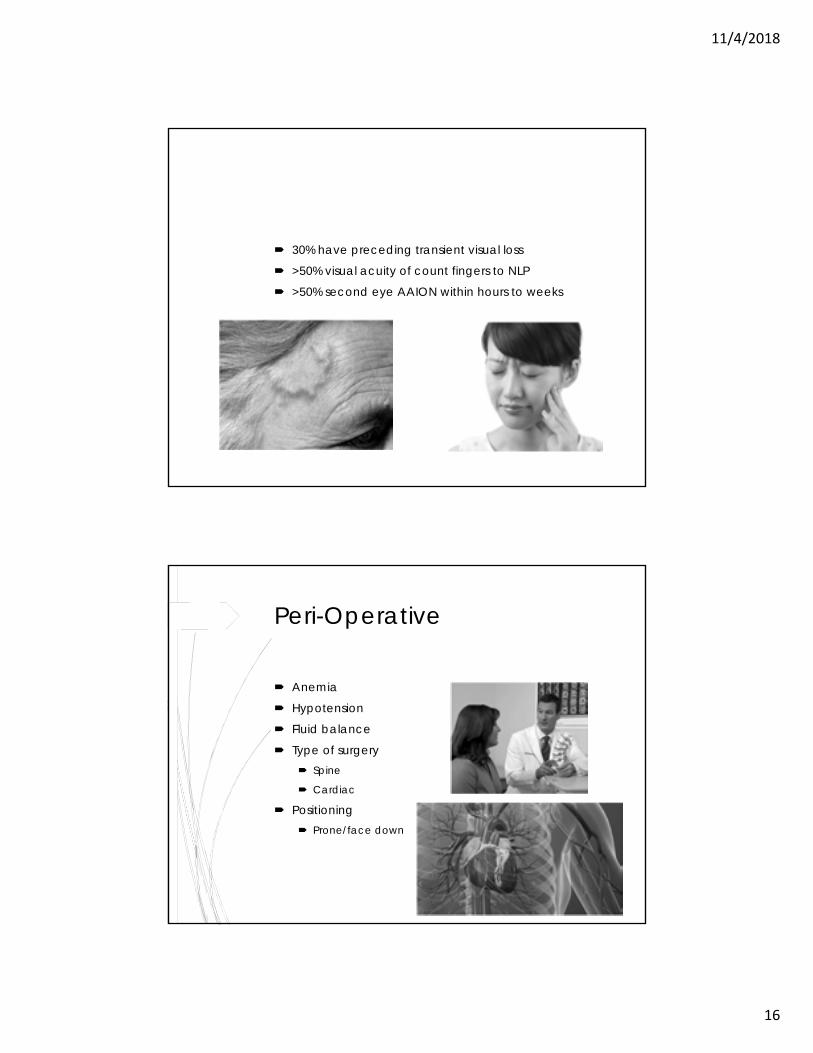
30% have preceding transient visual loss
>50% visual acuity of count fingers to NLP
>50% second eye AAION within hours to weeks
Peri-Operative
Anemia
Hypotension
Fluid balance
Type of surgery Spine
Cardiac
Positioning Prone/face down
11/4/2018
17
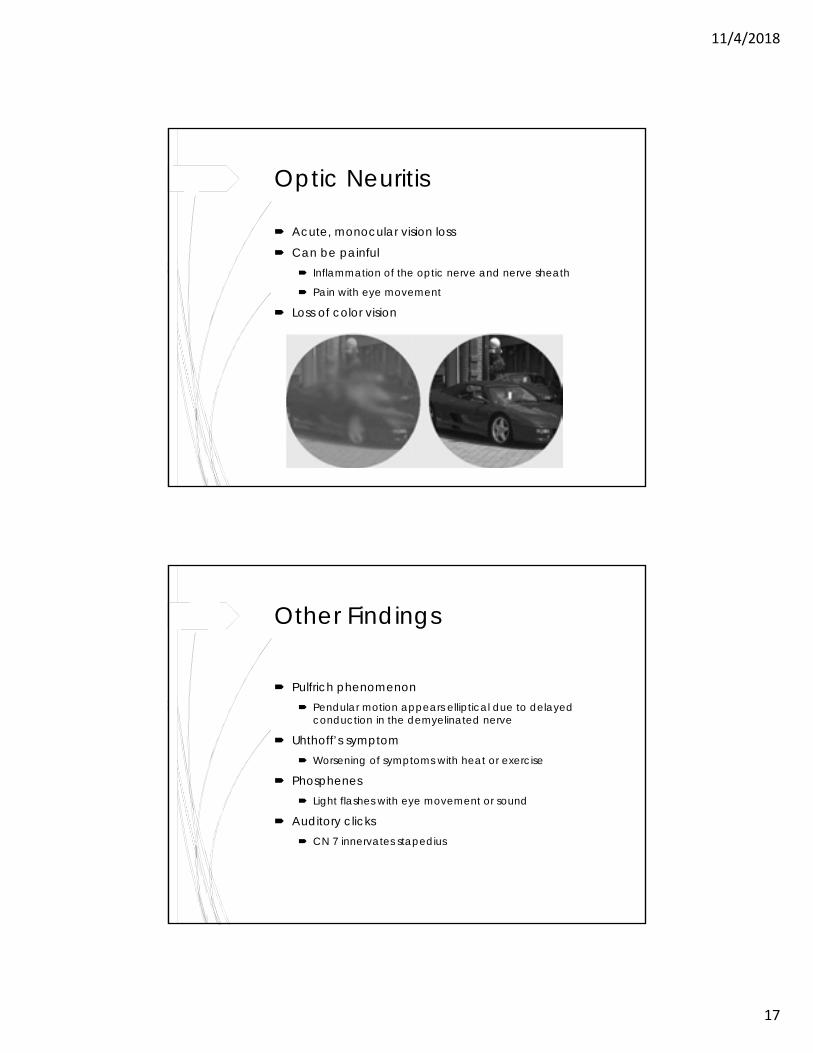
Optic Neuritis
Acute, monocular vision loss
Can be painful Inflammation of the optic nerve and nerve sheath
Pain with eye movement
Loss of color vision
Other Findings
Pulfrich phenomenon Pendular motion appears elliptical due to delayed
conduction in the demyelinated nerve
Uhthoff’s symptom Worsening of symptoms with heat or exercise
Phosphenes Light flashes with eye movement or sound
Auditory clicks CN 7 innervates stapedius
11/4/2018
18
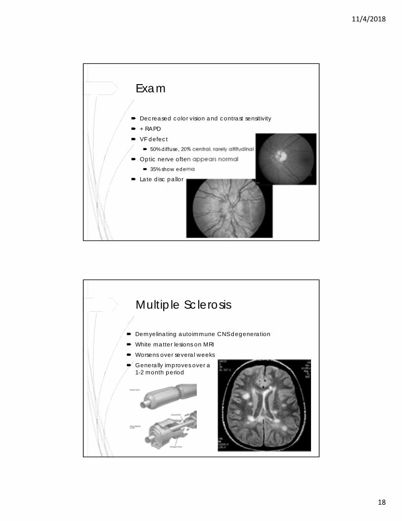
Exam
Decreased color vision and contrast sensitivity
+ RAPD
VF defect 50% diffuse, 20% central, rarely altitudinal
Optic nerve often appears normal 35% show edema
Late disc pallor
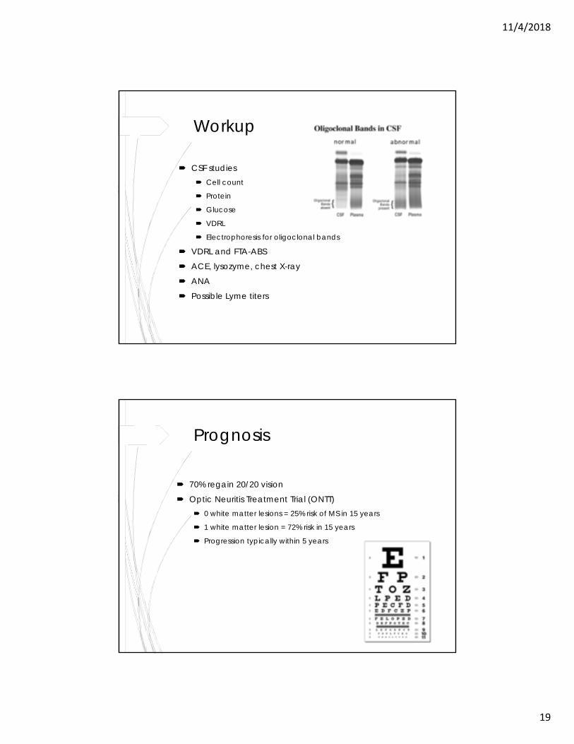
Multiple Sclerosis
Demyelinating autoimmune CNS degeneration
White matter lesions on MRI
Worsens over several weeks
Generally improves over a 1-2 month period
11/4/2018
19
Workup
CSF studies Cell count
Protein
Glucose
VDRL
Electrophoresis for oligoclonal bands
VDRL and FTA-ABS
ACE, lysozyme, chest X-ray
ANA
Possible Lyme titers
Prognosis
70% regain 20/20 vision
Optic Neuritis Treatment Trial (ONTT) 0 white matter lesions = 25% risk of MS in 15 years
1 white matter lesion = 72% risk in 15 years
Progression typically within 5 years
11/4/2018
20
Treatment
Based on findings of ONTT IV methylprednisolone 250 mg IV q6h x 3 days
PO prednisone 1 mg/kg/day x 11 days
Reduces symptoms by 1-2 weeks
No long term visual benefit
Oral prednisone alone increased risk of recurrence
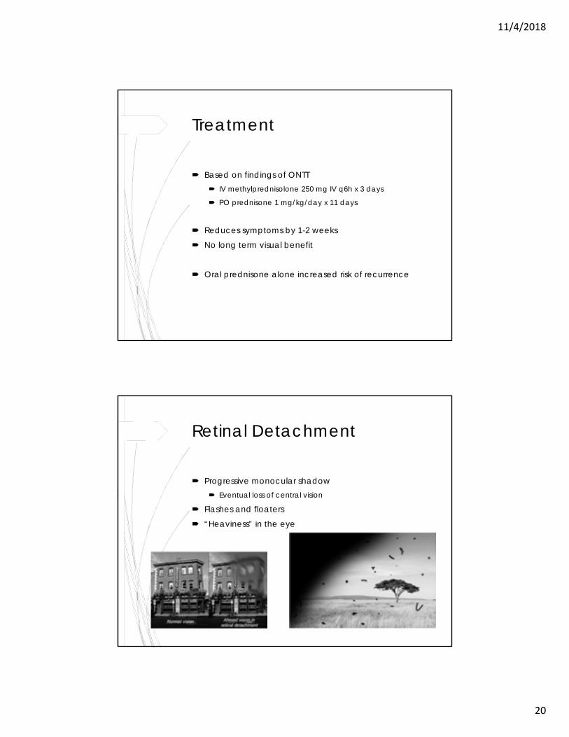
Retinal Detachment
Progressive monocular shadow Eventual loss of central vision
Flashes and floaters
“Heaviness” in the eye
11/4/2018
21
Types
1. Rhegmatogenous Breaks in the retina allow fluid to access the subretinal
space
2. Serous/Exudative Subretinal fluid collects due to damaged RPE and
increased choroidal vascular permeability
3. Tractional Fibrovascular proliferation pulls the retina away from the
RPE and choroid
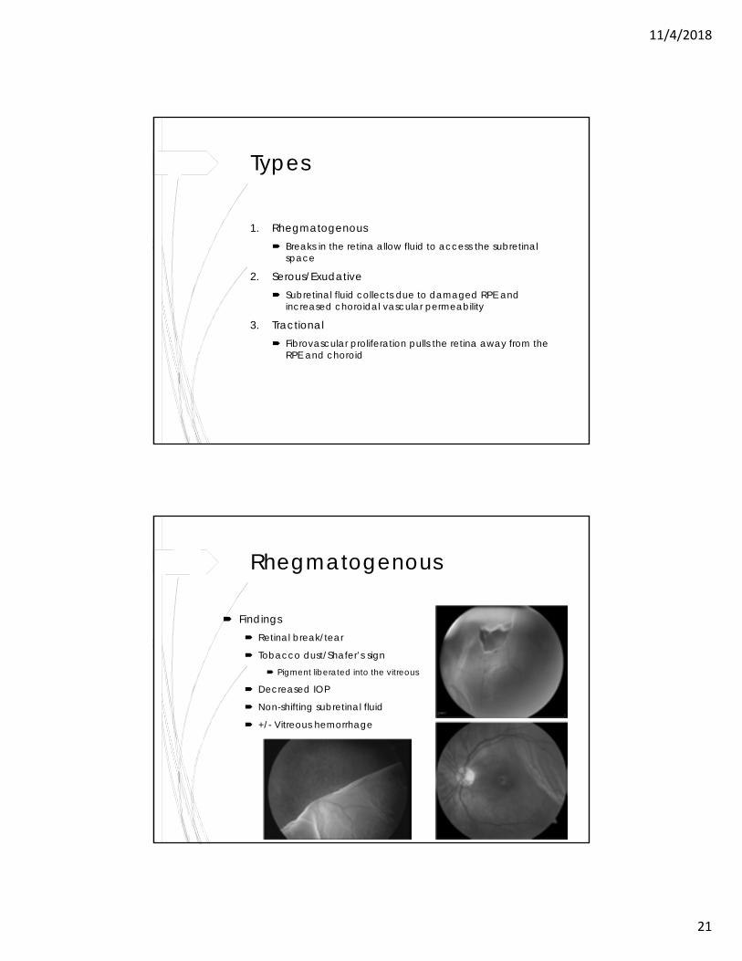
Rhegmatogenous
Findings Retinal break/tear
Tobacco dust/Shafer’s sign Pigment liberated into the vitreous
Decreased IOP
Non-shifting subretinal fluid
+/- Vitreous hemorrhage
11/4/2018
22
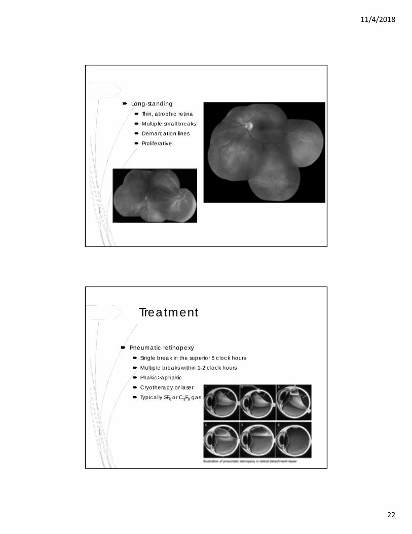
Long-standing Thin, atrophic retina
Multiple small breaks
Demarcation lines
Proliferative vitreoretinopathy
Treatment
Pneumatic retinopexy Single break in the superior 8 clock hours
Multiple breaks within 1-2 clock hours
Phakic>aphakic
Cryotherapy or laser
Typically SF6 or C3F8 gas
11/4/2018
23
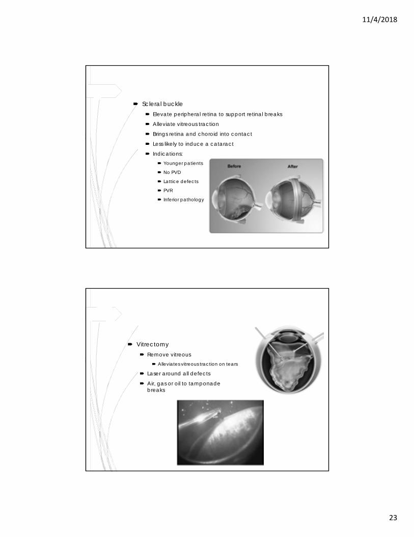
Scleral buckle Elevate peripheral retina to support retinal breaks
Alleviate vitreous traction
Brings retina and choroid into contact
Less likely to induce a cataract
Indications: Younger patients
No PVD
Lattice defects
PVR
Inferior pathology
Vitrectomy Remove vitreous
Alleviates vitreous traction on tears
Laser around all defects
Air, gas or oil to tamponade retinal breaks
11/4/2018
24
Thank You
Questions?
11/4/2018
1
Interesting Retina Clinic CasesDaniel K. Bennett, MD
Case 1
11/4/2018
2
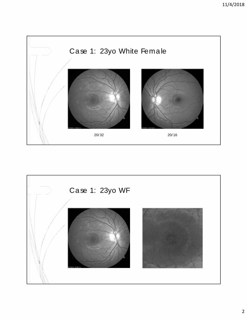
Case 1: 23yo White Female
20/32 20/16
Case 1: 23yo WF
11/4/2018
3
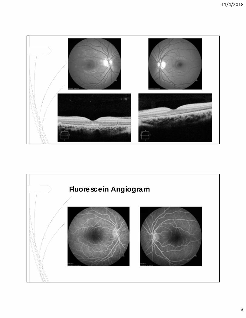
Fluorescein Angiogram
11/4/2018
4
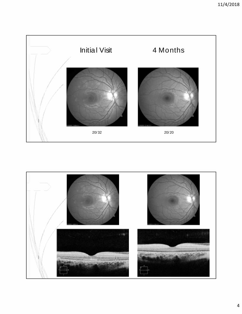
Initial Visit
20/32 20/20
4 Months
11/4/2018
5
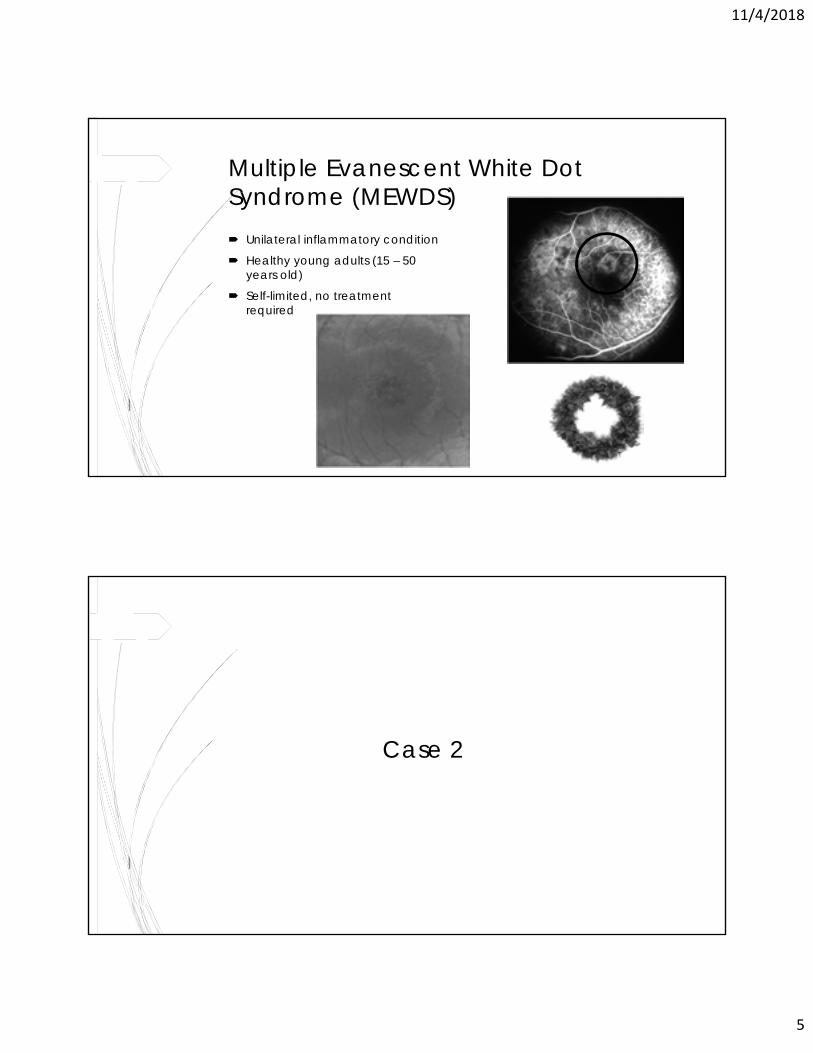
Multiple Evanescent White Dot Syndrome (MEWDS) Unilateral inflammatory condition
Healthy young adults (15 – 50 years old)
Self-limited, no treatment required
Case 2
11/4/2018
6
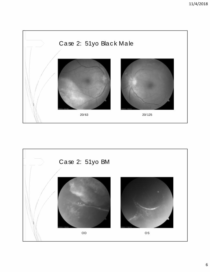
Case 2: 51yo Black Male
20/63 20/125
Case 2: 51yo BM
OD OS
11/4/2018
7
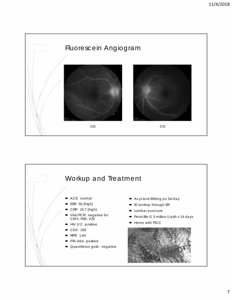
Fluorescein Angiogram
OD OS
Workup and Treatment
ACE: normal ESR: 92 (high) CRP: 15.7 (high) Viral PCR: negative for
CMV, HSV, VZV HIV 1/2: positive CD4: 159 RPR: 1:64 FTA-Abs: positive Quantiferon gold: negative
Acyclovir 800mg po 5x/day
ID workup through ER
Lumbar puncture
Penicillin G 3 million U q4h x 14 days
Home with PICC
11/4/2018
8
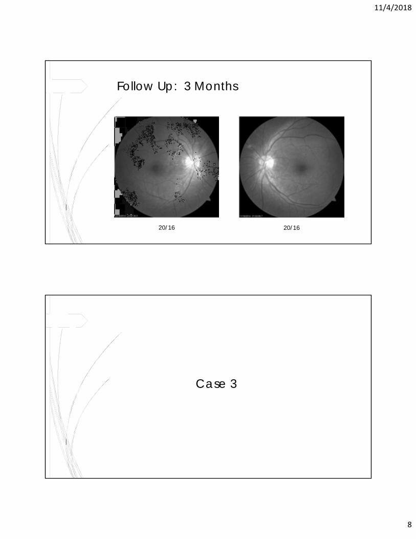
Follow Up: 3 Months
20/16 20/16
Case 3
11/4/2018
9
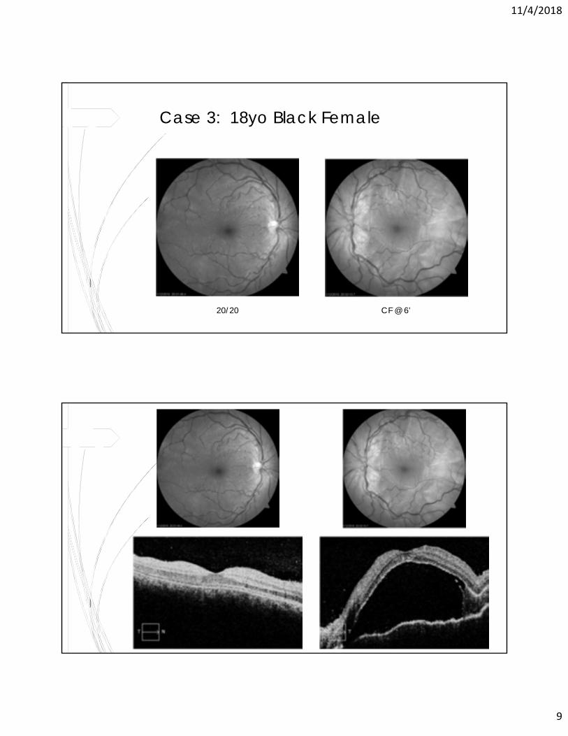
Case 3: 18yo Black Female
20/20 CF @ 6’
11/4/2018
10
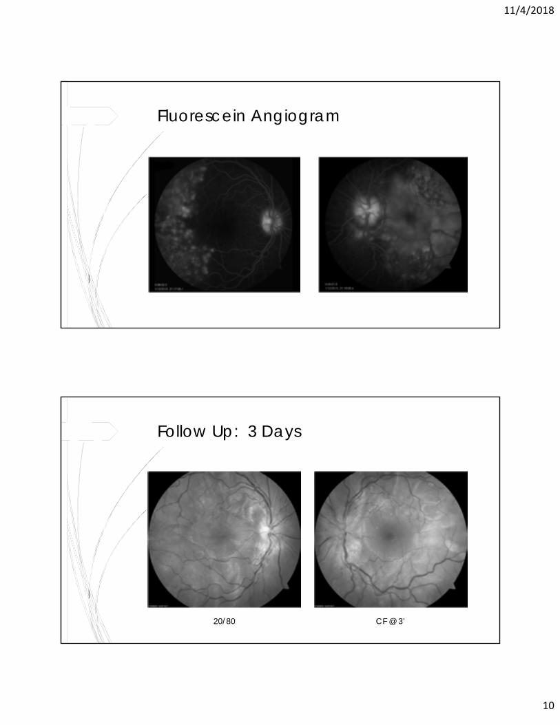
Fluorescein Angiogram
Follow Up: 3 Days
20/80 CF @ 3’
11/4/2018
11
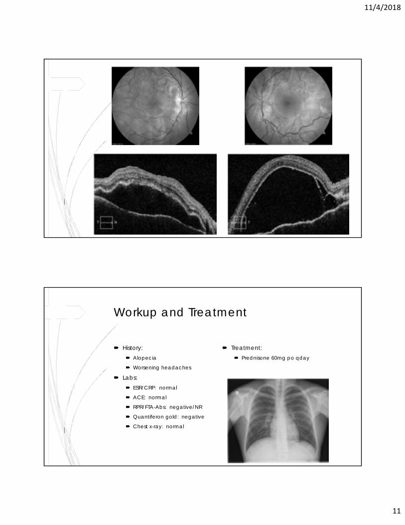
Workup and Treatment
History: Alopecia
Worsening headaches
Labs: ESR/CRP: normal
ACE: normal
RPR/FTA-Abs: negative/NR
Quantiferon gold: negative
Chest x-ray: normal
Treatment: Prednisone 60mg po qday
11/4/2018
12
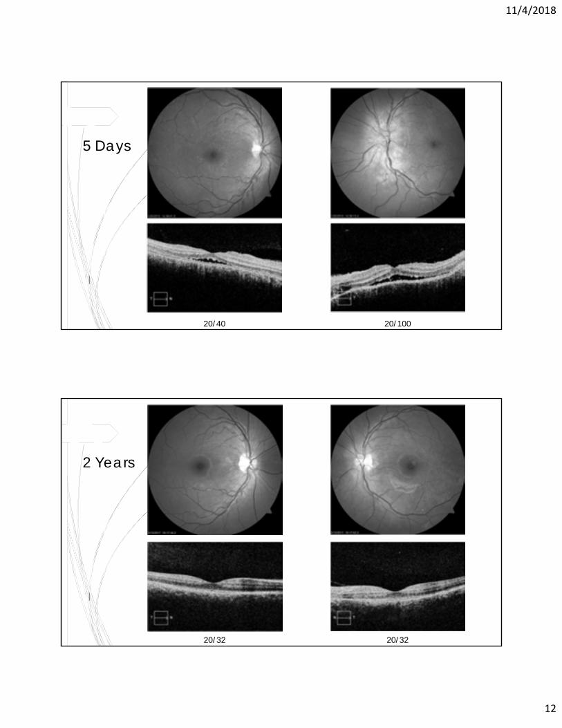
5 Days
20/40 20/100
2 Years
20/32 20/32
11/4/2018
13
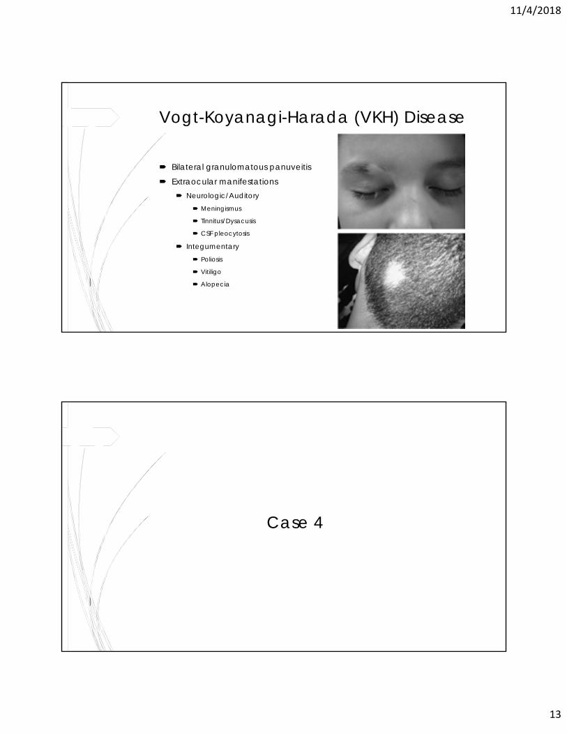
Vogt-Koyanagi-Harada (VKH) Disease
Bilateral granulomatous panuveitis
Extraocular manifestations Neurologic/Auditory
Meningismus
Tinnitus/Dysacusis
CSF pleocytosis
Integumentary Poliosis
Vitiligo
Alopecia
Case 4
11/4/2018
14
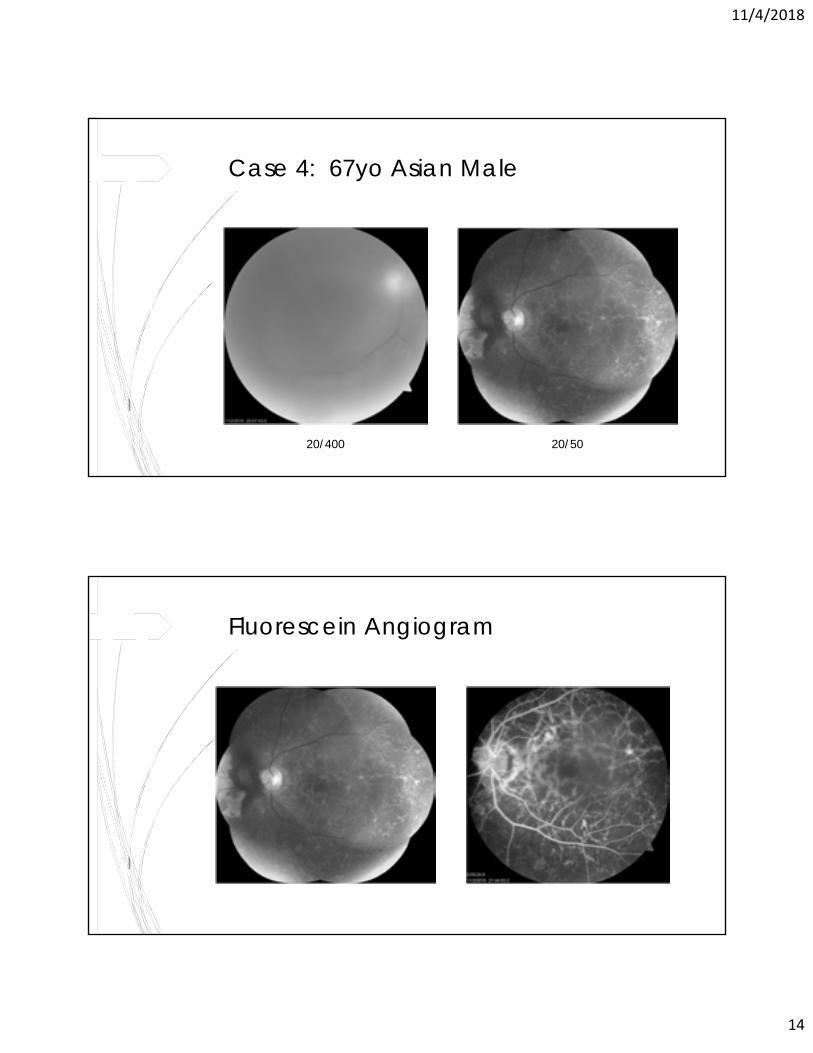
Case 4: 67yo Asian Male
20/400 20/50
Fluorescein Angiogram
11/4/2018
15
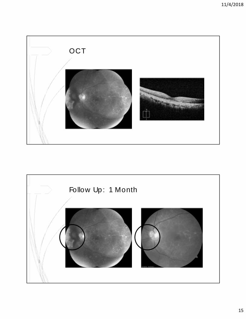
OCT
Follow Up: 1 Month
11/4/2018
16
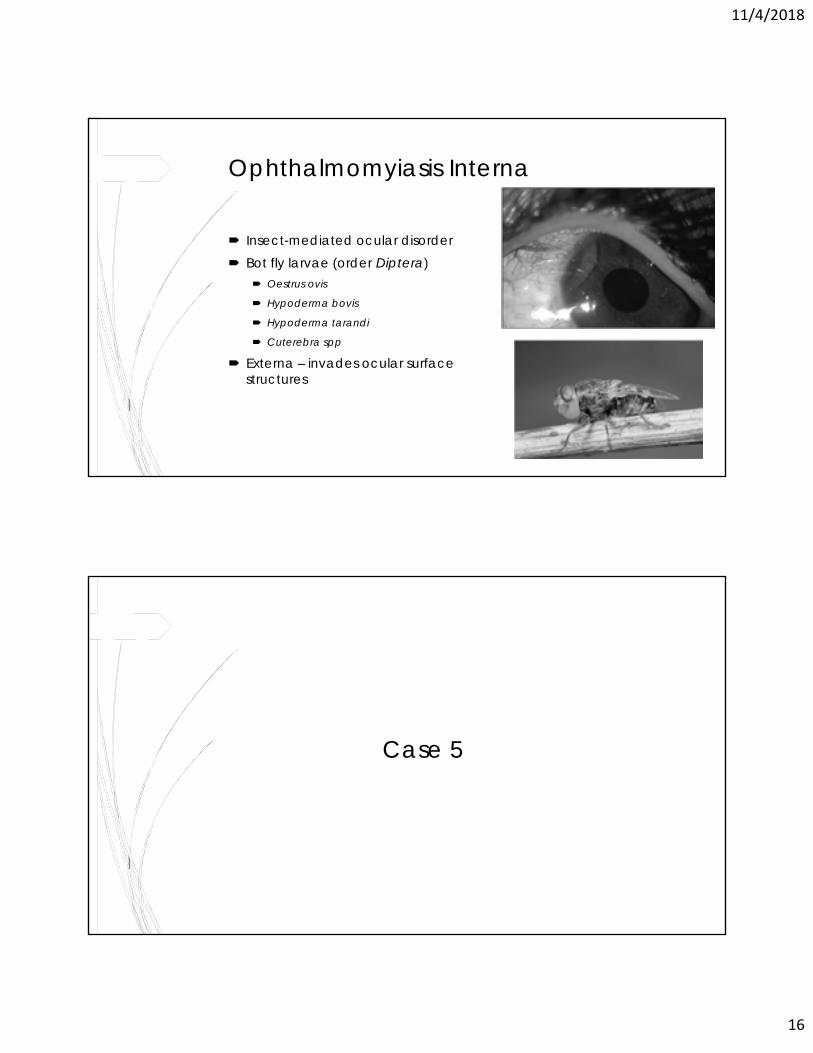
Ophthalmomyiasis Interna
Insect-mediated ocular disorder Bot fly larvae (order Diptera)
Oestrus ovis
Hypoderma bovis
Hypoderma tarandi
Cuterebra spp
Externa – invades ocular surface structures
Case 5
11/4/2018
17
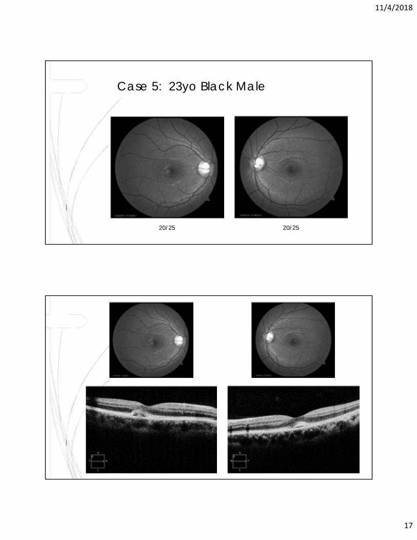
Case 5: 23yo Black Male
20/25 20/25
11/4/2018
18
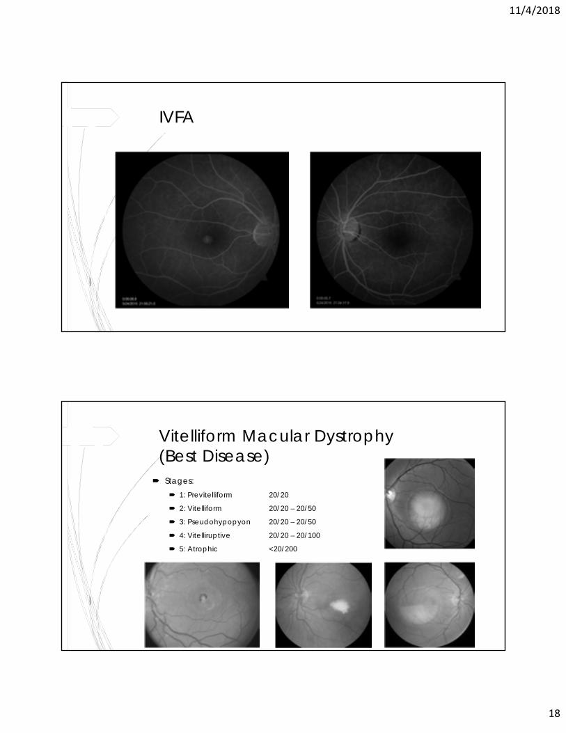
IVFA
Vitelliform Macular Dystrophy (Best Disease)
Stages: 1: Previtelliform 20/20
2: Vitelliform 20/20 – 20/50
3: Pseudohypopyon 20/20 – 20/50
4: Vitelliruptive 20/20 – 20/100
5: Atrophic <20/200
11/4/2018
19
Case 6
Case 6: 24yo White Female
No visual complaints, retinal lesion discovered on routine exam
No constitutional symptoms or systemic findings
No significant ocular/systemic family history
11/4/2018
20
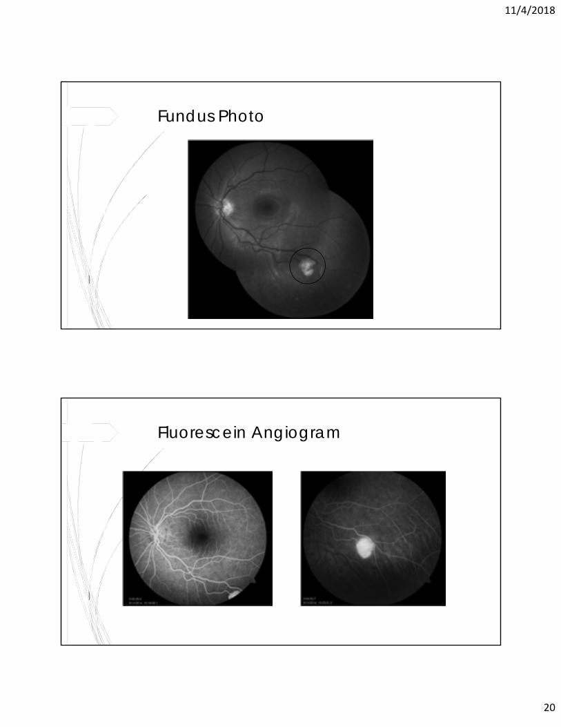
Fundus Photo
Fluorescein Angiogram
11/4/2018
21
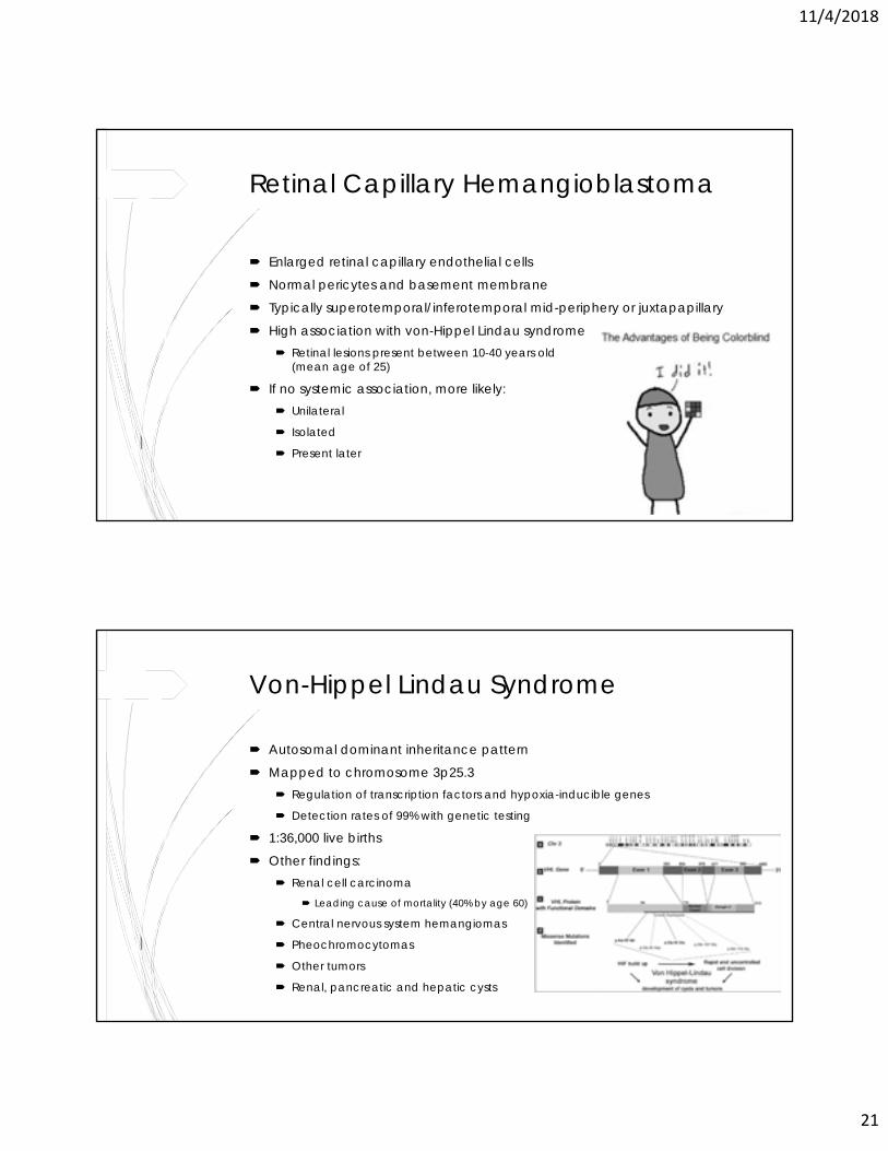
Retinal Capillary Hemangioblastoma
Enlarged retinal capillary endothelial cells
Normal pericytes and basement membrane
Typically superotemporal/inferotemporal mid-periphery or juxtapapillary
High association with von-Hippel Lindau syndrome Retinal lesions present between 10-40 years old
(mean age of 25)
If no systemic association, more likely: Unilateral
Isolated
Present later
Von-Hippel Lindau Syndrome
Autosomal dominant inheritance pattern
Mapped to chromosome 3p25.3 Regulation of transcription factors and hypoxia-inducible genes
Detection rates of 99% with genetic testing
1:36,000 live births
Other findings: Renal cell carcinoma
Leading cause of mortality (40% by age 60)
Central nervous system hemangiomas
Pheochromocytomas
Other tumors
Renal, pancreatic and hepatic cysts
11/4/2018
22
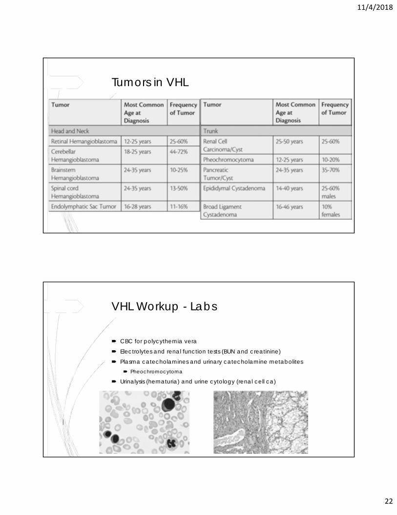
Tumors in VHL
VHL Workup - Labs
CBC for polycythemia vera
Electrolytes and renal function tests (BUN and creatinine)
Plasma catecholamines and urinary catecholamine metabolites Pheochromocytoma
Urinalysis (hematuria) and urine cytology (renal cell ca)
11/4/2018
23
VHL Workup - Imaging
Ophthalmic ultrasound
Abdominal ultrasound Renal and pancreatic lesions
Cysts of the epididymis and broad ligament
Abdominal CT and MRI Renal, pancreatic and adrenal gland lesions
Brain CT
CNS MRI
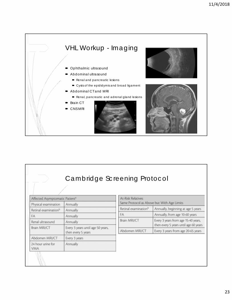
Cambridge Screening Protocol
11/4/2018
24
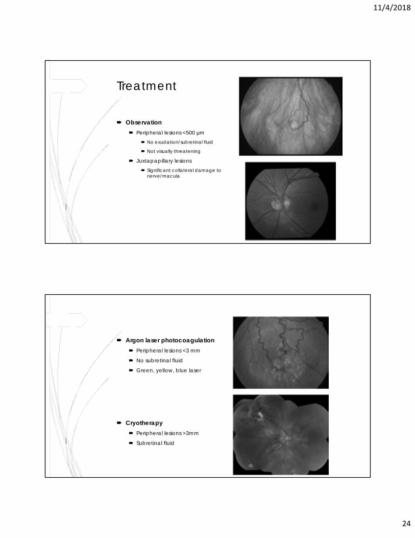
Treatment
Observation Peripheral lesions <500 µm
No exudation/subretinal fluid
Not visually threatening
Juxtapapillary lesions Significant collateral damage to
nerve/macula
Argon laser photocoagulation Peripheral lesions <3 mm
No subretinal fluid
Green, yellow, blue laser
Cryotherapy Peripheral lesions >3mm
Subretinal fluid
11/4/2018
25
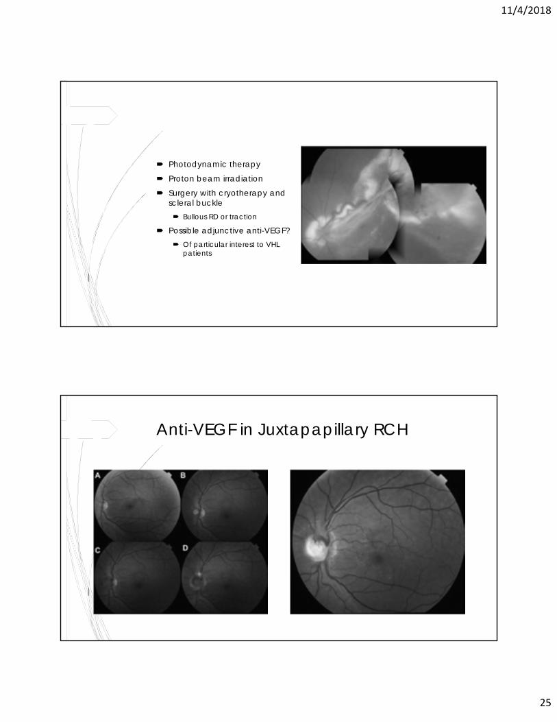
Photodynamic therapy
Proton beam irradiation
Surgery with cryotherapy and scleral buckle Bullous RD or traction
Possible adjunctive anti-VEGF? Of particular interest to VHL
patients
Anti-VEGF in Juxtapapillary RCH
11/4/2018
26
Prognosis
Guarded >25% of eyes with permanent vision loss
20% with VA <20/100 in at least one eye
Variables Number of lesions
Size
Location
Degree of exudative or traction retinal detachment
Case 7
11/4/2018
27
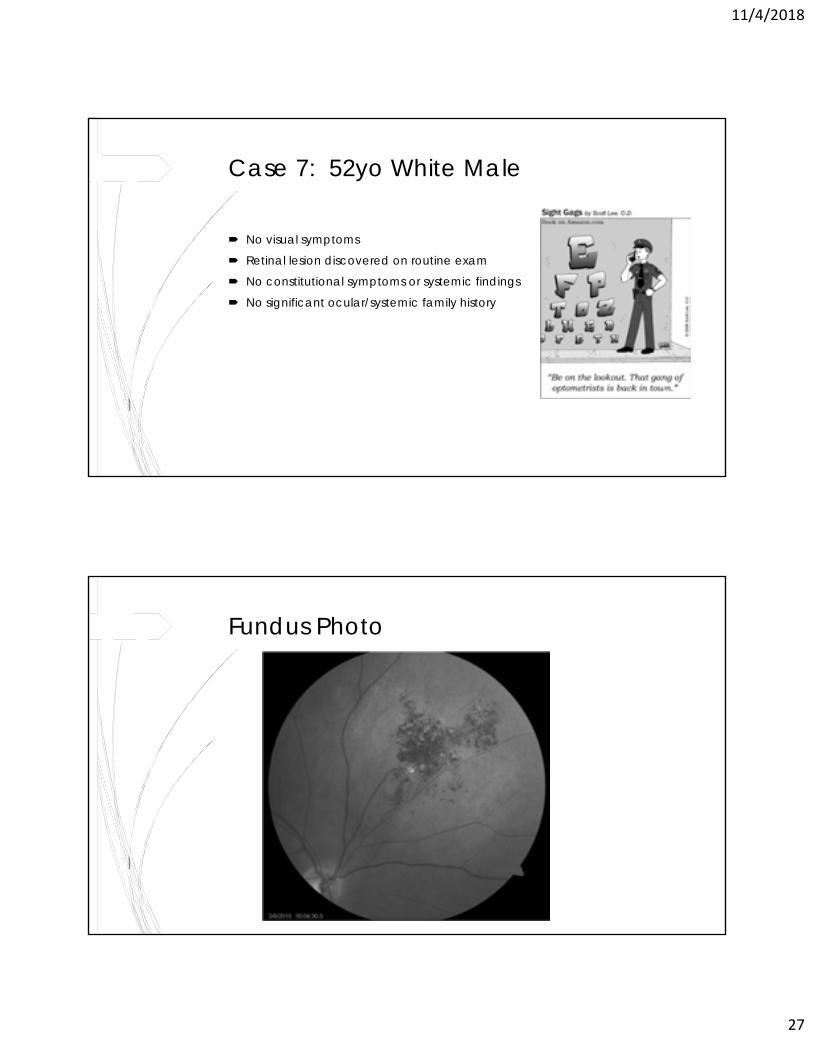
Case 7: 52yo White Male
No visual symptoms
Retinal lesion discovered on routine exam
No constitutional symptoms or systemic findings
No significant ocular/systemic family history
Fundus Photo
11/4/2018
28
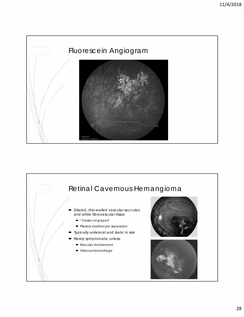
Fluorescein Angiogram
Retinal Cavernous Hemangioma
Dilated, thin-walled vascular saccules and white fibrovascular tissue “Cluster-of-grapes”
Plasma-erythrocyte separation
Typically unilateral and static in size
Rarely symptomatic unless: Macular involvement
Vitreous hemorrhage
11/4/2018
29
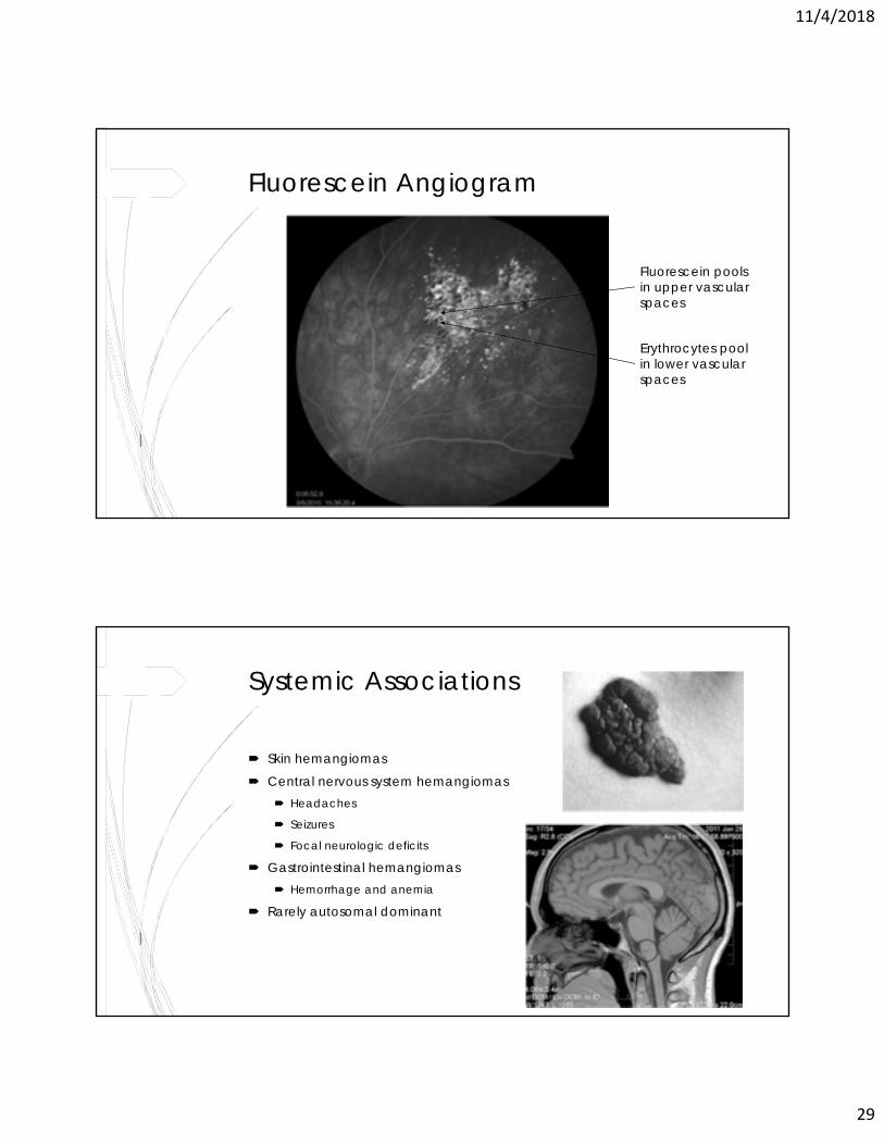
Fluorescein Angiogram
Fluorescein pools in upper vascular spaces
Erythrocytes pool in lower vascular spaces
Systemic Associations
Skin hemangiomas
Central nervous system hemangiomas Headaches
Seizures
Focal neurologic deficits
Gastrointestinal hemangiomas Hemorrhage and anemia
Rarely autosomal dominant
11/4/2018
30
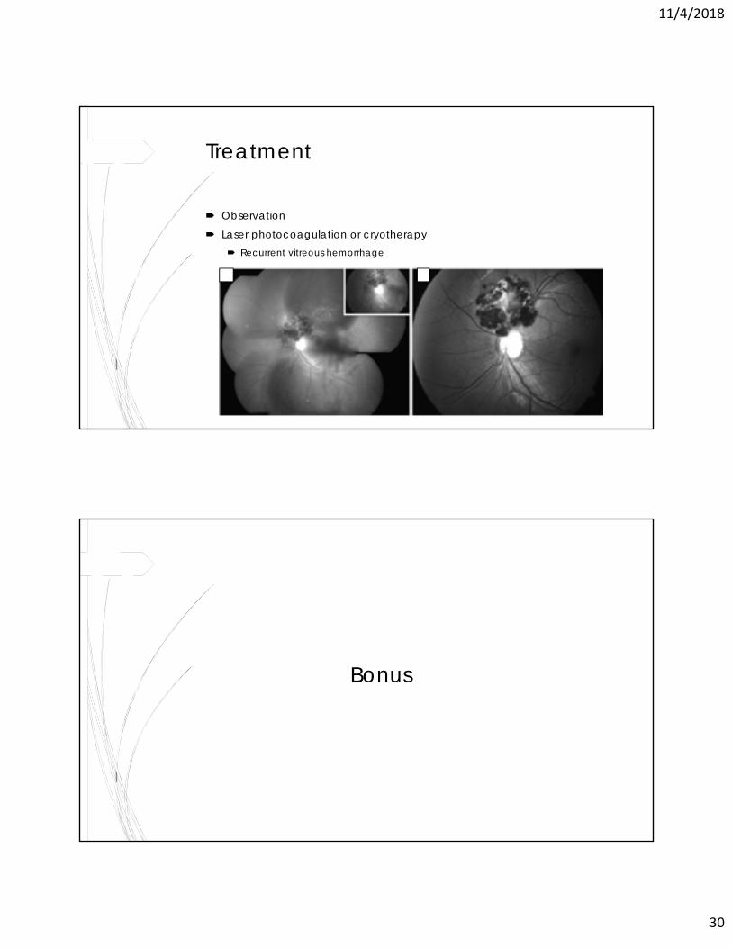
Treatment
Observation
Laser photocoagulation or cryotherapy Recurrent vitreous hemorrhage
Bonus
11/4/2018
31
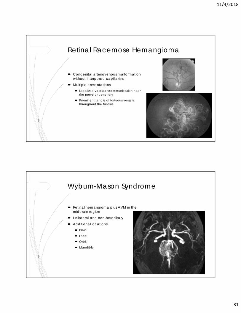
Retinal Racemose Hemangioma
Congenital arteriovenous malformation without interposed capillaries
Multiple presentations: Localized vascular communication near
the nerve or periphery
Prominent tangle of tortuous vessels throughout the fundus
Wyburn-Mason Syndrome
Retinal hemangioma plus AVM in the midbrain region
Unilateral and non-hereditary
Additional locations: Brain
Face
Orbit
Mandible
11/4/2018
32
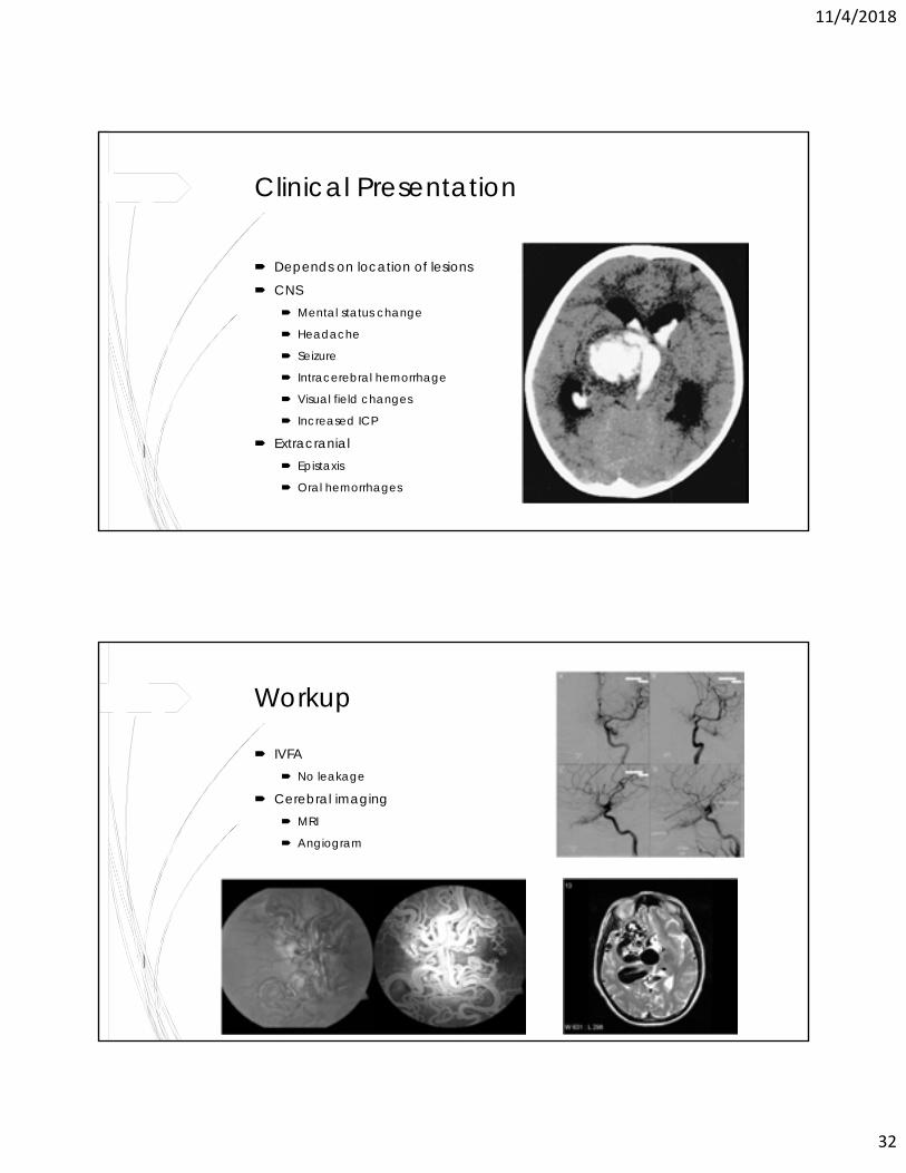
Clinical Presentation
Depends on location of lesions
CNS Mental status change
Headache
Seizure
Intracerebral hemorrhage
Visual field changes
Increased ICP
Extracranial Epistaxis
Oral hemorrhages
Workup
IVFA No leakage
Cerebral imaging MRI
Angiogram
11/4/2018
33
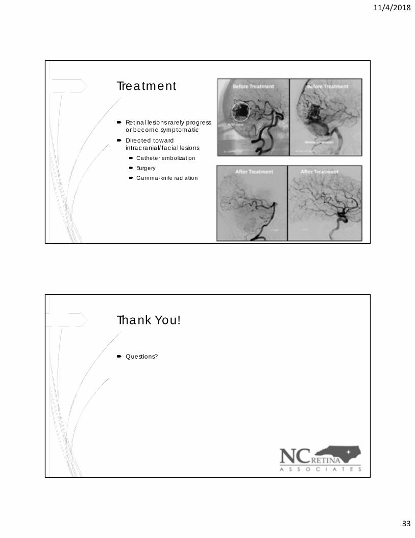
Treatment
Retinal lesions rarely progress or become symptomatic
Directed toward intracranial/facial lesions Catheter embolization
Surgery
Gamma-knife radiation
Thank You!
Questions?