Embed Size (px)
Citation preview
Macrophages Are Eliminated from the Injured Peripheral Nerve viaLocal Apoptosis and Circulation to Regional Lymph Nodesand the Spleen
Tanja Kuhlmann,1 Andreas Bitsch,2 Christine Stadelmann,1 Heike Siebert,1 and Wolfgang Bruck1
1Department of Neuropathology, Charite, Campus Virchow-Klinikum, 13353 Berlin, Germany, and 2Department ofNeurology, University of Gottingen, 37075 Gottingen, Germany
The present study investigated the fate of macrophages inperipheral nerves undergoing Wallerian degeneration, espe-cially their disappearance from the injured nerves after phago-cytosis of axonal and myelin debris. Wallerian degenerationwas induced in adult male C57Bl/6 mice by transecting the rightsciatic nerve. Five days after transection, the male sciaticnerves were transplanted into female recipient mice by placingthem exactly parallel to the host sciatic nerves. Nerves of thefemale recipient mice were also transected to induce break-down of the blood–nerve barrier in the host animal. Apoptosiswas assessed by morphological, immunohistochemical (acti-vated caspase-3), and molecular (DNA fragmentation) methodsin transplanted, recipient, and in control nerves. A subpopula-tion of macrophages within the degenerating nerves died lo-cally by apoptosis in each experiment. The fate of the male
macrophages within the transplanted nerves and the host or-ganism was investigated by in situ hybridization with aY-chromosome-specific DNA probe (145SC5). In situ hybridiza-tion specifically stained cells within the transplanted malenerve. Y-chromosome-positive cells were detected not onlyinside the transplanted nerve, but also inside the female hostnerve, the perineurial tissue, the local perineurial blood vessels,draining lymph nodes and the spleen of the female host, sug-gesting hematogenous as well as lymphatic elimination of mac-rophages from the injured nerve. These data indicate that localapoptosis and systemic elimination via circulation to the locallymph nodes and the spleen are involved in the disappearanceof macrophages from the injured peripheral nervous system.
Key words: macrophage; elimination; nervous system; apo-ptosis; migration; Y-chromosome probe; transplantation
The mechanisms that affect and regulate macrophage infiltrationof peripheral nerves during Wallerian degeneration have exten-sively been investigated in recent years (Stoll and Hartung, 1992;Griffin and Hoffman, 1993; Bruck, 1997). Disconnection of theaxon through traumatic, toxic, degenerative, ischemic, or meta-bolic damage leads to calcium-dependent axonal degeneration,retraction of Schwann cell cytoplasm from myelin sheaths, andformation of myelin ovoids within the first 24 hr (Beuche andFriede, 1984; Griffin and Hoffman, 1993; George et al., 1995).These events are followed by intense Schwann cell proliferationand recruitment of hematogenous and resident macrophages,which remove the degenerated myelin (Stoll et al., 1989;Fernandez-Valle et al., 1995; Bruck et al., 1996a; Bruck, 1997).Factors of axonal origin, complement components, adhesion mol-ecules, and degenerating myelin itself attract macrophages tomigrate into transected nerves (Bruck and Friede, 1990, 1991;Griffin et al., 1992; Bruck et al., 1995; Brown et al., 1997; Vou-gioukas et al., 1998).
So far, no studies have been published that investigate themechanisms or pathways leading to the disappearance of macro-phages from the damaged peripheral nerves. From other experi-mental models of tissue injury such as experimental autoimmune
encephalomyelitis (EAE) or myocardial infarction it is known thatlocal apoptosis is involved in the elimination of infiltrating inflam-matory cells (Nguyen et al., 1994, 1997; Smith et al., 1996; Take-mura et al., 1998). Immunologically, the elimination of macro-phages has been described as macrophage disappearance reaction;it is observed during delayed type hypersensitivity reactions andcan be inhibited or induced by various stimuli (Barth et al., 1995).
The purpose of the present study was to clarify the mechanismsby which macrophages are eliminated from the injured peripheralnerve tissue. The following two hypotheses were tested: (1)macrophages are eliminated by local apoptosis or (2) macro-phages migrate to local lymph nodes and the spleen. For theseexperiments, transected male sciatic nerves undergoing Walleriandegeneration were transplanted into female mice. The trans-planted nerves were massively laden with macrophages andplaced side-by-side to the host sciatic nerve, which was alsotransected. Apoptotic cells within the degenerating nerves wereidentified by morphologic criteria, DNA fragmentation (Bruck etal., 1996b), and the antibody CM-1, which recognizes the largesubunit of caspase-3 generated after activation (Srinivasan et al.,1998; Stadelmann et al., 1999). Activation of caspase-3 takesplace in the effector phase of the apoptotic process, and itsdetection therefore represents a reliable tool for the identificationof apoptotic cells. Transplanted macrophages were followed by insitu hybridization with a Y-chromosome-specific probe (Singh etal., 1987; Grounds et al., 1991).
MATERIALS AND METHODSAnimal surgery and transplantation. All animal surgery was done inaccordance with the German guidelines for animal experiments and was
Received Aug. 10, 2000; revised Feb. 15, 2001; accepted Feb. 23, 2001.Antiserum against activated caspase-3 was kindly provided by T. L. Deckwerth
and Anu Srinivasan. We thank Brigitte Maruschak and Stephanie Bunkowski forexcellent technical assistance.
Correspondence should be addressed to Dr. Wolfgang Bruck, Department ofNeuropathology, Charite, Campus Virchow-Klinikum, Augustenburger Platz 1,13353 Berlin, Germany. E-mail: [email protected] © 2001 Society for Neuroscience 0270-6474/01/213401-08$15.00/0
The Journal of Neuroscience, May 15, 2001, 21(10):3401–3408
officially approved by the county government of Braunschweig (Germa-ny). Wallerian degeneration was induced in 15 adult male and two femaleC57Bl/6 mice by transecting the right sciatic nerve under deep anesthesiawith Ketanest (50 mg/ml; Parke-Davis, Courbevoie, France; 0.6 mg/10gm body weight) and Rompun (2%; Bayer; 23.5 ml /10 gm body weight).The animals were decapitated under deep anesthesia 5 d after nervetransection. A uniform 5-mm-long piece of the degenerated sciatic nervewas immediately explanted and transplanted into 15 female or two maleC57Bl/6 mice, respectively. The transplanted nerves were placed side-by-side to the host sciatic nerve, which was also transected to inducebreakdown of the blood–nerve barrier. The transection sites of both thetransplanted and the recipient nerve were aligned and fixed by a musclesuture. On days after transplantation (DPT) 2, 5, 10, 20, and 40, corre-sponding to days 7, 10, 15, etc. after transection, the mice were perfusedunder deep anesthesia through the left cardiac ventricle with PBS fol-lowed by 4% paraformaldehyde. Each experimental group consisted ofthree animals. Degenerating host and donor sciatic nerves, the contralat-eral nontransected host sciatic nerve, regional and para-aortal lymphnodes, as well as the spleen were removed from each animal, post-fixedin 4% paraformaldehyde, and embedded in paraffin wax. Additionally,three control animals were used in which transected sciatic nerves wereallowed to degenerate for 6 d in situ in the absence of any transplantationprocedure to control for effects of nerve transplantation on cell invasionor macrophage apoptosis. The 5-mm-thick sections of all tissues werestained with hematoxylin–eosin (H&E).
Immunohistochemistry. Immunohistochemistry was performed by us-ing an avidin–biotin complex (ABC) technique on serial sections fromthe transected donor and host nerves. After deparaffinization, intrinsicperoxidase activity was blocked by incubation with 5% H2O2 in PBS for20 min. Nonspecific antibody binding was inhibited with 10% fetal calfserum (FCS) in PBS for 25 min. The sections were stained with mono-clonal antibodies against the macrophage antigens F4/80 (160 kDa gly-coprotein; Serotec, Oxford, UK) and Mac-3 (PharMingen, San Diego,CA) at a dilution of 1:50 and 1:200, respectively. For further immuno-cytochemical staining we used polyclonal antisera or monoclonal anti-bodies directed against T cells (CD3; Serotec; dilution 1:400), the S-100antigen (Dako, Glostrup, Denmark; dilution 1: 50) and activatedcaspase-3 (CM-1; kindly provided by T. L. Deckwerth and A. Srinivasan,Idun Pharmaceuticals, La Jolla, CA; dilution 1:5000). The CM-1 anti-body is specific for the large subunit of caspase-3 and thus for theactivated enzyme (Srinivasan et al., 1998). The slides were incubatedovernight at 4°C. Microwave pretreatment (five times for 3 min at 800 W)was applied for the CD3, Mac-3, and CM-1 antibodies. Secondary anti-bodies were biotinylated anti-rat or anti-rabbit Ig (Dako) applied at adilution of 1:200 for 60 min followed by incubation with the ABCcomplex (Vector Laboratories, Burlingame, CA) for 1 hr using diamino-benzidine (DAB) as chromogen. The primary antibody was omitted incontrol sections.
For double immunohistochemistry immunofluorescence procedureswere used. The binding of the primary antibodies (CM-1, Mac-3) wasvisualized using Oregon green (Mobitec)- or Cy3 (Jackson ImmunoRe-search, West Grove, PA)-coupled secondary reagents. The slides wereexamined on a Zeiss confocal laser-scanning microscope (LSM 510).
Semithin sections. Two C57Bl/6 mice were decapitated under deepanesthesia 10 d after transection of the right sciatic nerve. The distalnerve stumps were removed and immediately fixed in 2.5% glutaralde-hyde and post-fixed in 1% osmium tetroxide. Specimens were dehydratedin graded concentrations of alcohol, passed through propylene oxide, andembedded in Araldite. Semithin sections (1 mm) were stained withRichardson’s staining solution (1% Azur II, 2% Methyleneblue, 1%Borax).
In situ tailing. The in situ tailing technique (IST) was performed asdescribed in detail elsewhere (Gold et al., 1993). Sections were deparaf-finized and then incubated for 1 hr at 37°C in a reaction mix containing10 ml of 53 tailing buffer, 2 ml of cobalt chloride, 1 ml of digoxigeninlabeling mixture (nucleotides), and 6 U of terminal transferase. Distilledwater was added to give a total volume of 50 ml. Sections were washed inTBS, followed by incubation with alkaline phosphatase-labeled anti-digoxigenin antibody (diluted 1:250 in distilled water containing 10% FCS)for 1 hr at room temperature. The color reaction was developed withnitroblue tetrazolium (NBT) and 5-bromo-4-chloro-3-indolylphosphate(BCIP). All reagents, enzymes, and antibodies were purchased fromBoehringer Mannheim (Mannheim, Germany). IST-positive macrophageswere detected by double immunohistochemistry with the antibody F4/80 asdescribed above.
Probe synthesis and in situ hybridization. A DNA probe specific for themouse Y-chromosome was generated by PCR. A 722 bp fragment of themouse Y-chromosome was amplified from a plasmid containing a 1.5 kbsequence of the 145SC5 mouse Y-chromosome (a generous gift of L.Singh) (Singh et al., 1994). The PCR reaction mixture consisted of 40 ngof DNA, 5 ml of 103 buffer, 1.5 mM MgCl, 5 ml of PCR DIG ProbeSynthesis Mix (2 mM dATP, dCTP, dGTP, 1.3 mM dTTP, and 0.7 mMDIG-11-dUTP), 2.5 ml of each primer (10 pmol/ml) (forward primer:59-GTG TCT GGT GTA AAC GGG CA; reverse primer: 59-ACT TTTTGG ATC CAT CAT CTC T) and 2.5 U of polymerase mixture (ExpandHigh Fidelity PCR System) in a total volume of 50 ml. All reagents werepurchased from Boehringer Mannheim. The PCR profile consisted of 35cycles of denaturation at 94°C for 1 min, annealing at 55°C for 30 sec andan extension at 72°C for 1 min. An initial denaturation step for 5 min anda prolonged extension step at 72°C for 10 min after the last cyclecompleted the PCR program. The amplification product was transferredto a 1% agarose gel. After electrophoresis the specific band was cut andpurified by the Qiagen (Hilden, Germany) Extraction kit according tothe manufacturer’s protocol.
Nonradioactive in situ hybridization was performed as described ear-lier (Breitschopf et al., 1992). Slides were dewaxed in xylene and rehy-drated. Proteinase K digestion (50 mg/ml) was performed at 37°C for 20min. Sections were dehydrated in graded alcohol and chloroform. Thehybridization mix was denatured in boiling water for 10 min and con-sisted of 53 SSC, 5% dextran sulfate, 0.01% salmon sperm DNA, 0.02%SDS, and the probe at a concentration of 0.2%. The slides with thehybridization mix were denatured for 5 min at 95°C. Hybridization wasperformed at 42°C for 12–16 hr. Sections were washed in 13 and 23 SSCat 37°C for 30 min each. Nonspecific antibody binding was prevented byincubation in Boehringer’s blocking reagent with 10% FCS for 15 min.An anti-digoxigenin antibody conjugated to alkaline phosphatase(Boehringer Mannheim) was used at a dilution of 1:250 for 2–3 hr. Thereaction product was visualized with NBT–BCIP. The signal was devel-oped between 4 and 8 hr. As positive and negative controls, male andfemale mouse brain tissue was included in each in situ hybridization.
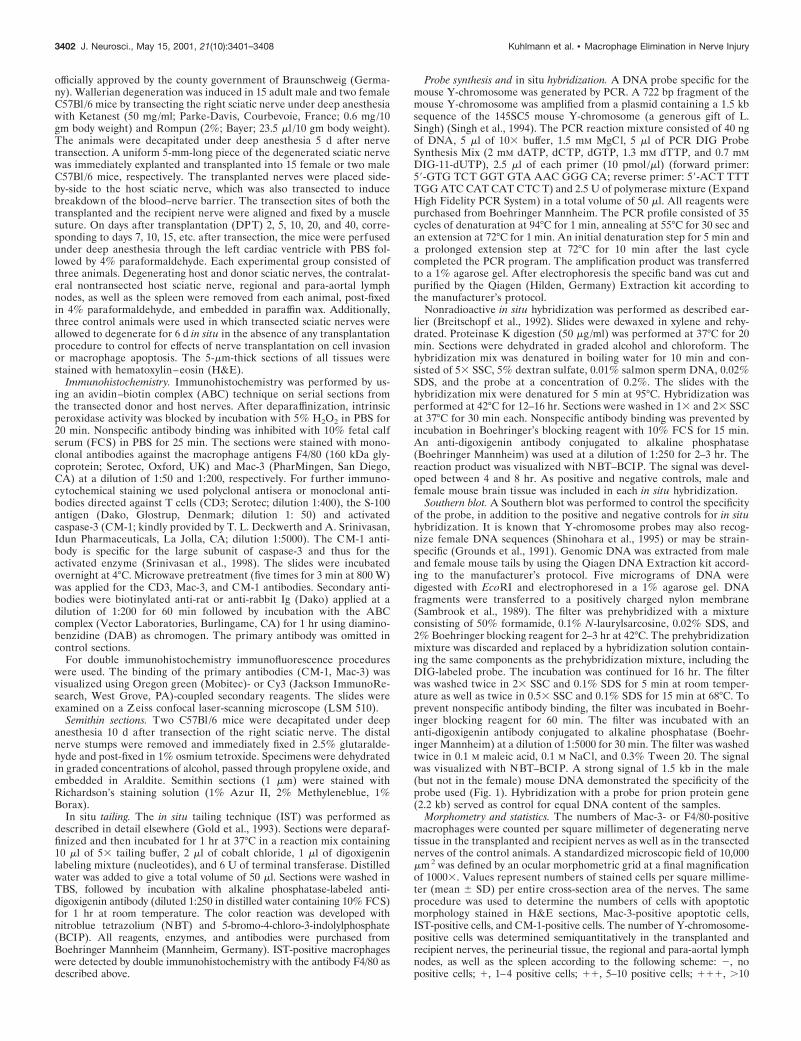
Southern blot. A Southern blot was performed to control the specificityof the probe, in addition to the positive and negative controls for in situhybridization. It is known that Y-chromosome probes may also recog-nize female DNA sequences (Shinohara et al., 1995) or may be strain-specific (Grounds et al., 1991). Genomic DNA was extracted from maleand female mouse tails by using the Qiagen DNA Extraction kit accord-ing to the manufacturer’s protocol. Five micrograms of DNA weredigested with EcoRI and electrophoresed in a 1% agarose gel. DNAfragments were transferred to a positively charged nylon membrane(Sambrook et al., 1989). The filter was prehybridized with a mixtureconsisting of 50% formamide, 0.1% N-laurylsarcosine, 0.02% SDS, and2% Boehringer blocking reagent for 2–3 hr at 42°C. The prehybridizationmixture was discarded and replaced by a hybridization solution contain-ing the same components as the prehybridization mixture, including theDIG-labeled probe. The incubation was continued for 16 hr. The filterwas washed twice in 23 SSC and 0.1% SDS for 5 min at room temper-ature as well as twice in 0.53 SSC and 0.1% SDS for 15 min at 68°C. Toprevent nonspecific antibody binding, the filter was incubated in Boehr-inger blocking reagent for 60 min. The filter was incubated with ananti-digoxigenin antibody conjugated to alkaline phosphatase (Boehr-inger Mannheim) at a dilution of 1:5000 for 30 min. The filter was washedtwice in 0.1 M maleic acid, 0.1 M NaCl, and 0.3% Tween 20. The signalwas visualized with NBT–BCIP. A strong signal of 1.5 kb in the male(but not in the female) mouse DNA demonstrated the specificity of theprobe used (Fig. 1). Hybridization with a probe for prion protein gene(2.2 kb) served as control for equal DNA content of the samples.
Morphometry and statistics. The numbers of Mac-3- or F4/80-positivemacrophages were counted per square millimeter of degenerating nervetissue in the transplanted and recipient nerves as well as in the transectednerves of the control animals. A standardized microscopic field of 10,000mm 2 was defined by an ocular morphometric grid at a final magnificationof 10003. Values represent numbers of stained cells per square millime-ter (mean 6 SD) per entire cross-section area of the nerves. The sameprocedure was used to determine the numbers of cells with apoptoticmorphology stained in H&E sections, Mac-3-positive apoptotic cells,IST-positive cells, and CM-1-positive cells. The number of Y-chromosome-positive cells was determined semiquantitatively in the transplanted andrecipient nerves, the perineurial tissue, the regional and para-aortal lymphnodes, as well as the spleen according to the following scheme: 2, nopositive cells; 1, 1–4 positive cells; 11, 5–10 positive cells; 111, .10
3402 J. Neurosci., May 15, 2001, 21(10):3401–3408 Kuhlmann et al. • Macrophage Elimination in Nerve Injury
positive cells. The Mann–Whitney U test and the ANOVA test were usedfor statistical analysis. A p value , 0.05 was considered significant.
RESULTSMorphology of the transected transplant andrecipient nervesWallerian degeneration induces recruitment of monocytes fromthe circulation into the damaged nerves. Earlier experiments
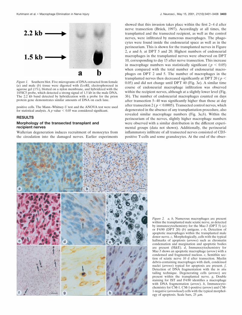
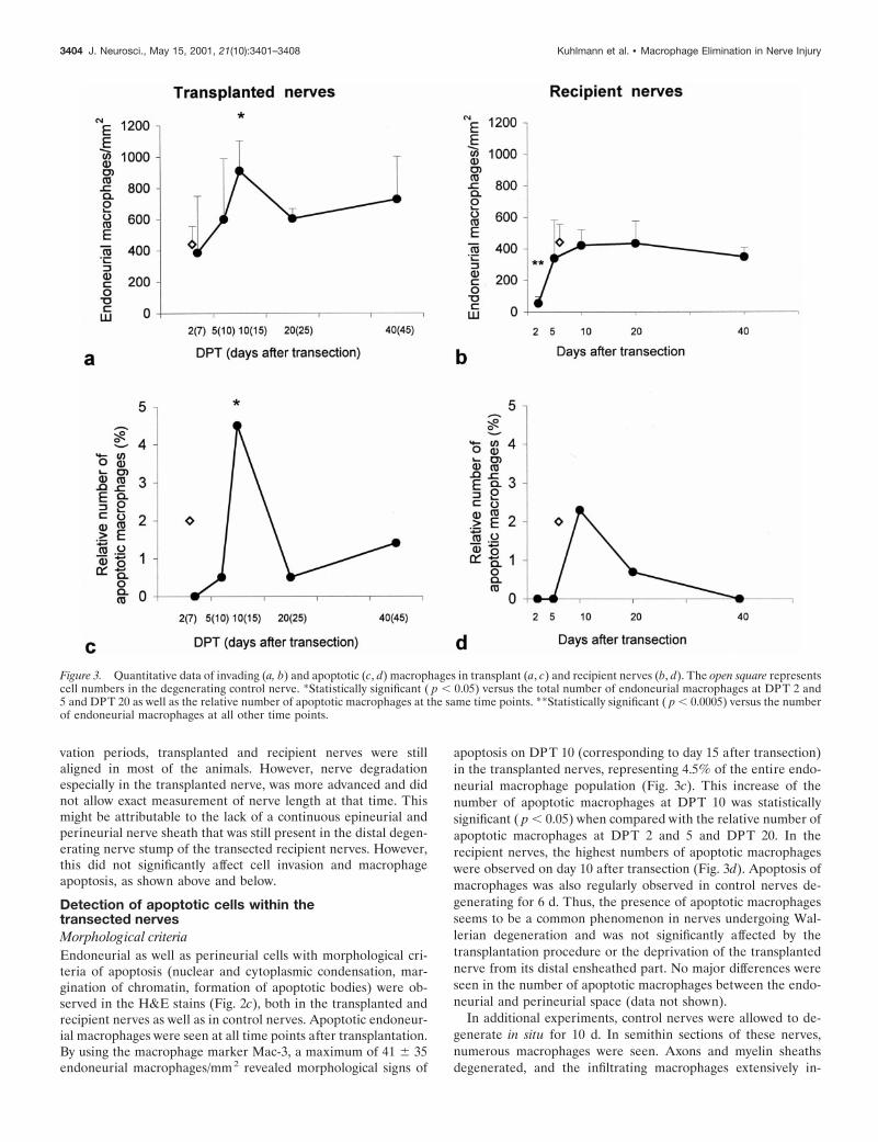
showed that this invasion takes place within the first 2–4 d afternerve transection (Bruck, 1997). Accordingly at all times, thetransplanted and the transected recipient, as well as the controlnerves, were infiltrated by numerous macrophages. The phago-cytes were found inside the endoneurial space as well as in theperineurium. This is shown for the transplanted nerves in Figure2, a and b, at DPT 5 and 20. Highest numbers of endoneurialmacrophages in the transplanted nerves were observed on DPT10, corresponding to day 15 after nerve transection. This increasein macrophage numbers was statistically significant ( p , 0.05)when compared with the total number of endoneurial macro-phages on DPT 2 and 5. The number of macrophages in thetransplanted nerves then decreased significantly at DPT 20 ( p ,0.05) and did not change until DPT 40 (Fig. 3a). A similar timecourse of endoneurial macrophage infiltration was observedwithin the recipient nerves, although at a slightly lower level (Fig.3b). The number of endoneurial macrophages counted on daysafter transection 5–40 was significantly higher than those at dayafter transection 2 ( p , 0.0005). Transected control nerves, whichdegenerated in the absence of any transplantation procedure, alsorevealed similar macrophage numbers (Fig. 3a,b). Within theperineurium of the nerves, slightly higher macrophage numberswere observed with a similar distribution in the different experi-mental groups (data not shown). Additionally, the perineurialinflammatory infiltrate of all transected nerves consisted of CD3-positive T-cells and some granulocytes. At the end of the obser-
Figure 2. a, b, Numerous macrophages are presentwithin the transplanted male sciatic nerve, as detectedby immunocytochemistry for the Mac-3 (DPT 5) (a)or F4/80 (DPT 20) (b) antigens. c–h, Detection ofapoptotic macrophages within the transplanted maledonor nerve. c, Morphologically, cells with the typicalhallmarks of apoptosis (arrows) such as chromatincondensation and margination and apoptotic bodiesare present (H&E). d, Immunocytochemistry forMac-3 shows an apoptotic macrophage (arrow) with acondensed and fragmented nucleus. e, Semithin sec-tion of sciatic nerve 10 d after transection. Myelindebris-containing macrophages with dark, condensednuclei (arrows) typical for apoptosis are present. f,Detection of DNA fragmentation with the in situtailing technique. Degenerating cells (arrows) arepresent within the transplanted nerve. g, Doublestaining for IST and F4/80 identifies a macrophagewith DNA fragmentation (arrow). h, Immunocyto-chemistry for CM-1. CM-1-positive (arrow) and CM-1-negative (arrowhead) cells with the typical morphol-ogy of apoptosis. Scale bars, 25 mm.
Figure 1. Southern blot. Five micrograms of DNA extracted from female(a) and male (b) tissue were digested with EcoRI, electrophoresed inagarose gel (1%), blotted on a nylon membrane, and hybridized with the145SC5 probe, which detected a strong signal of 1.5 kb in the male DNA.The 2.2 kb band detected by hybridization with a probe for the prionprotein gene demonstrates similar amounts of DNA on each lane.
Kuhlmann et al. • Macrophage Elimination in Nerve Injury J. Neurosci., May 15, 2001, 21(10):3401–3408 3403
vation periods, transplanted and recipient nerves were stillaligned in most of the animals. However, nerve degradationespecially in the transplanted nerve, was more advanced and didnot allow exact measurement of nerve length at that time. Thismight be attributable to the lack of a continuous epineurial andperineurial nerve sheath that was still present in the distal degen-erating nerve stump of the transected recipient nerves. However,this did not significantly affect cell invasion and macrophageapoptosis, as shown above and below.
Detection of apoptotic cells within thetransected nervesMorphological criteriaEndoneurial as well as perineurial cells with morphological cri-teria of apoptosis (nuclear and cytoplasmic condensation, mar-gination of chromatin, formation of apoptotic bodies) were ob-served in the H&E stains (Fig. 2c), both in the transplanted andrecipient nerves as well as in control nerves. Apoptotic endoneur-ial macrophages were seen at all time points after transplantation.By using the macrophage marker Mac-3, a maximum of 41 6 35endoneurial macrophages/mm2 revealed morphological signs of
apoptosis on DPT 10 (corresponding to day 15 after transection)in the transplanted nerves, representing 4.5% of the entire endo-neurial macrophage population (Fig. 3c). This increase of thenumber of apoptotic macrophages at DPT 10 was statisticallysignificant ( p , 0.05) when compared with the relative number ofapoptotic macrophages at DPT 2 and 5 and DPT 20. In therecipient nerves, the highest numbers of apoptotic macrophageswere observed on day 10 after transection (Fig. 3d). Apoptosis ofmacrophages was also regularly observed in control nerves de-generating for 6 d. Thus, the presence of apoptotic macrophagesseems to be a common phenomenon in nerves undergoing Wal-lerian degeneration and was not significantly affected by thetransplantation procedure or the deprivation of the transplantednerve from its distal ensheathed part. No major differences wereseen in the number of apoptotic macrophages between the endo-neurial and perineurial space (data not shown).
In additional experiments, control nerves were allowed to de-generate in situ for 10 d. In semithin sections of these nerves,numerous macrophages were seen. Axons and myelin sheathsdegenerated, and the infiltrating macrophages extensively in-
Figure 3. Quantitative data of invading (a, b) and apoptotic (c, d) macrophages in transplant (a, c) and recipient nerves (b, d). The open square representscell numbers in the degenerating control nerve. *Statistically significant ( p , 0.05) versus the total number of endoneurial macrophages at DPT 2 and5 and DPT 20 as well as the relative number of apoptotic macrophages at the same time points. **Statistically significant ( p , 0.0005) versus the numberof endoneurial macrophages at all other time points.
3404 J. Neurosci., May 15, 2001, 21(10):3401–3408 Kuhlmann et al. • Macrophage Elimination in Nerve Injury
gested the myelin fragments. Some of these macrophages con-tained a condensed and fragmented nucleus, changes typical forapoptosis (Fig. 2e). Additionally, a few macrophages were ob-served containing apoptotic bodies in their cytoplasms, whichpossibly represents phagocytosis of apoptotic cells bymacrophages.
Detection of DNA fragmentationNumerous cells with signs of DNA fragmentation were identifiedby the IST technique. IST-positive cells were observed in theendoneurium of transplanted as well as recipient nerves at alltime points investigated (Fig. 2f). Cells with DNA fragmentationwere also detected within the control nerves. Double staining ofIST and the macrophage-specific antigen F4/80 revealed a sub-population of macrophages that were IST-positive (Fig. 2g). Sim-ilar numbers of IST-positive macrophages were seen in recipientand control nerves (data not shown).
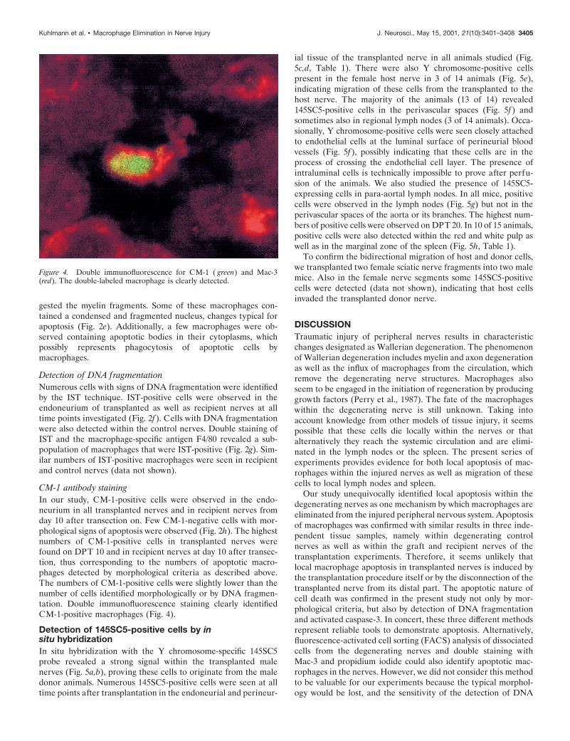
CM-1 antibody stainingIn our study, CM-1-positive cells were observed in the endo-neurium in all transplanted nerves and in recipient nerves fromday 10 after transection on. Few CM-1-negative cells with mor-phological signs of apoptosis were observed (Fig. 2h). The highestnumbers of CM-1-positive cells in transplanted nerves werefound on DPT 10 and in recipient nerves at day 10 after transec-tion, thus corresponding to the numbers of apoptotic macro-phages detected by morphological criteria as described above.The numbers of CM-1-positive cells were slightly lower than thenumber of cells identified morphologically or by DNA fragmen-tation. Double immunofluorescence staining clearly identifiedCM-1-positive macrophages (Fig. 4).
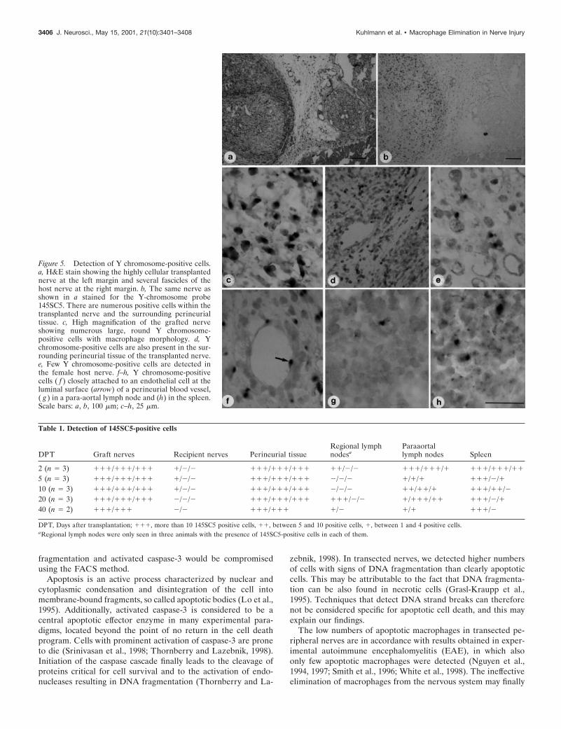
Detection of 145SC5-positive cells by insitu hybridizationIn situ hybridization with the Y chromosome-specific 145SC5probe revealed a strong signal within the transplanted malenerves (Fig. 5a,b), proving these cells to originate from the maledonor animals. Numerous 145SC5-positive cells were seen at alltime points after transplantation in the endoneurial and perineur-
ial tissue of the transplanted nerve in all animals studied (Fig.5c,d, Table 1). There were also Y chromosome-positive cellspresent in the female host nerve in 3 of 14 animals (Fig. 5e),indicating migration of these cells from the transplanted to thehost nerve. The majority of the animals (13 of 14) revealed145SC5-positive cells in the perivascular spaces (Fig. 5f) andsometimes also in regional lymph nodes (3 of 14 animals). Occa-sionally, Y chromosome-positive cells were seen closely attachedto endothelial cells at the luminal surface of perineurial bloodvessels (Fig. 5f), possibly indicating that these cells are in theprocess of crossing the endothelial cell layer. The presence ofintraluminal cells is technically impossible to prove after perfu-sion of the animals. We also studied the presence of 145SC5-expressing cells in para-aortal lymph nodes. In all mice, positivecells were observed in the lymph nodes (Fig. 5g) but not in theperivascular spaces of the aorta or its branches. The highest num-bers of positive cells were observed on DPT 20. In 10 of 15 animals,positive cells were also detected within the red and white pulp aswell as in the marginal zone of the spleen (Fig. 5h, Table 1).
To confirm the bidirectional migration of host and donor cells,we transplanted two female sciatic nerve fragments into two malemice. Also in the female nerve segments some 145SC5-positivecells were detected (data not shown), indicating that host cellsinvaded the transplanted donor nerve.
DISCUSSIONTraumatic injury of peripheral nerves results in characteristicchanges designated as Wallerian degeneration. The phenomenonof Wallerian degeneration includes myelin and axon degenerationas well as the influx of macrophages from the circulation, whichremove the degenerating nerve structures. Macrophages alsoseem to be engaged in the initiation of regeneration by producinggrowth factors (Perry et al., 1987). The fate of the macrophageswithin the degenerating nerve is still unknown. Taking intoaccount knowledge from other models of tissue injury, it seemspossible that these cells die locally within the nerves or thatalternatively they reach the systemic circulation and are elimi-nated in the lymph nodes or the spleen. The present series ofexperiments provides evidence for both local apoptosis of mac-rophages within the injured nerves as well as migration of thesecells to local lymph nodes and spleen.
Our study unequivocally identified local apoptosis within thedegenerating nerves as one mechanism by which macrophages areeliminated from the injured peripheral nervous system. Apoptosisof macrophages was confirmed with similar results in three inde-pendent tissue samples, namely within degenerating controlnerves as well as within the graft and recipient nerves of thetransplantation experiments. Therefore, it seems unlikely thatlocal macrophage apoptosis in transplanted nerves is induced bythe transplantation procedure itself or by the disconnection of thetransplanted nerve from its distal part. The apoptotic nature ofcell death was confirmed in the present study not only by mor-phological criteria, but also by detection of DNA fragmentationand activated caspase-3. In concert, these three different methodsrepresent reliable tools to demonstrate apoptosis. Alternatively,fluorescence-activated cell sorting (FACS) analysis of dissociatedcells from the degenerating nerves and double staining withMac-3 and propidium iodide could also identify apoptotic mac-rophages in the nerves. However, we did not consider this methodto be valuable for our experiments because the typical morphol-ogy would be lost, and the sensitivity of the detection of DNA
Figure 4. Double immunofluorescence for CM-1 ( green) and Mac-3(red). The double-labeled macrophage is clearly detected.
Kuhlmann et al. • Macrophage Elimination in Nerve Injury J. Neurosci., May 15, 2001, 21(10):3401–3408 3405
fragmentation and activated caspase-3 would be compromisedusing the FACS method.
Apoptosis is an active process characterized by nuclear andcytoplasmic condensation and disintegration of the cell intomembrane-bound fragments, so called apoptotic bodies (Lo et al.,1995). Additionally, activated caspase-3 is considered to be acentral apoptotic effector enzyme in many experimental para-digms, located beyond the point of no return in the cell deathprogram. Cells with prominent activation of caspase-3 are proneto die (Srinivasan et al., 1998; Thornberry and Lazebnik, 1998).Initiation of the caspase cascade finally leads to the cleavage ofproteins critical for cell survival and to the activation of endo-nucleases resulting in DNA fragmentation (Thornberry and La-
zebnik, 1998). In transected nerves, we detected higher numbersof cells with signs of DNA fragmentation than clearly apoptoticcells. This may be attributable to the fact that DNA fragmenta-tion can be also found in necrotic cells (Grasl-Kraupp et al.,1995). Techniques that detect DNA strand breaks can thereforenot be considered specific for apoptotic cell death, and this mayexplain our findings.
The low numbers of apoptotic macrophages in transected pe-ripheral nerves are in accordance with results obtained in exper-imental autoimmune encephalomyelitis (EAE), in which alsoonly few apoptotic macrophages were detected (Nguyen et al.,1994, 1997; Smith et al., 1996; White et al., 1998). The ineffectiveelimination of macrophages from the nervous system may finally
Figure 5. Detection of Y chromosome-positive cells.a, H&E stain showing the highly cellular transplantednerve at the left margin and several fascicles of thehost nerve at the right margin. b, The same nerve asshown in a stained for the Y-chromosome probe145SC5. There are numerous positive cells within thetransplanted nerve and the surrounding perineurialtissue. c, High magnification of the grafted nerveshowing numerous large, round Y chromosome-positive cells with macrophage morphology. d, Ychromosome-positive cells are also present in the sur-rounding perineurial tissue of the transplanted nerve.e, Few Y chromosome-positive cells are detected inthe female host nerve. f–h, Y chromosome-positivecells ( f ) closely attached to an endothelial cell at theluminal surface (arrow) of a perineurial blood vessel,( g ) in a para-aortal lymph node and (h) in the spleen.Scale bars: a, b, 100 mm; c–h, 25 mm.
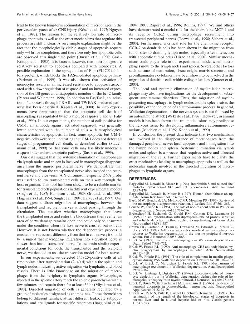
Table 1. Detection of 145SC5-positive cells
DPT Graft nerves Recipient nerves Perineurial tissueRegional lymphnodesa
Paraaortallymph nodes Spleen
2 (n 5 3) 111/111/111 1/2/2 111/111/111 11/2/2 111/111/1 111/111/11
5 (n 5 3) 111/111/111 1/2/2 111/111/111 2/2/2 1/1/1 111/2/110 (n 5 3) 111/111/111 1/2/2 111/111/111 2/2/2 11/11/1 111/11/220 (n 5 3) 111/111/111 2/2/2 111/111/111 111/2/2 1/111/11 111/2/140 (n 5 2) 111/111 2/2 111/111 1/2 1/1 111/2
DPT, Days after transplantation; 111, more than 10 145SC5 positive cells, 11, between 5 and 10 positive cells, 1, between 1 and 4 positive cells.aRegional lymph nodes were only seen in three animals with the presence of 145SC5-positive cells in each of them.
3406 J. Neurosci., May 15, 2001, 21(10):3401–3408 Kuhlmann et al. • Macrophage Elimination in Nerve Injury
lead to the known long-term accumulation of macrophages in theperivascular spaces after CNS injury (Kosel et al., 1997; Nguyenet al., 1997). The reasons for the relatively low rate of macro-phage apoptosis as well as the exact mechanisms that regulate thisprocess are still unclear. A very simple explanation might be thefact that the morphologically visible stages of apoptosis requireonly ;4 hr for completion, and therefore only few apoptotic cellsare observed at a single time point (Bursch et al., 1990; Grasl-Kraupp et al., 1995). It is known, however, that macrophages arerelatively resistant to apoptosis compared with monocytes. Apossible explanation is the upregulation of Flip (FLICE inhibi-tory protein), which blocks the FAS-mediated apoptotic pathway(Perlman et al., 1999). It was also shown that activation ofmonocytes results in an increased resistance to apoptosis associ-ated with a downregulation of caspase-8 and an increased expres-sion of the Bfl-gene, an antiapoptotic member of the bcl-2 family(Perera and Waldmann, 1998). In addition to Fas/FasL, an induc-tion of apoptosis through TRAIL- and TWEAK-mediated path-ways has been described (Kaplan et al., 2000). In vitro experi-ments have demonstrated that the apoptotic cell death ofmacrophages is regulated by activation of caspases 3 and 8 (Fahyet al., 1999). In our experiments, the number of cells positive forCM-1, an antibody against activated caspase-3, was generallylower compared with the number of cells with morphologicalcharacteristics of apoptosis. In fact, some apoptotic but CM-1-negative cells were seen, indicating that CM-1 does not detect allstages of programmed cell death, as described earlier (Stadel-mann et al., 1999) or that some cells may less likely undergo acaspase-independent apoptotic pathway (Susin et al., 1999).
Our data suggest that the systemic elimination of macrophagesvia lymph nodes and spleen is involved in macrophage disappear-ance from the injured peripheral nerve. We demonstrate thatmacrophages from the transplanted nerve also invaded the recip-ient nerve and vice versa. A Y-chromosome-specific DNA probewas used to follow transplanted cells on their way through thehost organism. This tool has been shown to be a reliable markerfor transplanted cell populations in different experimental models(Singh et al., 1987; Hampson et al., 1989; Grounds et al., 1991;Hagenaars et al., 1994; Singh et al., 1994; Harvey et al., 1997). Ourdata suggest a direct migration of macrophages between thetransplanted and recipient nerves without passing through thecirculation. The question whether macrophages that leavethe transplanted nerve and enter the bloodstream then reenter anarea of nerve damage remains open and could only be answeredunder the condition when the host nerve is crushed but not cut.However, it is not known whether the degenerative process incrushed nerves occurs differently from that in cut nerves; it shouldbe assumed that macrophage migration into a crushed nerve isslower than into a transected nerve. To ascertain similar experi-mental conditions for both, the transplanted and the recipientnerve, we decided to use the transection model for both nerves.
In our experiments, we detected 145SC5-positive cells at alltime points after transplantation (2–40 d) within the spleen andlymph nodes, indicating active migration via lymphatic and bloodvessels. There is little knowledge on the migration of macro-phages from the periphery to lymphatic organs. Macrophagesinjected in the splenic artery reach the splenic parenchyma withinfew minutes and remain there for at least 36 hr (Miyakawa et al.,1990). Directed migration of cells is generally regulated by agroup of molecules designated as chemokines. These chemokinesbelong to different families, attract different leukocyte subpopu-lations, and are ligands for specific receptors (Baggiolini et al.,
1994, 1997; Raport et al., 1996; Rollins, 1997). We and othershave demonstrated a crucial role for the chemokine MCP-1 andits receptor CCR2 during macrophage recruitment intotransected peripheral nerves (Toews et al., 1998; Siebert et al.,2000). Furthermore, a critical role for the chemokine receptorCCR-7 on dendritic cells has been shown in the migration fromtumor sites to draining lymph nodes, especially after interactionwith apoptotic tumor cells (Hirao et al., 2000). Similar mecha-nisms could play a role in our experimental model when macro-phages move to the lymph nodes and spleen. Several other factorssuch as state of maturation, tissue origin, matrix structure, andproinflammatory cytokines have been shown to be involved in themigration of dendritic cells within collagen lattices (Gunzer et al.,2000).
The local and systemic elimination of myelin-laden macro-phages may also have implications for the development of subse-quent autoimmune disorders. Thus, the migration of myelin-presenting macrophages to lymph nodes and the spleen raises thepossibility of the induction of an autoimmune process. In general,Wallerian degeneration in the nervous system is not followed byan autoimmune attack (Wekerle et al., 1986). However, in animalmodels it has been shown that traumatic lesions may predisposethe nervous tissue for developing inflammatory autoimmune re-actions (Maehlen et al., 1989; Konno et al., 1990).
In conclusion, the present data indicate that two mechanismsare involved in the disappearance of macrophages from thedamaged peripheral nerve: local apoptosis and immigration intothe lymph nodes and spleen. Systemic elimination via lymphnodes and spleen is a process that requires active and directedmigration of the cells. Further experiments have to clarify theexact mechanisms leading to macrophage apoptosis as well as themolecular signals involved in the directed migration of macro-phages to lymphatic organs.
REFERENCESBaggiolini M, Dewald B, Moser B (1994) Interleukin-8 and related che-
motactic cytokines—CXC and CC chemokines. Adv Immunol55:97–179.
Baggiolini M, Dewald B, Moser B (1997) Human chemokines: an up-date. Annu Rev Immunol 15:675–705.
Barth MW, Hendrzak JA, Melnicoff MJ, Morahan PS (1995) Review ofthe macrophage disappearance reaction. J Leukoc Biol 57:361–367.
Beuche W, Friede RL (1984) The role of non-resident cells in Walleriandegeneration. J Neurocytol 13:767–796.
Breitschopf H, Suchanek G, Gould RM, Colman DR, Lassmann H(1992) In situ hybridization with digoxigenin-labeled probes: sensitiveand reliable detection method applied to myelinating rat brain. ActaNeuropathol 84:581–587.
Brown HC, Castano A, Feam S, Townsend M, Edwards G, Streuli C,Perry VH (1997) Adhesion molecules involved in macrophage re-sponses to Wallerian degeneration in the murine peripheral nervoussystem. Eur J Neurosci 9:2057–2063.
Bruck W (1997) The role of macrophages in Wallerian degeneration.Brain Pathol 7:741–752.
Bruck W, Friede RL (1990) Anti-macrophage CR3 antibody blocks my-elin phagocytosis by macrophages in vitro. Acta Neuropathol80:415–418.
Bruck W, Friede RL (1991) The role of complement in myelin phago-cytosis during PNS Wallerian degeneration. J Neurol Sci 103:182–187.
Bruck W, Bruck Y, Maruschak B, Friede RL (1995) Mechanisms ofmacrophage recruitment in Wallerian degeneration. Acta Neuropathol89:363–367.
Bruck W, Huitinga I, Dijkstra CD (1996a) Liposome-mediated mono-cyte depletion during Wallerian degeneration defines the role of he-matogenous phagocytes in myelin removal. J Neurosci Res 46:477–484.
Bruck Y, Bruck W, Kretzschmar HA, Lassmann H (1996b) Evidence forneuronal apoptosis in pontosubicular neuron necrosis. NeuropatholAppl Neurobiol 22:23–29.
Bursch W, Paffe S, Putz B, Barthel G, Schulte-Hermann R (1990) De-termination of the length of the histological stages of apoptosis innormal liver and in altered hepatic foci of rats. Carcinogenesis11:847–853.
Kuhlmann et al. • Macrophage Elimination in Nerve Injury J. Neurosci., May 15, 2001, 21(10):3401–3408 3407
Fahy RJ, Doseff AI, Wewers MD (1999) Spontaneous human monocyteapoptosis utilizes a caspase-3-dependent pathway that is blocked byendotoxin and is independent of caspase-1. J Immunol 163:1755–1762.
Fernandez-Valle C, Bunge RP, Bunge BM (1995) Schwann cells degrademyelin and proliferate in the absence of macrophages: evidence from invitro studies of Wallerian degeneration. J Neurocytol 24:667–679.
George EB, Glass JD, Griffin JW (1995) Axotomy-induced axonal de-generation is mediated by calcium influx through ion-specific channels.J Neurosci 15:6445–6452.
Gold R, Schmied M, Rothe G, Zischler H, Breitschopf H, Wekerle H,Lassmann H (1993) Detection of DNA fragmentation in apoptosis:application of in situ nick translation to cell culture systems and tissuesections. J Histochem Cytochem 41:1023–1030.
Grasl-Kraupp B, Ruttkay-Nedecky B, Koudelka H, Bukowkska K, BurschW, Schulte-Herrmann R (1995) In situ detection of fragmented DNA(TUNEL assay) fails to discriminate among apoptosis, necrosis andautolytic cell death: a cautionary note. Hepatology 21:1465–1468.
Griffin JW, Hoffman PN (1993) Degeneration and regeneration in theperipheral nervous system. In: Peripheral neuropathy (van Dyck PJ,Thomas PK, Griffin JW, Low PA, Poduslo JF, eds), pp 361–376.Philadelphia: W.B. Saunders.
Griffin JW, George R, Lobato C, Tyor WR, Yan LC, Glass JD (1992)Macrophage responses and myelin clearance during Wallerian degen-eration: relevance to immune-mediated demyelination. J Neuroimmu-nol 40:153–166.
Grounds MD, Lai MC, Fan Y, Codling JC, Beilharz MW (1991) Trans-plantation in the mouse model - the use of a Y-chromosome-specificDNA clone to identify donor cells in situ. Transplantation52:1101–1105.
Gunzer M, Friedl P, Niggemann B, Brocker E-B, Kampgen E, Zanker KS(2000) Migration of dendritic cells within 3-D collagen lattices isdependent on tissue origin, state of maturation, and matrix structureand is maintained by proinflammatory cytokines. J Leukoc Biol67:622–629.
Hagenaars CE, Kawilarang-de Haas EWM, Hazekamp J, Wiegant J,Nijweide PJ (1994) Osteoclast development in the coculture system ofperiostless metatarsal bones and hemopoietic cells studied by in situhybridization with a probe for Y chromosomes. Calcif Tissue Int54:170–174.
Hampson IN, Spooncer E, Dexter TM (1989) Evaluation of a mouse Ychromosome probe for assessing marrow transplantation. Exp Hematol17:313–315.
Harvey AR, Symons NA, Pollett MA, Brooker GJF, Bartlett PF (1997)Fate of adult neural precursors grafted to adult cortex monitored witha Y-chromosome marker. NeuroReport 8:3939–3943.
Hirao M, Onai N, Hiroishi K, Watkins SC, Matsushima K, Robbins PD,Lotze MT, Tahara H (2000) CC chemokine receptor-7 on dendriticcells is induced after interaction with apoptotic tumor cells: critical rolein migration from the tumor site to draining lymph nodes. Cancer Res60:2209–2217.
Kaplan MJ, Ray D, Mo R-R, Yung RL, Richardson BC (2000) TRAIL(Apo2 ligand) and TWEAK (Apo3 ligand) mediate CD4 1 T cell killingof antigen-presenting macrophages. J Immunol 164:2897–2904.
Konno H, Yamamoto T, Suzuki H, Iwasaki Y, Ohara Y, Terunuma H,Harata N (1990) Targeting of adoptively transferred experimental al-lergic encephalitis lesion at the sites of Wallerian degeneration. ActaNeuropathol 80:521–526.
Kosel S, Egensberger R, Bise K, Arbogast S, Mehraein P, Graeber MB(1997) Long-lasting perivascular accumulation of major histocompat-ibility complex class II positive lipophages in the spinal cord of strokepatients: possible relevance for the immune privilege of the brain. ActaNeuropathol 94:532–538.
Lo AC, Houenou LJ, Oppenheim RW (1995) Apoptosis in the nervoussystem: morphological features, methods, pathology, and prevention.Arch Histol Cytol 58:139–149.
Maehlen J, Olsson T, Zachau A, Klareskog L, Kristensson K (1989)Local enhancement of major histocompatibility complex (MHC) classI and II expression and cell infiltration in experimental allergic enceph-alomyelitis around axotomized motor neurons. J Neuroimmunol23:125–132.
Miyakawa K, Matsuno K, Ohmori J, Kotani M (1990) Localization inthe rat spleen of carbon-laden macrophages introduced into the splenicartery: a subpopulation of macrophages entering the white pulp. AnatRec 227:464–474.
Nguyen KB, McCombe PA, Pender MP (1994) Macrophage apoptosis
in the central nervous system in experimental autoimmune encephalo-myelitis. J Autoimmun 7:145–152.
Nguyen KB, McCombe PA, Pender MP (1997) Increased apoptosis ofT-lymphocytes and macrophages in the central and peripheral nervoussystem of Lewis rats with experimental autoimmune encephalomyelitistreated with dexamethasone. J Neuropathol Exp Neurol 56:58–69.
Perera LP, Waldmann TA (1998) Activation of human monocytes in-duces differential resistance to apoptosis with rapid down-regulation ofcaspase-8/FLICE. Proc Natl Acad Sci USA 95:14308–14313.
Perlman H, Pagliari LJ, Georganas C, Mano T, Walsh K, Pope RM(1999) FLICE-inhibitory protein expression during macrophage differ-entiation confers resistance to Fas-mediated apoptosis. J Exp Med190:1679–1688.
Perry VH, Brown MC, Gordon S (1987) The macrophage response tocentral and peripheral nerve injury. A possible role for macrophages inregeneration. J Exp Med 165:1218–1223.
Raport CJ, Schweickart VL, Chantry D, Eddy RL Jr, Shows TB, GodiskaR, Gray PW (1996) New members of the chemokine receptor genefamily. J Leukoc Biol 59:18–23.
Rollins BJ (1997) Chemokines. Blood 90:909–928.Sambrook J, Fritsch EF, Maniatis T (1989) Molecular cloning. A labo-
ratory manual. Cold Spring Harbor Laboratory Press.Shinohara S, Sawada T, Nishioka Y, Tohma S, Kisaki T, Inoue T, Ando
K, Ikeda M, Fujii H, Ito K (1995) Differential expression of Fasantigen and bcl-2 protein on CD41 T cells, CD81 T cells, and mono-cytes. Cell Immunol 163:303–308.
Siebert H, Sachse A, Kuziel WA, Maeda N, Bruck W (2000) The che-mokine receptor CCR2 is involved in macrophage recruitment to theinjured peripheral nervous system. J Neuroimmunol 110:177–185.
Singh L, Matsukuma S, Jones KW (1987) The use of Y-chromosome-specific repeated DNA sequences in the analysis of testis developmentin an XX/XY mouse. Development 101S:143–149.
Singh L, Panicker SG, Nagaraj R, Majumdar KC (1994) Banded kraitminor-satellite (Bkm)-associated Y chromosome-specific repetitiveDNA in mouse. Nucleic Acids Res 22:2289–2295.
Smith T, Schmied M, Hewson AK, Lassmann H, Cuzner ML (1996)Apoptosis of T cells and macrophages in the central nervous system ofintact and adrenalectomized Lewis rats during experimental allergicencephalomyelitis. J Autoimmun 9:167–174.
Srinivasan A, Roth KA, Sayers RO, Shindler KS, Wong AM, Fritz LC,Tomaselli KJ (1998) In situ immunodetection of activated caspase-3in apoptotic neurons in the developing nervous system. Cell DeathDiffer 5:1004–1016.
Stadelmann C, Deckwerth TL, Srinivasan A, Bancher C, Bruck W,Jellinger K, Lassmann H (1999) Activation of caspase-3 in singleneurons and autophagic vacuoles of granulovacuolar degeneration inAlzheimer’s disease. Evidence for apoptotic cell death. Am J Pathol155:1459–1466.
Stoll G, Hartung H-P (1992) The role of macrophages in degenerationand immune-mediated demyelination of the peripheral nervous system.Adv Neuroimmunol 2:163–179.
Stoll G, Griffin JW, Li CY, Trapp BD (1989) Wallerian degeneration inthe peripheral nervous system: participation of both Schwann cells andmacrophages in myelin degradation. J Neurocytol 18:671–683.
Susin SA, Lorenzo HK, Zamzami N, Marzo I, Snow BE, Brothers GM,Mangion J, Jacotot E, Constantini P, Loeffler M, Larochette N,Goodlett DR, Aebersold R, Siderovski DP, Penninger JM, Kroemer G(1999) Molecular characterization of mitochondrial apoptosis-inducing factor. Nature 397:441–446.
Takemura G, Ohno M, Hayakawa Y, Misao J, Kanoh M, Ohno A, UnoY, Minatoguchi S, Fujiwara T, Fujiwara H (1998) Role of apoptosis inthe disappearance of infiltrated and proliferated interstitial cells aftermyocardial infarction. Circ Res 82:1130–1138.
Thornberry NA, Lazebnik Y (1998) Caspases: enemies within. Science281:1312–1316.
Toews AD, Barrett C, Morell P (1998) Monocyte chemoattractant pro-tein 1 is responsible for macrophage recruitment following injury tosciatic nerve. J Neurosci Res 53:260–267.
Vougioukas VI, Roeske S, Michel U, Bruck W (1998) Wallerian degen-eration in ICAM-1-deficient mice. Am J Pathol 152:241–249.
Wekerle H, Linington C, Lassmann H, Meyermann R (1986) Cellularimmune reactivity within the CNS. Trends Neurosci 9:271–277.
White CA, McCombe PA, Pender MP (1998) Microglia are more sus-ceptible than macrophages to apoptosis in the central nervous system inexperimental autoimmune encephalomyelitis through a mechanism notinvolving Fas (CD95). Int Immunol 10:935–941.
3408 J. Neurosci., May 15, 2001, 21(10):3401–3408 Kuhlmann et al. • Macrophage Elimination in Nerve Injury