Embed Size (px)
Citation preview

Antn. rheum. Dis. (1975), 34, 38
Macrophage-lymphocyte clustering inrheumatoid arthritis
F. W. S. WEBB, M. BAKER, R. WEISBART, R. BLUESTONE, AND L. GOLDBERGFrom the Department of Medicine, Rheumatology Division, University of California at Los Angeles, and theWadsworth Veterans Administration Hospital, Los Angeles, California
Webb, F. W. S., Baker, M., Weisbart, R., Bluestone, R., and Goldberg, L. (1975). Annals ofthe Rheumatic Diseases, 34, 38. Macrophage-lymphocyte clustering in rheumatoid arthritis.The cells in synovial fluid from patients with rheumatoid arthritis contain a smallpercentage of macrophages. Such macrophages were isolated and cultured alone andwith homologous and heterologous lymphocytes for 24 hours, in an attempt to identifypossible contact between living lymphocytes and macrophages. Such contact was found,with clustering of lymphocytes around macrophages, and was particularly well shownby scanning electron microscopy.
Although some kind of autoallergic process has beenput forward to explain the remarkable chronicityof the inflammatory process in such conditions asrheumatoid arthritis, recent work has, on the otherhand, suggested that very small amounts of foreignantigen may persist in an active form in experimentalmodels which resemble human rheumatoid disease.For example, Webb, Ford, and Glynn (1971) wereable to show that minute amounts of antigen per-sisting in the joints of normal rabbits gave rise to achronic synovitis if these rabbits were later immunizedto the original antigen. This work was extended(Consden, Doble, Glynn, and Nind, 1971) andapproximately 0-001% of the original amount ofantigen injected intra-articularly in rabbits wasshown to persist for up to 6 months.The assumption that some at least of the antigen
persisting in the normal rabbit knee may rest with thesynovial membrane macrophages has been confirmedby Webb, Goldberg, Bluestone, and Pearson (1972),who were able to show that antigen can be identifiedon the surface of rabbit synovial fluid macrophages,these cells probably being shed directly from thesynovial membrane into the joint cavity. The amountsof antigen persisting in the synovial fluid macro-phages are similar to the very small amounts per-sisting in macrophages in other experimentalsystems.
This and other work discussing the role of macro-phages in antigen handling and the passage ofretained antigenic information by cell to cell contactto lymphocytes has been reviewed recently byUnanue (1972).
With this background, a search was made for thepresence of macrophage-lymphocyte contact in thesynovial fluid of patients with rheumatoid arthritisand allied conditions. Although most of the cellsfound in these synovial effusions are polymorpho-nuclear neutrophil leucocytes whose role appearsto be to phagocytose complement-fixing immunecomplexes in the synovial fluid (see Zvaifler, 1973, forreview), a proportion of the cells are macrophages,that is, mononuclear phagocytic cells. It was assumedthat some of these macrophages, probably a smallproportion, would be derived directly from thesynovial membrane itself. It was also thought thatthese macrophages might bear on their surface an asyet unidentified primary antigen to which the patient'slymphocytes might be specifically sensitive. If so, itwas anticipated that macrophage-lymphocyte cluster-ing would be observed.
Materials and methods
MACROPHAGE PREPARATIONSynovial fluid was aspirated from the joints of six patientswith rheumatoid arthritis. The fluid was defibrinated usingglass beads and mixed with an equal volume of Hanks'sbalanced salt solution (HBSS). After centrifugation at140 g for 10 min, the cell button was washed in Hanks'ssolution and resuspended.
Cell counts were then carried out. When more than 20%of macrophages were found, the cells were washed threetimes before being used. When a small percentage ofmacrophages was present, macrophage-rich populationswere prepared by flotation on bovine serum albumin(Cline and Lehrer, 1968).
Accepted for publication May 24, 1974.Requests for reprints to: Dr. F. W. S. Webb, Dept. of Rheumatology, Ipswich Hospital, Anglesea Road, Ipswich, Suffolk.
copyright. on 24 June 2018 by guest. P
rotected byhttp://ard.bm
j.com/
Ann R
heum D
is: first published as 10.1136/ard.34.1.38 on 1 February 1975. D
ownloaded from
Macrophage-lymphocyte clustering in rheumatoid arthritis 39
MACROPHAGE IDENTIFICATIONMacrophages were identified as large cells with abundantcytoplasm up to 25 pum in diameter, containing a singleindented nucleus usually with two nucleoli. These cellsadhered quickly to glass and migrated over the glasssurface, rapidly taking up latex and colloidal carbonparticles. These phagocytic cells also formed rosetteswith human type 0 erythrocytes sensitized with anti-Rh0 antibody, showing that they possessed surface receptorsfor IgG as has been shown previously for peripheralblood macrophages (Huber, Douglas, and Fudenberg,1968).
MACROPHAGE CULTUREMacrophages at a concentration of 1 x 10⁶ cells/mlwere suspended in medium 199 containing 10% fetalcalf serum, penicillin 100 units/ml, and streptomycin100 units/ml. The osmolality of this culture medium was280 mOsm/l. The macrophages in their medium werecultured on glass coverslips in Sykes-Moore chambers at37°C for one hour, to give the macrophages time to stickto the glass. The chambers were then gently emptied to getrid of nonadherent cells, dead cells, and debris. Onechamber was then refilled with medium alone, anotherwith lymphocytes from the patient's blood, and threefurther chambers filled with lymphocytes taken fromhealthy laboratory personnel who showed no evidence ofarthritis. The concentration of lymphocytes in the mediumused to refill the chambers was approximately 2 x 10⁶/ml.LYMPHOCYTE PREPARATIONPeripheral blood was defibrinated with glass beads andcentrifuged at 400 g for 15 min. The buffy coat cellswere then removed and mixed with autologous serum andallowed to sediment for 4 hours in 1 g at 37°C (Weisbart,Webb, Bluestone, and Goldberg, 1972).
EXAMINATION OF CULTURESThese were examined from time to time by phase contrastmicroscopy and after 24 hours the coverslips were removed
from the chambers, gently washed in fresh culture medium,and fixed in methanol for Giemsa staining, or in 2%glutaraldehyde in medium 199 for scanning electronmicroscopy.
PREPARATION FOR THE SCANNING ELECTRONMICROSCOPEAfter fixation in 2% glutaraldehyde for 30 min, cover-slips were rinsed twice in Hanks's solution and post-fixedwith 1 % osmium tetroxide for 30 min, and rinsed twicein Hanks's solution. The coverslips were then dehydratedin graded alcohols for 10 min at each grade, and thenfrom absolute alcohol through graded alcohol/amylacetate mixtures to absolute amyl acetate. The coverslipwas then transferred to a critical point drying apparatus(Boyde and Wood, 1969), taking care not to allow theamyl acetate to evaporate before the apparatus wasfilled with liquid CO2. Gas was blown off until free fromthe smell of amyl acetate when the chamber was sealedand heated gently, to bring the pressure in the chamber to1500 lb/in2. The gas was then gently released and thecoverslip removed and coated in a vacuum chamberfirst with carbon and then with gold to provide a very thinconducting surface over the cells and coverslip. Theprepared coverslip was then examined in a Cambridgestereoscan scanning electron microscope.
Results
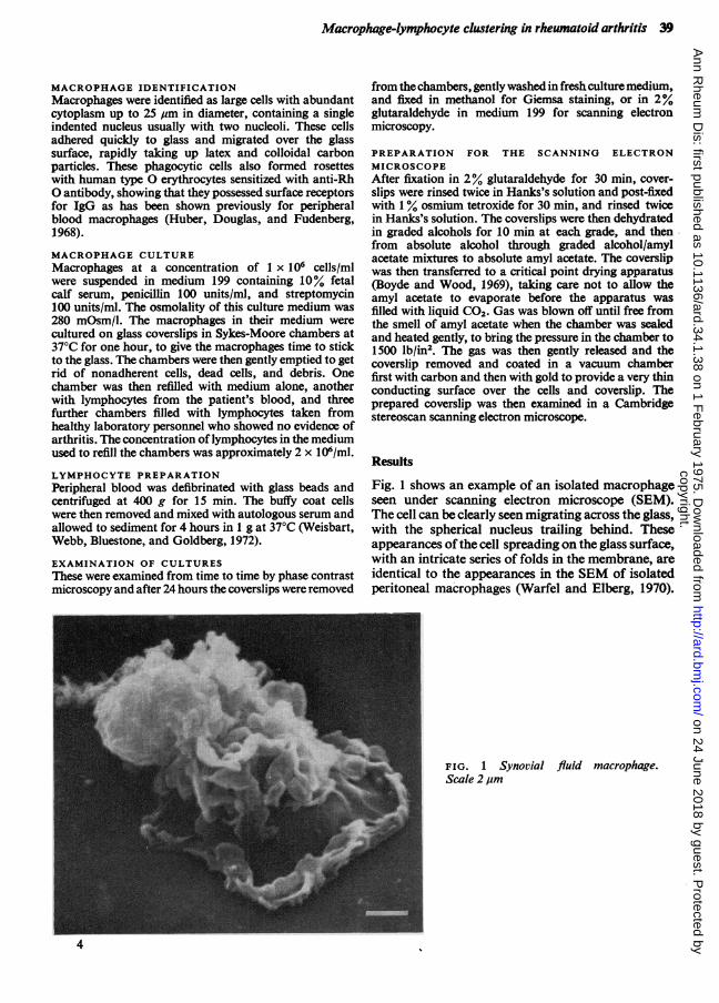
Fig. 1 shows an example of an isolated macrophageseen under scanning electron microscope (SEM).The cell can be clearly seen migrating across the glass,with the spherical nucleus trailing behind. Theseappearances of the cell spreading on the glass surface,with an intricate series of folds in the membrane, areidentical to the appearances in the SEM of isolatedperitoneal macrophages (Warfel and Elberg, 1970).
FIG. 1 Synovial fluid macrophage.Scale 2pum
copyright. on 24 June 2018 by guest. P
rotected byhttp://ard.bm
j.com/
Ann R
heum D
is: first published as 10.1136/ard.34.1.38 on 1 February 1975. D
ownloaded from

40 Annals of the Rheumatic Diseases
Similar appearances were also identified in the livingcell by phase and interference contrast microscopy.
Moreover, individual living cells were fixed inglutaraldehyde and re-examined afterwards, whenno change in the appearance of the fixed cells couldbe seen. There is therefore every reason to supposethat the preparation of the cells for the SEM leadto very little or no distortion from the living state inculture. Clear evidence of clustering of sphericallymphocytes around macrophages was seen, asshown in Fig. 2. Such clustering could also beidentified in methanol-fixed Giemsa stained prepara-
tions, but the clear identification of the sphericallymphocytes clustered on the macrophage surfacewas only seen under the SEM.
Isolated lymphocytes cultured on their own werealso examined (Fig. 3) and shown to be sphericalcells 3-4 pm in diameter. Recent work (Polliack,Lampen, Clarkson, De Harven, Bentwich, Siegal,and Kunkel, 1973), conducted after our study,suggests that the lymphocytes bearing villi are B orbursa-derived cells, while the smooth surfaced cellswithout villi ale T or thymus-derived lymphocytes.
Fig. 4 shows two lymphocytes in contact with a
FIG. 2 Cluster of 5 lympho-cytes around a synovial fluidmacrophage. Scale 2 ,um
F I G. 3 Two lymphocytesshowing villi. Scale 2 lum
copyright. on 24 June 2018 by guest. P
rotected byhttp://ard.bm
j.com/
Ann R
heum D
is: first published as 10.1136/ard.34.1.38 on 1 February 1975. D
ownloaded from

Macrophage-lymphocyte clustering in rheumatoid arthritis 41
FIG. 4 Cluster of 2 lympho-cytes about a macrophage.Scale 5 ,um
macrophage, where the lymphocytes are showingleaf-like villi on their surface. These lymphocyte-macrophage clusters were also seen in synovialfluid cells cultured after washing at 1 hour and withoutthe addition ofpurified peripheral blood lymphocytes.
It was originally thought that only homologouslymphocytes would show such contact with synovialfluid macrophages. However, it rapidly became clearthat the lymphocytes from the three normal donorswould also undergo clustering with synovial fluidmacrophages. This is presumably because in additionto the IgG receptors known to be present on macro-pages, macrophages retain on their surface a largevariety of different antigens in small amounts, towhich the surface immunoglobulins of the B cellsand also probably those less accessible immuno-globulins on the T cell surface, can make contact.
Therefore, although it is still possible that thesynovial fluid macrophage may carry small amountsof some as yet undetected primary antigen whichleads to specific contact with lymphocytes in andaround the synovial cavity, the finding of clustersas shown here does not allow identification of theseparticular lymphocytes nor of the proposed primaryantigen, while on the other hand the use of the SEMgives clear evidence of cell-cell contact which inconventionally stained preparations might be takenas chance overlying of one cell upon another duringfixation.We thank Mr. Zane Price for his help and advice on thepreparation of material for the SEM. This work wassupported in part by USPHS Grant GM15759, theSouthern California Arthritis Foundation, and theArthritis and Rheumatism Council of Great Britain.
References
BOYDE, A., AND WOOD, C. (1969) J. Microscopy, 90, 221 (The preparation of animal tissues for surface-scanningelectron microscopy)
CLINE, M. J., AND LEHRER, R. I. (1968) Blood, 32, 423 (Phagocytosis by human monocytes)CONSDEN, R., DOBLE, A., GLYNN, L. E., AND NIND, A. P. (1971) Ann. rheum. Dis., 30, 307 (Production of a chronic
arthritis with ovalbumin. Its retention in the rabbit knee joint)HUBER, H., DOUGLAS, S. D., AND FUDENBERG, H. H. (1968) Immunology, 17, 7 (The IgG receptor: an
immunological marker for the characterization of mononuclear cells)POLLIACK, A., LAMPEN, N., CLARKSON, B. D., DE HARVEN, E., BENTWICH, Z., SIEGAL, P., AND KUNKEL, H. G. (1973)
J. exp. Med., 138, 607 (Identification of human B and T lymphocytes by scanning electron microscopy)UNANUE, E. R. (1972) Advanc. Immunol., 15, 95 (The regulatory role of macrophages in antigenic stimulation)WARFEL, A. H., AND ELBERG, S. S. (1970) Science, 170, 446 (Macrophage membranes viewed through a scanning
electron microscope)
copyright. on 24 June 2018 by guest. P
rotected byhttp://ard.bm
j.com/
Ann R
heum D
is: first published as 10.1136/ard.34.1.38 on 1 February 1975. D
ownloaded from

42 Annals of the Rheumatic Diseases
WEBB, F. W. S., FORD, P. M., AND GLYNN, L. E. (1971) Brit. J. exp. Path., 52, 31 (Persistence of antigen in rabbitsynovial membrane)
-, GOLDBERG, L. S., BLUESTONE, R., AND PEARSON, C. M. (1972) Ibid., 53, 608 (Retention of antigen by rabbitsynovial macrophages)
WEISBART, R. H., WEBB, F. W., BLUESTONE, R., AND GOLDBERG, L. S. (1972) Vox Sang., 23, 478 (A simplified methodfor lymphocyte separation)
ZVAIFLER, N. J. (1973) Adv. Immunol., 16, 265 (The immunopathology ofjoint inflammation in rheumatoidarthritis)
copyright. on 24 June 2018 by guest. P
rotected byhttp://ard.bm
j.com/
Ann R
heum D
is: first published as 10.1136/ard.34.1.38 on 1 February 1975. D
ownloaded from