Embed Size (px)
Citation preview
Macrophage arginase-1 controls bacterial growth andpathology in hypoxic tuberculosis granulomasMaría A. Duque-Correaa, Anja A. Kühlb, Paulo C. Rodriguezc, Ulrike Zedlera, Sandra Schommer-Leitnera,Martin Raoa, January Weiner IIIa, Robert Hurwitzd, Joseph E. Quallse,f, George A. Kosmiadig, Peter J. Murraye,f,Stefan H. E. Kaufmanna,1,2, and Stephen T. Reecea,1,3
aDepartment of Immunology and dCentral Support Unit Biochemistry, Max Planck Institute for Infection Biology, 10117 Berlin, Germany; bDepartment ofImmunopathology, Charité - University Medicine Berlin, 12200 Berlin, Germany; cDepartment of Microbiology, Stanley S. Scott Cancer Center, Louisiana StateUniversity Health Sciences Center, New Orleans, LA 70112; Departments of eInfectious Diseases and fImmunology, St. Jude Children’s Research Hospital,Memphis, TN 38105; and gImmunology and Thoracic Surgery, Central Tuberculosis Research Institute, Moscow 107564, Russian Federation
Edited* by Carl F. Nathan, Weill Medical College of Cornell University, New York, NY, and approved August 21, 2014 (received for review May 13, 2014)
Lung granulomas develop uponMycobacterium tuberculosis (Mtb)infection as a hallmark of human tuberculosis (TB). They are struc-tured aggregates consisting mainly of Mtb-infected and -unin-fected macrophages and Mtb-specific T cells. The production ofNO by granuloma macrophages expressing nitric oxide synthase-2(NOS2) via L-arginine and oxygen is a key protective mechanismagainst mycobacteria. Despite this protection, TB granulomas areoften hypoxic, and bacterial killing via NOS2 in these conditionsis likely suboptimal. Arginase-1 (Arg1) also metabolizes L-argininebut does not require oxygen as a substrate and has been shown toregulate NOS2 via substrate competition. However, in other infec-tious diseases in which granulomas occur, such as leishmaniasisand schistosomiasis, Arg1 plays additional roles such as T-cell reg-ulation and tissue repair that are independent of NOS2 suppres-sion. To address whether Arg1 could perform similar functions inhypoxic regions of TB granulomas, we used a TB murine granu-loma model in which NOS2 is absent. Abrogation of Arg1 expres-sion in macrophages in this setting resulted in exacerbated lunggranuloma pathology and bacterial burden. Arg1 expression in hyp-oxic granuloma regions correlated with decreased T-cell proliferation,suggesting that Arg1 regulation of T-cell immunity is involved in dis-ease control. Our data argue that Arg1 plays a central role in thecontrol of TB when NOS2 is rendered ineffective by hypoxia.
The generic term “granuloma” describes an organized aggregateof immune and other cells formed in response to persistent
stimuli of noninfectious or infectious origin (1–3). Granulomasdevelop in tuberculosis (TB), leprosy, schistosomiasis, and leish-maniasis and function to contain and sometimes destroy the etio-logic agent (1–3). The precise role of granulomas in protectionagainst TB remains elusive (1, 4–6). Although Mycobacteriumtuberculosis (Mtb)-infected macrophages within granulomas areendowed with antimycobacterial defenses induced by the actionof T cells, the long-term persistence of Mtb in the face of strongimmune responses suggests that these mechanisms could be bothevaded and harnessed by Mtb or that they function inefficientlyin the granuloma environment (5, 7–9).Two enzymes associated with key macrophage functions in TB
granulomas are nitric oxide synthase-2 (NOS2) and arginase-1(Arg1), which compete for the same substrate, L-arginine. Thepredominance of either enzyme spatially influences macrophageactivation in different granuloma environments (8, 10–13). In thepresence of oxygen, NOS2 metabolizes L-arginine into L-citrul-line and nitric oxide (NO), which is associated with killing ofintracellular pathogens (10, 13–15). Arg1 hydrolyzes L-arginine,producing urea and L-ornithine, which can be further metabo-lized to downstream products such as polyamines; Arg1 activityis associated with anti-inflammatory responses (10, 12, 13, 16).Knowledge of the immune and inflammatory functions of NOS2and Arg1 comes largely from murine studies (12, 13). Productionof NO and other reactive nitrogen intermediates (RNIs) is akey protective mechanism against Mtb infection in mice because
NOS2-deficient (Nos2−/−) mice infected withMtb via aerosol andi.v. routes succumb to disease (17–21). Like NOS2, Arg1 is in-duced in murine and human macrophages upon infection withMtb, and NOS2 activity in Mtb infection is hampered by Arg1expression (22–24). Murine studies argue that Arg1 participatesin dampening effective immunity against Mtb, because aerosolMtb-infected mice that are deficient in macrophage-specific Arg1have lower bacterial loads and smaller cellular infiltrates in thelungs than similarly infected WT mice (22). Moreover, Mtbaerosol infection of transgenic mice that overexpress IL-10 andIL-13 in macrophages results in increased pulmonary expressionof Arg1, correlating with increased bacterial loads as comparedwith WT mice (25, 26). In these mouse models, Arg1 expressionwas linked to reduced production of RNIs, suggesting thatL-arginine depletion by Arg1 suppresses NOS2 activity and,therefore, Mtb killing (22, 25). However, evidence from murinemodels of parasitic infection argues that Arg1 can have at leasttwo other functions in addition to suppressing NOS2. First,macrophage Arg1 can reduce L-arginine availability to T cells,which are L-arginine auxotrophic, and can restrict local T-cellproliferation (10, 13, 27–29). Arginine restriction is an essential
Significance
Tuberculosis (TB) granulomas represent sites of both bacterialcontainment and tissue pathology. Macrophage killing of My-cobacterium tuberculosis (Mtb) in granulomas to contain in-fection must be regulated to prevent collateral tissue damage.Nitric oxide synthase-2 (NOS2) and arginase-1 (Arg1), macro-phage enzymes metabolizing L-arginine, play key roles in thisprocess. NOS2 produces reactive nitrogen intermediates tokill Mtb, whereas Arg1 regulates NOS2 activity via substratecompetition. Arg1 activity could predominate in hypoxic regionsof granulomas where NOS2 activity likely is suboptimal. Herewe show that Arg1 plays a central role in restricting bacterialgrowth and restraining tissue damage within granulomas in TBand other chronic inflammatory diseases. These findings point tothe modulation of Arg1 activity as a potential host-directedtherapy for TB.
Author contributions: M.A.D.-C., S.H.E.K., and S.T.R. designed research; M.A.D.-C., A.A.K.,U.Z., S.S.-L., M.R., R.H., and S.T.R. performed research; P.C.R., J.E.Q., G.A.K., and P.J.M.contributed new reagents/analytic tools; M.A.D.-C., A.A.K., M.R., J.W., R.H., and S.T.R.analyzed data; and M.A.D.-C., J.W., P.J.M., S.H.E.K., and S.T.R. wrote the paper.
The authors declare no conflict of interest.
*This Direct Submission article had a prearranged editor.
Freely available online through the PNAS open access option.1S.H.E.K. and S.T.R. contributed equally to this work.2To whom correspondence should be addressed. Email: [email protected] address: BioNTech RNA Pharmaceuticals GmbH, 55131 Mainz, Germany.
This article contains supporting information online at www.pnas.org/lookup/suppl/doi:10.1073/pnas.1408839111/-/DCSupplemental.
E4024–E4032 | PNAS | Published online September 8, 2014 www.pnas.org/cgi/doi/10.1073/pnas.1408839111
regulatory mechanism to restrain excessive T-cell responses inschistosomiasis granulomas, in which a Th2 response predom-inates and NOS2 expression is minimal (30). Moreover, depletionof L-arginine by Arg1 during Leishmania infection impairs Th1responses required for parasite killing and subsequent lesionhealing (31, 32). Second, products of L-arginine metabolism byArg1 can affect parasite fitness directly. In particular, L-ornithineand polyamines promote the proliferation of Leishmania andPlasmodium parasites (33–35) but also directly block helminthicmotility (36).Because of the dominant NOS2 response during murine TB,
determining the relative contributions of the Arg1 functions thatare independent of NOS2 suppression in mice during Mtb in-fection remains technically challenging.Here we devised a way to dissect the role of Arg1-expressing cells
in TB lung granulomas in the absence of NOS2 by using the gran-uloma model of Nos2−/− mice dermally infected with Mtb. Nos2−/−
mice dermally infected with Mtb, but not aerosol Mtb-infectedNos2−/− mice, recapitulate several features of TB granuloma pa-thology in humans, including hypoxia and caseation in the centralregions of lung granulomas (37). We identified Arg1-expressing
macrophages in hypoxic TB granulomas in the lungs of dermallyMtb-infected Nos2−/− mice that were analogous to TB lung granu-lomas in humans and nonhuman primates (NHPs) (11, 38). Wedemonstrate a critical role for Arg1-expressing macrophages inmaintaining the ability of granulomas to control bacterial rep-lication and to prevent pathology and provide evidence that thisrole involves spatial modulation of T-cell responses in specificgranuloma microenvironments. Hence, our results indicate regu-lation by Arg1 in granulomas is important for balancing the controlof Mtb growth and immunopathology during TB.
ResultsArg1 Is Expressed in TB Lung Granulomas of Infected Nos2−/− Miceand Is Associated with Necrosis. Arg1-expressing cells have beenidentified in TB lung granulomas in humans and NHPs (11, 38).Recently, we described the formation of human-like TB granu-lomas in Nos2−/− mice when Mtb bacilli disseminated to the lungvia hematogenous spread from a primarily lymphatic focus afterinfection of the ear dermis (Fig. S1A) (37). Dermally Mtb-infected (hereafter referred to simply as “infected”) Nos2−/−
mice, but not WT mice, demonstrated culturable Mtb and mainly
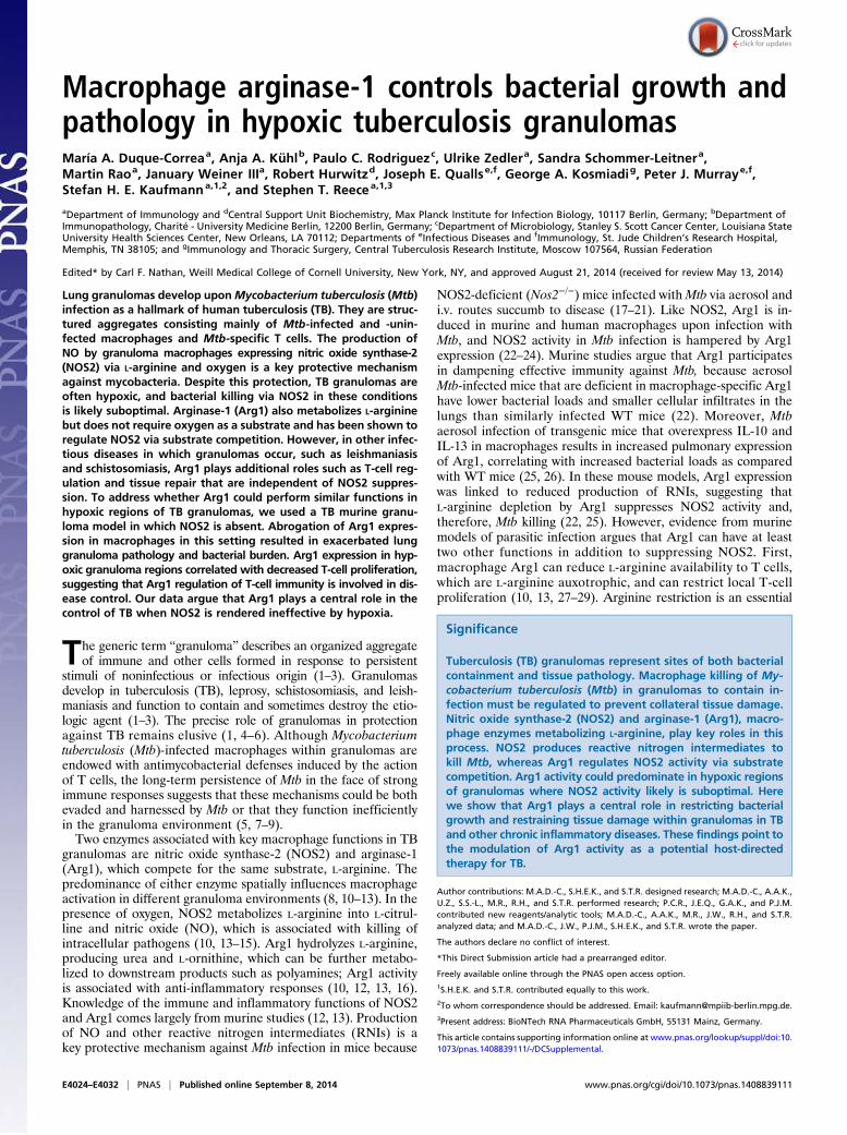
Fig. 1. Arg1 expression in lung granulomas of Mtb-infected Nos2−/− mice and association with necrosis. (A) Lung cfus from infected WT, Nos2−/−, and IFN-γ–blocked Nos2−/− mice at day 56 p.i. Data are shown as median and interquartile range; n = 6 mice; **P = 0.0022 (Mann–Whitney test). ND, not detectable. (Band C) Number of total granulomas (B) and number of necrotic granulomas (C) per lung section in infected WT (n = 72), Nos2−/− (n = 66), and IFN-γ–blockedNos2−/− (n = 62) mice at day 56 p.i. Data are shown as median and interquartile range; P < 0.0001 (Gaussian approximation; Kruskal–Wallis test and Dunn’spost test). (D) Arg1 expression in lung homogenates from infected WT, Nos2−/−, and IFN-γ–blocked Nos2−/− mice at day 56 p.i. Protein (20 μg) was immu-noblotted with anti-Arg1 and anti–β-actin. (E) IHC staining of lung tissue from infected WT and Nos2−/− mice with anti-Arg1 and isotype at day 56 p.i. (F)Number of Arg1+ granulomas per lung section in infected WT (n = 72), Nos2−/− ( n = 64), and IFN-γ–blocked Nos2−/− (n = 62) mice at day 56 p.i. Data areshown as median and interquartile range. P < 0.0001 (Gaussian approximation; Kruskal–Wallis test and. Dunn’s post test). (G) Representative Arg1 staining ofa lung section from an IFN-γ–blocked Nos2−/− mouse at day 56 p.i. (H) Correlation between the number of necrotic and Arg1+ granulomas in the lungs ofinfected IFN-γ–blocked Nos2−/− mice. Spearman correlation; P = 0.0001. Data are representative of two independent experiments. **P < 0.005; ***P < 0.0005.
Duque-Correa et al. PNAS | Published online September 8, 2014 | E4025
IMMUNOLO
GYAND
INFLAMMATION
PNASPL
US
nonnecrotizing granulomas, but also occasional necrotic granu-lomas, in the lungs at day 56 postinfection (p.i.) (Fig. 1 A–C)(37). When infected Nos2−/− mice were subjected to temporaryantibody-mediated blocking of IFN-γ at days 14 and 21 p.i. (Fig.S1), they showed significantly increased bacterial burden andnumbers of granulomas in the lungs at day 56 p.i., with a greaterproportion demonstrating hypoxia and central caseation, classi-cal features of human granuloma pathology (Fig. 1 A–C) (37).We investigated whether Arg1 was expressed in lung granulomasin this model. First, we evaluated whether Arg1 expression wasincreased in the lungs of infected mice. Using immunoblotting(Fig. 1D), we detected increased expression of Arg1 in the lungof infected Nos2−/− and IFN-γ–blocked Nos2−/− mice as com-pared with WT mice at day 56 p.i.We identified Arg1+ cells occasionally in nonnecrotizing granu-
lomas and consistently in caseous necrotic granulomas in infectedNos2−/− mice (Fig. 1E and Fig. S2 A and B). Arg1 was abundant inthe region around the necrotic core of the granulomas but wasundetectable in lungs of infected WT mice (Fig. 1E and Fig. S2 Aand B). Furthermore, infected IFN-γ–blocked Nos2−/− mice hadsignificantly elevated numbers of Arg1+ lung granulomas as com-pared with untreated infected Nos2−/− mice (Fig. 1 F and G).Analysis using Spearman’s correlation coefficient demonstrated asignificant positive association (r = 0.7933) between the numbers ofnecrotic and Arg1+ granulomas in the lungs of infected IFN-γ–blocked Nos2−/− mice (Fig. 1H), suggesting Arg1 expressioncorrelated with exacerbated lung granuloma pathology.Previously, hypoxic [pimonidazole-positive (PIMO+)] cells were
identified in regions surrounding the necrotic center of TB lunggranulomas in infected Nos2−/− mice (37), the region where Arg1+
cells are located. Because hypoxic conditions are well-knowninducers of Arg1 in macrophages (39, 40), we investigated whetherArg1+ cells stained for PIMO. PIMO enters cells in tissues with lowO2 partial pressure and forms adducts with thiol groups withinproteins; these adducts can be detected by immunohistochemistry(IHC) (Fig. S3). By comparing Arg1 and PIMO staining in strictlyconsecutive sections from the same granuloma, we demonstratedthat cells expressing Arg1 did not themselves stain for PIMO butlocated proximally to hypoxic cells localized at the necrotic centerof caseous granulomas in Nos2−/− mice (Fig. S3). Taken together,our findings demonstrate Arg1 is expressed in TB lung granulomasof infected Nos2−/− mice, particularly within the regions encirclingthe central necrotic core of caseous hypoxic granulomas.
Macrophages Are the Main Cell Type Expressing Arg1 in TB LungGranulomas of Infected Nos2−/− Mice. To identify which cellsexpressed Arg1, we stained sections of lungs of infected Nos2−/−
mice at day 56 p.i. with antibodies specific for the macrophagemarker CD68 and Arg1 using immunofluorescence (IF). Cellsexpressing Arg1 in both nonnecrotic and necrotic granulomas inlungs of infected Nos2−/− mice coexpressed CD68 (Fig. S4A). Insome caseous granulomas, Arg1 expression did not colocalizeprecisely with CD68 in the necrotic core (Fig. S4A). Becausethese regions are largely acellular, Arg1 may also be extracellularin this pathophysiological context.To characterize the phenotype of Arg1-expressing cells fur-
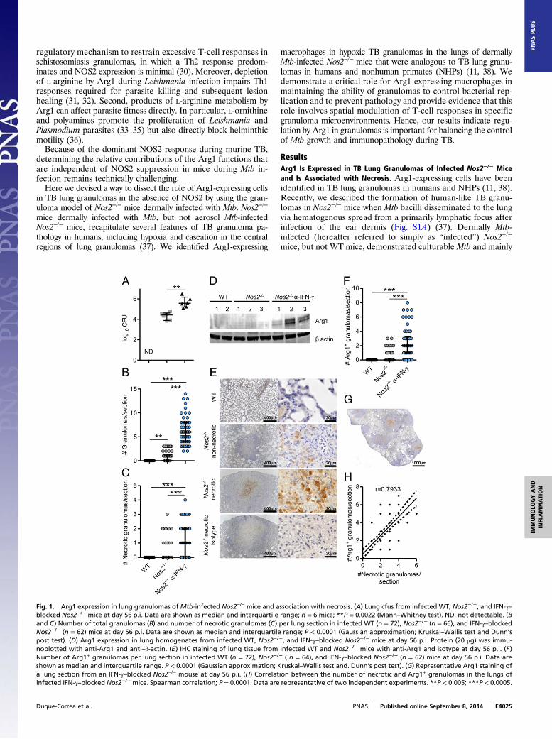
ther, cells were isolated from lungs of infected Nos2−/− and IFN-γ–blocked Nos2−/− mice and were stained with antibodies forneutrophil, inflammatory macrophage, monocyte, alveolar mac-rophage, and dendritic cell surface markers and for intracellularArg1 (Fig. 2A and Fig. S4 B and D). Notably, we observed asignificant decrease in the percentages of neutrophils, alveo-lar macrophages, and monocytes in lungs of infected IFN-γ–blocked Nos2−/− mice as compared with infected Nos2−/− mice(Fig. 2A). Supporting our IF data (Fig. S4A), inflammatory mac-rophages were the main cellular population expressing Arg1 in thelungs of infected Nos2−/− mice and IFN-γ–blocked Nos2−/− mice.Small percentages of neutrophils and monocytes were positive
for Arg1 also (Fig. 2B and Fig. S4 C and D). These data suggestrecruited monocytes are induced to express Arg1 in the lung andlater differentiate into inflammatory macrophages while contin-uously expressing Arg1. Moreover, because murine neutrophilsdo not express Arg1, it is possible that Arg1+ neutrophils havephagocytosed apoptotic Arg1-expressing macrophages or in-ternalized the enzyme from the extracellular milieu. Althougharginases are intracellular enzymes, they can be released intothe extracellular environment when the cellular membrane isbreached and retain activity upon release (41). The high level ofmacrophage necrosis in caseous granulomas in infected Nos2−/−
mice could explain the observation of extracellular Arg1 in ourmodel. To confirm our enzyme-localization data in human tissue,we stained lung sections from patients with cavitary TB andtuberculoma. Based on morphology, Arg1+ cells were classifiedas alveolar macrophages (Fig. S5A), multinucleated giant cells(Fig. S5B), and tissue macrophages (Fig. S5 C and D), con-firming a recent report on Arg1 expression in human TB (38).Together, these data indicate macrophages are the main cell typeexpressing Arg1 in TB lung granulomas.
Fig. 2. Macrophages are the main cell type expressing Arg1 in lungs fromMtb-infected Nos2−/− mice. (A) Single-cell lung suspensions prepared at day 56p.i. were analyzed by flow cytometry to detect the frequency of the indicatedimmune cell populations in the lungs of infected Nos2−/− and IFN-γ–blockedNos2−/− mice. (B) Percentages of Arg1+ cells in neutrophils, inflammatorymacrophages, and monocytes in lungs of infected Nos2−/− and IFN-γ–blockedNos2−/− mice at day 56 p.i. Histograms show Arg1 expression in neutrophils,inflammatory macrophages, and monocytes. The black line depicts Nos2−/−
mice (n = 12), and the blue line depicts IFN-γ–blocked Nos2−/− mice (n = 14).Data are shown as median and interquartile range. Mann–Whitney test. Dataare representative of two independent experiments. *P < 0.05; **P < 0.01.
E4026 | www.pnas.org/cgi/doi/10.1073/pnas.1408839111 Duque-Correa et al.
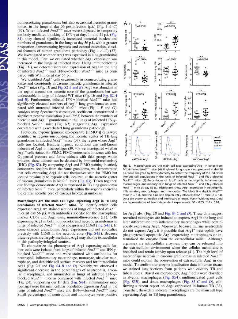
Arg1 Restricts L-Arginine Concentrations and Increases the Abundance ofPolyamines in the Lungs of Infected Nos2−/− Mice. Arginases hydrolyzeL-arginine to produce urea and L-ornithine, which then can bemetabolized further to polyamines (spermine, spermidine, and pu-trescine), which are important regulators of cellular proliferationand immunosuppression (10, 13, 42). Arginase activity also candeplete L-arginine, restricting antigen-specific T-cell proliferation(10, 13, 42). To dissect the relative contribution of these twopathways in TB in the absence of NOS2, we first quantified arginaseactivity and L-arginine metabolites in the lungs of infected mice.Arginase activity in the lungs of infected Nos2−/− and IFN-γ–blocked Nos2−/− mice (Fig. 3A) was associated with a reduction intotal L-arginine concentrations and increased amounts of L-orni-thine, putrescine, and spermidine (Fig. 3B). Thus, the elevated ex-pression and activity of Arg1 in the lungs of infected Nos2−/− andIFN-γ–blocked Nos2−/− mice resulted in a local increase in poly-amine concentrations and depletion of L-arginine during TB. Wenext assessed the relative importance of the ornithine-productionversus arginine-depletion pathways in TB.
Metabolites Derived from L-Arginine Do Not Affect Mtb Growth inVitro. The exogenous supply of metabolites derived from L-argininesupports the proliferation of protozoan parasites such as Leish-mania and Plasmodium (33–35). In contrast, Arg1-generated orni-thine and polyamines are directly antihelminthic (36). We nextaddressed whether a similar addition of these metabolites affectedproliferation of Mtb in vitro. We therefore cultured Mtb strainH37Rv in Sauton’s minimal medium supplemented with con-centrations of L-arginine, L-ornithine, putrescine, spermidine,and spermine and followed bacterial growth for 1, 2, 4, and 7 d ofculture. We did not find that these metabolites had any influence onMtb growth in vitro (Fig. S6). These data suggest that the observeddecrease in detectable L-arginine and increase in L-ornithine, pu-trescine, and spermidine in the lungs of infected IFN-γ–blockedNos2−/− mice was unlikely to affect Mtb burden in the lung.
Abrogation of Macrophage Arg1 Exacerbates Mtb Growth andPathology in TB Lung Granulomas. Having ruled out direct effectsof ornithine or polyamines on Mtb growth, we next turned toa genetic system to evaluate the consequences of Arg1 deletionon immune cells and TB granuloma maintenance. To this end,we created mice lacking Arg1 in macrophages on a completeNOS2-deficient background [Arg1flox/flox; Tie2-Cre; Nos2−/−
mice, hereafter referred to as “Arg1-Nos2 double knockout(DKO) mice”]. Although the Tie2-Cre deleter is found in allhematopoietic and endothelial cells, Arg1 is expressed mainly bymacrophages in our model, and thus Arg1-Nos2 DKO mice allowus to delete Arg1 specifically in macrophages (Fig. S1B) (22).Arg1flox/flox; Tie2-Cre mice were not used here, because they donot represent an appropriate control for the NOS2-deficient gran-uloma model. Mtb infection of Arg1flox/flox; Tie2-Cre mice has beenreported previously (22).Histological analyses of Arg1-Nos2 DKO mice dermally infected
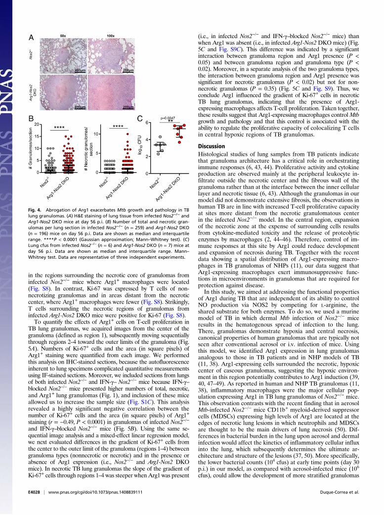
with Mtb demonstrated striking granuloma enlargement as com-pared with Nos2−/− mice at day 56 p.i. (Fig. 4A). In addition, weobserved a significant increase in both the total number of granu-lomas and the number of necrotic granulomas in the lungs of in-fected Arg1-Nos2 DKO mice in comparison with infected Nos2−/−
mice (Fig. 4B), and these increases were linked with an increase inthe total bacterial burden in the lung (Fig. 4C). Based on the resultsfrom the temporary blocking of IFN-γ in infected Nos2−/− mice, wepredicted that IFN-γ–blocked infected Arg1-Nos2DKOmice wouldshow exacerbated pathology and bacterial growth as compared withinfected nonblocked Arg1-Nos2 DKO mice. When we subjectedinfected Arg1-Nos2 DKO mice to IFN-γ blocking, we observedextreme proteolysis in the lungs, elevated numbers of necroticgranulomas, and exceedingly high bacterial counts at day 56 p.i.(Fig. S7). This extensive pathology warranted cessation of the
experiments according to veterinary guidelines to keep animal suf-fering within reasonable limits. Taken together, our findings in-dicate that Arg1 abrogation affects the ability to controlMtb growthand pathology in granulomas in the lung.
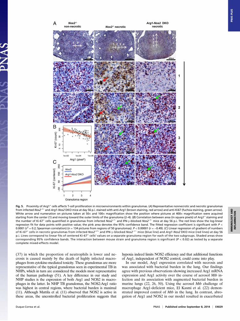
Arg1-Expressing Macrophages Correlate with Restrained T-Cell Responsesin TB Lung Granulomas. We hypothesized that the increased gran-uloma pathology and inability to control Mtb growth in thelungs of infected Arg1-Nos2 DKO mice could be associatedwith a role of Arg1 in blocking T-cell proliferation in granulomamicroenvironments. Therefore, we first determined whether thepresence of Arg1+ cells correlated with reduced T-cell proliferationby staining TB lung granulomas for Arg1, CD3e to designateT cells, and Ki-67, an established marker of cellular proliferation.T cells stained for CD3e lacked Ki-67 expression when localized
Fig. 3. Arg1 metabolizes L-arginine, producing L-ornithine and polyamines inlungs of Mtb-infected Nos2−/− mice. (A) Arginase activity measured by the con-version of L-arginine into L-ornithine. (B) Amounts of L-arginine, L-ornithine,putrescine, and spermidine detected by HPLC in lung homogenates of infectedWT (n = 7), Nos2−/− (n = 6), and IFN-γ–blocked Nos2−/− (n = 6) mice at day 56 p.i.Data are shown as median and interquartile range. Kruskal–Wallis test andDunn’s post test. Data are representative of two independent experiments. *P <0.05; **P < 0.005; ***P < 0.0005.
Duque-Correa et al. PNAS | Published online September 8, 2014 | E4027
IMMUNOLO
GYAND
INFLAMMATION
PNASPL
US
in the regions surrounding the necrotic core of granulomas frominfected Nos2−/− mice where Arg1+ macrophages were located(Fig. S8). In contrast, Ki-67 was expressed by T cells of non-necrotizing granulomas and in areas distant from the necroticcenter, where Arg1+ macrophages were fewer (Fig. S8). Strikingly,T cells surrounding the necrotic regions of granulomas frominfected Arg1-Nos2 DKO mice were positive for Ki-67 (Fig. S8).To quantify the effects of Arg1+ cells on T-cell proliferation in
TB lung granulomas, we acquired images from the center of thegranuloma (defined as region 1), subsequently moving sequentiallythrough regions 2–4 toward the outer limits of the granuloma (Fig.5A). Numbers of Ki-67+ cells and the area (in square pixels) ofArg1+ staining were quantified from each image. We performedthis analysis on IHC-stained sections, because the autofluorescenceinherent to lung specimens complicated quantitative measurementsusing IF-stained sections. Moreover, we included sections from lungsof both infected Nos2−/− and IFN-γ– Nos2−/− mice because IFN-γ–blocked Nos2−/− mice presented higher numbers of total, necrotic,and Arg1+ lung granulomas (Fig. 1), and inclusion of these miceallowed us to increase the sample size (Fig. S1C). This analysisrevealed a highly significant negative correlation between thenumber of Ki-67+ cells and the area (in square pixels) of Arg1+
staining (r = −0.49, P < 0.0001) in granulomas of infected Nos2−/−
and IFN-γ–blocked Nos2−/− mice (Fig. 5B). Using the same se-quential image analysis and a mixed-effect linear regression model,we next evaluated differences in the gradient of Ki-67+ cells fromthe center to the outer limit of the granuloma (regions 1–4) betweengranuloma types (nonnecrotic or necrotic) and in the presence orabsence of Arg1 expression (i.e., Nos2−/− and Arg1-Nos2 DKOmice). In necrotic TB lung granulomas the slope of the gradient ofKi-67+ cells through regions 1–4 was steeper when Arg1 was present
(i.e., in infected Nos2−/− and IFN-γ–blocked Nos2−/− mice) thanwhen Arg1 was absent (i.e., in infected Arg1-Nos2 DKO mice) (Fig.5C and Fig. S9C). This difference was indicated by a significantinteraction between granuloma region and Arg1 presence (P <0.05) and between granuloma region and granuloma type (P <0.02). Moreover, in a separate analysis of the two granuloma types,the interaction between granuloma region and Arg1 presence wassignificant for necrotic granulomas (P < 0.02) but not for non-necrotic granulomas (P = 0.35) (Fig. 5C and Fig. S9). Thus, weconclude Arg1 influenced the gradient of Ki-67+ cells in necroticTB lung granulomas, indicating that the presence of Arg1-expressing macrophages affects T-cell proliferation. Taken together,these results suggest that Arg1-expressing macrophages control Mtbgrowth and pathology and that this control is associated with theability to regulate the proliferative capacity of colocalizing T cellsin central hypoxic regions of TB granulomas.
DiscussionHistological studies of lung samples from TB patients indicatethat granuloma architecture has a critical role in orchestratingimmune responses (6, 43, 44). Proliferative activity and cytokineproduction are observed mainly at the peripheral leukocyte in-filtrate outside the necrotic center and the fibrous wall of thegranuloma rather than at the interface between the inner cellularlayer and necrotic tissue (6, 43). Although the granulomas in ourmodel did not demonstrate extensive fibrosis, the observations inhuman TB are in line with increased T-cell proliferative capacityat sites more distant from the necrotic granulomatous centerin the infected Nos2−/− model. In the central region, expansionof the necrotic zone at the expense of surrounding cells resultsfrom cytokine-mediated toxicity and the release of proteolyticenzymes by macrophages (2, 44–46). Therefore, control of im-mune responses at this site by Arg1 could reduce developmentand expansion of necrosis during TB. Together with the recentdata showing a spatial distribution of Arg1-expressing macro-phages in TB granulomas of NHPs (11), our data suggest thatArg1-expressing macrophages exert immunosuppressive func-tions in microenvironments in granulomas that are required forprotection against disease.In this study, we aimed at addressing the functional properties
of Arg1 during TB that are independent of its ability to controlNO production via NOS2 by competing for L-arginine, theshared substrate for both enzymes. To do so, we used a murinemodel of TB in which dermal Mtb infection of Nos2−/− miceresults in the hematogenous spread of infection to the lung.There, granulomas demonstrate hypoxia and central necrosis,canonical properties of human granulomas that are typically notseen after conventional aerosol or i.v. infection of mice. Usingthis model, we identified Arg1 expression in lung granulomasanalogous to those in TB patients and in NHP models of TB(11, 38). Arg1-expressing cells surrounded the necrotic, hypoxiccenter of caseous granulomas, suggesting the hypoxic environ-ment in this region potentially contributes to Arg1 induction (39,40, 47–49). As reported in human and NHP TB granulomas (11,38), inflammatory macrophages were the major cellular pop-ulation expressing Arg1 in TB lung granulomas of Nos2−/− mice.This observation contrasts with the recent finding that in aerosolMtb-infected Nos2−/− mice CD11b+ myeloid-derived suppressorcells (MDSCs) expressing high levels of Arg1 are located at theedges of necrotic lung lesions in which neutrophils and MDSCsare thought to be the main drivers of lung necrosis (50). Dif-ferences in bacterial burden in the lung upon aerosol and dermalinfection would affect the kinetics of inflammatory cellular influxinto the lung, which subsequently determines the ultimate ar-chitecture and structure of the lesions (37, 50). More specifically,the lower bacterial counts (104 cfus) at early time points (day 30p.i.) in our model, as compared with aerosol-infected mice (106
cfus), could allow the development of more stratified granulomas
Fig. 4. Abrogation of Arg1 exacerbates Mtb growth and pathology in TBlung granulomas. (A) H&E staining of lung tissue from infected Nos2−/− andArg1-Nos2 DKO mice at day 56 p.i. (B) Number of total and necrotic gran-ulomas per lung section in infected Nos2−/− (n = 259) and Arg1-Nos2 DKO(n = 196) mice on day 56 p.i. Data are shown as median and interquartilerange. ****P < 0.0001 (Gaussian approximation; Mann–Whitney test). (C)Lung cfus from infected Nos2−/− (n = 6) and Arg1-Nos2 DKO (n = 7) mice atday 56 p.i. Data are shown as median and interquartile range. Mann–Whitney test. Data are representative of three independent experiments.
E4028 | www.pnas.org/cgi/doi/10.1073/pnas.1408839111 Duque-Correa et al.
(37) in which the proportion of neutrophils is lower and ne-crosis is caused mainly by the death of highly infected macro-phages from cytokine-mediated toxicity. These granulomas are morerepresentative of the typical granulomas seen in experimental TB inNHPs, which in turn are considered the models most representativeof the human pathology (51). A key difference in our study andNHP studies is the expression of both Arg1 and NOS2 in macro-phages in the latter. In NHP TB granulomas, the NOS2:Arg1 ratiowas highest in central regions, where bacterial burden is maximal(11). Although Mattila et al. (11) observed that NOS2 is active inthese areas, the uncontrolled bacterial proliferation suggests that
hypoxia indeed limits NOS2 efficiency and that additional functionsof Arg1, independent of NOS2 control, could come into play.In our model, Arg1 expression correlated with necrosis and
was associated with bacterial burden in the lung. Our findingsagree with previous observations showing increased Arg1 mRNAexpression and Arg1 activity over the course of aerosol Mtb in-fection and its association with augmented bacterial burden inmurine lungs (22, 26, 50). Using the aerosol Mtb challenge ofmacrophage Arg1-deficient mice, El Kasmi et al. (22) demon-strated improved control of Mtb in the lung. In contrast, abro-gation of Arg1 and NOS2 in our model resulted in exacerbated
Fig. 5. Proximity of Arg1+ cells affects T-cell proliferation in microenvironments within granulomas. (A) Representative nonnecrotic and necrotic granulomasfrom infected Nos2−/− and Arg1-Nos2 DKOmice at day 56 p.i. stained with anti-Arg1 (brown staining, red arrow) and anti-Ki67 (fuchsia staining, green arrow).White arrow and numeration on pictures taken at 50× and 100× magnification show the position where pictures at 400× magnification were acquiredstarting from the center (1) and moving toward the outer limits of the granuloma (2–4). (B) Correlation between area (in square pixels) of Arg1+ staining andthe number of Ki-67+ cells quantified in granulomas from infected Nos2−/− and IFN-γ–blocked Nos2−/− mice at day 56 p.i. The red lines show the log-linearregression fit for data points with positive value; the pink area denotes the 95% confidence band. The fitted regression coefficient is significant with P <0.0001 (r2 = 0.2; Spearman correlation) (n = 134 pictures from regions of 50 granulomas). P < 0.00001 (r = −0.49). (C) Linear regression of gradient of numbersof Ki-67+ cells in necrotic granulomas from infected Nos2−/− and IFN-γ–blocked Nos2−/− mice (blue line) and Arg1-Nos2 DKO mice (red lines) at day 56p.i. Lines correspond to linear fits of centered Ki-67+ cells’ values on a separate granuloma region for each of the two subgroups. Shaded areas showcorresponding 95% confidence bands. The interaction between mouse strain and granuloma region is significant (P < 0.02) as tested by a separatecomplete mixed-effects model.
Duque-Correa et al. PNAS | Published online September 8, 2014 | E4029
IMMUNOLO
GYAND
INFLAMMATION
PNASPL
US
granuloma pathology and bacterial growth as compared withNOS2 deficiency alone. These data can be reconciled by addi-tional functions of Arg1 in the absence of NOS2, in a systemwhere Arg1 is expressed spatially in an organized granuloma en-vironment. We did not detect a direct effect of metabolites de-rived from L-arginine onMtb growth in vitro, suggesting that Arg1does not restrict Mtb growth directly. However, these metabolitesmay require additional tissue-specific factors to exert this effect.Uncontrolled T-cell responses to mycobacterial antigens could
contribute to immunopathology driven by lung tissue damage dur-ing TB. For instance, patients coinfected with HIV and Mtb, whopresent low counts of CD4+ T cells, lack caseating granulomas(46, 52). Immune reconstitution in these patients by antiretroviraltherapy results in the increased development of lung pathologyassociated with TB (46, 52). We identified increased numbers ofproliferating T cells in areas around the necrotic core of granulo-mas in absence of Arg1 expression, suggesting that Arg1-expressingmacrophages would be ideally positioned to contribute to the localregulation of T-cell proliferation in this microenvironment, whichin TB lung granulomas is a key battlefield between host and mi-crobe. A caveat of alternative approaches for studying the effects ofArg1 on T-cell proliferation and the activation of lung T cells is thatthe granuloma architecture must be destroyed to obtain the cells.Thus, we consider histological analysis to be more informativeabout the local effects of Arg1. Arg1 activity in the lung frominfected Nos2−/− mice resulted in local depletion of L-arginine andproduction of L-ornithine and polyamines. Both the anti-inflam-matory effects of polyamines and the depletion of L-arginine fromthe extracellular milieu could be responsible for suppressed T-cellresponses. Moreover, additional environmental features of thegranuloma caseous center, such as oxygen and glucose gradients,could contribute to the suppression of T-cell proliferation.Our results agree with similar suppressive roles of Arg1-
expressing macrophages in other chronic infectious diseases.During infections with the intracellular parasites Leishmania sp.,Schistosoma mansoni, and Trypanosoma sp., Arg1 expression/ac-tivity was induced concomitantly with increasing parasite load(31, 53–55). Moreover, Arg1-expressing cells accumulate in andaround S. mansoni granulomas (30, 55, 56) and in skin lesionsfrom patients who have cutaneous leishmaniasis (57); these lesionsoften are necrotic and hypoxic (58, 59). In murine S. mansoniinfection, Arg1-expressing macrophages restrain Th2 cytokine-driven inflammation and fibrosis (30, 60). Notably, duringLeishmania infection in mice, Arg1 contributes to the failure toheal persistent lesions, which is a consequence of impaired T-cellresponses to L. major resulting from local depletion of L-arginineby arginase (32). Taken together, these observations indicate thatthe control of T-cell responses by Arg1 is a common mechanism ofimmunosuppression in different chronic infectious diseases.In conclusion, our data demonstrate that Arg1-expressing
macrophages in hypoxic and necrotic regions of TB granulomasplay a crucial role in controlling both Mtb growth and TB pa-thology, at least in part by restraining T-cell responses, in-dependently of NO production. Hence, our data argue thatcontrol of Mtb must be balanced regionally with a continuousrestriction of T-cell–mediated immunopathology.
Materials and MethodsEthics Statement. All animal experiments were approved by the local ethicscommittee of the German authorities, the State Office of Health and SocialAffairs Berlin (approval no. G0055/88), and the Animal Care and Use Com-mittee of St. Jude Children’s Research Hospital (approval no. 267). Formalin-fixed and paraffin-embedded tissue samples, taken from patients who un-derwent treatment for tuberculosis at the Central Tuberculosis ResearchInstitute in Moscow were retrieved from the archives of Immunology andThoracic Surgery, Central Tuberculosis Research Institute. The use of humanbiopsies as anonymous samples was based on informed patient consent andapproved by the ethics commission of Charité— Universitätsmedizin Berlin (61).
Experimental Animals. C57BL/6 (WT) mice were obtained from Charles RiverLaboratories, Nos2−/− (B6.129P2-Nos2tm1Lau/J) mice were obtained from theJackson Laboratory, and Arg1flox/flox; Tie2-Cre; Nos2−/− C57BL/6 mice werebred in our facilities at the Max Planck Institute for Infection Biology, Berlin.Infected mice were maintained at biosafety level 3, under specific pathogen-free conditions.
Infection with Mtb. The Mtb strain H37Rv (ATCC) was cultured in Mid-dlebrook 7H9 broth (BD) supplemented with 0.05% (vol/vol) Tween 80 andMiddlebrook ADC Enrichment (BD) until midlog phase (OD600 of 0.6–0.8).Bacteria were harvested, resuspended in PBS, and frozen at –80 °C until use.For dermal infections, mice were anesthetized by i.p. administration ofketamine (65 mg/kg), acepromazine (2 mg/kg), and xylazine (11 mg/kg), and104 Mtb in a volume of 20 μL PBS was injected into the ear dermis.
In Vivo Cytokine Blocking. For cytokine blocking, infected Nos2−/− mice re-ceived 500 μg mAb purified from XMG1.2 (anti–IFN-γ) hybridomas (ATCC) i.p.in PBS on days 14 and 21 p.i. as described previously (37).
Enumeration of cfus. Mice were killed at day 56 p.i., and lungs were removedaseptically and homogenized in 1 mL PBS containing 0.05% Tween 80 (vol/vol).Homogenates were then diluted in PBS containing 0.05% Tween 80 (vol/vol)and were plated onto Middlebrook 7H11 agar plates supplemented withMiddlebrook OADC Enrichment (BD). The cfus were enumerated after 3–4 wkof incubation at 37 °C.
Histology. Formalin-fixed and paraffin-embedded tissue samples were takenfrom patients with TB who underwent selective thoracic surgery at theDepartment of Thoracic Surgery of the Central Tuberculosis Research In-stitute, Moscow, Russia.
At the time points described, mice were killed, and lungs were removedaseptically and fixed in PBS containing 4% paraformaldehyde (PFA) (wt/vol)overnight at room temperature. Sections of formalin-fixed, paraffin-embedded tissue (2- to 3-μm thick) were cut, deparaffinized, and sub-jected to H&E staining (for IHC) or immunostaining (for IF).
For immunostaining, sections were subjected to a heat-induced epitoperetrieval step using citrate buffer (pH6) (S2369; Dako) or Target RetrievalSolution (pH9) (S2367; Dako). Slides were rinsed with distilled water, washedin Tris-buffered saline (pH 7.5), and incubated for 1 h at room temperatureor overnight at 4 °C with primary antibodies against Arg1 (19/Arginase I:610708; Becton Dickinson; 1:100), N-20 (sc-18351; Santa Cruz BiotechnologyInc.; 1:100), and H-52 (sc-18351; Santa Cruz Biotechnology Inc.; 1:100) for IHCand against CD3 (A0452; Dako; 1:40), Ki67 antigen (clone TEC-3, M7249;Dako; 1:50), Arg1 (N-20; sc-18351; Santa Cruz Biotechnology Inc.; 1:20), andCD68 (MCA1957T; Abd Serotec; 1:50) for IF. For detection, secondary anti-bodies donkey anti-rabbit Alexa 488 (711-545-152; Jackson Immuno-Research; 1:100; excitation at 493 nm, emission at 519 nm), donkey anti-ratCy3 (712-165-153; Jackson ImmunoResearch; 1:100; excitation at 550 nm,emission at 570 nm), donkey anti-goat Alexa 647 (705-605-147; JacksonImmunoResearch; 1:100; excitation at 651 nm, emission at 667 nm), bio-tinylated rabbit anti-rat (312-065-045; Dianova; 1:200), rabbit anti–goat-HRP(305-035-045; Dianova; 1:200), and goat anti–mouse-peroxidase (115-035-166; Jackson ImmunoResearch; 1:1,000) were used for 1 h at room temper-ature. Peroxidase was detected with diaminobenzidine substrate and thechromogen system (K3467; Dako). Alkaline phosphatase was revealed byFast Red as a chromogen (K0625; Dako). Nuclei were stained with hema-toxylin for IHC and with Hoechst (excitation at 346 nm, emission at 460 nm)for IF. Negative controls were performed by omitting primary antibodies,using corresponding isotypes, and (specifically for Arg1) staining tissue fromArg1-Nos2 DKO mice. Granuloma hypoxia was evaluated on lung sectionsusing the hypoxia marker PIMO kit Hydroxyprobe-1 (Natural Pharmacia In-ternational), according to the manufacturer’s instructions.
Microscopy. IHC samples were visualized using a Zeiss AxioImager Z1 microscopeequipped with a CCD AxioCam at 25 °C. Images were processed with AxioVisionsoftware (Zeiss). IF samples were visualized using a Leica DM R upright epi-fluorescence microscope at 25 °C. Images were captured using a NikonDXM1200F camera and Nikon ACT-1 software. Images were processed usingAdobe PhotoShop CS3. Granulomas were quantified in H&E-stained sections ofthe whole left lobe of the lungs. A minimum of 16 individual sections from eachmouse in each group (n = 6) was used to evaluate total granulomas per section.Caseous granulomas were defined as containing a pink central acellular necroticregion surrounded by a granulomatous inflammatory infiltrate. A minimum of16 individual sections containing granulomas was evaluated for Arg1 stainingper mouse from each group (n = 6), and an analogous section was H&E stained
E4030 | www.pnas.org/cgi/doi/10.1073/pnas.1408839111 Duque-Correa et al.
to confirm nonnecrotizing or caseous phenotype. To quantify Ki-67+ and Arg1+
staining in granulomas, an image at 100× magnification was taken to orientatethe position of images at 400× magnification that were acquired starting fromthe center and moving toward the outer limits of the granuloma. Next, in eachimage at 400× magnification the total number of Ki-67+ cells and the area (insquare pixels) of Arg1+ staining were quantified using AutMESS software (Zeiss).
RNA Isolation and Quantitative RT-PCR. For in vivo RNA isolation, lungs wereaseptically removed from infected mice at days 28 and 56 p.i. and werehomogenized in TRIzol total RNA isolation reagent (Invitrogen). RNA wasisolated from TRIzol via chloroform extraction and treatment with ethanoland was dissolved in RNase-free water. RNA was analyzed for quality andquantified using a NanoDrop 1000 Spectrophotometer (Thermo FischerScientific). One microgram of total RNA was reverse-transcribed usingSuperScript III Reverse Transcriptase (Invitrogen), and 10 ng cDNA was subjectedto quantitative RT-PCR using primers for Arg1 5′-GGAATCTGCATGGGCAA-CCTGTGT-3′ and 5′AGGGTCTACGTCTCGCAAGCCA-3′. Uptake of SYBR Green(Applied Biosystems) was measured using an ABI PRISM 7900 thermocycler(Applied Biosystems). Cycle threshold values were normalized to those obtainedfor GAPDH, and 2–ΔΔCT was used to calculate change in relative mRNA ex-pression between groups.
Isolation of Lung Leukocytes. Mice were killed, and cells were isolated fromlungs of individual mice. Lungs were cut into small pieces and incubated for30 min at 37 °C in 5% CO2 in Roswell Park Memorial Institute (RPMI)-10complete medium (Invitrogen) containing 2 mM L-glutamine, 1 mM sodiumpyruvate, 10 mM Hepes buffer, and 10% heat-inactivated FCS supplementedwith 0.7 mg/mL blend of collagenase VIII (Sigma), 0.3 mg/mL collagenase D(Roche), and 0.03 mg/mL DNase I (Roche). Lung pieces were pressed through40-μm cell strainers. Red blood cells were lysed in ammonium chloridebuffer, cells were washed with PBS containing 0.2% BSA, and 2 × 10⁶ cellsper well were added to 96-well tissue culture plates (Nunc).
Arg1 Intracellular Staining. Cells were blocked with rat serum and αCD16/αCD32 mAb and were surfaced stained with antibodies against CD11b(M1/70; BD Pharmingen), CD11c (HL3; eBioscience), F4/80 (BM8; eBioscience),and Ly6G (1A8; BD Pharmingen). Then cells were fixed with 2% PFA in PBS,permeabilized in saponin buffer, and stained with anti-Arg1 (RandD Sys-tems). From each sample, 50–100,000 leukocyte-gated cells were acquiredusing a FACS Canto II (BD Pharmingen) and were analyzed using BDFACSDiva and FlowJo software.
Protein Extracts. Lungs were removed aseptically from mice and homoge-nized in 1 mL tissue lysis buffer containing PBS Tween 20 0.05% (vol/vol) andinhibitors of proteases [Mini cOmplete EDTA-free tablets (Roche)] andphosphatases [PhosSTOP tablets (Roche)]. The homogenate was centrifugedat 14,000 × g. Supernatants then were centrifuged in 0.22-μm SPIN-X filtertubes (Corning). Proteins were quantified using the BCA Protein Assay Kit(Thermo Scientific).
Arginase Activity Assay. Arginase activity was determined in protein extractsby measuring the conversion of L-arginine into L-ornithine. In brief, 20 μg ofprotein was added to 25 μL of 10 mM MnCl2. This mixture was heated at55–60 °C for 20 min to activate arginase. Then, 150 μL of carbonate buffer(100 mmol/L) (Sigma) and 50 μL of L-arginine (100 mmol/L) (Sigma) wereadded, and the solution was incubated at 37 °C for 20 min. The hydrolysisreaction from L-arginine to L-ornithine was identified by a colorimetric assayafter the addition of ninhydrin solution and incubation at 95 °C for 1 h.
Western Blot. Arg1 (N-20; sc-18351; Santa Cruz Biotechnology) and β-actin(AC-15; ab6276-100; Abcam) expression was detected by immunoblot using20 μg of protein extracts. Protein extracts were electrophoresed in 12% Tris-
Glycine gels (Bio-Rad), transferred to PVDF membranes, and immunoblottedwith the appropriate antibodies.
HPLC. Amounts of L-arginine, L-ornithine, putrescine, and spermidine inprotein extracts were determined by use of HPLC, as described elsewhere(62). In brief, protein extracts were deproteinized and derivatized, and theneach sample was tested. A standard curve with known concentrations ofeach molecule was run for each experiment.
Evaluation ofMtb Growth in the Presence of L-Arginine Metabolites.Mtb strainH37Rv (ATCC) was cultured in Middlebrook 7H9 broth (BD) supplementedwith 0.05% (vol/vol) Tween 80 and Middlebrook ADC Enrichment (BD) untilmidlog phase at 37 °C (OD600 of 0.6–0.8, corresponding to an approximatecell density of 108 cells/mL). Bacteria were harvested by centrifugation,washed with 1× PBS, resuspended in Sauton’s medium supplemented with0.05% (vol/vol) Tween 80, and cultured at 37 °C until reaching an OD600 of0.5. Then, bacteria were cultured at an OD600 of 0.05 in Sauton’s mediumcontaining serial dilutions of L-arginine (200 μM, 100 μM, 50 μM), L-ornithine(200 μM, 100 μM, 50 μM), putrescine (10 μM, 1 μM, 0.1 μM), spermidine(10 μM, 1 μM, 0.1 μM), and spermine (24 μM, 16 μM, 4 μM). After 1, 2, 4, and7 d, bacterial growth was evaluated by measurement of OD600 and byplating serial dilutions onto Middlebrook 7H11 agar plates and countingthe cfus after 3 wk of incubation at 37 °C.
Statistics. Statistical analyses were carried out using Prism 5 software(GraphPad) and the R package nlme (63). Individual comparisons were per-formed using the Mann–Whitney test. Multiple group comparisons wereperformed using the Kruskal–Wallis test, followed by Dunn’s multiplecomparison test to compare individual groups. The association between thenumber of necrotic and Arg1+ granulomas enumerated in the lungs ofinfected IFN-γ–blocked Nos2−/− mice was estimated by use of a regressionmodel and Spearman’s correlation coefficient. Nonparametric tests in-cluding the Mann–Whitney test, the Kruskal–Wallis test, and Spearman’scorrelation were performed when the data were not normal. Accordingly,graphs illustrating these data show the median and the interquartile range.For statistical analysis of the quantification of Ki-67+ cells and the area (insquare pixels) of Arg1+ staining in TB lung granulomas, linear models werecalculated using the R package nlme (63). The significance of the effects ofgranuloma type (nonnecrotic or necrotic), presence and absence of Arg1expression (i.e., mouse strain: Nos2−/− and Arg1-Nos2 DKO mice), andgranuloma region and of their interactions was computed using a mixed-effect (type III) linear model (64). In principle, a linear regression model isfitted to each granuloma (with granuloma region as the independent vari-able and the quantification of the Ki-67+ cells as the dependent variable)and then compares the set of models from granulomas of Nos2−/− mice withthe set of models from granulomas of Arg1-Nos2 DKO mice. This techniquemakes it possible to account for the substantial granuloma-specific vari-ability. For purposes of visualization only (Fig. 5C), to account for thegranuloma-specific variability, Ki-67 values were centered relative to thevalue in the first region measured, and a simple linear regression fit wascalculated for each combination of granuloma type and mouse strain, sep-arately. All statistical procedures, R code necessary to replicate the resultsand figures, raw data, and results of statistical analyses are availableupon request.
ACKNOWLEDGMENTS. We thank Diane Schad for graphical assistance andMary Louise Grossman for help in preparing the manuscript. This workwas supported by the European Union’s Seventh Framework Programme(EU FP7) Collaborative Projects NEWTBVAC (Grant HEALTH-F3-2009-241745), SysteMTb (Grant HEALTH-F4-2010-241587), and (FP7/2007-2013)Grant 280873 ADITEC., CORE Grant P30 CA21765, National Institutes ofHealth Grant AI097485, The Hartwell Foundation, and The American Leba-nese Syrian Associated Charities.
1. Ramakrishnan L (2012) Revisiting the role of the granuloma in tuberculosis. Nat Rev
Immunol 12(5):352–366.2. Reece ST, Kaufmann SH (2012) Floating between the poles of pathology and pro-
tection: Can we pin down the granuloma in tuberculosis? Curr Opin Microbiol 15(1):
63–70.3. Spector WG (1969) The granulomatous inflammatory exudate. Int Rev Exp Pathol
8:1–55.4. Bold TD, Ernst JD (2009) Who benefits from granulomas, mycobacteria or host? Cell
136(1):17–19.5. Paige C, Bishai WR (2010) Penitentiary or penthouse condo: The tuberculous granu-
loma from the microbe’s point of view. Cell Microbiol 12(3):301–309.6. Russell DG (2007) Who puts the tubercle in tuberculosis? Nat Rev Microbiol 5(1):39–47.
7. Dorhoi A, Reece ST, Kaufmann SH (2011) For better or for worse: The immune re-
sponse against Mycobacterium tuberculosis balances pathology and protection. Im-
munol Rev 240(1):235–251.8. Flynn JL, Chan J, Lin PL (2011) Macrophages and control of granulomatous in-
flammation in tuberculosis. Mucosal Immunol 4(3):271–278.9. Russell DG, Cardona PJ, Kim MJ, Allain S, Altare F (2009) Foamy macrophages and the
progression of the human tuberculosis granuloma. Nat Immunol 10(9):943–948.10. Grohmann U, Bronte V (2010) Control of immune response by amino acid metabo-
lism. Immunol Rev 236:243–264.11. Mattila JT, et al. (2013) Microenvironments in tuberculous granulomas are delineated
by distinct populations of macrophage subsets and expression of nitric oxide synthase
and arginase isoforms. J Immunol 191(2):773–784.
Duque-Correa et al. PNAS | Published online September 8, 2014 | E4031
IMMUNOLO
GYAND
INFLAMMATION
PNASPL
US
12. Murray PJ, Wynn TA (2011) Protective and pathogenic functions of macrophagesubsets. Nat Rev Immunol 11(11):723–737.
13. Peranzoni E, et al. (2007) Role of arginine metabolism in immunity and immunopa-thology. Immunobiology 212(9-10):795–812.
14. Fang FC (2004) Antimicrobial reactive oxygen and nitrogen species: Concepts andcontroversies. Nat Rev Microbiol 2(10):820–832.
15. Bogdan C (2001) Nitric oxide and the immune response. Nat Immunol 2(10):907–916.16. Martinez FO, Helming L, Gordon S (2009) Alternative activation of macrophages: An
immunologic functional perspective. Annu Rev Immunol 27:451–483.17. Jung YJ, LaCourse R, Ryan L, North RJ (2002) Virulent but not avirulent Mycobacte-
rium tuberculosis can evade the growth inhibitory action of a T helper 1-dependent,nitric oxide Synthase 2-independent defense in mice. J Exp Med 196(7):991–998.
18. MacMicking JD, et al. (1997) Identification of nitric oxide synthase as a protectivelocus against tuberculosis. Proc Natl Acad Sci USA 94(10):5243–5248.
19. Nathan C (2008) Microbiology. An antibiotic mimics immunity. Science 322(5906):1337–1338.
20. Yang CS, Yuk JM, Jo EK (2009) The role of nitric oxide in mycobacterial infections.Immune Netw 9(2):46–52.
21. Ralph AP, Kelly PM, Anstey NM (2008) L-arginine and vitamin D: Novel adjunctiveimmunotherapies in tuberculosis. Trends Microbiol 16(7):336–344.
22. El Kasmi KC, et al. (2008) Toll-like receptor-induced arginase 1 in macrophagesthwarts effective immunity against intracellular pathogens. Nat Immunol 9(12):1399–1406.
23. Hart BE, Tapping RI (2012) Differential trafficking of TLR1 I602S underlies host pro-tection against pathogenic mycobacteria. J Immunol 189(11):5347–5355.
24. Qualls JE, et al. (2010) Arginine usage in mycobacteria-infected macrophages dependson autocrine-paracrine cytokine signaling. Sci Signal 3(135):ra62.
25. Schreiber T, et al. (2009) Autocrine IL-10 induces hallmarks of alternative activation inmacrophages and suppresses antituberculosis effector mechanisms without compro-mising T cell immunity. J Immunol 183(2):1301–1312.
26. Heitmann L, et al. (2014) The IL-13/IL-4Rα axis is involved in tuberculosis-associatedpathology. J Pathol.
27. Bronte V, Zanovello P (2005) Regulation of immune responses by L-arginine metab-olism. Nat Rev Immunol 5(8):641–654.
28. Rodríguez PC, Ochoa AC (2008) Arginine regulation by myeloid derived suppressorcells and tolerance in cancer: Mechanisms and therapeutic perspectives. Immunol Rev222:180–191.
29. Stempin CC, Dulgerian LR, Garrido VV, Cerban FM (2010) Arginase in parasitic in-fections: Macrophage activation, immunosuppression, and intracellular signals.J Biomed Biotechnol 2010:683485.
30. Pesce JT, et al. (2009) Arginase-1-expressing macrophages suppress Th2 cytokine-driven inflammation and fibrosis. PLoS Pathog 5(4):e1000371.
31. Kropf P, et al. (2005) Arginase and polyamine synthesis are key factors in the regu-lation of experimental leishmaniasis in vivo. FASEB J 19(8):1000–1002.
32. Modolell M, et al. (2009) Local suppression of T cell responses by arginase-inducedL-arginine depletion in nonhealing leishmaniasis. PLoS Negl Trop Dis 3(7):e480.
33. Colotti G, Ilari A (2011) Polyamine metabolism in Leishmania: From arginine to try-panothione. Amino Acids 40(2):269–285.
34. Niemand J, Louw AI, Birkholtz L, Kirk K (2012) Polyamine uptake by the intra-erythrocytic malaria parasite, Plasmodium falciparum. Int J Parasitol 42(10):921–929.
35. Tavares J, Ouaissi A, Lin PK, Tomás A, Cordeiro-da-Silva A (2005) Differential effectsof polyamine derivative compounds against Leishmania infantum promastigotesand axenic amastigotes. Int J Parasitol 35(6):637–646.
36. Esser-von Bieren J, et al. (2013) Antibodies trap tissue migrating helminth larvae andprevent tissue damage by driving IL-4Rα-independent alternative differentiation ofmacrophages. PLoS Pathog 9(11):e1003771.
37. Reece ST, et al. (2010) Serine protease activity contributes to control of Mycobacte-rium tuberculosis in hypoxic lung granulomas in mice. J Clin Invest 120(9):3365–3376.
38. Pessanha AP, Martins RA, Mattos-Guaraldi AL, Vianna A, Moreira LO (2012) Arginase-1expression in granulomas of tuberculosis patients. FEMS Immunol Med Microbiol 66(2):265–268.
39. Lewis JS, Lee JA, Underwood JC, Harris AL, Lewis CE (1999) Macrophage responses tohypoxia: Relevance to disease mechanisms. J Leukoc Biol 66(6):889–900.
40. Louis CA, et al. (1998) Distinct arginase isoforms expressed in primary and trans-formed macrophages: Regulation by oxygen tension. Am J Physiol 274(3 Pt 2):R775–R782.
41. Morris CR, et al. (2004) Decreased arginine bioavailability and increased serum argi-nase activity in asthma. Am J Respir Crit Care Med 170(2):148–153.
42. Soda K (2011) The mechanisms by which polyamines accelerate tumor spread. J ExpClin Cancer Res 30:95.
43. Ulrichs T, et al. (2004) Human tuberculous granulomas induce peripheral lymphoidfollicle-like structures to orchestrate local host defence in the lung. J Pathol 204(2):217–228.
44. Ulrichs T, Kaufmann SH (2006) New insights into the function of granulomas in hu-man tuberculosis. J Pathol 208(2):261–269.
45. Kaplan G, et al. (2003) Mycobacterium tuberculosis growth at the cavity surface:A microenvironment with failed immunity. Infect Immun 71(12):7099–7108.
46. Elkington PT, D’Armiento JM, Friedland JS (2011) Tuberculosis immunopathology:The neglected role of extracellular matrix destruction. Sci Transl Med 3(71):ps6.
47. Murray PJ, Wynn TA (2011) Obstacles and opportunities for understanding macro-phage polarization. J Leukoc Biol 89(4):557–563.
48. El Kasmi KC, et al. (2014) Adventitial fibroblasts induce a distinct proinflammatory/profibrotic macrophage phenotype in pulmonary hypertension. J Immunol 193(2):597–609.
49. Colegio OR, et al. (2014) Functional polarization of tumour-associated macrophagesby tumour-derived lactic acid. Nature, 10.1038/nature13490.
50. Obregón-Henao A, Henao-Tamayo M, Orme IM, Ordway DJ (2013) Gr1(int)CD11b+myeloid-derived suppressor cells in Mycobacterium tuberculosis infection. PLoS ONE8(11):e80669.
51. Lin PL, et al. (2014) Sterilization of granulomas is common in active and latent tu-berculosis despite within-host variability in bacterial killing. Nat Med 20(1):75–79.
52. Meintjes G, et al.; International Network for the Study of HIV-associated IRIS (2008)Tuberculosis-associated immune reconstitution inflammatory syndrome: Case defi-nitions for use in resource-limited settings. Lancet Infect Dis 8(8):516–523.
53. Duleu S, et al. (2004) Mouse strain susceptibility to trypanosome infection: An argi-nase-dependent effect. J Immunol 172(10):6298–6303.
54. Gobert AP, et al. (2000) L-Arginine availability modulates local nitric oxide productionand parasite killing in experimental trypanosomiasis. Infect Immun 68(8):4653–4657.
55. Hesse M, et al. (2001) Differential regulation of nitric oxide synthase-2 and arginase-1by type 1/type 2 cytokines in vivo: Granulomatous pathology is shaped by the patternof L-arginine metabolism. J Immunol 167(11):6533–6544.
56. Barron L, et al. (2013) Role of arginase 1 from myeloid cells in th2-dominated lunginflammation. PLoS ONE 8(4):e61961.
57. Abebe T, et al. (2012) Local increase of arginase activity in lesions of patients withcutaneous leishmaniasis in Ethiopia. PLoS Negl Trop Dis 6(6):e1684.
58. Araújo AP, Frezza TF, Allegretti SM, Giorgio S (2010) Hypoxia, hypoxia-induciblefactor-1α and vascular endothelial growth factor in a murine model of Schistosomamansoni infection. Exp Mol Pathol 89(3):327–333.
59. Arrais-Silva WW, Paffaro VA, Jr, Yamada AT, Giorgio S (2005) Expression of hypoxia-inducible factor-1alpha in the cutaneous lesions of BALB/c mice infected with Leish-mania amazonensis. Exp Mol Pathol 78(1):49–54.
60. Herbert DR, et al. (2010) Arginase I suppresses IL-12/IL-23p40-driven intestinal in-flammation during acute schistosomiasis. J Immunol 184(11):6438–6446.
61. Ulrichs T, et al. (2005) Differential organization of the local immune response inpatients with active cavitary tuberculosis or with nonprogressive tuberculoma. J InfectDis 192(1):89–97.
62. Zea AH, et al. (2005) Arginase-producing myeloid suppressor cells in renal cell carci-noma patients: A mechanism of tumor evasion. Cancer Res 65(8):3044–3048.
63. Pinheiro J, Bates D, DebRoy S, Sarkar D, and R Development Core Team (2013) nlme:Linear and nonlinear mixed effects models. R package. Version 3.1-109. (R De-velopment Core Team, Vienna).
64. Pinheiro J, Bates D (2000) Mixed Effects Models in S and S-plus (Springer, New York),1st Ed, pp 1–528.
E4032 | www.pnas.org/cgi/doi/10.1073/pnas.1408839111 Duque-Correa et al.