Embed Size (px)
Citation preview
1
Published online before print August 25, 2010,
doi: 10.1098/rsif.2010.0306
J. R. Soc. Interface 6 April 2011 vol. 8 no. 57 590-600
Macromolecular Dynamics in Red Blood Cells Investigated
Using Neutron Spectroscopy
Andreas Maximilian Stadler1,*, Lambert van Eijck2, Franz Demmel3, and Gerhard Artmann4
1 Research Centre Jülich, 52425 Jülich, Germany
2 Institut Laue-Langevin, 38042 Grenoble, France
3 Rutherford Appleton Laboratory, Didcot OX11 0QX, United Kingdom
4 Institute of Bioengineering, Aachen University of Applied Science, 52428 Jülich, Germany
* corresponding author: [email protected]
2
Abstract
We present neutron scattering measurements on the dynamics of hemoglobin (Hb) in human
red blood cells in vivo. Global and internal Hb dynamics were measured in the ps to ns time-
and Å length-scale using quasielastic neutron backscattering spectroscopy. We observed the
cross-over from global Hb short-time to long-time self-diffusion. Both short- and long-time
diffusion coefficients agree quantitatively with predicted values from hydrodynamic theory of
non-charged hard-sphere suspensions when a bound water fraction of around
0.23g H2O/ g Hb is taken into account. The higher amount of water in the cells facilitates
internal protein fluctuations in the ps time-scale when compared to fully hydrated Hb powder.
Slower internal dynamics of Hb in red blood cells in the ns time-range were found to be rather
similar to results obtained with fully hydrated protein powders, solutions and E. coli cells.
Keywords:
hemoglobin, red blood cells, neutron spectroscopy, protein dynamics, macromolecular
diffusion
3
Introduction
Ongoing research is dedicated to obtaining a coherent picture of the interactions and
dynamical properties of proteins in their physiological environment. Cells are highly complex
objects which are composed of organelles, tens of thousands of different proteins, RNA and
DNA, lipids, polysaccharides and many other chemical components. Red blood cells (RBC)
in this sense are exceptional. They are highly specialized and relatively simple in their
composition with the main macromolecular component hemoglobin (Hb) making of 92% of
the dry weight. The concentration of Hb in RBC is c=0.33 g/ml with a corresponding volume
fraction of φ=0.25 (Krueger and Nossal 1988). The hydrodynamic radius of Hb is ~32 Å
(Digel et al. 2006), and the average distance between Hb molecules is in the order of 1 nm
(Krueger and Nossal 1988). RBC are therefore particularly well suited model systems to study
the physical properties of concentrated protein solutions in-vivo.
From a biological point of view the properties of human RBC are interesting to study as well
as they exhibit a variety of remarkable properties. RBC have been shown to undergo a
passage transition through narrow micropipettes at body temperature (Artmann et al. 1998).
The single cells were aspirated with a micropipette (diameter of the pipette tip ~1.5 µm) and
at temperatures lower than body temperature all cells blocked the pipette. Above body
temperature all aspirated RBC passed the narrow micropipette tip easily without any apparent
resistance. The passage temperature was 36.3 ± 0.3 °C being remarkably close to human body
temperature (Artmann et al. 1998). It was found that the passage behavior is caused by a
reduction of the viscosity of the concentrated Hb solution in the RBC (Artmann et al. 1998).
The loss of viscosity and the passage transition in the micropipette experiments were found to
be connected to perturbations and partial unfolding of the structure of Hb at body temperature
(Artmann et al. 2004). Further studies revealed that the structural perturbation of Hb at body
temperature leads to Hb aggregation above ~37 °C (Digel et al. 2006), and concomitantly
RBC release cytosolic cell water to the outside blood plasma as observed in colloid osmotic
pressure measurements (Artmann et al. 2009). A direct correlation between the structural
perturbation temperature of Hb and the body temperature of a large variety of different
species was reported, which further supported the biological relevance of the effect (Digel et
4
al. 2006; Zerlin et al. 2007). It was speculated that the partial loss of Hb structure causes an
increase in surface hydrophobicity, which might result in stronger protein-protein interactions
and thus lead to protein aggregation above body temperature (Digel et al. 2006; Stadler et al.
2008a; Stadler et al. 2009).
Krueger and co-authors studied the interactions of Hb in RBC and concentrated solution and
demonstrated that a hard sphere potential plus screened electrostatics can approximately
describe the protein-protein interaction potential (Krueger et al. 1990; Krueger and Nossal
1988). The same results was obtained later also for concentrated myoglobin solutions
(Longeville et al. 2003). It might be of interest to note that studies on concentrated solutions
of crystallins and of lysozyme demonstrated that a delicate balance between hard sphere and
weak attractive interactions are crucial for the stability of these concentrated protein solutions.
(Cardinaux et al. 2007; Dorsaz et al. 2009; Stradner et al. 2007). In further experiments
Doster and Longeville examined the diffusion of Hb in RBC using neutron spin-echo
spectroscopy (Doster and Longeville 2007). The authors had the idea to interpret the diffusion
of Hb in RBC using the theory of colloidal diffusion at high concentration. The neutron spin-
echo technique is sensitive to molecular motions occurring in the ns and nm time- and length-
scale. Doster and Longeville compared the measured short-time and long-time self-diffusion
coefficient of Hb to theoretical calculations of non-charged hard sphere suspensions with
direct and hydrodynamic interactions (Doster and Longeville 2007). It was necessary to
include the hydration shell as a hydrodynamic coat to release the discrepancy with colloidal
theory. Furthermore, it was deduced that hydrodynamic and not direct interactions dominate
Hb diffusion at high concentration.
Without hydration water, proteins would neither fold correctly (Chaplin 2006; Cheung et al.
2002; Dobson et al. 1998) nor acquire the conformational flexibility, which is considered
relevant for biological activity (Rupley and Careri 1991). Motions in proteins occur over a
very large range of time-scales from fast reorientations of amino acid side chains in the ps
range, to slower motions of the protein backbone in the ns time-scale and to very slow
processes of protein subunits and folding processes in the µs and ms range (McCammon and
Harvey 1987). Fast fluctuations in the ps and ns time-scale are considered to act as lubricant
and to enable much slower physiologically important motions (Brooks et al. 1988). Hydration
dependent internal protein dynamics has been studied with incoherent neutron scattering in
5
several model systems mainly as hydrated powders, including myoglobin (Doster 2008;
Doster et al. 1989), lysozyme (Cornicchi et al. 2005; Marconi et al. 2008; Paciaroni et al.
2005), and α-amylase (Fitter 1999, 2003; Fitter and Heberle 2000). In incoherent neutron
scattering experiments, the single particle motions of hydrogen (H) atoms are detected. H
atoms are indicators of average protein dynamics as they constitute ~50% of the atoms and
are uniformly distributed in the macromolecules (Gabel et al. 2002). Hydration water not only
enables protein dynamics but participates actively in protein function. Around 60 additional
water molecules are bound in the hydration layer of the oxygenated form of Hb as compared
to the deoxygenated state of Hb (Colombo et al. 1992). The additional water molecules were
found to be thermodynamically important for regulation of Hb activity. The study of Colombo
and coworkers was done in aqueous solution at a Hb concentration of 64 mg/ml. Further
studies revealed that binding of the extra water molecules is the rate-limiting step of Hb
activity (Salvay et al. 2003). Therefore, it is an important question if protein dynamics is
adapted to the specific hydration level in cells.
In this manuscript, we present a study of Hb dynamics in RBC in the ps to ns time- and Å
length-scale using high-resolution quasielastic neutron scattering (QENS). The aim of the
study is to demonstrate how QENS allows the measurement and separation of internal protein
dynamics and global macromolecular diffusion in whole cells. The QENS technique provides
complementary information to fluorescent correlation spectroscopy (Schwille et al. 1999;
Wawrezinieck et al. 2005) or neutron spin-echo spectroscopy (Doster and Longeville 2007;
Lal et al. 2010; Le Coeur and Longeville 2008) which are sensitive to different time-space
windows of protein fluctuations.
6
Material and Methods
Sample preparation
Samples of human venous blood from healthy adults were drawn with tubes containing
heparin to prevent blood coagulation. RBC samples were prepared as described in Stadler et
al. (Stadler et al. 2008a). During the sample preparation, the RBC were gassed with CO to
increase the stability of Hb and the glycocalyx matrix was removed. The cells were washed
several times with D2O HEPES buffer (137 mM NaCl, 4 mM KCl, 1.8 mM CaCl2, 0.8 mM
Na2HPO4, 0.2 mM NaH2PO4, 0.7 mM MgSO4, 8.4 mM HEPES, and 4 mM NaOH) at pD=7.4
and 290 mOsm to reduce the neutron scattering contribution of the buffer. The washing steps
were repeated until the level of H2O was estimated to be below 0.1 vol%. The shape of the
cells was checked with optical microscopy after the washing steps. The cell pellet was sealed
in a flat aluminum sample holder of 0.2 mm thickness for the neutron scattering experiment.
It was checked by weighing that there occurred no loss of sample material during the
experiment.
Dynamic light scattering experiments
Samples for the dynamic light scattering experiments were prepared from a blood drop taken
from the finger tip. The RBC were washed with H2O HEPES buffer and lysed with distilled
water. The sample for dynamic light scattering experiments was not gassed with CO. Before
the dynamic light scattering experiments, the dilute Hb solution in H2O buffer (0.1M KCl,
61.3 mM K2HPO4, 5.33 mM KH2PO4, pH 7.4, 290-300 mOsm) was centrifuged at 20000
relative centrifugal force and filtered using 0.25 µm nitrocellulose filters. UV/VIS absorption
spectroscopy was used to determine the concentration of the Hb solution. The Hb was found
to be in the oxy-state as evidenced by the characteristic bands in the absorption spectrum, and
the protein concentration was 0.4 mg/ml. The protein concentration was determined using
7
extinction coefficients of 13.8 mM-1*cm-1 at 541 nm and 128 mM-1*cm-1 at 405 nm for oxy-
Hb, the molar concentration is per heme group (Antonini and Brunori 1970). Dynamic light
scattering of dilute human Hb solution was measured on a Wyatt DAWN-EOS instrument
(Wyatt Technology, Santa Barbara, CA) and corrected for temperature dependent D2O
viscosity using literature values (Cho et al. 1999). The diffusion coefficients were calculated
using the ASTRA 5 software package from the manufacturer. Around 5 ml of sample was
measured per experiment.
Neutron scattering experiments
Neutron scattering was measured on the high-resolution neutron backscattering spectrometers
IN10 and IN16 at the ILL (http://www.ill.eu/instruments-support/instruments-
groups/yellowbook/) and on IRIS at the ISIS spallation source
(http://www.isis.stfc.ac.uk/instruments/iris/). To minimize multiple scattering, RBC samples
with high transmissions were used (0.95 on IN16 and IRIS, 0.9 on IN10). The instruments
IRIS, IN10 and IN16 are characterized by energy resolutions ∆E of 17, 1 and 0.9 µeV
(FWHM), respectively, which correspond to slowest observable motions in the order of
Et ∆=∆ /h ~40 ps and ~1 ns, respectively. Neutron scattering was measured in the range of
0.49 ≤ q ≤ 1.6 Å-1 on IN16, 0.5 ≤ q ≤ 1.45 Å-1 on IN10 and 0.48 ≤q ≤ 1.6 Å-1 on IRIS, where q
is the modulus of the scattering vector. The instrumental energy resolution was determined
with a vanadium measurement. The scattering contribution of the empty aluminum sample
holder was subtracted from the measured data. Neutron detectors were grouped on IN16 and
IRIS to obtain better statistics. Incoherent scattering of D2O solvent contributes partially to
the measured intensities: Free and interfacial water dynamics are out of the Å-ns space and
time window of IN10 and IN16 and contribute only as a flat background to the measured
spectra (Tehei et al. 2007). Experimental data is dominated by Hb motions on the IRIS
spectrometer, and the incoherent contribution of D2O on IRIS is estimated to be smaller than
4% at q<1.3 Å-1 (Stadler et al. 2008a). Gaspar and coworkers evaluated the coherent and
incoherent scattering contributions of concentrated protein solutions in D2O solvent (Gaspar
et al. 2010). In a completely dry myoglobin powder the authors found an incoherent scattering
fraction of ~90% and a coherent scattering fraction of ~10% between 0.5 and 1.5 Å-1. For a
8
concentrated myoglobin solution of 360 mg/ml, the authors reported an incoherent scattering
fraction of around 80% and a coherent scattering fraction of around 20% between 0.5 and
1.5 Å-1. The coherent scattering fraction of D2O in the 360 mg/ml solution therefore has to be
~10% in that scattering vector range. In RBC the protein concentration is 330 mg/ml and the
values should be comparable.
QENS data analysis
The scattering function of internal protein dynamics ( )ω,qSI can be written in simplified
form as an elastic term and a single Lorenzian that represents internal protein diffusive
motions (Gabel et al. 2002)
( ) ( ) ( ) ( )( ) ( )( )22
11,
q
qqAqAqS
I
II Γ+
Γ⋅⋅−+⋅=ωπ
ωδω . (1)
The prefactor A(q) is called Elastic Incoherent Structure Factor (EISF), q is the modulus of
the scattering vector, and ΓI(Q) are the Half-Widths at Half-Maximum (HWHM) of the
Lorentzian. The q-dependence of the EISF contains information about the geometry of
localized motions, and the scattering vector dependence of ΓI(q) informs about the diffusion
coefficients and residences times of diffusive motions.
Global macromolecular diffusion consists of translational and rotational diffusion of the
protein. The scattering function of global protein diffusion ( )ω,qSG is the convolution of the
scattering functions of translational and rotational diffusion assuming that rotational and
translational diffusion are uncorrelated. It was shown theoretically by Perez and co-workers
that rotational diffusion of a protein leads to an additional broadening of the measured
HWHM (Perez et al. 1999). The scattering function ( )ω,qSG could be approximated by a
single Lorentzian with the half-widths ΓG(q),
9
( ) ( )( )22
1,
q
qqS
G
GG Γ+
Γ⋅=ωπ
ω . (2)
The line-widths and the apparent diffusion coefficient Dapp of the protein are related by
( ) 2qDq appG ⋅=Γ (Perez et al. 1999). The apparent diffusion coefficient Dapp was compared to
D0, which is the translational diffusion coefficient of the protein at infinite dilution. The
identical value of 27.10
=DDapp was obtained for myoglobin and hemoglobin (Perez et al.
1999; Stadler et al. 2008a). The calculation of the contributions of rotational and translational
diffusion to the measured spectra is described in the appendix.
Furthermore, it is assumed that internal protein dynamics and global protein diffusion are
uncorrelated in concentrated protein solutions. The scattering function ),( ωqS then is the
convolution between ( )ω,qSI and ( )ω,qSG , ( ) ( ) ( )ωωω ,,, qSqSqS GI ⊗= (Bee 1988). The
scattering function reads as
( ) ( ) ( ) ( )( )
( ) ( ) ( )( ) ( )[ ]
+Γ+ΓΓ+Γ
⋅−++Γ
Γ⋅⋅><−=
222222 1
exp,ωπωπ
ωqq
qqqA
q
qqAqxqS
IG
IG
G
G (3)
where the exponential represents a Debye-Waller factor for fast molecular vibrations. ( )ω,qS
plus linear background was convoluted with the instrumental resolution function and fitted to
the measured QENS spectra in the energy range of -14 ≤ E ≤ +14 µeV for IN16,
-12.4 ≤ E ≤ +12.4 µeV for IN10 and -0.5 ≤ E ≤ +0.5 meV for IRIS using the DAVE software
package (Azuah et al. 2009).
Gaspar and coworkers demonstrated that the half-widths of internal motions ΓI(q) are a
weaker parameter compared to the EISF (Gaspar et al. 2008). The authors could fit measured
QENS spectra equally well with constant or freely varying line-widths as a function of the
scattering vector. As a test we fixed the line-widths of internal motions to the q-independent
average value of ΓI(q)=0.2meV. The obtained line-widths ΓG(q) of global Hb diffusion were
then found to increase linearly with q2 as expected but did not intercept zero at q2�0. A non
zero intercept at q2�0 of global protein diffusion is difficult to interpret with global Hb
10
diffusion. On the other hand, when the line-widths ΓI(q) were allowed to vary freely, we
obtained line widths ΓG(q) that pass through zero as expected for global protein diffusion.
Results and Discussion
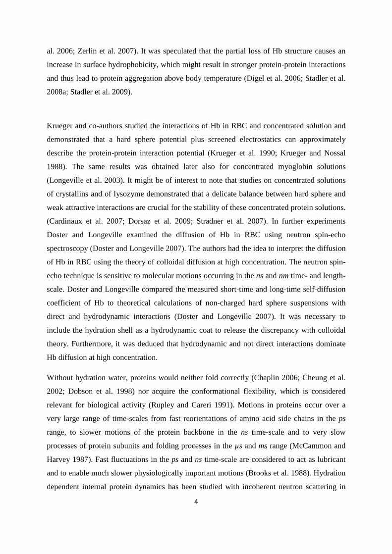
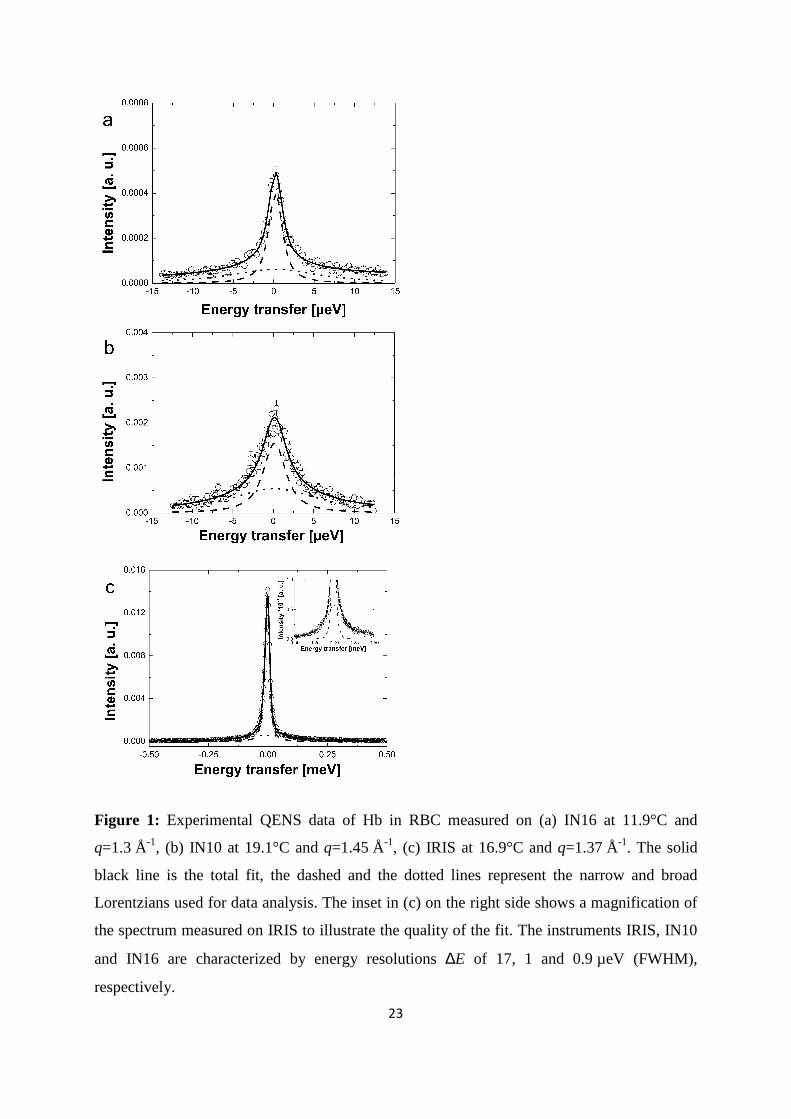
In the following we present and discuss the results of our experiments. Typical QENS data
measured on the neutron spectrometers IN16, IN10, and on IRIS are shown in Figure 1. The
measured spectra were well described with a narrow and a broad Lorentzian for global
macromolecular diffusion and internal Hb dynamics, respectively. First, we discuss the results
about global Hb diffusion. Our interpretation follows the ideas of Doster and Longeville
(Doster and Longeville 2007).
Global macromolecular diffusion
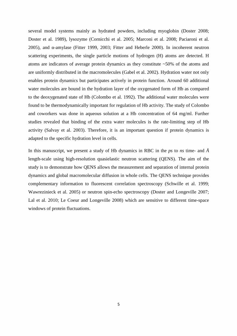
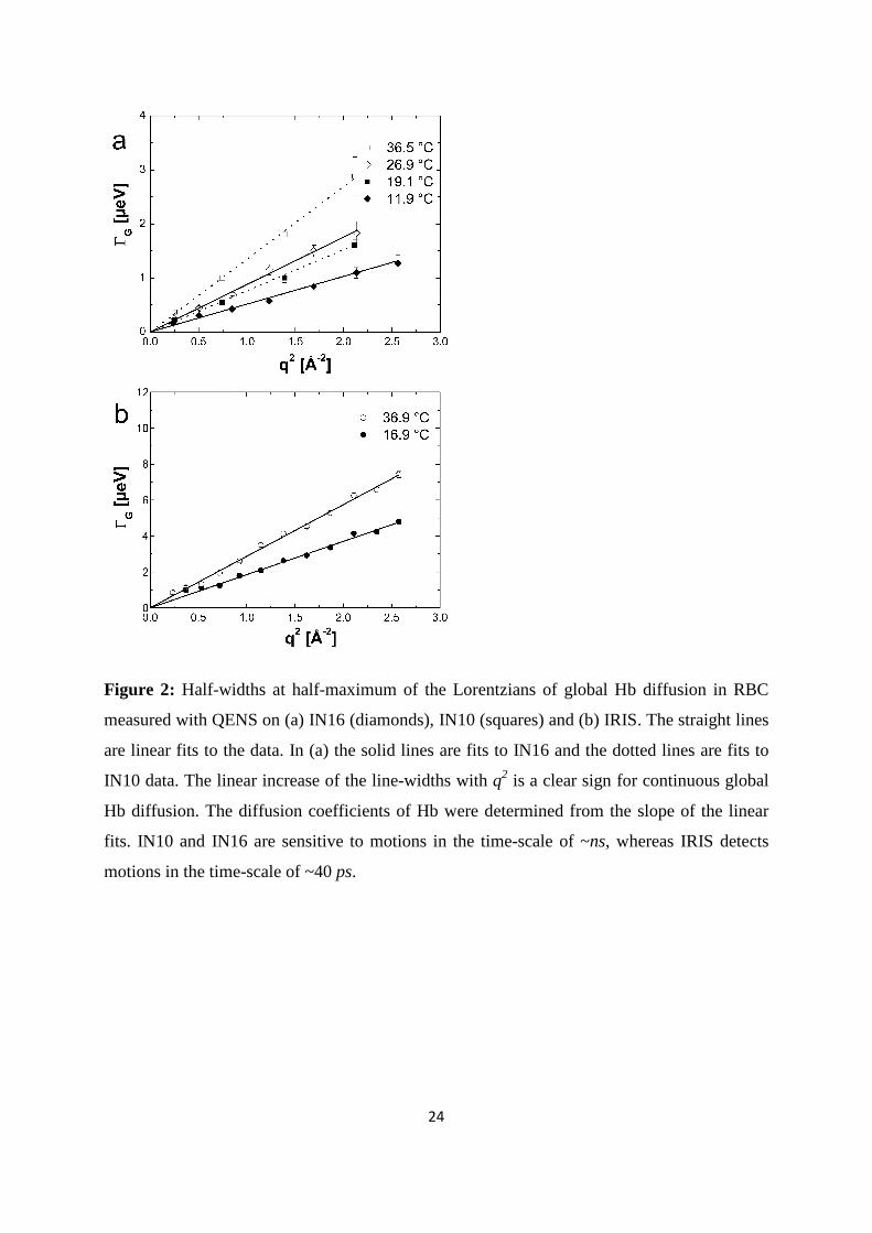
The measured half-widths at half-maximum (HWHM) for global Hb diffusion are presented
in Figure 2. Apparent diffusion coefficients Dapp were determined according to
( ) 2qDq appG ⋅=Γ in the range of 0.24≤q2≤2.56 Å-2 for IN10 and IN16 data, and in the range
of 0.72≤q2≤2.57 Å-2 for IRIS data. The line-widths ( )qGΓ of global Hb diffusion increase
linearly with q2 up to around 2.6 Å-2. This behavior is a clear sign for continuous global
diffusion of Hb. The Dapp contain both a component of translational and rotational diffusion
of Hb. It was shown previously (Perez et al. 1999; Stadler et al. 2008a) that rotational
diffusion of Hb leads to an additional broadening of the spectra by the factor 1.27. Therefore,
the apparent diffusion coefficients Dapp were divided by 1.27 to obtain the global translational
diffusion coefficient D of Hb. The essential steps in the calculation of the contributions of
rotational and translational diffusion to the experimental spectra are outlined in the appendix.
All obtained values of D are compared in Figure 3. The line-widths obtained from the
measurements with IN16 and IN10 intercept zero (Figure 2 a), which indicates that on time-
scales of ~1 ns global Hb diffusion does not sense confinement of neighboring proteins. The
( )qGΓ measured on IRIS appear to converge towards a plateau at small q2 and low
temperature (Figure 2 b). The feature indicates a cage effect of the neighboring molecules on
11
Hb diffusion in the ps time-scale and was observed before (Stadler et al. 2008a). Multiple
scattering might lead to a deviation from linear behavior at small q2. However, as the
transmission of the sample was 0.95 multiple scattering should be completely negligible. An
alternative explanation could be that small uncertainties of the resolution function might result
in a plateau at small q2, as the HWHM are only ~10% of the energy resolution of IRIS. An
observation time dependent diffusion coefficient is obtained. Unruh and coworkers observed a
similar phenomenon (Unruh et al. 2008). The authors studied the motions in liquid medium-
chain n-alkanes using QENS with observation times from 1.1 ps to 900 ps and molecular
dynamics simulations. The study revealed a time dependent diffusion coefficient, and there
was no need to use the obtained half-widths at low-resolution for the analysis of the high-
resolution data. To check the validity of our interpretations we have also performed a
complementary analysis in time-space (see Supplementary Material). The obtained diffusion
coefficients in time- and in energy-space are identical within the error bars. In the case that
the diffusion coefficient measured with IRIS would be visible with IN16/IN10, we would
obtain a mixture in time-space of the IRIS and the IN16/IN10 energy-space results. This is not
the case and our check therefore demonstrates the validity of our analysis.
Studies on average macromolecular dynamics in E. coli cells (Jasnin et al. 2008) and in
concentrated myoglobin solutions (Busch et al. 2007) using high-resolution neutron
backscattering spectroscopy reported that the measured line-widths of global macromolecular
diffusion deviate from linear behavior and tend towards saturation at large q2. Jump-diffusion
of the macromolecules was discussed as a possible explanation (Busch et al. 2007; Jasnin et
al. 2008), as this mechanism would result in a saturation of the line-widths at large q2. On the
other hand, a distribution of diffusion coefficients could also be responsible for the deviation
of the line-widths from linear behavior (Busch et al. 2007). Importantly, any kind of non-
localized diffusion leads to line-widths that tend towards zero with ( ) 2qDq appG ⋅=Γ at small
q2-values. Jasnin and co-workers studied average macromolecular dynamics in E. coli using
the IRIS spectrometer (Jasnin et al. 2008). Global macromolecular diffusion in E. coli was too
slow and could not be resolved with IRIS. Prokaryotic cells, such as E. coli, are very complex
objects which contain a vast amount of large macromolecular assemblies, such as ribosomes
with a molecular mass of 2.5 MDa. Average macromolecular dynamics in E. coli are therefore
difficult to attribute to a certain component. Hb is the main macromolecular component of
RBC with a rather small molecular mass of 65 kDa. It is reasonable to assume that global
12
diffusion of Hb is significantly faster than that of large macromolecular complexes in E. coli,
which would explain why Hb global diffusion in RBC is visible on IRIS.
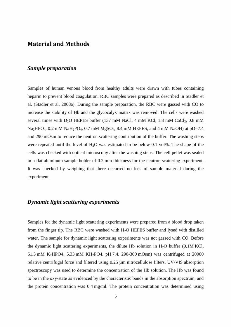
Tokuyama and Oppenheim evaluated the short-time SSD and long-time L
SD self-diffusion
coefficients of concentrated non-charged hard-sphere suspensions with hydrodynamic and
direct interactions as a function of the volume fraction φ and of the diffusion coefficient at
infinite dilution D0 (Tokuyama and Oppenheim 1994). Short-time self-diffusion corresponds
to particles that move in a static configuration of the neighboring particles at times t<τD, with
the structural relaxation time τD. The long-time limit of self-diffusion is reached at t>τD. The
values of the short- and long-time self-diffusion coefficients are equal only in dilute solution.
At higher concentrations short-time self-diffusion is always faster than long-time self-
diffusion. We measured the diffusion coefficient D0 of Hb at infinite dilution with dynamic
light scattering. The theoretical values of SSD and LSD of Hb at a volume fraction of φ=0.25
( 056.0 DD SS ⋅= , 028.0 DD L
S ⋅= ) are given in Figure 3 (Tokuyama and Oppenheim 1994). It
is obvious that the measured diffusion coefficients are too small and do not agree with the
theoretical values. Full hydration of myoglobin corresponds to a value of h~0.39g H2O/ g Mb
(Rupley and Careri 1991). It is believed that the critical hydration to allow the onset of
anharmonic motions in myoglobin (Mb) is around hMb=0.35g H2O/g Mb (Doster et al. 1989).
Hb has a larger radius of gyration than Mb (Hb: RG~24Å, Mb: RG~16Å) and a smaller surface
to volume ratio S/V (Longeville et al. 2003; Schelten et al. 1972). Approximating Hb and Mb
as spherical particles, the critical hydration of Hb should be around
hHb=(S/V)Hb / (S/V)Mb*hMb=16Å/24Å*0.35g H2O/ g Mb=0.23g H2O/ g Hb. It is known that the
density of protein hydration water is ~10% larger than bulk solvent (Svergun et al. 1998). The
partial specific volume ν of Hb plus hydration water is then
( ) gmlgml /98.0/1.1/23.075.0 =+=ν , which corresponds to an effective volume fraction
of Hb plus hydration water of 32.0=⋅= νφ c with the concentration c=0.33 g/ml of Hb in
RBC (DeMoll et al. 2007; Doster and Longeville 2007). The measured self-diffusion
coefficients of Hb with IN16, IN10 and IRIS agree with high accuracy with the theoretical
values of Hb plus hydration shell ( 00.45 DD SS ⋅= , 00.18 DD L
S ⋅= ) (Tokuyama and
Oppenheim 1994). In a study on the short-time limit of Hb diffusion in RBC we estimated
that the structural relaxation time τD is in the order of several hundred ps (Stadler et al.
13
2008a). Therefore, IRIS is sensitive to motions which are faster than τD and short-time self-
diffusion is detected. The high-resolution instruments IN16 and IN10, and neutron spin echo
spectroscopy (Doster and Longeville 2007) measure motions which are longer than τD, and
the long-time limit of Hb self-diffusion is observed. We showed previously that although
~90% of cell water in RBC has properties similar to bulk water, a small fraction of ~10%
cellular water exhibits strongly reduced dynamics and was attributed to water molecules
which are bound to the surface of Hb (Stadler et al. 2008b). The ratio of water per Hb in RBC
is h~2.3g H2O/ g Hb and a ~10% fraction corresponds to a bound water fraction of
~0.23g H2O/ g Hb which is identical to the value reported in this article. Furthermore, Doster
and Longeville measured the diffusion of Hb in whole red blood cells using spin echo
spectroscopy in the ns and nm time- and length-scale (Doster and Longeville 2007). The
authors demonstrated that the presence of the hydration shell leads to a reduction of the
diffusion coefficient of Hb. Garcia de la Torre calculated hydrodynamic properties of proteins
from atomic structures (Garcia de la Torre 2001). It was demonstrated by comparing
calculated and experimental values that a hydration shell of h~0.27g H2O/ g Hb is bound to
the surface of Hb. Our result is reasonably close to that value.
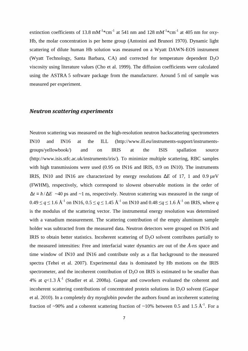
Internal hemoglobin dynamics
We now turn our attention to the results concerning internal protein dynamics. Detailed
information about protein internal motions can be extracted from the scattering vector
dependence of the quasielastic broadening and the Elastic Incoherent Structure Factor
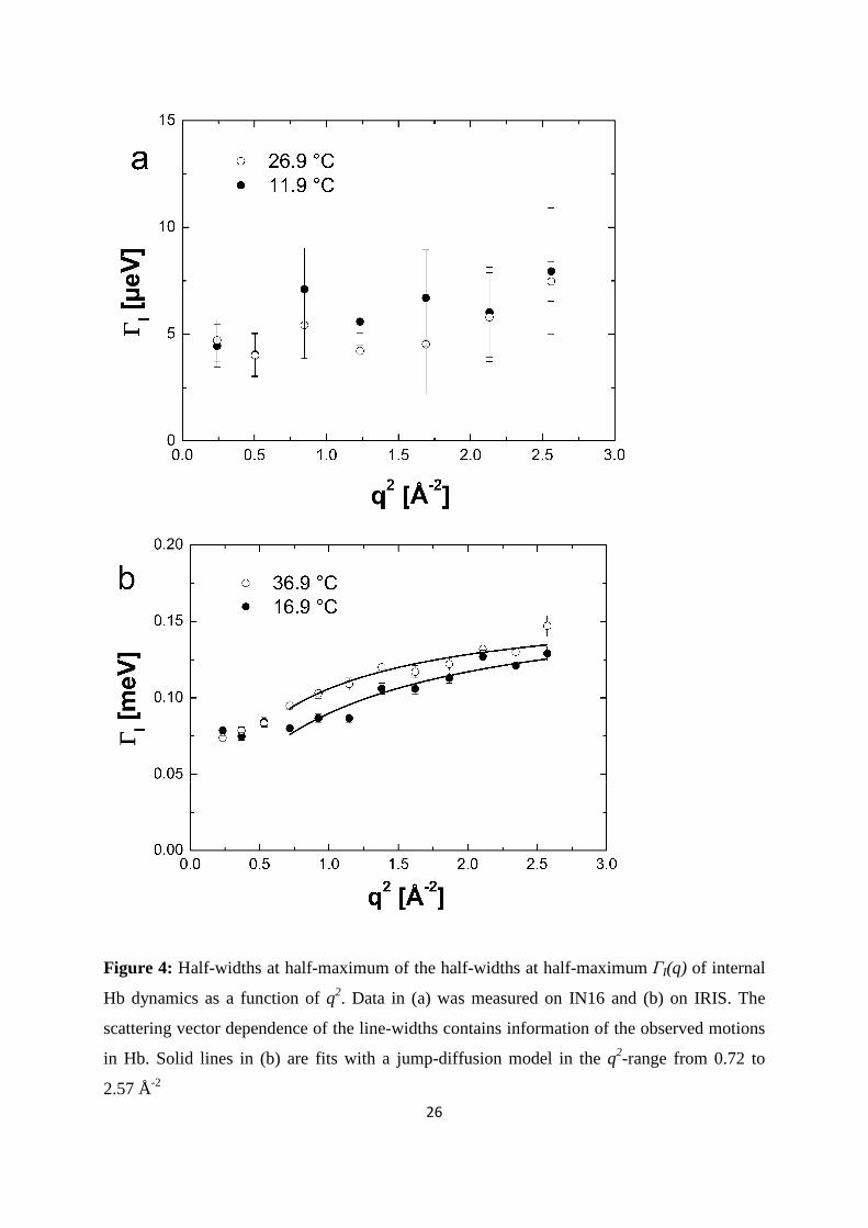
(EISF). The HWHM of internal protein dynamics measured on IN16 and on IRIS are given in
Figure 4 (a) and (b). The ( )qIΓ measured with IRIS show typical behavior of localized jump-
diffusion. The half-widths tend towards a constant value at small q2, which indicates
confining effects of local boundaries. Diffusive jumps with a finite jump-length lead to a
plateau in the line-widths at large scattering vectors. In the q2-range of 0.72 and 2.57 Å-2 the
( )ω,qIΓ measured on IRIS could be well described with a jump-diffusion model given by
( )τ
ω2
2
1,
qD
qDq
I
II +
=Γ (Bee 1988). The parameters of the jump-diffusion model are the
residence time before a jump τ and the jump-diffusion coefficient DI of protein internal
14
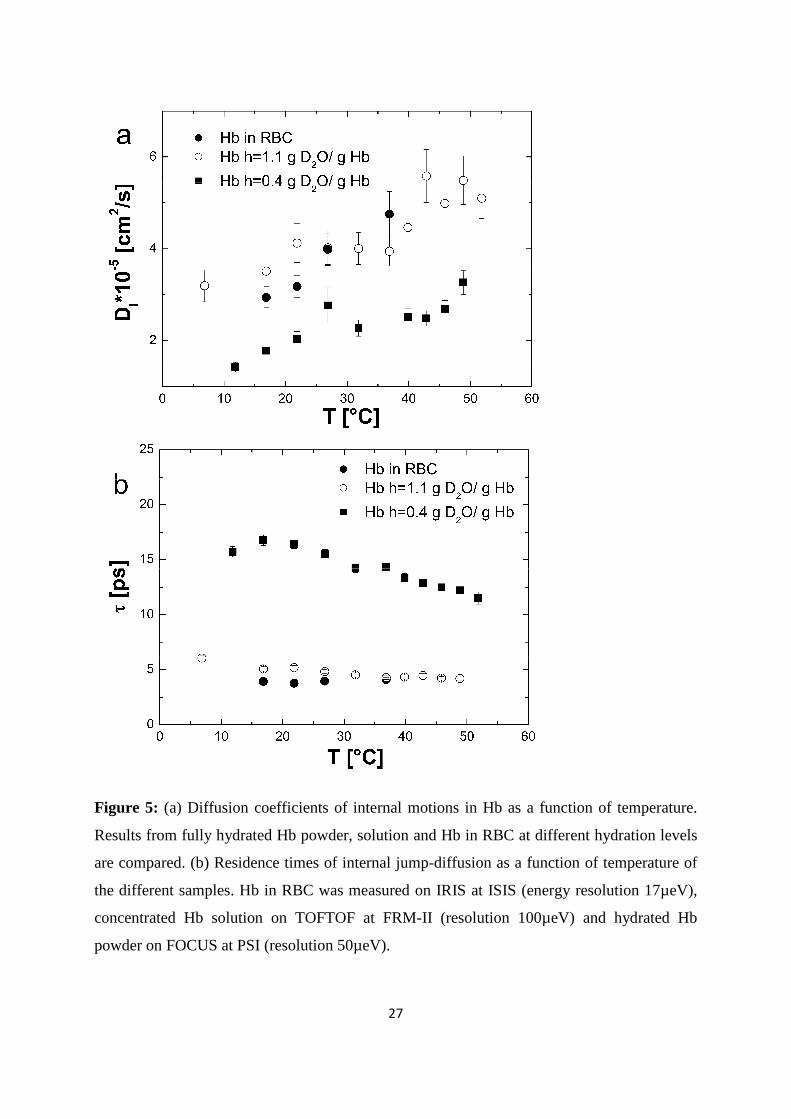
motions. In Figure 5, the jump-diffusion coefficients and the residence times of internal Hb
dynamics in RBC are compared to jump-diffusion coefficients and residence times of internal
Hb dynamics as hydrated powder (h=0.4 g D2O/ g Hb) and as concentrated solution (h=1.1 g
D2O/ g Hb). The corresponding hydration level in RBC is h~2.5 g D2O/ g Hb. The results of
the experiments with the hydrated Hb powder and concentrated Hb solution have been
published before and are given here for comparison (Stadler et al. 2009). The Hb powder and
solution were measured on neutron time-of-flight spectrometers with energy resolutions of 50
and 100 µeV, respectively. All data were analyzed in the same way. The results demonstrate
that an increase in the hydration level from one hydration shell in the Hb powder to around 3
hydration layers in the concentrated Hb solution increases the jump-diffusion coefficients and
strongly reduces the residence times of internal protein dynamics in the ps time-scale. A
further increase in the hydration level to around 6 hydration layers per Hb in whole RBC does
neither enhance the jump-diffusion coefficients nor reduce significantly the residence times as
compared to the concentrated Hb sample. The rate of internal jump-diffusion in the ps time-
scale appears to be already fully developed in the concentrated Hb solution. The observed
motions in the ps range could correspond to diffusive jumps of amino acid side chains and
attached methyl groups (Fitter et al. 1996).
The half-widths of internal protein dynamics from the experiment on IN16 are independent of
the scattering vector within the error bars, as shown in Figure 4(a), and have average values of
5.8 ± 1.4 µeV at 11.9 °C and 6.2 ± 1.0 µeV at 26.9 °C. The line-widths determined with IN10
are 5.5 µeV at 19.1 °C and 4.2 µeV at 36.5 °C. The ( )qIΓ on each individual spectrum of
IN10 had large errors. The average value was used for all spectra at one temperature and held
constant during fitting of IN10 data; the obtained values are rather imprecise and are given
only for completeness. The line-widths obtained on IN16 and IN10 are in agreement with
other studies which investigated protein dynamics in the ns time-scale using high-resolution
quasielastic neutron scattering. Fitter and co-workers studied hydrated bacteriorhodopsin and
obtained half-widths of 5.5 µeV (Fitter et al. 1997), Orecchini and co-workers investigated
hydrated β-lactoglobulin powder and found half-widths of 16 µeV (Orecchini et al. 2002),
Busch et al. found line-widths of 10 µeV of myoglobin in concentrated solution (Busch et al.
2007), and Jasnin et al. measured average dynamics in whole E. coli and obtained line-widths
of ~7 µeV (Jasnin et al. 2008). If we exclude the lactoglobulin case, the values of the
measured line-widths are rather similar although the hydration levels in the investigated
15
systems are different. We recall that correlation times τ and line-widths Γ are inversely related
by Γ= /1corτ . This seems to indicate that correlation times of motions in globular and
membrane proteins in the ns time-scale are rather similar in hydrated protein powders,
solutions and in whole cells. As the observed line-widths on IN16 and IN10 are independent
of the scattering vector, a different class of motions is observed using the high-resolution
instruments. Rotational motions lead to line-widths which are independent of the scattering
vector (Bee 1988), and the observed dynamics might be attributed to slow rotations of side
chains or relaxations of the protein backbone.
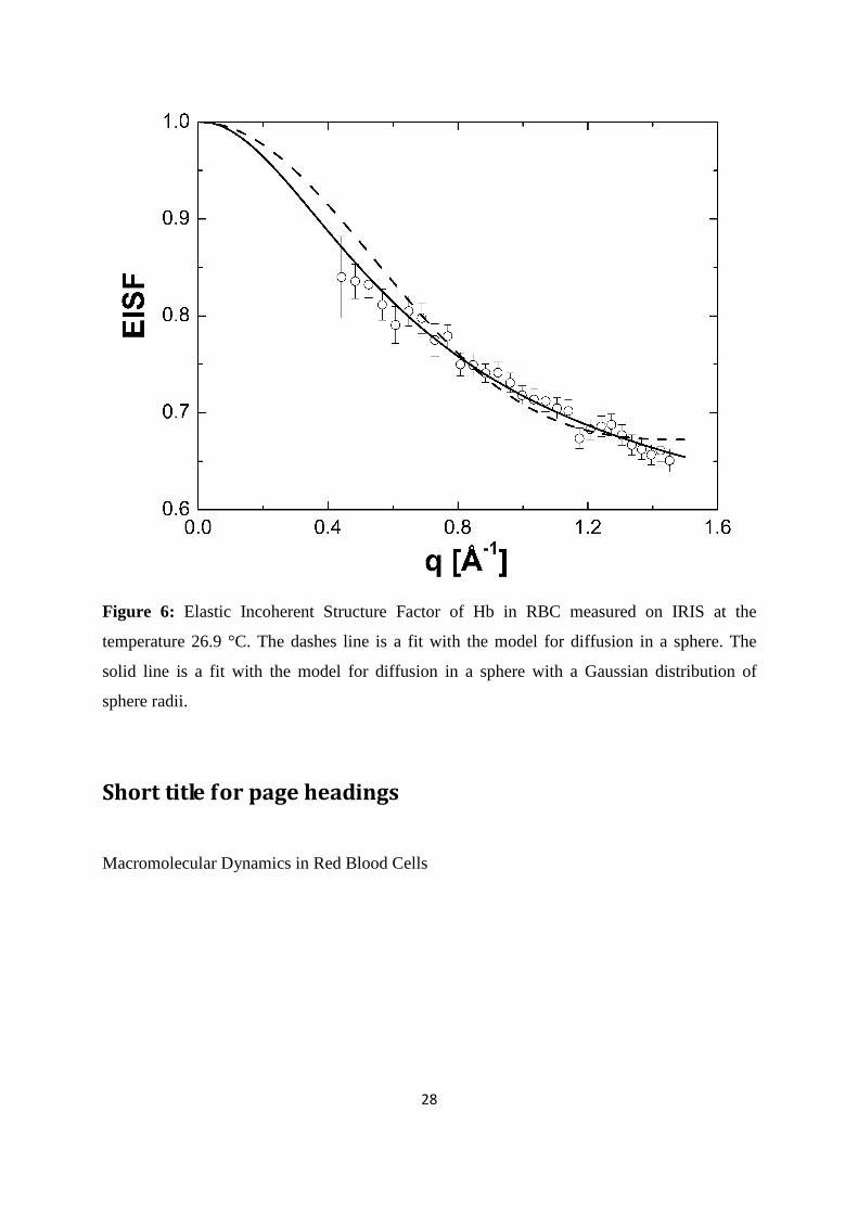
Information about the geometry of motions can be extracted from the measured EISF. Only
four and six data points are available on IN10 and IN16, respectively. This is too few and
does not allow an accurate analysis. Therefore, we limit our discussion to the results of the
experiment using the IRIS spectrometer. The EISF obtained with IRIS at 26.9 °C is shown in
Figure 6. The EISF was interpreted with the model of Volino and Dianoux for diffusion in a
sphere (Volino and Dianoux 1980). The diffusion in a sphere model can be written as
( ) ( ) ( ) 2
10
31
⋅−+=
qa
qajppqA , where j1(qa) is the first-order spherical Bessel function of the
first kind, a is the sphere radius, and A0(q) is the EISF. The hydrogen atoms which appear
immobile and mobile within the instrumental energy resolution are represented by the
fractions p and (1-p), respectively. The obtained sphere radius a increases from 2.8 ± 0.1 Å at
16.9 °C to 3.3 ± 0.1 Å at 36.9 °C. The immobile fraction p has got the average value of 0.67.
These values reasonable agree with results on macromolecular dynamics in E. coli which
found a=3.1 Å and p=0.61 at 6.9 °C; a=3.4 Å and p=0.56 at 26.9 °C (Jasnin et al. 2008).
To take into account of the heterogeneity of internal protein dynamics Perez and co-workers
extended the diffusion in a sphere model and introduced a Gaussian distribution of sphere
radii f(a) instead of a single sphere (Perez et al. 1999). The Gaussian distribution is defined as
( ) ( )2
2
2exp
2
2σπσ
aaf −= , with the standard deviation σ as free parameter. The mean value
of the sphere radius is given by π
σ 2ˆ =a . A neutron scattering study using specific isotope
labeling in order to investigate the dynamics of specific amino acids in bacteriorhodopsin
demonstrated the heterogeneity of internal protein dynamics (Wood et al. 2008). The obtained
average sphere radius â increases from â=2.1 ± 0.1 Å at 16.9 °C to â=3.0 ± 0.2 Å at 36.9 °C.
16
The immobile fraction p has an average value of 0.50 and increases only slightly with
temperature from p=0.47 ± 0.02 at 16.9°C to p=0.55 ± 0.01 at 36.9°C. Using the same model
we have quantified the average amplitudes of motion in concentrated Hb solution (Stadler et
al. 2009). The average sphere radius was found to increase from â=2.3 Å at 6.9 °C to â=2.6 Å
at 36.9 °C, while the immobile fraction was constant with temperature p=0.38 (Stadler et al.
2009). Within the error bars the obtained average sphere radii of Hb in RBC and of Hb in
concentrated solution are similar when we exclude the value at 36.9 °C, which is larger in Hb
in RBC than in the Hb solution. Although the energy resolutions of the instruments used for
both experiments are different (17µeV and 100µeV, respectively) the observed motions look
similar. This might either be due to the fact that the same motions are seen using the IRIS and
the time-of-flight spectrometers, or that different classes of motions in the order of 40ps and
several ps are similar. The second possibility would also imply that the corresponding
hierarchical structures in the energy landscape are similar.
The model for diffusion in a sphere approximately describes the measured EISF. Better fits
can be obtained when a Gaussian distribution of sphere radii is used. It should be noted that
both the diffusion in a sphere model and the Gaussian distribution can only be simple and
rough representations for the heterogeneity of internal protein dynamics. In any case, models
are never wrong, they are just more or less appropriate.
Conclusion
In summary, we measured the global self-diffusion and internal dynamics of Hb in RBC, in
vivo, using high-resolution quasielastic neutron backscattering spectroscopy. It is
demonstrated that global protein diffusion and internal dynamics can be separated and
interpreted quantitatively. The cross-over from the short to the long time limit of Hb self-
diffusion could be observed. It is demonstrated that the diffusion of Hb at high concentration
in RBC can be described with concepts of colloid physics. Experimental data is in
quantitative agreement with hydrodynamic theory of non-charged hard-sphere suspensions
when it is assumed that the hydration shell moves with the protein. It is shown that interfacial
protein hydration water has a strong influence on global protein diffusion under physiological
17
conditions in cells. The same result was obtained by Doster and Longeville using spin-echo
spectroscopy (Doster and Longeville 2007). Experiments with whole RBC using micropipette
aspiration and colloidal osmotic pressure measurements (Artmann et al. 2009; Artmann et al.
1998) indicated that the cellular environment might have similarities to a colloidal gel. It was
suggested that the trigger for the formation of the gel could be Hb-Hb interactions, which are
influenced by the molecular properties of Hb (Digel et al. 2006; Zerlin et al. 2007). Recently,
we studied Hb-Hb interactions in concentrated solution using small angle neutron scattering
and could show that Hb molecules associate into a large-scale superstructure at high
concentration (Stadler et al. 2010). In this article, we observe a slowing down of the atomistic
diffusion of Hb, which might indeed lead to gel-like properties on a macroscopic scale. It is
demonstrated how incoherent neutron scattering can contribute to the understanding of
cellular phenomena on a macroscopic scale.
Internal Hb dynamics was also measured and could be separated from global Hb diffusion.
The internal motions of Hb were compared to results obtained with hydrated powder and
solution samples. Different types of motions were brought into focus by using neutron
spectrometers with specific energy resolutions. Hydration water was found to have a strong
influence on motions in the ps time-scale. Jump-diffusion coefficients of internal Hb
fluctuations are significantly enhanced and residence times of the internal diffusive jumps are
reduced in RBC as compared to fully hydrated Hb powder. Slower internal dynamics of Hb in
RBC in the ns time-range were found to be rather similar to results obtained with fully
hydrated protein powders, solutions and E. coli cells. Still missing is a combined analysis of
the data measured with different spectrometers, which should be done in a future publication.
Future work might also be dedicated to investigate protein dynamics in whole cells under
different environmental conditions.
Acknowledgements
The author (A.M.S.) thanks Georg Büldt for continuous support. We also thank Giuseppe
Zaccai for valuable discussion and critical reading of the manuscript.
18
Appendix
Global Hb diffusion: Contribution of rotational and
translational diffusion
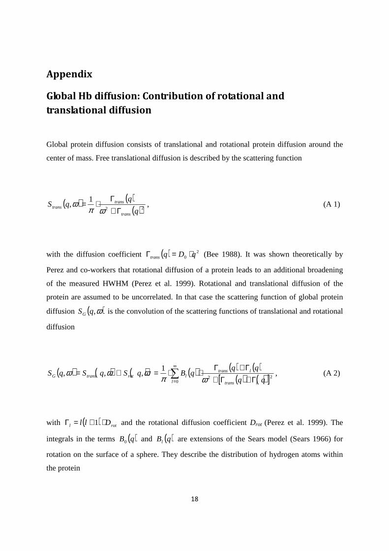
Global protein diffusion consists of translational and rotational protein diffusion around the
center of mass. Free translational diffusion is described by the scattering function
( ) ( )( )22
1,
q
qqS
trans
transtrans Γ+
Γ⋅=ωπ
ω , (A 1)
with the diffusion coefficient ( ) 20 qDqtrans ⋅=Γ (Bee 1988). It was shown theoretically by
Perez and co-workers that rotational diffusion of a protein leads to an additional broadening
of the measured HWHM (Perez et al. 1999). Rotational and translational diffusion of the
protein are assumed to be uncorrelated. In that case the scattering function of global protein
diffusion ( )ω,qSG is the convolution of the scattering functions of translational and rotational
diffusion
( ) ( ) ( ) ( ) ( ) ( )( ) ( )[ ]∑
∞
= Γ+Γ+Γ+Γ
⋅⋅=⊗=0
22
1,,,
l ltrans
ltranslrottransG
qqqBqSqSqS
ωπωωω , (A 2)
with ( ) rotl Dll ⋅+=Γ 1 and the rotational diffusion coefficient Drot (Perez et al. 1999). The
integrals in the terms ( )qB0 and ( )qBl are extensions of the Sears model (Sears 1966) for
rotation on the surface of a sphere. They describe the distribution of hydrogen atoms within
the protein
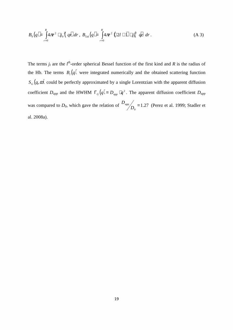
19
( ) ( )∫=
⋅=R
r
drqrjrqB0
20
20 4π , ( ) ( ) ( )drqrjlrqB
R
r
ll ∫=
≥ ⋅+⋅=0
221 124π . (A 3)
The terms j l are the l th-order spherical Bessel function of the first kind and R is the radius of
the Hb. The terms ( )qBl were integrated numerically and the obtained scattering function
( )ω,qSG could be perfectly approximated by a single Lorentzian with the apparent diffusion
coefficient Dapp and the HWHM ( ) 2qDq appG ⋅=Γ . The apparent diffusion coefficient Dapp
was compared to D0, which gave the relation of 27.10
=DDapp (Perez et al. 1999; Stadler et
al. 2008a).
20
References
Antonini E, Brunori M (1970) Hemoglobin. Ann. Rev. Biochem. 39:977-1042
Artmann GM, Burns L, Canaves JM, Temiz-Artmann A, Schmid-Schonbein GW, Chien S, Maggakis-
Kelemen C (2004) Circular dichroism spectra of human hemoglobin reveal a reversible
structural transition at body temperature. Eur. Biophys. J. 33:490-496
Artmann GM, Digel I, Zerlin KF, Maggakis-Kelemen C, Linder P, Porst D, Kayser P, Stadler AM, Dikta G,
Temiz Artmann A (2009) Hemoglobin senses body temperature. Eur. Biophys. J. 38:589-600
Artmann GM, Kelemen C, Porst D, Büldt G, Chien S (1998) Temperature transitions of protein
properties in human red blood cells. Biophys. J. 75:3179-83
Azuah RT, Kneller LR, Qiu Y, Tregenna-Piggott PLW, Brown CM, Copley JRD, Dimeo RM (2009) DAVE:
A comprehensive software suite for the reduction, visualization, and analysis of low energy
neutron spectroscopic data. J. Res. Natl. Inst. Stan. Technol. 114:341-358
Bee M (1988) Quasielastic neutron scattering. Principles and Applications in Solid State Chemistry,
Biology and Materials Science. Adam Hilger, Bristol and Philadelphia
Brooks CL, Karplus M, Pettitt BM (1988) Proteins : a Theoretical Perspectives of Dynamics, Structures,
and Thermodynamics, vol 71. John Wiley & Sons, New York
Busch S, Doster W, Longeville S, Garcia Sakai V, Unruh T (2007) Microscopic protein diffusion at high
concentration. In: Sokol PE, Kaiser H, Baxter D, Pynn R, Bossev D, Leuschner M (eds).
Materials Research Society, pp 107-114
Cardinaux F, Gibaud T, Stradner A, Schurtenberger P (2007) Interplay between spinodal
decomposition and glass formation in proteins exhibiting short-range attractions. Phys. Rev.
Lett. 99:118301
Chaplin M (2006) Opinion - Do we underestimate the importance of water in cell biology? Nat. Rev.
Mol. Cell Biol. 7:861-866
Cheung MS, Garcia AE, Onuchic JN (2002) Protein folding mediated by solvation: Water expulsion and
formation of the hydrophobic core occur after the structural collapse. Proc. Natl. Acad. Sci.
U. S. A. 99:685-690
Cho CH, Urquidi J, Singh S, Wilse Robinson G (1999) Thermal offset viscosities of liquid H2O, D2O, and
T2O. J. Phys. Chem. B 103:1991-1994
Colombo MF, Rau DC, Parsegian VA (1992) Protein Solvation in Allosteric Regulation - a Water Effect
on Hemoglobin. Science 256:655-659
Cornicchi E, Onori G, Paciaroni A (2005) Picosecond-time-scale fluctuations of proteins in glassy
matrices: The role of viscosity. Phys. Rev. Lett. 95:158104
DeMoll E, Cox DJ, Daniel E, Riggs AF (2007) Apparent specific volume of human hemoglobin: Effect of
ligand state and contribution of heme. Anal. Biochem. 363:196-203
Digel I, Maggakis-Kelemen C, Zerlin KF, Linder P, Kasischke N, Kayser P, Porst D, Temiz Artmann A,
Artmann GM (2006) Body temperature-related structural transitions of monotremal and
human hemoglobin. Biophys. J. 91:3014-21
Dobson CM, Sali A, Karplus M (1998) Protein folding: A perspective from theory and experiment.
Angew. Chem., Int. Ed. 37:868-893
Dorsaz N, Thurston GM, Stradner A, Schurtenberger P, Foffi G (2009) Colloidal characterization and
thermodynamic stability of binary eye lens protein mixtures. J. Phys. Chem. B 113:1693-709
Doster W (2008) The dynamical transition of proteins, concepts and misconceptions. Eur. Biophys. J.
37:591-602
Doster W, Cusack S, Petry W (1989) Dynamical transition of myoglobin revealed by inelastic neutron
scattering. Nature 337:754-6
Doster W, Longeville S (2007) Microscopic diffusion and hydrodynamic interactions of hemoglobin in
red blood cells. Biophys. J. 93:1360-1368
21
Fitter J (1999) The temperature dependence of internal molecular motions in hydrated and dry
alpha-amylase: The role of hydration water in the dynamical transition of proteins. Biophys.
J. 76:1034-1042
Fitter J (2003) A measure of conformational entropy change during thermal protein unfolding using
neutron spectroscopy. Biophys. J. 84:3924-3930
Fitter J, Heberle J (2000) Structural equilibrium fluctuations in mesophilic and thermophilic alpha-
amylase. Biophys. J. 79:1629-1636
Fitter J, Lechner RE, Buldt G, Dencher NA (1996) Internal molecular motions of bacteriorhodopsin:
Hydration-induced flexibility studied by quasielastic incoherent neutron scattering using
oriented purple membranes. Proc. Natl. Acad. Sci. U. S. A. 93:7600-7605
Fitter J, Lechner RE, Dencher NA (1997) Picosecond molecular motions in bacteriorhodopsin from
neutron scattering. Biophys. J. 73:2126-37
Gabel F, Bicout D, Lehnert U, Tehei M, Weik M, Zaccai G (2002) Protein dynamics studied by neutron
scattering. Q. Rev. Biophys. 35:327-367
Garcia de la Torre J (2001) Hydration from hydrodynamics. General considerations and applications
of bead modelling to globular proteins. Biophys. Chem. 93:159-170
Gaspar AM, Appavou MS, Busch S, Unruh T, Doster W (2008) Dynamics of well-folded and natively
disordered proteins in solution: a time-of-flight neutron scattering study. Eur. Biophys. J.
37:573-582
Gaspar AM, Busch S, Appavou MS, Haeussler W, Georgii R, Su YX, Doster W (2010) Using polarization
analysis to separate the coherent and incoherent scattering from protein samples. Biochim.
Biophys. Acta, Proteins Proteomics 1804:76-82
http://www.ill.eu/instruments-support/instruments-groups/yellowbook/
http://www.isis.stfc.ac.uk/instruments/iris/
Jasnin M, Moulin M, Haertlein M, Zaccai G, Tehei M (2008) In vivo measurement of internal and
global macromolecular motions in E. coli. Biophys. J. 95:857-864
Krueger S, Chen SH, Hofrichter J, Nossal R (1990) Small angle neutron scattering studies of HbA in
concentrated solutions. Biophys. J. 58:745-57
Krueger S, Nossal R (1988) SANS studies of interacting hemoglobin in intact erythrocytes. Biophys. J.
53:97-105
Lal J, Fouquet P, Maccarini M, Makowski L (2010) Neutron Spin-Echo Studies of Hemoglobin and
Myoglobin: Multiscale Internal Dynamics. J. Mol. Biol. 397:423-435
Le Coeur C, Longeville S (2008) Microscopic protein diffusion at high concentration by neutron spin-
echo spectroscopy. Chem. Phys. 345:298-304
Longeville S, Doster W, Kali G (2003) Myoglobin in crowded solutions: structure and diffusion. Chem.
Phys. 292:413-424
Marconi M, Cornicchi E, Onori G, Paciaroni A (2008) Comparative study of protein dynamics in
hydrated powders and in solutions: A neutron scattering investigation. Chem. Phys. 345:224-
229
McCammon JA, Harvey SC (1987) Dynamics of Proteins and Nuclear Acids
Cambridge University Press, Cambridge, UK
Orecchini A, Paciaroni A, Bizzarri AR, Cannistraro S (2002) Dynamics of different hydrogen classes in
beta-lactoglobulin: A quasielastic neutron scattering investigation. J. Phys. Chem. B 106:7348-
7354
Paciaroni A, Cinelli S, Cornicchi E, De Francesco A, Onori G (2005) Fast fluctuations in protein
powders: The role of hydration. Chem. Phys. Lett. 410:400-403
Perez J, Zanotti JM, Durand D (1999) Evolution of the Internal Dynamics of Two Globular Proteins
from Dry Powder to Solution. Biophys. J. 77:454-469
Rupley JA, Careri G (1991) Protein Hydration and Function. Adv. Prot. Chem. 41:37-172
22
Salvay AG, Grigera JR, Colombo MF (2003) The role of hydration on the mechanism of allosteric
regulation: in situ measurements of the oxygen-linked kinetics of water binding to
hemoglobin. Biophys. J. 84:564-70
Schelten J, Schlecht P, Schmatz W, Mayer A (1972) Neutron Small Angle Scattering of Hemoglobin. J.
Biolog. Chem. 247:5436-5441
Schwille P, Haupts U, Maiti S, Webb WW (1999) Molecular dynamics in living cells observed by
fluorescence correlation spectroscopy with one- and two-photon excitation. Biophys. J.
77:2251-65
Sears VF (1966) THEORY OF COLD NEUTRON SCATTERING BY HOMONUCLEAR DIATOMIC LIQUIDS: II.
HINDERED ROTATION. Can. J. Phys. 44:1299-1311
Stadler AM, Digel I, Artmann GM, Embs JP, Zaccai G, Buldt G (2008a) Hemoglobin dynamics in red
blood cells: correlation to body temperature. Biophys. J. 95:5449-61
Stadler AM, Digel I, Embs JP, Unruh T, Tehei M, Zaccai G, Büldt G, Artmann GM (2009) From Powder
to Solution: Hydration Dependence of Human Hemoglobin Dynamics Correlated to Body
Temperature. Biophys. J. 96:5073-5081
Stadler AM, Embs JP, Digel I, Artmann GM, Unruh T, Buldt G, Zaccai G (2008b) Cytoplasmic water and
hydration layer dynamics in human red blood cells. J. Am. Chem. Soc. 130:16852-3
Stadler AM, Schweins R, Zaccai G, Lindner P (2010) Observation of a Large-Scale Superstructure in
Concentrated Hemoglobin Solutions by Using Small Angle Neutron Scattering. J. Phys. Chem.
Lett. 1:1805-1808
Stradner A, Foffi G, Dorsaz N, Thurston G, Schurtenberger P (2007) New Insight into Cataract
Formation: Enhanced Stability through Mutual Attraction. Phys. Rev. Lett. 99:198103
Svergun DI, Richard S, Koch MH, Sayers Z, Kuprin S, Zaccai G (1998) Protein hydration in solution:
experimental observation by x-ray and neutron scattering. Proc. Natl. Acad. Sci. U. S. A.
95:2267-72
Tehei M, Franzetti B, Wood K, Gabel F, Fabiani E, Jasnin M, Zamponi M, Oesterhelt D, Zaccai G,
Ginzburg M, Ginzburg BZ (2007) Neutron scattering reveals extremely slow cell water in a
Dead Sea organism. Proc. Natl. Acad. Sci. U. S. A. 104:766-71
Tokuyama M, Oppenheim I (1994) Dynamics of hard-sphere suspensions. Phys. Rev. E 50:R16-R19
Unruh T, Smuda C, Busch S, Neuhaus J, Petry W (2008) Diffusive motions in liquid medium-chain n-
alkanes as seen by quasielastic time-of-flight neutron spectroscopy. J. Chem. Phys.
129:121106
Volino F, Dianoux AJ (1980) Neutron Incoherent-Scattering Law for Diffusion in a Potential of
Spherical-Symmetry - General Formalism and Application to Diffusion inside a Sphere. Mol.
Phys. 41:271-279
Wawrezinieck L, Rigneault H, Marguet D, Lenne PF (2005) Fluorescence correlation spectroscopy
diffusion laws to probe the submicron cell membrane organization. Biophys. J. 89:4029-4042
Wood K, Grudinin S, Kessler B, Weik M, Johnson M, Kneller GR, Oesterheit D, Zaccai G (2008)
Dynamical heterogeneity of specific amino acids in bacteriorhodopsin. J. Mol. Biol. 380:581-
591
Zerlin KFT, Kasischke N, Digel I, Maggakis-Kelemen C, Artmann AT, Porst D, Kayser P, Linder P,
Artmann GM (2007) Structural transition temperature of hemoglobins correlates with
species' body temperature. Eur. Biophys. J. 37:1-10
23
Figure 1: Experimental QENS data of Hb in RBC measured on (a) IN16 at 11.9°C and
q=1.3 Å-1, (b) IN10 at 19.1°C and q=1.45 Å-1, (c) IRIS at 16.9°C and q=1.37 Å-1. The solid
black line is the total fit, the dashed and the dotted lines represent the narrow and broad
Lorentzians used for data analysis. The inset in (c) on the right side shows a magnification of
the spectrum measured on IRIS to illustrate the quality of the fit. The instruments IRIS, IN10
and IN16 are characterized by energy resolutions ∆E of 17, 1 and 0.9 µeV (FWHM),
respectively.
24
Figure 2: Half-widths at half-maximum of the Lorentzians of global Hb diffusion in RBC
measured with QENS on (a) IN16 (diamonds), IN10 (squares) and (b) IRIS. The straight lines
are linear fits to the data. In (a) the solid lines are fits to IN16 and the dotted lines are fits to
IN10 data. The linear increase of the line-widths with q2 is a clear sign for continuous global
Hb diffusion. The diffusion coefficients of Hb were determined from the slope of the linear
fits. IN10 and IN16 are sensitive to motions in the time-scale of ~ns, whereas IRIS detects
motions in the time-scale of ~40 ps.
25
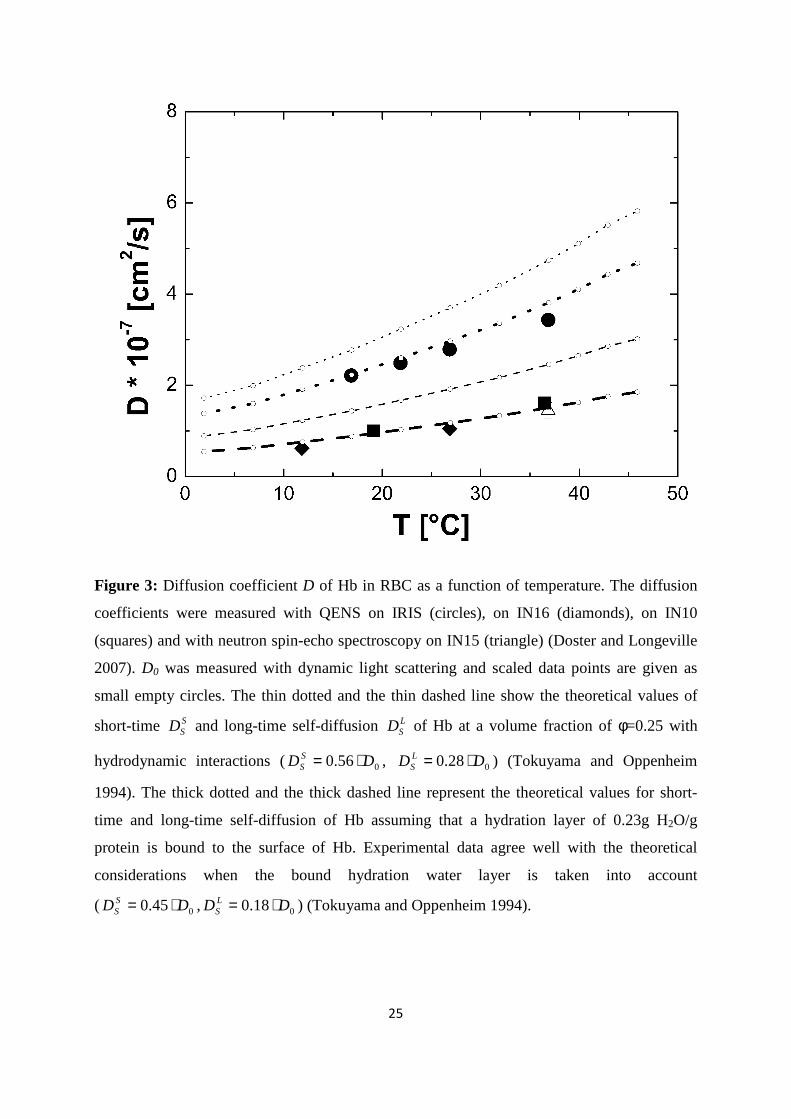
Figure 3: Diffusion coefficient D of Hb in RBC as a function of temperature. The diffusion
coefficients were measured with QENS on IRIS (circles), on IN16 (diamonds), on IN10
(squares) and with neutron spin-echo spectroscopy on IN15 (triangle) (Doster and Longeville
2007). D0 was measured with dynamic light scattering and scaled data points are given as
small empty circles. The thin dotted and the thin dashed line show the theoretical values of
short-time SSD and long-time self-diffusion L
SD of Hb at a volume fraction of φ=0.25 with
hydrodynamic interactions ( 056.0 DD SS ⋅= , 028.0 DD L
S ⋅= ) (Tokuyama and Oppenheim
1994). The thick dotted and the thick dashed line represent the theoretical values for short-
time and long-time self-diffusion of Hb assuming that a hydration layer of 0.23g H2O/g
protein is bound to the surface of Hb. Experimental data agree well with the theoretical
considerations when the bound hydration water layer is taken into account
( 00.45 DD SS ⋅= , 00.18 DD L
S ⋅= ) (Tokuyama and Oppenheim 1994).
26
Figure 4: Half-widths at half-maximum of the half-widths at half-maximum ΓI(q) of internal
Hb dynamics as a function of q2. Data in (a) was measured on IN16 and (b) on IRIS. The
scattering vector dependence of the line-widths contains information of the observed motions
in Hb. Solid lines in (b) are fits with a jump-diffusion model in the q2-range from 0.72 to
2.57 Å-2
27
Figure 5: (a) Diffusion coefficients of internal motions in Hb as a function of temperature.
Results from fully hydrated Hb powder, solution and Hb in RBC at different hydration levels
are compared. (b) Residence times of internal jump-diffusion as a function of temperature of
the different samples. Hb in RBC was measured on IRIS at ISIS (energy resolution 17µeV),
concentrated Hb solution on TOFTOF at FRM-II (resolution 100µeV) and hydrated Hb
powder on FOCUS at PSI (resolution 50µeV).
28
Figure 6: Elastic Incoherent Structure Factor of Hb in RBC measured on IRIS at the
temperature 26.9 °C. The dashes line is a fit with the model for diffusion in a sphere. The
solid line is a fit with the model for diffusion in a sphere with a Gaussian distribution of
sphere radii.
Short title for page headings
Macromolecular Dynamics in Red Blood Cells