Embed Size (px)
Citation preview
693
http://journals.tubitak.gov.tr/veterinary/
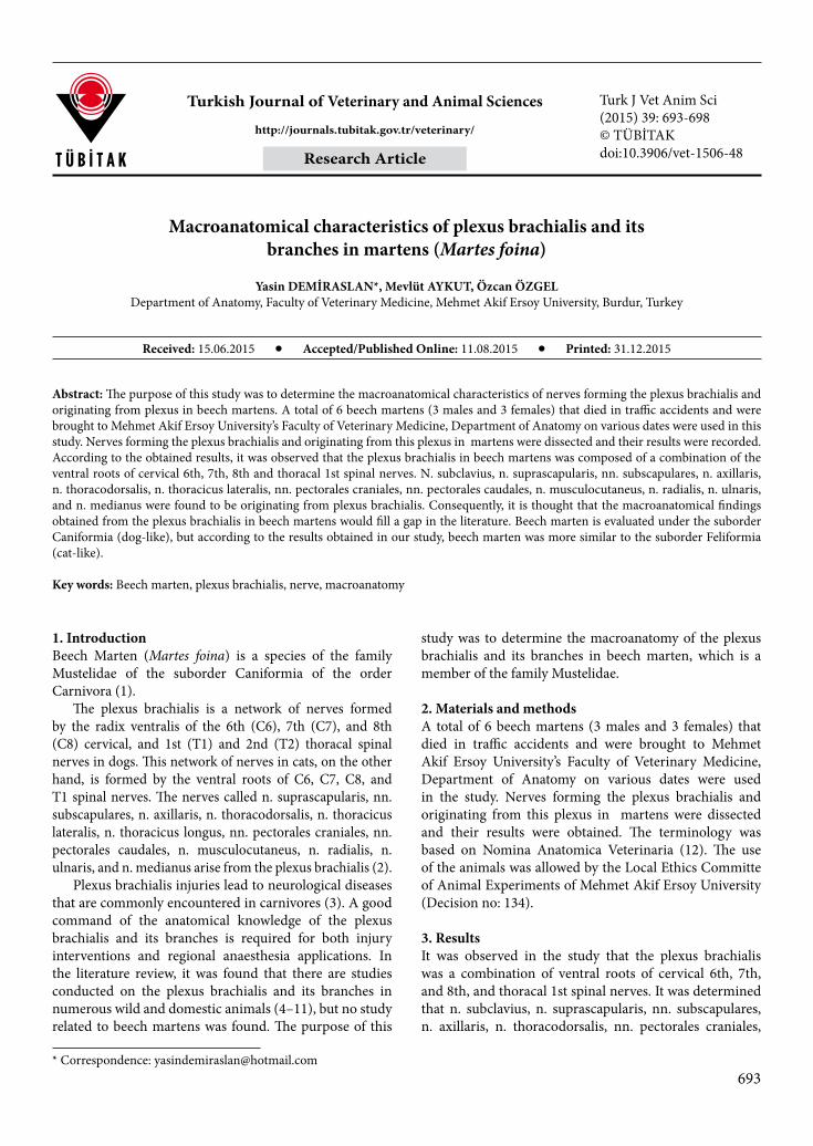
Turkish Journal of Veterinary and Animal Sciences Turk J Vet Anim Sci(2015) 39: 693-698© TÜBİTAKdoi:10.3906/vet-1506-48
Macroanatomical characteristics of plexus brachialis and its branches in martens (Martes foina)
Yasin DEMİRASLAN*, Mevlüt AYKUT, Özcan ÖZGELDepartment of Anatomy, Faculty of Veterinary Medicine, Mehmet Akif Ersoy University, Burdur, Turkey
* Correspondence: [email protected]
1. Introduction Beech Marten (Martes foina) is a species of the family Mustelidae of the suborder Caniformia of the order Carnivora (1).
The plexus brachialis is a network of nerves formed by the radix ventralis of the 6th (C6), 7th (C7), and 8th (C8) cervical, and 1st (T1) and 2nd (T2) thoracal spinal nerves in dogs. This network of nerves in cats, on the other hand, is formed by the ventral roots of C6, C7, C8, and T1 spinal nerves. The nerves called n. suprascapularis, nn. subscapulares, n. axillaris, n. thoracodorsalis, n. thoracicus lateralis, n. thoracicus longus, nn. pectorales craniales, nn. pectorales caudales, n. musculocutaneus, n. radialis, n. ulnaris, and n. medianus arise from the plexus brachialis (2).
Plexus brachialis injuries lead to neurological diseases that are commonly encountered in carnivores (3). A good command of the anatomical knowledge of the plexus brachialis and its branches is required for both injury interventions and regional anaesthesia applications. In the literature review, it was found that there are studies conducted on the plexus brachialis and its branches in numerous wild and domestic animals (4–11), but no study related to beech martens was found. The purpose of this
study was to determine the macroanatomy of the plexus brachialis and its branches in beech marten, which is a member of the family Mustelidae.
2. Materials and methods A total of 6 beech martens (3 males and 3 females) that died in traffic accidents and were brought to Mehmet Akif Ersoy University’s Faculty of Veterinary Medicine, Department of Anatomy on various dates were used in the study. Nerves forming the plexus brachialis and originating from this plexus in martens were dissected and their results were obtained. The terminology was based on Nomina Anatomica Veterinaria (12). The use of the animals was allowed by the Local Ethics Committe of Animal Experiments of Mehmet Akif Ersoy University (Decision no: 134).
3. ResultsIt was observed in the study that the plexus brachialis was a combination of ventral roots of cervical 6th, 7th, and 8th, and thoracal 1st spinal nerves. It was determined that n. subclavius, n. suprascapularis, nn. subscapulares, n. axillaris, n. thoracodorsalis, nn. pectorales craniales,
Abstract: The purpose of this study was to determine the macroanatomical characteristics of nerves forming the plexus brachialis and originating from plexus in beech martens. A total of 6 beech martens (3 males and 3 females) that died in traffic accidents and were brought to Mehmet Akif Ersoy University’s Faculty of Veterinary Medicine, Department of Anatomy on various dates were used in this study. Nerves forming the plexus brachialis and originating from this plexus in martens were dissected and their results were recorded. According to the obtained results, it was observed that the plexus brachialis in beech martens was composed of a combination of the ventral roots of cervical 6th, 7th, 8th and thoracal 1st spinal nerves. N. subclavius, n. suprascapularis, nn. subscapulares, n. axillaris, n. thoracodorsalis, n. thoracicus lateralis, nn. pectorales craniales, nn. pectorales caudales, n. musculocutaneus, n. radialis, n. ulnaris, and n. medianus were found to be originating from plexus brachialis. Consequently, it is thought that the macroanatomical findings obtained from the plexus brachialis in beech martens would fill a gap in the literature. Beech marten is evaluated under the suborder Caniformia (dog-like), but according to the results obtained in our study, beech marten was more similar to the suborder Feliformia (cat-like).
Key words: Beech marten, plexus brachialis, nerve, macroanatomy
Received: 15.06.2015 Accepted/Published Online: 11.08.2015 Printed: 31.12.2015
Research Article
694
DEMİRASLAN et al. / Turk J Vet Anim Sci
nn. pectorales caudales, n. thoracicus lateralis, n. musculocutaneus, n. radialis, n. ulnaris, and n. medianus originated from the plexus brachialis (Figures 1–5).
It was observed that n. subclavius was formed by the ventral root of C6 spinal nerve and reached from the medial to the lateral surface in the cranial middle level of the scapula. It was determined that the nerve divided into two branches, and one of these branches reached m. brachiocephalicus, whereas the other one dispersed into the cutaneous muscle of the shoulder area.
It was observed that n. suprascapularis was formed by the ventral root of C6 spinal nerve and proceeded towards the distal 1/3 level of the scapula right after its origin. While the first branch of the nerve, which divided into two parts in this level, spread into m. subscapularis and m. supraspinatus, the other branch continued its course. Also, this branch divided into two more branches and one of these branches gave two more branches to the rear side of m. supraspinatus. The other branch gave branches to m. infraspinatus and m. deltoideus after reaching the lateral surface of the scapula from collum scapulae and finally ended by giving cutaneous branches.
It was found that nn. subscapulares was formed by the ventral root of C6 and C7 spinal nerves, consisted of two parts, and ended by dispersing in m. subscapularis and m. teres major.
N. axillaris was observed to be formed by the ventral roots of C7 spinal nerve. Rr. musculares were found to primarily arise from this nerve. Rr. musculares were found to be dividing into three branches, two of which reached the rear side of m. subscapularis and the other one reached
m. teres major. The continuing n. axillaris was observed to be passing between m. teres major and m. subscapularis and reaching the lateral part of the shoulder area from the caudal of the collum scapulae. At this level, the nerve was found to be reaching an end, giving one branch to the shoulder joint capsule, and one branch for m. deltoideus and n. cutaneus antebrachii cranialis.
N. thoracodorsalis was formed by the ventral roots of C7 and C8 spinal nerves and dispersed in m. latissimus dorsi as three branches.
N. thoracicus lateralis was formed by the ventral roots of C7 and C8 spinal nerves and ended in the cutaneous muscle in the shoulder area.
N. thoracicus longus was found to be formed by the ventral roots of C7 and C8 spinal nerves and ended branching in m. serratus ventralis.
Nn. pectorales craniales were found to be shaped by the ventral root of C7 spinal nerve and spread into the pectoral muscles as two branches.
Nn. pectorales caudales were found to be formed by the ventral roots of C7 and C8 spinal nerves and ended in m. cutaneus trunci and m. pectoralis ascendens.
N. musculocutaneus was found to be originating from the ventral root of C7 spinal nerve and giving r. muscularis proximalis et distalis in the extremitas proximalis level of the humerus. These branches were observed to be dispersing in m. biceps brachii. The continuing n. musculocutaneus proceeded distally, reached n. cutaneus antebrachii medialis, which innerved craniomedial skin and fascia of the leg, and finally reached an end after connecting to r. superficialis of n. radialis in articulatio carpi level.
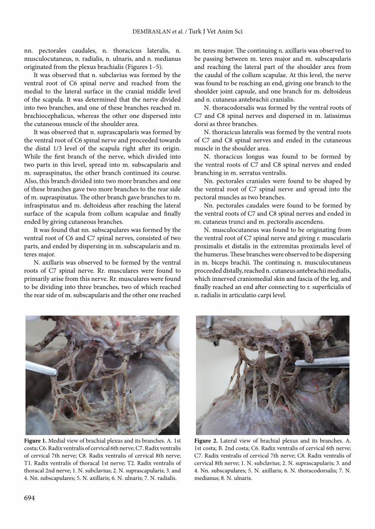
Figure 1. Medial view of brachial plexus and its branches. A. 1st
costa; C6. Radix ventralis of cervical 6th nerve; C7. Radix ventralis of cervical 7th nerve; C8. Radix ventralis of cervical 8th nerve; T1. Radix ventralis of thoracal 1st nerve; T2. Radix ventralis of thoracal 2nd nerve; 1. N. subclavius; 2. N. suprascapularis; 3. and 4. Nn. subscapulares; 5. N. axillaris; 6. N. ulnaris; 7. N. radialis.
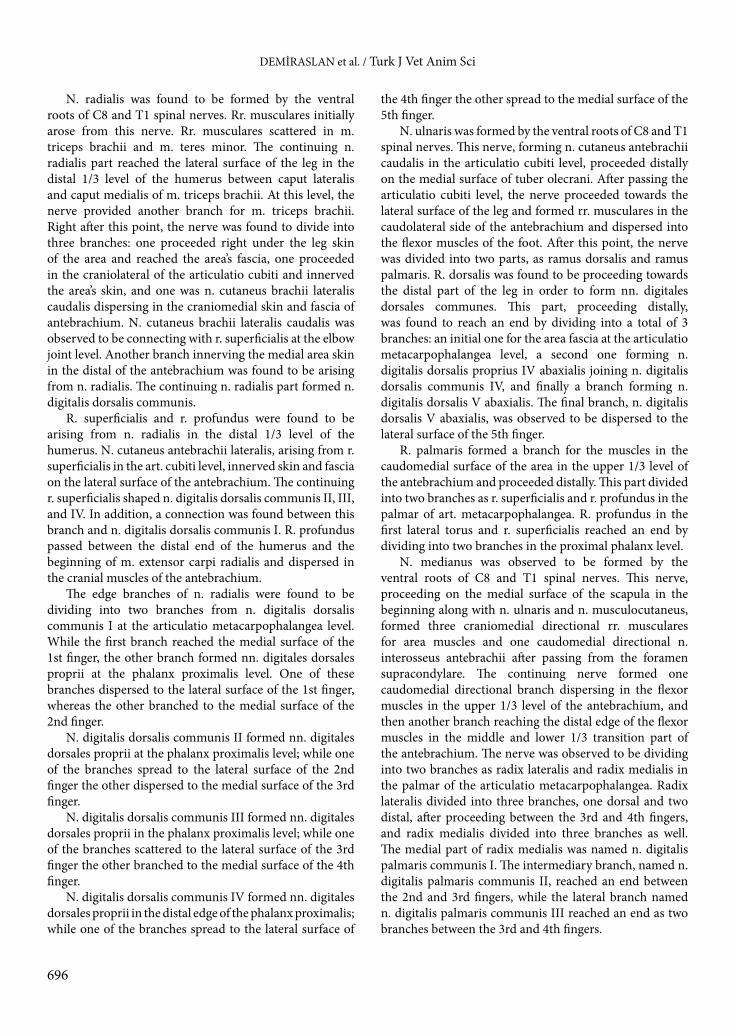
Figure 2. Lateral view of brachial plexus and its branches. A. 1st costa; B. 2nd costa; C6. Radix ventralis of cervical 6th nerve; C7. Radix ventralis of cervical 7th nerve; C8. Radix ventralis of cervical 8th nerve; 1. N. subclavius; 2. N. suprascapularis; 3. and 4. Nn. subscapulares; 5. N. axillaris; 6. N. thoracodorsalis; 7. N. medianus; 8. N. ulnaris.

695
DEMİRASLAN et al. / Turk J Vet Anim Sci
Figure 3. Medial view of some nerves of brachial plexus. A. Humerus; b. supracondylar foramen; 1. N. radialis; 2. and 6. N. medianus; 3. N ulnaris; 4. and 5. Rr. musculares of n. medianus; 7. and 8. Rr. musculares of n. ulnaris.
Figure 4. Dorsal view of some nerves on hand. 1. R. superficialis of n. radialis; 2. and 3. N. digitalis dorsalis communis I; 4. N. digitalis dorsalis communis II; 5. N. digitalis dorsalis communis III; 6. N. digitalis dorsalis communis IV; 7. N. digitalis dorsalis proprius IV abaxialis; 8. R. dorsalis of n. ulnaris; 9. N. digitalis dorsalis V abaxialis.
Figure 5. Palmar view of some nerves under hand. 1. Radix medialis of n. medianus; 2. N. digitalis palmaris communis I; 3. N. digitalis palmaris communis II; 4. N. digitalis palmaris communis III; 5. R. superficialis of n. ulnaris; 6. R. dorsalis of n. ulnaris; 7. N. digitalis dorsalis proprius IV abaxialis; 8. N. digitalis dorsalis V abaxialis.
696
DEMİRASLAN et al. / Turk J Vet Anim Sci
N. radialis was found to be formed by the ventral roots of C8 and T1 spinal nerves. Rr. musculares initially arose from this nerve. Rr. musculares scattered in m. triceps brachii and m. teres minor. The continuing n. radialis part reached the lateral surface of the leg in the distal 1/3 level of the humerus between caput lateralis and caput medialis of m. triceps brachii. At this level, the nerve provided another branch for m. triceps brachii. Right after this point, the nerve was found to divide into three branches: one proceeded right under the leg skin of the area and reached the area’s fascia, one proceeded in the craniolateral of the articulatio cubiti and innerved the area’s skin, and one was n. cutaneus brachii lateralis caudalis dispersing in the craniomedial skin and fascia of antebrachium. N. cutaneus brachii lateralis caudalis was observed to be connecting with r. superficialis at the elbow joint level. Another branch innerving the medial area skin in the distal of the antebrachium was found to be arising from n. radialis. The continuing n. radialis part formed n. digitalis dorsalis communis.
R. superficialis and r. profundus were found to be arising from n. radialis in the distal 1/3 level of the humerus. N. cutaneus antebrachii lateralis, arising from r. superficialis in the art. cubiti level, innerved skin and fascia on the lateral surface of the antebrachium. The continuing r. superficialis shaped n. digitalis dorsalis communis II, III, and IV. In addition, a connection was found between this branch and n. digitalis dorsalis communis I. R. profundus passed between the distal end of the humerus and the beginning of m. extensor carpi radialis and dispersed in the cranial muscles of the antebrachium.
The edge branches of n. radialis were found to be dividing into two branches from n. digitalis dorsalis communis I at the articulatio metacarpophalangea level. While the first branch reached the medial surface of the 1st finger, the other branch formed nn. digitales dorsales proprii at the phalanx proximalis level. One of these branches dispersed to the lateral surface of the 1st finger, whereas the other branched to the medial surface of the 2nd finger.
N. digitalis dorsalis communis II formed nn. digitales dorsales proprii at the phalanx proximalis level; while one of the branches spread to the lateral surface of the 2nd finger the other dispersed to the medial surface of the 3rd finger.
N. digitalis dorsalis communis III formed nn. digitales dorsales proprii in the phalanx proximalis level; while one of the branches scattered to the lateral surface of the 3rd finger the other branched to the medial surface of the 4th finger.
N. digitalis dorsalis communis IV formed nn. digitales dorsales proprii in the distal edge of the phalanx proximalis; while one of the branches spread to the lateral surface of
the 4th finger the other spread to the medial surface of the 5th finger.
N. ulnaris was formed by the ventral roots of C8 and T1 spinal nerves. This nerve, forming n. cutaneus antebrachii caudalis in the articulatio cubiti level, proceeded distally on the medial surface of tuber olecrani. After passing the articulatio cubiti level, the nerve proceeded towards the lateral surface of the leg and formed rr. musculares in the caudolateral side of the antebrachium and dispersed into the flexor muscles of the foot. After this point, the nerve was divided into two parts, as ramus dorsalis and ramus palmaris. R. dorsalis was found to be proceeding towards the distal part of the leg in order to form nn. digitales dorsales communes. This part, proceeding distally, was found to reach an end by dividing into a total of 3 branches: an initial one for the area fascia at the articulatio metacarpophalangea level, a second one forming n. digitalis dorsalis proprius IV abaxialis joining n. digitalis dorsalis communis IV, and finally a branch forming n. digitalis dorsalis V abaxialis. The final branch, n. digitalis dorsalis V abaxialis, was observed to be dispersed to the lateral surface of the 5th finger.
R. palmaris formed a branch for the muscles in the caudomedial surface of the area in the upper 1/3 level of the antebrachium and proceeded distally. This part divided into two branches as r. superficialis and r. profundus in the palmar of art. metacarpophalangea. R. profundus in the first lateral torus and r. superficialis reached an end by dividing into two branches in the proximal phalanx level.
N. medianus was observed to be formed by the ventral roots of C8 and T1 spinal nerves. This nerve, proceeding on the medial surface of the scapula in the beginning along with n. ulnaris and n. musculocutaneus, formed three craniomedial directional rr. musculares for area muscles and one caudomedial directional n. interosseus antebrachii after passing from the foramen supracondylare. The continuing nerve formed one caudomedial directional branch dispersing in the flexor muscles in the upper 1/3 level of the antebrachium, and then another branch reaching the distal edge of the flexor muscles in the middle and lower 1/3 transition part of the antebrachium. The nerve was observed to be dividing into two branches as radix lateralis and radix medialis in the palmar of the articulatio metacarpophalangea. Radix lateralis divided into three branches, one dorsal and two distal, after proceeding between the 3rd and 4th fingers, and radix medialis divided into three branches as well. The medial part of radix medialis was named n. digitalis palmaris communis I. The intermediary branch, named n. digitalis palmaris communis II, reached an end between the 2nd and 3rd fingers, while the lateral branch named n. digitalis palmaris communis III reached an end as two branches between the 3rd and 4th fingers.
697
DEMİRASLAN et al. / Turk J Vet Anim Sci
No difference was observed between sexes in terms of the course of the plexus brachialis and its branches in the study.
4. Discussion The plexus brachialis and its branches in beech martens were examined macroanatomically in this study. Beech marten is taxonomically evaluated under the suborder Caniformia (dog-like) (1). However, according to the results obtained in our study, beech marten was more similar to the suborder Feliformia (cat-like). This similarity was supported by two findings of the study: the first one was that, as in cats, the plexus brachialis was formed by the ventral roots of C6, C7, C8, and T1 spinal nerves; and the second was the presence of foramen supracondylare and the capability of n. medianus to pass through this hole, as in Felidae.
Getty (2) and Tıpırdamaz and Erden (11) reported that the plexus brachialis in dogs was formed by the ventral roots of C6, C7, C8, T1, and T2, and Getty (2) and Aslan (4) reported that in cats it was formed by the ventral roots of C6, C7, C8, and T1 spinal nerves. In our study, the plexus brachialis was observed to be formed by a combination of the ventral roots of C6, C7, C8, and T1 spinal nerves.
In this study, n. subclavius, n. suprascapularis, nn. subscapulares, n. axillaris, n. thoracodorsalis, nn. pectorales craniales, nn. pectorales caudales, n. thoracicus lateralis, n. musculocutaneus, n. radialis, n. ulnaris, and n. medianus were found to be originating from the plexus brachialis. According to the results, all nerves except for n. subclavius were in parallel with the literature (2,4,11). As specified also in Nomina Anatomica Veterinaria (12), n. subclavius was found to be formed by the ventral root of C6 spinal nerve and reached an end in the cutaneous muscle in the shoulder area and m. brachiocephalicus.
While Tıpırdamaz and Erden (11) reported that n. suprascapularis was formed by the ventral roots of C6 and C7 spinal nerves in dogs, Aslan (4) reported that it was formed by the ventral root of C6 spinal nerve in cats. In our study, n. suprascapularis originated from the ventral root of the C6 spinal nerve.
While Getty (2) reported that nn. subscapulares innerved m. subscapularis and m. teres major in dogs, and m. latissimus dorsi in cats. In our study, n. subscapularis in beech martens was found to have a similar innervation area of the same nerve in dogs.
While Getty (2) and Tıpırdamaz and Erden (11) reported that n. axillaris was formed by the ventral roots of C7 and C8 spinal nerves in dogs, Getty (2) and Aslan (4) reported that it was formed by the ventral roots of C6 and C7 spinal nerves in cats. In our study, n. axillaris in beech martens was found to be formed by the ventral root of C7 spinal nerve. Sanchez et al. (3) reported that the final
branches of n. axillaris jointed n. radialis. However, in our study, no such result was found.
While Getty (2) reported that n. musculocutaneus was formed by the ventral roots of C7 spinal nerve in dogs, Getty (2) and Aslan (4) reported that it was formed by the ventral roots of C6 and C7 spinal nerves in cats. In our study, n. musculocutaneus in beech martens was found to be formed by the ventral roots of C7 spinal nerve. It was reported in the literature that in dogs (2,11), pumas, jaguars, and domestic cats (3) n. musculocutaneus is connected to n. medianus, n. cutaneus antebrachii medialis, and r. superficialis of n. radialis. However, in our study, only one connection was found between n. musculocutaneus and r. superficialis of n. radialis.
Tıpırdamaz and Erden (11) emphasized that in dogs nn. pectorales craniales were formed by the ventral roots of C8 and T1; nn. pectorales caudales and n. thoracicus lateralis were formed by the ventral roots of C8, T1, and T2; n. thoracodorsalis was formed by the ventral root of C8; and n. thoracicus longus was formed by the ventral roots of C7 and C8 spinal nerves. Aslan (4) specified that in domestic cats, nn. pectorales craniales originated from the ventral roots of C7 and T1, nn. pectorales caudales and n. thoracicus lateralis originated from the ventral roots of C8 and T1, and n. thoracodorsalis and n. thoracicus longus originated from the ventral roots of C7 and C8 spinal nerves. In our study, it was found that nn. pectorales craniales were formed by the ventral root of C7, and n. pectorales caudales, n. thoracicus lateralis, n. thoracodorsalis, and n. thoracicus longus were formed by the ventral roots of C7 and C8 spinal nerves.
While Getty (2) reported that n. radialis was formed by the ventral roots of C7, C8, and T1 in cats and dogs, Tıpırdamaz and Erden (11) reported that it was formed by the ventral roots of C7 and C8 spinal nerves in dogs. In our study, n. radialis was found to be formed by the ventral roots of C8 and T1 spinal nerves in beech martens. The course of n. radialis was in parallel with the literature. However, a connection was found between n. digitalis dorsalis proprius I abaxialis of n. digitalis dorsalis communis I and n. digitalis dorsalis proprius I axialis of n. digitalis dorsalis communis II. This information was not previously encountered (2,4,11).
While Getty (2) and Tıpırdamaz and Erden (11) reported that n. ulnaris was formed by the ventral roots of C8, T1, and T2 in dogs, Getty (2) reported that it was formed by the ventral roots of C8 and T1 spinal nerves in cats. In our study, the origin of n. ulnaris in beech martens was found to be similar to cats. Sanchez et al. (3) and Aslan (4) reported a connection between the final branches of n. ulnaris and n. medianus in domestic cats; at the same time, Sanchez et al. (3) reported a connection between r. dorsalis of n. ulnaris and r. superficialis of n. radialis in domestic
698
DEMİRASLAN et al. / Turk J Vet Anim Sci
cats, pumas, and jaguars. However, in our study, no such result was found.
While Getty (2) and Tıpırdamaz and Erden (11) reported that n. medianus was formed by the ventral roots of C8, T1, and T2 in dogs, Getty (2) reported that it was formed by the ventral roots of C7, C8, and T1 spinal nerves in cats. In our study, n. medianus was formed by the ventral roots of C8 and T1 spinal nerves. In the course of n. medianus, the differences between the branches of radix lateralis and radix medialis attracted our attention. It has been reported in the literature (2) that radix lateralis, which is one of the edge branches of n. medianus, forms n. digitalis palmaris communis II, III, and IV in cats and
dogs. Radix medialis was reported to form n. digitalis palmaris I abaxialis and n. digitalis palmaris communis I. In our study, it was observed that radix medialis formed n. digitalis palmaris communis I, II, and III.
Consequently, the macroanatomy of the plexus brachialis and its branches in beech martens, which are not very well known in terms of their anatomy, physiology, nutrition, and care, as well as preclinical and clinic terms, was determined. We are of the opinion that the results of this study could be used for both comparisons between species and for the regional and traumatic neurological conditions of beech martens.
References
1. ITIS. Integrated Taxonomic Information System Martes foina, (Erxleben 1777). Washington, DC, USA: ITIS; 2015.
2. Getty R. Sisson and Grossman’s the Anatomy of the Domestic Animals (Volume 2). 5th ed. Philadelphia, PA, USA: Saunders; 1975.
3. Sanchez HL, Silva LB, Rafasquino ME, Mateo AG, Zuccolilli GO, Portiansky EL, Alonso CR. Anatomical study of the forearm and hand nerves of the domestic cat (Felis catus), puma (Puma concolor) and jaguar (Panthera onca). Anat Histol Embryol 2013; 42: 99–104.
4. Aslan K. The comparative macroanatomic investigation on the brachial plexus of the nativecat (Felis domestica) and White New Zealand Rabbit (Oryctolagus cuniculus). J Fac Vet Med Istanbul Univ 1994; 20: 197–208 (in Turkish).
5. Aydın A, Karan M. The spinal nerves forming the brachial plexus in mole-rats (Spalax leucodon).Vet Med-Czech 2012; 57: 430–433.
6. Aydın A. Nerves originating from brachial plexus in the porcupine (Hystrix cristata). Vet Med-Czech 2004; 49: 123–128.
7. Aydın A. Brachial plexus of the porcupine (Hystrix cristata). Vet Med-Czech 2003; 48: 301–304.
8. Çevik Demirkan A, Özdemir V, Demirkan I, Türkmenoğlu I. Gross morphological features of plexus brachialis in the chinchilla (Chinchilla lanigera). J S Afr Vet Assoc 2007; 78: 21–24.
9. Dursun N, Tıpırdamaz S, Gezici M. Macroanatomic investigations on the brachial plexus in Kangal dogs. Selçuk Univ Vet Bil Derg 1994; 10: 78–80 (in Turkish).
10. Souza PR, Cardoso JR, Araujo LBM, Moreiral PC, Cruz VS, Araujo EG. Gross anatomy of the brachial plexus in the giant Anteater (Myrmecophaga tridactyla). Anat Histol Embryol 2014; 43: 341–345.
11. Tıpırdamaz S, Erden H. Macroanatomic investigations on the brachial plexus of the dogs. Selçuk Univ Vet Bil Derg 1988; 4: 317–332 (in Turkish).
12. International Committee on Veterinary Gross Anatomical Nomenclature. Nomina Anatomica Veterinaria (N.A.V.), 5th ed. Ghent, Belgium: World Association of Veterinary Anatomists; 2012.