Embed Size (px)
Citation preview

m-calpain Activation Is Regulated by Its MembraneLocalization and by Its Binding to Phosphatidylinositol4,5-Bisphosphate*□S
Received for publication, March 15, 2010, and in revised form, August 16, 2010 Published, JBC Papers in Press, August 20, 2010, DOI 10.1074/jbc.M110.123604
Ludovic Leloup‡, Hanshuang Shao‡, Yong Ho Bae§, Bridget Deasy¶, Donna Stolz�, Partha Roy‡§, and Alan Wells‡§**1
From the Departments of ‡Pathology, §Bioengineering, ¶Orthopedic Surgery, and �Cell Biology and Physiology, University ofPittsburgh and **Pittsburgh Veterans Affairs Medical Center, Pittsburgh, Pennsylvania 15261
m-calpain plays a critical role in cell migration enabling rearde-adhesion of adherent cells by cleaving structural compo-nents of the adhesion plaques. Growth factors and chemokinesregulate keratinocyte, fibroblast, and endothelial cell migrationby modulating m-calpain activity. Growth factor receptorsactivatem-calpain secondary to phosphorylation on serine 50by ERK. Concurrently, activated m-calpain is localized to itsinner membrane milieu by binding to phosphatidylinositol 4,5-bisphosphate (PIP2). Opposing this, CXCR3 ligands inhibit cellmigration by blocking m-calpain activity secondary to a PKA-mediated phosphorylation in the C2-like domain. The failure ofm-calpain activation in the absence of PIP2 points to a key reg-ulatory role, although whether this PIP2-mediated membranelocalization is regulatory for m-calpain activity or merely servesas a docking site for ERK phosphorylation is uncertain. Herein,we report the effects of two CXCR3 ligands, CXCL11/IP-9/I-TACandCXCL10/IP-10, on the EGF- andVEGF-induced redis-tribution of m-calpain in human fibroblasts and endothelialcells. The two chemokines block the tail retraction and, thus, themigration withinminutes, preventing and reverting growth fac-tor-induced relocalization of m-calpain to the plasma mem-brane of the cells. PKA phosphorylation ofm-calpain blocks thebinding of the protease to PIP2. Unexpectedly, we found thatthis was due to membrane anchorage itself and not merely ser-ine 50 phosphorylation, as the farnesylation-induced anchorageofm-calpain triggers a strong activation of this protease, leadingnotably to an increased cell death. Moreover, the ERK and PKAphosphorylations have no effect on this membrane-anchoredm-calpain.However, the presence of PIP2 is still required for theactivationof the anchoredm-calpain. In conclusion,wedescribea novel mechanism of m-calpain activation by interaction withthe plasma membrane and PIP2 specifically, this phosphoinosi-tide acting as a cofactor for the enzyme. The phosphorylation ofm-calpain by ERK and PKA by growth factors and chemokines,respectively, act in cells to regulate the enzyme only indirectlyby controlling its redistribution.
Calpains are intracellular cysteine proteases involved innumerous physiological and pathological phenomena, such asembryo development and tumor invasion (1). Among the 15members of the calpain family, the two ubiquitous calpains,calpain 1 and calpain 2, are the best described. They form withthe calpain small subunit 1 (calpain S1) two heterodimers,�-calpain for calpain 1 and m-calpain for calpain 2, that arestrongly involved in the regulation of cell motility. Cell migra-tionwas previously described as a four-stage process: cellmem-brane protrusion, adhesion to the substrate, contraction of thecell body, and finally, release of the adhesion contacts at the rearof the cell (2–4). Cell migration is, thus, governed by a succes-sion of adhesion and de-adhesion steps, and a balance betweenthese two processes is required for an optimal cell movement(5, 6). Previous studies have shown that the ubiquitous calpainsregulate these adhesion and de-adhesion steps. Indeed, �-cal-pain is involved in the formation of the adhesion complexes atthe front of themigrating cell, notably by regulating the activityof Rho GTPases (7, 8) and promoting adhesion turnover (9). Atthe rear end of the cell, m-calpain allows cell detachment bycleaving proteins constituent of the adhesion complexes, suchas the focal adhesion kinase, talin, vinculin, and �-actinin (10–12). The role played by m-calpain is crucial as the de-adhesionof the cell rear was shown to be rate-limiting in both hapto- andchemokinetic motility (13, 14).Cell migration is critical for skin wound healing, a complex
process that consists of a succession of highly orchestrated andregulated events (15). The first stages of woundhealing (inflam-matory and regenerative phases) require the in-migration ofkeratinocytes, fibroblasts, and endothelial cells to repopulatethe wound and create new blood vessels (16–18). The growthfactors EGF, VEGF, and PDGF control these first steps by stim-ulating themigration of these cells (19–21). Thismassive influxof cells leads to a hypercellular wound bed containing a pro-visional and immature matrix synthesized by the migrating fi-broblasts. The maturation of the wound occurs during thefinal stage, the resolving phase. Different signals are requiredto stop the proliferation and the migration of the cells and toinduce the contraction of the matrix by the fibroblasts (22).Previous studies have highlighted the crucial role played byCXCR3 ligands in this “stop healing” process (9, 23, 25, 26).Indeed, the ELR-negative CXC chemokines IP-9 (CXCL11 orI-TAC) and IP-10 (CXCL10) inhibit the growth factor-inducedmotility of fibroblasts and endothelial cells and also induce theapoptosis of these supernumerary cells (27). These chemokines
* This work was supported, in whole or in part, by National Institutes of HealthNIGMS grant GM069668.
□S The on-line version of this article (available at http://www.jbc.org) containssupplemental Figs. S1–S8 and Movie 1.
1 To whom correspondence should be addressed: University of Pittsburgh,Scaife Hall S713, 3550 Terrace St., Pittsburgh, PA 15261. Tel.: 412-624-0973;Fax: 412-624-8946; E-mail: [email protected].
THE JOURNAL OF BIOLOGICAL CHEMISTRY VOL. 285, NO. 43, pp. 33549 –33566, October 22, 2010Printed in the U.S.A.
OCTOBER 22, 2010 • VOLUME 285 • NUMBER 43 JOURNAL OF BIOLOGICAL CHEMISTRY 33549
by guest on March 14, 2019
http://ww
w.jbc.org/
Dow
nloaded from
thereby induce the contraction of the matrix (28) and restorethe physiologic paucicellularity, leading to the formation of astrong and organized dermis.As ubiquitous calpains are centrally involved in cell mi-
gration, their regulation is crucial during wound healing.The regulation of �-calpain is well known, and the calciuminfluxes are mainly responsible for the activation of thisenzyme (9). On the opposite, the regulation of m-calpain re-mains unclear. The calcium concentrations required for itsactivation in vitro are supraphysiological and not consistentwith cell survival (estimated between 400 and 800 �M (1)).Recent studies have highlighted the roles played by phosphory-lations inm-calpain regulation particularly during wound heal-ing process. The growth factors secreted during the first stepsof wound closure promote cell migration by activating m-cal-pain. EGF and other classical growth factors stimulate m-cal-pain activity by inducing the enzyme phosphorylation on theserine 50 residue via the ERK/MAPK pathway (29, 30). Thequestion remained as to why m-calpain leads to detachmentprimarily at the rear. It was recently determined that asymmet-ric phosphoinositide distribution directs this polarity (31). EGFinduces the activation of PLC�1 at the front of the cells, leadingto the degradation of phosphatidylinositol 4,5-bisphosphate(PIP2)2and, thus, to the formation of a gradient toward the rear(32). The phosphorylation of m-calpain by ERK allows theinteraction of the domain III of this enzyme with PIP2. m-cal-pain is, thus, relocalized to the rear of the cells, more particu-larly at the membrane, close to the adhesion complexes. Inter-estingly, in the absence of PIP2, growth factors cannot activatem-calpain in living cells, although whether this is due to failureto be ERK phosphorylated is still uncertain.The CXCR3 ligands IP-9 and IP-10 secreted during the re-
solving phase of wound healing block the growth factor-in-duced migration by inhibiting m-calpain activity (23–25). Thisinhibition is due to the phosphorylation of the protease by PKAon the serine 369 (33). By inhibiting the growth factor-inducedm-calpain activity, IP-9 and IP-10 prevent cell retraction, lead-ing to the contraction and the maturation of the wound matrix(34). Although the serine 369 phosphorylation is modeled to“freeze” m-calpain into a conformation that prevents the activecleft from cleaving proteins, the dominance of this inhibitoryphosphorylation and its effects on m-calpain localization areunknown. Moreover, the importance of the localization of theenzyme at the membrane and the role played by PIP2 (simpledocking site or a cofactor) in the m-calpain activation processare still not completely understood.In this study we asked whether the effects of the CXCR3
ligands on migration were immediate and dominant over EGFand VEGF and whether these chemokines have any effect onthe redistribution of m-calpain induced by these growth fac-tors. We observed that the inhibition of the growth factor-induced migration by IP-9 and IP-10 is immediate and dom-inant over EGF and VEGF effects. Moreover, the redis-
tribution of m-calpain at the rear and at the cell membraneinduced by these growth factors is prevented and even revertedby these two chemokines. These effects are not due to a changein PIP2 localization but to the PKA-mediated inhibition of theinteraction betweenm-calpain and this phosphoinositide. Thisled us to query the role of the membrane localization of m-cal-pain in its activation process; we bypassed the PIP2-dependentmembrane localization by directly anchoring the enzyme at theplasma membrane of the cells. Farnesylation-induced anchor-age ofm-calpain at the plasmamembrane induced a very strongactivation of the enzyme. This anchored m-calpain does notrequire the ERKphosphorylation to be activated and is resistantto PKA phosphorylation. Interestingly, this anchored m-cal-pain still requires the presence of PIP2 to be activated. Takentogether, our results clearly show that m-calpain activity ismainly regulated by its relocalization at the plasma membraneand by its binding to PIP2, whereas the ERK and PKAphosphor-ylations appear to only indirectly regulate the enzyme activityin living cells by controlling its redistribution. This represents aquantal and unexpected advance of our understanding ofmolecular control of m-calpain activation.
EXPERIMENTAL PROCEDURES
Materials and Reagents—Dulbecco’s modified Eagle’s medium(DMEM), �-minimum essential medium, and fetal bovineserum (FBS) were purchased from Cellgro. The MCDB131medium was purchased from Invitrogen. The growth factorsEGF and VEGF were purchased from BD Biosciences andSigma, respectively, and the chemokines IP-9 and IP-10 werefrom PeproTech Inc. The primary antibodies used for theimmunoblots were purchased from Santa Cruz Biotechnologyfor m-calpain and GFP, from Sigma for actin, GAPDH, andvinculin, and from Cell Signaling Technology for PLC�1 (totaland phosphorylated on Tyr-783). The HRP-conjugated sec-ondary antibodies were purchased from BIOSOURCE. Con-cerning the immunofluorescent staining, the primary antibod-ies were purchased from Calbiochem (m-calpain) and Echelon(phosphatidylinositol 4,5-bisphosphate), and the secondaryantibodies were from Invitrogen. The anti-GFP antibody usedfor immunoprecipitation was also purchased from Invitrogen.TRITC-phalloidin and DAPI were purchased from Sigma. Thepurified PKA and PKI (PKA inhibitor) were purchased fromPromega, and purified m-calpain was from Calbiochem. Thephosphoinositides and the active human ERK1were purchasedfrom Sigma. Finally, the MicroSpin columns S-200 HR werepurchased from GE Healthcare.Cell Culture—Human fibroblasts Hs68, human microvascu-
lar endothelial cells, andmurine fibroblasts NR6WTwere used.TheHs68 fibroblastswere grown inDMEMsupplementedwith10% FBS. The murine fibroblasts were grown in �-minimumessentialmediumplus 7.5% of FBS, and human endothelial cellswere grown in MCDB131 supplemented with 10% of FBS. Forthe different experiments, cells were incubated overnight inquiescentmedia (0.5%dialyzed FBS (dFBS) forHs68, 0.1%dFBSfor human microvascular endothelial and NR6 WT cells). Ascell migration and EGF responsiveness decreases with cell age(35), all cells were used as early passage (�10).
2 The abbreviations used are: PIP2, phosphatidylinositol 4,5-bisphosphate;PKI, cAMP-dependent protein kinase inhibitor; PLC�1, phospholipase Cgamma 1; VEGF, vascular endothelial growth factor; TRITC, tetramethyl-rhodamine isothiocyanate.
m-calpain Is Regulated by Its Plasma Membrane Localization
33550 JOURNAL OF BIOLOGICAL CHEMISTRY VOLUME 285 • NUMBER 43 • OCTOBER 22, 2010
by guest on March 14, 2019
http://ww
w.jbc.org/
Dow
nloaded from
Cell Treatments—EGF was used at 1 nM for human fibro-blasts and at 10 nM for human endothelial cells. VEGF used totreat the endothelial cells was added at 200 ng/ml. The CXCR3ligands IP-9 and IP-10 were used at 15 and 200 ng/ml for fibro-blasts and endothelial cells, respectively. Insulin, used to acti-vate farnesyltransferase, was used at 100 nM for 20 or 120 min(36).In Vitro Wound Healing Assays—Confluent cells were incu-
bated overnight in quiescentmedium. Themonolayer was thenscraped with a pipette tip to create an acellular area. Pictures ofthe created wounds were taken. The cells were then treatedwith EGF or VEGF alone or in combination with IP-9 or IP-10.For combination treatments, the chemokines were added 4 hbefore or after the growth factors. After an overnight incuba-tion (18 h; before proliferation), pictures of the wound weretaken. Thewound closurewas estimatedwithMetaMorph soft-ware (Molecular Devices) by measuring the area of the woundsbefore and after incubation.Continuous Cell Tracking Experiments—For cell tracking
experiments, human fibroblasts and endothelial cells, incu-bated in quiescent media overnight, were visualized every 4min. EGF was first added to the media. IP-9 or IP-10 was added40min later for fibroblasts and 75min later for endothelial cells.The velocity of the cells was determined with MetaMorphsoftware.Cell Footprint Isolation—The apical (dorsal) aspect of cells
was removed by a modification of a method described pre-viously (37). Quiescent cells plated on collagen-coated cov-erslips were treated overnight with EGF, IP-9, or IP-10 aloneor in combination. For combination treatments, IP-9 andIP-10 were added 4 h before or after EGF. After the overnightincubation, the cells were washed with PBS and with MES-buffered saline (MBS; 20 mM MES ((pH 5.5), 135 mM NaCl, 0.5mM CaCl2, 1 mM MgCl2). The cells were then coated with a 1%solution of cationic colloidal silica (silica prepared as a 30%stock colloid as described previously (38)). After washing withMBS, the cells were coated with 1% polyacrylic acid in MBS(stock 25% aqueous polyacrylic acid solution, 100,000 averagemolecular weight; Sigma). The cells were washed with MBSand then swelled in hypotonic lysis buffer (2.5 mM imidazole(pH 7.0) supplemented with protease inhibitors (1:100 dilu-tion, protease mixture set V, Calbiochem)) for 30 min. Cellswere then unroofed by squirting themonolayerwith lysis bufferthrough a 5-ml syringe fitted with a blunted, flattened 18-gaugeneedle. The degree of unroofing was monitored by observingcells with an inverted phase-contrast microscope. The isolatedfootprints were then used to visualize m-calpain by immuno-staining or to quantify m-calpain by immunoblot. For immu-nostaining, the footprints were washed once using MBS andfixed using a 2% paraformaldehyde solution prepared in PBS.For immunoblot, the footprints were washed using PBS, andthe proteinswere extractedwith 1� SDS sample loading buffer.Immunofluorescent Staining—The cells and the footprints
were washed using PBS and fixed with a 2% paraformaldehydesolution (in PBS). The cells were permeabilized with a 0.1%Triton-X solution for 5 min. The cells or the footprints werethen blocked with non-immune goat serum (5% in PBS). Afterwashing, the cells or the footprints were incubated for 2 h with
the primary antibodies (against calpain 2 subunit, phospha-tidylinositol 4,5-bisphosphate, vinculin, or calpain S1) dilutedat 1:100. Then they were washed and incubated with the sec-ondary antibodies (Alexa Fluor 488 or 594, 1:500 dilution, 1 h).Actin filaments were stained by incubating the cells or the foot-prints for 40 min with TRITC-phalloidin. DAPI was used tostain the cell nuclei. The coverslips were mounted using Gelva-tol and observed using a fluorescent microscope. For the quan-tification of the fluorescence of the rear and of the front parts ofthe cells, the pictureswere analyzed usingMetaMorph software(20 cells were analyzed for each condition).Generation of a Phosphomimetic Variant of m-calpain
(S369E)—To mimic PKA phosphorylation of m-calpain, theserine 369 was replaced by a glutamic acid. The plasmid encod-ing the GFP-WT-m-calpain (described previously (33)) wasmutated using a PCR-based mutagenesis kit (Stratagene) andprimers that encoded the mutation (5�-GAA CTG GAG GCGGGGCGAGACCGCGGGAGGTTGC-3� and 5�-GCAACCTCC CGC GGT CTC GCC CCG CCT CCA GTT C-3�). Thegenerated mutant referred to as GFP-S369E-m-calpain wasconfirmed by DNA sequencing.Construction of the CAAX- and SAAX-calpain 2 Plasmids—
The K-Ras farnesylation sequence CAAX (and the negativecontrol SAAX) was added to the gene encoding the humancalpain 2 subunit by three successive PCR. The plasmid encod-ing the human WT-calpain 2 was used as a template. The cal-pain 2 gene containing the CAAX and SAAX sequences wasinserted in the plasmid pEGFP-C1 (obtained from the Prof.Chang Lu) usingHindIII restriction enzyme (NewEnglandBio-labs). The mutation of the serine 50 to alanine was performedusing a PCR-based mutagenesis kit (Stratagene) and primersthat encoded the mutation (5�-TCC AGG ACC CGG CCTTCCCGGCCAT-3� and 5�-ATGGCCGGGAAGGCCGGGTCC TGGA-3�). The generated mutants referred to as CAAX-calpain 2 S50A and SAAX-calpain 2 S50A were confirmed byDNA sequencing.Transfection of the NR6WT Fibroblasts—NR6WT fibro-
blasts seeded in antibiotic-free medium were transfectedwith the different plasmids using Lipofectamine 2000 (In-vitrogen). The cells transfected with the plasmids encodingthe WT-, the ST369AA-, and the S369E-m-calpain wereincubated overnight in the presence of 15�MMDL28170 (Cal-biochem) to reduce cell death due to calpain overexpression.For the expression of the CAAX- and SAAX-calpain 2, the cellswere transfected at the same time with the plasmids and a mixof two siRNAs directed against the murine calpain 2 (IDTDNA). The cells transfected with the plasmid encoding theCAAX-calpain 2 were incubated overnight in the presence of5 �M calpain inhibitor MDL28170 (Calbiochem, EMD Bio-sciences) to reduce the cell death due to the very strong activityof this mutated m-calpain. After the overnight incubation, thecells expressing CAAX- and SAAX-calpain 2 were washed withPBS and treated for 20 or 120 min with insulin.Immunoprecipitation of the GFP m-calpains—The trans-
fected cells were washed and treated for 30 min with EGF withandwithout IP-10 to induce the phosphorylations ofm-calpain.IP-10 was added at the same time as EGF. After the incubation,the proteins were extracted using radioimmune precipitation
m-calpain Is Regulated by Its Plasma Membrane Localization
OCTOBER 22, 2010 • VOLUME 285 • NUMBER 43 JOURNAL OF BIOLOGICAL CHEMISTRY 33551
by guest on March 14, 2019
http://ww
w.jbc.org/
Dow
nloaded from
assay buffer plus protease inhibitors, centrifuged, and incu-bated overnightwith an anti-GFP antibody (0.5�g, Invitrogen).The proteins were then incubated with protein A-Sepharosebeads. The beads were washedwith radioimmune precipitationassay buffer, and the GFP-m-calpains were eluted using GentleAg/Ab elution buffer (Pierce). The purified GFP-m-calpainswere then used for PIP2 binding assay (using liposomes).In Vitro Phosphorylation of m-calpain by ERK1 and PKA—
Purified m-calpain (from porcine kidney; Calbiochem) wasphosphorylated in vitro using PKA (Promega) and/or activeERK1 (Sigma). For ERK1 phosphorylation, 2.5 �g of m-calpainwere incubated with 0.1 �g of active ERK1 in the followingbuffer: 20 mM MOPS, pH 7.2, 25 mM �-glycerolphosphate, 1mM sodium orthovanadate, 1 mM DTT, 100 �M ATP, and 15mM MgCl2. After 30 min of incubation at 30 °C, m-calpain wasused for a PIP2 binding assay or for in vitro phosphorylation byPKA. For PKA phosphorylation, m-calpain (2.5 �g) was incu-batedwith 25 units of PKA for 2 h in the following buffer: 50mM
Tris-HCl (pH 7.5), 7 mM MgCl2, 1 mM DTT, 0.06% CHAPS, 50�MATP. Sampleswere also treatedwith PKAandPKI (2mM) asnegative control. After a 2-h incubation at 30 °C,m-calpain wasused for PIP2 binding assay (using liposomes).m-calpain Binding to Phospholipid-containing Liposomes—
Preparation of the liposomes and assessment of m-calpain-phospholipid binding were performed as previously reported(31, 39). Briefly, liposomes were prepared from a crude bovinebrain extract of phosphoinositides that contains 20–40% di-and triphosphoinositide (aminimumof 5 to 10%of each). Bind-ing ofm-calpain (in vitro phosphorylatedm-calpain or GFP-m-calpain purified by immunoprecipitation) to lipid vesicles wasassayed by spin column size exclusion chromatography onMicroSpin S200 HR columns (GE Healthcare). The amount ofprotein in elute was detected by SDS-gel electrophoresis fol-lowed by immunoblotting. The quantification of the band den-sity was carried out with ImageJ software.In Vitro Calpain Activity Assay—The GFP m-calpains, im-
munoprecipitated and eluted in non-denaturing conditions,were desalted using the Zeba Desalt Spin Columns purchasedfrom Pierce Thermo Scientific. The activity of the desaltedsamples was assessed using the calpain activity assay kit fromBioVision. The fluorescence was quantified using a SpectraFluor plate reader (Tecan).Cell Survival Assay—The NR6WT fibroblasts were trans-
fected with the empty plasmid (GFP) or with the plasmidsencoding the CAAX/SAAX-calpain 2. For the transfection withCAAX-calpain 2 or CAAX-calpain 2 S50A, the cells were incu-bated overnightwith 5�MMDL28170 to protect them from thestrong activity of these m-calpain mutants. After the overnightincubation, the transfected cellswerewashed several timeswithPBS to remove the calpain inhibitor used for the CAAX-calpain2. They were then treated with 100 nM insulin for 20 min. Thecells were washed to remove the insulin and incubated in reg-ular medium. Pictures of three different fields were taken at thebeginning of the experiment and after 3 and 6 h. The GFP cellswere then counted for each time point, and cell survival wasexpressed as a percentage of the cells counted at the beginningof the experiment.
PIP2 Depletion—To deplete PIP2, the NR6WT cells, trans-fected with the empty plasmid or with the plasmids encodingthe CAAX- or SAAX-calpain 2, were treated withmedium con-taining butanol-1 (1.5%) for 45 min (31). Butanol-2 was used asa negative control. Insulin (100 nM) was added 20 min beforethe end of the incubation to induce the activation of the farne-syltransferase. After the treatments, calpain activity was as-sessed by observing vinculin degradation or by using t-Boc-LM-CMAC (Calbiochem). The cells remained viable during theshort duration of this treatment (31).t-Butoxycarbonyl (Boc) Assay—To quantify calpain activity,
cells were incubated for 20min in the presence of 25mM t-Boc-LM-CMAC. After incubation, the cells were washed threetimes with PBS, and fluorescence was quantified using a platereader (SpectraFluor, Tecan).Statistical Analysis—All comparisons were performed
with Student’s t test with minimum significance deemed tobe p � 0.05.
RESULTS
CXCR3 Ligands Override Growth Factor-induced Migrationof Fibroblasts and Endothelial Cells—Previous studies haveshown that the CXCR3 ligands IP-9/CXCL11 and IP-10/CXCL10 inhibit the migration of fibroblasts and endothelialcells induced by EGF andVEGF, respectively (23, 40). However,little is known concerning the efficiency, rapidity, and domi-nance of the migration inhibition; for instance, could theseligands stop ongoing locomotion. To have a better understand-ing of this phenomenon, the migration of human fibroblasts(Hs68) and human dermal microvascular endothelial cellstreated with EGF or VEGF was quantified by wound healingassays in the presence of IP-9 or IP-10. These two chemokineswere added to the media 4 h before (pretreatment) or after thegrowth factors. The results showed that the stimulation offibroblast and endothelial cell migration observed after EGFtreatment was almost totally inhibited when the cells were pre-treated with IP-9 and IP-10 (Fig. 1, A and B). A similar decre-ment in in vitro “wound” closure was also observed when thetwo CXCR3 ligands were added 4 h after EGF, although notto the full extent as when added before EGF. This non-sig-nificant difference in the extent of inhibition was deemed aspossibly due to 4 h of treatment with EGF alone. In bothsituations no differences were observed between IP-9 andIP-10, as would expected as they act via the commonCXCR3.Similar data were obtained with endothelial cells treatedwith VEGF and IP-10 (Fig. 1C). These results show that theeffects of IP-9 and IP-10 are likely dominant over EGF andVEGF stimulation of migration and disrupt ongoing eventsrather than preventing subsequent activation by growth fac-tors. To probe this postulate, live cells were tracked. Thevelocities of untreated fibroblasts and endothelial cells werequantified before the addition of EGF and subsequentlyIP-10. The results demonstrate that the fibroblast migrationis stimulated within minutes by EGF. In the same manner,IP-10 reduces the motility back toward pre-EGF speedswithin minutes (Fig. 1D). The velocity of the cells, increasedfrom 0.54 to 0.99 �m/min by EGF, was decreased to 0.6�m/min by IP-10 (Fig. 1E). Similar results were observed
m-calpain Is Regulated by Its Plasma Membrane Localization
33552 JOURNAL OF BIOLOGICAL CHEMISTRY VOLUME 285 • NUMBER 43 • OCTOBER 22, 2010
by guest on March 14, 2019
http://ww
w.jbc.org/
Dow
nloaded from

using endothelial cells (Fig. 1, F and G). Observing the migra-tion of these cells shows thatCXCR3-mediated inhibition of themigration is due to the inability to retract the cell tail (Fig. 1Hand supplemental Movie 1). The extension of the lamellipodia
seems unaffected by CXCR3 ligands. Taken together, theseresults show that the effects of IP-9 and IP-10 on cell migrationand retraction are immediate and dominant over EGF andVEGF in both human fibroblasts and endothelial cells.
FIGURE 1. Inhibition of growth factor-mediated migration by IP-9 and IP-10. Wound healing assays were carried out to study the effects of CXCR3 ligandson the growth factor-mediated migration of human fibroblasts (Hs68, A) and endothelial cells (B and C). The cells were grown until 80 –100% confluence. After24 h of quiescence, the cells were scraped using a pipette tip to create a wound and treated with EGF (A and B) or VEGF (C). The chemokines IP-9 and IP-10 wereadded 4 h before or after the growth factors. The wound closure was quantified using Metamorph software by comparing the wound areas before and after theovernight treatments. The results were expressed as percentage (100% for untreated cells). Experiments were repeated three times, and the error scale barsshow S.D. * and **, significantly different from untreated cells and cells treated with growth factors, respectively (p � 0.05). The effects of IP-10 on EGF-mediatedmigration were also studied by cell-tracking assays with fibroblasts (D and E) and endothelial cells (F, G, and H). Pictures of the untreated cells were taken every4 min for 4 h. EGF was added to the quiescent medium after 75 min. IP-10 was then added to the medium 75 min after EGF for endothelial cells and 40 min afterEGF for fibroblasts. The velocities of at least 50 migrating cells were determined for each time point using Metamorph software (D and F). The average velocitiesof the cells were also calculated for each treatment (E and G). The error scale bars show S.E. Pictures illustrating the effects of EGF and IP-10 on an endothelial cellare shown in H. The white arrow shows the rear of the cell, impossible to retract after IP-10 treatment.
m-calpain Is Regulated by Its Plasma Membrane Localization
OCTOBER 22, 2010 • VOLUME 285 • NUMBER 43 JOURNAL OF BIOLOGICAL CHEMISTRY 33553
by guest on March 14, 2019
http://ww
w.jbc.org/
Dow
nloaded from
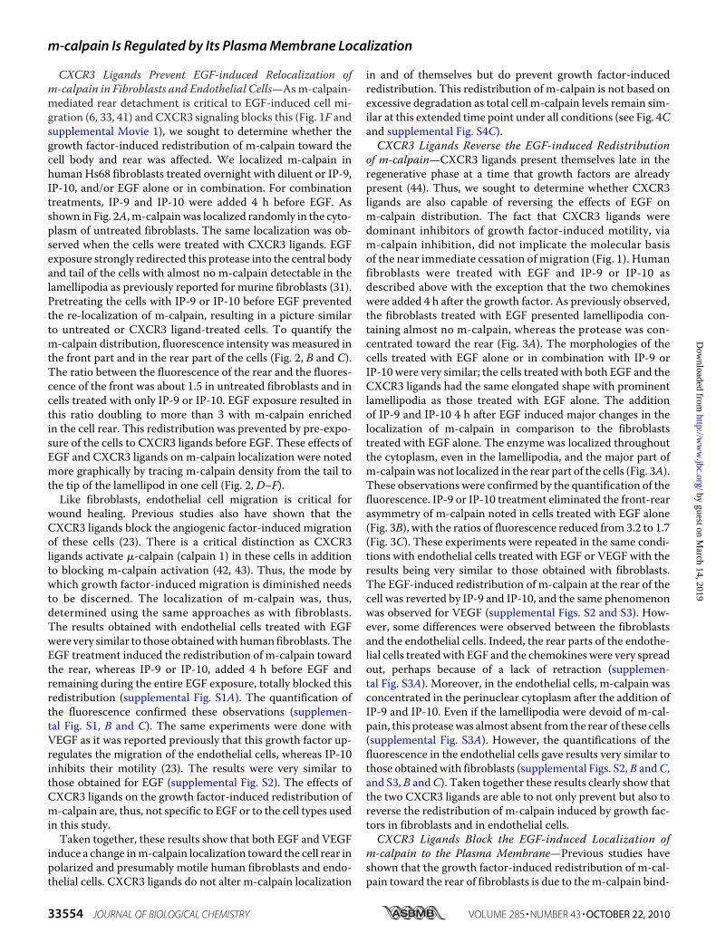
CXCR3 Ligands Prevent EGF-induced Relocalization ofm-calpain in Fibroblasts and Endothelial Cells—Asm-calpain-mediated rear detachment is critical to EGF-induced cell mi-gration (6, 33, 41) and CXCR3 signaling blocks this (Fig. 1F andsupplemental Movie 1), we sought to determine whether thegrowth factor-induced redistribution of m-calpain toward thecell body and rear was affected. We localized m-calpain inhuman Hs68 fibroblasts treated overnight with diluent or IP-9,IP-10, and/or EGF alone or in combination. For combinationtreatments, IP-9 and IP-10 were added 4 h before EGF. Asshown in Fig. 2A, m-calpainwas localized randomly in the cyto-plasm of untreated fibroblasts. The same localization was ob-served when the cells were treated with CXCR3 ligands. EGFexposure strongly redirected this protease into the central bodyand tail of the cells with almost no m-calpain detectable in thelamellipodia as previously reported for murine fibroblasts (31).Pretreating the cells with IP-9 or IP-10 before EGF preventedthe re-localization of m-calpain, resulting in a picture similarto untreated or CXCR3 ligand-treated cells. To quantify them-calpain distribution, fluorescence intensity was measured inthe front part and in the rear part of the cells (Fig. 2, B and C).The ratio between the fluorescence of the rear and the fluores-cence of the front was about 1.5 in untreated fibroblasts and incells treated with only IP-9 or IP-10. EGF exposure resulted inthis ratio doubling to more than 3 with m-calpain enrichedin the cell rear. This redistribution was prevented by pre-expo-sure of the cells to CXCR3 ligands before EGF. These effects ofEGF and CXCR3 ligands on m-calpain localization were notedmore graphically by tracing m-calpain density from the tail tothe tip of the lamellipod in one cell (Fig. 2, D–F).
Like fibroblasts, endothelial cell migration is critical forwound healing. Previous studies also have shown that theCXCR3 ligands block the angiogenic factor-induced migrationof these cells (23). There is a critical distinction as CXCR3ligands activate �-calpain (calpain 1) in these cells in additionto blocking m-calpain activation (42, 43). Thus, the mode bywhich growth factor-induced migration is diminished needsto be discerned. The localization of m-calpain was, thus,determined using the same approaches as with fibroblasts.The results obtained with endothelial cells treated with EGFwere very similar to those obtainedwith human fibroblasts. TheEGF treatment induced the redistribution of m-calpain towardthe rear, whereas IP-9 or IP-10, added 4 h before EGF andremaining during the entire EGF exposure, totally blocked thisredistribution (supplemental Fig. S1A). The quantification ofthe fluorescence confirmed these observations (supplemen-tal Fig. S1, B and C). The same experiments were done withVEGF as it was reported previously that this growth factor up-regulates the migration of the endothelial cells, whereas IP-10inhibits their motility (23). The results were very similar tothose obtained for EGF (supplemental Fig. S2). The effects ofCXCR3 ligands on the growth factor-induced redistribution ofm-calpain are, thus, not specific to EGF or to the cell types usedin this study.Taken together, these results show that both EGF and VEGF
induce a change inm-calpain localization toward the cell rear inpolarized and presumably motile human fibroblasts and endo-thelial cells. CXCR3 ligands do not alter m-calpain localization
in and of themselves but do prevent growth factor-inducedredistribution. This redistribution of m-calpain is not based onexcessive degradation as total cell m-calpain levels remain sim-ilar at this extended time point under all conditions (see Fig. 4Cand supplemental Fig. S4C).CXCR3 Ligands Reverse the EGF-induced Redistribution
of m-calpain—CXCR3 ligands present themselves late in theregenerative phase at a time that growth factors are alreadypresent (44). Thus, we sought to determine whether CXCR3ligands are also capable of reversing the effects of EGF onm-calpain distribution. The fact that CXCR3 ligands weredominant inhibitors of growth factor-induced motility, viam-calpain inhibition, did not implicate the molecular basisof the near immediate cessation of migration (Fig. 1). Humanfibroblasts were treated with EGF and IP-9 or IP-10 asdescribed above with the exception that the two chemokineswere added 4 h after the growth factor. As previously observed,the fibroblasts treated with EGF presented lamellipodia con-taining almost no m-calpain, whereas the protease was con-centrated toward the rear (Fig. 3A). The morphologies of thecells treated with EGF alone or in combination with IP-9 orIP-10 were very similar; the cells treated with both EGF and theCXCR3 ligands had the same elongated shape with prominentlamellipodia as those treated with EGF alone. The additionof IP-9 and IP-10 4 h after EGF induced major changes in thelocalization of m-calpain in comparison to the fibroblaststreated with EGF alone. The enzyme was localized throughoutthe cytoplasm, even in the lamellipodia, and the major part ofm-calpainwas not localized in the rear part of the cells (Fig. 3A).These observations were confirmed by the quantification of thefluorescence. IP-9 or IP-10 treatment eliminated the front-rearasymmetry of m-calpain noted in cells treated with EGF alone(Fig. 3B), with the ratios of fluorescence reduced from 3.2 to 1.7(Fig. 3C). These experiments were repeated in the same condi-tions with endothelial cells treated with EGF or VEGF with theresults being very similar to those obtained with fibroblasts.The EGF-induced redistribution of m-calpain at the rear of thecell was reverted by IP-9 and IP-10, and the same phenomenonwas observed for VEGF (supplemental Figs. S2 and S3). How-ever, some differences were observed between the fibroblastsand the endothelial cells. Indeed, the rear parts of the endothe-lial cells treatedwith EGF and the chemokines were very spreadout, perhaps because of a lack of retraction (supplemen-tal Fig. S3A). Moreover, in the endothelial cells, m-calpain wasconcentrated in the perinuclear cytoplasm after the addition ofIP-9 and IP-10. Even if the lamellipodia were devoid of m-cal-pain, this proteasewas almost absent from the rear of these cells(supplemental Fig. S3A). However, the quantifications of thefluorescence in the endothelial cells gave results very similar tothose obtainedwith fibroblasts (supplemental Figs. S2,B andC,and S3, B andC). Taken together these results clearly show thatthe two CXCR3 ligands are able to not only prevent but also toreverse the redistribution of m-calpain induced by growth fac-tors in fibroblasts and in endothelial cells.CXCR3 Ligands Block the EGF-induced Localization of
m-calpain to the Plasma Membrane—Previous studies haveshown that the growth factor-induced redistribution of m-cal-pain toward the rear of fibroblasts is due to them-calpain bind-
m-calpain Is Regulated by Its Plasma Membrane Localization
33554 JOURNAL OF BIOLOGICAL CHEMISTRY VOLUME 285 • NUMBER 43 • OCTOBER 22, 2010
by guest on March 14, 2019
http://ww
w.jbc.org/
Dow
nloaded from
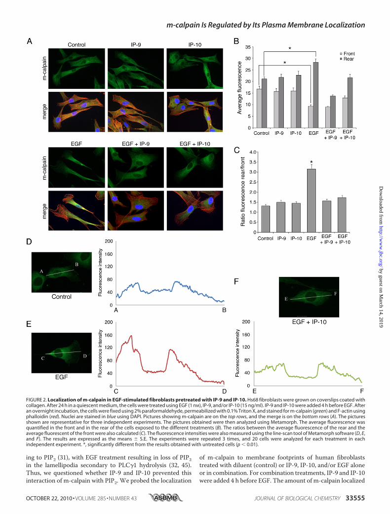
ing to PIP2 (31), with EGF treatment resulting in loss of PIP2in the lamellipodia secondary to PLC�1 hydrolysis (32, 45).Thus, we questioned whether IP-9 and IP-10 prevented thisinteraction of m-calpain with PIP2. We probed the localization
of m-calpain to membrane footprints of human fibroblaststreated with diluent (control) or IP-9, IP-10, and/or EGF aloneor in combination. For combination treatments, IP-9 and IP-10were added 4 h before EGF. The amount of m-calpain localized
FIGURE 2. Localization of m-calpain in EGF-stimulated fibroblasts pretreated with IP-9 and IP-10. Hs68 fibroblasts were grown on coverslips coated withcollagen. After 24 h in a quiescent medium, the cells were treated using EGF (1 nM), IP-9, and/or IP-10 (15 ng/ml). IP-9 and IP-10 were added 4 h before EGF. Afteran overnight incubation, the cells were fixed using 2% paraformaldehyde, permeabilized with 0.1% Triton X, and stained for m-calpain (green) and F-actin usingphalloidin (red). Nuclei are stained in blue using DAPI. Pictures showing m-calpain are on the top rows, and the merge is on the bottom rows (A). The picturesshown are representative for three independent experiments. The pictures obtained were then analyzed using Metamorph. The average fluorescence wasquantified in the front and in the rear of the cells exposed to the different treatments (B). The ratios between the average fluorescence of the rear and theaverage fluorescent of the front were also calculated (C). The fluorescence intensities were also measured using the line-scan tool of Metamorph software (D, E,and F). The results are expressed as the means � S.E. The experiments were repeated 3 times, and 20 cells were analyzed for each treatment in eachindependent experiment. *, significantly different from the results obtained with untreated cells (p � 0.01).
m-calpain Is Regulated by Its Plasma Membrane Localization
OCTOBER 22, 2010 • VOLUME 285 • NUMBER 43 JOURNAL OF BIOLOGICAL CHEMISTRY 33555
by guest on March 14, 2019
http://ww
w.jbc.org/
Dow
nloaded from

to the plasma membrane was unchanged in the fibroblaststreatedwith IP-9 or IP-10 alone (Fig. 4A). As expected (31), EGFtreatment increases the amount of this protease localized to themembrane in both cell types. However, the increase of m-cal-pain induced by EGF was prevented when the cells were pre-treated with IP-9 or IP-10. Indeed, the amount of m-calpain inthe footprints of pretreated fibroblasts was similar to the con-trol, as validated by immunoblotting of footprint-associatedm-calpain (Fig. 4B). These effects of EGF, IP-9, and IP-10 werenot due to changes in the levels of cellular m-calpain (Fig. 4C).To determine whether the CXCR3 ligands were also able toreverse the effects of EGF, the same experiments were per-formedwith fibroblasts treatedwith IP-9 and IP-10 4 h after theaddition of EGF. As shown in Fig. 4D, the amount of m-calpainlocalized at the membrane of the fibroblasts treated with EGFand IP-9 or IP-10 was similar to the control. m-calpain, trans-located to the membrane by the EGF stimulation, was thusremoved from the membrane by IP-9 and IP-10. These obser-vations were confirmed by immunoblots (Fig. 4E). The sameexperiments were performed with endothelial cells in the sameconditions and gave very similar results (supplemental Fig. S4).Taken together these results clearly show that CXCR3 ligandsare able to prevent and also revert the EGF-induced transloca-
tion of m-calpain to the membrane of human fibroblasts andendothelial cells.CXCR3 Ligands Do Not Affect EGF-induced Distribution of
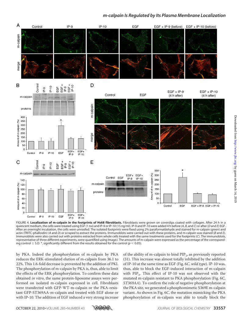
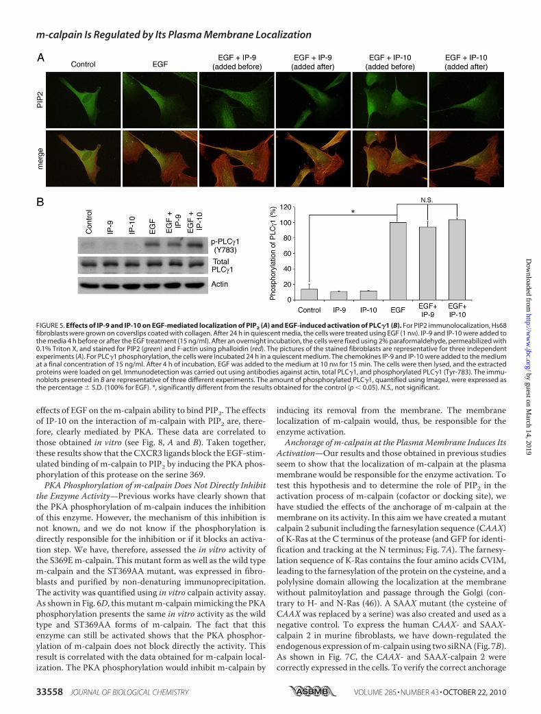
PIP2 and Activation of PLC�1—As previously stated, thegrowth factor-induced redistribution of m-calpain at the rearand at the membrane of the cells is due to the binding of theenzyme to PIP2. The altered localization of m-calpain could,thus, be secondary to a redistribution of PIP2 induced by theCXCR3 ligands. We, therefore, determined whether PIP2 dis-tribution was altered by IP-9 or IP-10. PIP2 was visualized byantibody detection in human fibroblasts treated with EGF andIP-9 or IP-10 (added 4 h before or after the growth factor). Thegrowth factor and chemokine concentrations used were thesame than those used for the previous experiments. The resultsshow that PIP2 was localized randomly in untreated fibroblasts(Fig. 5). The addition of EGF induced the concentration of PIP2at the rear of the cells. However, and in contrast to the resultsobserved form-calpain, IP-9, added before or after EGF, had noeffects on the localization of PIP2 induced by the growth factor.Similarly, the EGF-induced localization of PIP2 at the rear of thecell remains unchanged in the presence of IP-10. The sameresults were obtained with endothelial cells treated in the sameconditions (supplemental Fig. S5). Previous studies have shownthat the asymmetric localization of PIP2 in the cells is due to thedepletion of this phosphoinositide at the front of the cell byPLC�1. This enzyme is phosphorylated and, thus, activatedafter EGF treatment. The CXCR3 ligands had no effect on thephosphorylation of PLC�1 induced by EGF (Fig. 5B). Thesechemokines are, thus, unable to block the EGF-mediated acti-vation of PLC�1. This result confirms the data obtained byimmunolocalization of PIP2.
Taken together, these results showed that the effects of theCXCR3 ligands on EGF-induced localization of m-calpain arenot due to changes in the localization of PIP2. IP-9 and IP-10could be able to block the interaction between m-calpain andPIP2 allowed by EGF, thus preventing and also reversing theredistribution of m-calpain observed in the presence of EGF.PKA Phosphorylation of m-calpain Induced by CXCR3
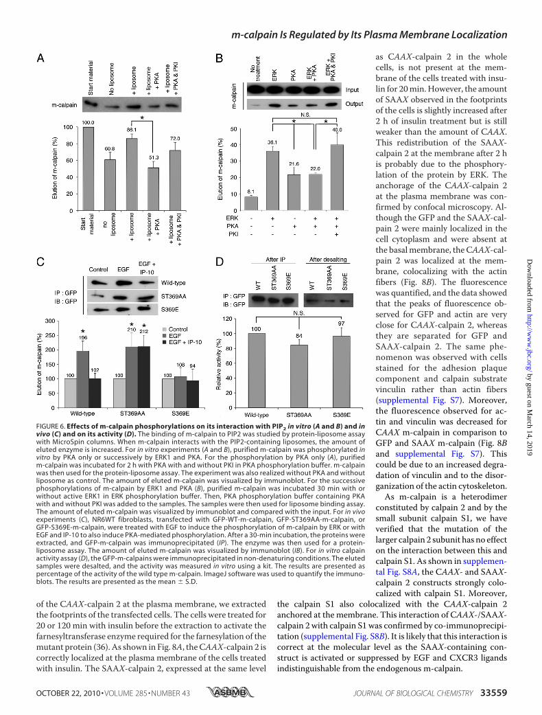
Ligands Blocks the Interaction between m-calpain and PIP2—As previously stated, CXCR3 ligands inhibit m-calpain activityby inducing the phosphorylation of m-calpain by PKA (33). Toparse the effects of this phosphorylation of m-calpain ability tobind PIP2, we performed a protein-liposome assaywith purifiedm-calpain treated in vitrowith PKA. As shown in Fig. 6A, the invitro phosphorylation of m-calpain by PKA strongly reducedthe binding of m-calpain to PIP2-containing liposomes. Indeedthe elution of m-calpain (due to the binding of m-calpain to theliposomes) was reduced from 86 to 51% after PKA phosphory-lation. This inhibition of m-calpain binding to PIP2 was abro-gated in the presence of the PKA specific inhibitor PKI. In vivothe CXCR3 ligands intervene after the growth factors; it is,thus, important to know if the stimulation of the interactionbetween m-calpain and PIP2 induced by ERK can be blockedby the PKA phosphorylation of m-calpain. To this aim, theability of m-calpain to bind PIP2 was studied after the suc-cessive phosphorylations of the protease by ERK1 and PKA.As shown in Fig. 6B, the strong stimulation of the binding ofm-calpain to PIP2 induced by ERK1 phosphorylation is limited
FIGURE 3. Localization of m-calpain in fibroblasts treated with IP-9 andIP-10 4 h after the addition of EGF. Hs68 fibroblasts were grown on cover-slips coated with collagen. After 24 h in a quiescent medium, the cells weretreated using EGF (1 nM). Four hours after this first treatment, IP-9 and IP-10were added to the medium (15 ng/ml). After an overnight incubation, thecells were fixed using 2% paraformaldehyde, permeabilized with 0.1% TritonX, and stained for m-calpain (green) and F-actin using phalloidin (red). Nucleiwere stained in blue using DAPI. The pictures obtained with the differenttreatments (A) were analyzed using Metamorph, and the average fluores-cence was quantified in the front and in the rear of the cells (B). The ratiosbetween the rear and the front were also calculated (C). The results areexpressed as the means � S.E. The experiments were repeated three times,and 20 cells were analyzed for each treatment in each independent experi-ment. *, **, #, significantly different from the results obtained for the cellstreated with EGF alone (p � 0.01).
m-calpain Is Regulated by Its Plasma Membrane Localization
33556 JOURNAL OF BIOLOGICAL CHEMISTRY VOLUME 285 • NUMBER 43 • OCTOBER 22, 2010
by guest on March 14, 2019
http://ww
w.jbc.org/
Dow
nloaded from
by PKA. Indeed the phosphorylation of m-calpain by PKAreduces the ERK-stimulated elution of m-calpain from 36.1 to22%. This 1.6-fold decrease is prevented by the addition of PKI.The phosphorylation of m-calpain by PKA is, thus, able to limitthe effects of the ERK phosphorylation. To confirm these dataobtained in vitro, the same protein-liposome assays were per-formed on isolated m-calpain expressed in cell. Fibroblastswere transfected with GFP-WT-m-calpain or the PKA-resis-tant GFP-ST369AA-m-calpain and treated with EGF alone orwith IP-10. The addition of EGF induced a very strong increase
of the ability of m-calpain to bind PIP2, as previously reported(31). This increase was almost totally inhibited by the additionof IP-10 at the same time as EGF (Fig. 6C,wild type). IP-10 was,thus, able to block the EGF-induced interaction of m-calpainwith PIP2. This effect of IP-10 was not observed with themutated m-calpain resistant to PKA phosphorylation (Fig. 6C,ST369AA). To confirm the role of negative phosphorylation atthe PKA site, we generated a phosphomimetic S369Em-calpainvariant. As shown in Fig. 6C, the mutation mimicking the PKAphosphorylation of m-calpain was able to totally block the
FIGURE 4. Localization of m-calpain in the footprints of Hs68 fibroblasts. Fibroblasts were grown on coverslips coated with collagen. After 24 h in aquiescent medium, the cells were treated using EGF (1 nM) and IP-9 or IP-10 (15 ng/ml). IP-9 and IP-10 were added 4 h before (A, B, and C) or after (D and E) EGF.After an overnight incubation, the cells were unroofed. The isolated footprints were fixed using 2% paraformaldehyde and stained for m-calpain (green) andactin (TRITC-phalloidin) (A and D) or scraped to extract the proteins. Immunoblots were carried out with these proteins, and m-calpain was stained (B and E).Immunoblots were also carried out with proteins extracted from whole cells treated with the same treatments used for the footprints (C). The immunoblots,representative of three different experiments, were quantified using ImageJ. The amounts of m-calpain were expressed as the percentage of the correspond-ing control � S.D. *, significantly different from the results obtained for the control (p � 0.05).
m-calpain Is Regulated by Its Plasma Membrane Localization
OCTOBER 22, 2010 • VOLUME 285 • NUMBER 43 JOURNAL OF BIOLOGICAL CHEMISTRY 33557
by guest on March 14, 2019
http://ww
w.jbc.org/
Dow
nloaded from
effects of EGF on them-calpain ability to bind PIP2. The effectsof IP-10 on the interaction of m-calpain with PIP2 are, there-fore, clearly mediated by PKA. These data are correlated tothose obtained in vitro (see Fig. 8, A and B). Taken together,these results show that the CXCR3 ligands block the EGF-stim-ulated binding of m-calpain to PIP2 by inducing the PKA phos-phorylation of this protease on the serine 369.PKA Phosphorylation of m-calpain Does Not Directly Inhibit
the Enzyme Activity—Previous works have clearly shown thatthe PKA phosphorylation of m-calpain induces the inhibitionof this enzyme. However, the mechanism of this inhibition isnot known, and we do not know if the phosphorylation isdirectly responsible for the inhibition or if it blocks an activa-tion step. We have, therefore, assessed the in vitro activity ofthe S369Em-calpain. This mutant form as well as the wild typem-calpain and the ST369AA mutant, was expressed in fibro-blasts and purified by non-denaturing immunoprecipitation.The activity was quantified using in vitro calpain activity assay.As shown in Fig. 6D, thismutantm-calpainmimicking the PKAphosphorylation presents the same in vitro activity as the wildtype and ST369AA forms of m-calpain. The fact that thisenzyme can still be activated shows that the PKA phosphor-ylation of m-calpain does not block directly the activity. Thisresult is correlated with the data obtained for m-calpain local-ization. The PKA phosphorylation would inhibit m-calpain by
inducing its removal from the membrane. The membranelocalization of m-calpain would, thus, be responsible for theenzyme activation.Anchorage of m-calpain at the PlasmaMembrane Induces Its
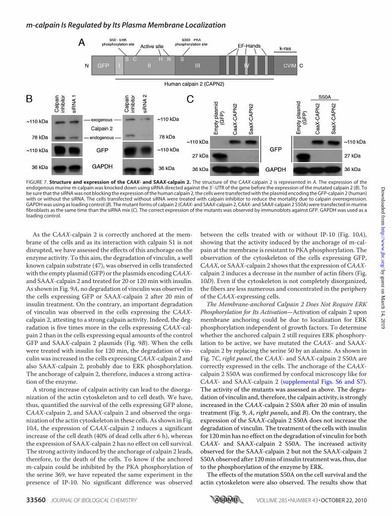
Activation—Our results and those obtained in previous studiesseem to show that the localization of m-calpain at the plasmamembrane would be responsible for the enzyme activation. Totest this hypothesis and to determine the role of PIP2 in theactivation process of m-calpain (cofactor or docking site), wehave studied the effects of the anchorage of m-calpain at themembrane on its activity. In this aim we have created a mutantcalpain 2 subunit including the farnesylation sequence (CAAX)of K-Ras at the C terminus of the protease (and GFP for identi-fication and tracking at the N terminus; Fig. 7A). The farnesy-lation sequence of K-Ras contains the four amino acids CVIM,leading to the farnesylation of the protein on the cysteine, and apolylysine domain allowing the localization at the membranewithout palmitoylation and passage through the Golgi (con-trary to H- and N-Ras (46)). A SAAX mutant (the cysteine ofCAAX was replaced by a serine) was also created and used as anegative control. To express the human CAAX- and SAAX-calpain 2 in murine fibroblasts, we have down-regulated theendogenous expression ofm-calpain using two siRNA (Fig. 7B).As shown in Fig. 7C, the CAAX- and SAAX-calpain 2 werecorrectly expressed in the cells. To verify the correct anchorage
FIGURE 5. Effects of IP-9 and IP-10 on EGF-mediated localization of PIP2 (A) and EGF-induced activation of PLC�1 (B). For PIP2 immunolocalization, Hs68fibroblasts were grown on coverslips coated with collagen. After 24 h in quiescent media, the cells were treated using EGF (1 nM). IP-9 and IP-10 were added tothe media 4 h before or after the EGF treatment (15 ng/ml). After an overnight incubation, the cells were fixed using 2% paraformaldehyde, permeabilized with0.1% Triton X, and stained for PIP2 (green) and F-actin using phalloidin (red). The pictures of the stained fibroblasts are representative for three independentexperiments (A). For PLC�1 phosphorylation, the cells were incubated 24 h in a quiescent medium. The chemokines IP-9 and IP-10 were added to the mediumat a final concentration of 15 ng/ml. After 4 h of incubation, EGF was added to the medium at 10 nM for 15 min. The cells were then lysed, and the extractedproteins were loaded on gel. Immunodetection was carried out using antibodies against actin, total PLC�1, and phosphorylated PLC�1 (Tyr-783). The immu-noblots presented in B are representative of three different experiments. The amount of phosphorylated PLC�1, quantified using ImageJ, were expressed asthe percentage � S.D. (100% for EGF). *, significantly different from the results obtained for the control (p � 0.05). N.S., not significant.
m-calpain Is Regulated by Its Plasma Membrane Localization
33558 JOURNAL OF BIOLOGICAL CHEMISTRY VOLUME 285 • NUMBER 43 • OCTOBER 22, 2010
by guest on March 14, 2019
http://ww
w.jbc.org/
Dow
nloaded from
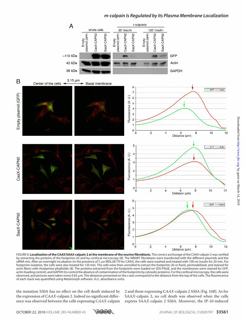
of the CAAX-calpain 2 at the plasma membrane, we extractedthe footprints of the transfected cells. The cells were treated for20 or 120 min with insulin before the extraction to activate thefarnesyltransferase enzyme required for the farnesylation of themutant protein (36). As shown in Fig. 8A, theCAAX-calpain 2 iscorrectly localized at the plasma membrane of the cells treatedwith insulin. The SAAX-calpain 2, expressed at the same level
as CAAX-calpain 2 in the wholecells, is not present at the mem-brane of the cells treated with insu-lin for 20min.However, the amountof SAAX observed in the footprintsof the cells is slightly increased after2 h of insulin treatment but is stillweaker than the amount of CAAX.This redistribution of the SAAX-calpain 2 at the membrane after 2 his probably due to the phosphory-lation of the protein by ERK. Theanchorage of the CAAX-calpain 2at the plasma membrane was con-firmed by confocal microscopy. Al-though the GFP and the SAAX-cal-pain 2 were mainly localized in thecell cytoplasm and were absent atthe basalmembrane, theCAAX-cal-pain 2 was localized at the mem-brane, colocalizing with the actinfibers (Fig. 8B). The fluorescencewas quantified, and the data showedthat the peaks of fluorescence ob-served for GFP and actin are veryclose for CAAX-calpain 2, whereasthey are separated for GFP andSAAX-calpain 2. The same phe-nomenon was observed with cellsstained for the adhesion plaquecomponent and calpain substratevinculin rather than actin fibers(supplemental Fig. S7). Moreover,the fluorescence observed for ac-tin and vinculin was decreased forCAAX m-calpain in comparison toGFP and SAAX m-calpain (Fig. 8Band supplemental Fig. S7). Thiscould be due to an increased degra-dation of vinculin and to the disor-ganization of the actin cytoskeleton.As m-calpain is a heterodimer
constituted by calpain 2 and by thesmall subunit calpain S1, we haveverified that the mutation of thelarger calpain 2 subunit has no effecton the interaction between this andcalpain S1. As shown in supplemen-tal Fig. S8A, the CAAX- and SAAX-calpain 2 constructs strongly colo-calized with calpain S1. Moreover,
the calpain S1 also colocalized with the CAAX-calpain 2anchored at the membrane. This interaction of CAAX-/SAAX-calpain 2 with calpain S1 was confirmed by co-immunoprecipi-tation (supplemental Fig. S8B). It is likely that this interaction iscorrect at the molecular level as the SAAX-containing con-struct is activated or suppressed by EGF and CXCR3 ligandsindistinguishable from the endogenous m-calpain.
FIGURE 6. Effects of m-calpain phosphorylations on its interaction with PIP2 in vitro (A and B) and invivo (C) and on its activity (D). The binding of m-calpain to PIP2 was studied by protein-liposome assaywith MicroSpin columns. When m-calpain interacts with the PIP2-containing liposomes, the amount ofeluted enzyme is increased. For in vitro experiments (A and B), purified m-calpain was phosphorylated invitro by PKA only or successively by ERK1 and PKA. For the phosphorylation by PKA only (A), purifiedm-calpain was incubated for 2 h with PKA with and without PKI in PKA phosphorylation buffer. m-calpainwas then used for the protein-liposome assay. The experiment was also realized without PKA and withoutliposome as control. The amount of eluted m-calpain was visualized by immunoblot. For the successivephosphorylations of m-calpain by ERK1 and PKA (B), purified m-calpain was incubated 30 min with orwithout active ERK1 in ERK phosphorylation buffer. Then, PKA phosphorylation buffer containing PKAwith and without PKI was added to the samples. The samples were then used for liposome binding assay.The amount of eluted m-calpain was visualized by immunoblot and compared with the input. For in vivoexperiments (C), NR6WT fibroblasts, transfected with GFP-WT-m-calpain, GFP-ST369AA-m-calpain, orGFP-S369E-m-calpain, were treated with EGF to induce the phosphorylation of m-calpain by ERK or withEGF and IP-10 to also induce PKA-mediated phosphorylation. After a 30-min incubation, the proteins wereextracted, and GFP-m-calpain was immunoprecipitated (IP). The enzyme was then used for a protein-liposome assay. The amount of eluted m-calpain was visualized by immunoblot (IB). For in vitro calpainactivity assay (D), the GFP-m-calpains were immunoprecipitated in non-denaturing conditions. The elutedsamples were desalted, and the activity was measured in vitro using a kit. The results are presented aspercentage of the activity of the wild type m-calpain. ImageJ software was used to quantify the immuno-blots. The results are presented as the mean � S.D.
m-calpain Is Regulated by Its Plasma Membrane Localization
OCTOBER 22, 2010 • VOLUME 285 • NUMBER 43 JOURNAL OF BIOLOGICAL CHEMISTRY 33559
by guest on March 14, 2019
http://ww
w.jbc.org/
Dow
nloaded from
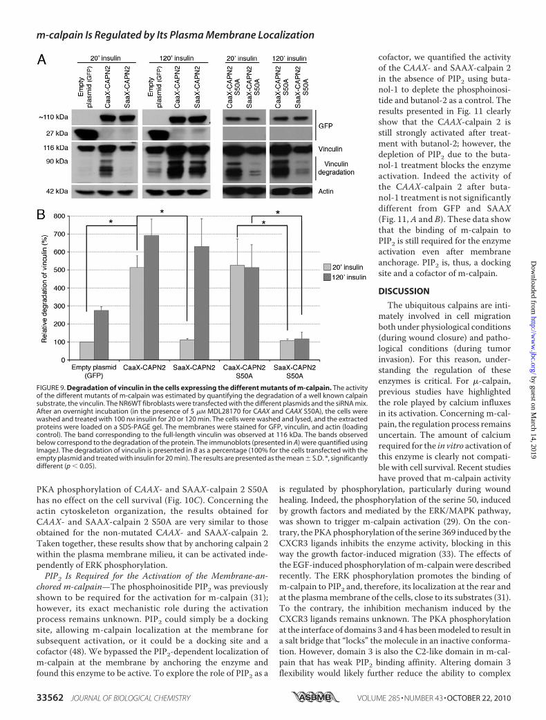
As the CAAX-calpain 2 is correctly anchored at the mem-brane of the cells and as its interaction with calpain S1 is notdisrupted, we have assessed the effects of this anchorage on theenzyme activity. To this aim, the degradation of vinculin, a wellknown calpain substrate (47), was observed in cells transfectedwith the empty plasmid (GFP) or the plasmids encodingCAAX-and SAAX-calpain 2 and treated for 20 or 120min with insulin.As shown in Fig. 9A, no degradation of vinculin was observed inthe cells expressing GFP or SAAX-calpain 2 after 20 min ofinsulin treatment. On the contrary, an important degradationof vinculin was observed in the cells expressing the CAAX-calpain 2, attesting to a strong calpain activity. Indeed, the deg-radation is five times more in the cells expressing CAAX-cal-pain 2 than in the cells expressing equal amounts of the controlGFP and SAAX-calpain 2 plasmids (Fig. 9B). When the cellswere treated with insulin for 120 min, the degradation of vin-culin was increased in the cells expressing CAAX-calpain 2 andalso SAAX-calpain 2, probably due to ERK phosphorylation.The anchorage of calpain 2, therefore, induces a strong activa-tion of the enzyme.A strong increase of calpain activity can lead to the disorga-
nization of the actin cytoskeleton and to cell death. We have,thus, quantified the survival of the cells expressing GFP alone,CAAX-calpain 2, and SAAX-calpain 2 and observed the orga-nization of the actin cytoskeleton in these cells. As shown in Fig.10A, the expression of CAAX-calpain 2 induces a significantincrease of the cell death (40% of dead cells after 6 h), whereasthe expression of SAAX-calpain 2 has no effect on cell survival.The strong activity induced by the anchorage of calpain 2 leads,therefore, to the death of the cells. To know if the anchoredm-calpain could be inhibited by the PKA phosphorylation ofthe serine 369, we have repeated the same experiment in thepresence of IP-10. No significant difference was observed
between the cells treated with or without IP-10 (Fig. 10A),showing that the activity induced by the anchorage of m-cal-pain at themembrane is resistant to PKA phosphorylation. Theobservation of the cytoskeleton of the cells expressing GFP,CAAX, or SAAX-calpain 2 shows that the expression of CAAX-calpain 2 induces a decrease in the number of actin fibers (Fig.10D). Even if the cytoskeleton is not completely disorganized,the fibers are less numerous and concentrated in the peripheryof the CAAX-expressing cells.The Membrane-anchored Calpain 2 Does Not Require ERK
Phosphorylation for Its Activation—Activation of calpain 2 uponmembrane anchoring could be due to localization for ERKphosphorylation independent of growth factors. To determinewhether the anchored calpain 2 still requires ERK phosphory-lation to be active, we have mutated the CAAX- and SAAX-calpain 2 by replacing the serine 50 by an alanine. As shown inFig. 7C, right panel, the CAAX- and SAAX-calpain 2 S50A arecorrectly expressed in the cells. The anchorage of the CAAX-calpain 2 S50A was confirmed by confocal microscopy like forCAAX- and SAAX-calpain 2 (supplemental Figs. S6 and S7).The activity of the mutants was assessed as above. The degra-dation of vinculin and, therefore, the calpain activity, is stronglyincreased in the CAAX-calpain 2 S50A after 20 min of insulintreatment (Fig. 9, A, right panels, and B). On the contrary, theexpression of the SAAX-calpain 2 S50A does not increase thedegradation of vinculin. The treatment of the cells with insulinfor 120min has no effect on the degradation of vinculin for bothCAAX- and SAAX-calpain 2 S50A. The increased activityobserved for the SAAX-calpain 2 but not the SAAX-calpain 2S50Aobserved after 120min of insulin treatmentwas, thus, dueto the phosphorylation of the enzyme by ERK.The effects of the mutation S50A on the cell survival and the
actin cytoskeleton were also observed. The results show that
FIGURE 7. Structure and expression of the CAAX- and SAAX-calpain 2. The structure of the CAAX-calpain 2 is represented in A. The expression of theendogenous murine m-calpain was knocked down using siRNA directed against the 3�-UTR of the gene before the expression of the mutated calpain 2 (B). Tobe sure that the siRNA was not blocking the expression of the human calpain 2, the cells were transfected with the plasmid encoding the GFP-calpain 2 (human)with or without the siRNA. The cells transfected without siRNA were treated with calpain inhibitor to reduce the mortality due to calpain overexpression.GAPDH was using as loading control (B). The mutant forms of calpain 2 (CAAX- and SAAX-calpain 2, CAAX- and SAAX-calpain 2 S50A) were transfected in murinefibroblasts as the same time than the siRNA mix (C). The correct expression of the mutants was observed by immunoblots against GFP. GAPDH was used as aloading control.
m-calpain Is Regulated by Its Plasma Membrane Localization
33560 JOURNAL OF BIOLOGICAL CHEMISTRY VOLUME 285 • NUMBER 43 • OCTOBER 22, 2010
by guest on March 14, 2019
http://ww
w.jbc.org/
Dow
nloaded from
the mutation S50A has no effect on the cell death induced bythe expression of CAAX-calpain 2. Indeed no significant differ-ence was observed between the cells expressing CAAX-calpain
2 and those expressing CAAX-calpain 2 S50A (Fig. 10B). As forSAAX-calpain 2, no cell death was observed when the cellsexpress SAAX-calpain 2 S50A. Moreover, the IP-10-induced
FIGURE 8. Localization of the CAAX/SAAX-calpain 2 at the membrane of the murine fibroblasts. The correct anchorage of the CAAX-calpain 2 was verifiedby extracting the proteins of the footprints (A) and by confocal microscopy (B). The NR6WT fibroblasts were transfected with the different plasmids and thesiRNA mix. After an overnight incubation (in the presence of 5 �M MDL28170 for CAAX), the cells were washed and treated with 100 nM insulin for 20 min. Forfootprints isolation, the cells were also treated for 120 min. The cells were then unroofed to extract the footprints (A) or fixed, permeabilized, and stained foractin fibers with rhodamine-phalloidin (B). The proteins extracted from the footprints were loaded on SDS-PAGE, and the membranes were stained for GFP,actin (loading control), and GAPDH (to control the absence of contamination of the footprints by cytosolic proteins). For the confocal microscopy, the cells wereobserved, and pictures were taken every 0.63 �m. The distances presented on the x axis correspond to the distance from the top of the cells. The fluorescenceof each stack was quantified using Metamorph software. A.U., absorbance units.
m-calpain Is Regulated by Its Plasma Membrane Localization
OCTOBER 22, 2010 • VOLUME 285 • NUMBER 43 JOURNAL OF BIOLOGICAL CHEMISTRY 33561
by guest on March 14, 2019
http://ww
w.jbc.org/
Dow
nloaded from
PKA phosphorylation of CAAX- and SAAX-calpain 2 S50Ahas no effect on the cell survival (Fig. 10C). Concerning theactin cytoskeleton organization, the results obtained forCAAX- and SAAX-calpain 2 S50A are very similar to thoseobtained for the non-mutated CAAX- and SAAX-calpain 2.Taken together, these results show that by anchoring calpain 2within the plasma membrane milieu, it can be activated inde-pendently of ERK phosphorylation.PIP2 Is Required for the Activation of the Membrane-an-
chored m-calpain—The phosphoinositide PIP2 was previouslyshown to be required for the activation for m-calpain (31);however, its exact mechanistic role during the activationprocess remains unknown. PIP2 could simply be a dockingsite, allowing m-calpain localization at the membrane forsubsequent activation, or it could be a docking site and acofactor (48). We bypassed the PIP2-dependent localization ofm-calpain at the membrane by anchoring the enzyme andfound this enzyme to be active. To explore the role of PIP2 as a
cofactor, we quantified the activityof the CAAX- and SAAX-calpain 2in the absence of PIP2 using buta-nol-1 to deplete the phosphoinosi-tide and butanol-2 as a control. Theresults presented in Fig. 11 clearlyshow that the CAAX-calpain 2 isstill strongly activated after treat-ment with butanol-2; however, thedepletion of PIP2 due to the buta-nol-1 treatment blocks the enzymeactivation. Indeed the activity ofthe CAAX-calpain 2 after buta-nol-1 treatment is not significantlydifferent from GFP and SAAX(Fig. 11, A and B). These data showthat the binding of m-calpain toPIP2 is still required for the enzymeactivation even after membraneanchorage. PIP2 is, thus, a dockingsite and a cofactor of m-calpain.
DISCUSSION
The ubiquitous calpains are inti-mately involved in cell migrationboth under physiological conditions(during wound closure) and patho-logical conditions (during tumorinvasion). For this reason, under-standing the regulation of theseenzymes is critical. For �-calpain,previous studies have highlightedthe role played by calcium influxesin its activation. Concerning m-cal-pain, the regulation process remainsuncertain. The amount of calciumrequired for the in vitro activation ofthis enzyme is clearly not compati-ble with cell survival. Recent studieshave proved that m-calpain activity
is regulated by phosphorylation, particularly during woundhealing. Indeed, the phosphorylation of the serine 50, inducedby growth factors and mediated by the ERK/MAPK pathway,was shown to trigger m-calpain activation (29). On the con-trary, the PKAphosphorylation of the serine 369 induced by theCXCR3 ligands inhibits the enzyme activity, blocking in thisway the growth factor-induced migration (33). The effects ofthe EGF-induced phosphorylation ofm-calpain were describedrecently. The ERK phosphorylation promotes the binding ofm-calpain to PIP2 and, therefore, its localization at the rear andat the plasmamembrane of the cells, close to its substrates (31).To the contrary, the inhibition mechanism induced by theCXCR3 ligands remains unknown. The PKA phosphorylationat the interface of domains 3 and 4 has beenmodeled to result ina salt bridge that “locks” the molecule in an inactive conforma-tion. However, domain 3 is also the C2-like domain in m-cal-pain that has weak PIP2 binding affinity. Altering domain 3flexibility would likely further reduce the ability to complex
FIGURE 9. Degradation of vinculin in the cells expressing the different mutants of m-calpain. The activityof the different mutants of m-calpain was estimated by quantifying the degradation of a well known calpainsubstrate, the vinculin. The NR6WT fibroblasts were transfected with the different plasmids and the siRNA mix.After an overnight incubation (in the presence of 5 �M MDL28170 for CAAX and CAAX S50A), the cells werewashed and treated with 100 nM insulin for 20 or 120 min. The cells were washed and lysed, and the extractedproteins were loaded on a SDS-PAGE gel. The membranes were stained for GFP, vinculin, and actin (loadingcontrol). The band corresponding to the full-length vinculin was observed at 116 kDa. The bands observedbelow correspond to the degradation of the protein. The immunoblots (presented in A) were quantified usingImageJ. The degradation of vinculin is presented in B as a percentage (100% for the cells transfected with theempty plasmid and treated with insulin for 20 min). The results are presented as the mean � S.D. *, significantlydifferent (p � 0.05).
m-calpain Is Regulated by Its Plasma Membrane Localization
33562 JOURNAL OF BIOLOGICAL CHEMISTRY VOLUME 285 • NUMBER 43 • OCTOBER 22, 2010
by guest on March 14, 2019
http://ww
w.jbc.org/
Dow
nloaded from
with PIP2. Moreover, the removal of m-calpain from the mem-brane, where its substrates are located, would be a very efficientway to block cell retraction and subsequently cell migration.Therefore, our first aim was to identify the effects of the
CXCR3 ligands on the growth factor-induced relocalizationof m-calpain at the membrane. Moreover, it was unknownwhether IP-9 and IP-10 were dominant over the growth fac-tors and, thus, if their effects are immediate. Our data showthat the EGF-induced migration of fibroblasts is stronglyinhibited by the chemokines within minutes after their addi-tion (Fig. 1, A, D, and E). Importantly, similar results wereobtained with endothelial cells treated with EGF or VEGF,
showing that this phenomenon is not specific for fibroblastsand EGF (Fig. 1, B, C, E, and F). These data prove that theCXCR3 chemokines are able to override the growth factor-induced stimulation of cell migration and that these inhibi-tory effects are almost immediate. This inhibition of cellmotility is at least in part due to preventing cell retraction, asobserved by cell tracking within minutes after the addition ofthe chemokines (Fig. 1H and supplemental Movie 1). Thisresult agrees with previous studies showing that IP-9 andIP-10 are able to inhibit m-calpain, which is critically in-volved in cell retraction by cleaving several components of theadhesion complexes (11). Moreover, we found that CXCR3
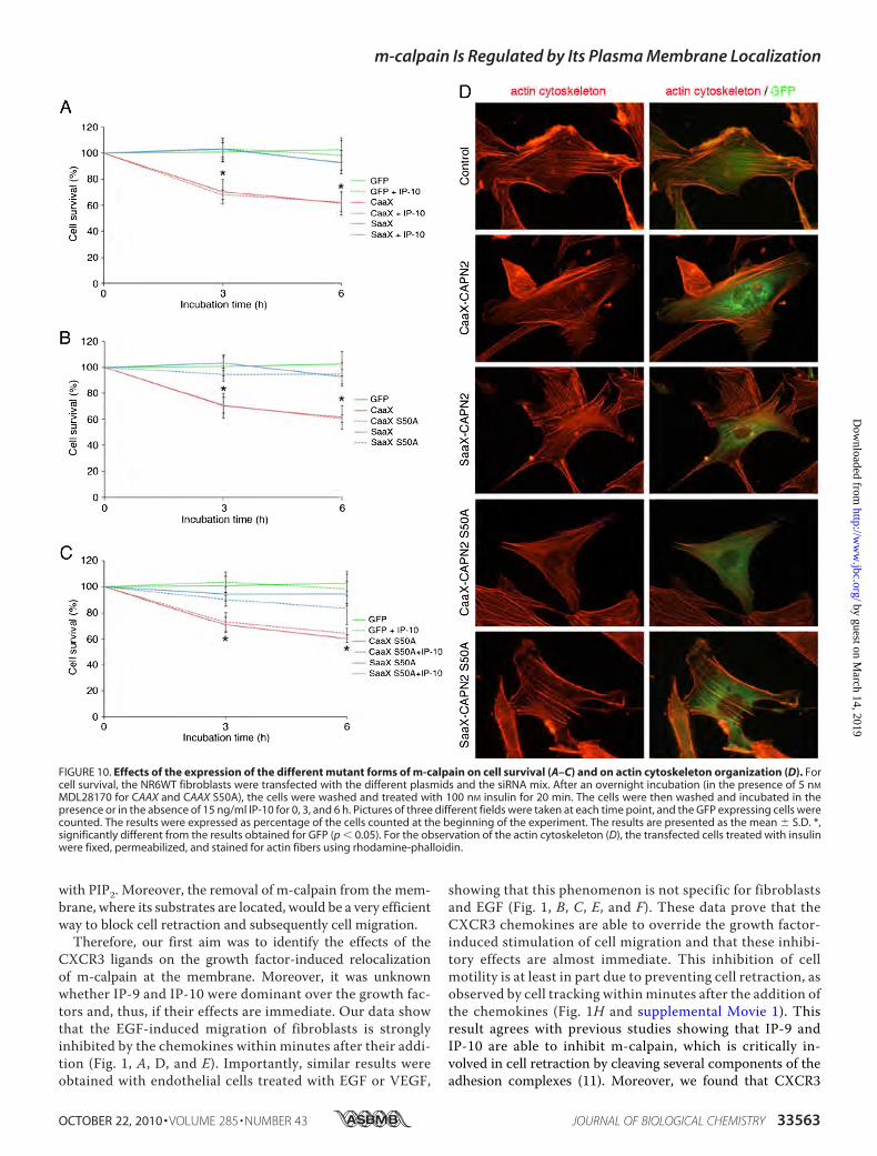
FIGURE 10. Effects of the expression of the different mutant forms of m-calpain on cell survival (A–C) and on actin cytoskeleton organization (D). Forcell survival, the NR6WT fibroblasts were transfected with the different plasmids and the siRNA mix. After an overnight incubation (in the presence of 5 nM
MDL28170 for CAAX and CAAX S50A), the cells were washed and treated with 100 nM insulin for 20 min. The cells were then washed and incubated in thepresence or in the absence of 15 ng/ml IP-10 for 0, 3, and 6 h. Pictures of three different fields were taken at each time point, and the GFP expressing cells werecounted. The results were expressed as percentage of the cells counted at the beginning of the experiment. The results are presented as the mean � S.D. *,significantly different from the results obtained for GFP (p � 0.05). For the observation of the actin cytoskeleton (D), the transfected cells treated with insulinwere fixed, permeabilized, and stained for actin fibers using rhodamine-phalloidin.
m-calpain Is Regulated by Its Plasma Membrane Localization
OCTOBER 22, 2010 • VOLUME 285 • NUMBER 43 JOURNAL OF BIOLOGICAL CHEMISTRY 33563
by guest on March 14, 2019
http://ww
w.jbc.org/
Dow
nloaded from
ligands not only prevented growth factor-induced m-calpainrelocalization in fibroblasts (Fig. 2) but also revertedmembranelocalization when added after the growth factors (Fig. 3). Simi-lar results were obtained with endothelial cells treated withEGF or with the angiogenic growth factor VEGF, showingthat the effects of CXCR3 chemokines are generalizable(supplemental Figs. S1–S3). It is also interesting to note thatthe chemokines have no effect on the shape of the growthfactor-stimulated cells. Indeed, the cell polarity remainedwith lamellipodia still present at the front of the cells (Fig. 3).By inhibiting the localization of m-calpain at the rear of thecells without blocking the formation of the lamellipodia, theCXCR3 ligands inhibit cell retraction (and, thus, productivelocomotion), whereas still allowing for matrix contractionrequired for dermal maturation. This would not be possibleif both cell retraction and lamellipodia formation wereblocked (28). Relocalization of m-calpain to the membranein the cell body and rear results from increased binding ofm-calpain to PIP2 (31). Our results show that the CXCR3chemokines prevent and even revert this EGF-induced local-ization of m-calpain at the plasma membrane of both fibro-blasts and endothelial cells (Fig. 4 and supplemental Fig. S4).The chemokines are, thus, able to separate m-calpain from itssubstrates, preventing the cleavage of the adhesion complexes.This phenomenon is not due to a modification of the PIP2asymmetry that determines cell front from rear (Fig. 5A). In
agreement with this, the chemo-kines have no effect on the activa-tion of PLC�1 by EGF, which isresponsible for the PIP2 asymmetricrelocation away from the lamellipo-dia (Fig. 5B). It was, thus, interestingto study the effects of the CXCR3ligands on the ability ofm-calpain tobind PIP2. Our liposome bindingassays showed that the phosphory-lation of m-calpain by PKA reducedthe interaction between m-calpainand PIP2 even when m-calpain waspreviously phosphorylated by ERK1(Fig. 6, A and B). Moreover, thetreatment of the cells with IP-10reduced the EGF-stimulated abilityof m-calpain to bind PIP2. Thiseffect is clearly due to the PKAphosphorylation of m-calpain onthe serine 369, as IP-10 has no effecton the PKA-resistant mutant formof m-calpain (ST369AA). It is alsopossible to prevent the effects ofEGF by mimicking this phosphory-lation with the S369E-m-calpain(Fig. 6C). By blocking the interac-tion between PIP2 and m-calpain,the PKA phosphorylation of m-cal-pain is, thus, clearly responsible forthe effects of the CXCR3 ligands onm-calpain redistribution at the rear
and at the plasma membrane of the cells.Our results and those obtained in previous studies seem to
show that the PIP2-dependent localization of m-calpain atthe membrane could be responsible for its activation andthat the phosphorylations would regulate m-calpain activityonly indirectly. Second, we have, thus, studied the effects ofthe anchorage of m-calpain at the membrane on its activity.To this aim, we have created a mutant calpain 2 contain-ing the K-Ras farnesylation sequence (CAAX, Fig. 7A). TheCAAX-calpain 2 was correctly anchored at the plasma mem-brane, whereas the negative control SAAX-calpain 2 wasmainly cytoplasmic and totally absent from themembrane (Fig.8, A and B, and supplemental Fig. S7). This artificial and forcedlocalization of calpain 2 at the cell membrane triggered a verystrong activation of the enzyme and subsequently the degrada-tion of its substrates (Fig. 9). As an important control, theSAAX-calpain 2 presents no increased activity when the cellsare treated with insulin for only 20 min. The activity of theSAAX-calpain 2 is increased only after a 120-min insulin treat-ment because of ERK phosphorylation. The increased activityobserved with CAAX-calpain 2 induced the death of the cellsexpressing this mutant protease and disorganized the actincytoskeleton (Fig. 10, A andD), probably by degrading the pro-tein of the adhesion complexes. The activity of the anchoredcalpain 2 is independent of the ERK phosphorylation. Indeed,the mutation of the serine 50 to alanine has no effect on the
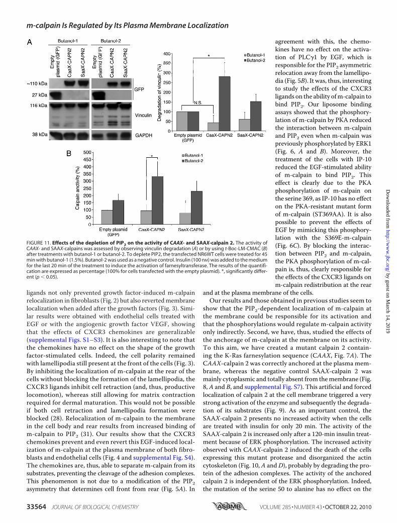
FIGURE 11. Effects of the depletion of PIP2 on the activity of CAAX- and SAAX-calpain 2. The activity ofCAAX- and SAAX-calpains was assessed by observing vinculin degradation (A) or by using t-Boc-LM-CMAC (B)after treatments with butanol-1 or butanol-2. To deplete PIP2, the transfected NR6WT cells were treated for 45min with butanol-1 (1.5%). Butanol-2 was used as a negative control. Insulin (100 nM) was added to the mediumfor the last 20 min of the treatment to induce the activation of farnesyltransferase. The results of the quantifi-cation are expressed as percentage (100% for cells transfected with the empty plasmid). *, significantly differ-ent (p � 0.05).
m-calpain Is Regulated by Its Plasma Membrane Localization
33564 JOURNAL OF BIOLOGICAL CHEMISTRY VOLUME 285 • NUMBER 43 • OCTOBER 22, 2010
by guest on March 14, 2019
http://ww
w.jbc.org/
Dow
nloaded from
activity of theCAAX-calpain 2 and on the subsequent cell death(Figs. 9 and 10B). On the contrary, the mutation S50A blocksthe insulin-induced activation of the SAAX-calpain 2. In thesamemanner the CAAX-calpain 2 is resistant to the PKA phos-phorylation. Indeed the addition of IP-10 has no effect on thecell death resulting fromCAAX-calpain 2 expression. Very sim-ilar results were also obtainedwhen the cells expressing CAAX-calpain 2 S50Awere treatedwith IP-10.However, the activationof the CAAX-calpain 2 still requires the presence of PIP2 (Fig.11). This phosphoinositide was previously shown to be crucialfor the activation of m-calpain (31); however, it was unclear if itwas only a docking site necessary for themembrane localizationor also a cofactor allowing m-calpain activity. Our data showthat even anchored at the membrane and, thus, not relying onPIP2 binding to be localized at the membrane, m-calpain stillrequires the presence of PIP2 to be activated. These very inter-esting results show clearly that PIP2 acts a cofactor of m-cal-pain, crucial for the enzyme activation. The hypothesis of PIP2being the final activating factor is intellectually satisfying as thecharged head group of PIP2 may subsume the electrostaticstructural rearrangement directed by calcium at supraphysi-ological levels (24).On the surface these findings and the resulting model of
the Ser-50 and Ser-369 phosphorylations on m-calpain con-trolling activation only indirectly due to regulation of local-ization within the plasma membrane milieu of PIP2 siteswould appear to conflict with earlier reports from our ownstudies (29, 49). Although we do not have a structural basisfor this seeming discrepancy, the main reasons for divergentfindings are likely the difference between activation in cellsand activation of bacterially produced calpains. As m-cal-pain is multiply, although substoichiometrically phosphory-lated on numerous serines and threonine in cells (1), theseother post-translational modifications absent in bacteriallyexpressed m-calpain may further constrain this potent andirreversible signaling protease (7). Intriguingly, we were notable to structurally model the Ser-50 phosphorylation as thisis in a highly disordered domain, suggesting that phosphor-ylation allows activation by either removing steric hindranceto the active cleft or, more likely, based on the observed data,that phosphorylation serves to help bind the m-calpain to itsmembrane cofactor PIP2. Still, the molecular structural basisof this activation is speculative at present and awaits exper-imentation beyond the scope of the present missive.In conclusion, our results clearly show that the localiza-
tion of m-calpain at the plasma membrane and its binding toPIP2 are the keystone of its activation process. The ERKphosphorylation of m-calpain induces the enzyme localiza-tion at the membrane by promoting the binding betweenm-calpain on PIP2 leading to its activation. On the contrary,PKA phosphorylation, induced by CXCR3 ligands, blocksthe binding to PIP2 and, thus, induces the removal of m-cal-pain from themembrane and its inactivation. The phosphor-ylations of m-calpain have no direct effect on the enzymeactivity. They control indirectly the activation process bycontrolling the redistribution of the protease, thereforeallowing, preventing, or reverting its activation.
Acknowledgments—We thank members of the Wells and Roy labora-tories and Stephen Chirieleison for helpful comments and technicalassistance. We also thank Prof. Chang Lu of Purdue University forproviding the plasmid pEGFP-C1.
REFERENCES1. Goll, D. E., Thompson, V. F., Li, H., Wei, W., and Cong, J. (2003) Physiol.
Rev. 83, 731–8012. Lauffenburger, D. A., and Horwitz, A. F. (1996) Cell 84, 359–3693. Ridley, A. J., Schwartz, M. A., Burridge, K., Firtel, R. A., Ginsberg, M. H.,
Borisy, G., Parsons, J. T., and Horwitz, A. R. (2003) Science 302,1704–1709
4. Sheetz, M. P., Felsenfeld, D., Galbraith, C. G., and Choquet, D. (1999)Biochem. Soc. Symp. 65, 233–243
5. Cox, E. A., and Huttenlocher, A. (1998)Microsc. Res. Tech. 43, 412–4196. Hautaniemi, S., Kharait, S., Iwabu, A., Wells, A., and Lauffenburger, D. A.
(2005) Bioinformatics 21, 2027–20357. Glading, A., Lauffenburger, D. A., and Wells, A. (2002) Trends Cell Biol.
12, 46–548. Kulkarni, S., Saido, T. C., Suzuki, K., and Fox, J. E. (1999) J. Biol. Chem. 274,
21265–212759. Satish, L., Blair, H. C., Glading, A., andWells, A. (2005)Mol. Cell. Biol. 25,
1922–194110. Cooray, P., Yuan, Y., Schoenwaelder, S. M., Mitchell, C. A., Salem, H. H.,
and Jackson, S. P. (1996) Biochem. J. 318, 41–4711. Franco, S. J., Rodgers, M. A., Perrin, B. J., Han, J., Bennin, D. A., Critchley,
D. R., and Huttenlocher, A. (2004) Nat. Cell Biol. 6, 977–98312. Palecek, S. P., Huttenlocher, A., Horwitz, A. F., and Lauffenburger, D. A.
(1998) J. Cell Sci. 111, 929–94013. Glading, A., Chang, P., Lauffenburger, D. A., and Wells, A. (2000) J. Biol.
Chem. 275, 2390–239814. Huttenlocher, A., Ginsberg, M. H., and Horwitz, A. F. (1996) J. Cell Biol.
134, 1551–156215. Broughton, G. 2nd, Janis, J. E., and Attinger, C. E. (2006) Plast. Reconstr.
Surg. 117, Suppl. 7, 1e-S–32e-S16. Diegelmann, R. F., and Evans, M. C. (2004) Front Biosci. 9, 283–28917. Li, J., Zhang, Y. P., andKirsner, R. S. (2003)Microsc. Res. Tech. 60, 107–11418. Raja, Sivamani, K., Garcia, M. S., and Isseroff, R. R. (2007) Front Biosci. 12,
2849–286819. Imanishi, J., Kamiyama, K., Iguchi, I., Kita, M., Sotozono, C., and Ki-
noshita, S. (2000) Prog. Retin. Eye Res. 19, 113–12920. Kunimoto, B. T. (1999) Ostomy. Wound Manage. 45, 56–6421. Wells, A., Gupta, K., Chang, P., Swindle, S., Glading, A., and Shiraha, H.
(1998)Microsc. Res. Tech. 43, 395–41122. Nedelec, B., Ghahary, A., Scott, P. G., and Tredget, E. E. (2000)Hand Clin.
16, 289–30223. Bodnar, R. J., Yates, C. C., and Wells, A. (2006) Circ. Res. 98, 617–62524. Moldoveanu, T., Hosfield, C. M., Jia, Z., Elce, J. S., and Davies, P. L. (2001)
Biochim. Biophys. Acta 1545, 245–25425. Yates, C. C., Whaley, D., Y.-Chen, A., Kulesekaran, P., Hebda, P. A., and
Wells, A. (2008) Am. J. Pathol. 173, 643–65226. Yates, C. C., Whaley, D., Hooda, S., Hebda, P. A., Bodnar, R. J., andWells,
A. (2009)Wound Repair Regen. 17, 34–4127. Shao,H., Yi, X.M., andWells, A. (2008)WoundRepair Regen. 16, 551–55828. Allen, F. D., Asnes, C. F., Chang, P., Elson, E. L., Lauffenburger, D. A., and
Wells, A. (2002)Wound Repair Regen. 10, 67–7629. Glading, A., Bodnar, R. J., Reynolds, I. J., Shiraha, H., Satish, L., Potter,
D. A., Blair, H. C., and Wells, A. (2004)Mol. Cell. Biol. 24, 2499–251230. Glading, A., Uberall, F., Keyse, S. M., Lauffenburger, D. A., and Wells, A.
(2001) J. Biol. Chem. 276, 23341–2334831. Shao, H., Chou, J., Baty, C. J., Burke, N. A., Watkins, S. C., Stolz, D. B., and
Wells, A. (2006)Mol. Cell. Biol. 26, 5481–549632. Chou, J., Stolz, D. B., Burke, N. A.,Watkins, S. C., andWells, A. (2002) Int.
J. Biochem. Cell Biol. 34, 776–79033. Shiraha, H., Glading, A., Chou, J., Jia, Z., and Wells, A. (2002) Mol. Cell.
Biol. 22, 2716–2727
m-calpain Is Regulated by Its Plasma Membrane Localization
OCTOBER 22, 2010 • VOLUME 285 • NUMBER 43 JOURNAL OF BIOLOGICAL CHEMISTRY 33565
by guest on March 14, 2019
http://ww
w.jbc.org/
Dow
nloaded from
34. Smith, K. D.,Wells, A., and Lauffenburger, D. A. (2006) Exp. Cell Res. 312,1970–1982
35. Shiraha, H., Gupta, K., Drabik, K., andWells, A. (2000) J. Biol. Chem. 275,19343–19351
36. Goalstone, M. L., and Draznin, B. (1996) J. Biol. Chem. 271, 27585–2758937. Stolz, D. B., and Jacobson, B. S. (1992) J. Cell Sci. 103, 39–5138. Chaney, L. K., and Jacobson, B. S. (1983) J. Biol. Chem. 258, 10062–1007239. Tompa, P., Emori, Y., Sorimachi, H., Suzuki, K., and Friedrich, P. (2001)
Biochem. Biophys. Res. Commun. 280, 1333–133940. Shiraha, H., Glading, A., Gupta, K., and Wells, A. (1999) J. Cell Biol. 146,
243–25441. Kharait, S., Hautaniemi, S., Wu, S., Iwabu, A., Lauffenburger, D. A., and
Wells, A. (2007) BMC Syst. Biol. 1, 942. Bodnar, R. J., Yates, C. C., Rodgers, M. E., Du, X., and Wells, A. (2009)
J. Cell Sci. 122, 2064–207743. Satish, L., Yager, D., and Wells, A. (2003) J. Invest. Dermatol. 120,
1110–111744. Satish, L., Babu,M., Tran, K. T., Hebda, P. A., andWells, A. (2004)Wound
Repair Regen. 12, 183–19245. Chou, J., Burke, N. A., Iwabu, A., Watkins, S. C., andWells, A. (2003) Exp.
Cell Res. 287, 47–5646. Hancock, J. F. (2003) Nat. Rev. Mol. Cell Biol. 4, 373–38447. Serrano, K., andDevine, D. V. (2004)CellMotil. Cytoskeleton 58, 242–25248. Pontremoli, S.,Melloni, E., Sparatore, B., Salamino, F.,Michetti,M., Sacco,
O., and Horecker, B. L. (1985) Biochem. Biophys. Res. Commun. 129,389–395
49. Smith, S. D., Jia, Z., Huynh, K. K.,Wells, A., and Elce, J. S. (2003) FEBS Lett.542, 115–118
m-calpain Is Regulated by Its Plasma Membrane Localization
33566 JOURNAL OF BIOLOGICAL CHEMISTRY VOLUME 285 • NUMBER 43 • OCTOBER 22, 2010
by guest on March 14, 2019
http://ww
w.jbc.org/
Dow
nloaded from

Roy and Alan WellsLudovic Leloup, Hanshuang Shao, Yong Ho Bae, Bridget Deasy, Donna Stolz, Partha
Binding to Phosphatidylinositol 4,5-Bisphosphatem-calpain Activation Is Regulated by Its Membrane Localization and by Its
doi: 10.1074/jbc.M110.123604 originally published online August 20, 20102010, 285:33549-33566.J. Biol. Chem.
10.1074/jbc.M110.123604Access the most updated version of this article at doi:
Alerts:
When a correction for this article is posted•
When this article is cited•
to choose from all of JBC's e-mail alertsClick here
Supplemental material:
http://www.jbc.org/content/suppl/2010/09/01/M110.123604.DC1
http://www.jbc.org/content/285/43/33549.full.html#ref-list-1
This article cites 49 references, 18 of which can be accessed free at
by guest on March 14, 2019
http://ww
w.jbc.org/
Dow
nloaded from